Article

Clinical and Radiological Diagnosis of Oroantral Communication: A Retrospective Outpatient Study


1
Department of Oral-Maxillo-Facial Surgery “Arsenie Guțan”, Faculty of Stomatology, Nicolae Testemițanu State University of Medicine and Pharmacy, MD-2004 Chisinau, Moldova
2
Municipal Dental Center, MD-2001 Chisinau, Moldova
3
Department of Otorhinolaryngology, Nicolae Testemițanu State University of Medicine and Pharmacy, MD-2004 Chisinau, Moldova
4
Scientific Center of Medicines, Nicolae Testemițanu State University of Medicine and Pharmacy, MD-2025 Chisinau, Moldova
*
Author to whom correspondence should be addressed.
Sinusitis 2026, 10(1), 14; https://doi.org/10.3390/sinusitis10010014
Submission received: 19 March 2026 / Revised: 3 June 2026 / Accepted: 9 June 2026 / Published: 11 June 2026

Abstract

Background: Oroantral communication (OAC) represents a pathological communication between the oral cavity and the maxillary sinus, most commonly occurring after extraction of posterior maxillary teeth. Despite being considered a rare complication, it remains clinically significant due to the risk of chronic sinus pathology. Methods: This study was conducted within the “Arsenie Guțan” Department of Oral and Maxillofacial Surgery at “Nicolae Testemițanu” State University of Medicine and Pharmacy. We evaluated a cohort of 31 patients with OAC treated in an outpatient setting at the Municipal Dental Center in Chișinău during 2022. Results: The first molar was the most frequently involved tooth (45.16%), with molars accounting for 83.88% of cases. Immediate post-extraction diagnosis was achieved in 90.32% of patients. Imaging was required in selected cases, with orthopantomography (OPG) used in 83.87% and computed tomography (CT) in 9.67% of patients. Conclusions: Early diagnosis based on combined clinical and radiographic assessment remains essential for successful management of OAC. Prompt intervention reduces the risk of maxillary sinus infection and chronic fistula formation.

1. Introduction

Oroantral communication (OAC) is defined as an unnatural pathological connection between the maxillary sinus (MS) and the oral cavity [1,2,3,4]. This condition represents a significant challenge in oral and maxillofacial surgery, as its incidence and the severity of the MS floor trauma are influenced by a complex interplay of demographic, socioeconomic, and racial factors, as well as the specific therapeutic approach and the quality of initial care provided [1,5]. Socioeconomic and demographic factors may influence the incidence of oroantral communication indirectly through delayed presentation for dental treatment, limited access to preventive dental care, poor oral hygiene, and increased prevalence of advanced dental disease requiring extraction of posterior maxillary teeth. When the lesion complexity involves adjacent anatomical structures, interdisciplinary collaboration between oral and maxillofacial (OMF) surgeons and ENT specialists becomes essential [6,7,8,9,10,11,12,13,14,15,16]. A precise understanding of this pathology, particularly its relationship with the respiratory system and pathologies located at the interface between oral and ENT regions, is crucial for establishing an accurate diagnostic and therapeutic protocol [1].
The anatomical proximity of the maxillary posterior teeth roots to the sinus floor is the primary predisposing factor for OAC. Typically, these roots are separated from the MS floor by a dense cortical bone of varying thickness; however, in certain clinical cases, they are only delimited by the thin mucoperiosteum [6,7,9,17,18,19].
V. Topalo (2008) highlighted that the anatomical peculiarities of the posterior maxillary sectors—characterized by low bone density (D3, D4) and alveolar bone volume reduction due to tooth loss and subsequent sinus pneumatization—pose significant difficulties in maintaining or restoring dental arch integrity.
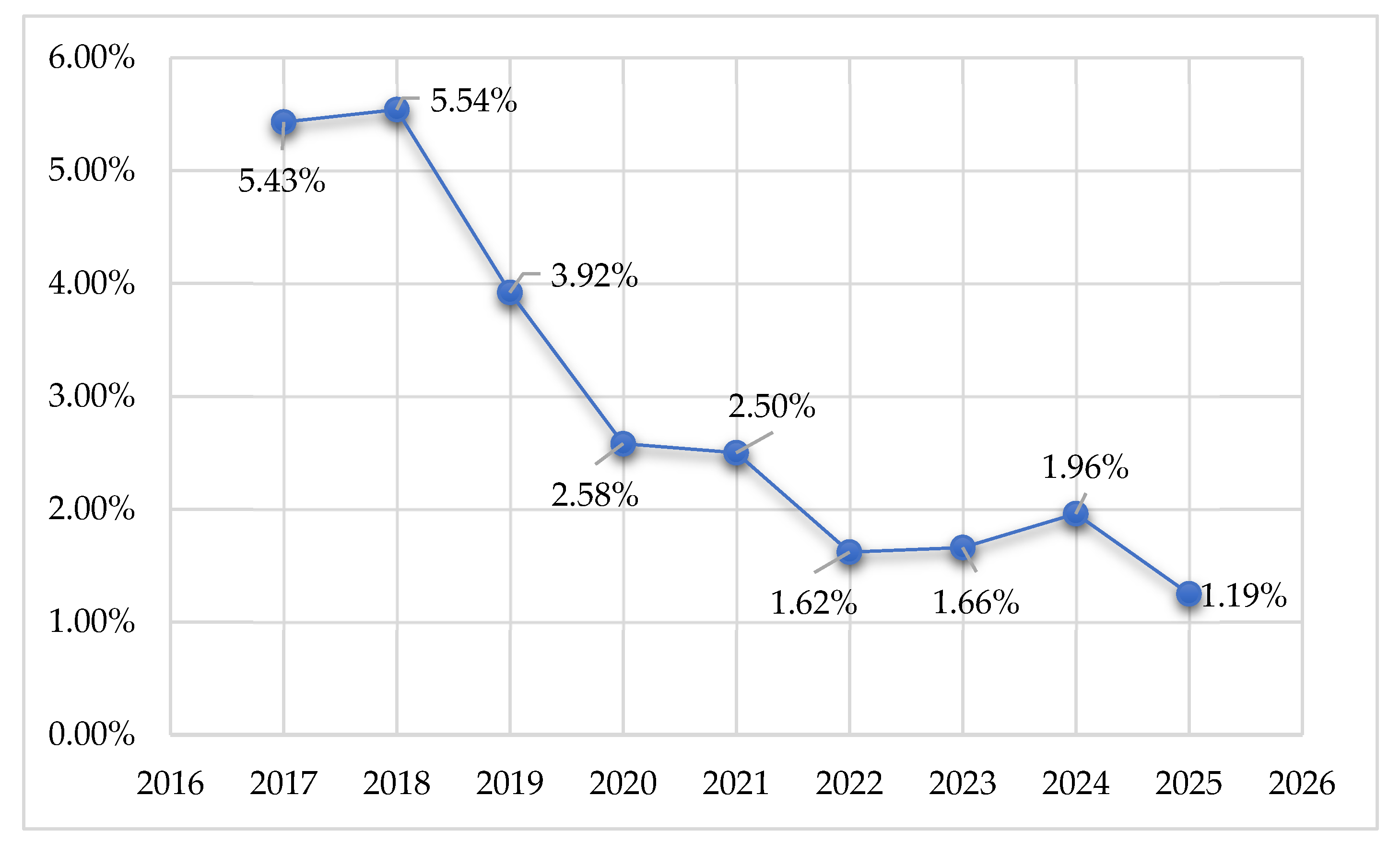
In terms of local epidemiology and clinical trends, the incidence of these conditions has shown a notable evolution in recent years. In 2017, these affections accounted for 5.43% of the total patients treated in the OMF Surgery department. Subsequent years recorded 5.54% in 2018, 3.92% in 2019, 2.58% in 2020, 2.50% in 2021, 1.62% in 2022, 1.66% in 2023, 1.96% in 2024, and 1.19% in 2025. Consequently, the incidence of inflammatory processes of the maxillary sinus decreased significantly from 5.54% to 1.19% (t = 14.64; p < 0.001) registering a total reduction of 4.29%. Similar epidemiological observations have been reported in the contemporary literature, where improvements in preventive dental care, earlier diagnosis, and advances in oral surgical techniques have contributed to a reduction in severe odontogenic complications and oroantral communications [20,21,22,23,24,25,26].
The etiology of OAC is predominantly iatrogenic, often resulting from alveolar process trauma during dental extractions, endodontic treatments, or surgical maneuvers involving the upper teeth [9,15,16,17,27,28,29,30,31]. While OAC is considered a relatively rare complication, the literature reports varying incidence rates ranging from 0.31% to 13%, with the occurrence frequency depending heavily on the specific tooth type being removed [25,26]. Despite the wide variety of surgical plastic methods available, the management of OAC remains a subject of intense debate among specialists. Recent studies by Parvini et al. and Salian et al. indicate that 20% of OAC cases are misdiagnosed, and only 33% achieve successful healing after the initial treatment [30].
The clinical diagnosis of OAC must be established promptly during the examination of the post-extraction socket. Pathognomonic signs include the passage of air between the MS and the oral cavity, rhinolalia, and unilateral fluid reflux through the nasal cavity. Diagnostic maneuvers such as the Valsalva test and careful probing with a buttoned stylus remain gold standards, though they must be performed with caution to prevent sinus infection or further tissue damage [12,28,32,33]. Modern imaging, including Computed Tomography (CT) and Cone–Beam Computed Tomography (CBCT), has become an essential adjuvant tool for identifying OAC and assessing the state of MS soft tissues [21,22,27]. Recent otorhinolaryngological studies further emphasize the importance of CBCT and CT imaging in identifying odontogenic sinus pathology, retained foreign bodies, and inflammatory mucosal changes associated with oroantral communication [21,29].
The aim of this study was to evaluate the clinical and paraclinical diagnostic protocols of oroantral communication in an outpatient setting and to identify key risk factors associated with its occurrence [9,15,16,17,18].

2. Materials and Methods

2.1. Study Design and Setting

This retrospective study was conducted at the “Arsenie Guțan” Department of Oral and Maxillofacial Surgery and Oral Implantology of the “Nicolae Testemițanu” State University of Medicine and Pharmacy [23]. The retrospective analysis included patients treated between January 2022 and December 2022. Clinical data were collected from patients treated within the surgical department of the Municipal Dental Center in Chisinau. Data normality was assessed prior to statistical testing to ensure the validity of parametric analyses.

2.2. Patient Selection and Data Collection

A total of 31 patients diagnosed with accidental oroantral communication (OAC) were included in the study. Data were extracted from the patients’ medical observation charts, focusing on several key clinical and demographic parameters [12,24]. Clinical assessment included evaluation of air passage through the extraction socket, Valsalva maneuver, fluid reflux through the nasal cavity, socket probing, and post-extraction symptoms suggestive of maxillary sinus involvement.
Demographics and Social Status: Age, sex, environment (urban/rural), and educational level, number of children [11,19,34,35].
Clinical Profile: Primary diagnosis, etiology of the communication, the specific causal tooth involved, and the presence or absence of foreign bodies within the maxillary sinus [36,37,38,39,40].
Paraclinical Examinations: Analysis of radiographic findings and other diagnostic imaging used during the treatment process [2,3,35,41].
To ensure a comprehensive theoretical framework, a systematic review of the literature was performed using international medical databases, including PubMed, Medline, and general academic search engines, targeting recent scientific advancements in OMF surgery [7,20,25,27].

2.3. Statistical Analysis

Data processing was performed using Microsoft Excel (Microsoft Office 2010 package). Descriptive statistics were calculated, and results were expressed as percentages and mean values ± standard deviation. Quantitative results were evaluated and presented in the form of descriptive tables and comparative diagrams [30]. Statistical significance was determined using the t-Student test, ensuring the accuracy of the comparative findings presented in the results section [22,26,29,31].

3. Results

3.1. Analysis of Demographic and Socio-Economic Determinants

The study involved 31 patients, where a statistically significant prevalence of the female sex was recorded, representing 67.75% (±8.98; n = 21) of the total sample, compared to 32.25% (±8.98; n = 10) for males (t = 2.80, p < 0.01). When analyzing the age distribution, the decade 31–40 years showed the highest susceptibility, accounting for 51.6% (n = 16) of cases. Interestingly, over 60% of patients with OAC were aged between 30 and 60 years, with a calculated mean age of 42.94 years.
Regarding the residential environment, a significant difference was observed: 87.1% (±5.95) of subjects were from the urban sector (Chisinau), while only 12.9% (±5.84) resided in rural areas (t = 9.41, p < 0.001). Socio-economic status, evaluated through education and family structure, showed that 83.87% of patients had completed formal education, and 71% were parents, suggesting that socioeconomic factors may influence access to dental care and treatment-seeking behavior (Table 1).

3.2. Topography of “Sinus Teeth” and Clinical Manifestations

All patients presented with chronic periodontitis prior to the OAC incident [9,15]. The first molar (M1) was identified as the most frequent causal tooth in 45.16% of cases, followed by the second molar (22.58%) and the third molar (16.13%). Collectively, molars accounted for 83.88% of the total communications, while premolars represented only 16.12% (t = 6.37 p < 0.001) (see Figure 1).
Recent radiological studies have demonstrated a strong association between periapical lesions of posterior maxillary teeth and maxillary sinus mucosal thickening, emphasizing the importance of cone–beam computed tomography (CBCT) in evaluating odontogenic sinus pathology [1].

3.3. Diagnostic Protocols and Paraclinical Findings

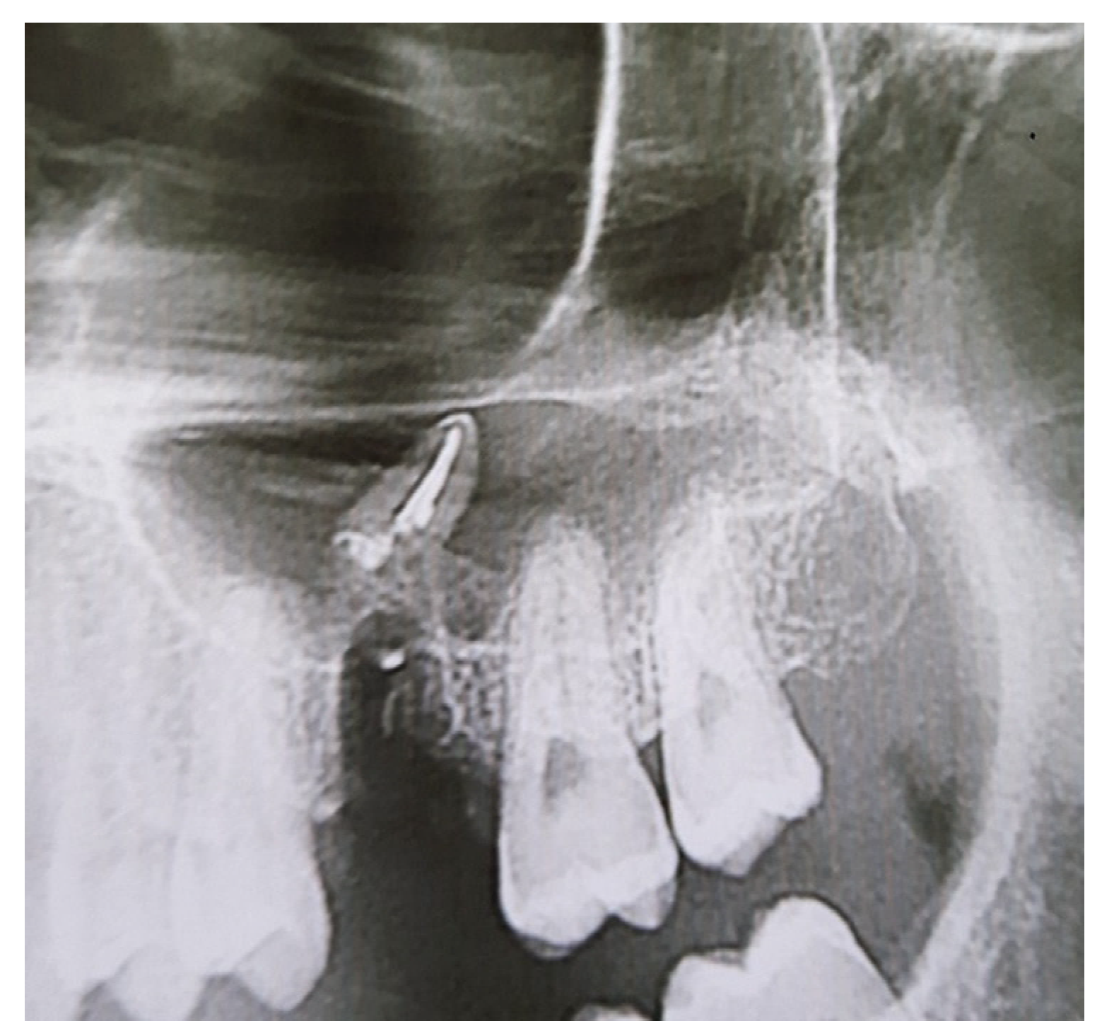
The Kruchinsky and Filippenko classification remains a useful clinical tool for categorizing oroantral communications according to their etiology, duration, and associated maxillary sinus involvement. Its application facilitates standardized diagnosis and supports clinical decision-making regarding conservative versus surgical management [36,37,38]. Based on the Kruchinsky and Filippenko classification [38], the clinical diagnosis of acute OAC was established in 90.32% of cases immediately post-extraction. However, in 9.68% (n = 3) of patients, the communication was complicated by the propulsion of foreign bodies (roots or dental fragments) into the maxillary sinus. In all three cases, foreign bodies were identified immediately after extraction during clinical examination and subsequently confirmed radiologically (Figure 2).
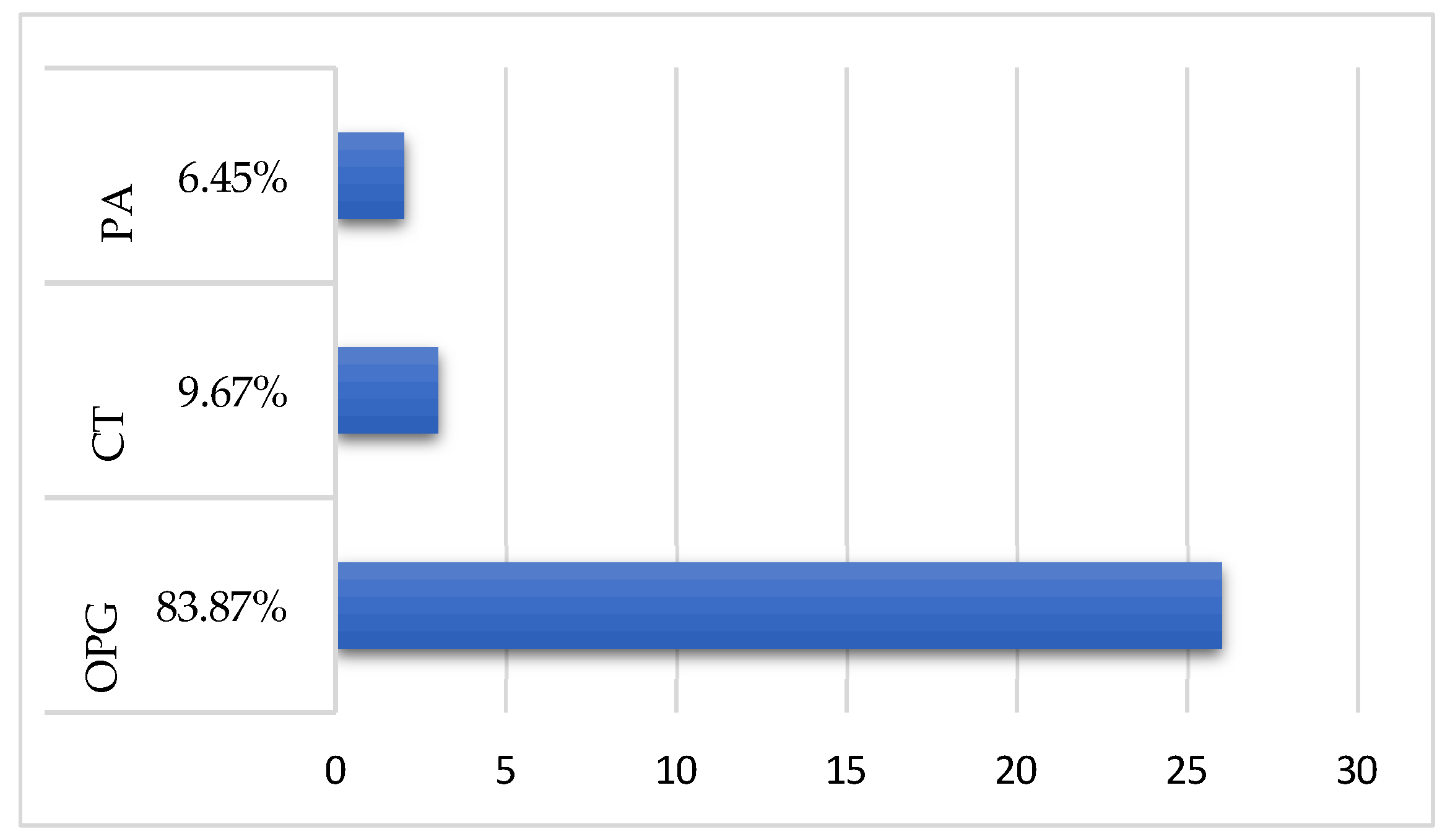
High-performance imaging was mandatory for these cases; Orthopantomography (OPG) was the primary tool for 26 patients (83.87%), while Computed Tomography (CT) was utilized for 3 patients (9.67%) (see Figure 3). Only 6.45% of cases were assessed using traditional periapical radiography (t = 12.22; p < 0.001).

4. Discussion

The clinical and socioeconomic dynamics of oroantral communications (OAC) analyzed in this study highlight the complex nature of this complication. Our findings provide a deeper understanding of why certain patient groups are more vulnerable and how diagnostic protocols must adapt to prevent chronicity.

4.1. Epidemiological Evolution and Demographic Trends

The significant decrease in the incidence of inflammatory processes of the maxillary sinus observed in our data—from 5.54% in 2018 to 1.19% in 2025 (t = 14.64; p < 0.001)—reflects a positive trend in the prevention and management of oral complications within the OMF department (Figure 4).
Regarding gender distribution, our study recorded a notable female prevalence (67.75%). Similar findings were reported in previous epidemiological studies of odontogenic sinus pathology [26]. However, this remains a point of academic debate, as other literature indicates a slight male predilection with a ratio of 1:1.52 [27,40], suggesting that local health-seeking behaviors and gender-specific bone density patterns may influence these statistics.

4.2. Socio-Economic Determinants of Dental Morbidity

A notable finding of this study is the association between socioeconomic factors and the risk of oroantral communication (OAC). Our data indicate that 87.1% of patients originated from urban areas, which may reflect greater access to specialized dental services compared with rural regions [18,31].
In addition, our findings suggest a relationship between financial constraints and dental morbidity. Previous studies have reported that lower educational levels and economic pressures are associated with reduced tooth retention [19]. Although 83.87% of the patients in our cohort had completed formal education, the financial demands associated with larger families (71% of patients had children) may influence both the timing and quality of dental care.
Delayed treatment frequently results in the extraction of severely compromised posterior maxillary teeth (“sinus teeth”), increasing the risk of accidental perforation of the sinus floor during surgical procedures.

4.3. The “Sinus Tooth” Concept and Anatomical Risk

The anatomical proximity of the maxillary sinus floor to the posterior maxillary teeth remains the primary risk factor for the development of oroantral communication. In our study, the first molar was identified as the most frequent causal tooth (45.16%). The hierarchy of “sinus teeth” identified in the present study was as follows: first molar, second molar, second premolar, third molar, first premolar, and canine.
The predominance of molars (83.88%) compared with premolars (16.12%) (t = 6.37; p < 0.001) highlights the vulnerability of the posterior maxillary region, where low bone density (D3/D4) and sinus pneumatization are most pronounced [34]. These findings are consistent with previous studies demonstrating that the roots of maxillary molars and premolars are often separated from the sinus floor by a thin bony plate or may even protrude into the sinus cavity, significantly increasing the risk of oroantral communication following extraction [42].
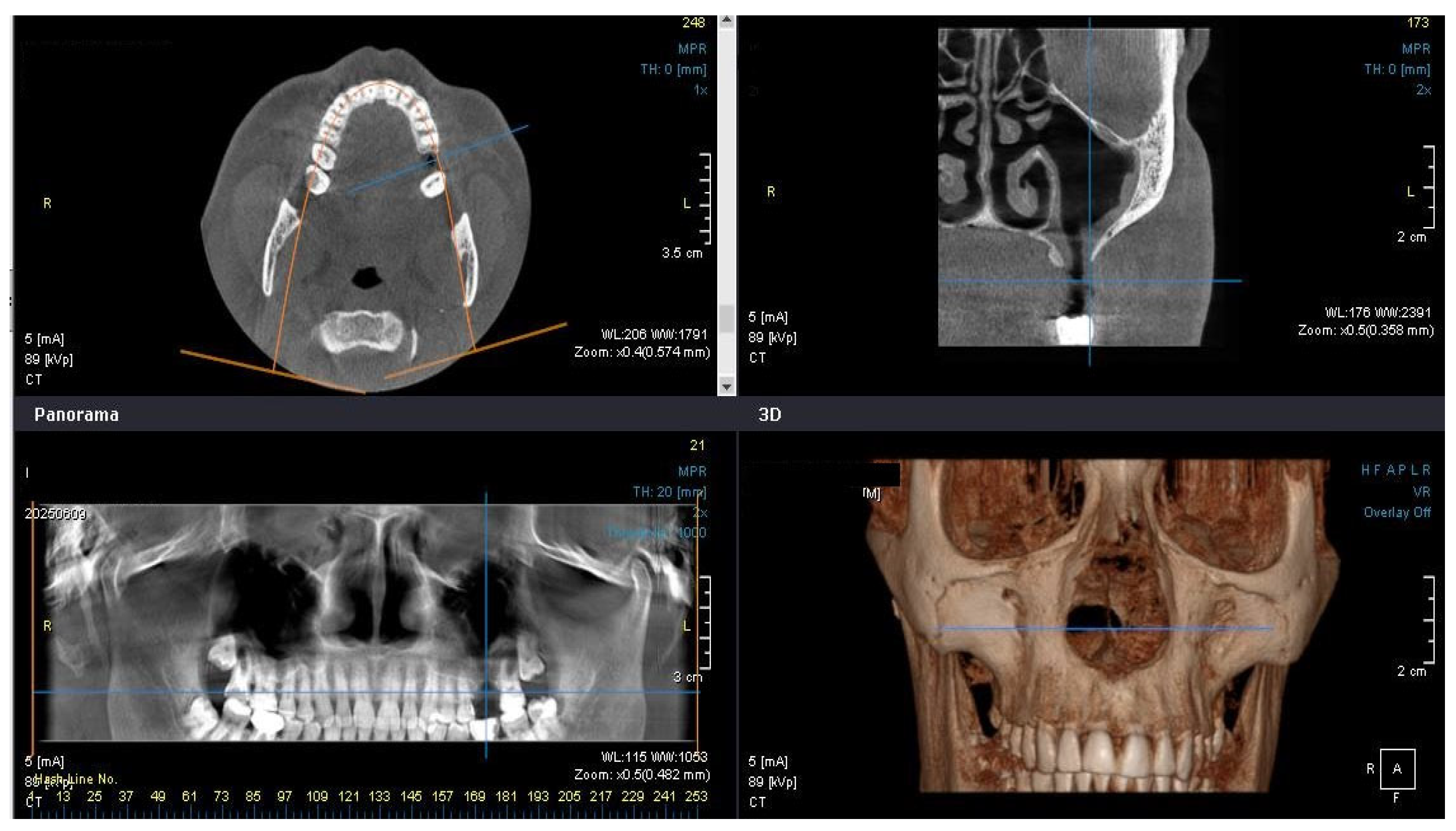
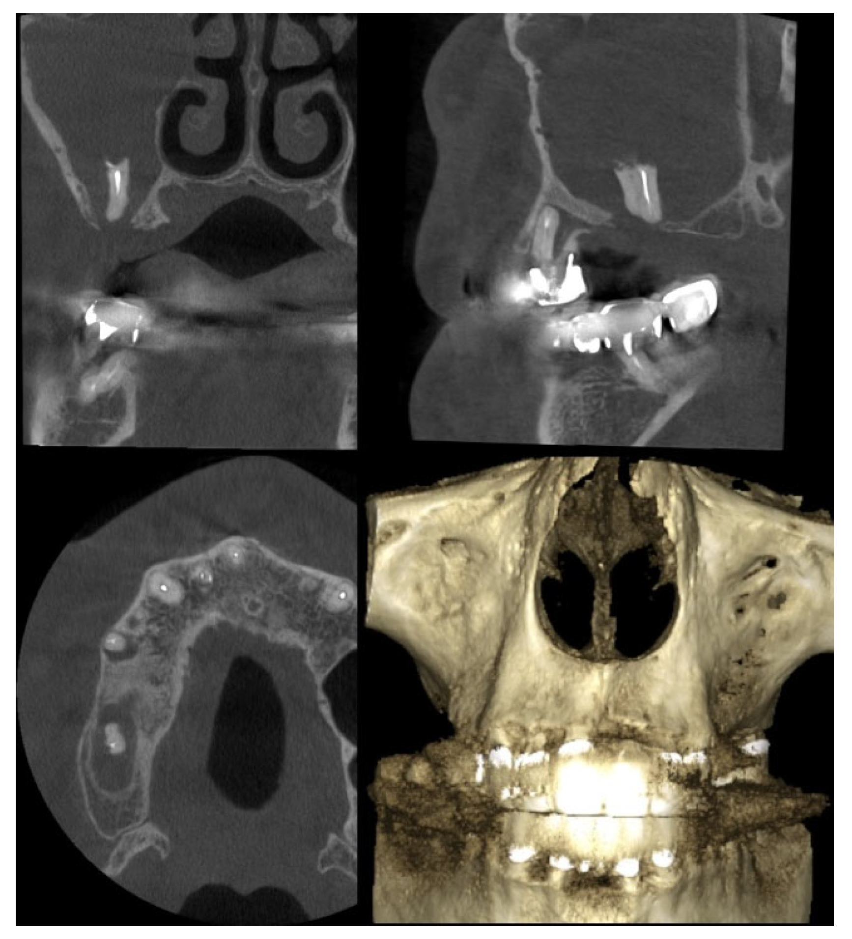
Recent otorhinolaryngological studies emphasize the importance of interdisciplinary management in odontogenic sinus pathology. CBCT imaging may reveal disruption of the sinus floor and retained foreign bodies associated with odontogenic pathology (Figure 5).
Zuska et al. further emphasized that CT findings may be more important than symptoms alone when establishing the indication for endoscopic sinus surgery in odontogenic sinus disease [29]. Simuntis et al. demonstrated a significant association between periapical lesions of posterior maxillary teeth and maxillary sinus mucosal thickening using CBCT analysis [1]. Yun et al. reported that odontogenic maxillary sinusitis associated with oroantral fistulae frequently requires combined surgical treatment, including fistula closure and endoscopic sinus surgery, particularly in chronic cases resistant to conservative management [8]. More recent studies also highlight the diagnostic value of CBCT in identifying odontogenic sinusitis of endodontic origin and preventing chronic sinus complications [21]. Yoo et al. further demonstrated that radiological imaging plays a central role in identifying odontogenic causes of sinus disease and in guiding interdisciplinary surgical treatment planning [16]. Furthermore, CT severity assessment using the Lund–Mackay score may assist in determining the need for surgical intervention in odontogenic chronic rhinosinusitis, particularly in cases with extension beyond the maxillary sinus and involvement of the ostiomeatal complex [29]. These findings support the concept that oroantral communication should not be regarded solely as a dental complication (Table 2). From an otorhinolaryngological perspective, persistent communication may represent the initial stage of odontogenic maxillary sinus disease, requiring coordinated diagnostic and therapeutic management by both dental and ENT specialists.
In these high-risk anatomical areas, pre-operative imaging—such as orthopantomography (OPG) or cone–beam computed tomography (CBCT)—plays a crucial role in assessing the thickness of the bone septa separating the oral cavity from the maxillary sinus and in reducing the risk of accidental oroantral communication [9]. In addition, the recent literature highlights the value of computed tomography in the evaluation of sinus floor integrity, retained root fragments, and associated maxillary sinus pathology [42,43]. Three-dimensional CBCT evaluation may further assist preoperative planning in complex cases (Figure 6).

4.4. Transition from OAC to OAF and Chronic Odontogenic Sinusitis

The distinction between a primary oroantral communication (OAC) and a secondary oroantral fistula (OAF) represents an important clinical consideration supported by our findings. As reported by Timofeev (2007), an OAC typically persists as a simple communication for approximately 7–10 days before epithelialization leads to the formation of a chronic fistulous tract [40]. Other authors, including Szabo, suggest that this transition may occur even more rapidly, with epithelial migration beginning within 48–72 h and complete epithelialization occurring by day 7–8 [36]. Recent studies further support this rapid progression, emphasizing that early epithelialization significantly increases the risk of chronic fistula formation if the communication is not managed promptly [44].
In our study, foreign bodies were detected in 9.68% of cases, a finding of particular clinical concern. The presence of root fragments or endodontic materials may prevent spontaneous closure and often requires immediate surgical intervention to avoid the development of chronic maxillary sinusitis. The extensive use of high-performance imaging in our protocol (93.55%) proved essential for the early detection of such complications, enabling timely management before the establishment of permanent pathological tracts.
Advanced radiological involvement identified on CT may indicate progression toward chronic inflammatory sinus disease requiring combined ENT and dental surgical management [29,41,44].
Delayed management of oroantral communication, particularly in the presence of retained root fragments or foreign bodies, may trigger a persistent inflammatory response that prevents spontaneous closure and promotes epithelialization of the communication, ultimately resulting in chronic oroantral fistula formation.
If left untreated, chronic oroantral communication may facilitate persistent bacterial contamination of the maxillary sinus and progression toward chronic odontogenic rhinosinusitis. In prolonged cases, chronic inflammatory changes and impaired sinus drainage may contribute to persistent sinonasal disease and increase the risk of secondary complications. The recent literature suggests that retained dental materials and foreign bodies may act as persistent inflammatory stimuli within the maxillary sinus, promoting chronic mucosal alterations, impaired sinus drainage, and progression toward chronic odontogenic rhinosinusitis [22,29,41]. Consequently, radiological follow-up is recommended in selected cases to identify early sinonasal complications. Such complications often require combined ENT and maxillofacial surgical management and may significantly increase patient morbidity [22,29]. This progression underlines the importance of early closure of oroantral communications before irreversible sinonasal inflammatory changes become established.

5. Conclusions

The results of this study highlight the importance of early identification and prompt management of oroantral communications (OAC) following dental extractions in the posterior maxillary region. Our analysis of the current literature suggests that evolving diagnostic methods are essential to address the limitations of traditional protocols and to resolve existing clinical challenges. The close anatomical relationship between the maxillary sinus and posterior teeth remains the principal risk factor, particularly in molar extractions where bone density is reduced and sinus pneumatization is common.
Effective diagnosis relies on a synergy between pre-operative radiographic evaluation and careful post-extraction clinical assessment, including meticulous exploration of the socket with a buttoned stylus and a gentle Valsalva maneuver. Early detection allows for immediate surgical management, which is vital to prevent maxillary sinus infection and the subsequent development of chronic, epithelialized oroantral fistulas.
Ultimately, the integration of systematic imaging protocols (OPG/CT) and standardized clinical examination in outpatient oral surgery will significantly improve patient outcomes and minimize long-term complications associated with accidental OAC. Close collaboration between oral-maxillofacial surgeons, dentists, and ENT specialists is essential for preventing progression toward chronic odontogenic sinus disease and improving long-term patient outcomes.
Future studies involving larger multicenter cohorts and long-term follow-up are needed to better define the relationship between oroantral communication, odontogenic sinusitis, and chronic sinonasal complications.

Study Limitations

This study has several limitations. The relatively small sample size and the retrospective design may limit the generalizability of the results. Additionally, the data were collected from a single center, which may introduce selection bias. Further multicenter studies with larger patient cohorts are needed to confirm these findings.
The absence of long-term follow-up data limits the evaluation of chronic fistula formation and long-term treatment outcomes.

Author Contributions

D.H. was responsible for data collection, clinical investigation, and surgical procedures. V.C. (Vasile Cabac) contributed to data collection and provided methodological supervision of the study. V.C. (Victoria Ciobanu) and M.D. performed the data analysis and were primarily responsible for manuscript preparation and writing of the original draft. N.C. supervised the study and critically revised the manuscript. S.P. contributed to manuscript editing, correspondence, and final approval of the submitted version. All authors have read and agreed to the published version of the manuscript.

Funding

This research received no external funding.

Institutional Review Board Statement

The study was conducted in accordance with the Declaration of Helsinki and approved by the Ethical Committee of the “Nicolae Testemițanu” State University of Medicine and Pharmacy (Nr. 3, date 24 March 2025).

Informed Consent Statement

The need for patient consent was waived by the Institutional Review Board due to the retrospective and anonymized nature of the study.

Data Availability Statement

The datasets used and analyzed during the current study are available from the corresponding author upon reasonable request, subject to privacy and ethical restrictions.

Conflicts of Interest

The authors declare no conflicts of interest.

Abbreviations

The following abbreviations are used in this manuscript:
OACOroantral communication
OAFOroantral fistula
MSMaxillary sinus
OMFOral and maxillofacial
OPGOrthopantomography
CTComputed tomography
CBCTCone–beam computed tomography
PAPeriapical radiography

References

  1. Simuntis, R.; Kubilius, R.; Vaitkus, S. Association between Maxillary Posterior Teeth Periapical Odontogenic Lesions and Maxillary Sinus Mucosal Thickening: A Cone-Beam CT Study. Sinusitis 2020, 4, 3. [Google Scholar] [CrossRef]
  2. Azzouzi, A.; Hallab, L.; Chbicheb, S. Diagnosis and Management of oroantral fistula: Case series and review. Int. J. Surg. Case Rep. 2022, 97, 107436. [Google Scholar] [CrossRef]
  3. Fatani, B.; Fatani, A.; Alomar, A. Oroantral Communication and Fistula: A Review of the Literature. Saudi J. Oral Dent. Res. 2020, 5, 575–581. [Google Scholar] [CrossRef]
  4. Bolintineanu, S.; Monica, V.; Şișu, A.; Pop, E.; Roșu, L.; Stoican, L.; Pustai, A.; Matu, C.; Petrescu, C. Anatomia Omului. Volumul VI: CAP ŞI GÂT, Revised ed.; Editura “Victor Babeș”: Timișoara, Romania, 2018; p. 32. [Google Scholar]
  5. Bucur, A. Managementul Afecțiunilor Chirurgicale Oro-Maxilo-Faciale; Editura Didactică și Pedagogică: București, Romania, 2012; p. 264. [Google Scholar]
  6. Burlibaşa, C. Chirurgie Orală şi Maxilofacială; Editura Medicală: Bucureşti, Romania, 2007; pp. 653–697. [Google Scholar]
  7. Bereczki-Temistocle, D.L.; Gurzu, S.; Jung, I.; Cosarca, A.; Beresescu, G.; Golu, V.; Petrovan, C.; Ormenisan, A. Selecting the Best Surgical Treatment Methods in Oroantral Communications. Int. J. Environ. Res. Public Health 2022, 19, 14543. [Google Scholar] [CrossRef]
  8. Yun, P.T.; Seo, Y.J.; Jeong, S.K. Odontogenic Maxillary Sinusitis: Therapeutic Management of Cases with Oroantral Fistulae. Sinusitis 2021, 5, 6. [Google Scholar] [CrossRef]
  9. Chele, N.; Motelica, G.; Zănoagă, O.; Slabari, E. Extracția Dentară—Tehnici, Accidente și Complicații; Editura “Știința”: Chișinău, Moldova, 2022; p. 188. [Google Scholar]
  10. Chouikh, F.; Dierks, E.J. The Buccal Fat Pad Flap. Oral Maxillofac. Surg. Clin. 2021, 33, 177–184. [Google Scholar] [CrossRef] [PubMed]
  11. Duzh, A.N.; Aliamovskii, V.V.; Sokolova, O.R. Influence of oral health literacy of parents on the dental status of children. J. New Med. Technol. 2019, 5, 66–71. [Google Scholar]
  12. Ionescu, E. Manual pentru Rezidențiat. In Stomatologie; Editura Universitară “Carol Davila”: București, Romania, 2021; Volume 2, pp. 623–625. [Google Scholar]
  13. Fronie, A. Curs de Chirurgie Maxilo-Facială: Traumatismele și Supurațiile în Regiunea OMF; Editura Sitech: Craiova, Romania, 2014; pp. 193–199. [Google Scholar]
  14. Gundy, S. Etiology and Management of Oroantral Fistula. Ann. Otolaryngol. Rhinol. 2016, 3, 1122. [Google Scholar]
  15. Sabatino, L.; Pierri, M.; Iafrati, F.; Di Giovanni, S.; Moffa, A.; De Benedetto, L.; Passarelli, P.C.; Casale, M. Odontogenic Sinusitis from Classical Complications and Its Treatment: Our Experience. Antibiotics 2023, 12, 390. [Google Scholar] [CrossRef]
  16. Yoo, S.H.; Jung, H.J.; Park, S.K.; Mo, J.H. Clinical and Radiological Analysis of Odontogenic Sinusitis. J. Clin. Med. 2025, 14, 2821. [Google Scholar] [CrossRef]
  17. Hîțu, D.; Chele, N.; Cabac, V.; Mighic, A.; Bădărău, L. Maxillary Sinusitis of Odontogenic Origin. Eur. Online J. Complement. Altern. Med. 2021, 6, 11–16. [Google Scholar] [CrossRef]
  18. Hîțu, D.; Chele, N.; Șcerbatiuc, D.; Cabac, V.; Bădărău, L.; Mogîldea, M. Maxillary sinusitis of tumor origin. Bull. Acad. Sci. Mold. Med. Sci. 2021, 1, 148–154. [Google Scholar]
  19. Jalilova, G.I. Socio-demographic status of patient with secondary partial edentulism. Bull. Contemp. Clin. Med. 2020, 13, 22–26. [Google Scholar] [CrossRef] [PubMed]
  20. Khandelwal, P.; Hajira, N. Management of Oroantral Communication and Fistula: Various Surgical Options. World J. Plast. Surg. 2017, 6, 3–8. [Google Scholar]
  21. Rolla, M.; Pinto, K.P.; Sassone, L.M.; Silva, E.J.N.L. Odontogenic Sinusitis of Endodontic Origin: Successful Management Through Endodontic Treatment. Sinusitis 2025, 9, 1. [Google Scholar] [CrossRef]
  22. Martu, C.; Martu, M.-A.; Maftei, G.-A.; Diaconu-Popa, D.A.; Radulescu, L. Odontogenic Sinusitis: From Diagnosis to Treatment Possibilities—A Narrative Review of Recent Data. Diagnostics 2022, 12, 1600. [Google Scholar] [CrossRef]
  23. Negru, N.; Hîțu, D.; Zugrav, V.; Carajeleascov, V.; Juc, D.; Gatman, D. Management of oro-maxillofacial conditions associated with endocrine pathologies in outpatient surgery. Rev. Sci. Health Mold. 2022, 29, 519. [Google Scholar]
  24. Nistor, A.M.; Diaconeasa, M.A.; Julea, I.; Nicoale, T.; Cuculici, G.P.; Ghiorghiu, A. Manual de Chirurgie Orală, Anatomie, Patologie și Tehnici Chirurgicale; Editura Medicală Callistro: București, Romania, 2017. [Google Scholar]
  25. Parvini, P.; Obreja, K.; Begic, A.; Schwarz, F.; Becker, J.; Sader, R.; Salti, L. Decision-making in closure of oroantral communication and fistula. Int. J. Implant Dent. 2019, 5, 13. [Google Scholar] [CrossRef]
  26. Pawlik, P.; Stanek, A.; Wyganowska-Świątkowska, M.; Błochowiak, K. The epidemiological pattern of oroantral communication—A retrospective study. Eur. J. Clin. Exp. Med. 2019, 17, 38–44. [Google Scholar] [CrossRef]
  27. Shahrour, R.; Shah, P.; Withana, T.; Jung, J.; Syed, A.Z. Oroantral communication, its causes, complications, treatments and radiographic features: A pictorial review. Imaging Sci. Dent. 2021, 51, 307–311. [Google Scholar] [CrossRef]
  28. de Resende, F.S.; De Moraes, A.J.S.A.; Valladares Trindade, A.C.; Izolani Neto, O.; Neves Barbosa, C.C.; Cecilio Barbosa, O.L. Oroantral communication closure through Bichat ball technique: A literature review. Braz. J. Surg. Clin. Res. 2018, 23, 97–99. [Google Scholar]
  29. Zuska, K.; Fuksa, J.; Knotek, M.; Sisák, M.; Schalek, P. Role of the Lund–Mackay Score in Determining Surgical Indication in Odontogenic Chronic Rhinosinusitis. Sinusitis 2026, 10, 7. [Google Scholar] [CrossRef]
  30. Salian, K.K.K.; Eachempati, P.; Kumbargere, N.S.; Shetty, N.; Soe, M.; Aggarwal, H.; Mathew, R.J. Interventions for treating oroantral communications and fistulae due to dental procedures. Cochrane Database Syst. Rev. 2018, 8, CD011784. [Google Scholar]
  31. Saibene, A.M.; Pipolo, C.; Borloni, R.; Felisati, G. ENT and dentist cooperation in the management of odontogenic sinusitis. A review. Acta Otorhinolaryngol. Ital. 2021, 41, S116. [Google Scholar] [CrossRef]
  32. Timoşca, G.; Burlibaşa, C. Chirurgie Oro-Maxilofacială; Universitas: Chişinău, Moldova, 1992; pp. 265–298. [Google Scholar]
  33. Topalo, V.; Atamni, F.; Sîrbu, D.; Gumeniuc, A.; Dobrovolschi, O.; Suharschi, I. Maxillary sinus floor elevation via crestal approach with simultaneous dental implant installation. Bull. Acad. Sci. Mold. 2008, 2, 90–94. [Google Scholar]
  34. Tintiuc, D.; Negară, A.; Grejdian, T.; Blaja-Lisnic, N.; Celac, A.; Marjine, L.; Lavric, A.; Bădan, V. Opinions regarding the lifestyle and health status of long-lived people. Bull. Acad. Sci. Mold. Med. Sci. 2011, 2, 212–215. [Google Scholar]
  35. Ebenezer, V.; Balakrishnan; Rakeshmohan; Asir, V.D. Management of Oro Antral Communication: A Review. Eur. J. Mol. Clin. Med. 2020, 7, 6382. [Google Scholar]
  36. Afanasyev, V.V. Surgical Stomatology; GEOTAR-Media: Moscow, Russia, 2019; p. 398. [Google Scholar]
  37. Bernadskiy, Y.I. Fundamentals of Surgical Stomatology; Medical Literature: Moscow, Russia, 2003; pp. 58–60, 160–162. [Google Scholar]
  38. Kruchinskiy, G.V.; Filippenko, V.I. Odontogenic Maxillary Sinusitis; Vysheyshaya Shkola: Minsk, Belarus, 1991; p. 167. [Google Scholar]
  39. Mukhin, P.N. Plastic Surgery of Oroantral Communication. Ph.D. Thesis, Moscow, Russia, 2012. [Google Scholar]
  40. Timofeev, A. Fundamentals of Maxillofacial Surgery and Surgical Stomatology; Medical Information Agency: Moscow, Russia, 2007; pp. 274–293. [Google Scholar]
  41. Gâta, A.; Toader, C.; Valean, D.; Trombitaș, V.E.; Albu, S. Role of Endoscopic Sinus Surgery and Dental Treatment in the Management of Odontogenic Sinusitis. J. Clin. Med. 2021, 10, 2712. [Google Scholar] [CrossRef] [PubMed]
  42. Alomari, F.; Kota, M.Z.; Khan, A.A.G.; Alshowail, L.Y.M.; Alamer, S.S.; Alshahrani, S.A.; Alasmari, N.A.; Alwan, S.H.H.; Alqahtani, A.M. Oroantral communication and fistula: Diagnosis and management. Saudi Dent. J. 2025, 37, 59. [Google Scholar] [CrossRef]
  43. Oliva, S.; Lorusso, F.; Scarano, A.; D’Amario, M.; Murmura, G. Imaging in oroantral communication and fistula: Diagnostic considerations. Dent. J. 2024, 12, 147. [Google Scholar] [CrossRef]
  44. Verma, R.R.; Verma, R. Oro-Antral Fistulas and Their Management: Our Experience. Indian J. Otolaryngol. Head Neck Surg. 2021, 74, 1576–1583. [Google Scholar] [CrossRef]
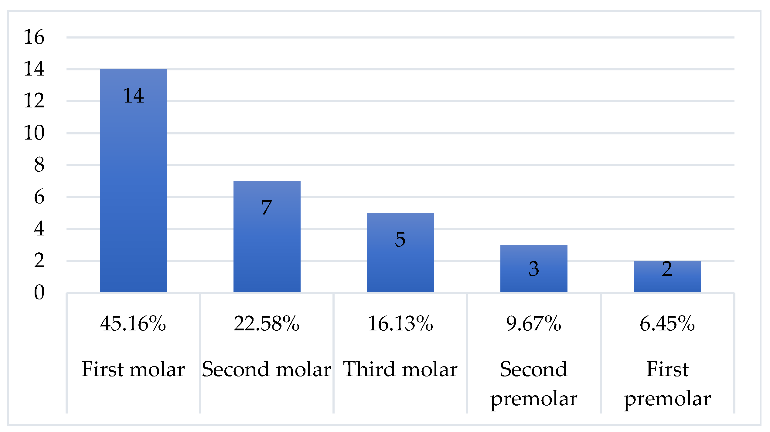
Figure 1. Distribution of teeth associated with oroantral communication in the study.
Sinusitis 10 00014 g001
Figure 2. Panoramic radiograph demonstrating displacement of a dental root fragment into the maxillary sinus following extraction of a posterior maxillary molar.
Sinusitis 10 00014 g002
Figure 3. Imaging methods used for the diagnosis of oroantral communication in the study group.
Sinusitis 10 00014 g003
Figure 4. Evolutionary trend of maxillary sinus inflammatory processes incidence at the OMF Surgery Department (2017–2025).
Sinusitis 10 00014 g004
Figure 5. Cone–beam computed tomography (CBCT) demonstrating disruption of the maxillary sinus floor associated with oroantral communication and retained dental root fragment.
Sinusitis 10 00014 g005
Figure 6. Three-dimensional CBCT reconstruction demonstrating bony defect of the maxillary sinus floor associated with oroantral communication.
Sinusitis 10 00014 g006
Table 1. Demographic and Socio-Economic Characteristics of the Study Group.
CharacteristicCategoryFrequency (n)Percentage (%)Statistical
Significance
Gender Female2167.75% ± 8.98t = 2.80; p < 0.01
Male1032.25% ± 8.98
Age group<20 years26.45%Mean Age: 42.94
21–30 years929.00%
31–40 years1651.60%
41–50 years39.70%
>51 years13.25%
ResidenceUrban (Chișinău)2787.1% ± 5.95t = 9.41; p < 0.001
Rural412.9% ± 5.84
EducationHigher/Vocational2683.87% ± 7.37t = 6.16; p < 0.001
Secondary/Incomplete516.13% ± 7.36
Family statusWith children2271.0% ± 8.69t = 3.48; p < 0.001
Without children929.0% ± 8.69
Table 2. Comparison of recent studies addressing odontogenic sinusitis and oroantral communication.
StudyMain Finding
Simuntis et al. [1]Periapical lesions associated with sinus mucosal thickening
Yun et al. [8]OAF may require combined OMF + ESS treatment
Yoo et al. [16]Imaging essential for interdisciplinary management
Rolla et al. [21]Endodontic treatment may resolve odontogenic sinusitis
Zuska et al. [29]Lund–Mackay score useful for surgical indication
Disclaimer/Publisher’s Note: The statements, opinions and data contained in all publications are solely those of the individual author(s) and contributor(s) and not of MDPI and/or the editor(s). MDPI and/or the editor(s) disclaim responsibility for any injury to people or property resulting from any ideas, methods, instructions or products referred to in the content.