Article

Antibacterial Effect of Nanosilver Fluoride and Silver Diamine Fluoride Against Streptococcus mutans: An In Vitro Study


by
Carlos Alonso Alvarez-Marín
1,
Norma Leticia Robles-Bermeo
2,*,
Rogelio José Scougall-Vilchis
2,*,
Raúl Alberto Morales-Luckie
3,
María Guadalupe González-Pedroza
4 and
Nayeli Lovera-Rojas
2
1
PhD Program in Health Sciences, Advanced Studies and Research Center in Dentistry Dr. Keisaburo Miyata, School of Dentistry, Autonomous University of the State of Mexico, Toluca 50130, Mexico
2
Academic Area, Advanced Studies and Research Center in Dentistry Dr. Keisaburo Miyata, School of Dentistry, Autonomous University of the State of Mexico, Av. Jesus Carranza Esquina Paseo Tollocan S.N., Colonia Universidad CP, Toluca 50130, Mexico
3
Department of Materials Science, Center for Research in Sustainable Chemistry (CCIQS), Autonomous University of the State of México (UAEMex), Toluca 50200, Mexico
4
Department of Biotechnology, Faculty of Science, Autonomous University of the State of México (UAEMex), Toluca 50200, Mexico
*
Authors to whom correspondence should be addressed.
Appl. Nano 2026, 7(2), 14; https://doi.org/10.3390/applnano7020014
Submission received: 17 March 2026 / Revised: 30 April 2026 / Accepted: 21 May 2026 / Published: 1 June 2026
(This article belongs to the Collection Feature Papers for Applied Nano)

Abstract

Introduction: Silver diamine fluoride (SDF) is a colorless solution used at different concentrations. It is a topical treatment used on caries lesions having as its main properties being cariostatic, remineralizing and antibacterial. Nanosilver fluoride (NSF) is effective as a cariostatic without a staining effect on the tooth surface as in the case of SDF, which generates a black stain on the treated surface. This NSF has been shown to exhibit low toxicity and continue to exhibit antimicrobial properties. Purpose: To compare the antibacterial effect of silver diamine fluoride and nanosilver fluoride against Streptococcus mutans. Methods: The NSF was prepared by starting with the synthesis of silver nanoparticles with chitosan and then adding it to a sodium fluoride (NaF) solution. The compared groups were SDF, NSF, AgNPs, NaF, and chlorhexidine. The antibacterial effect will be measured using the Kirby–Bauer microbiological technique. Results: The analysis of results was obtained using the ANOVA statistical test. A significant difference was obtained in the comparison between the groups with a value of p = 0.001. Subsequently, Tukey’s test was applied, obtaining significant differences between all the groups compared against the SDF, obtaining greater results in this group. Conclusions: Both silver diamine fluoride and nanosilver fluoride exhibit strong antibacterial activity at commercially recommended concentrations, supporting their use as optimal dental materials in both interceptive and preventive caries treatments.

Graphical Abstract

1. Introduction

Dental caries is a multifactorial disease arising from the metabolism of sugars by bacteria within the dental biofilm. This process generates polymeric substances that stabilize the biofilm’s pH and facilitate bacterial adhesion. Additionally, acid production during sugar metabolism promotes the demineralization of dental hard tissues, increasing their structural solubility [1]. The development of dental caries is accelerated by the presence of fermentable carbohydrates, which promote the proliferation of cariogenic bacteria, including Streptococcus mutans [2].
Dental caries management has increasingly focused on minimally invasive techniques designed to protect healthy tooth tissue, preserve pulp vitality, and maintain the tooth until natural exfoliation occurs [3]. One of the most commonly used minimally invasive treatments is silver diamine fluoride (SDF), a colorless, odorless, alkaline solution containing silver and fluoride ions that interact with ammonia within the solution [4].
The antibacterial activity of SDF is attributed to its silver ions, which inhibit bacterial enzymatic activity and prevent microbial replication through condensation of bacterial DNA [5].
Another mechanism of action of SDF involves the occlusion of dentinal tubules by the deposition of silver particles, which prevents acid penetration and diffusion, while also reducing the presence of Streptococcus mutans within the tubules [6,7]. Although SDF is an effective minimally invasive treatment due to its remineralizing, cariostatic, and antibacterial properties, its use in pediatric dentistry is limited by its main drawback: the formation of black stains on treated carious surfaces [8].
The incorporation of nanotechnology into dentistry has introduced innovative approaches to caries management. Currently, nanotechnology strategies in dentistry can be classified into three main groups: antimicrobial systems, remineralizing agents, and controlled drug release systems [9]. In the antimicrobial field, metallic and metal oxide nanoparticles (such as silver, zinc oxide, and copper oxide) have demonstrated high efficacy in disrupting oral biofilms and combating specific pathogens such as Streptococcus mutans [10]. On the other hand, remineralizing systems employ biomimetic materials such as nano-hydroxyapatite and nanometric amorphous calcium phosphate, which promote the structural recovery of dentinal tubules and enamel at a microscopic level. Furthermore, nanotechnology has introduced into dentistry smart nanocarriers, such as chitosan nanoparticles and pH-sensitive systems, which allow for strictly localized and controlled therapeutic release under acidogenic conditions [9].
Silver nanoparticles (AgNPs), such as SDF, exhibit antibacterial activity against cariogenic bacteria. When combined with fluoride, they form a remineralizing solution known as nanosilver fluoride (NSF) [5]. Unlike SDF, NSF prevents black staining on carious surfaces, thereby eliminating SDF’s primary drawback and enhancing the effectiveness and appeal of minimally invasive treatments [11].
The mechanisms of action of the antibacterial effect of silver nanoparticles have not been fully explained. However, the following mechanisms of action have been reported in the literature: (1) disruption of the bacterial cell wall and cytoplasmic membrane; (2) ribosomal denaturation by inhibiting protein synthesis; (3) disruption of ATP production; (4) membrane rupture by species that react to the presence of oxygen; (5) interference in DNA replication; (6) denaturation of the cell wall membrane; (7) perforation of the cytoplasmic membrane [11,12]. This study aimed to compare the antibacterial effects of NSF and SDF against Streptococcus mutans.

2. Materials and Methods

2.1. Synthesis and Characterization of Silver Nanoparticles

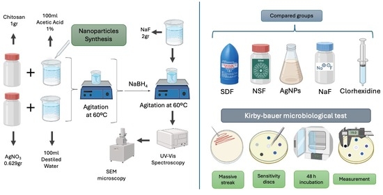
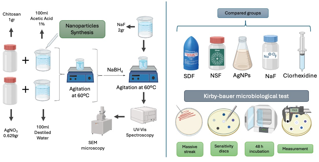
The NSF solution was prepared through the chemical synthesis of AgNPs. Sodium borohydride (NaBH4) served as the reducing agent, while chitosan (1 g) was used as a stabilizer. The chitosan was first dissolved in 100 mL of 1% acetic acid under continuous stirring until fully solubilized. Separately, 0.645 g of AgNO3 was dissolved in 100 mL of distilled water. The two solutions were then combined to initiate the synthesis of AgNPs. The mixture was stirred at 60 °C until the characteristic color of the nanoparticles appeared. The synthesized AgNPs were characterized using Scanning Electron Microscopy (SEM) (CIQTEK 3200, Hefei, China) and TEM (JEOL 2010, Akishima, Japan). A complementary characterization, UV–visible spectroscopy (Velab VE-5100UV, Pharr, TX, USA), was performed. Finally, sodium fluoride (NaF) was added to the solution to complete the NSF formulation. The final concentrations were 600 ppm for AgNPs and 5000 ppm for NaF.

2.2. Microbiological Test

The Kirby–Bauer microbiological technique was used, employing Mueller–Hinton (MH) agar and a certified strain of Streptococcus mutans (ATCC 353384) as the culture medium. Bacterial suspensions were prepared by dispersing colonies in sterile saline in a glass tube and vortexing until a turbidity equivalent to 0.5 on the McFarland scale was achieved [12].
Sterilized absorbent paper disks (6 mm in diameter) were impregnated with each treatment—NSF, SDF, AgNPs, NaF, chlorhexidine (positive control), and saline solution (negative control)—with each procedure performed in triplicate. Plates were incubated for 24 h at 37 °C under microaerophilic conditions. Inhibition zones were measured in millimeters using a digital vernier caliper (Mitutoyo CD-6” ASX, Tokyo, Japan) [12,13].

2.3. Minimum Inhibitory Concentration

As a complementary test, the minimum inhibitory concentration (MIC) was determined using the microdilution technique in a 96-well plate, with each treatment tested in triplicate. Initially, 100 μL of MH broth and 10 μL of bacterial suspension, standardized to 0.5 McFarland turbidity, were added to each well. Treatments were applied in serial dilutions, transferring 100 μL from wells with the highest concentration to the next well in the series. Plates were incubated at 37 °C for 24 h, and results were read using a microplate reader [12].

3. Results

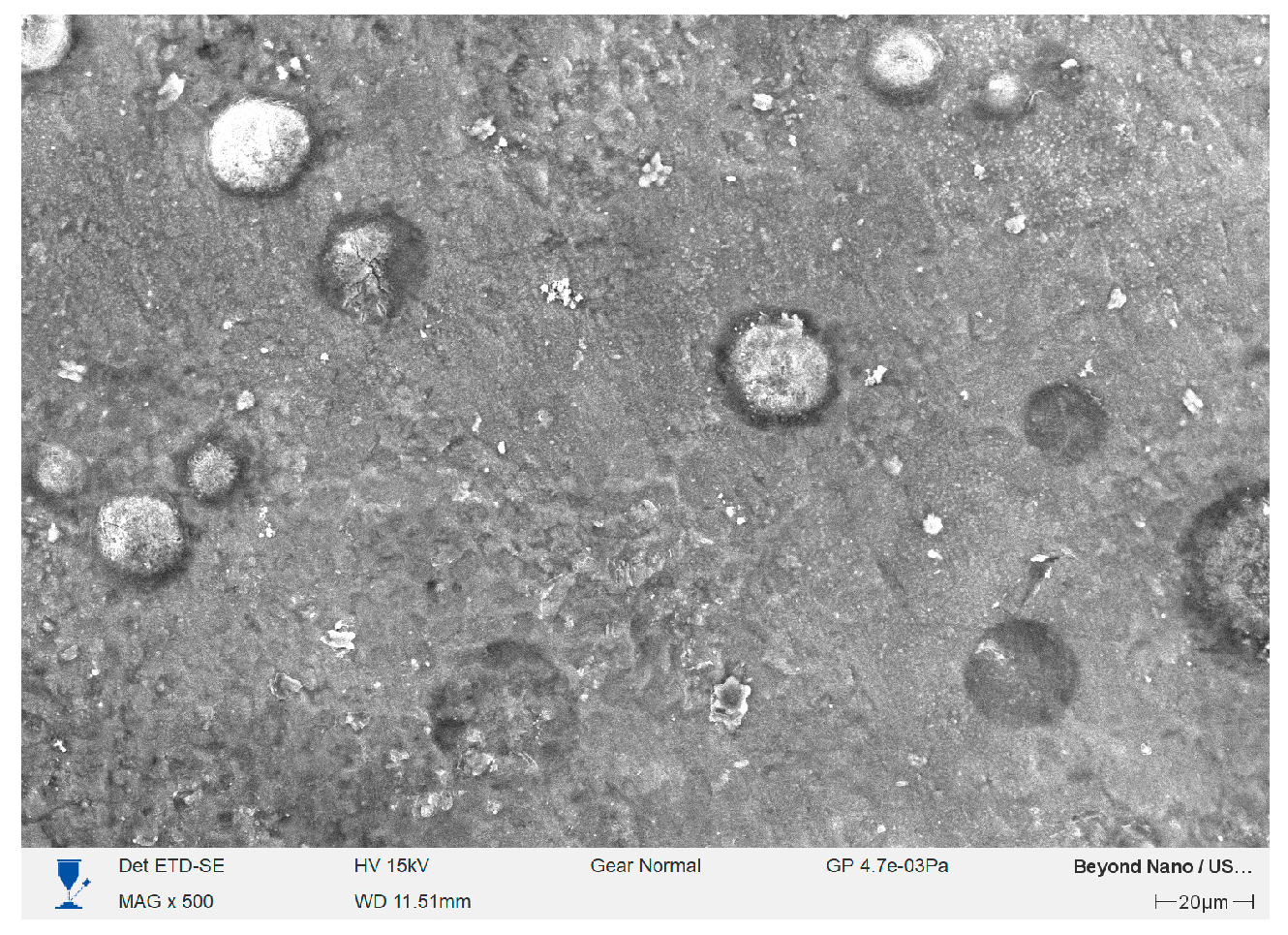
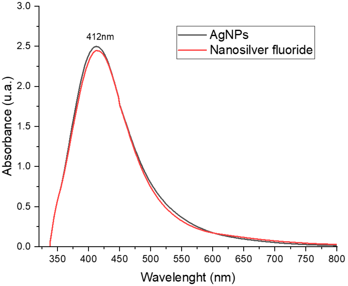
The synthesized AgNPs were characterized using Scanning Electron Microscopy (SEM) and TEM obtaining images of spherical particles with a homogeneous distribution (Figure 1). The data obtained in the micrography indicate an average size of 55 nm in diameter. A complementary characterization UV–visible spectroscopy was performed. An absorbance peak at 412 nm was observed, consistent with typical AgNPs (Figure 2).
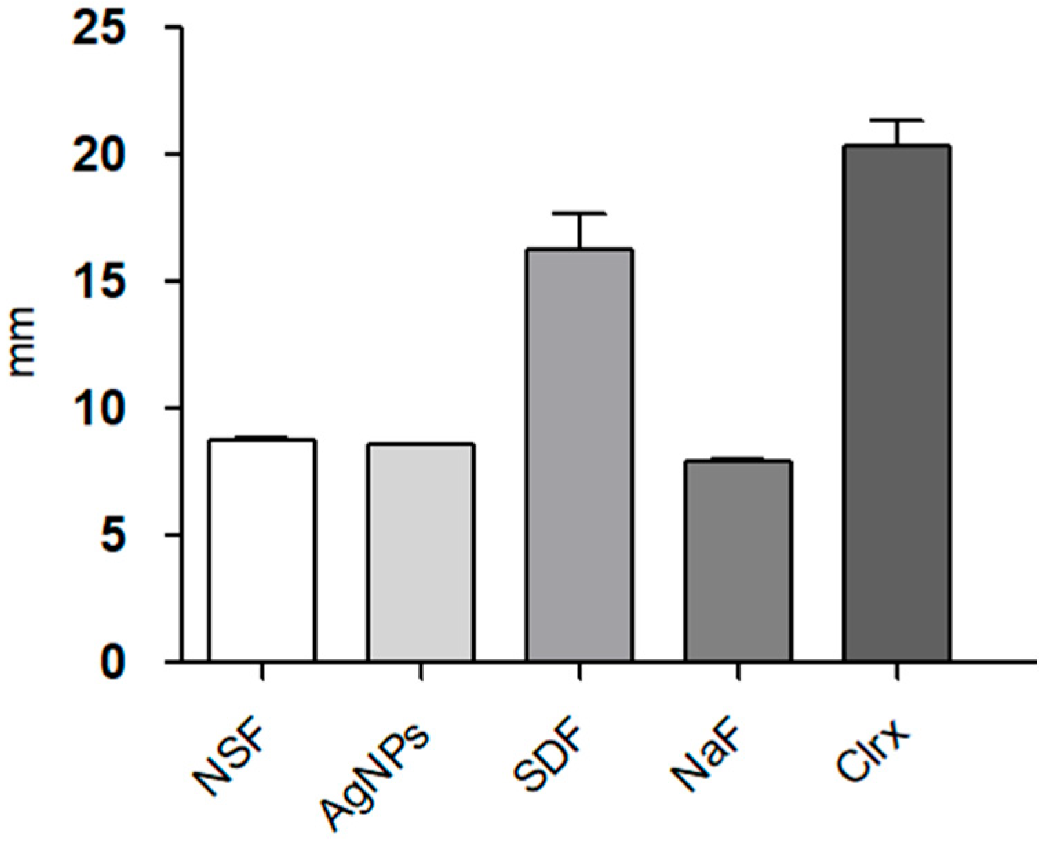
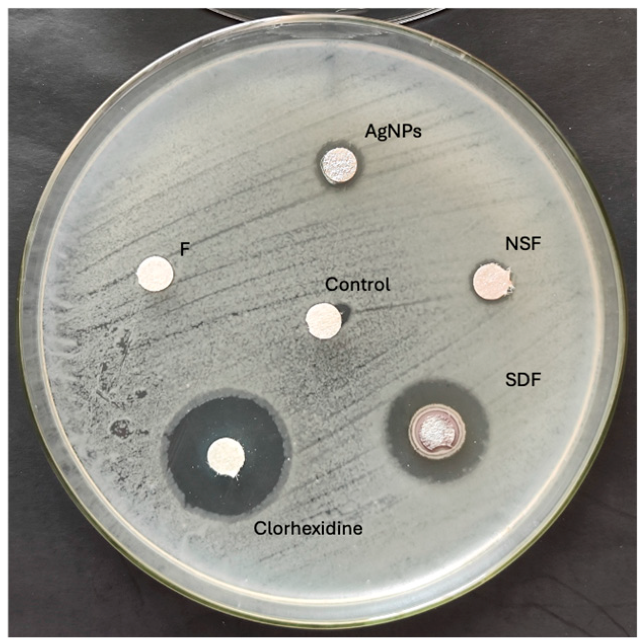
Descriptive statistics, including mean and standard deviation, were calculated for all groups. Inferential analysis using one-way ANOVA revealed a statistically significant difference among the groups (p = 0.001). The largest inhibition zones were observed in the positive control group (chlorhexidine), followed by the SDF group (Figure 3). Tukey’s multiple comparison test was subsequently performed to evaluate pairwise differences between groups, with the results summarized in Table 1.
Tukey’s post hoc analysis revealed statistically significant differences among the majority of the evaluated groups. The positive control (chlorhexidine) exhibited the highest antibacterial activity. Furthermore, SDF displayed significantly greater efficacy compared to NSF, AgNPs and sodium fluoride groups. Conversely, no statistically significant differences were observed among NSF, AgNPs, and NaF groups.
The inhibition zones observed in the groups containing silver particles (NSF, AgNPs, SDF) had well-defined edges, with no bacterial colonies present within the clear area. In contrast, the zone produced by the NaF group showed diffuse boundaries, with colonies appearing from a radius of 7 mm. The inhibition zones obtained are shown in Figure 4.
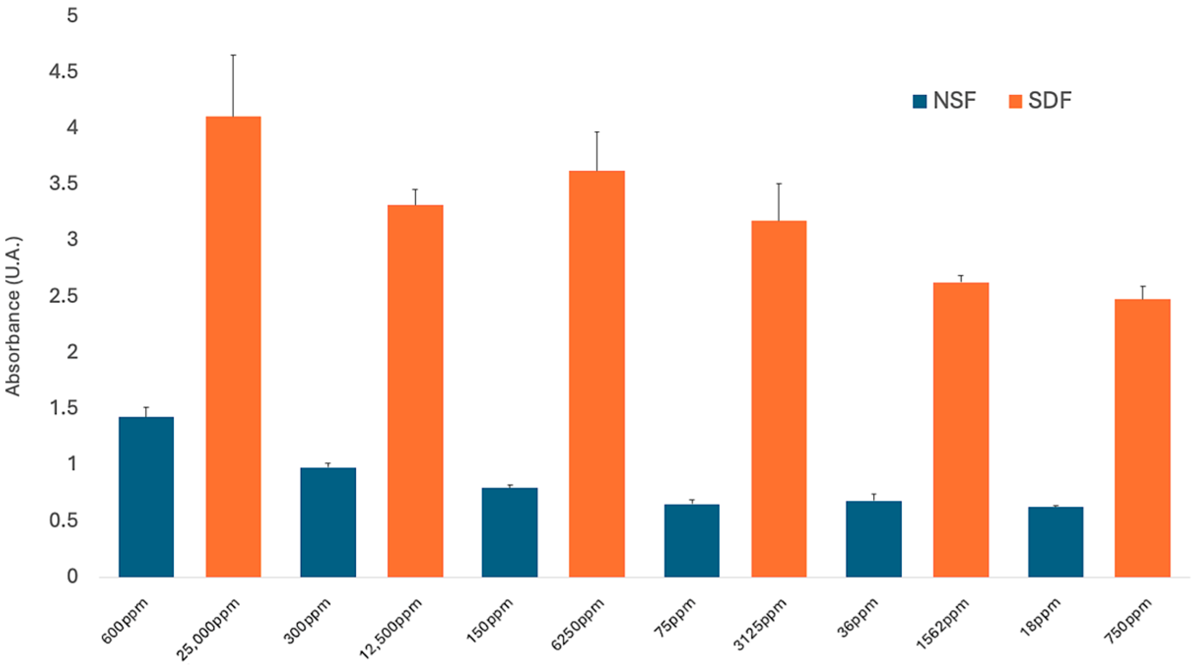
The results of the minimum inhibitory concentration test comparing NSF and SDF are shown in Figure 5. Concentrations were gradually diluted and expressed in parts per million (ppm), starting from a maximum of 600 ppm for NSF and 25,000 ppm for SDF. Notably, bacterial inhibition was observed even at a 3% dilution, corresponding to 18 ppm for NSF and 750 ppm for SDF.
The data obtained from the microplate reader reveals a distinct absorbance profile of NSF and SDF. The absorbance results are attributed to the intrinsic turbidity of the bacterial proliferation. A significant change was noted in the effective concentration ranges. Both compounds have antibacterial effectiveness even at minimal doses, preserving their stability against Streptococcus mutans.

4. Discussion

The high prevalence of dental caries is a global health problem, hence the need for dental treatments that can be applied easily and without sophisticated equipment is increasing [14,15,16]. The use of silver diamine fluoride and nanosilver fluoride are effective options as minimally invasive treatments for the treatment and prevention of caries [17,18,19,20,21]. Crystal and Niederman, in 2019, mentioned the benefits of applying silver diamine fluoride as a minimally invasive dental treatment compared to conventional treatments [16]. These include safe treatment, effectiveness, application time, patient comfort, and accessibility [10].
Several studies demonstrate the effectiveness of using AgNPs in dental materials, such as adhesive systems, composite and acrylic resins, root canal fillings, and implant appliances [10,21]. This study sought to compare the antibacterial properties of AgNPs in a minimally invasive treatment with a material containing macroscopic silver particles (SDF).
Antibacterial tests were performed using the Kirby–Bauer microbiological technique. This technique has been one of the most widely used over time by different authors due to its standardized methodology, specifically in treatments with silver nanoparticles, as in the case of the study conducted by Muhammad et al. who in 2024 conducted tests to verify the antibacterial effectiveness of AgNPs against different bacteria [22].
Elchaghaby MA, Rashad S, and Wassef NM in 2024 used the Kirby–Bauer microbiological technique as a complement to determining the minimum inhibitory concentration (MIC) [23]. In the case of the study here presented, microbiological techniques were performed separately, with similar results despite the methodological differences.
The Kirby–Bauer microbiological technique was used to compare the antibacterial effects of SDF and NSF and each of the separate components of nanosilver fluoride (silver nanoparticles and sodium fluoride) to determine whether bacterial inhibition increased or decreased when the components were mixed. This methodology is consistent with that performed by Sharma P. et al. [24].
The results obtained in the present study indicate that when evaluating the antibacterial effect of silver diamine fluoride and nanosilver fluoride, SDF exhibits a significantly greater antibacterial effect compared to nanosilver fluoride. These results differ from those obtained by N. Ammar et al. in 2022 [3]. In their study, they performed different measurements than the present study. They report that the antibacterial effectiveness is greater with NSF in its initial application. However, after one month, the antibacterial effectiveness was practically equal between both agents.
According to the existing literature, the superior outcomes demonstrated by the SDF group relative to the NSF group are likely associated with elevated silver concentrations. It has been established that as the concentration (in ppm) increases, the diameter of the inhibition zones expands, reflecting a more potent antibacterial response against S. mutans. This evidence corroborates the inter-group differences observed in the current study, confirming that higher concentrations of silver particles significantly enhanced the antibacterial efficacy of SDF [3,25,26].
The main difference in concentration requirements between NSF and SDF stems from the optimized pharmacodynamics of nanotechnology. While traditional SDF relies on a macro-complex of silver and fluoride ions, NSF utilizes silver nanoparticles (AgNPs) characterized by an exceptionally high surface-area-to-volume ratio. This structural advantage facilitates a more efficient release of Ag ions, allowing for the disruption of Streptococcus mutans cell membranes at a fraction of the dosage required by SDF. Targino et al., as well as the results obtained by the present study, demonstrate that the NSF formulation is effective against S. mutans at concentrations lower than SDF while offering a superior safety profile regarding cytotoxicity [27].
In addition to the above and with greater clinical significance, the ability of the NSF to inhibit bacterial growth without the characteristic dark staining associated with SDF provides vital clinical support as an aesthetic alternative for caries management as a minimally interventional treatment, and this is corroborated in the literature evaluations [28].
The antibacterial effect of NSF has been primarily associated with the presence of silver nanoparticles in its formulation. However, different studies have shown that the antibacterial effect of its other components (chitosan and sodium fluoride) generates a cumulative antibacterial effect and, in combination with its remineralizing effect, an anticariogenic [29,30]. The above is confirmed by the results obtained when performing the antibacterial test on sodium fluoride, obtaining positive results vs. S. mutans.
Nanosilver fluoride has been incorporated for study at different concentrations from 256 [3], 400 [30], 600 or 1500 ppm [9]. However, some studies such as the one carried out by Akyildiz and Sönmezbasan [20,31] based its composition on what was described by Targino et al. in 2014 [32] in which a concentration of AgNPs of 376 ppm is incorporated. In each of the previously mentioned concentrations, favorable results have been obtained in the antimicrobial activity against Streptococcus mutans, which supports the results obtained in the present research, in which a concentration of AgNPs of 600 ppm was incorporated as an average concentration of those mentioned in the literature.
The measurement of the MIC was performed using the agar microdilution method, and this methodology is commonly used to observe the minimum concentration at which a compound continues to be effective against a bacterial agent. The study conducted by Targino et al. coincides with the methodology used in this study [27]. The minimum effective concentration reported by Targino et al. [27] of SDF and NSF was 50 ppm. However, in the present study, the test was performed at a lower concentration (18 ppm) obtaining positive results of bacterial inhibition, thus making it possible to use it as a dental material against the main caries-generating bacterium even at low concentrations.
The cytotoxicity profile of these dental materials remains a critical subject of academic discourse. In vitro evidence indicates that SDF significantly reduces the viability of fibroblasts and pulpal cells, potentially inducing cellular necrosis. Moreover, SDF has been documented to exhibit prolonged cytotoxic potential, attributed to the retention and sustained release of these highly reactive ions within the tissues [33].
Conversely, NSF mitigates these cytotoxic concerns through the integration of nanotechnology and biological stabilizers, functioning at substantially lower silver concentrations. In contrast to the ammonia used in SDF, the chitosan incorporated into NSF serves as a highly biocompatible physicochemical interface. This natural polymeric coating not only prevents nanoparticle agglomeration but also buffers the direct interaction between the metallic core and host cells. Consequently, this ensures significantly higher cell viability rates exceeding 70–80% compared to those observed with SDF [27].
Another important aspect to consider is the difference in the chemical composition of the solutions. SDF is an alkaline solution in which high concentrations of silver and fluoride are combined, and these tend to form unstable salts that precipitate rapidly. Ammonia acts as a ligand, binding to silver ions and forming a highly stable complex known as the diamine silver ion. This ion reduces the oxidative potential of silver, preventing premature precipitation of the salts and maintaining a stable ionic concentration [34].
NSF, on the other hand, utilizes silver particles in the nanometric range. Due to their high surface energy, metallic nanoparticles have a strong tendency to agglomerate, which would negate their antibacterial properties at the nanoscale [35]. For this reason, chitosan has been used for its properties as a nanoparticle stabilizer, in addition to functioning as a coating agent. Chitosan encapsulates newly formed silver nanoparticles, stabilizing them against aggregation, inhibiting growth, and ensuring particle size at the nanoscale (approximately 100 nm) [35]. Both stabilizing agents (ammonia and chitosan) have been confirmed as effective according to the solution components, making them suitable alternatives for the preservation of SDF and NSF with their structure appropriate for use [27,34].

5. Conclusions

The present study demonstrates that both silver diamine fluoride and nanosilver fluoride are antibacterial agents with high effectiveness at the commercial concentrations recommended by the literature, making them optimal dental materials for use in both interceptive and preventive treatments for caries, in addition to testing the effectiveness of NSF at low doses against the main cavity-causing bacteria, S. mutans.

Author Contributions

N.L.R.-B. designed the research study. C.A.A.-M. performed the experimental process and manuscript drafting. R.A.M.-L. provided help and advice on the development of silver nanoparticles and nanosilver fluoride. M.G.G.-P. helped with the development of the antibacterial effect of NSF and SDF against Streptococcus mutans. R.J.S.-V. analyzed the data. N.L.-R. critically reviewed the manuscript. All authors have read and agreed to the published version of the manuscript.

Funding

This research received no external funding.

Institutional Review Board Statement

The study was conducted in accordance with the Declaration of Helsinki, and approved by the Ethics Committee of the Institute of Studies About the University (IESU) of the Autonomous University of the State of Mexico (2024/P04, 9 October 2024).

Informed Consent Statement

Each participant signed their informed consent and had detailed knowledge of the process that the donated sample would take.

Data Availability Statement

The data supporting the results and conclusions presented here are included and available in the content of this article.

Acknowledgments

I thank each of the people who supported the development of this project, especially my teachers, for sharing and giving me their knowledge, time and support. I also thank M. en C.M. Alan Peñaloza for his support in the laboratory for the development of the project.

Conflicts of Interest

The authors declare no conflicts of interest.

References

  1. Uribe, S.E.; Innes, N.; Maldupa, I. The global prevalence of early childhood caries: A systematic review with meta-analysis using the WHO diagnostic criteria. Int. J. Paediatr. Dent. 2021, 31, 817–830. [Google Scholar] [CrossRef] [Scilit]
  2. Pollick, H. The Role of Fluoride in the Prevention of Tooth Decay. Pediatr. Clin. N. Am. 2018, 65, 923–940. [Google Scholar] [CrossRef] [Scilit]
  3. Ammar, N.; El-Tekeya, M.M.; Essa, S.; Essawy, M.M.; Talaat, D.M. Antibacterial effect and impact on caries activity of nanosilver fluoride and silver diamine fluoride in dentin caries of primary teeth: A randomized controlled clinical trial. BMC Oral Health 2022, 22, 657. [Google Scholar] [CrossRef] [Scilit] [PubMed]
  4. Torres, P.J.; Phan, H.T.; Bojorquez, A.K.; Garcia-Godoy, F.; Pinzon, L.M. Minimally invasive techniques used for caries management in dentistry. A review. J. Clin. Pediatr. Dent. 2021, 45, 224–232. [Google Scholar] [CrossRef] [Scilit] [PubMed]
  5. de Almeida Piovesan, É.T.; Alves, J.B.; Ribeiro, C.D.P.V.; Massignan, C.; Bezerra, A.C.B.; Leal, S.C. Is silver diamine fluoride effective in reducing dentin hypersensitivity? A systematic review. J. Dent. Res. Dent. Clin. Dent. Prospect. 2023, 17, 63–70. [Google Scholar] [CrossRef] [Scilit] [PubMed]
  6. Surendranath, P.; Krishnappa, S.; Srinath, S. Silver Diamine Fluoride in Preventing Caries: A Review of Current Trends. Int. J. Clin. Pediatr. Dent. 2022, 15, 247–251. [Google Scholar] [CrossRef] [Scilit]
  7. Hamama, H.H.; Yiu, C.K.; Burrow, M.F. Effect of silver diamine fluoride and potassium iodide on residual bacteria in dentinal tubules. Aust. Dent. J. 2015, 60, 80–87. [Google Scholar] [CrossRef] [Scilit]
  8. Duangthip, D.; Fung, M.; Wong, M.; Chu, C.; Lo, E. Adverse Effects of Silver Diamine Fluoride Treatment among Preschool Children. J. Dent. Res. 2017, 97, 395–401. [Google Scholar] [CrossRef] [Scilit]
  9. Yin, I.X.; Xu, V.W.; Xu, G.Y.; Yu, O.Y.; Niu, J.Y.; Chu, C.H. Synthesis and Application of Silver Nanoparticles for Caries Management: A Review. Pharmaceuticals 2024, 17, 1264. [Google Scholar] [CrossRef] [Scilit]
  10. Yin, I.X.; Zhang, J.; Zhao, I.S.; Mei, M.L.; Li, Q.; Chu, C.H. The Antibacterial Mechanism of Silver Nanoparticles and Its Application in Dentistry. Int. J. Nanomed. 2020, 15, 2555–2562. [Google Scholar] [CrossRef] [Scilit]
  11. Espíndola-Castro, L.; Rosenblatt, A.; Galembeck, A.; Monteiro, G. Dentin Staining Caused by Nano-Silver Fluoride: A Comparative Study. Oper. Dent. 2020, 45, 435–441. [Google Scholar] [CrossRef] [Scilit] [PubMed]
  12. Ferreira, G.L.S.; Bezerra, L.M.D.; Ribeiro, I.L.A.; Castro, R.D. Susceptibility of cariogenic microorganisms to phytoconstituents. Brazlian J. Biol. 2018, 78, 691–696. [Google Scholar] [CrossRef] [Scilit]
  13. Jorgensen, J.H.; Ferraro, M.J. Antimicrobial Susceptibility Testing: A Review of General Principles and Contemporary Practice. Med. Microbiol. 2009, 49, 1749–1755. [Google Scholar] [CrossRef] [Scilit]
  14. Luo, B.; Liang, N.; Townsend, J.; Lo, E.; Chu, C.; Duangthip, D. Sugar substitutes on caries prevention in permanent teeth among children and adolescents: A systematic review and meta-analysis. J. Dent. 2014, 146, 105069. [Google Scholar] [CrossRef] [Scilit]
  15. Kazeminia, M.; Abdi, A.; Shohaimi, S.; Jalali, R.; Vaisi-raygani, A.; Salari, N. Dental caries in primary and permanent teeth in children’s worldwide, 1995 to 2019: A systematic review and meta-analysis. Head Face Med. 2020, 16, 22. [Google Scholar] [CrossRef] [Scilit]
  16. Crystal, Y.O.; Niederman, R. Evidence-Based Dentistry Update on Silver Diamine Fluoride. Dent. Clin. North Am. 2019, 63, 45–68. [Google Scholar] [CrossRef] [Scilit]
  17. Subbiah, G.; Gopinathan, N. Is Silver Diamine Fluoride Effective in Preventing and Arresting Caries in Elderly Adults? A Systematic Review. J. Int. Soc. Prev. Community Dent. 2018, 8, 191–199. [Google Scholar] [CrossRef] [Scilit] [PubMed]
  18. Contractor, I.A.; Girish, M.S.; Indira, M.D. Silver Diamine Fluoride: Extending the spectrum of Preventive Dentistry, a literature review. Pediatr. Dent. J. 2021, 31, 17–24. [Google Scholar] [CrossRef] [Scilit]
  19. Mei, M.L.; Ito, L.; Chu, C.H.; Lo, E.C.M.; Zhang, C.F. Prevention of dentine caries using silver diamine fluoride application followed by Er: YAG laser irradiation: An in vitro study. Lasers Med. Sci. 2014, 29, 1785–1791. [Google Scholar] [CrossRef] [Scilit]
  20. Puppala, N.; Nagireddy, V.R.; Reddy, D.; Kondamadugu, S.; Mareddy, A.; Chris, A. Nanosilver Fluoride—A Paradigm Shift for Arrest in Dental Caries in Primary Teeth of Schoolchildren: A Randomized Controlled Clinical Trial. Int. J. Clin. Pediatr. Dent. 2019, 12, 484–490. [Google Scholar] [CrossRef] [Scilit] [PubMed]
  21. Mallineni, S.K.; Sakhamuri, S.; Kotha, S.L.; AlAsmari, A.R.G.M.; AlJefri, G.H.; Almotawah, F.N.; Mallineni, S.; Sajja, R. Silver Nanoparticles in Dental Applications: A Descriptive Review. Bioengineering 2023, 10, 327. [Google Scholar] [CrossRef] [Scilit]
  22. Muhammad, A.; Asma, A.; Sakina, M.; Waheed, M.; Ishfaq, H.; Shaheen, B.; Nazir, R.; Ali, N.; Ali, E.A.; Bari, A. Phyto Extract Mediated Synthesis of Silver Nanoparticles (AgNPs) and Their Biological Activities. BioMed Res. Int. 2024, 2022, 9845022. [Google Scholar]
  23. Elchaghaby, M.A.; Rashad, S.; Wassef, N.M. Bioactivity and antibacterial effect of star anise biosynthesized silver nanoparticles against Streptococcus mutans: An in vitro study. BMC Complement. Med. Ther. 2024, 24, 259. [Google Scholar] [CrossRef] [Scilit]
  24. Sharma, P.; Dhawan, P.; Rajpal, S.K.; Sharma, R. A Comparison of Antimicrobial Efficacy of Silver-based Preventive Restorations (Silver Nitrate, Silver Diamine Fluoride, and Silver Nanoparticles) against Streptococcus mutans Monospecies Biofilm Model. J. Clin. Pediatr. Dent. 2023, 16, 13–19. [Google Scholar] [CrossRef] [Scilit]
  25. Alvarez-Marín, C.A.; Robles-Bermeo, N.L.; Hassan Moustafa, W.H.; Medina-Solís, C.E. Antibacterial Effects of Silver Diamine Fluoride with and without Potassium Iodide against Streptococcus mutans. Contemp. Clin. Dent. 2024, 15, 22–26. [Google Scholar] [CrossRef] [Scilit] [PubMed]
  26. Yan, I.G.; Zheng, F.M.; Gao, S.S.; Duangthip, D.; Lo, E.C.M.; Chu, C.H. Ion Concentration of Silver Diamine Fluoride Solutions. Int. Dent. J. 2022, 72, 779–784. [Google Scholar] [CrossRef] [Scilit] [PubMed]
  27. Targino, A.G.R.; Flores, M.A.P.; Dos Santos, V.E.; De Godoy Bené Bezerra, F.; De Luna Freire, H.; Galembeck, A.; Rosenblatt, A. An innovative approach to treating dental decay in children. A new anti-caries agent. J. Mater. Sci. Mater. Med. 2014, 25, 2041–2047. [Google Scholar] [CrossRef] [Scilit]
  28. K, A.C.; Ninawe, N.; Sawant, S.; A Dongarwar, R.; Khade, A.V.; Badhe, H. Comparative Evaluation of the Cariostatic and Antibacterial Efficacy of Nano Silver Fluoride with Silver Diamine Fluoride in Children with Special Health Care Needs: A Randomized Controlled Clinical Trial. Cureus 2025, 17, e86611. [Google Scholar] [CrossRef] [Scilit]
  29. Pushpalatha, C.; Bharkhavy, K.V.; Shakir, A.; Augustine, D.; Sowmya, S.V.; Bahammam, H.A.; Bahammam, S.A.; Albar, N.H.M.; Zidane, B.; Patil, S. The Anticariogenic Efficacy of Nano Silver Fluoride. Front. Bioeng. Biotechnol. 2022, 10, 931327. [Google Scholar] [CrossRef] [Scilit]
  30. Zhao, I.S.; Yin, I.X.; Mei, M.L.; Lo, E.-C.-M.; Tang, J.; So, L.Y.; Chu, C.H. Remineralising Dentine Caries Using Sodium Fluoride with Silver Nanoparticles: An In Vitro Study. Int. J. Nanomed. 2020, 15, 2829–2839. [Google Scholar] [CrossRef] [Scilit] [PubMed]
  31. Akyildiz, M.; Sönmez, I.S. Comparison of Remineralising Potential of Nano Silver Fluoride, Silver Diamine Fluoride and Sodium Fluoride Varnish on Artificial Caries: An In Vitro Study. Oral Health Prev. Dent. 2019, 17, 469–477. Available online: http://www.ncbi.nlm.nih.gov/pubmed/31268047 (accessed on 20 March 2026).
  32. Elias, V.; Vasconcelos, A.; Targino, A.G.R.; Flores, M.A.P.; Galembeck, A.; Caldas, A.F., Jr.; Rosenblatt, A. A New “Silver-Bullet” to treat caries in children–Nano Silver Fluoride: A randomised clinical trial. J. Dent. 2014, 42, 945–951. [Google Scholar]
  33. Sruthi, S.; Ganesh, J.; Kumar, R.S. Cytotoxic Effect of Three Different Silver Diamine Fluoride: An In-Vitro Study. Ann. Med. Health Sci. Res. 2021, 11, 56–60. [Google Scholar]
  34. Alshamrani, M.H.; Alajlan, R.A.; Alsaadi, W.E.; Alabdulmohsen, A.M.; Abuthnain, M.; Mourão, C.F.; Lowenstein, A. Functional Coating Effects of Silver Diamine Fluoride (SDF) on Artificial Caries Lesions: A Microhardness-Based Evaluation. Materials 2025, 18, 3889. [Google Scholar] [CrossRef] [Scilit] [PubMed]
  35. Zameer, M.; Birajdar, S.B.; Peeran, S.W.; Basheer, S.N.; Peeran, S.A.; Reddy, A. Nanosilver fluoride as a caries arresting agent: A narrative review. Contemp. Pediatr. Dent. 2021, 2, 1–13. [Google Scholar] [CrossRef] [Scilit]
Figure 1. Scanning Electron Microscopy (SEM) micrograph of synthesized chitosan-functionalized silver nanoparticles. (500× magnification, 15 kV). A uniform distribution of spherical nanoparticles is observed within the chitosan matrix. This morphology, characterized by minimal agglomeration of the silver cores, indicates a stable nanocomposite system designed for sustained release.
Applnano 07 00014 g001
Figure 2. UV-Vis spectroscopy of silver nanoparticles. Peak wavelength at 412 nm. Black wave: Spectroscopy of silver nanoparticles. Red wave: Spectroscopy of nanosilver fluoride.
Applnano 07 00014 g002
Figure 3. Comparison of antibacterial activity, measured as inhibition zone diameter (mm), of nanosilver fluoride (NSF), silver nanoparticles (AgNPs), silver diamine fluoride (SDF), sodium fluoride (NaF), and chlorhexidine (Clrx) against Streptococcus mutans.
Applnano 07 00014 g003
Figure 4. Inhibition zones of the different groups on a certified strain of Streptococcus mutans.
Applnano 07 00014 g004
Figure 5. Minimum inhibitory concentration (MIC) comparison between nanosilver fluoride (NSF) and silver diamine fluoride (SDF) across serial microdilutions up to 3% of their initial concentrations (18 ppm and 750 ppm respectably).
Applnano 07 00014 g005
Table 1. Multiple comparisons between groups using Tukey’s test.
GroupsMean Diffp Value95% Cl of Diff
NSF vs. AgNPs0.19670.998−1.991 to 2.385
NSF vs. SDF−7.437<0.001 *−9.625 to −5.249
NSF vs. NaF0.89330.673−1.295 to 3.081
NSF vs. Clrx−11.53<0.001 *−13.71 to −9.339
AgNPs vs. SDF−7.633<0.001 *−9.821 to −5.445
AgNPs vs. NaF0.69670.828−1.941 to 2.885
AgNPs vs. Clrx−11.72<0.001 *−13.91 to −9.535
SDF vs. NaF8.330<0.001 *6.142 to 10.52
SDF vs. Clrx−4.0900.0008 *−6.27 to −1.902
NaF vs. Clrx−12.42<0.001 *−14.61 to −10.23
Results of Tukey’s multiple comparison test between antimicrobial groups. Mean difference (Mean Diff), statistical significance (p < 0.05), and 95% confidence interval (CI) are reported. * Comparison between groups with statistically significant differences.
Disclaimer/Publisher’s Note: The statements, opinions and data contained in all publications are solely those of the individual author(s) and contributor(s) and not of MDPI and/or the editor(s). MDPI and/or the editor(s) disclaim responsibility for any injury to people or property resulting from any ideas, methods, instructions or products referred to in the content.