Review

Quantum Biology Research Meets Pathophysiology and Therapeutic Mechanisms: A Biomedical Perspective

1
Cardiology Research Laboratory, Department of Cardiology, Istituto Auxologico Italiano, IRCCS, 20145 Milan, Italy
2
Department of Human Science and Quality of Life Promotion, Università Telematica San Raffaele Roma, 00166 Rome, Italy
3
Department of Physics, State University of Milano, 20133 Milan, Italy
4
School of Medicine, The University of Jordan, Amman 11942, Jordan
5
Center for Cardiac Arrhythmias of Genetic Origin, Istituto Auxologico Italiano, IRCCS, 20095 Milan, Italy
6
Department of Medicine and Surgery, University of Milano-Bicocca, 20126 Milan, Italy
7
Sleep Disorders Center & Department of Cardiology, San Luca Hospital, Istituto Auxologico Italiano, IRCCS, 20145 Milan, Italy
8
Department of Neurology and Laboratory of Neuroscience, Istituto Auxologico Italiano, IRCCS, 20145 Milan, Italy
9
“Dino Ferrari” Center, Department of Pathophysiology and Transplantation, Università degli Studi di Milano, 20122 Milan, Italy
*
Authors to whom correspondence should be addressed.
Quantum Rep. 2022, 4(2), 148-172; https://doi.org/10.3390/quantum4020011
Submission received: 15 February 2022 / Revised: 16 March 2022 / Accepted: 24 March 2022 / Published: 4 April 2022
(This article belongs to the Special Issue Recent Advances in Quantum Biology)

Abstract

:
The recent advances of quantum biology suggest a potential role in biomedical research. Studies related to electromagnetic fields, proton pumping in mitochondrial respiratory chain, quantum theory of T-cell receptor (TCR)-degeneracy, theories on biophotons, pyrophosphates or tubulin as possible carriers for neural information, and quantum properties of ions and protons, might be useful for understanding mechanisms of some serious immune, cardiovascular, and neural pathologies for which classic biomedical research, based on biochemical approach, is struggling to find new therapeutic strategies. A breakthrough in medical knowledge is therefore needed in order to improve the understanding of the complex interactions among various systems and organs typical of such pathologies. In particular, problems related to immune system over-activation, to the role of autonomic nervous system (ANS) dysfunction in the obstructive sleep apnea (OSA) syndrome, to the clinical consequences of ion channels dysfunction and inherited cardiac diseases, could benefit from the new perspective provided by quantum biology advancement. Overall, quantum biology might provide a promising biophysical theoretic system, on which to base pathophysiology understanding and hopefully therapeutic strategies. With the present work, authors hope to open a constructive and multidisciplinary debate on this important topic.

Graphical Abstract

1. Introduction

Quantum biology, a new branch of physics, seems to provide new concepts which might have an impact for a potential breakthrough in biomedical research. Quantum biology is defined as the field of studies applying quantum mechanics and theoretical physical chemistry to biological questions for which classical physics cannot provide an accurate description [1]. In fact, when biological systems began to be investigated on nanoscales, it was realized that a quantum mechanical description was necessary, in order to have a full characterization of the subsystem behaviour. Such a precise description would be useful to understand the pathophysiology of various pathological disorders. The foundations of quantum biology can be traced back to the work of scientists such as Dicke in 1954 on superfluorescence [2], and Emilio Del Giudice and co-workers during the eighties who advanced the development of the theory of water coherent dipole interactions [3,4]. They again proposed a pioneering model to explain Raman spectra of active metabolic cell processes [3] through collective quantum-based mechanism.
Later, during the nineties, Mari Jibu and co-workers also set analytically the basic theoretical ideas of quantum biology of the brain [5]. Afterwards, S. Kobe and co-workers verified the hypothesis of Del Giudice and Jibu experimentally by studying the nanocrystalization of calcium carbonate in magnetic fields [6,7], and later, in a series of papers, A.C. Cefalas and co-workers further advanced the theoretical and experimental notions of quantum biology by interpreting coherent memory and energy storage, both needed in quantum biology [8,9,10].
With the advancement of technology like single-molecule spectroscopy or time-resolved microscopy, quantum biology has further developed to a point where a crucial discovery was made: unlike what was believed, quantum coherence can be maintained inside the warm and noisy environment of the living cell for a sufficiently long time to allow quantum reactions (see Table 1 for definitions of quantum physics) [11]. In particular, the groundbreaking work of Engel and colleagues on photosynthesis [12,13] revealed that one of the basic functions allowing life in our planet is possible thanks to the quantum property of superposition; the electronic quantum coherence occurring during excitation energy transfer in a photosynthetic system has proved to be significantly long-lived (more than 660 femtoseconds), despite the noisy environments at room temperature. Similarly, studies in the field of the functioning of enzymes [14] and avian compass of migratory birds [15] allowed what has been called “the Dawn of Quantum Biology” [16].
At this point an important question arises: could the non-trivial quantum effects of biological systems have a significant influence on biomedical pathology and therapy? Especially considering “that classical mechanics cannot explain the stable operation of the cell, as well as any over-cell structures” (Melkikh and Khrennikov [17]).
The discoveries that gave rise to quantum biology are largely due to advances in physics [3,4] and physical chemistry [16], rather than those in biomedicine, especially because biomedical research needs to rapidly find effective therapies for patients. Moreover, the typical study programme of biomedical students does not include an in-depth understanding of higher mathematics or quantum physics. A gap has therefore formed between the fundamental discoveries of quantum biology and quantum chemistry and the awareness of their importance by the biomedical world. Filling this gap will require a lot of work and the creation of multidisciplinary teams and specific programs.
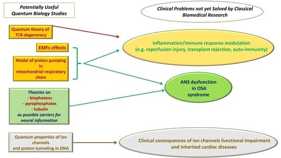
The following paragraphs will summarize advances of quantum studies relevant for biology on low frequency electromagnetic fields, on proton pumping in mitochondrial respiratory chain, on quantum theory of TCR-degeneracy, on theories on biophotons, pyrophosphates, or tubulin as possible carriers for neural information, and on quantum properties of ion channels and proton tunneling in DNA.
These findings might open new perspectives on the following problems that preclinical and clinical research has not been able to solve so far: problems related to inflammation and immune response, the relationship between OSA syndrome and ANS dysfunction, clinical consequences of ion channels’ functional impairment, and of inherited cardiac diseases.
In fact, such pathological processes involve a complex crosstalk among systems and organs [18,19,20,21,22], which makes it difficult the development of new effective therapies and would require a multiorgan approach (Figure 1, Table 2). Several therapeutic strategies and mechanisms of action are under investigation. Each is considered promising, but has not yet proved conclusive despite the great potential inherent; the recent hypothesis and discoveries of quantum biology could potentially open new perspectives on such biomedical issues.
In this review, authors do not mean to give any “answer” or “clear hypothesis” on possible quantum biology involvement in biomedical research, but rather, they hope to open a debate on whether quantum biology is developed enough to provide biomedical research a promising biophysical theoretic system, on which to base the search of answers not yet provided by current biochemical approach.

2. Methods

For this narrative review, electronic databases were used: PubMed, Scopus, and Google Scholar. Search terms: quantum biology, electromagnetic fields, quantum properties of protons and ions, information transmission in neurons, DNA point mutations, immune dysfunction, cardiovascular disease, neural dysfunction, stem cells, reactive oxygen species (ROS), OSA syndrome. Being one of the first reviews on this topic, the authors did not indicate any time limits. The inclusion criteria were: articles in English about quantum biology and focused on: problems related to inflammation and immune response, ANS dysfunction, clinical consequences of ion channels’ functional impairment and of inherited cardiac diseases, any study design, and articles published in peer-review journals. Exclusion criteria: articles about quantum mechanics that did not have some relevance to biology. Two independent authors (L.C. and V.R.) performed the research on an electronic database. In total, 201 articles of quantum biology were obtained, and 54 out of them were relevant and therefore were analysed (Figure 2). Three authors (L.C., A.B.Q., and V.R.) independently screened the title and abstract and in case of disagreements, a fourth author (N.L.) participated in achieving consensus. Data were extracted independently by six authors (L.C., A.B.Q., A.G., V.R., N.L., A.F.) including the following information: author and publication year, aim, results, conclusion. Any disagreement among the reviewers was resolved by consensus through meetings.

3. Results

3.1. Low Frequency Electromagnetic Fields

Interesting studies [23,24,25,26,27,28] described the effects of low frequency electromagnetic fields (EMFs) on stem cells and on ROS production, and hypothesized a future EMFs application in medical therapies. Usselman and colleagues [28] described an indirect methodology for investigating possible non-trivial quantum effects on mammal cells: they applied weak magnetic fields on rat pulmonary arterial smooth muscle cells (rPASMC) and, through Electron Paramagnetic Resonance (EPR) Spectroscopy and fluorescence techniques, observed a decrease of superoxide (O2•−) and an increase of hydrogen peroxide (H2O2) concentrations.
O2•− and H2O2 are often involved in oxidative stress, but have also a physiological role inside the cell. A possible physical modulation of their concentration inside a living cell is therefore of great interest. The mechanism that links O2•− consumption and H2O2 production is due to spin-correlated radical-pair behaviour (SCRPM) under radio frequency (RF) magnetic fields. In quantum mechanics, electron spin is not a classic mechanical rotation of a sphere, because it is hard to think in terms of its radius and angular velocity, being an intrinsic characteristic of the particle. Furthermore, in any atom the position of electron around the nucleus arises an angular momentum with quantized values in intensity and direction. The sum of orbital angular momentum and spin gives the total angular momentum of the atom that can interact with an external magnetic field. The radical-pair (O2•− and H2O2) mechanism describes the way a magnetic field, interacting with angular momentum quantization of the atoms, can affect the kinetics of reactions, also organic. This means that magnetic effect at quantum scale level influenced the O2•− and H2O2 production inside a mammalian living cell, thus showing a non-trivial quantum effect on cell metabolism and function. Authors hypothesized that the radical pair underwent “intersystem crossing” between singlet and triplet states and an applied properly tuned RF oscillating magnetic field (Zeeman resonance) could ultimately affect the relative amount of singlet (H2O2) and triplet (O2•−) reaction products (the reader can find a clear visualization of the “intersystem crossing” in the paper of Usselman et al. in Figure 2, page 2 [23]). These resonance effects on ROS product yields are considered a key manifestation of quantum biology. In summary, this study demonstrated that magnetic fields applied in living cells can modulate their oxidative state, through a direct action at quantum scale level, indicating a non-trivial quantum effect at biochemical level. This study has been developed, confirming the results in HUVEC (Human Umbilical Vein Endothelial Cells) and highlighting the implications for public health [23,27].
Interestingly, magnetic fields were also found to have an impact on stem cells functioning and differentiation; Gaetani and colleagues [25] found that cardiac stem cells from human bioptic specimens showed a significant increase in the expression of cardiac markers, at transcriptional, translational, and phenotypical levels, after 5 days of exposure to extremely low-frequency electromagnetic fields where a combination of static and alternate EMFs, tuned to Ca2+ ion cyclotron energy resonance (ICR), was applied to trigger the specific differentiation [25,29]. One of the possible explanations [30,31] is the hypothesis that EMFs could interact with endogenous alternate current electric fields in resistance-capacitive biological systems in a resonant manner.
Ventura et al. [24], for their part, showed that proper delivery of energy by electromagnetic field (at 2.4 GigaHz) by means of radioelectric devices was able to tune stem cell multipotency, to reprogram human skin fibroblasts into cardiac-, neuronal-, and skeletal muscle-like cells, and to revert stem cell senescence [32,33,34,35].

3.2. Proton Pumping in Mitochondrial Respiratory Chain and Quantum Theory of TCR-Degeneracy

-
Mitochondrial respiratory chain.
A mathematical model describing proton pumping in mitochondrial respiratory chain, published by Friedman and colleagues in this special issue [36], might contribute to enhance knowledge on mitochondria functioning, which is also responsible for ROS production. Mitochondria are essential for storing chemical energy in the form of adenosine triphosphate (ATP) [37,38], but the physical mechanism of the electron–proton energy remains extremely difficult to describe in detail. Friedman et al. [36] suggested a model, based on quantum mechanics calculations [36], with elastic forces facilitating proton transfer in mitochondrial complex 1 and found that the pumping of protons against the gradient can be obtained by means of conformation-mediated energy supply from electrons.
The proton pump of complex 1 of the respiratory chain is modelled as having a piston that has a positive charge at the two ends. The movements of the piston, assisted by the elastic forces, change the energy level in the system, allowing electron transportation. This represents a simple model that explains the pumping function of complex 1 to transport the protons from the side of lower concentration to the side of the higher concentration. Authors think that their model constitutes the underlying physical mechanism of electrostatic wave propagation along the membrane arm of complex I and the consequent proton pumping promoted by this wave and suggested that their unified model of respiratory chain functionality can provide a coherent framework for further research in the field.
-
Quantum theory of TCR-degeneracy.
TCR degeneracy [39] is defined as the property of a single TCR in the immune system of an organism of recognizing more than a million different peptides and interacting with several ligands, usually molecules present in the membrane of cells not belonging to the organism itself (e.g., external pathogens or cells belonging to a transplanted organ). The key point of this phenomenon is the protein-protein interaction between an antigenic peptide-major histocompatibility complex (pMHC), present in the “external” cells, and a TCR present in the organism’s immune system [40]; this TCR-pMHC interaction controls the function of self-nonself discrimination and regulates the probability of an immune reaction against microbic pathogens or against a transplanted organ.
Different theoretical approaches have been proposed to explain how this interaction can impact the downstream signaling in T cells and the immune system activation, but none of these were able to give a comprehensive understanding of this crucial interaction among cells. Hence, a quantum mechanical-based approach was adopted by Antipas et al. [41] to explain the TCR degeneracy, and to explain their different immunological avidity (Table 2). Quantum chemistry calculations were at the base of these authors’ studies.
They applied quantum mechanics to the interactions between the TCR and pMHC complex, considering the electronic structure of the proteins, including spin density which is varied according to the protonation of the N terminus in the peptide structure. In this perspective, there is “a cloud” of possible charge concentrations between the two peptides, at the level of sub-atomic structure, which modulates TCR degeneracy according to the specific situation and insult (for an interesting visualization of the concept, the reader is addressed to the Figure 1 of the reference Antipas et al. 2015 [41]). In authors’ works [41,42,43,44], statistics and quantum chemical calculations were shown to predict immunological responses.
In particular, the quantum approach was applied on Tax peptides (Table 2) from the human T-cell leukemia virus type 1 (HTLV-1), which show different immunogenic activity even though they share near-identical stereochemistry and interact with the same TCR. The quantum approach was able to explain how antigens like Tax peptides have different degrees of immunogenicity with small differences in the chemical structures. It solved this paradox by digging deep to the atomic structure and electronic features of the antigen’s peptide structure, which is intrinsically reflected by the principles of quantum mechanics. The quantum mechanical calculations, taking into consideration the electronic structure of the primary peptide structure of the antigen, indicated that atomic coordination in the TCR-pMHC complex reflects the immunological functionality of the different variants of Tax antigen. The work of Antipas and colleagues is based on the modern chemistry, which is based entirely on quantum mechanics.

3.3. Theories on Biophotons, Pyrophosphates, or Tubulin as Possible Carriers for Neural Information

Physicists and quantum biologists proposed some hypotheses which could provide new perspectives on how neural information is carried through the brain, toward ANS and finally to the organs. Three potential “carriers”, with quantum properties, have been suggested:
  • biophotons (an experimentally confirmed optical communication among cells [45,46])
  • pyrophosphate in the “Posner molecule”
  • microtubules, present in the cell structural skeleton
Biophotons: Kumar and colleagues [47] explored the question on whether biophotons [45,46] might carry information in the brain, in addition to the known electro-chemical signals, and proposed that myelinated axons might serve as potential biophotons waveguides. Since optical communication can also transmit quantum information, it has been considered a possible role of superposition or entanglement in neural functions (Table 1) [47]. The problem of environmental decoherence, which might rapidly destroy quantum effects, seemed to be partly addressed by the evidence that nuclear spins can have tens of milliseconds coherence times in the brain and that, at room temperature, it was demonstrated to be a long-lived nuclear spin entanglement [48,49]. The thesis of photon entanglement through the brain is also supported by Shi and colleagues [50] who found the photon coherence preservation after propagation through rat brain tissue slices.
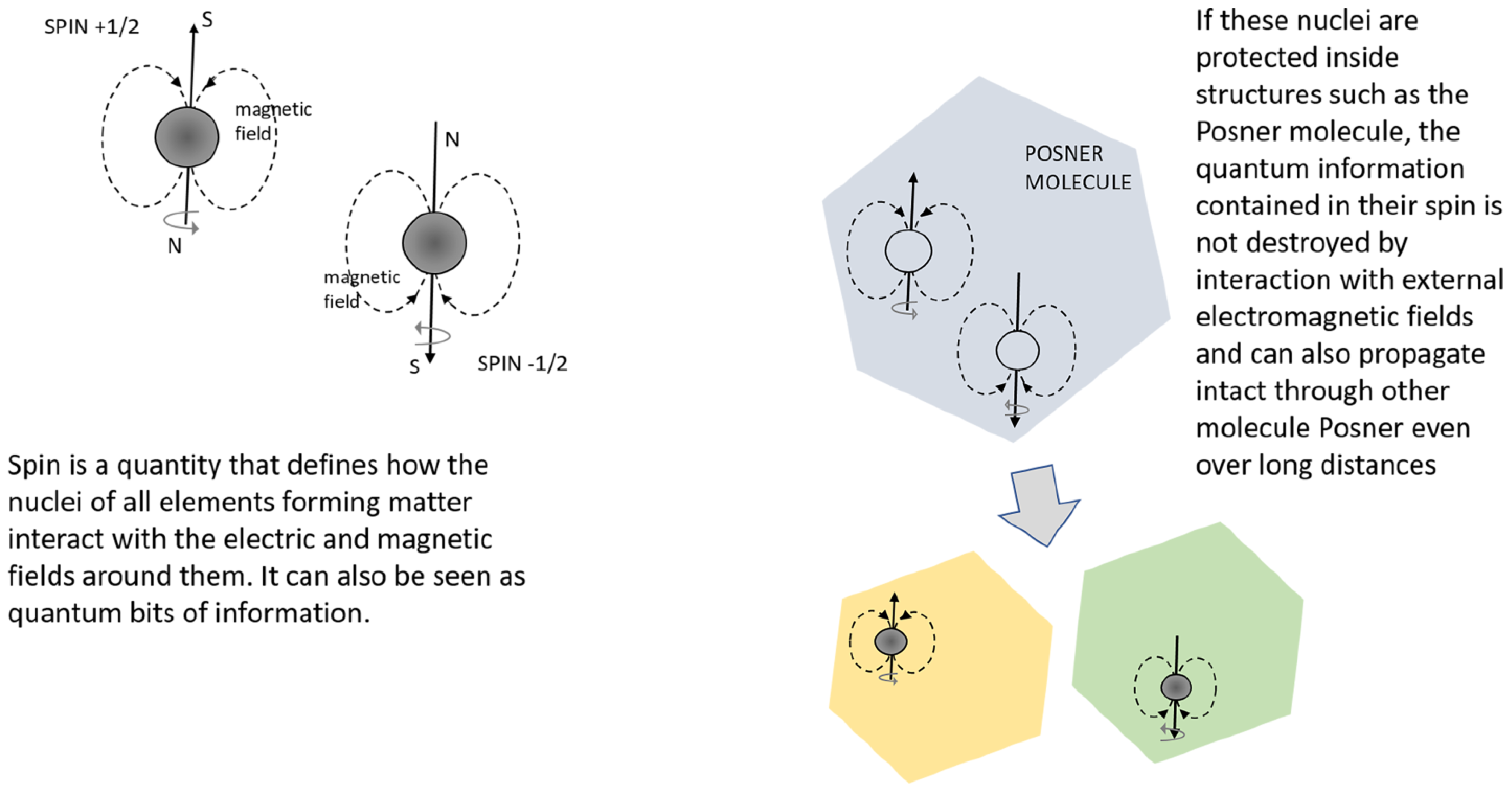
Pyrophosphate: Fisher [51] hypothesized that the function of carrying information might be performed by a “Posner molecule” Ca9(PO4)6, whose mechanism has been recently explained by Swift et al. [52]. In particular, pyrophosphate, involved in several biochemical reactions in the cell [53,54], would produce two quantum entangled phosphates after enzymatic hydrolysis: their incorporation into calcium phosphate Posner molecules gives the quantum entangled property to the Posner molecules. A calcium-mediated glutamate release from presynaptic neurons and non-local quantum correlations in post-synaptic firing among neurons would therefore occur, allowing the information to be propagated intact over long distances (Figure 3) [55].
Microtubules, one of the major components of the cell structural skeleton, were also proposed to be potential carriers of brain information, considering the quantum property of tubulin to be in two superposition states. In fact, in 2013, Sahu and colleagues [56] described the microtubule as a memory-switching element with multiple symmetries and demonstrated how every single protein acts as a memory storage unit. In particular, the microtubule was dropped in a pre-prepared chip, with an atomic force microscope tip to measure the conductivity of a single microtubule, purified from eventual impurities.
Accordingly, when a voltage bias was applied on a single microtubule, a nearly perfect square relationship between current and voltage was obtained. This implies that the conductivity of a single microtubule changed suddenly at a certain voltage bias. This electrical behaviour shows the memory storage and switching capabilities of the microtubule. Moreover, the orientation of the tubulin proteins on the surface of the microtubule, which determines the dipolar direction of the tubulin, changed with the applied voltage bias, indicating that the protein arrangement symmetry was related to the conducting state of the microtubule. This correlation between the dipolar direction and the conducting states provided insights about how a level with a multi-superimposed state originated and survived.
This seems to corroborate the theory of consciousness named Orchestrated Objective Reduction (Orch-OR) conceived by Nobel laureate Sir Roger Penrose [57] and Stuart Hameroff in the mid-1990s [58]. This theory considers the mind as a quantum computer able to manage multilevel layers of information and the according reactions and suggests that quantum states might extend through the nervous system by entanglement between adjacent neurons through gap junctions. There is an important open debate around this theory, which by exploring the nature of mind and consciousness, is certainly revolutionary, but deserves to be mentioned. It is theoretical and, as the other mathematical models, needs to be experimentally demonstrated.

3.4. Quantum Properties of Ion Channels and Proton Tunneling in the DNA

-
Ion Channels.
Mathematical models of ion channels provided by quantum calculations give molecular descriptions of their structures at a sub-atomic level that can shed light to some aspects of channels function, involving ion conduction and electrical properties of the channels. The works that exploited quantum mechanics in this field can be classified into two main categories:
(1)
The studies focused on the selectivity filter of voltage-gated channels (the narrowest part of the conduction pathway through the pore of the channels), which discriminates between different ions.
(2)
The studies focused on the intracellular hydrophobic gate, which regulates the ion flow and the overall conductance of the channel.
The studies of point (1) implemented the quantum concept of non-locality on ions while moving in the selectivity filter, to explain the mechanism behind the filter selectivity and its ability to discriminate between ions at a high flow rate: Summhammer, Salari, et al. [59,60,61,62], by using the Berneche–Roux model of the bacterial KcsA model channel, mathematically demonstrated solutions of the Schrödinger equation representing the interaction of a single potassium ion within the surrounding carbonyl dipoles. They showed that if the quantum wave behaviour of potassium ions is considered, the wave-packet can propagate in the selectivity filter, demonstrating a quantum non-local distribution of potassium ions, which become highly delocalized in the filter region of proteins at warm temperatures [59,60,61,62]. Interestingly, this quantum mechanical approach could provide a consistent explanation of the ability of the selectivity filter to differentiate between ions such as between potassium and sodium ions. The explanation states that the different magnitude of kinetic energy loss (“the cooling effect”) requires a different oscillation frequency of the carbonyl oxygen groups that line the selectivity filter. Hence, it is expected that each ion has a different magnitude of the cooling effect and unique oscillation frequency of carbonyl oxygen groups [59,60,61,62]. Authors suggested that, from a mathematical point of view, it is no longer possible to consider an ion to be at one exact position at a given time and that some features of quantum dynamics, like non-local effects, seem to be essential to resolve the discrepancy between calculations reported from classical thermodynamics, and transition rates of ions through channel proteins that have been experimentally observed.
The first study of point (2) was published by Qaswal in 2019 [63] and focused on the intracellular hydrophobic gate of the channels, attempting to apply the quantum concept of tunneling to this cell structure. In this study, the closed gate is illustrated as a potential barrier that should impede the passage of ions; however, ions have a non-zero probability to pass through the closed gate via quantum tunneling. Classically, the closed gate forms an energy barrier whose energy is higher than the kinetic energy of passing ions; hence, the closed channel blocks the permeation of ions and the conductance of the channel will be zero, due to an absent flow of ions through the closed gate. However, applying the principle of quantum tunneling on the closed gate and its corresponding ions, it has been found that ions can pass through the closed gate via quantum tunneling and this passage is a probabilistic event [63]. The probability of quantum tunneling of ions through the closed gate can be calculated by the following equation [63,64,65]:
P = e 2 8 m 3 w g ( g E K ) 3
where m is the mass of the ion, is the reduced Planck constant, w is the length of the gate, g is the barrier energy of the closed gate, and EK is the kinetic energy of ions when they reach the closed gate.
From Equation (1), the tunneling probability of ions is inversely related to the mass of the ion, the length of the gate, and the energy of the gate, while it is proportional to the kinetic energy of the ion. The unique feature of quantum tunneling transport is that the ions do not have to get higher kinetic energy than the energy of the gate to pass. Actually, the ions have a probabilistic passage through the gate at any value of kinetic energy according to Equation (1). The quantum tunneling of ions enables ion channels to get a quantum conductance that can affect the membrane potential [63,64,65].
It has been found that sodium ions and potassium ions have low quantum conductance values at normal physiological parameters, if they are compared with values of classical conductance when the channel is open [63]. Moreover, these low quantum conductance values were not able to affect the resting membrane potential of excitable tissues [63]. This indicates that the quantum behaviour of sodium and potassium ions, which are the main ions that determine the resting membrane potential, not only does not affect normal physiology, being compatible with normal cellular functions during the resting membrane potential, but even allows for a better explanation for several observed physiological and pathological phenomena [66,67].
Further interesting works [64,65] introduced a new concept called “quantum electrochemical equilibrium” which includes, in addition to chemical and electrical (voltage) gradients, a third gradient called “quantum gradient”. This concept can explain the potential therapeutic effects of lithium and magnesium ions [68,69,70] (the reader is addressed to these references for details).
-
Proton tunneling.
Proton tunneling is a specific quantum effect involving the instantaneous disappearance of a proton in one site of a potential barrier and the appearance of the same proton at the other side. Electron tunneling is a well-known phenomenon. However, a proton has a mass about 2000 times more than that of an electron, therefore, the probability of tunneling is lower; nevertheless, proton tunneling still occurs and it has been observed [14]. Proton tunneling is usually associated to hydrogen bonds, present in many molecules, including DNA. When a hydrogen atom loses its electron in a bond, it become equivalent to a proton, which can tunnel through a barrier. Slocombe and colleagues [71] made calculations about the proton transfer in DNA nucleobases, obtaining a quantum-corrected rate equation, which can justify quantum tunnelling of the protons. They described the tautomeric double proton transfer reactions in the DNA base pairs at computational level, and considered this mechanism one of the possible explanations of spontaneous point mutations, being the transition state stable enough to be involved in the replication processes. The first who proposed the proton tunneling model for the origin of spontaneous point mutations in the DNA base pairs was, however, Löwdin in 1966 [72,73]. Löwdin suggested that the genetic alphabet stored in the DNA double helix depends upon the stability of the hydrogen-bonded base pairs and the motion of the proton(s) along these hydrogen bonds [73]. Each proton belonging to a given hydrogen bond between the A-T or G-C base pairs may be transferred via quantum mechanical tunneling from the position corresponding to the normal base pair to the position corresponding to the “rare” one, the latter implying a lesion of the genetic alphabet [72]. The formation of mismatches that may produce spontaneous mutations in the genetic code has been hypothesised to be due to the formation and pairing of rare tautomers of the DNA bases [72], especially during the DNA replication step [71]. Although the replisome is subjected to high fidelity checkpoints during DNA replication, the quantum micro-effect of proton tunneling may create a chance for the introduction of a mismatch in a strand, which will be then maintained in the double helix of the following DNA copies. Furthermore, the tunneling time of protons in the base-pair nucleotide has been calculated [74]. Two types have been obtained: the first one is the dwell time and the other one is the entropic time. According to the authors, the entropic time seems more relevant because it becomes shorter as the energy barrier decreases. This time requires an experimental confirmation, but it sounds acceptable because the obtained entropic time of proton tunneling is within the order of 10 12 s, which is congruent with time scales of the molecular transitions.
In the next paragraphs, we will try to figure out if the quantum models detailed above may help current medicine to unravel some pathophysiological aspects still unclear, and may be considered in those fields where biochemical and pharmacological approaches struggle to understand mechanisms of action and to find new effective therapies.

3.5. Problems Related to Inflammation and Immune Response

3.5.1. Stem Cells Therapy

The works on low frequency electromagnetic fields (Section 3.1) and on proton pumping in mitochondrial respiratory chains (Section 3.2) might help in resolving the two main challenges of stem cell transplantation therapy: a—the differentiation and the efficiency of transplanted stem cells, and b—the control of inflammation through modulation of ROS metabolism [23,24,25,27,28].
Stem cells have been used as possible therapy to replace and regenerate injured cells thus recovering, at least in part, organ functions [75,76,77,78,79,80]. The Nobel Prize winning discovery of induced pluripotent stem cells (iPSCs) represented a turning point in the field of stem cells [81], since iPSCs are obtained by reprogramming any adult somatic cells [82], which possess almost the same pluripotency property as embryonic stem cells. At present, stem cells represent a potentially resolving approach [78,80,83,84,85]; nevertheless, the inflammatory microenvironment present in the diseased organ, caused by, among others, ROS [86,87,88,89], can cause massive stem cell death [75,76]. Moreover, residual undifferentiated stem cells may form tumors and proliferate [90]. Finally, very often, there is a low efficiency or incomplete reprogramming of transplanted stem cells [79], despite progress being made [82,91].
The studies of Gaetani and Ventura [24,25,32,34,35] suggested that EMFs applied on stem cells before transplant might improve both the expression of organ markers and an effective reprogramming of stem cells, also reverting their senescence. Future experiments performed on preclinical models, like 3D organoids or laboratory animals, could lead to a possible clinical application. The works of Usselman and colleagues [28] could be applied to decrease the production of superoxide (O2•−), thus reducing the inflammatory-related stem cell death. Finally, the work of Friedman et al. [36] on proton pumping in mitochondrial respiratory chains might improve our knowledge of how ROS are produced in mitochondria, thus opening possible applications in ROS modulation.
However, it is important to take into account that these studies are in vitro or represent a mathematical prediction, and that the mass of biological material (i.e., organs and tissues) surrounding the cellular system might reduce the effects observed in the cited papers.

3.5.2. Anti-Inflammatory Agents and Transplants for Organ Failures

Growing evidence indicates a role played by immunity in pathologies not directly related to infection events [19,21,92,93,94,95,96,97,98,99]. In heart or brain ischemia, for example, the beneficial effects of reperfusion after an ischemic event may be compromised by what is called reperfusion injury, an exaggerated innate immune response triggered by a rapid oxygen availability after a period of hypoxia [92,98,100,101,102,103], which is characterized by an increase of ROS and toxic molecules damaging lipids, DNA, RNA, and proteins [18,104]. Despite considerable efforts and undoubted progress, effective anti-inflammatory and anti-oxidant therapies, mitigating the inflammatory response without compromising the repair mechanism, are not yet available. The main issue was a poor translation between pre-clinical studies and clinical results. For example, the anti-inflammatory agent Etanercept and the anti-oxidant molecules superoxide dismutase and catalase, failed to show the therapeutic effects seen in animals when given to patients [105,106,107]. These experiences suggest an insufficient understanding of the mechanisms of action of immune response, essential to achieve effective pharmacological modulation. In this regard, the quantum biology studies on ROS production [36] and modulation [28] might help research aimed at understanding one of the mechanisms of action of immune response: oxidative stress. An immediate clinical application of EMFs on humans suffering oxidative stress after reperfusion injury is perhaps still too far, but theoretically speaking, multidisciplinary teams might create and patent EMFs-based devices able to reduce inflammation-related ROS production.
Another extremely important issue medical research has to deal with is the problem of organ rejection after transplant. Solid organ transplantation is a lifesaving therapy for patients with end-stage organ failure, like kidney, heart, liver, lung, or pancreas extreme dysfunction. Unfortunately, about 50% of patients undergo organ rejection within ten years after transplant and the problem of organ donors’ availability is a significant limitation point [108]. In the organ rejection, the donor’s organ undergoes severe inflammation, fibrosis, and, finally, damage and rejection [108,109,110]. As described previously (Section 3.2), TCR-degeneracy is the key point of this process. Despite active research [111,112], there are no therapies that can directly prevent recruitment of several inflammatory cells including T cells, and the promising results obtained by targeting the adaptive immune response did not allow an optimal long-term graft survival rate [39,40,113]. If confirmed, the described work of Antipas and colleagues [41,42,43,44] on the quantum behaviour of the TCR-pMHC complex could start pioneering studies on mechanisms of actions responsible for organ rejection and for other immune dysfunctions including auto-immune diseases, where an organ is attacked by its own immune system. These studies should be deepened, with the aim of describing in detail the mechanism of action of TCR-degeneracy and TCR-pMHC complex interaction, in order to find a way to modulate it. The turning point would be to “inform” TCR that the transplanted tissue or the organ affected by autoimmune pathology, is not a harmful external pathogen, but a tissue to be accepted as “self”.

3.6. OSA and Alterered Cardiovascular Autonomic Regulation

OSA is a sleep-disordered breathing characterized by frequent episodes of complete or partial occlusion of the upper airway, causing intermittent hypoxia during sleep and sleep fragmentation. Such frequent cycles of hypoxia and reoxygenation have been associated with enhanced sympathetic activation, inflammatory activation, and increased oxidative stress, leading to cytokines and free radical production [114]. Furthermore, experimental evidence in animals demonstrated that intermittent hypoxia can activate an oxidative cascade (ROS-Carbon Monoxide-H2S signaling cascade) potentially affecting important physiological parameters like blood pressure through the stimulation of carotid body neural activity [115].
Among the changes induced by OSA-related intermittent hypoxia, altered cardiovascular autonomic regulation represents the main physiopathological basis for the hypothesised causal relationship between OSA and cardiovascular diseases. In fact, patients with OSA exhibit baroreflex sensitivity (BRS) reduction, increased heart rate variability, and elevated blood pressure, indicating ANS dysfunction [116,117].
Furthermore, OSA can cause debilitating symptoms like restless sleep, daytime sleepiness, and mood-related suffering such as irritability, anxiety, and depression [118]. Such symptoms can be the consequence of an impaired nervous system regulation: Hilton and colleagues [119] showed that OSA syndrome depresses waking vagal tone, which may compromise the myocardium during periods of heightened sympathetic activity.
Thus, a better understanding of the autonomic regulation of the cardiovascular system in OSA could unhide potential additional mechanisms and help researchers to explain the increased incidence of cardiovascular disorders in sleep apnoea/hypopnoea syndrome patients.
In particular, there are two important questions related to ANS modulation in OSA that deserve to be answered:
(1)
despite numerous studies demonstrating the association between OSA and hypertension, it is still unclear why effective OSA treatment with continuous positive airway pressure (CPAP) does not consistently improve blood pressure;
(2)
it is unclear why a percentage of OSA patients, who are adherent to OSA treatment, continue to suffer from residual excessive daytime sleepiness.
In order to address these two issues, it would be essential to quantify the activities of the sympathetic and parasympathetic nervous systems in order to better understand ANS dysfunction in each tissue and district of the organism. In detail, it would be helpful, from a pathophysiological perspective, to trace the information carried by neurons from the brain stem to the periphery and vice-versa and to study how ANS maintains homeostasis in different organs and tissues. At present, ANS can be measured indirectly through heart rate variability or catecholamines secretion [120], whilst a direct non-invasive tracing is currently unavailable.
With the current knowledge on nervous system functioning, based on the well-known electro-chemical signals, it is difficult to find solutions for the above-mentioned clinical problems. Therefore, new theories arose in order to understand more in depth how the nervous system works; in particular, how neural information is carried along the systems.
Among the experimental theories that can be applied to directly measure ASN activity, biophotons, the Posner molecule, and microtubules deserve to be mentioned (see Paragraph 3.3 [47,51,56].
Biophoton emission is an ultra-weak emission of optical light, namely of electromagnetic energy, which is found to be a common phenomenon in biological systems and in humans, where it is also correlated with pathological conditions [121] and ROS generation [45,46]. One of the most important roles of this ultra-weak optical emission in living organisms (comparable to the light emitted by a candle far away) seems to be related with information transfer [122]. Nevertheless, these ultra-weak signals can be measured only on the surface of the organisms. If future research confirms biophotons as carriers of neural information “inside” the nervous system, and finds a way to trace them, it might be possible to measure and analyze cardiovascular autonomic regulation in many different cardio-respiratory disorders.
The Posner molecule is a cluster having a central Ca2+ ion surrounded by six phosphate PO43− anions. According to Fisher and Weingarten [51,55], when pyrophosphate (a molecule widely present in the organism) undergoes hydrolysis, two molecules of phosphate are produced and the result is that the nuclear spins of their phosphorus remain predominantly quantum entangled and become incorporated into Posner molecules. This process would produce quantum entangled Posner molecules which can propagate intact information through other Posner molecules: for example, inside glutamatergic neurons, even over long distances. A device able to detect it might follow information through the nervous system and would help in those situations where neurological functions are strictly related to pathological symptoms, as happens in OSA [123].
Microtubules are structures constituted by polymers of tubulins, which are the base of the cytoskeleton, providing structure and shape to eukaryotic cells. As described above, the tubulin seemed to have the quantum property of storing information and of instantaneously propagating it through superposition and entangled states. Should neural information be carried by this way, an eventual device tracing such a transmission might provide a detailed “image” of the entire crosstalk between ANS and organs. This might deepen the knowledge of the neuropathology of sleep-wake disorders, which involve brainstem, hypothalamus, and basal forebrain [123].
Although different as far as theoretical bases are concerned, these three theories could in part coexist with the aim of revolutionizing the medical technology available to clinicians and allowing new means to further explore ANS alterations.

3.7. Ion Channels Dysfunction and Inherited Cardiac Diseases

-
Ion channels dysfunction.
Ion channels are pore-forming proteins inserted in the cell membrane, which allow ions, typically Ca++, Na+, K+, Cl, to cross the membrane. They are present in the plasma membranes of all cells and have multiple essential functions, including the regulation of membrane potential through the control of the ions flow crossing. There are many types of ion channels, and the crossing of ions through the membrane is one of the key functions of the most important biological processes such as cardiac, skeletal, and smooth muscle contraction, nutrients transport, or neurotransmitter release [124,125]. Disruption of their normal functions, caused by genetic mutations or by other mechanisms like the autoimmune attack on the ion channel [126,127,128], can have devastating, often fatal consequences for the organism; cystic fibrosis, long QT syndromes (LQTS), sudden infant death syndrome, calmodulinopathy, cardiac arrhythmias, and amyotrophic lateral sclerosis (ALS), are all characterized by ion channels abnormalities (interestingly, in ALS, much evidence suggests that therapies targeting ion channels may be able to contrast motor neurons’ excitotoxicity) [129,130,131,132,133,134].
For this reason, ion channels are frequently the target of drugs and therapies [128,135]; however, for many ion channels disorders, there are no pharmacological therapies available.
At this stage of quantum biology studies, there are only mathematical models that would provide a new theory on ion channels functioning and related mechanisms of actions. Recent theoretical models [64,66,67] have suggested that different factors like hypoxia or inflammation implicated in the pathogenesis of excitability disorders, such as cardiac arrhythmias, epilepsy, and chronic pain disorders, can decrease the energy of the ion channel gate. In their calculations, authors showed that the gate-energy decrease caused by such factors can significantly augment the quantum tunneling to the degree that sodium, potassium ions, and protons will be able to depolarize the resting membrane potential. The state of depolarized membrane potential represents a state of hyper-excitability that predisposes cardiac cells to develop arrhythmias, and neurons to produce epilepsy and pain disorders.
Additionally, the model of quantum tunneling has been applied on protons during acidosis and its related pathologies [66]. The mathematical calculations have shown that protons quantum tunneling through the closed sodium channels can depolarize the membrane potential, hence, cardiac arrhythmias or even cardiac arrest could happen. As the tunneling probability implies that the quantum particle has a non-zero probability to pass through a barrier, it is important to assess whether the event of quantum tunneling falls within the time scale of the biological function. Particularly, the quantum tunneling through the closed channels should result in a tunneling time sufficient to produce tunneling currents congruent with those that affect the membrane potential of cells. The usual unitary currents of ion channels that can affect the membrane potential are measured within pico-amperes (pA = 10 12 A) or even less and this current is generated when the ion passes within a time scale of 10 7 s. However, longer passage time and thus lower current can change the membrane potential. Accordingly, the tunneling time through the closed gate can be calculated by the following equation
t t u n n e l i n g = h E K P
where h is the Planck constant, E K is the kinetic energy of the ion, and P is the tunneling probability. To reach the time scale of 10 7 s, the tunneling probability should be within the order of 10 7 , which has been demonstrated mathematically and theoretically in these papers [63,64,65,66,67,68,69,70].
Finally, the quantum-tunneling-induced membrane depolarization may be the universal mechanism behind the cellular effects of lithium ion including its therapeutic effect to treat bipolar disorder [68,70].
If the mechanisms of quantum behaviours of ions and protons through the channels are confirmed, they will probably improve the understanding of mechanisms of action, likely leading to technological repercussions in the search for therapies that currently do not exist.
-
Inherited Cardiac Diseases.
Rare diseases with a genetic origin are often caused by a point mutation in the DNA, i.e., the substitution of a single nucleotide (base) at the DNA level resulting in an amino acid change in the corresponding protein. Although these changes are microscopic, they may have a clinical devastating impact depending on the position of the substitution and on the role of the protein.
In the field of inherited cardiac diseases, there are at least two emblematic disorders that should be mentioned for their high risk of sudden cardiac death and partial response to therapy: the arrhythmogenic cardiomyopathy (ACM) and the calmodulinopathy [135,136,137].
The genetic bases of ACM relate to the junction structures that allow a tight connection among adjacent cardiomyocytes and their simultaneous beating in the heart. Mutations in the genes coding for junctional proteins underlie the development of ACM [138,139,140]. ACM disease affects apparently healthy young people, including athletes and sporty people, and the first manifestation could sometimes be sudden cardiac death. All the therapies currently employed in ACM patients partially alleviate symptoms and the risk of sudden cardiac death but are not able to prevent the progression of the disease [138].
The other fitting example concerns the “calmodulinopathy”, caused by a point mutation in any of the three genes encoding calmodulin (CaM), which is a Ca2+ signalling protein ubiquitously expressed, which, in the heart, modulates different ion channels and participates in several cellular processes [141].
Calmodulinopathy usually manifests at an early age (around 4 years old), with extremely severe and often fatal symptoms [142]. The mechanistic implications of CaM mutations have basically been studied in vitro, since no animal models are available. The impairment of two different cardiac ion channels interacting with calmodulin (the L-type Ca2+ channel Cav1.2 and the ryanodine receptor 2) is responsible for the main disease phenotypes observed: the LQTS and the Catecolaminergic Polymorphic Ventricular Tachycardia [143,144]. One of the major issues in calmodulinopathy is the lack of effective therapies able to prevent life-threatening arrhythmias, despite several therapeutic strategies, both pharmacological and surgical, having been tested [145,146,147], and despite the continuous efforts of precision medicine research [143].
Overall, inherited arrhythmia syndromes with a high risk of sudden cardiac death demand a deeper understanding of the molecular mechanisms underlying the disease, to explain why some penetrant genetic mutations have such a severe manifestation. We think that a major comprehension of these biological phenomena should start from basic inter-disciplinary research that is essential both for translational applications and for the search of effective therapies.
Despite the fact that a clinical application of quantum biology findings to the rare genetic diseases is too far away, some theories explaining the generation of spontaneous mutations based on the quantum effect deserve to be deepened. In particular, proton tunneling (Paragraph 3.4) has been proposed as one of the possible mechanisms that may be responsible for the spontaneous formation of point mutations in the DNA [71,72].
The magnitude of the biological molecules and the distances of atoms involved in the basic chemical bonds within these molecules suggests the possibility that non-trivial quantum effects may underlie the well-known physical-chemical effects already described in this context. However, the extent of quantum effect for the effective formation of a point mutation with respect to other forces/mechanisms, like deamination pathway or the polarisation interactions of aqueous solutions [71], is not known. In addition, the modelling systems employed in different studies remain inevitably at a computational level, and refer to a simplified reproduction of the different chemical bonds within a molecule, missing important aspects related to the complexity of biological molecules and to their 3D structures.

4. Conclusions and Perspectives

The idea that quantum physics may have a role in biology is not recent: Bohr, Frohlich, Schrödinger, and Penrose dealt with physics aspects of life in a series of lectures and publications [57,148,149,150]; Dicke, Del Giudice, Jibu, Kobe, and Cefalas laid the foundation of basic concepts needed in quantum biology [2,3,4,5,6,7,8,9,10], and quantum biology has rapidly been extended to the human cell [23,71].
Quantum studies, including mathematical models and experimental investigations, appear potentially useful to better approach some clinical problems.
In particular, research on the effect of EMFs on living cells and ROS production, studies on proton pumping in mitochondrial respiratory chains, and a quantum approach to TCR-degeneracy, might be useful to better understand the mechanisms of action of inflammatory-related organ injury, transplant rejection, and oxidative stress in OSA.
Quantum hypotheses on biophotons, pyrophosphates, and tubulin as possible carriers of neural information could provide new perspectives to improve the knowledge of the impact of sleep-disordered breathing on ANS functioning and of neuroscience in general. Finally, studies on quantum properties of ion channels and proton tunneling in the DNA might, respectively, improve the knowledge about ion channels functioning and DNA mutations occurrence, implicated in ion channels and inherited cardiac diseases.
Other studies on biological information in the fields of consciousness and evolution of the species have seriously considered the quantum approach [5,11,151,152], making extremely clear how the remarkable quantum physics successes are now being entered into the biomedical field. As an example, the recent article of Yang and colleagues [153], which came out while this review was in preparation, directly observed an ultrafast hydrogen bond strengthening in liquid water when a 100-nm water sample was excited with an approximately 3315 cm−1 infrared laser pulse. Experimental results indicated that when the water molecules were hit by photons, they generated specific vibrations. Initially, these vibrations led the hydrogen atoms to attract those of oxygen and immediately after they led to the opposite action: the hydrogen atoms pushed oxygen atoms away with force, thus expanding the space between the molecules. This phenomenon is known as the so-called nuclear quantum effect, which was thought to be at the center of many strange properties of water, but it was never been directly observed [153]. A thorough revision of this recent article is beyond the scope of the present work; however, this discovery shows how advances in quantum mechanics are approaching the “core” of biology itself: all living organisms are made up of water for a percentage ranging from about 70% to over 90%. Many human pathologies like auto-immune diseases or organ ischemic injury compromise, for example, connective tissue function and organ extracellular matrix, which contain a high percentage of water [154,155]. If further studies on quantum properties of water demonstrate non-trivial effects on living cells, perspectives for biomedical research will be remarkable. The same can be said for studies on proton tunneling in enzyme reactions [156,157]: if they are experimentally confirmed and observed in multicellular organisms or even in superior animals, they would have incredible implications for biomedical research.
In conclusion, in the present review, authors described the challenges facing the biomedical community when addressing clinical problems related to immune response, ANS and OSA, ion channels dysfunction, and genetic diseases.
In the light of these difficulties, an innovative and promising approach is required and quantum biology might provide the necessary new theoretical basis.
In view of the accumulated experimental, theoretical, and computational evidence on the influence of quantum effects on the cell physiology, it seems clear that a substantial amount of research needs to begin to uncover the interface between the quantum phenomena at the sub-cellular nanoscale level and the biochemical effects observed at macroscopic scale. Accordingly, specific experimental tests performed by multidisciplinary teams would represent a great opportunity and might provide the scientific platform for concrete applications of quantum biology resources to biomedical research and clinical practice.

Limitations

Living systems are macroscopic, complex, hot, wet, and noisy; therefore, there is the important challenge of verifying whether quantum coherence is maintained as the complexity of the living system increases, and what type of mechanisms might protect quantum effects against the harsh environments in the biological systems. The question whether complex living entities, like pluricellular organisms or even animals, can manifest non-trivial quantum effects remains under a strong debate [158]. In particular, Tegmark [159] disagreed with Sir Roger Penrose and colleagues’ theory of the brain as a quantum computer on the base of calculation of neural decoherence rates, which suggested that cognitive processes can be considered as a classical and not as a quantum system. Davies in 2004 [160] addressed the issue of “decoherence evasion” which should be reached by the biological system through a quasi-isolation from the decohering environment, and the possibility of existence, inside the living entity, of decoherence-free subspaces. An interesting debate on the topic was published in 2008 [161]. Despite part of these issues being addressed by the experimental works cited in this review, a definitive evidence of non-trivial quantum effects in complex organisms, in particular in humans, is still lacking. Further studies are needed, especially when a possible medical application is considered.

Author Contributions

Conceptualization: L.C. methodology L.C. and D.R. software N.L. and A.B.Q.; validation: L.C., V.R., N.L., A.B.Q., A.F., C.L. and M.P.; formal analysis: V.R., N.L., A.B.Q. and A.F.; investigation L.C., A.B.Q., V.R. and N.L.; resources: G.P. and V.S.; writing—original draft preparation: L.C.; writing—review and editing: L.C., V.R., N.L., A.B.Q., A.G., D.R., C.L., M.P., P.B., V.S. and G.P.; supervision: L.C. and G.P. All authors have read and agreed to the published version of the manuscript.

Funding

This research received no external funding.

Institutional Review Board Statement

The studies involving living organisms cited in this review were conducted according to the guidelines of the Declaration of Helsinki, and approved by the Institutional Review Board (or Ethics Committee) of the different Institutions.

Acknowledgments

Authors are grateful to Lidia Cova for her precious support.

Conflicts of Interest

V.S. received compensation for consulting services and/or speaking activities from AveXis, Cytokinetics, Italfarmaco, Liquidweb S.r.l., and Novartis Pharma AG. He receives or has received research support form the Italian Ministry of Health, AriSLA, and E-Rare Joint Transnational Call. He is in the Editorial Board of Amyotrophic Lateral Sclerosis and Frontotemporal Degeneration, European Neurology, America Journal of Neurodegenerative Diseases, Frontiers in Neurology. L.C. received compensation for consulting services as Scientific Member of Animal Welfare Body of ENVIGO-(Bresso-Milan-Italy). She is Topic Board Member of International Journal of Molecular Sciences (MDPI) and Topic Editor of Frontiers in Physiology: Integrative Physiology—Clinical and Translational Physiology. The funders had no role in the design of the study; in the collection, analyses, or interpretation of data; in the writing of the manuscript, or in the decision to publish the results.

References

  1. Al-Khalili, J. Life on the Edge: The Coming of Age of Quantum Biology; Bantam Press: London, UK, 2014; ISBN 0593069323. [Google Scholar]
  2. Dicke, R.H. Coherence in Spontaneous Radiation Processes. Phys. Rev. 1954, 93, 99–110. [Google Scholar] [CrossRef] [Green Version]
  3. Del Giudice, E.; Doglia, S.; Milani, M. A Collective Dynamics in Metabolically Active Cells. Phys. Scr. 1982, 26, 232–238. [Google Scholar] [CrossRef]
  4. Del Giudice, E.; Preparata, G.; Vitiello, G. Water as a Free Electric Dipole Laser. Phys. Rev. Lett. 1988, 61, 1085–1088. [Google Scholar] [CrossRef] [PubMed]
  5. Jibu, M.; Yasue, K. Volume 18, Numbers 2 and 3, Spring and Summer (special issue): Understanding Tomorrow’s Mind: Advances in Chaos Theory, Quantum, Theory, and Consciousness in Psychology by Larry Vandervert (Editor) American Nonlinear Systems. J. Mind Behav. 1990, 11, 247–258. [Google Scholar]
  6. Kobe, S.; Dražić, G.; Cefalas, A.C.; Sarantopoulou, E.; Stražišar, J. Nucleation and crystallization of CaCO3 in applied magnetic fields. Cryst. Eng. 2002, 5, 243–253. [Google Scholar] [CrossRef]
  7. Kobe, S.; Dražić, G.; McGuiness, P.J.; Meden, T.; Sarantopoulou, E.; Kollia, Z.; Cefalas, A.C. Control over nanocrystalization in turbulent flow in the presence of magnetic fields. Mater. Sci. Eng. C 2003, 23, 811–815. [Google Scholar] [CrossRef]
  8. Cefalas, A.C.; Kobe, S.; Dražic, G.; Sarantopoulou, E.; Kollia, Z.; Stražišar, J.; Meden, A. Nanocrystallization of CaCO3 at solid/liquid interfaces in magnetic field: A quantum approach. Appl. Surf. Sci. 2008, 254, 6715–6724. [Google Scholar] [CrossRef]
  9. Cefalas, A.C.; Sarantopoulou, E.; Kollia, Z.; Riziotis, C.; Dražic, G.; Kobe, S.; Stražišar, J.; Meden, A. Magnetic Field Trapping in Coherent Antisymmetric States of Liquid Water Molecular Rotors. J. Comput. Theor. Nanosci. 2010, 7, 1800–1805. [Google Scholar] [CrossRef]
  10. Cefalas, A.C.; Gavriil, V.; Ferraro, A.; Kollia, Z.; Sarantopoulou, E. Dynamics and Physics of Integrin Activation in Tumor Cells by Nano-Sized Extracellular Ligands and Electromagnetic Fields. Methods Mol. Biol. 2021, 2217, 197–233. [Google Scholar] [PubMed]
  11. Marais, A.; Adams, B.; Ringsmuth, A.K.; Ferretti, M.; Gruber, J.M.; Hendrikx, R.; Schuld, M.; Smith, S.L.; Sinayskiy, I.; Krüger, T.P.J.; et al. The future of quantum biology. J. R. Soc. Interface 2018, 15, 20180640. [Google Scholar] [CrossRef] [PubMed] [Green Version]
  12. Engel, G.S.; Calhoun, T.R.; Read, E.L.; Ahn, T.K.; Mančal, T.; Cheng, Y.C.; Blankenship, R.E.; Fleming, G.R. Evidence for wavelike energy transfer through quantum coherence in photosynthetic systems. Nature 2007, 446, 782–786. [Google Scholar] [CrossRef] [PubMed]
  13. Collini, E.; Wong, C.Y.; Wilk, K.E.; Curmi, P.M.G.; Brumer, P.; Scholes, G.D. Coherently wired light-harvesting in photosynthetic marine algae at ambient temperature. Nature 2010, 463, 644–647. [Google Scholar] [CrossRef] [PubMed]
  14. Masgrau, L.; Roujeinikova, A.; Johannissen, L.O.; Hothi, P.; Basran, J.; Ranaghan, K.E.; Mulholland, A.J.; Sutcliffe, M.J.; Scrutton, N.S.; Leys, D. Atomic description of an enzyme reaction dominated by proton tunneling. Science 2006, 312, 237–241. [Google Scholar] [CrossRef] [PubMed] [Green Version]
  15. Gauger, E.M.; Rieper, E.; Morton, J.J.L.; Benjamin, S.C.; Vedral, V. Sustained Quantum Coherence and Entanglement in the Avian Compass. Phys. Rev. Lett. 2011, 106, 040503. [Google Scholar] [CrossRef] [PubMed] [Green Version]
  16. Ball, P. Physics of life: The dawn of quantum biology. Nature 2011, 474, 272–274. [Google Scholar] [CrossRef] [Green Version]
  17. Melkikh, A.V.; Khrennikov, A. Nontrivial quantum and quantum-like effects in biosystems: Unsolved questions and paradoxes. Prog. Biophys. Mol. Biol. 2015, 119, 137–161. [Google Scholar] [CrossRef] [PubMed]
  18. Dusi, V.; Ghidoni, A.; Ravera, A.; De Ferrari, G.M.; Calvillo, L. Chemokines and Heart Disease: A Network Connecting Cardiovascular Biology to Immune and Autonomic Nervous Systems. Mediat. Inflamm. 2016, 2016, 5902947. [Google Scholar] [CrossRef] [Green Version]
  19. Calvillo, L.; Gironacci, M.M.; Crotti, L.; Meroni, P.L.; Parati, G. Neuroimmune crosstalk in the pathophysiology of hypertension. Nat. Rev. Cardiol. 2019, 16, 476–490. [Google Scholar] [CrossRef] [PubMed]
  20. Calvillo, L.; Parati, G. Immune System and Mind-Body Medicine—An Overview. In Brain and Heart Dynamics; Springer: Cham, Switzerland, 2019; pp. 97–115. [Google Scholar] [CrossRef]
  21. Tracey, K.J. The Inflammatory Reflex. Nature 2002, 420, 853–859. [Google Scholar] [CrossRef] [PubMed]
  22. Levine, G.N.; Cohen, B.E.; Commodore-Mensah, Y.; Fleury, J.; Huffman, J.C.; Khalid, U.; Labarthe, D.R.; Lavretsky, H.; Michos, E.D.; Spatz, E.S.; et al. Psychological Health, Well-Being, and the Mind-Heart-Body Connection: A Scientific Statement from the American Heart Association. Circulation 2021, 143, E763–E783. [Google Scholar] [CrossRef] [PubMed]
  23. Usselman, R.J.; Chavarriaga, C.; Castello, P.R.; Procopio, M.; Ritz, T.; Dratz, E.A.; Singel, D.J.; Martino, C.F. The Quantum Biology of Reactive Oxygen Species Partitioning Impacts Cellular Bioenergetics. Sci. Rep. 2016, 6, 38543. [Google Scholar] [CrossRef] [Green Version]
  24. Ventura, C.; Bianchi, F.; Cavallini, C.; Olivi, E.; Tassinari, R. The use of physical energy for tissue healing. Eur. Heart J. Suppl. 2015, 17, A69–A73. [Google Scholar] [CrossRef] [Green Version]
  25. Gaetani, R.; Ledda, M.; Barile, L.; Chimenti, I.; De Carlo, F.; Forte, E.; Ionta, V.; Giuliani, L.; D’Emilia, E.; Frati, G.; et al. Differentiation of human adult cardiac stem cells exposed to extremely low-frequency electromagnetic fields. Cardiovasc. Res. 2009, 82, 411–420. [Google Scholar] [CrossRef] [Green Version]
  26. Albaqami, M.; Hammad, M.; Pooam, M.; Procopio, M.; Sameti, M.; Ritz, T.; Ahmad, M.; Martino, C.F. Arabidopsis cryptochrome is responsive to Radiofrequency (RF) electromagnetic fields. Sci. Rep. 2020, 10, 11260. [Google Scholar] [CrossRef] [PubMed]
  27. Sherrard, R.M.; Morellini, N.; Jourdan, N.; El-Esawi, M.; Arthaut, L.D.; Niessner, C.; Rouyer, F.; Klarsfeld, A.; Doulazmi, M.; Witczak, J.; et al. Low-intensity electromagnetic fields induce human cryptochrome to modulate intracellular reactive oxygen species. PLoS Biol. 2018, 16, e2006229. [Google Scholar] [CrossRef] [Green Version]
  28. Usselman, R.J.; Hill, I.; Singel, D.J.; Martino, C.F. Spin biochemistry modulates reactive oxygen species (ROS) production by radio frequency magnetic fields. PLoS ONE 2014, 9, e93065. [Google Scholar] [CrossRef] [Green Version]
  29. Means, A.R. Calcium, calmodulin and cell cycle regulation. FEBS Lett. 1994, 347, 1–4. [Google Scholar] [CrossRef] [Green Version]
  30. Liboff, A.R. Electric-field ion cyclotron resonance. Bioelectromagnetics 1997, 18, 85–87. [Google Scholar] [CrossRef]
  31. Lednev, V.V. Possible mechanism for the influence of weak magnetic fields on biological systems. Bioelectromagnetics 1991, 12, 71–75. [Google Scholar] [CrossRef] [PubMed]
  32. Maioli, M.; Rinaldi, S.; Santaniello, S.; Castagna, A.; Pigliaru, G.; Delitala, A.; Bianchi, F.; Tremolada, C.; Fontani, V.; Ventura, C. Radioelectric asymmetric conveyed fields and human adipose-derived stem cells obtained with a nonenzymatic method and device: A novel approach to multipotency. Cell Transplant. 2014, 23, 1489–1500. [Google Scholar] [CrossRef] [PubMed]
  33. Lints, T.J.; Parsons, L.M.; Hartley, L.; Lyons, I.; Harvey, R.P. Nkx-2.5: A novel murine homeobox gene expressed in early heart progenitor cells and their myogenic descendants. Development 1993, 119, 419–431. [Google Scholar] [CrossRef] [PubMed]
  34. Ventura, C.; Maioli, M. Opioid Peptide Gene Expression Primes Cardiogenesis in Embryonal Pluripotent Stem Cells. Circ. Res. 2000, 87, 189–194. [Google Scholar] [CrossRef] [PubMed] [Green Version]
  35. Ventura, C.; Zinellu, E.; Maninchedda, E.; Maioli, M. Dynorphin B is an agonist of nuclear opioid receptors coupling nuclear protein kinase C activation to the transcription of cardiogenic genes in GTR1 embryonic stem cells. Circ. Res. 2003, 92, 623–629. [Google Scholar] [CrossRef] [PubMed] [Green Version]
  36. Friedman, J.; Mourokh, L.; Vittadello, M. Mechanism of proton pumping in complex i of the mitochondrial respiratory chain. Quantum Rep. 2021, 3, 425–434. [Google Scholar] [CrossRef]
  37. Murphy, M.P. How mitochondria produce reactive oxygen species. Biochem. J. 2009, 417, 1–13. [Google Scholar] [CrossRef] [PubMed] [Green Version]
  38. Andrieux, P.; Chevillard, C.; Cunha-Neto, E.; Nunes, J.P.S. Mitochondria as a Cellular Hub in Infection and Inflammation. Int. J. Mol. Sci. 2021, 22, 11338. [Google Scholar] [CrossRef] [PubMed]
  39. Cohn, M. An in depth analysis of the concept of “polyspecificity” assumed to characterize TCR/BCR recognition. Immunol. Res. 2008, 40, 128–147. [Google Scholar] [CrossRef] [PubMed] [Green Version]
  40. Ayala García, M.A.; González Yebra, B.; López Flores, A.L.; Guaní Guerra, E. The Major Histocompatibility Complex in Transplantation. J. Transplant. 2012, 2012, 842141. [Google Scholar] [CrossRef]
  41. Antipas, G.S.E.; Germenis, A.E. The quantum chemical causality of pMHC-TCR biological avidity: Peptide atomic coordination data and the electronic state of agonist N termini. Data Br. 2015, 3, 180–184. [Google Scholar] [CrossRef] [PubMed] [Green Version]
  42. Antipas, G.S.E.; Germenis, A.E. Atomic Coordination Reflects Peptide Immunogenicity. Front. Mol. Biosci. 2016, 2, 77. [Google Scholar] [CrossRef] [PubMed] [Green Version]
  43. Antipas, G.S.E.; Germenis, A.E. Quantum chemical calculations predict biological function: The case of T cell receptor interaction with a peptide/MHC class I. Front. Chem. 2015, 3, 9. [Google Scholar] [CrossRef] [PubMed] [Green Version]
  44. Germenis, A.E.; Manoussakis, M.N.; Georgios, S.E. The Dawn of Quantum Immunology. SRL Immunol. Immunother. 2016, 1, 3–6. [Google Scholar]
  45. Kobayashi, M.; Kikuchi, D.; Okamura, H. Imaging of ultraweak spontaneous photon emission from human body displaying diurnal rhythm. PLoS ONE 2009, 4, e0006256. [Google Scholar] [CrossRef] [Green Version]
  46. Cifra, M.; Pospíšil, P. Ultra-weak photon emission from biological samples: Definition, mechanisms, properties, detection and applications. J. Photochem. Photobiol. B Biol. 2014, 139, 2–10. [Google Scholar] [CrossRef] [PubMed]
  47. Kumar, S.; Boone, K.; Tuszyński, J.; Barclay, P.; Simon, C. Possible existence of optical communication channels in the brain. Sci. Rep. 2016, 6, 36508. [Google Scholar] [CrossRef] [PubMed] [Green Version]
  48. Warren, W.S.; Ahn, S.; Mescher, M.; Garwood, M.; Ugurbil, K.; Richter, W.; Rizi, R.R.; Hopkins, J.; Leigh, J.S. MR imaging contrast enhancement based on intermolecular zero quantum coherences. Science 1998, 281, 247–251. [Google Scholar] [CrossRef] [PubMed]
  49. Dolde, F.; Jakobi, I.; Naydenov, B.; Zhao, N.; Pezzagna, S.; Trautmann, C.; Meijer, J.; Neumann, P.; Jelezko, F.; Wrachtrup, J. Room-temperature entanglement between single defect spins in diamond. Nat. Phys. 2013, 9, 139–143. [Google Scholar] [CrossRef] [Green Version]
  50. Shi, L.; Galvez, E.J.; Alfano, R.R. Photon Entanglement through Brain Tissue. Sci. Rep. 2016, 6, 37714. [Google Scholar] [CrossRef] [PubMed] [Green Version]
  51. Fisher, M.P.A. Quantum cognition: The possibility of processing with nuclear spins in the brain. Ann. Phys. 2015, 362, 593–602. [Google Scholar] [CrossRef] [Green Version]
  52. Swift, M.W.; Van de Walle, C.G.; Fisher, M.P.A. Posner molecules: From atomic structure to nuclear spins. Phys. Chem. Chem. Phys. 2018, 20, 12373–12380. [Google Scholar] [CrossRef] [PubMed] [Green Version]
  53. Terkeltaub, R.A. Inorganic pyrophosphate generation and disposition in pathophysiology. Am. J. Physiol. Physiol. 2001, 281, C1–C11. [Google Scholar] [CrossRef] [PubMed]
  54. Prosdocimo, D.A.; Wyler, S.C.; Romani, A.M.; O’Neill, W.C.; Dubyak, G.R. Regulation of vascular smooth muscle cell calcification by extracellular pyrophosphate homeostasis: Synergistic modulation by cyclic AMP and hyperphosphatemia. Am. J. Physiol. Physiol. 2010, 298, C702–C713. [Google Scholar] [CrossRef] [PubMed] [Green Version]
  55. Weingarten, C.P.; Doraiswamy, P.M.; Fisher, M.P.A. A New Spin on Neural Processing: Quantum Cognition. Front. Hum. Neurosci. 2016, 10, 541. [Google Scholar] [CrossRef] [PubMed] [Green Version]
  56. Sahu, S.; Ghosh, S.; Hirata, K.; Fujita, D.; Bandyopadhyay, A. Multi-level memory-switching properties of a single brain microtubule. Appl. Phys. Lett. 2013, 102, 123701. [Google Scholar] [CrossRef]
  57. Penrose, R. The Emperor’s New Mind: Concerning Computers, Minds, and the Laws of Physics; Oxford University Press: Oxford, UK, 1989. [Google Scholar]
  58. Hameroff, S.; Penrose, R. Consciousness in the universe. Phys. Life Rev. 2014, 11, 39–78. [Google Scholar] [CrossRef] [Green Version]
  59. Summhammer, J.; Salari, V.; Bernroider, G. A quantum-mechanical description of ion motion within the confining potentials of voltage-gated ion channels. J. Integr. Neurosci. 2012, 11, 123–135. [Google Scholar] [CrossRef] [PubMed] [Green Version]
  60. Salari, V.; Naeij, H.; Shafiee, A. Quantum Interference and Selectivity through Biological Ion Channels. Sci. Rep. 2017, 7, 41625. [Google Scholar] [CrossRef] [Green Version]
  61. Summhammer, J.; Sulyok, G.; Bernroider, G. Quantum dynamics and non-local effects behind ion transition states during permeation in membrane channel proteins. Entropy 2018, 20, 558. [Google Scholar] [CrossRef] [Green Version]
  62. Summhammer, J.; Sulyok, G.; Bernroider, G. Quantum mechanical coherence of K+ ion wave packets increases conduction in the KcsA ion channel. Appl. Sci. 2020, 10, 4250. [Google Scholar] [CrossRef]
  63. Qaswal, A.B. Quantum tunneling of ions through the closed voltage-gated channels of the biological membrane: A mathematical model and implications. Quantum Rep. 2019, 1, 219–225. [Google Scholar] [CrossRef] [Green Version]
  64. Qaswal, A.B.; Ababneh, O.; Khreesha, L.; Al-Ani, A.; Suleihat, A.; Abbad, M. Mathematical modeling of ion quantum tunneling reveals novel properties of voltage-gated channels and quantum aspects of their pathophysiology in excitability-related disorders. Pathophysiology 2021, 28, 116–154. [Google Scholar] [CrossRef]
  65. Qaswal, A.B. Quantum electrochemical equilibrium: Quantum version of the Goldman–Hodgkin–Katz equation. Quantum Rep. 2020, 2, 266–277. [Google Scholar] [CrossRef]
  66. Ababneh, O.; Qaswal, A.B.; Alelaumi, A.; Khreesha, L.; Almomani, M.; Khrais, M.; Khrais, O.; Suleihat, A.; Mutleq, S.; Al-Olaimat, Y.; et al. Proton quantum tunneling: Influence and relevance to acidosis-induced cardiac arrhythmias/cardiac arrest. Pathophysiology 2021, 28, 400–436. [Google Scholar] [CrossRef] [PubMed]
  67. Qaswal, A.B. The Role of Quantum Tunneling of Ions in the Pathogenesis of the Cardiac Arrhythmias Due to Channelopathies, Ischemia, and Mechanical Stretch. Biophysics 2021, 66, 637–641. [Google Scholar] [CrossRef]
  68. Qaswal, A.B. Lithium Stabilizes the Mood of Bipolar Patients by Depolarizing the Neuronal Membrane via Quantum Tunneling through the Sodium Channels. Clin. Psychopharmacol. Neurosci. 2020, 18, 214–218. [Google Scholar] [CrossRef] [PubMed]
  69. Qaswal, A.B. Magnesium ions depolarize the neuronal membrane via quantum tunneling through the closed channels. Quantum Rep. 2020, 2, 57–63. [Google Scholar] [CrossRef] [Green Version]
  70. Khreesha, L.; Qaswal, A.B.; Al Omari, B.; Albliwi, M.A.; Ababneh, O.; Albanna, A.; Abunab, A.; Iswaid, M.; Alarood, S.; Guzu, H.; et al. Quantum Tunneling-Induced Membrane Depolarization Can Explain the Cellular Effects Mediated by Lithium: Mathematical Modeling and Hypothesis. Membranes 2021, 11, 851. [Google Scholar] [CrossRef] [PubMed]
  71. Slocombe, L.; Al-Khalili, J.S.; Sacchi, M. Quantum and classical effects in DNA point mutations: Watson–Crick tautomerism in AT and GC base pairs. Phys. Chem. Chem. Phys. 2021, 23, 4141–4150. [Google Scholar] [CrossRef] [PubMed]
  72. Kryachko, E.S. The origin of spontaneous point mutations in DNA via Löwdin mechanism of proton tunneling in DNA base pairs: Cure with covalent base pairing. Int. J. Quantum Chem. 2002, 90, 910–923. [Google Scholar] [CrossRef]
  73. Löwdin, P.-O. Quantum Genetics and the Aperiodic Solid: Some Aspects on the Biological Problems of Heredity, Mutations, Aging, and Tumors in View of the Quantum Theory of the DNA Molecule. Adv. Quantum Chem. 1966, 2, 213–360. [Google Scholar]
  74. Çelebi, G.; Özçelik, E.; Vardar, E.; Demir, D. Time delay during the proton tunneling in the base pairs of the DNA double helix. Prog. Biophys. Mol. Biol. 2021, 167, 96–103. [Google Scholar] [CrossRef] [PubMed]
  75. Yu, H.; Lu, K.; Zhu, J.; Wang, J. Stem cell therapy for ischemic heart diseases. Br. Med. Bull. 2017, 121, 135–154. [Google Scholar] [CrossRef] [Green Version]
  76. Hao, L.; Zou, Z.; Tian, H.; Zhang, Y.; Zhou, H.; Liu, L. Stem cell-based therapies for ischemic stroke. Biomed. Res. Int. 2014, 2014, 468748. [Google Scholar] [CrossRef] [PubMed] [Green Version]
  77. Hu, G.; Li, Q.; Niu, X.; Hu, B.; Liu, J.; Zhou, S.; Guo, S.; Lang, H.; Zhang, C.; Wang, Y.; et al. Exosomes secreted by human-induced pluripotent stem cell-derived mesenchymal stem cells attenuate limb ischemia by promoting angiogenesis in mice. Stem Cell Res. Ther. 2015, 6, 10. [Google Scholar] [CrossRef] [Green Version]
  78. Danieli, P.; Malpasso, G.; Ciuffreda, M.C.; Cervio, E.; Calvillo, L.; Copes, F.; Pisano, F.; Mura, M.; Kleijn, L.; de Boer, R.A.; et al. Conditioned Medium from Human Amniotic Mesenchymal Stromal Cells Limits Infarct Size and Enhances Angiogenesis. Stem Cells Transl. Med. 2015, 4, 448–458. [Google Scholar] [CrossRef] [PubMed]
  79. Cova, L.; Silani, V. Amyotrophic lateral sclerosis: Applications of stem cells—An update. Stem Cells Cloning Adv. Appl. 2010, 3, 145–156. [Google Scholar] [CrossRef] [Green Version]
  80. Kasai-Brunswick, T.H.; Carvalho, A.B.; Campos de Carvalho, A.C. Stem cell therapies in cardiac diseases: Current status and future possibilities. World J. Stem Cells 2021, 13, 1231–1247. [Google Scholar] [CrossRef] [PubMed]
  81. Takahashi, K.; Tanabe, K.; Ohnuki, M.; Narita, M.; Ichisaka, T.; Tomoda, K.; Yamanaka, S. Induction of Pluripotent Stem Cells from Adult Human Fibroblasts by Defined Factors. Cell 2007, 131, 861–872. [Google Scholar] [CrossRef] [PubMed] [Green Version]
  82. Stadtfeld, M.; Hochedlinger, K. Induced pluripotency: History, mechanisms, and applications. Genes Dev. 2010, 24, 2239–2263. [Google Scholar] [CrossRef] [PubMed] [Green Version]
  83. Kleiman, R.J.; Engle, S.J. Human inducible pluripotent stem cells: Realization of initial promise in drug discovery. Cell Stem Cell 2021, 28, 1507–1515. [Google Scholar] [CrossRef] [PubMed]
  84. Mura, M.; Bastaroli, F.; Corli, M.; Ginevrino, M.; Calabrò, F.; Boni, M.; Crotti, L.; Valente, E.M.; Schwartz, P.J.; Gnecchi, M. Generation of the human induced pluripotent stem cell (hiPSC) line PSMi006-A from a patient affected by an autosomal recessive form of long QT syndrome type 1. Stem Cell Res. 2020, 42, 101658. [Google Scholar] [CrossRef] [PubMed]
  85. Laflamme, M.A.; Chen, K.Y.; Naumova, A.V.; Muskheli, V.; Fugate, J.A.; Dupras, S.K.; Reinecke, H.; Xu, C.; Hassanipour, M.; Police, S.; et al. Cardiomyocytes derived from human embryonic stem cells in pro-survival factors enhance function of infarcted rat hearts. Nat. Biotechnol. 2007, 25, 1015–1024. [Google Scholar] [CrossRef] [PubMed]
  86. Singh, A.; Kukreti, R.; Saso, L.; Kukreti, S. Oxidative stress: A key modulator in neurodegenerative diseases. Molecules 2019, 24, 1583. [Google Scholar] [CrossRef] [PubMed] [Green Version]
  87. Li, X.; Fang, P.; Mai, J.; Choi, E.T.; Wang, H.; Yang, X.F. Targeting mitochondrial reactive oxygen species as novel therapy for inflammatory diseases and cancers. J. Hematol. Oncol. 2013, 6, 1. [Google Scholar] [CrossRef] [Green Version]
  88. Muller, F. The nature and mechanism of superoxide production by the electron transport chain: Its relevance to aging. Age 2000, 23, 227–253. [Google Scholar] [CrossRef] [PubMed] [Green Version]
  89. Heusch, G. Myocardial ischaemia–reperfusion injury and cardioprotection in perspective. Nat. Rev. Cardiol. 2020, 17, 773–789. [Google Scholar] [CrossRef] [PubMed]
  90. Lee, A.S.; Tang, C.; Rao, M.S.; Weissman, I.L.; Wu, J.C. Tumorigenicity as a clinical hurdle for pluripotent stem cell therapies. Nat. Med. 2013, 19, 998–1004. [Google Scholar] [CrossRef] [PubMed] [Green Version]
  91. Maida, C.D.; Norrito, R.L.; Daidone, M.; Tuttolomondo, A.; Pinto, A. Neuroinflammatory mechanisms in ischemic stroke: Focus on cardioembolic stroke, background, and therapeutic approaches. Int. J. Mol. Sci. 2020, 21, 6454. [Google Scholar] [CrossRef]
  92. Calvillo, L.; Vanoli, E.; Andreoli, E.; Besana, A.; Omodeo, E.; Gnecchi, M.; Zerbi, P.; Vago, G.; Busca, G.; Schwartz, P.J. Vagal Stimulation, through its Nicotinic Action, Limits Infarct Size and the Inflammatory Response to Myocardial Ischemia and Reperfusion. J. Cardiovasc. Pharmacol. 2011, 58, 500–507. [Google Scholar] [CrossRef] [Green Version]
  93. Swirski, F.K. From clonal haematopoiesis to the CANTOS trial. Nat. Rev. Cardiol. 2018, 15, 79–80. [Google Scholar] [CrossRef]
  94. Piancone, F.; La Rosa, F.; Marventano, I.; Saresella, M.; Clerici, M. The Role of the Inflammasome in Neurodegenerative Diseases. Molecules 2021, 26, 953. [Google Scholar] [CrossRef] [PubMed]
  95. Li, R.; Wen, A.; Lin, J. Pro-inflammatory cytokines in the formation of the pre-metastatic niche. Cancers 2020, 12, 3752. [Google Scholar] [CrossRef] [PubMed]
  96. Béland, L.-C.; Markovinovic, A.; Jakovac, H.; De Marchi, F.; Bilic, E.; Mazzini, L.; Kriz, J.; Munitic, I. Immunity in amyotrophic lateral sclerosis: Blurred lines between excessive inflammation and inefficient immune responses. Brain Commun. 2020, 2, fcaa124. [Google Scholar] [CrossRef] [PubMed]
  97. Papismadov, N.; Krizhanovsky, V. Natural killers of cognition. Nat. Neurosci. 2021, 24, 2–4. [Google Scholar] [CrossRef]
  98. Frangogiannis, N.G.; Youker, K.A.; Rossen, R.D.; Gwechenberger, M.; Lindsey, M.H.; Mendoza, L.H.; Michael, L.H.; Ballantyne, C.M.; Wayne Smith, C.; Entman, M.L. Cytokines and the Microcirculation in Ischemia and Reperfusion. J. Mol. Cell Cardiol. 1998, 30, 2567–2576. [Google Scholar] [CrossRef] [PubMed]
  99. Ridker, P.M.; Everett, B.M.; Thuren, T.; MacFadyen, J.G.; Chang, W.H.; Ballantyne, C.; Fonseca, F.; Nicolau, J.; Koenig, W.; Anker, S.D.; et al. Antiinflammatory Therapy with Canakinumab for Atherosclerotic Disease. N. Engl. J. Med. 2017, 377, 1119–1131. [Google Scholar] [CrossRef] [PubMed]
  100. Shi, H. Cerebral tissue oxygenation and oxidative brain injury during ischemia and reperfusion. Front. Biosci. 2007, 12, 1318. [Google Scholar] [CrossRef] [PubMed] [Green Version]
  101. Zhou, T.; Prather, E.R.; Garrison, D.E.; Zuo, L. Interplay between ROS and Antioxidants during Ischemia-Reperfusion Injuries in Cardiac and Skeletal Muscle. Int. J. Mol. Sci. 2018, 19, 417. [Google Scholar] [CrossRef] [PubMed] [Green Version]
  102. Calvillo, L.; Masson, S.; Salio, M.; Pollicino, L.; De Angelis, N.; Fiordaliso, F.; Bai, A.; Ghezzi, P.; Santangelo, F.; Latini, R. In vivo cardioprotection by N-acetylcysteine and isosorbide 5-mononitrate in a rat model of ischemia-reperfusion. Cardiovasc. Drugs Ther. 2003, 17, 199–208. [Google Scholar] [CrossRef]
  103. Frangogiannis, N.G. Chemokines in the ischemic myocardium: From inflammation to fibrosis. Inflamm. Res. 2004, 53, 585–595. [Google Scholar] [CrossRef] [PubMed]
  104. Schieber, M.; Chandel, N.S. ROS function in redox signaling and Oxidative Stress. Curr. Biol. 2014, 24, 453–462. [Google Scholar] [CrossRef] [PubMed] [Green Version]
  105. Masson, S.; Pietro Maggioni, A.; Ghezzi, P.; Bertini, R.; Latini, R.; Calvillo, L. Cardiac protection by pharmacological modulation of inflammation. Expert Opin. Investig. Drugs 2001, 10, 1913–1924. [Google Scholar] [CrossRef] [PubMed]
  106. Mann, D.L.; Mcmurray, J.J.V.; Packer, M.; Swedberg, K.; Borer, J.S.; Colucci, W.S.; Djian, J.; Drexler, H.; Feldman, A.; Kober, L.; et al. Clinical Investigation and Reports Targeted Anticytokine Therapy in Patients with Chronic Heart Failure. Circulation 2004, 109, 1594–1602. [Google Scholar] [CrossRef] [Green Version]
  107. Seyhan, A.A. Lost in translation: The valley of death across preclinical and clinical divide—Identification of problems and overcoming obstacles. Transl. Med. Commun. 2019, 4, 18. [Google Scholar] [CrossRef] [Green Version]
  108. Valenzuela, N.M.; Reed, E.F. Antibody-mediated rejection across solid organ transplants: Manifestations, mechanisms, and therapies. J. Clin. Investig. 2017, 127, 2492–2504. [Google Scholar] [CrossRef] [PubMed] [Green Version]
  109. Kenta, I.; Takaaki, K. Molecular Mechanisms of Antibody-Mediated Rejection and Accommodation in Organ Transplantation. Nephron 2021, 144 (Suppl. 1), 2–6. [Google Scholar] [CrossRef] [PubMed]
  110. Siu, J.H.Y.; Surendrakumar, V.; Richards, J.A.; Pettigrew, G.J. T cell allorecognition pathways in solid organ transplantation. Front. Immunol. 2018, 9, 2548. [Google Scholar] [CrossRef] [PubMed] [Green Version]
  111. Sykes, M.; Sachs, D.H. Transplanting organs from pigs to humans. Sci. Immunol. 2019, 4, eaau6298. [Google Scholar] [CrossRef] [PubMed]
  112. Lu, T.; Yang, B.; Wang, R.; Qin, C. Xenotransplantation: Current Status in Preclinical Research. Front. Immunol. 2020, 10, 3060. [Google Scholar] [CrossRef] [PubMed]
  113. Ochando, J.; Fayad, Z.A.; Madsen, J.C.; Netea, M.G.; Mulder, W.J.M. Trained immunity in organ transplantation. Am. J. Transplant. 2020, 20, 10–18. [Google Scholar] [CrossRef] [PubMed] [Green Version]
  114. Javaheri, S.; Barbe, F.; Campos-Rodriguez, F.; Dempsey, J.A.; Khayat, R.; Javaheri, S.; Malhotra, A.; Martinez-Garcia, M.A.; Mehra, R.; Pack, A.I.; et al. Sleep Apnea: Types, Mechanisms, and Clinical Cardiovascular Consequences. J. Am. Coll. Cardiol. 2017, 69, 841–858. [Google Scholar] [CrossRef]
  115. Yuan, G.; Peng, Y.-J.; Khan, S.A.; Nanduri, J.; Singh, A.; Vasavda, C.; Semenza, G.L.; Kumar, G.K.; Snyder, S.H.; Prabhakar, N.R. H2S production by reactive oxygen species in the carotid body triggers hypertension in a rodent model of sleep apnea. Sci. Signal. 2016, 9, ra80. [Google Scholar] [CrossRef] [PubMed] [Green Version]
  116. Teresa, M.; Rovere, L., Jr.; Marcus, F.I.; Mortara, A.; Schwartz, P.J.; Commentary, S. Baroreflex sensitivity and heart-rate variability in prediction of total cardiac mortality after myocardial infarction. Lancet 1998, 351, 478–484. [Google Scholar]
  117. Cortelli, P.; Lombardi, C.; Montagna, P.; Parati, G. Baroreflex modulation during sleep and in obstructive sleep apnea syndrome. Auton. Neurosci. Basic Clin. 2012, 169, 7–11. [Google Scholar] [CrossRef] [PubMed]
  118. Garvey, J.F.; Pengo, M.F.; Drakatos, P.; Kent, B.D. Epidemiological aspects of obstructive sleep apnea. J. Thorac. Dis. 2015, 7, 920–929. [Google Scholar] [CrossRef] [PubMed]
  119. Hilton, M.F.; Chappell, M.J.; Bartlett, W.A.; Malhotra, A.; Beattie, J.M.; Cayton, R.M. The sleep apnoea/hypopnoea syndrome depresses waking vagal tone independent of sympathetic activation. Eur. Respir. J. 2001, 17, 1258–1266. [Google Scholar] [CrossRef] [PubMed] [Green Version]
  120. Bisogni, V.; Pengo, M.F.; Maiolino, G.; Rossi, G.P. The sympathetic nervous system and catecholamines metabolism in obstructive sleep apnoea. J. Thorac. Dis. 2016, 8, 243–254. [Google Scholar] [CrossRef] [PubMed]
  121. Yang, M.; Ding, W.; Liu, Y.; Fan, H.; Bajpai, R.P.; Fu, J.; Pang, J.; Zhao, X.; Han, J. Ultra-weak photon emission in healthy subjects and patients with type 2 diabetes: Evidence for a non-invasive diagnostic tool. Photochem. Photobiol. Sci. 2017, 16, 736–743. [Google Scholar] [CrossRef] [PubMed]
  122. Fels, D. Physical Non-Contact Communication between Microscopic Aquatic Species: Novel Experimental Evidences for an Interspecies Information Exchange. J. Biophys. 2016, 2016, 7406356. [Google Scholar] [CrossRef] [Green Version]
  123. Schwartz, J.; Roth, T. Neurophysiology of Sleep and Wakefulness: Basic Science and Clinical Implications. Curr. Neuropharmacol. 2008, 6, 367–378. [Google Scholar] [CrossRef] [PubMed] [Green Version]
  124. Abdul Kadir, L.; Stacey, M.; Barrett-Jolley, R. Emerging Roles of the Membrane Potential: Action Beyond the Action Potential. Front. Physiol. 2018, 9, 1661. [Google Scholar] [CrossRef] [PubMed]
  125. Alexander, S.; Mathie, A.; Peters, J. Ion channels. Br. J. Phamacol. 2011, 164, S137–S174. [Google Scholar] [CrossRef]
  126. Schwartz, P.J.; Gnecchi, M.; Dagradi, F.; Castelletti, S.; Parati, G.; Spazzolini, C.; Sala, L.; Crotti, L. From patient-specific induced pluripotent stem cells to clinical translation in long QT syndrome Type 2. Eur. Heart J. 2019, 40, 1832–1836. [Google Scholar] [CrossRef]
  127. Conte Camerino, D. Grand challenge for ion channels: An underexploited resource for therapeutics. Front. Pharmacol. 2010, 1, 113. [Google Scholar] [CrossRef] [PubMed] [Green Version]
  128. Conte Camerino, D.; Tricarico, D.; Desaphy, J.F. Ion Channel Pharmacology. Neurotherapeutics 2007, 4, 184–198. [Google Scholar] [CrossRef] [Green Version]
  129. O’Sullivan, B.P.; Freedman, S.D. Cystic fibrosis. Lancet 2009, 373, 1891–1904. [Google Scholar] [CrossRef]
  130. Wang, D.W.; Desai, R.R.; Crotti, L.; Arnestad, M.; Insolia, R.; Pedrazzini, M.; Ferrandi, C.; Vege, A.; Rognum, T.; Schwartz, P.J.; et al. Cardiac sodium channel dysfunction in sudden infant death syndrome. Circulation 2007, 115, 368–376. [Google Scholar] [CrossRef] [Green Version]
  131. Calvillo, L.; Spazzolini, C.; Vullo, E.; Insolia, R.; Crotti, L.; Schwartz, P.J. Propranolol prevents life-threatening arrhythmias in LQT3 transgenic mice: Implications for the clinical management of LQT3 patients. Heart Rhythm 2014, 11, 126–132. [Google Scholar] [CrossRef] [PubMed] [Green Version]
  132. LoRusso, E.; Hickman, J.J.; Guo, X. Ion channel dysfunction and altered motoneuron excitability in ALS. Neurol. Disord. Epilepsy J. 2019, 3, 124. [Google Scholar]
  133. Yamamoto, Y.; Makiyama, T.; Harita, T.; Sasaki, K.; Wuriyanghai, Y.; Hayano, M.; Nishiuchi, S.; Kohjitani, H.; Hirose, S.; Chen, J.; et al. Allele-specific ablation rescues electrophysiological abnormalities in a human iPS cell model of long-QT syndrome with a CALM2 mutation. Hum. Mol. Genet. 2017, 26, 1670–1677. [Google Scholar] [CrossRef] [PubMed]
  134. Kanai, K.; Kuwabara, S.; Misawa, S.; Tamura, N.; Ogawara, K.; Nakata, M.; Sawai, S.; Hattori, T.; Bostock, H. Altered axonal excitability properties in amyotrophic lateral sclerosis: Impaired potassium channel function related to disease stage. Brain 2006, 129, 953–962. [Google Scholar] [CrossRef] [PubMed] [Green Version]
  135. Kotta, M.C.; Sala, L.; Ghidoni, A.; Badone, B.; Ronchi, C.; Parati, G.; Zaza, A.; Crotti, L. Calmodulinopathy: A Novel, Life-Threatening Clinical Entity Affecting the Young. Front. Cardiovasc. Med. 2018, 5, 175. [Google Scholar] [CrossRef]
  136. Corrado, D.; Mark, S.; Link, H.C. Arrhythmogenic right ventricular cardiomyopathy. N. Engl. J. Med. 2017, 376, 61–72. [Google Scholar] [CrossRef]
  137. Towbin, J.A.; McKenna, W.J.; Abrams, D.J.; Ackerman, M.J.; Calkins, H.; Darrieux, F.C.C.; Daubert, J.P.; de Chillou, C.; DePasquale, E.C.; Desai, M.Y.; et al. 2019 HRS expert consensus statement on evaluation, risk stratification, and management of arrhythmogenic cardiomyopathy. Heart Rhythm 2019, 16, e301–e372. [Google Scholar] [CrossRef] [PubMed] [Green Version]
  138. Corrado, D.; Basso, C.; Judge, D.P. Arrhythmogenic cardiomyopathy. Circ. Res. 2017, 121, 785–802. [Google Scholar] [CrossRef] [PubMed] [Green Version]
  139. Ghidoni, A.; Elliott, P.M.; Syrris, P.; Calkins, H.; James, C.A.; Judge, D.P.; Murray, B.; Barc, J.; Probst, V.; Schott, J.J.; et al. Cadherin 2-Related Arrhythmogenic Cardiomyopathy. Circ. Genom. Precis. Med. 2021, 14, e003097. [Google Scholar] [CrossRef] [PubMed]
  140. De Bortoli, M.; Postma, A.V.; Poloni, G.; Calore, M.; Minervini, G.; Mazzotti, E.; Rigato, I.; Ebert, M.; Lorenzon, A.; Vazza, G.; et al. Whole-Exome Sequencing Identifies Pathogenic Variants in TJP1 Gene Associated with Arrhythmogenic Cardiomyopathy. Circ. Genom. Precis. Med. 2018, 11, e002123. [Google Scholar] [CrossRef] [PubMed] [Green Version]
  141. Ben-Johny, M.; Dick, I.E.; Sang, L.; Limpitikul, W.B.; Kang, P.W.; Niu, J.; Banerjee, R.; Yang, W.; Babich, J.S.; Issa, J.B.; et al. Towards a Unified Theory of Calmodulin Regulation (Calmodulation) of Voltage-Gated Calcium and Sodium Channels. Curr. Mol. Pharmacol. 2015, 8, 188–205. [Google Scholar] [CrossRef] [PubMed] [Green Version]
  142. Crotti, L.; Spazzolini, C.; Tester, D.J.; Ghidoni, A.; Baruteau, A.E.; Beckmann, B.M.; Behr, E.R.; Bennett, J.S.; Bezzina, C.R.; Bhuiyan, Z.A.; et al. Calmodulin mutations and life-threatening cardiac arrhythmias: Insights from the International Calmodulinopathy Registry. Eur. Heart J. 2019, 40, 2964–2975. [Google Scholar] [CrossRef] [PubMed]
  143. Limpitikul, W.B.; Dick, I.E.; Tester, D.J.; Boczek, N.J.; Limphong, P.; Yang, W.; Choi, M.H.; Babich, J.; Disilvestre, D.; Kanter, R.J.; et al. A Precision Medicine Approach to the Rescue of Function on Malignant Calmodulinopathic Long-QT Syndrome. Circ. Res. 2017, 120, 39–48. [Google Scholar] [CrossRef]
  144. Hwang, H.S.; Nitu, F.R.; Yang, Y.; Walweel, K.; Pereira, L.; Johnson, C.N.; Faggioni, M.; Chazin, W.J.; Laver, D.; George, A.L.; et al. Divergent regulation of ryanodine receptor 2 calcium release channels by arrhythmogenic human calmodulin missense mutants. Circ. Res. 2014, 114, 1114–1124. [Google Scholar] [CrossRef] [PubMed] [Green Version]
  145. Priori, S.G.; Wilde, A.A.; Horie, M.; Cho, Y.; Behr, E.R.; Berul, C.; Blom, N.; Brugada, J.; Chiang, C.E.; Huikuri, H.; et al. Executive summary: HRS/EHRA/APHRS expert consensus statement on the diagnosis and management of patients with inherited primary arrhythmia syndromes. Europace 2013, 15, 1389–1406. [Google Scholar] [CrossRef] [PubMed]
  146. Schwartz, P.J.; Priori, S.G.; Cerrone, M.; Spazzolini, C.; Odero, A.; Napolitano, C.; Bloise, R.; De Ferrari, G.M.; Klersy, C.; Moss, A.J.; et al. Left Cardiac Sympathetic Denervation in the Management of High-Risk Patients Affected by the Long-QT Syndrome. Circulation 2004, 109, 1826–1833. [Google Scholar] [CrossRef] [PubMed] [Green Version]
  147. De Ferrari, G.M.; Dusi, V.; Spazzolini, C.; Bos, J.M.; Abrams, D.J.; Berul, C.I.; Crotti, L.; Davis, A.M.; Eldar, M.; Kharlap, M.; et al. Clinical Management of Catecholaminergic Polymorphic Ventricular Tachycardia. Circulation 2015, 131, 2185–2193. [Google Scholar] [CrossRef] [PubMed] [Green Version]
  148. Bohr, N. Light and life. Nature 1933, 131, 421–423. [Google Scholar] [CrossRef] [Green Version]
  149. Fröhlich, H. Long-range coherence and energy storage in biological systems. Int. J. Quantum Chem. 1968, 2, 641–649. [Google Scholar] [CrossRef]
  150. Schrodinger, E. What is Life? With Mind and Matter and Autobiographical Sketches; Cambridge University Press: Cambridge, UK, 1992. [Google Scholar]
  151. Asano, M.; Basieva, I.; Khrennikov, A.; Ohya, M.; Tanaka, Y.; Yamato, I. Quantum Information Biology: From Information Interpretation of Quantum Mechanics to Applications in Molecular Biology and Cognitive Psychology. Found. Phys. 2015, 45, 1362–1378. [Google Scholar] [CrossRef] [Green Version]
  152. Lloyd, S. A quantum of natural selection. Nat. Phys. 2009, 5, 164–166. [Google Scholar] [CrossRef]
  153. Yang, J.; Dettori, R.; Nunes, J.P.F.; List, N.H.; Biasin, E.; Centurion, M.; Chen, Z.; Cordones, A.A.; Deponte, D.P.; Heinz, T.F.; et al. Direct observation of ultrafast hydrogen bond strengthening in liquid water. Nature 2021, 596, 531–535. [Google Scholar] [CrossRef] [PubMed]
  154. McKee, T.J.; Perlman, G.; Morris, M.; Komarova, S.V. Extracellular matrix composition of connective tissues: A systematic review and meta-analysis. Sci. Rep. 2019, 9, 10542. [Google Scholar] [CrossRef] [PubMed] [Green Version]
  155. Silva, A.C.; Pereira, C.; Fonseca, A.C.R.G.; Pinto-do-Ó, P.; Nascimento, D.S. Bearing My Heart: The Role of Extracellular Matrix on Cardiac Development, Homeostasis, and Injury Response. Front. Cell Dev. Biol. 2021, 8, 621644. [Google Scholar] [CrossRef] [PubMed]
  156. Brookes, J.C. Quantum effects in biology: Golden rule in enzymes, olfaction, photosynthesis and magnetodetection. Proc. R. Soc. A Math. Phys. Eng. Sci. 2017, 473, 20160822. [Google Scholar] [CrossRef] [PubMed] [Green Version]
  157. Vardi-Kilshtain, A.; Nitoker, N.; Major, D.T. Nuclear quantum effects and kinetic isotope effects in enzyme reactions. Arch. Biochem. Biophys. 2015, 582, 18–27. [Google Scholar] [CrossRef] [PubMed]
  158. Bordonaro, M.; Ogryzko, V. Quantum biology at the cellular level—Elements of the research program. Biosystems 2013, 112, 11–30. [Google Scholar] [CrossRef] [PubMed] [Green Version]
  159. Tegmark Max Importance of quantum decoherence in brain processes. Phys. Rev. 2000, E61, 4194–4206.
  160. Davies, P.C.W. Does quantum mechanics play a non-trivial role in life? Biosystems 2004, 78, 69–79. [Google Scholar] [CrossRef] [PubMed]
  161. Abbott, D.; Gea-Banacloche, J.; Davies, P.C.W.; Hameroff, S.; Zeilinger, A.; Eisert, J.; Wiseman, H.; Bezrukov, S.M.; Frauenfelder, H. Plenary debate: Quantum effects in biology: Trivial or not? Fluct. Noise Lett. 2008, 08, C5–C26. [Google Scholar] [CrossRef]
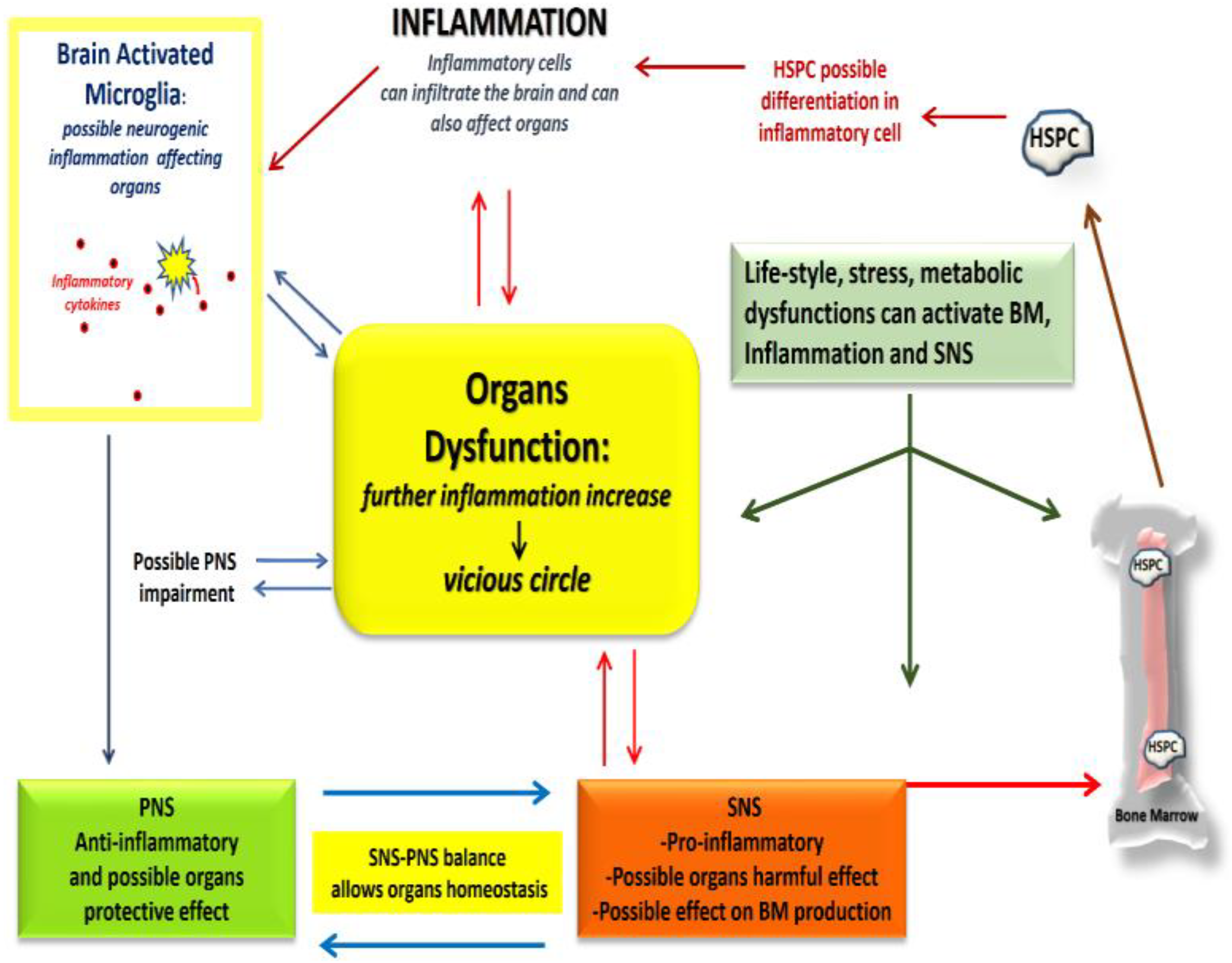
Figure 1. Complex crosstalk involved in organs dysfunction. The figure illustrates an example of the complex cross-talk existing among organs. It is based on studies describing neuro-immune pathways which can create a vicious circle: this phenomenon occurs when there is reciprocal crosstalk among the nervous system, immune system, and organs and can over-activate both immune or neuro-immune reactions, making it extremely difficult to understand the triggering events and consequently the precise mechanisms of action. This negatively affects the discovery of new therapies. For example, sympathetic nervous system (SNS) activation can induce extravasation of haematopoietic stem and progenitor cells (HSPCs) from the bone marrow. HSPCs can differentiate in haematopoietic, repairing, or inflammatory cells. Inflammatory cells can reach the brain, differentiate into microglia (brain inflammatory cells), and start cytokine production (molecules responsible for inflammatory response), leading to neuro-inflammation with consequences for organ homeostasis and likely affecting para-sympathetic nervous system (PNS). Neuro-inflammation in the brain and circulating inflammatory cells can also directly affect PNS and organs. A multi-organ approach is therefore required, but current biochemical approach still fails to explain this complex cross-talk, preventing the development of new effective therapies. Quantum biology research might open new perspectives in exploring and decode this cross-talk network.
Quantumrep 04 00011 g001
Figure 2. Method of studies selection.
Quantumrep 04 00011 g002
Figure 3. Posner molecule mechanism description: spin values for elementary particles (electron, proton, neutron). In a nucleus the total nuclear spin is the combination of spin of each nucleon and their angular momentum inside the nucleus. Spin is an intrinsic angular momentum of subatomic particles. Nuclei with spin value different from 0, interact with the electric and magnetic fields around them. It can also be seen as quantum bits of information. Phosphorus exists in the monoisotopic form 31P with spin ½ so a couple of such atoms in a molecule can be entangled. The quantum information contained in their spin protected inside structures, such as the Posner molecule, is not destroyed by environmental interaction and it can also propagate through other molecule Posner keeping information intact even over long distances (with respect to neurons’ distances in the cerebral tissue) in a relatively long time period (millisecond).
Quantumrep 04 00011 g003
Table 1. Divulgative Explanations for «Outsiders» of Quantum Concepts.
Quantum particle is an object that behaves both as a particle and as a wave. Typically, subatomic particles like electrons have this property. For these objects, classical physics (e.g., Newton’s and Maxwell’s laws) cannot describe classical parameters (for example, position and speed) and a completely new physic (quantum mechanics) is needed. Quantum mechanics provides, with a good approximation, a description of the physical properties of such objects.
Quantum coherence is the condition necessary to a particle for maintaining its quantum behaviour (for example, the counter-intuitive state of being in two states at the same time). Quantum coherence is related to the concept that sub-atomic particles have wave-like properties. In order to maintain coherence, environmental conditions around the particle must be very stable and meet specific requirements.
Quantum Superposition is a quantum mathematical description that represents the non-locality of the particle based on its wave-like properties. Accordingly, the particle can be present at multiple locations at the same time. As an intuitive image, it is like having one stone able to hit many birds simultaneously with one shot.
Quantum Tunnelling is the phenomenon where a quantum object tunnels through a barrier that it cannot surmount, for example, for adverse thermodynamic conditions. This is counterintuitive because it is like spookily passing through a thick and tall wall instead of overcoming it.
Quantum Entanglement is a quantum phenomenon that describes the instantaneous interaction between two particles, which were previously in contact, when they are pushed apart from each other. Regardless of the distance between them, the two objects (e.g., electrons in a previously covalent bond), remain “in contact” in a so-called entangled state. It has been observed that the two particles change their spin in response to the spin changing of the other instantaneously. For example, given that a particle A has entangled with a particle B, if particle A is found to have spin-up, then particle B must have spin-down. Later, if particle A is found to have spin-down, regardless of distance, particle B immediately changes in a spin-up particle.
Table 2. Biomedical terms and abbreviations.
AvidityThe measure of the total binding strength of an antibody at every binding site
Tax peptidesHuman T-cell leukemia virus type 1 (HTLV-1) is the etiological agent of an aggressive form of T-cell disorders leading to cancer.
Tax peptides are the oncoproteins inside the HTLV-1 which play a crucial role in the immortalization of malignant T-cells.
CVDsCardiovascular diseases
ANSAutonomic Nervous System
SNSSympathetic Nervous System
PNSParasympathetic Nervous System
HSPCsHaematopoietic Stem and Progenitor Cells
BRSThe Baroreflex Sensitivity index is defined as the change in interbeat interval in milliseconds per unit change in blood pressure.
It measures the control on the heart rate. Alterations of the BRS contribute to the reduction of parasympathetic activity and to the increase of sympathetic activity with possible harmful effect on cardiovascular system.
CVCardiovascular
ATPAdenosine Triphosphate
ACMArrhythmogenic Cardiomyopathy
Publisher’s Note: MDPI stays neutral with regard to jurisdictional claims in published maps and institutional affiliations.