
Review

Hydrogel-Integrated Nanotheranostic Platforms for Localized Diagnosis and Therapy
1
Department of Chemistry and Bioscience, Kumoh National Institute of Technology, 61 Daehak-ro, Gumi 39177, Republic of Korea
2
Department of Biomedical Engineering, Northwestern University, Evanston, IL 60208, USA
*
Authors to whom correspondence should be addressed.
J. Nanotheranostics 2026, 7(2), 10; https://doi.org/10.3390/jnt7020010
Submission received: 6 March 2026 / Revised: 13 April 2026 / Accepted: 18 April 2026 / Published: 23 April 2026

Abstract

Nanotheranostic platforms integrating diagnostic and therapeutic functions within a single system have attracted significant attention in precision medicine. However, conventional nanotheranostics based on systemic administration often suffer from off-target accumulation, limited retention at disease sites, and dose-limiting toxicity. To address these limitations, hydrogel-integrated nanotheranostic systems have emerged as a promising strategy for achieving localized diagnosis and therapy with improved spatial control and safety. This review provides a comprehensive overview of recent advances in hydrogel–nanomaterial nanotheranostic platforms, focusing on their design principles, diagnostic capabilities, and therapeutic applications. We discuss the complementary roles of hydrogels and nanomaterials, where hydrogels function as localized reservoirs and tissue interfaces, and nanomaterials provide imaging and therapeutic functionalities. Key integration strategies including physical encapsulation, chemical conjugation, and in situ nanoparticle formation are systematically compared. We further summarize localized diagnostic modalities such as real-time imaging and therapy monitoring, and highlight research-driven applications in cancer treatment, inflammation and infection management, and tissue regeneration. Finally, major translational challenges and future perspectives toward personalized, image-guided local theranostics are discussed. Overall, hydrogel-based nanotheranostic platforms represent a versatile approach for next-generation localized precision medicine.

1. Introduction

Nanotheranostics, which integrates diagnostic and therapeutic functionalities within a single nanoscale platform, has rapidly evolved as a cornerstone of precision and personalized medicine. By enabling simultaneous disease diagnosis, real-time monitoring of pathological progression, and on-demand therapeutic intervention, nanotheranostic systems offer a powerful approach to overcome the limitations of conventional sequential diagnosis–treatment paradigms. Advances in nanomaterial synthesis and surface engineering have led to the development of multifunctional nanoparticles capable of incorporating imaging agents, therapeutic cargos, and targeting moieties into a unified construct, thereby expanding their applications in cancer therapy, inflammatory diseases, infectious disorders, and tissue regeneration [1,2]. Despite these advances, the majority of nanotheranostic platforms have been designed for systemic administration, most commonly via intravenous injection. Following systemic delivery, nanotheranostic agents often suffer from nonspecific biodistribution and off-target accumulation, particularly in clearance organs such as the liver and spleen, which substantially reduces delivery efficiency to diseased tissues. This limited targeting efficiency is further compounded by biological barriers, including heterogeneous vascular permeability, elevated interstitial pressure in solid tumors, and rapid renal or hepatic clearance [3,4]. As a result, high systemic doses are often required to achieve therapeutic efficacy, increasing the risk of dose-limiting toxicity and long-term safety concerns associated with nanomaterial accumulation [5]. To address these challenges, localized therapeutic strategies have emerged as an attractive alternative to systemic nanotheranostics. By confining diagnostic and therapeutic agents to specific anatomical sites, localized delivery enables higher local concentrations while minimizing systemic exposure and adverse effects [6,7]. This approach is particularly beneficial for spatially confined diseases, such as solid tumors, post-surgical resection margins, chronic wounds, localized infections, and inflammatory lesions. In addition, localized therapy allows sustained treatment and repeated therapeutic action at the disease site, thereby improving therapeutic precision and patient compliance [8]. Hydrogels have gained increasing attention as enabling matrices for localized nanotheranostic platforms owing to their unique structural, mechanical, and biological properties. As highly hydrated three-dimensional polymer networks, hydrogels can closely mimic the extracellular matrix of native tissues, facilitating intimate tissue integration and prolonged retention at the target site. Importantly, hydrogels can function as local reservoirs for nanotheranostic agents, protecting encapsulated nanoparticles from premature degradation and clearance while enabling controlled and sustained release [8,9]. Their versatile processability further allows hydrogels to be engineered into injectable systems, adhesive platforms, or patch-type constructs, thereby broadening their applicability across diverse biomedical settings [10,11]. Beyond serving as passive carriers, hydrogels can actively participate in diagnostic and therapeutic processes. Functional hydrogels that respond to physiological or externally applied stimuli—such as pH, enzymes, reactive oxygen species, temperature, or light—enable dynamic regulation of nanoparticle release and therapeutic activation in response to disease-specific microenvironments [12,13,14]. When combined with nanomaterials possessing imaging or therapeutic functionalities, these hydrogel–nanoparticle hybrid systems offer spatiotemporally controlled diagnosis and therapy with enhanced precision compared to conventional systemic approaches. In this review, we provide a comprehensive overview of hydrogel-integrated nanotheranostic platforms designed for localized diagnosis and therapy. We first outline the fundamental design principles governing the integration of hydrogels and nanomaterials, followed by a discussion of localized diagnostic modalities and therapeutic applications enabled by these hybrid systems [15,16,17]. We then highlight recent advances in functional hydrogel designs that enhance nanotheranostic performance and conclude by addressing current challenges, translational barriers, and future perspectives for the clinical development of localized hydrogel-based nanotheranostic technologies.

2. Design Principles of Hydrogel-Integrated Nanotheranostics

2.1. Role of Hydrogels

Hydrogels play a central role in hydrogel-integrated nanotheranostic platforms by serving as multifunctional matrices that enable localized retention, mechanical integration with tissues, and controlled presentation of diagnostic and therapeutic agents. As three-dimensional polymeric networks capable of retaining large amounts of water, hydrogels can be engineered to remain confined at the target site, thereby providing a stable and localized microenvironment for nanotheranostic functions [8,18,19]. This local retention capability is essential for maximizing diagnostic signal intensity and therapeutic efficacy while minimizing systemic dissemination and off-target effects. A key advantage of hydrogels lies in their ability to mechanically match surrounding biological tissues. By tuning polymer composition, crosslinking density, and network architecture, hydrogels can closely mimic the viscoelastic properties of native tissues, including soft tumors, skin, cartilage, and wound beds [20,21]. Mechanical compatibility promotes intimate tissue integration, mitigates foreign-body responses, and enhances the stability of localized nanotheranostic systems. The importance of mechanical matching is further supported by studies demonstrating that cellular behavior and tissue responses are highly sensitive to matrix stiffness and elasticity [22], underscoring the relevance of hydrogel mechanics in long-term localized applications. In addition to mechanical support, hydrogels function as local reservoirs that enable sustained and controlled release of nanotheranostic agents. Encapsulation of nanoparticles within hydrogel matrices can significantly reduce burst release and slow diffusion-driven clearance, allowing therapeutic and diagnostic agents to be released over extended periods in a predictable manner. Release kinetics can be precisely modulated through hydrogel network design, including crosslink density, degradability, and responsiveness to environmental cues, thereby enabling fine control over therapeutic dosing and diagnostic signal persistence at localized disease sites [9,12,23]. The versatility of hydrogels further allows them to be engineered into diverse delivery formats, including injectable, adhesive, and patch-type systems, each offering distinct advantages for localized nanotheranostics. Injectable hydrogels enable minimally invasive administration and in situ gelation, allowing conformal filling of irregular tissue defects or post-surgical cavities [24,25]. Adhesive hydrogels provide robust attachment to biological surfaces, ensuring stable localization under dynamic physiological conditions and making them particularly suitable for skin, mucosal, and organ-surface applications [26,27,28]. Representative studies include an injectable tissue-adhesive hydrogel containing Ag-doped Mo2C-derived polyoxometalate nanoparticles for infected skin wounds, which achieved strong tissue adhesion together with photothermal/chemodynamic antibacterial therapy and accelerated wound healing [29]. In an oral mucosal model, a wet-adhesive NbC/TA–GelMA hydrogel maintained adhesion in the moist oral environment and enabled sustained antibacterial activity and mucosal repair under near-infrared irradiation [30]. In addition, an injectable adhesive albumin nanoparticle-incorporated hydrogel was reported for localized tumor treatment, demonstrating firm tissue attachment and prolonged local drug retention at the treatment site [31]. These representative studies illustrate that adhesive hydrogels are not only mechanically stable at wet tissue interfaces but also functionally versatile across skin, mucosal, and localized tumor applications Patch-type hydrogel systems are well suited for transdermal and wound-based theranostics, where continuous contact, localized monitoring, and sustained therapeutic delivery are required. Collectively, these characteristics position hydrogels as more than passive carriers in nanotheranostic platforms. By simultaneously providing localized retention, mechanical compatibility, controlled release, and adaptable delivery formats, hydrogels establish a functional interface between nanomaterials and biological tissues. This integrative role forms the foundation for the rational design of hydrogel-based nanotheranostic systems aimed at localized diagnosis and therapy.

2.2. Role of Nanomaterials

Nanomaterials constitute the functional core of hydrogel-integrated nanotheranostic platforms by enabling active diagnostic signal generation and therapeutic action at localized disease sites. While hydrogels primarily provide structural support, spatial confinement, and controlled release, nanomaterials are responsible for imparting imaging contrast and therapeutic efficacy. Owing to their tunable physicochemical properties, nanomaterials can be engineered to interact with diverse imaging modalities and therapeutic mechanisms, thereby complementing the structural and regulatory roles of hydrogels within integrated nanotheranostic systems [2]. As diagnostic agents, nanomaterials have been widely utilized to support multiple imaging modalities, including fluorescence imaging, magnetic resonance imaging (MRI), photoacoustic imaging, and computed tomography (CT). Fluorescent nanomaterials, such as dye-loaded polymeric nanoparticles and aggregation-induced emission nanostructures, provide high sensitivity and real-time visualization capabilities, making them suitable for monitoring hydrogel localization and nanoparticle distribution [32,33]. MRI-active nanomaterials, including iron oxide nanoparticles and gadolinium-based nanostructures, offer deep tissue penetration and high spatial resolution, enabling noninvasive and longitudinal monitoring of hydrogel-based systems implanted in vivo [34,35]. In addition, nanomaterials designed for photoacoustic or CT imaging, often based on plasmonic or high-atomic-number components, provide complementary diagnostic information by combining optical, acoustic, and radiographic contrast mechanisms [36,37]. When incorporated into hydrogels, these imaging nanomaterials benefit from prolonged local retention, allowing sustained diagnostic signal generation and real-time assessment of therapeutic progression. Nanomaterials also play a critical role as therapeutic agents within hydrogel-integrated nanotheranostic platforms. Drug-loaded nanoparticles enable the localized and controlled delivery of chemotherapeutics, anti-inflammatory agents, or antimicrobial compounds while protecting encapsulated cargos from premature degradation [4,38,39]. Beyond conventional drug delivery, nanomaterials can directly mediate physical therapeutic modalities, such as photothermal therapy, by converting externally applied stimuli into localized therapeutic effects. Plasmonic nanoparticles and carbon-based nanomaterials have demonstrated efficient light-to-heat conversion under near-infrared irradiation, enabling precise ablation of diseased tissues with minimal damage to surrounding healthy structures [40,41]. Furthermore, nanomaterials have emerged as powerful tools for localized immunomodulation, allowing the delivery of antigens, adjuvants, or immune checkpoint modulators to reshape local immune microenvironments and enhance therapeutic efficacy while minimizing systemic immune-related toxicity [42,43]. A defining advantage of nanomaterials in nanotheranostic platforms is their inherent capacity for multifunctionality, enabling the integration of diagnostic and therapeutic functions within a single nanoscale construct. Multifunctional nanomaterials can simultaneously provide imaging contrast, therapeutic payload delivery, and stimulus-responsive therapeutic activation, thereby enabling image-guided therapy and real-time monitoring of treatment response. Within hydrogel-integrated systems, this multifunctionality allows a clear division of labor: hydrogels maintain spatial localization and regulate release kinetics, while nanomaterials execute diagnostic and therapeutic tasks. This modular yet synergistic integration enhances design flexibility and enables the development of advanced nanotheranostic platforms tailored for specific clinical applications requiring localized diagnosis, therapy, and treatment monitoring.

2.3. Integration Strategies of Hydrogel–Nanomaterial Systems

The integration strategy between hydrogels and nanomaterials is a critical determinant of the stability, functionality, and performance of hydrogel-based nanotheranostic platforms. Effective integration governs nanoparticle distribution, retention, release kinetics, and functional accessibility, ultimately influencing diagnostic signal fidelity and therapeutic efficacy. Broadly, hydrogel–nanomaterial integration strategies can be classified into physical encapsulation, chemical conjugation, and in situ nanoparticle formation, each offering distinct advantages and design considerations (Table 1). Physical encapsulation represents the most straightforward and widely adopted integration strategy, in which preformed nanoparticles are physically entrapped within the hydrogel network during gelation or post-fabrication loading. This approach preserves the intrinsic physicochemical properties of nanomaterials while allowing facile system assembly and modular design. Encapsulated nanoparticles are retained within the hydrogel matrix through steric hindrance and diffusional constraints, enabling sustained local presence and controlled release governed primarily by hydrogel mesh size, crosslinking density, and degradation behavior [8,19]. Physical encapsulation is particularly attractive for multifunctional nanotheranostic systems, as it allows independent optimization of hydrogel and nanoparticle components without complex chemical modification. Chemical conjugation provides a more robust and stable integration strategy by covalently linking nanomaterials to hydrogel polymer chains or crosslinkers. Through covalent bonding or strong affinity interactions, chemically conjugated nanomaterials exhibit reduced mobility and enhanced retention within the hydrogel matrix, minimizing burst release and nanoparticle leaching [44,45,46]. This strategy enables precise spatial control over nanoparticle distribution and facilitates long-term localization of diagnostic and therapeutic functions. However, chemical conjugation requires careful consideration of reaction conditions and linker chemistry to preserve nanomaterial functionality and avoid unintended alterations in hydrogel mechanical or biological properties. In situ nanoparticle formation offers an alternative integration approach in which nanomaterials are synthesized directly within the hydrogel matrix from molecular or ionic precursors. This strategy enables uniform nanoparticle dispersion, intimate hydrogel–nanomaterial interfaces, and strong confinement of nanomaterials within the polymer network [47,48]. In situ formation is particularly advantageous for generating metal, metal oxide, or coordination-based nanoparticles with controlled size and spatial distribution, while simultaneously simplifying fabrication workflows. Moreover, the hydrogel environment can serve as a template or nanoreactor that regulates nanoparticle nucleation and growth, thereby enhancing functional reproducibility. Nevertheless, reaction compatibility with biological systems and residual precursor toxicity must be carefully addressed for biomedical applications. Collectively, these integration strategies provide a versatile toolbox for engineering hydrogel-based nanotheranostic platforms with tailored structural and functional characteristics. The choice of integration method should be guided by the intended diagnostic modality, therapeutic mechanism, required release profile, and translational considerations. Rational selection and optimization of hydrogel–nanomaterial integration strategies are therefore essential for achieving reliable, effective, and clinically relevant localized nanotheranostic systems (Figure 1).

2.4. Comparison of Hydrogel-Based and Non-Hydrogel Localized Systems

Hydrogel-based localized systems offer several distinctive advantages over non-hydrogel localized platforms, particularly in applications requiring anatomical conformity, minimally invasive placement, and spatiotemporally controlled release. Owing to their high water content, soft tissue-like mechanics, and tunable network structures, hydrogels can conform to irregular defect sites, support in situ gelation after injection, and provide sustained local retention with reduced mechanical mismatch to surrounding tissues. These features make hydrogel matrices especially attractive for postoperative cavities, wound beds, joint spaces, and other anatomically complex lesions where close tissue contact and adaptable depot formation are important. In addition, hydrogels are readily engineered to incorporate stimuli-responsive, adhesive, degradable, or self-healing properties, allowing greater flexibility for integrating imaging and therapeutic nanomaterials into multifunctional localized theranostic platforms [49,50]. By contrast, non-hydrogel localized systems—including polymeric wafers, films, rods, solid implants, and microneedle-based platforms—can offer advantages in structural robustness, shape fidelity, and, in some settings, manufacturing simplicity or device-like precision. For example, solid implantable depots can provide defined geometry and prolonged release, while microneedles offer minimally invasive and painless access across barrier tissues such as skin. However, these systems may be less adaptable to irregular resection cavities or dynamic wet tissue surfaces, and rigid implants can show burst-weighted release behavior, limited conformability, or mechanical mismatch with soft tissues. In brain-localized delivery, for instance, polymeric wafers such as Gliadel have demonstrated the clinical feasibility of local implantation, yet their rigid structure, early burst release, and implantation-related complications have highlighted important limitations relative to softer and more conformable depot systems [7,51,52]. Overall, hydrogel-based systems are generally more advantageous when localized therapy requires conformal filling, soft tissue compatibility, injectable administration, and multifunctional responsiveness, whereas non-hydrogel systems may remain useful when higher mechanical integrity, predefined architecture, or barrier-penetrating delivery is prioritized. Thus, hydrogel and non-hydrogel localized systems should be viewed as complementary rather than mutually exclusive platforms, with the optimal choice depending on the anatomical site, therapeutic objective, and translational constraints of the intended application.

3. Localized Diagnostic Modalities Enabled by Hydrogel–Nano Systems

Localized diagnostic capability is a defining advantage of hydrogel–nanomaterial integrated systems, enabling continuous and spatially confined monitoring that is difficult to achieve with systemically administered nanotheranostic agents. By immobilizing or slowly releasing imaging-capable nanomaterials at the target site, hydrogels provide a stable diagnostic window for real-time visualization, assessment of drug release behavior, and tracking of therapeutic response. This localized diagnostic paradigm supports image-guided therapy while minimizing background signals and systemic interference. One of the primary diagnostic functions enabled by hydrogel–nano systems is real-time imaging of diseased tissues and implanted therapeutic platforms. Nanomaterials designed for fluorescence, magnetic resonance imaging (MRI), photoacoustic (PA) imaging, or computed tomography (CT) can be incorporated into hydrogels to generate persistent and localized imaging contrast (Table 2) [53,54]. To ensure clarity, the terms “local retention,” “spatial confinement,” and “site-specific localization” are used to describe related but distinct aspects of hydrogel-integrated systems. Specifically, local retention refers to the ability of the system to remain at the target site over time, spatial confinement describes the restriction of nanoparticle or drug distribution within a defined region, and site-specific localization indicates the precise targeting of the system to a particular anatomical or pathological site. Recent studies have shown that fluorescence-labeled hydrogel systems can enable real-time visualization of hydrogel localization, degradation, and payload release in vivo. For example, carbon nanodot-labeled injectable hydrogels were used for non-invasive tracking of hydrogel degradation, while zinc phthalocyanine-incorporated thermosensitive hydrogels enabled simultaneous monitoring of carrier degradation and drug release behavior through dual fluorescence imaging [55,56]. MRI-visible hydrogel platforms have also been developed using iron oxide-based nanocomposites, allowing longitudinal monitoring of hydrogel degradation and implant fate in vivo [57,58]. Furthermore, injectable polypeptide/Ag2S quantum dot hydrogels have demonstrated second near-infrared fluorescence and photoacoustic imaging capability for monitoring localized therapeutic platforms, while gold- or ytterbium nanoparticle-containing hydrogels have enabled CT-based localization and image-guided treatment assessment [59,60,61]. Unlike freely circulating nanoparticles, hydrogel-confined imaging agents exhibit prolonged residence at the target site, allowing continuous visualization of hydrogel placement, structural integrity, and spatial distribution over time. This capability is particularly valuable for post-surgical monitoring, localized tumor treatment, and wound-based theranostic applications, where precise localization is critical. Beyond static imaging, hydrogel–nano systems enable monitoring of drug release and therapeutic activation through signal-responsive nanomaterials. Changes in imaging signal intensity or contrast can be correlated with nanoparticle diffusion, hydrogel degradation, or drug release events, providing indirect yet powerful insights into therapeutic kinetics [62,63]. A potential challenge in hydrogel-integrated imaging systems is distinguishing whether the detected signal originates from nanoparticles retained within the hydrogel matrix or from those that have diffused into the surrounding tissue. Several strategies have been developed to address this issue. One approach involves the use of dual-labeling systems, in which the hydrogel matrix and embedded nanoparticles are independently labeled with distinct imaging probes, enabling spatial differentiation of the two components. For example, hydrogels labeled with fluorescent or radiotracers in combination with nanoparticle-based contrast agents have been used to simultaneously track hydrogel degradation and nanoparticle release in vivo [64]. Another strategy relies on monitoring signal evolution over time, as changes in fluorescence or MRI signal intensity can be correlated with hydrogel degradation and nanoparticle diffusion behavior [55,65]. In addition, multimodal imaging approaches, such as combining MRI with optical or photoacoustic imaging, provide complementary spatial and functional information that improves localization accuracy [66]. Collectively, these strategies enable more precise interpretation of imaging signals and enhance the reliability of hydrogel-based nanotheranostic systems for localized diagnosis and therapy. Importantly, the diagnostic reliability of hydrogel–nano systems depends not only on the initial imaging contrast but also on signal stability over time. In practice, signal persistence within hydrogels is strongly influenced by several coupled factors, including photobleaching or contrast decay of the imaging probe, diffusion or leakage of signal-generating nanomaterials from the matrix, hydrogel swelling, and bulk or enzymatic degradation of the hydrogel network itself [55,65,67,68,69]. Several studies have shown that decreases in fluorescence, MRI, CT, or photoacoustic signals can serve as useful surrogate indicators of hydrogel erosion or material clearance, but such changes must be interpreted carefully because signal loss may also arise from probe diffusion or attenuation rather than true matrix degradation alone [55,65,67,68,69]. For example, fluorescence-based tracking studies using covalently labeled or nanoparticle-loaded hydrogels demonstrated progressive signal decay during in vivo degradation, while also highlighting the importance of probe stability and tissue penetration depth for accurate longitudinal monitoring [55,65,67]. Similarly, CEST MRI and photoacoustic imaging have been used to quantify hydrogel persistence and degradation over time, showing that changes in signal intensity can correlate well with scaffold disintegration, volumetric loss, or local remodeling processes [68,69]. Therefore, signal stability should be considered a key design parameter in hydrogel-based nanotheranostics, and the choice of imaging agent, labeling strategy, and hydrogel–probe interaction must be optimized to distinguish true therapeutic or degradation-related changes from artifacts caused by diffusion, photophysical instability, or background attenuation. This concept has been directly demonstrated in several hydrogel-based systems. In fluorescence-guided platforms, signal attenuation has been correlated with hydrogel erosion, whereas dual-fluorescent designs have enabled separate visualization of carrier persistence and drug release in vivo. Similarly, MRI-traceable hydrogels containing iron oxide nanoparticles have been used to follow structural degradation and material fate over time, supporting noninvasive assessment of localized therapeutic depots [55,56,57,58]. For example, fluorescence quenching, MRI signal variation, or PA signal modulation can be engineered to respond to nanoparticle release or microenvironmental changes, enabling noninvasive monitoring of therapeutic payload delivery. The hydrogel matrix plays a crucial role in regulating these processes by controlling release rates and maintaining spatial confinement of signal-generating nanomaterials. Hydrogel-integrated nanotheranostic systems also support therapy response tracking, allowing clinicians and researchers to assess treatment efficacy in real time. Imaging-capable nanomaterials can report on therapeutic outcomes such as tumor regression, tissue regeneration, or inflammatory resolution by providing longitudinal imaging data from the treated site [2,70]. In addition to reporting material retention, recent hydrogel–nano systems have demonstrated the ability to connect localized imaging outputs with therapeutic progression. Representative examples include MRI-monitored cartilage-regenerative hydrogels, NIR fluorescence/photoacoustic-monitored chemo-photothermal hydrogels, and CT-visible hydrogel depots for image-guided local chemotherapy, all of which highlight the value of integrating diagnostic readouts with therapeutic evaluation in a single localized platform [58,59,60]. In photothermal or immunotherapeutic systems, imaging signals can be used to guide treatment timing, evaluate treatment-induced changes, and optimize therapeutic regimens. By combining localized retention with responsive imaging functionalities, hydrogel–nano platforms enable dynamic feedback between diagnosis and therapy, which is essential for precision and personalized treatment strategies. Collectively, these diagnostic modalities highlight the synergistic roles of hydrogels and nanomaterials in enabling localized, real-time, and functionally informative diagnostics. Hydrogels provide spatial control and signal stability, while nanomaterials generate modality-specific diagnostic outputs. This synergy underpins the growing interest in hydrogel-based nanotheranostic systems for image-guided localized therapy and longitudinal disease monitoring.

4. Therapeutic Applications

4.1. Cancer: Tumor Resection Site and Post-Surgical Recurrence Prevention

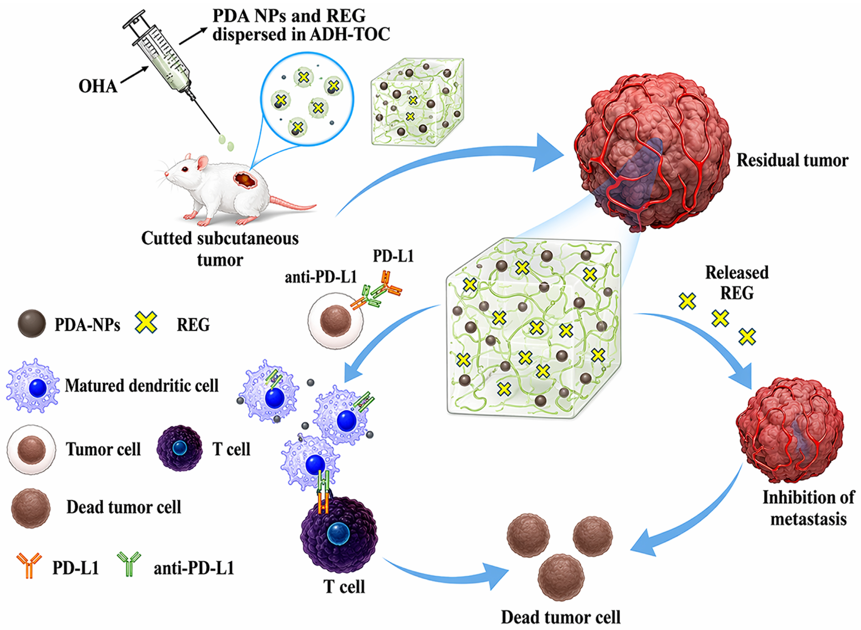
Localized cancer therapy is a particularly compelling application of hydrogel–nanomaterial integrated systems in the context of tumor resection and post-surgical recurrence prevention, where residual malignant cells at the surgical margin can drive local regrowth and metastasis. Systemic adjuvant therapy often fails to sustain sufficiently high drug concentrations at the surgical bed and is associated with dose-limiting toxicities, motivating approaches that confine therapeutic action to the resection site while enabling longitudinal monitoring of the treated area [7]. Hydrogel-based depots integrated with therapeutic nanomaterials can be placed directly into resection cavities or onto excision surfaces, conformally covering irregular geometries and providing sustained local exposure during the high-risk postoperative window. Representative recent work demonstrated an injectable multifunctional hydrogel enabling postoperative synergistic photothermal–chemodynamic therapy while simultaneously addressing bacterial infection at the surgical site, illustrating the value of integrating antitumor and wound-related functions into a single localized platform [71]. Such designs exemplify how hydrogel confinement can maintain therapeutic agents in situ while nanomaterials execute modality-specific antitumor actions. Beyond depot chemotherapy concepts, nanomaterial integration enables stimulus-triggered ablation and amplified local therapy tailored to residual disease. For example, an injectable therapeutic hydrogel incorporating CuO2@Au components provided a self-supplying/reactive therapeutic microenvironment and demonstrated effective inhibition in post-surgery recurrence models, highlighting how nanomaterials can extend beyond passive carriage to actively shape local therapeutic conditions [72]. In parallel, practical postoperative formats continue to advance: a sprayable thermosensitive hydrogel incorporating ultrathin CrB nanosheets achieved strong near-infrared photothermal performance and was reported to suppress postoperative tumor recurrence while also supporting wound healing and antibacterial activity, underscoring the translational appeal of conformal coverage for resection sites [73]. Hydrogel–nanomaterial platforms are also increasingly used to prevent recurrence by modulating the local immune microenvironment. More recent studies have further reinforced this postoperative strategy by demonstrating that polydopamine nanoparticle-incorporated injectable hydrogels can simultaneously suppress angiogenesis and stimulate tumoricidal immunity, thereby reducing postresection metastasis and recurrence. In parallel, polydopamine-doped supramolecular hydrogels have shown the ability to combine recurrence inhibition with enhanced wound repair, highlighting the value of integrating local antitumor activity with postoperative tissue recovery. In addition, hydrogel-localized mRNA lipid nanoparticle platforms have recently emerged as promising postoperative immunotherapeutic systems by promoting sustained local antigen expression, immune cell recruitment, and antigen presentation within a confined microenvironment [74,75,76]. An injectable polydopamine nanoparticle-incorporated hydrogel was reported as a promising postoperative strategy aimed at inhibiting metastasis/recurrence through combined mechanisms including immune activation (e.g., in combination with checkpoint blockade) [74] (Figure 2). Moreover, next-generation payloads such as mRNA lipid nanoparticles have been localized using hydrogel/microgel matrices (e.g., LiNx), enabling sustained local antigen expression/presentation and immune cell recruitment—an approach that aligns well with emerging postoperative vaccination and local immunotherapy paradigms [76]. Finally, localized resection-site strategies have expanded toward multifunctional composites that deliver therapeutic gases or redox modulators in situ. A sprayable alginate-based hydrogel composite incorporating a photocatalyst was applied to a postoperative breast cancer model to generate CO locally under light stimulation and to enhance clearance of residual tumor cells while aiming to reduce systemic toxicity, further illustrating the current trend toward practical, wound-covering postoperative local control systems [77]. Collectively, these recent studies support a clear direction in the field: hydrogel–nano systems are being engineered as multifunctional postoperative platforms that integrate recurrence suppression with wound repair, infection control, and immune activation—capabilities difficult to achieve with systemic nanomedicine alone [78]. A key design challenge in postoperative cancer settings is that the local tissue environment is highly heterogeneous, with irregular resection cavities, residual infiltrative tumor cells, inflammatory wound responses, and variable risks of bleeding or infection. Accordingly, hydrogel-based nanotheranostic systems for this application must balance conformal filling, local retention, therapeutic potency, and safety, while avoiding premature clearance or excessive toxicity to surrounding normal tissue [79,80]. From a clinical translation perspective, these factors directly affect reproducibility of intraoperative placement, dose uniformity within the surgical bed, and the ability to achieve reliable postoperative monitoring without compromising wound healing or surgical workflow [79,80].

4.2. Inflammation and Infection: Wounds, Arthritis, and Localized Infection

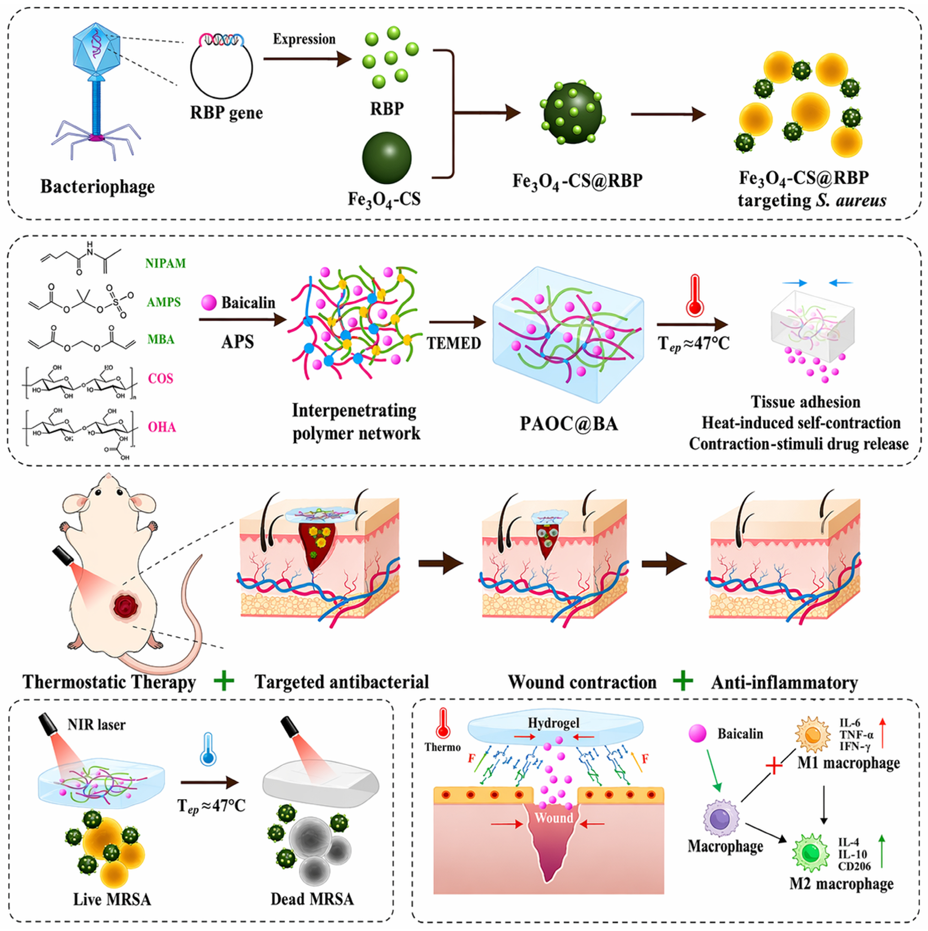
Hydrogel–nanomaterial integrated systems are increasingly applied to inflammatory and infectious diseases where localized and sustained therapy is essential for effective treatment. In wounds and localized infections, systemic antibiotics or anti-inflammatory drugs are often limited by poor penetration into necrotic tissue, biofilm-associated resistance, and systemic side effects. Locally applied hydrogel depots can maintain a moist protective interface and spatially confine functional nanomaterials, enabling high local antimicrobial and anti-inflammatory activity while reducing off-target exposure. In wound-healing and localized infection models, injectable or self-healing nanoparticle–hydrogel composites have been widely explored as multifunctional therapeutic platforms. For example, injectable self-healing hydrogels incorporating CuS nanoparticles demonstrated strong antibacterial efficacy under external stimulation while simultaneously supporting wound healing through sustained local retention and controlled release [81]. Recent work has further expanded this concept by introducing AIE-driven photothermal injectable hydrogels that adapt to irregular wound geometries and provide combined antibacterial activity, tissue adhesion, and regenerative support. These systems illustrate the growing shift from simple antibacterial depots toward multifunctional wound platforms capable of simultaneously controlling infection and promoting tissue reconstruction [82,83,84]. Recent designs increasingly emphasize multifunctional infection control, integrating photothermal or photodynamic antibacterial effects with tissue-regenerative support. An AIE-driven photothermal nanotrigger-activated injectable hydrogel dressing exemplifies this trend, achieving effective infection control and accelerated tissue repair in vivo [83]. Similarly, smart nanoparticle–hydrogel hybrid systems have been reported to synergistically regulate inflammation, bacterial burden, and regeneration, highlighting the shift toward integrated therapeutic modules rather than single-function antibacterial dressings [85] (Figure 3). For chronic and infected wounds, particularly in diabetic or immunocompromised settings, hydrogel–nanomaterial systems that combine photothermal and antioxidant functions have shown promising therapeutic outcomes. A recent study reported a photothermal and redox-active hydrogel that accelerated healing of infected diabetic wounds by simultaneously eradicating bacteria and mitigating excessive oxidative stress, underscoring the importance of local microenvironment modulation in wound therapy [86]. In addition, skin-adhesive antibacterial hydrogels incorporating photothermal nanomaterials have been developed for stable attachment to dynamic tissue surfaces, offering translationally attractive formats for localized infection control [87]. Localized inflammatory diseases such as arthritis further benefit from hydrogel-confined nanomaterial delivery due to the rapid clearance of drugs from joint cavities following intra-articular injection. Recent work demonstrated injectable nanoparticle-forming hydrogel depots capable of sustaining anti-inflammatory drug release for extended periods and significantly improving therapeutic outcomes in osteoarthritis models after a single administration [88]. More advanced systems have incorporated nanoparticle-loaded, stimulus-responsive self-healing hydrogels, enabling prolonged joint retention (exceeding several weeks) and combined anti-inflammatory and antioxidative effects, thereby addressing both symptom relief and disease progression [89]. Importantly, nanotheranostic functionality has begun to emerge in arthritis treatment through the integration of imaging or tracking capabilities. A recent Theranostics study introduced a self-healing radiopaque hyaluronic acid hydrogel, allowing longitudinal in vivo monitoring of hydrogel persistence within the joint space and supporting precision scheduling of therapeutic intervention [84]. For rheumatoid arthritis and other localized inflammatory lesions, hydrogel-based nano/microsphere delivery systems have also been reported to enable microenvironment-triggered activation of therapeutic agents. An in situ-activated phospholipid-mimic artemisinin prodrug delivered via an injectable hydrogel nano/microsphere platform demonstrated effective localized immunomodulation and disease suppression in rheumatoid arthritis models [90]. Collectively, these recent research advances highlight a unifying translational rationale across wounds, infection, and arthritis: hydrogels provide anatomical conformity, retention, and protection at inflammatory foci, while embedded nanomaterials supply potent antimicrobial or anti-inflammatory activity, stimulus-responsive control, and, in select systems, diagnostic trackability. Such synergistic integration positions hydrogel-based nanotheranostic platforms as powerful tools for the localized management of inflammation and infection. In inflammatory and infectious lesions, a major challenge is that the local microenvironment is often dominated by biofilm formation, excessive exudate, elevated reactive oxygen species, enzymatic degradation, and continuous mechanical disturbance, all of which can reduce hydrogel stability and compromise therapeutic persistence. In joint diseases such as osteoarthritis, rapid clearance from the synovial cavity and repetitive mechanical loading impose additional requirements for prolonged retention, mechanical resilience, and sustained bioactivity after intra-articular administration [91,92]. For clinical translation, these disease-specific constraints mean that successful systems must maintain efficacy under wet and dynamic tissue conditions, support predictable residence time, and avoid impairing native healing or joint function, which is essential for reducing repeat dosing and improving practical usability [91,92].

4.3. Tissue Regeneration: Regenerative Theranostics for Healing and Monitoring

Tissue regeneration represents a unique application domain for hydrogel–nanomaterial integrated systems, where therapeutic repair and longitudinal monitoring are inherently coupled. Unlike cancer or infection therapy, regenerative processes require sustained support over extended time scales and benefit from continuous assessment of healing progression, tissue integration, and functional recovery. Conventional regenerative biomaterials often focus solely on structural or biochemical support, whereas hydrogel-based nanotheranostic platforms enable simultaneous tissue healing and real-time monitoring, thereby advancing the concept of regenerative theranostics. Recent research has demonstrated that hydrogels incorporating imaging-capable or signal-responsive nanomaterials can actively participate in tissue repair while reporting local biological status. For example, injectable nanocomposite hydrogels containing fluorescence- or photoacoustic-active nanoparticles have been shown to support angiogenesis and tissue remodeling while enabling noninvasive visualization of material retention and degradation during regeneration [93,94]. Representative MRI-visible regenerative hydrogels have demonstrated that scaffold degradation and cartilage repair can be longitudinally tracked in vivo, providing direct information on both implant fate and regenerative progression. In particular, multiparametric MR imaging and MRI-monitorable GelMA-based systems have shown clear utility for correlating hydrogel persistence with cartilage healing outcomes [58,95,96]. Such systems provide valuable feedback on scaffold persistence and integration, which are critical parameters in regenerative outcomes but difficult to assess using conventional approaches. Beyond passive monitoring, regenerative nanotheranostic systems increasingly integrate microenvironment-responsive sensing to track healing-associated changes. Hydrogels embedded with nanoparticles responsive to pH, reactive oxygen species (ROS), or enzymatic activity have been reported to dynamically report inflammatory resolution and tissue maturation during wound and bone regeneration [97,98]. In these platforms, nanomaterial-derived signal changes correlate with key regenerative milestones, such as reduced oxidative stress or matrix remodeling, allowing healing progression to be assessed in real time without invasive sampling. Bone and cartilage regeneration provide particularly compelling examples of regenerative theranostics. Recent studies have described injectable or printable hydrogels incorporating osteoinductive nanoparticles that not only promote bone formation but also enable longitudinal imaging of scaffold distribution and mineralization processes [58,99]. For instance, nanomaterial-loaded hydrogels with radiopaque or MRI-visible components have been used to track defect filling and material persistence during bone repair, supporting precision control over regenerative interventions [100]. Similarly, nanocomposite hydrogels designed for cartilage repair have combined sustained delivery of regenerative cues with imaging-assisted evaluation of hydrogel residence and tissue integration [101]. Emerging work also highlights the integration of bioelectronic or mechano-responsive nanomaterials into hydrogels for regenerative monitoring. Conductive or mechanochromic nanoparticle-hydrogel composites have been explored to report mechanical loading, tissue strain, or functional recovery during regeneration, particularly in musculoskeletal and skin applications [102,103]. These systems illustrate a shift from static scaffolds toward interactive regenerative platforms capable of both guiding and sensing tissue repair processes. Collectively, recent advances demonstrate that hydrogel-based nanotheranostic systems are redefining regenerative medicine by enabling healing-coupled diagnostics. By combining sustained regenerative support with real-time, noninvasive monitoring of tissue status, these platforms offer a powerful framework for personalized and adaptive regenerative therapies, where treatment strategies can be dynamically adjusted based on in situ diagnostic feedback. In regenerative applications, one of the most important design challenges is to match hydrogel degradation, mechanical support, and signal persistence with the pace of tissue healing, since excessively rapid degradation may result in loss of structural support, whereas overly persistent scaffolds may hinder remodeling and tissue integration. These requirements become even more demanding in cartilage and osteochondral repair, where long-term mechanical loading, defect-specific geometry, and the need for longitudinal monitoring must all be considered simultaneously [58,104]. From a clinical translation standpoint, successful regenerative nanotheranostic platforms will therefore require reproducible degradation behavior, durable yet biocompatible mechanical performance, and imaging outputs that reflect meaningful healing progression rather than scaffold presence alone [58,104].

5. Advanced Functional Hydrogels

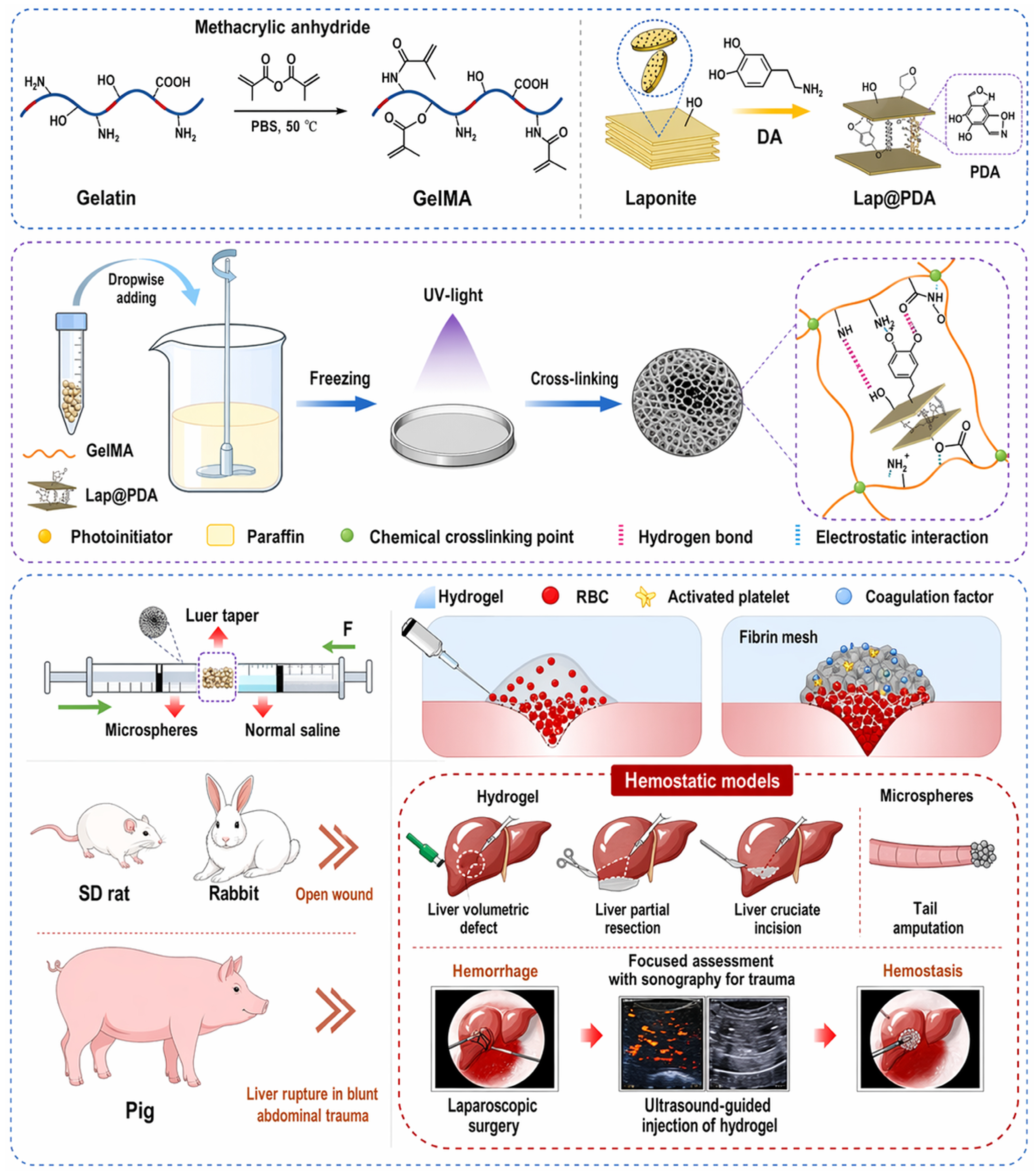
Advanced functional hydrogels have become a major driver for improving the performance and translational feasibility of hydrogel–nanomaterial nanotheranostic systems. Beyond serving as passive local depots, modern hydrogel matrices are increasingly engineered to be responsive, injectable/self-healing, adhesive, and degradable/bioresorbable, allowing spatiotemporal control over nanomaterial behavior and enabling more realistic clinical deployment. Recent work and focused syntheses highlight that such functions are typically combined within a single platform (e.g., injectable + self-healing + adhesive + degradable), reflecting a shift toward integrated “smart” materials rather than single-property gels. This design trend is increasingly supported by recent reviews and application-focused studies showing that multifunctional hydrogels are now engineered to integrate stimuli-responsiveness, injectability, self-healing behavior, adhesion, and monitoring capability within a single therapeutic platform rather than as isolated material properties. In particular, recent theranostic and wound-healing reviews have emphasized that such functional convergence is becoming a central design principle for clinically relevant hydrogel systems [105,106]. Stimuli-responsive hydrogels are particularly impactful for localized nanotheranostics because they enable condition-adaptive regulation of drug release and diagnostic signal dynamics. Endogenous triggers (pH, enzymes, ROS) and exogenous triggers (light/heat/ultrasound) can be used to modulate hydrogel network integrity and transport, thereby synchronizing nanomaterial exposure with disease microenvironments or externally applied activation [107,108]. Importantly, this responsiveness supports programmable release (minimizing burst, enabling on-demand dosing) while maintaining localized confinement—an advantage emphasized in recent analyses of stimuli-responsive hydrogel systems for targeted therapy and diagnosis [102]. In practice, responsive behavior is increasingly implemented in nanocomposite architectures where nanoparticles contribute not only as payloads but also as trigger transducers (e.g., photothermal conversion, catalytic redox modulation), enabling feedback-like control of local therapeutic conditions and potentially improving reproducibility in complex tissues [109,110]. Representative recent examples include injectable hydrogels in which near-infrared-responsive nanocomponents trigger in situ gelation, photothermal therapy, or combined tumor/anti-infection treatment after local administration. These systems illustrate how nanomaterials can function not merely as cargos but as active transducers that convert external light stimulation into gel formation and therapeutic activation, thereby improving local controllability and treatment precision [71,111]. Injectable and self-healing systems have similarly accelerated, as they directly address two translational bottlenecks: minimally invasive placement and mechanical robustness under dynamic physiological environments. Thermoresponsive or dynamic covalent networks allow hydrogels to be injected as flowable precursors and rapidly recover into stable depots, while self-healing behavior can preserve localization and function after deformation or minor damage. Recent reports describe thermoresponsive injectable self-healing hydrogels that accommodate nanoparticle/drug co-loading for synergistic localized therapy, illustrating how network dynamics can be tuned to balance injectability with sustained retention and controlled release [112]. Complementing disease-specific demonstrations, recent theranostic hydrogel systems also underscore how injectable architectures can be used to restore diseased microenvironments while enabling longitudinal assessment, supporting a general design logic of “minimally invasive placement + functional persistence” for localized nanotheranostics [110]. Adhesive hydrogels provide another step toward real-world usability by enabling stable attachment to wet, moving tissues (skin, mucosa, organ surfaces) and maintaining intimate tissue–material interfaces. This is particularly important for surface-accessible infections and wounds, where hydrogel detachment or dilution can rapidly degrade therapeutic performance. Recent work has demonstrated skin-adhesive antibacterial photothermal hydrogels, leveraging adhesion to sustain local residency and external activation for efficient infection control without systemic antibiotic burden—an approach directly aligned with localized theranostic concepts [113]. Recent adhesive hydrogel studies further show that robust tissue attachment is closely linked to therapeutic reliability under dynamic conditions. For example, injectable self-healing adhesive chitosan hydrogels have combined antioxidative, antibacterial, and hemostatic functions, while bioinspired adhesive skin-electronic hydrogel interfaces have extended this concept toward simultaneous monitoring and treatment. These advances support the view that adhesion is not only a retention feature but also a prerequisite for stable sensing, repeatable activation, and user-operable localized therapy [114,115]. In parallel, the broader adhesive-material direction is reinforced by recent high-impact demonstrations of adhesive wound-therapy constructs integrated with wearable activation modules, highlighting the converging trend of “adhesion + external controllability” for precision, patient-friendly interventions at the tissue interface [116]. Collectively, these advances position adhesion not as a cosmetic feature, but as a functional prerequisite for consistent dosing, reliable imaging/monitoring, and user-operable localized therapy. Finally, degradable/bioresorbable platforms are increasingly emphasized to address long-term safety, reduce the need for surgical retrieval, and align treatment duration with the healing timeline. Recent perspectives on biodegradable and injectable hydrogels highlight key choices in degradable chemistries and their implications for mechanical stability, release kinetics, and in vivo clearance—considerations that become even more critical when nanomaterials are integrated into the matrix [117]. This point is also supported by biodegradable pH- and temperature-responsive injectable hydrogel systems that have shown controlled in vivo gel formation and bioresorption behavior, underscoring the importance of matching degradation kinetics with therapeutic duration [118]. In localized nanotheranostic platforms, such degradability is especially relevant because it can reduce long-term material burden while preserving sufficient residence time for imaging, triggered therapy, and tissue repair [71,118]. Recent theranostic examples further demonstrate degradable nanocomposite systems designed for biological safety and eventual resorption, supporting the development of temporary yet functional depots for localized therapy and monitoring [119] (Figure 4). In addition, emerging designs explicitly incorporate “self-degradable” or programmed-degradation behaviors to synchronize therapeutic action (e.g., triggered ablation windows) with controlled disappearance of the platform, underscoring the trend toward time-resolved, lifecycle-aware nanotheranostic hydrogels. Taken together, these studies indicate that the next generation of hydrogel-based nanotheranostics is moving toward multifunctional, lifecycle-aware systems that combine adaptive responsiveness, minimally invasive placement, durable tissue interfacing, and programmed clearance within a single clinically deployable material framework. To further highlight the translational relevance of hydrogel–nanomaterial systems, it is important to consider clinically evaluated platforms that share key design principles with these integrated nanotheranostic strategies. While fully integrated hydrogel–nanotheranostic systems are still largely in the preclinical stage, several hydrogel-based delivery systems and nanoparticle-enabled diagnostic or therapeutic platforms have progressed into clinical studies or regulatory approval. For example, injectable and implantable hydrogel systems have been clinically investigated for localized drug delivery and tissue repair. A representative case is the polyethylene glycol (PEG)-based hydrogel sealant (DuraSeal®), which has received U.S. Food and Drug Administration (FDA) approval for surgical applications, demonstrating the clinical feasibility of in situ-forming hydrogel systems for localized retention and controlled delivery [120]. Similarly, alginate and collagen-based hydrogels have been evaluated in clinical settings for wound healing and tissue regeneration, highlighting their biocompatibility and translational potential [121,122]. On the nanomaterial side, several nanoparticle-based systems with diagnostic or therapeutic functions have advanced to clinical use. Superparamagnetic iron oxide nanoparticles (SPIONs), such as ferumoxytol, have been clinically approved and widely used as MRI contrast agents, demonstrating the clinical applicability of nanoparticle-enabled imaging technologies [123]. In addition, liposomal and polymeric nanoparticle formulations, including Doxil® (liposomal doxorubicin), have been approved for cancer therapy, validating the clinical potential of nanoparticle-mediated drug delivery systems [124]. More recently, emerging studies have begun to bridge hydrogel and nanomaterial technologies for localized clinical applications. For instance, nanoparticle-loaded hydrogel systems have been explored in early-stage clinical and translational studies for localized cancer therapy and wound treatment, aiming to combine sustained release, spatial confinement, and imaging capability [125,126]. Although these integrated systems are still under development, the successful clinical translation of individual hydrogel and nanomaterial components strongly supports the future clinical potential of hydrogel-based nanotheranostic platforms. Representative clinically evaluated or approved hydrogel- and nanomaterial-based platforms relevant to hydrogel-based nanotheranostic systems are summarized in Table 3.

6. Challenges and Translational Considerations

Despite the rapid progress of hydrogel–nanomaterial nanotheranostic platforms in preclinical research, several critical challenges must be addressed to enable successful clinical translation. These challenges span technical limitations, manufacturing and scalability issues, regulatory complexity, and long-term biosafety considerations, all of which are particularly relevant for hybrid systems that integrate soft biomaterials with functional nanomaterials. One major technical challenge lies in imaging depth and sensitivity, especially for localized diagnostic applications in deep or heterogeneous tissues. While fluorescence-based nanotheranostics offer high sensitivity, their limited tissue penetration restricts clinical applicability beyond superficial or intraoperative settings. Alternative modalities such as MRI, photoacoustic imaging, and CT provide improved depth penetration but often require higher nanomaterial loading to achieve sufficient contrast, potentially increasing material burden and safety concerns [53,127]. Moreover, signal attenuation over time due to nanoparticle diffusion, hydrogel degradation, or biological clearance complicates longitudinal monitoring, underscoring the need for imaging strategies that balance depth, sensitivity, and temporal stability [128]. Manufacturing and scalability represent another major bottleneck for translation. Many hydrogel–nano systems rely on multistep fabrication processes, including nanoparticle synthesis, surface functionalization, hydrogel crosslinking, and sterile formulation, which can be difficult to standardize and scale reproducibly [129]. Batch-to-batch variability in nanoparticle size, surface chemistry, and loading efficiency can significantly impact therapeutic performance and imaging readouts. Recent perspectives emphasize the importance of simplifying material compositions, adopting scalable synthesis methods, and implementing quality-by-design (QbD) principles early in development to facilitate large-scale production and regulatory acceptance [129,130]. Regulatory complexity is particularly pronounced for hydrogel–nanotheranostic systems, which are typically classified as combination products due to the integration of device-like biomaterials with drug and/or diagnostic components. The coexistence of structural hydrogels and functional nanomaterials complicates regulatory pathways, as safety, efficacy, and quality must be evaluated for each component as well as for the integrated system [131]. In addition, discrepancies between regulatory frameworks for drugs, devices, and combination products across regions further increase translational uncertainty. Recent analyses highlight that early engagement with regulatory agencies and a clear definition of the primary mode of action are essential for navigating approval pathways for multifunctional nanotheranostic platforms [131,132]. Long-term biosafety remains a central concern, particularly for nanomaterials retained locally for extended periods. While hydrogels are often designed to be biodegradable or bioresorbable, embedded nanomaterials may persist after matrix degradation, raising questions regarding chronic inflammation, accumulation, or delayed toxicity [133]. Long-term fate studies assessing nanomaterial degradation, clearance routes, and immunological responses are still limited, and standardized evaluation protocols are lacking. Recent studies and reviews emphasize the need for lifecycle-aware design, in which degradation of both hydrogel matrices and nanomaterials is coordinated to minimize long-term biological burden [133,134]. Collectively, these challenges highlight that successful translation of hydrogel-based nanotheranostic systems requires not only material innovation but also integration of imaging physics, manufacturing science, regulatory strategy, and safety assessment. Addressing these considerations through rational design, early standardization, and translationally informed evaluation will be critical for advancing hydrogel–nanomaterial nanotheranostics from promising laboratory concepts to clinically viable technologies.

7. Future Perspectives

Hydrogel–nanomaterial integrated nanotheranostic platforms are poised to play an increasingly important role in the evolution of personalized and localized medicine. As advances in materials science, imaging technologies, and data-driven decision-making converge, future nanotheranostic systems are expected to move beyond “one-size-fits-all” designs toward patient- and site-specific therapeutic strategies. Localized hydrogel-based platforms provide a natural foundation for personalization by allowing modulation of material composition, mechanical properties, degradation profiles, and nanomaterial payloads in response to individual disease characteristics and anatomical constraints [130,135]. One promising direction is the development of personalized local theranostics, in which hydrogel–nano systems are tailored to the biological and pathological features of a specific patient or lesion. Variations in tissue stiffness, inflammatory status, vascularization, and immune microenvironment can significantly influence therapeutic outcomes, particularly in localized diseases. Recent perspectives emphasize that adaptable hydrogel matrices combined with modular nanomaterials could enable customization of release kinetics, therapeutic intensity, and diagnostic sensitivity, thereby improving efficacy while minimizing adverse effects [136]. Integration with patient-derived data and emerging precision medicine frameworks is likely to further accelerate this trend. Another key future direction lies in image-guided smart release systems, where diagnostic feedback is directly coupled to therapeutic action. In such systems, imaging modalities not only visualize hydrogel placement or nanomaterial retention but also actively inform or trigger drug release and therapeutic activation. Advances in stimuli-responsive materials and imaging physics suggest that real-time feedback loops—linking imaging signals with programmable release from hydrogels—may enable adaptive dosing, optimized treatment timing, and dynamic adjustment of therapeutic regimens [137]. This closed-loop theranostic paradigm represents a significant conceptual shift from static delivery platforms toward responsive and self-regulating therapeutic systems. Despite these opportunities, the realization of clinical impact will depend on the establishment of clear clinical translation roadmaps. Future efforts must align material innovation with practical considerations, including scalable manufacturing, robust quality control, regulatory strategy, and clinically relevant endpoints. Recent translational analyses highlight the importance of early-stage engagement with clinicians and regulatory agencies, simplified system architectures, and rigorous long-term safety evaluation to bridge the gap between laboratory demonstrations and clinical implementation [133]. In particular, defining the primary mode of action and selecting clinically meaningful imaging and therapeutic outcomes will be critical for positioning hydrogel-based nanotheranostics within existing healthcare frameworks. Looking ahead, the successful translation of hydrogel–nanomaterial nanotheranostic systems will likely require interdisciplinary integration, combining materials engineering, imaging science, immunology, and regulatory expertise. By coupling localized therapeutic precision with real-time diagnostic insight and translationally informed design, future hydrogel-based nanotheranostic platforms have the potential to redefine how localized diseases are diagnosed, treated, and monitored in clinical practice [138].

8. Conclusions

Hydrogel–nanomaterial integrated nanotheranostic platforms represent a powerful strategy for achieving localized diagnosis and therapy with enhanced precision, safety, and therapeutic efficacy. By combining the structural and regulatory functions of hydrogels with the diagnostic and therapeutic capabilities of nanomaterials, these systems enable sustained local retention, controlled release, and real-time monitoring that are difficult to realize with conventional systemic approaches. Recent advances demonstrate that rational integration strategies and advanced hydrogel functionalities—such as stimuli responsiveness, injectability, adhesion, and degradability—have significantly expanded the applicability of nanotheranostics across cancer therapy, inflammation and infection management, and tissue regeneration. In these contexts, hydrogel-based localization not only improves therapeutic outcomes but also facilitates image-guided intervention and longitudinal assessment of treatment response, reinforcing the value of theranostic feedback loops. Despite substantial progress, challenges related to imaging depth, scalable manufacturing, regulatory complexity, and long-term biosafety remain critical barriers to clinical translation. Addressing these issues will require simplified and standardized system architectures, translationally informed design choices, and early engagement with regulatory frameworks. Importantly, future development should prioritize lifecycle-aware materials and clinically relevant imaging and therapeutic endpoints. Looking forward, the convergence of personalized material design, image-guided smart release systems, and translational roadmaps is expected to drive the next generation of localized nanotheranostic platforms. With continued interdisciplinary collaboration and a focus on clinical practicality, hydrogel–nanomaterial nanotheranostics have the potential to transition from promising experimental systems to impactful clinical technologies for precision localized medicine.

Author Contributions

Conceptualization, S.A.Y. and H.K.; writing—original draft preparation, J.P., S.A.Y. and H.K.; writing—review and editing, J.P., D.Y., T.K. and C.C.; supervision, S.A.Y. and H.K.; project administration, S.A.Y. and H.K.; funding acquisition, H.K. All authors have read and agreed to the published version of the manuscript.

Funding

This work was supported by the Korea Environmental Industry & Technology Institute (KEITI), funded by the Ministry of Climate, Energy and Environment of the Government of the Republic of Korea and the Gyeongsangbuk-do RISE (Regional Innovation System & Education) project (Regional Growth Innovation LAB unit).

Data Availability Statement

No new data were created or analyzed in this study. Data sharing is not applicable to this article.

Conflicts of Interest

The authors declare no conflicts of interest.

Abbreviations

The following abbreviations are used in this manuscript:
AIEAggregation-Induced Emission
BNCBacterial Nanocellulose
CNFCellulose Nanofibrils
CNCCellulose Nanocrystals
CTComputed Tomography
FLFluorescence Imaging
GdGadolinium
MRIMagnetic Resonance Imaging
NIRNear-Infrared
PAPhotoacoustic Imaging
PEGPoly(ethylene glycol)
PDAPolydopamine
ROSReactive Oxygen Species

References

  1. Janib, S.M.; Moses, A.S.; MacKay, J.A. Imaging and drug delivery using theranostic nanoparticles. Adv. Drug Deliv. Rev. 2010, 62, 1052–1063. [Google Scholar] [CrossRef] [PubMed]
  2. Lim, E.K.; Kim, T.; Paik, S.; Haam, S.; Huh, Y.M.; Lee, K. Nanomaterials for Theranostics: Recent Advances and Future Challenges. Chem. Rev. 2015, 115, 327–394. [Google Scholar] [CrossRef] [PubMed]
  3. Wilhelm, S.; Tavares, A.J.; Dai, Q.; Ohta, S.; Audet, J.; Dvorak, H.F.; Chan, W.C.W. Analysis of nanoparticle delivery to tumours. Nat. Rev. Mater. 2016, 1, 16014. [Google Scholar] [CrossRef]
  4. Blanco, E.; Shen, H.; Ferrari, M. Principles of nanoparticle design for overcoming biological barriers to drug delivery. Nat. Biotechnol. 2015, 33, 941–951. [Google Scholar] [CrossRef]
  5. Mayne, R.; Morgan, J.; Whiting, J.G.H.; Phillips, N.; Adamatzky, A. On measuring nanoparticle toxicity and clearance with. Sci. Rep. 2019, 9, 8957. [Google Scholar] [CrossRef]
  6. Park, K. Controlled drug delivery systems: Past forward and future back. J. Control. Release 2014, 190, 3–8. [Google Scholar] [CrossRef]
  7. Wolinsky, J.B.; Colson, Y.L.; Grinstaff, M.W. Local drug delivery strategies for cancer treatment: Gels, nanoparticles, polymeric films, rods, and wafers. J. Control. Release 2012, 159, 14–26. [Google Scholar] [CrossRef]
  8. Li, J.Y.; Mooney, D.J. Designing hydrogels for controlled drug delivery. Nat. Rev. Mater. 2016, 1, 16071. [Google Scholar] [CrossRef]
  9. Peppas, N.A.; Bures, P.; Leobandung, W.; Ichikawa, H. Hydrogels in pharmaceutical formulations. Eur. J. Pharm. Biopharm. 2000, 50, 27–46. [Google Scholar] [CrossRef]
  10. Stevanovic, M.; Jovic, M.; Filipovic, N.; Lukac, S.; Tomic, N.; Maneski, L.P.; Stojanovic, Z. Multifunctional Nanomaterial-Integrated Hydrogels for Sustained Drug Delivery: From Synthesis and Characterization to Biomedical Application. Gels 2025, 11, 892. [Google Scholar] [CrossRef]
  11. Zhianmanesh, M.; Khodaei, A.; Crago, M.; Lotz, O.; Naficy, S.; Dehghani, F.; Bilek, M.; Yavari, S.A.; Akhavan, B. Universal Method for Covalent Attachment of Hydrogels to Diverse Polymeric Surfaces for Biomedical Applications. Adv. Mater. 2026, 38, e03524. [Google Scholar] [CrossRef]
  12. Qiu, Y.; Park, K. Environment-sensitive hydrogels for drug delivery. Adv. Drug Deliv. Rev. 2012, 64, 49–60. [Google Scholar] [CrossRef]
  13. Liu, X.; Zhou, Q.; Yang, Y.; Wu, X.; Chen, J.; Wang, R.Q.; Chen, E.H. Hydrogels in cancer treatment: Mapping the future of precision drug delivery. Front. Immunol. 2025, 16, 1607240. [Google Scholar] [CrossRef] [PubMed]
  14. Lavrador, P.; Esteves, M.R.; Gaspar, V.M.; Mano, J.F. Stimuli-Responsive Nanocomposite Hydrogels for Biomedical Applications. Adv. Funct. Mater. 2021, 31, 2005941. [Google Scholar] [CrossRef]
  15. Appel, E.A.; del Barrio, J.; Loh, X.J.; Scherman, O.A. Supramolecular polymeric hydrogels. Chem. Soc. Rev. 2012, 41, 6195–6214. [Google Scholar] [CrossRef]
  16. Norouzi, M.; Nazari, B.; Miller, D.W. Injectable hydrogel-based drug delivery systems for local cancer therapy. Drug Discov. Today 2016, 21, 1835–1849. [Google Scholar] [CrossRef]
  17. Lima-Sousa, R.; Alves, C.G.; Melo, B.L.; Costa, F.J.P.; Nave, M.; Moreira, A.F.; Mendonça, A.G.; Correia, I.J.; de Melo-Diogo, D. Injectable hydrogels for the delivery of nanomaterials for cancer combinatorial photothermal therapy. Biomater. Sci. 2023, 11, 6082–6108. [Google Scholar] [CrossRef]
  18. Lovrak, M.; Picken, S.J.; Eelkema, R.; van Esch, J.H. Supramolecular Gluing of Polymeric Hydrogels. ChemNanoMat 2018, 4, 772–775. [Google Scholar] [CrossRef]
  19. Hoare, T.R.; Kohane, D.S. Hydrogels in drug delivery: Progress and challenges. Polymer 2008, 49, 1993–2007. [Google Scholar] [CrossRef]
  20. Lee, H.J. Recent Advances in Hydrogels for Tissue Engineering and Biomedical Therapeutics. Gels 2025, 11, 733. [Google Scholar] [CrossRef]
  21. Caló, E.; Khutoryanskiy, V.V. Biomedical applications of hydrogels: A review of patents and commercial products. Eur. Polym. J. 2015, 65, 252–267. [Google Scholar] [CrossRef]
  22. Discher, D.E.; Sweeney, L.; Sen, S.; Engler, A. Matrix elasticity directs stem cell lineage specification. Cell 2006, 126, 677–689. [Google Scholar] [CrossRef] [PubMed]
  23. Lin, C.C.; Metters, A.T. Hydrogels in controlled release formulations: Network design and mathematical modeling. Adv. Drug Deliv. Rev. 2006, 58, 1379–1408. [Google Scholar] [CrossRef] [PubMed]
  24. Ruel-Gariépy, E.; Leroux, J.C. In situ-forming hydrogels—Review of temperature-sensitive systems. Eur. J. Pharm. Biopharm. 2004, 58, 409–426. [Google Scholar] [CrossRef] [PubMed]
  25. Cheng, Y.; Zhang, H.T.; Wei, H.; Yu, C.Y. Injectable hydrogels as emerging drug-delivery platforms for tumor therapy. Biomater. Sci. 2024, 12, 1151–1170. [Google Scholar] [CrossRef]
  26. Chen, B.J.; Yu, R.J.; Wang, J.Q.; Feng, Y.X.; Zhang, Y.J.; Mao, Y.C.; Shan, C.X.; Wang, X.D. Biomaterials-Based Hydrogel with Superior Bio-Mimetic Ionic Conductivity and Tissue-Matching Softness for Bioelectronics. Adv. Funct. Mater. 2025, 36, e27495. [Google Scholar] [CrossRef]
  27. Li, S.N.; Cong, Y.; Fu, J. Tissue adhesive hydrogel bioelectronics. J. Mater. Chem. B 2021, 9, 4423–4443. [Google Scholar] [CrossRef]
  28. Zhang, W.L.; Zhang, Y.W.; Zhang, Y.C.; Dai, Y.; Xia, F.; Zhang, X.J. Adhesive and tough hydrogels: From structural design to applications. J. Mater. Chem. B 2021, 9, 5954–5966. [Google Scholar] [CrossRef]
  29. Huang, H.; Su, Y.; Wang, C.X.; Lei, B.; Song, X.J.; Wang, W.J.; Wu, P.; Liu, X.Y.; Dong, X.C.; Zhong, L.P. Injectable Tissue-Adhesive Hydrogel for Photothermal/Chemodynamic Synergistic Antibacterial and Wound Healing Promotion. ACS Appl. Mater. Inter. 2023, 15, 2714–2724. [Google Scholar] [CrossRef]
  30. Chen, J.Y.; Ren, J.Y.; Wu, Y.J.; Hu, N.; Zhao, F.; Zhang, L. Wet adhesive hydrogels based on niobium carbide for experimental research of oral mucosal impairment. RSC Adv. 2024, 14, 12935–12946. [Google Scholar] [CrossRef]
  31. Wang, T.R.; Ding, J.F.; Chen, Z.X.; Zhang, Z.; Rong, Y.; Li, G.; He, C.L.; Chen, X.S. Injectable, Adhesive Albumin Nanoparticle-Incorporated Hydrogel for Sustained Localized Drug Delivery and Efficient Tumor Treatment. ACS Appl. Mater. Inter. 2024, 16, 9868–9879. [Google Scholar] [CrossRef] [PubMed]
  32. Weissleder, R.; Nahrendorf, M.; Pittet, M.J. Imaging macrophages with nanoparticles. Nat. Mater. 2014, 13, 125–138. [Google Scholar] [CrossRef] [PubMed]
  33. Seo, Y.H.; Kim, S. Aggregation-induced emission nanoparticles with improved optical absorption for boosting fluorescence signal of tumors. Spectrochim. Acta A 2022, 280, 121534. [Google Scholar] [CrossRef] [PubMed]
  34. Zhang, H.; Liu, X.L.; Fan, H.M. Advances in magnetic nanoparticle-based magnetic resonance imaging contrast agents. Nano Res. 2023, 16, 12531–12542. [Google Scholar] [CrossRef]
  35. Saul, P.; Schröder, L.; Schmidt, A.B.; Hövener, J.B. Nanomaterials for hyperpolarized nuclear magnetic resonance and magnetic resonance imaging. Wires Nanomed. Nanobiotechnol. 2023, 15, e1879. [Google Scholar] [CrossRef]
  36. Yu, C.X.; Kong, L.H.; Tian, J.; Zhang, Y.; Jia, X.T.; Dang, W.L.; Xing, B.; Zhang, Q.Q.; Pang, X.C.; Hu, Z.M.; et al. Photoacoustic imaging-guided triple-responsive nanoparticles with tumor hypoxia relief for improving chemotherapy/photothermal/photodynamic synergistic therapy against breast cancer. Biomed. Pharmacother. 2023, 164, 114928. [Google Scholar] [CrossRef]
  37. Kubiliute, R.; Slektaite, A.; Burkanas, M.; Grigiene, R.; Rotomskis, R. Gold Nanoparticles as a Contrast Agent for X-Ray Imaging. In Proceedings of the International Conference “Medical Physics in the Baltic States”, Kaunas, Lithuania, 13–15 October 2011; pp. 27–30. [Google Scholar]
  38. Kamaly, N.; Yameen, B.; Wu, J.; Farokhzad, O.C. Degradable Controlled-Release Polymers and Polymeric Nanoparticles: Mechanisms of Controlling Drug Release. Chem. Rev. 2016, 116, 2602–2663. [Google Scholar] [CrossRef]
  39. Yuk, S.A.; Kim, H.; Abutaleb, N.S.; Dieterly, A.M.; Taha, M.S.; Tsifansky, M.D.; Lyle, L.T.; Seleem, M.N.; Yeo, Y. Nanocapsules modify membrane interaction of polymyxin B to enable safe systemic therapy of Gram-negative sepsis. Sci. Adv. 2021, 7, eabj1577. [Google Scholar] [CrossRef]
  40. Mammari, N.; Duval, R.E. Photothermal/Photoacoustic Therapy Combined with Metal-Based Nanomaterials for the Treatment of Microbial Infections. Microorganisms 2023, 11, 2084. [Google Scholar] [CrossRef]
  41. Huang, X.H.; Jain, P.K.; El-Sayed, I.H.; El-Sayed, M.A. Plasmonic photothermal therapy (PPTT) using gold nanoparticles. Laser Med. Sci. 2008, 23, 217–228. [Google Scholar] [CrossRef]
  42. Smith, J.D.; Morton, L.D.; Ulery, B.D. Nanoparticles as synthetic vaccines. Curr. Opin. Biotechnol. 2015, 34, 217–224. [Google Scholar] [CrossRef]
  43. Nzeteu, G.A.N.; Gibbs, B.F.; Kotnik, N.; Troja, A.; Bockhorn, M.; Meyer, N.H. Nanoparticle-based immunotherapy of pancreatic cancer. Front. Mol. Biosci. 2022, 9, 948898. [Google Scholar] [CrossRef] [PubMed]
  44. Lin, C.C.; Anseth, K.S. PEG Hydrogels for the Controlled Release of Biomolecules in Regenerative Medicine. Pharm. Res. 2009, 26, 631–643. [Google Scholar] [CrossRef] [PubMed]
  45. Shen, J.R.; Li, H.L.; Lu, C.F.; Yang, G.C.; Wang, F.Y.; Nie, J.Q.; Hu, X.J.; Dong, N.G.; Shi, J.W. Hydrolytically degradable POSS-PEG hybrid hydrogels prepared in aqueous phase with tunable mechanical properties, swelling ratio and degradation rate. React. Funct. Polym. 2018, 123, 91–96. [Google Scholar] [CrossRef]
  46. Zhang, W.; Wang, R.X.; Sun, Z.M.; Zhu, X.W.; Zhao, Q.; Zhang, T.F.; Cholewinski, A.; Yang, F.; Zhao, B.X.; Pinnaratip, R.; et al. Catechol-functionalized hydrogels: Biomimetic design, adhesion mechanism, and biomedical applications. Chem. Soc. Rev. 2020, 49, 433–464. [Google Scholar] [CrossRef]
  47. Rosi, N.L.; Mirkin, C.A. Nanostructures in biodiagnostics. Chem. Rev. 2005, 105, 1547–1562. [Google Scholar] [CrossRef]
  48. Qureshi, D.; Nayak, S.K.; Maji, S.; Anis, A.; Kim, D.; Pal, K. Environment sensitive hydrogels for drug delivery applications. Eur. Polym. J. 2019, 120, 109220. [Google Scholar] [CrossRef]
  49. Lu, P.L.; Ruan, D.X.; Huang, M.Q.; Tian, M.; Zhu, K.S.; Gan, Z.Q.; Xiao, Z.C. Harnessing the potential of hydrogels for advanced therapeutic applications: Current achievements and future directions. Signal Transduct. Target. Ther. 2024, 9, 166. [Google Scholar] [CrossRef]
  50. Alghamdi, M.; Gumbleton, M.; Newland, B. Local delivery to malignant brain tumors: Potential biomaterial-based therapeutic/adjuvant strategies. Biomater. Sci. 2021, 9, 6037–6051. [Google Scholar] [CrossRef]
  51. Bastiancich, C.; Bozzato, E.; Henley, I.; Newland, B. Does local drug delivery still hold therapeutic promise for brain cancer? A systematic review. J. Control. Release 2021, 337, 296–305. [Google Scholar] [CrossRef]
  52. Guillot, A.J.; Cordeiro, A.S.; Donnelly, R.F.; Montesinos, M.C.; Garrigues, T.M.; Melero, A. Microneedle-Based Delivery: An Overview of Current Applications and Trends. Pharmaceutics 2020, 12, 569. [Google Scholar] [CrossRef] [PubMed]
  53. Ni, D.L.; Bu, W.B.; Ehlerding, E.B.; Cai, W.B.; Shi, J.L. Engineering of inorganic nanoparticles as magnetic resonance imaging contrast agents. Chem. Soc. Rev. 2017, 46, 7438–7468. [Google Scholar] [CrossRef] [PubMed]
  54. Sun, I.C.; Ahn, C.H.; Kim, K.; Emelianov, S. Photoacoustic imaging of cancer cells with glycol-chitosan-coated gold nanoparticles as contrast agents. J. Biomed. Opt. 2019, 24, 121903. [Google Scholar] [CrossRef]
  55. Wang, L.; Li, B.Q.; Xu, F.; Li, Y.; Xu, Z.H.; Wei, D.Q.; Feng, Y.J.; Wang, Y.M.; Jia, D.C.; Zhou, Y. Visual degradation of injectable hydrogel by real-time and non-invasive tracking using carbon nanodots as fluorescent indicator. Biomaterials 2017, 145, 192–206. [Google Scholar] [CrossRef] [PubMed]
  56. Dong, X.; Wei, C.; Chen, H.L.; Qin, J.W.; Liang, J.; Kong, D.L.; Liu, T.J.; Lv, F. Real-Time Imaging Tracking of a Dual Fluorescent Drug Delivery System Based on Zinc Phthalocyanine-Incorporated Hydrogel. ACS Biomater. Sci. Eng. 2016, 2, 2001–2010. [Google Scholar] [CrossRef]
  57. Zhang, Y.; Sun, Y.; Yang, X.; Hilborn, J.; Heerschap, A.; Ossipov, D.A. Injectable In Situ Forming Hybrid Iron Oxide-Hyaluronic Acid Hydrogel for Magnetic Resonance Imaging and Drug Delivery. Macromol. Biosci. 2014, 14, 1249–1259. [Google Scholar] [CrossRef]
  58. Chen, Z.L.; Yan, C.G.; Yan, S.N.; Liu, Q.; Hou, M.R.; Xu, Y.K.; Guo, R. Non-invasive monitoring of hydrogel degradation and cartilage regeneration by multiparametric MR imaging. Theranostics 2018, 8, 1146–1158. [Google Scholar] [CrossRef]
  59. Jin, R.M.; Yang, X.Q.; Zhao, D.H.; Hou, X.L.; Li, C.Q.; Song, X.L.; Chen, W.; Wang, Q.; Zhao, Y.D.; Liu, B. An injectable hybrid hydrogel based on a genetically engineered polypeptide for second near-infrared fluorescence/photoacoustic imaging-monitored sustained chemo-photothermal therapy. Nanoscale 2019, 11, 16080–16091. [Google Scholar] [CrossRef]
  60. Keshavarz, M.; Moloudi, K.; Paydar, R.; Abed, Z.; Beik, J.; Ghaznavi, H.; Shakeri-Zadeh, A. Alginate hydrogel co-loaded with cisplatin and gold nanoparticles for computed tomography image-guided chemotherapy. J. Biomater. Appl. 2018, 33, 161–169. [Google Scholar] [CrossRef]
  61. Dong, Y.C.; Kumar, A.; Rosario-Berrios, D.N.; Si-Mohamed, S.; Hsu, J.C.; Nieves, L.M.; Douek, P.; Noel, P.B.; Cormode, D.P. Ytterbium Nanoparticle Contrast Agents for Conventional and Spectral Photon-Counting CT and Their Applications for Hydrogel Imaging. ACS Appl. Mater. Interfaces 2022, 14, 39274–39284. [Google Scholar] [CrossRef]
  62. Zingale, E.; Romeo, A.; Rizzo, S.; Cimino, C.; Bonaccorso, A.; Carbone, C.; Musumeci, T.; Pignatello, R. Fluorescent Nanosystems for Drug Tracking and Theranostics: Recent Applications in the Ocular Field. Pharmaceutics 2022, 14, 955. [Google Scholar] [CrossRef] [PubMed]
  63. Huang, J.; Wang, Z.R.; Krishna, S.; Hu, Q.; Xuan, M.; Xie, H.X. Environment-sensitive hydrogels as potential drug delivery systems for the treatment of periodontitis. Mater. Express 2020, 10, 975–985. [Google Scholar] [CrossRef]
  64. Yang, X.; Wang, J.; Ding, Z.K.; Lin, Q.C.; Zhuo, L.G.; Liao, W.; Zhao, Y.; Feng, Y.; Chen, Y.; Wei, H.Y.; et al. Dual-radiolabelling of an injectable hyaluronan-tyramine-bisphosphonate hybrid gel for and tracking. Carbohydr. Polym. 2020, 231, 115652. [Google Scholar] [CrossRef] [PubMed]
  65. Dong, Y.Q.; Jin, G.R.; Ji, C.C.; He, R.Y.; Lin, M.; Zhao, X.; Li, A.; Lu, T.J.; Xu, F. Non-invasive tracking of hydrogel degradation using upconversion nanoparticles. Acta Biomater. 2017, 55, 410–419, Correction in Acta Biomater. 2017, 59, 361. https://doi.org/10.1016/j.actbio.2017.07.015. [Google Scholar] [CrossRef]
  66. Dong, Y.C.; Bouché, M.; Uman, S.; Burdick, J.A.; Cormode, D.P. Detecting and Monitoring Hydrogels with Medical Imaging. ACS Biomater. Sci. Eng. 2021, 7, 4027–4047. [Google Scholar] [CrossRef]
  67. Wang, W.W.; Liu, J.J.; Li, C.; Zhang, J.; Liu, J.F.; Dong, A.J.; Kong, D.L. Real-time and non-invasive fluorescence tracking of degradation of the thermosensitive PEGlyated polyester hydrogel. J. Mater. Chem. B 2014, 2, 4185–4192. [Google Scholar] [CrossRef]
  68. Zhu, W.; Chu, C.Y.; Kuddannaya, S.; Yuan, Y.; Walczak, P.; Singh, A.; Song, X.L.; Bulte, J.W.M. In Vivo Imaging of Composite Hydrogel Scaffold Degradation Using CEST MRI and Two-Color NIR Imaging. Adv. Funct. Mater. 2019, 29, 1903753. [Google Scholar] [CrossRef]
  69. Shrestha, B.; Stojkova, K.; Yi, R.; Anastasio, M.A.; Ye, J.Y.; Brey, E.M. Gold nanorods enable noninvasive longitudinal monitoring of hydrogels with photoacoustic tomography. Acta Biomater. 2020, 117, 374–383. [Google Scholar] [CrossRef]
  70. Chen, J.M.; Sha, Q.L.; Rehman, M.U.; Wu, M.; Hu, Z.Y.; Wu, F.S. Organic Nanomaterials Based on Aza-Boron Dipyrromethene with Aggregation-Induced Emission for NIR-II Fluorescence Imaging-Guided Photothermal Therapy. ACS Appl. Nano Mater. 2024, 7, 23122–23132. [Google Scholar] [CrossRef]
  71. Huang, H.; Wang, X.R.; Wang, W.L.; Qu, X.Y.; Song, X.J.; Zhang, Y.W.; Zhong, L.P.; Yang, D.P.; Dong, X.C.; Zhao, Y.X. Injectable hydrogel for postoperative synergistic photothermal-chemodynamic tumor and anti-infection therapy. Biomaterials 2022, 280, 121289. [Google Scholar] [CrossRef]
  72. Hu, P.; Jia, Z.L.; Zhao, S.; Lin, K.P.; Yang, G.Y.; Guo, W.J.; Yu, S.L.; Cheng, J.J.; Du, G.H.; Shi, J.H. Injectable Therapeutic Hydrogel with H2O2 Self-Supplying and GSH Consumption for Synergistic Chemodynamic/Low-Temperature Photothermal Inhibition of Postoperative Tumor Recurrence and Wound Infection. Adv. Healthc. Mater. 2024, 13, 2401551. [Google Scholar] [CrossRef]
  73. Quan, S.H.; Pan, G.F.; Shah, S.W.A.; Yuan, H.; Zhao, Y.; Li, X.X.; Shen, H.L.; Xie, H.H.; Yang, H.; Shao, J.D. CrB MBene-infused sprayable hydrogel for NIR-I/II photothermal cancer treatment. Sci. China Chem. 2025, 69, 2059–2072. [Google Scholar] [CrossRef]
  74. Peng, N.; Du, Y.J.; Liu, J.H.; Li, D.Y.; Li, Y.H.; Deng, K.; Li, L.W.; Jia, P.Z.; Min, J.; Lin, J.M. Injectable Polydopamine Nanoparticle-Incorporated Hydrogels for Antiangiogenesis and Stimulating Tumoricidal Immunity to Inhibit Metastasis and Recurrence Postresection. ACS Appl. Mater. Inter. 2024, 16, 64447–64462. [Google Scholar] [CrossRef] [PubMed]
  75. Wang, X.Q.; Wu, B.B.; Zhang, Y.Q.; Dou, X.Q.; Zhao, C.L.; Feng, C.L. Polydopamine-doped supramolecular chiral hydrogels for postoperative tumor recurrence inhibition and simultaneously enhanced wound repair. Acta Biomater. 2022, 153, 204–215. [Google Scholar] [CrossRef] [PubMed]
  76. Zhu, Y.N.; Yao, Z.C.; Li, S.Y.; Ma, J.Y.; Wei, C.; Yu, D.; Stelzel, J.L.; Ni, B.Y.X.; Miao, Y.; Van Batavia, K.; et al. An mRNA lipid nanoparticle-incorporated nanofiber-hydrogel composite for cancer immunotherapy. Nat. Commun. 2025, 16, 5707. [Google Scholar] [CrossRef]
  77. Wang, Z.Y.; Zhu, J.X.; Mi, B.B.; Ni, M.; Xue, Y.M.; Deng, Y.L.; Chen, L.; Xu, X.Y.; Li, X.Y.; Liu, G.H.; et al. Photocatalytic CO-releasing spray hydrogel for in situ postoperative cancer treatment. Bioact. Mater. 2025, 53, 893–907. [Google Scholar] [CrossRef]
  78. Wang, Z.P.; Zhai, B.T.; Sun, J.; Zhang, X.F.; Zou, J.B.; Shi, Y.J.; Guo, D.Y. Recent advances of injectable in situ-forming hydrogels for preventing postoperative tumor recurrence. Drug Deliv. 2024, 31, 2400476. [Google Scholar] [CrossRef]
  79. Zhong, Z.Q.; Gan, L.; Feng, Z.Y.; Wang, W.H.; Pan, X.; Wu, C.B.; Huang, Y. Hydrogel local drug delivery systems for postsurgical management of tumors: And perspectives. Mater. Today Bio 2024, 29, 101308. [Google Scholar] [CrossRef]
  80. Askari, E.; Seyfoori, A.; Amereh, M.; Gharaie, S.S.; Ghazali, H.S.; Ghazali, Z.S.; Khunjush, B.; Akbari, M. Stimuli-Responsive Hydrogels for Local Post-Surgical Drug Delivery. Gels 2020, 6, 14. [Google Scholar] [CrossRef]
  81. Zhou, L.Q.; Chen, F.; Hou, Z.S.; Chen, Y.W.; Luo, X.L. Injectable self-healing CuS nanoparticle complex hydrogels with antibacterial, anti-cancer, and wound healing properties. Chem. Eng. J. 2021, 409, 128224. [Google Scholar] [CrossRef]
  82. Kong, Y.; Hou, Z.S.; Zhou, L.Q.; Zhang, P.F.; Ouyang, Y.W.; Wang, P.W.; Chen, Y.W.; Luo, X.L. Injectable Self-Healing Hydrogels Containing CuS Nanoparticles with Abilities of Hemostasis, Antibacterial activity, and Promoting Wound Healing. ACS Biomater. Sci. Eng. 2021, 7, 335–349. [Google Scholar] [CrossRef] [PubMed]
  83. Huang, X.X.; Zhu, X.L.; Gan, D.L.; Yu, Y.W.; Jiang, X.F.; Wang, M.Q. AIE-driven photothermal nanotrigger-activated multifunctional injectable hydrogel for maxillofacial infection control. Mater. Today Bio 2025, 34, 102247. [Google Scholar] [CrossRef] [PubMed]
  84. Said, M.; Tavakoli, C.; Dumot, C.; Toupet, K.; Olivier, C.; Gilles, A.; Maumus, M.; Dong, Y.C.; Collomb, N.; Auxenfans, C.; et al. A self-healing radiopaque hyaluronic acid hydrogel as a new injectable biomaterial for precision medicine in osteoarthritis. Theranostics 2025, 15, 4054–4073. [Google Scholar] [CrossRef] [PubMed]
  85. Wan, H.P.; Tan, H.R.; Zhao, X.H. SMart Nanoparticle-Hydrogel Hybrid System for Synergistic Eradication of Infection and Promotion of Wound Healing. Adv. Sci. 2025, 13, e17320. [Google Scholar] [CrossRef]
  86. Zeng, H.; Ying, Z.R.; Luo, X.; Tan, S.; Liu, X.H.; Zhao, X.Y.; He, S.S.; Chen, F.; Kulak, A.I.; Lu, B.Q. Gallic acid-modified bioglass with combined photothermal and antibacterial effects for the regeneration of infected diabetic wound. Compos. Part B—Eng. 2023, 257, 110668. [Google Scholar] [CrossRef]
  87. Mohanty, A.; Patra, S.; Bhutia, S.K.; Mohapatra, S. Skin-Adhesive Antibacterial Hydrogel Loaded with Biosynthesized CuS Nanoparticles for NIR Laser-Induced Wound Healing. ACS Appl. Bio Mater. 2026, 9, 412–422. [Google Scholar] [CrossRef]
  88. Seo, B.B.; Kwon, Y.; Kim, J.; Hong, K.H.; Kim, S.E.; Song, H.R.; Kim, Y.M.; Song, S.C. Injectable polymeric nanoparticle hydrogel system for long-term anti-inflammatory effect to treat osteoarthritis. Bioact. Mater. 2022, 7, 14–25. [Google Scholar] [CrossRef]
  89. Fan, H.X.; Zhao, J.S.; Gao, Z.C.; Mu, Y.L.; Zhang, X.M.; Pan, W.B.; Li, S.S.; He, H.B.; Gou, J.X.; Tang, X.; et al. Injectable self-healing hydrogel loaded PLGA nanoparticles for long-term osteoarthritis therapy. Biomater. Adv. 2026, 179, 214506. [Google Scholar] [CrossRef]
  90. Du, Y.W.; Li, C.; Zhang, Y.; Xiong, W.; Wang, F.; Wang, J.; Zhang, Y.Z.; Deng, L.F.; Li, X.S.; Chen, W.; et al. In Situ-Activated Phospholipid-Mimic Artemisinin Prodrug via Injectable Hydrogel Nano/Microsphere for Rheumatoid Arthritis Therapy. Research 2022, 2022, 0003. [Google Scholar] [CrossRef]
  91. Huang, C.; Dong, L.L.; Zhao, B.H.; Lu, Y.F.; Huang, S.R.; Yuan, Z.Q.; Luo, G.X.; Xu, Y.; Qian, W. Anti-inflammatory hydrogel dressings and skin wound healing. Clin. Transl. Med. 2022, 12, e1094. [Google Scholar] [CrossRef]
  92. Kalairaj, M.S.; Pradhan, R.; Saleem, W.; Smith, M.M.; Gaharwar, A.K. Intra-Articular Injectable Biomaterials for Cartilage Repair and Regeneration. Adv. Healthc. Mater. 2024, 13, 2303794. [Google Scholar] [CrossRef]
  93. Chen, Y.R.; Fan, Z.T.; Xu, W.Y.; Zhu, Z.Y.; Tan, Z.; Hu, Y.Q.; Kurzina, I.; Cherdyntseva, N.; Yang, W.J.; Wang, L.H. An injectable nanocomposite hydrogel with deep penetration ability for enhanced photothermal and chemotherapy. J. Colloid. Interf. Sci. 2025, 685, 268–279. [Google Scholar] [CrossRef] [PubMed]
  94. Sood, A.; Das, S.S.; Dev, A.; Bhardwaj, D.; Kumar, A.; Agrawal, G.; Han, S.S. Fluorescent nanocomposites loaded hydrogels as a theranostic platform for advanced healthcare applications: Recent trends and opportunities. Eur. Polym. J. 2023, 196, 112323. [Google Scholar] [CrossRef]
  95. Zhang, H.Y.; Fang, W.J.; Zhao, T.T.; Zhang, H.B.; Gao, L.; Li, J.Y.; Wang, R.J.; Xu, W.P. Real-Time MRI Monitoring of GelMA-Based Hydrogel-Loaded Kartogenin for Cartilage Regeneration. Front. Bioeng. Biotechnol. 2022, 10, 940735. [Google Scholar] [CrossRef] [PubMed]
  96. Pan, Y.Y.; Huang, L.; Sun, S.W.; Yan, Y.; Xu, H.; Yan, J.; Chen, H.B.; Li, N.; Tang, S.J.; Zhang, F.M. Injectable Photoacoustic-Traceable Prussian Blue Nano-hybridized Hydrogel Microspheres Loaded with Stem Cells for Promoting Diabetic Bone Regeneration. Adv. Healthc. Mater. 2026, e04006. [Google Scholar] [CrossRef]
  97. Ni, Z.P.; Yu, H.J.; Wang, L.; Huang, Y.D.; Lu, H.; Zhou, H.Y.; Liu, Q.X. Multistage ROS-Responsive and Natural Polyphenol-Driven Prodrug Hydrogels for Diabetic Wound Healing. ACS Appl. Mater. Inter. 2022, 14, 52643–52658. [Google Scholar] [CrossRef]
  98. Pi, Y.P.; Ganabady, K.; Celiz, A.D. Enzyme-responsive biomaterials for biomedical applications. Commun. Mater. 2025, 6, 263. [Google Scholar] [CrossRef]
  99. Groborz, O.; Kolouchova, K.; Parmentier, L.; Szabó, A.; Van Durme, B.; Dunlop, D.; Slanina, T.; Van Vlierberghe, S. Photoprintable Radiopaque Hydrogels for Regenerative Medicine. ACS Appl. Eng. Mater. 2024, 2, 811–817. [Google Scholar] [CrossRef]
  100. Lee, Y.; Kim, M.; Kim, N.; Byun, S.; Seo, S.; Han, J.Y. Injectable Hydrogel Systems for Targeted Drug Delivery: From Site-Specific Application to Design Strategy. Appl. Sci. 2025, 15, 11599. [Google Scholar] [CrossRef]
  101. Wei, W.; Ma, Y.Z.; Yao, X.D.; Zhou, W.Y.; Wang, X.Z.; Li, C.L.; Lin, J.X.; He, Q.L.; Leptihn, S.; Ouyang, H.W. Advanced hydrogels for the repair of cartilage defects and regeneration. Bioact. Mater. 2021, 6, 998–1011. [Google Scholar] [CrossRef]
  102. Ye, X.L.; Chen, Y.D.; Lv, C.H.; Ying, Y.B.; Ping, J.F.; Pan, J.M.; Lan, L.Y. formed hydrogels for soft bioelectronics. Mater. Horiz. 2025, 12, 9537–9555. [Google Scholar] [CrossRef] [PubMed]
  103. Guo, Q.Q.; Zhang, X.X. A review of mechanochromic polymers and composites: From material design strategy to advanced electronics application. Compos. Part B—Eng. 2021, 227, 109434. [Google Scholar] [CrossRef]
  104. Kang, Y.; Guan, Y.J.; Li, S.L. Innovative hydrogel solutions for articular cartilage regeneration: A comprehensive review. Int. J. Surg. 2024, 110, 7984–8001. [Google Scholar] [CrossRef] [PubMed]
  105. Tang, M.; Song, J.Z.; Zhang, S.Y.; Shu, X.L.; Liu, S.; Ashrafizadeh, M.; Ertas, Y.N.; Zhou, Y.; Lei, M. Innovative theranostic hydrogels for targeted gastrointestinal cancer treatment. J. Transl. Med. 2024, 22, 970. [Google Scholar] [CrossRef]
  106. Xiang, T.; Guo, Q.R.; Jia, L.H.; Yin, T.Y.; Huang, W.; Zhang, X.Y.; Zhou, S.B. Multifunctional Hydrogels for the Healing of Diabetic Wounds. Adv. Healthc. Mater. 2024, 13, 2301885. [Google Scholar] [CrossRef]
  107. Yilmaz, A.S.; Pepe, A.; Latifi, N.; Sawyer, G.W.; Dunne, M.R. Stimuli-responsive hydrogels in targeted cancer therapy: A systematic review of materials, mechanisms, and delivery strategies. Mater. Design 2026, 261, 115318. [Google Scholar] [CrossRef]
  108. Li, L.L.; Sun, X.D.; Guo, Y.C.; Cheng, W.; Shi, Y.; Pan, L.J. Recent Advances in Stimuli-Responsive Conductive Hydrogels for Smart Sensing and Actuation: Properties, Design Strategies, and Applications. Macromol. Mater. Eng. 2025, 310, e00097. [Google Scholar] [CrossRef]
  109. Yang, S.H.; Zheng, Z.; Zhang, M.; Tan, B.W.; Zhang, X.Y.; Chen, W.C.; Liao, J.F. Self-degradable nanoparticles hybrid hydrogel with cascade thermal control and spontaneous ion release for complex bone defect regeneration. Chem. Eng. J. 2025, 512, 162653. [Google Scholar] [CrossRef]
  110. Zhu, Y.F.; Liu, H.F.; Wu, P.; Chen, Y.; Deng, Z.M.; Cai, L.; Wu, M.H. Multifunctional injectable hydrogel system as a mild photothermal-assisted therapeutic platform for programmed regulation of inflammation and osteo-microenvironment for enhanced healing of diabetic bone defects in situ. Theranostics 2024, 14, 7140–7198. [Google Scholar] [CrossRef]
  111. Nosrati-Siahmazgi, V.; Abbaszadeh, S.; Musaie, K.; Eskandari, M.R.; Rezaei, S.; Xiao, B.; Ghorbani-Bidkorpeh, F.; Shahbazi, M.A. NIR-Responsive injectable hydrogel cross-linked by homobifunctional PEG for photo-hyperthermia of melanoma, antibacterial wound healing, and preventing post-operative adhesion. Mater. Today Bio 2024, 26, 101062. [Google Scholar] [CrossRef]
  112. Xu, S.C.; Qian, Z.Y.; Zhao, N.Y.; Yuan, W.Z. Thermoresponsive injectable self-healing hydrogel containing polydopamine-coated Fe/Mo-doped TiO2 nanoparticles for efficient synergistic sonodynamic-chemodynamic-photothermal-chemo therapy. J. Colloid. Interf. Sci. 2024, 654, 1431–1446. [Google Scholar] [CrossRef] [PubMed]
  113. Cheng, S.; Pan, M.; Hu, D.R.; Han, R.X.; Li, L.; Bei, Z.W.; Li, Y.C.; Sun, A.; Qian, Z.Y. Adhesive chitosan-based hydrogel assisted with photothermal antibacterial property to prompt mice infected skin wound healing. Chin. Chem. Lett. 2023, 34, 108276. [Google Scholar] [CrossRef]
  114. Guo, S.; Ren, Y.K.; Chang, R.; He, Y.M.; Zhang, D.; Guan, F.X.; Yao, M.H. Injectable Self-Healing Adhesive Chitosan Hydrogel with Antioxidative, Antibacterial, and Hemostatic Activities for Rapid Hemostasis and Skin Wound Healing. ACS Appl. Mater. Inter. 2022, 14, 34455–34469. [Google Scholar] [CrossRef] [PubMed]
  115. Ma, H.; Liu, Z.Y.; Lu, X.Q.; Zhang, S.T.; Tang, C.L.; Cheng, Y.F.; Zhang, H.; Liu, G.L.; Sui, C.; Ding, C.B.; et al. 3D printed multi-coupled bioinspired skin-electronic interfaces with enhanced adhesion for monitoring and treatment. Acta Biomater. 2024, 187, 183–198. [Google Scholar] [CrossRef]
  116. Lee, S.Y.; Jeon, S.; Kwon, Y.W.; Kwon, M.; Kang, M.S.; Seong, K.Y.; Park, T.E.; Yang, S.Y.; Han, D.W.; Hong, S.W.; et al. Combinatorial wound healing therapy using adhesive nanofibrous membrane equipped with wearable LED patches for photobiomodulation. Sci. Adv. 2022, 8, eabn1646. [Google Scholar] [CrossRef]
  117. Li, Y.; Yang, H.Y.; Lee, D.S. Biodegradable and Injectable Hydrogels in Biomedical Applications. Biomacromolecules 2022, 23, 609–618. [Google Scholar] [CrossRef]
  118. Le, T.M.D.; Nguyen, V.V.L.; Trinh, T.A.; Pham, N.S.; Lee, D.S.; Huynh, D.P. Sulfonamide functionalized amino acid-based pH- and temperature-sensitive biodegradable injectable hydrogels: Synthesis, physicochemical characterization and in vivo degradation kinetics. J. Appl. Polym. Sci. 2021, 138, 50488. [Google Scholar] [CrossRef]
  119. Liu, T.; Ma, P.; Jing, F.Y.; Tao, Y.H.; Lu, J.F.; Wei, D.D.; Yang, L.X.; Feng, F.L.; Li, Y.H.; Yuan, H.B.; et al. Injectable hydrogels based on mussel-inspired nanocomposite microspheres for non-compressible intra-abdominal hemorrhage control. Theranostics 2025, 15, 8509–8530. [Google Scholar] [CrossRef]
  120. Cosgrove, G.R.; Delashaw, J.B.; Grotenhuis, J.A.; Tew, J.M.; van Loveren, H.; Spetzler, R.F.; Payner, T.; Rosseau, G.; Shaffrey, M.E.; Hopkins, L.N.; et al. Safety and efficacy of a novel polyethylene glycol hydrogel sealant for watertight dural repair. J. Neurosurg. 2007, 106, 52–58. [Google Scholar] [CrossRef]
  121. Boateng, J.S.; Matthews, K.H.; Stevens, H.N.E.; Eccleston, G.M. Wound healing dressings and drug delivery systems: A review. J. Pharm. Sci. 2008, 97, 2892–2923. [Google Scholar] [CrossRef]
  122. Lee, K.Y.; Mooney, D.J. Alginate: Properties and biomedical applications. Prog. Polym. Sci. 2012, 37, 106–126. [Google Scholar] [CrossRef] [PubMed]
  123. Toth, G.B.; Varallyay, C.G.; Horvath, A.; Bashir, M.R.; Choyke, P.L.; Daldrup-Link, H.E.; Dosa, E.; Finn, J.P.; Gahramanov, S.; Harisinghani, M.; et al. Current and potential imaging applications of ferumoxytol for magnetic resonance imaging. Kidney Int. 2017, 92, 47–66. [Google Scholar] [CrossRef] [PubMed]
  124. Barenholz, Y. Doxil®—The first FDA-approved nano-drug: Lessons learned. J. Control. Release 2012, 160, 117–134. [Google Scholar] [CrossRef] [PubMed]
  125. Chao, Y.; Chen, Q.; Liu, Z. Smart Injectable Hydrogels for Cancer Immunotherapy. Adv. Funct. Mater. 2020, 30, 1902785. [Google Scholar] [CrossRef]
  126. Fan, D.Y.; Tian, Y.; Liu, Z.J. Injectable Hydrogels for Localized Cancer Therapy. Front. Chem. 2019, 7, 675. [Google Scholar] [CrossRef]
  127. Liu, C.H.; Grodzinski, P. Nanotechnology for Cancer Imaging: Advances, Challenges, and Clinical Opportunities. Radiol.-Imag. Cancer 2021, 3, e200052. [Google Scholar] [CrossRef]
  128. Kircher, M.F.; Willmann, J.K. Molecular Body Imaging: MR Imaging, CT, and US. Part I. Principles. Radiology 2012, 263, 633–643. [Google Scholar] [CrossRef]
  129. Park, K. The drug delivery field at the inflection point: Time to fight its way out of the egg. J. Control. Release 2017, 267, 2–14. [Google Scholar] [CrossRef]
  130. Mitchell, M.J.; Billingsley, M.M.; Haley, R.M.; Wechsler, M.E.; Peppas, N.A.; Langer, R. Engineering precision nanoparticles for drug delivery. Nat. Rev. Drug Discov. 2021, 20, 101–124. [Google Scholar] [CrossRef]
  131. Etheridge, M.L.; Campbell, S.A.; Erdman, A.G.; Haynes, C.L.; Wolf, S.M.; McCullough, J. The big picture on nanomedicine: The state of investigational and approved nanomedicine products. Nanomed-Nanotechnol. Biol. Med. 2013, 9, 1–14. [Google Scholar] [CrossRef]
  132. Ventola, C.L. Progress in Nanomedicine: Approved and Investigational Nanodrugs. Pharm. Ther. 2017, 42, 742–755. [Google Scholar]
  133. Nyström, A.M.; Fadeel, B. Safety assessment of nanomaterials: Implications for nanomedicine. J. Control. Release 2012, 161, 403–408. [Google Scholar] [CrossRef]
  134. Pelaz, B.; Alexiou, C.H.; Alvarez -Puebla, R.A.; Alves, F.; Andrews, A.M.; Ashraf, S.; Balogh, L.P.; Ballerini, L.; Bestetti, A.; Brendel, C.; et al. Diverse Applications of Nanomedicine. ACS Nano 2017, 11, 2313–2381. [Google Scholar] [CrossRef]
  135. Zhang, C.Y.; Yan, L.; Wang, X.; Zhu, S.; Chen, C.Y.; Gu, Z.J.; Zhao, Y.L. Progress, challenges, and future of nanomedicine. Nano Today 2020, 35, 101008. [Google Scholar] [CrossRef]
  136. Wicki, A.; Witzigmann, D.; Balasubramanian, V.; Huwyler, J. Nanomedicine in cancer therapy: Challenges, opportunities, and clinical applications. J. Control. Release 2015, 200, 138–157. [Google Scholar] [CrossRef]
  137. Samadzadeh, M.; Khosravi, A.; Zarepour, A.; Soufi, G.J.; Hekmatnia, A.; Zarrabi, A.; Iravani, S. Molecular imaging using (nano)probes: Cutting-edge developments and clinical challenges in diagnostics. RSC Adv. 2025, 15, 24696–24725. [Google Scholar] [CrossRef]
  138. Cabrera, F.J.; Wang, D.C.; Reddy, K.; Acharya, G.; Shin, C.S. Challenges and opportunities for drug delivery to the posterior of the eye. Drug Discov. Today 2019, 24, 1679–1684. [Google Scholar] [CrossRef]
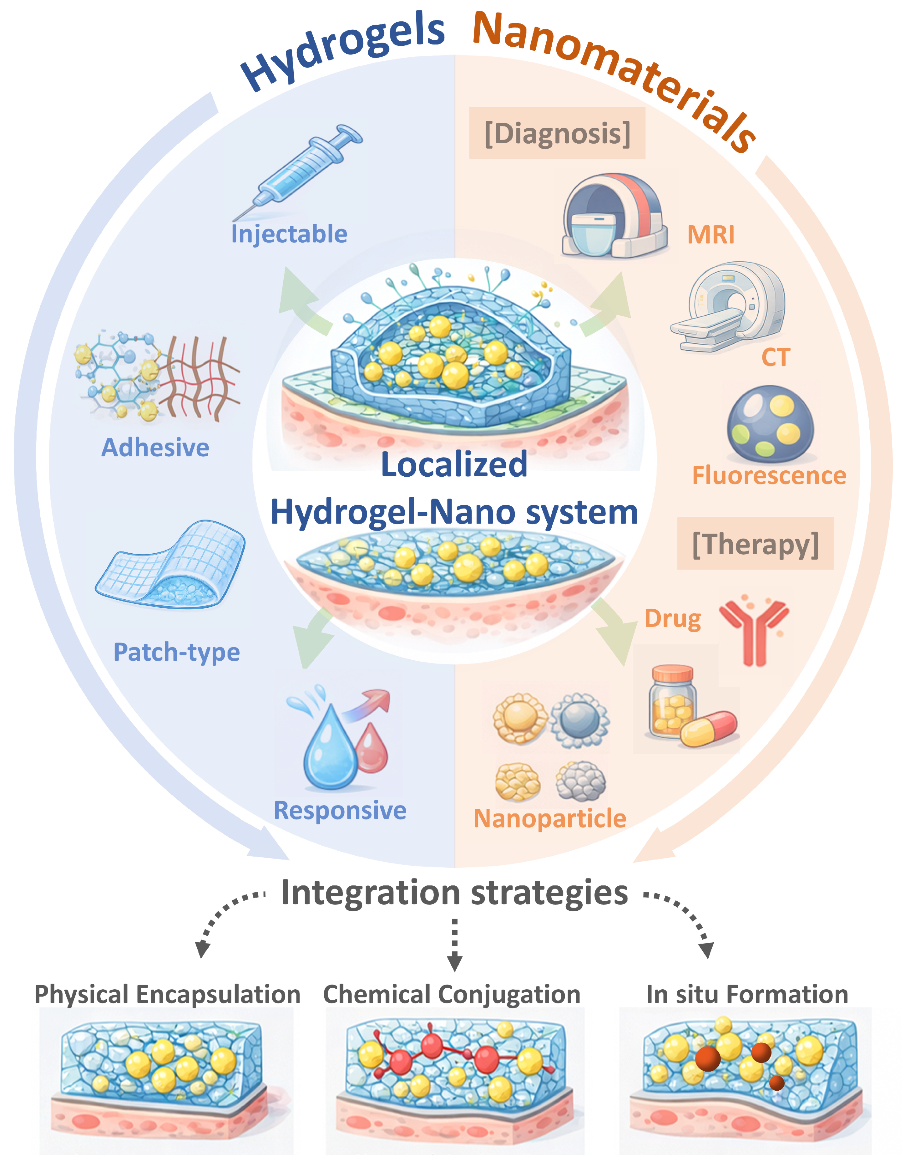
Figure 1. Hydrogel–Nanomaterial Design Principles for Localized Nanotheranostics. The arrows indicate the overall integration flow and functional interactions within the localized hydrogel–nano system, while the blue and red color schemes represent hydrogel-related and nanomaterial-related components, respectively.
Jnt 07 00010 g001
Figure 2. Schematic illustration of representative hydrogel–nanomaterial integrated systems designed for localized cancer therapy at tumor resection sites. Injectable hydrogel incorporating polydopamine nanoparticles and immune-modulatory components for inhibition of tumor recurrence and metastasis through localized therapy and immune activation. Reprinted with permission from Ref. [74]. Copyright 2024 American Chemical Society.
Jnt 07 00010 g002
Figure 3. Representative applications of hydrogel-integrated nanomaterials for localized inflammatory disease management and regenerative theranostics. Injectable thermoresponsive nanocomposite hydrogel enabling antibacterial photothermal therapy and wound contraction under external stimulation, while modulating inflammatory responses. Arrows indicate the sequential fabrication and therapeutic workflow, as well as the progression of wound healing, while different colors are used to distinguish the major components and functional processes of the hydrogel–nanomaterial system. Reproduced with permission from Ref. [85]. Copyright 2025 The Authors. Advanced Science published by Wiley-VCH GmbH.
Jnt 07 00010 g003
Figure 4. Overview of advanced functional hydrogel designs supporting hydrogel–nanomaterial nanotheranostic systems. Injectable and crosslinkable nanocomposite hydrogel fabrication strategy incorporating functional nanomaterials within a mechanically stable and stimuli-responsive polymer network. The legends in the figure body correspond to the indicated materials, interaction types, and biological components, including photoinitiator, paraffin, chemical crosslinking points, hydrogen bonds, electrostatic interactions, hydrogel, RBCs, activated platelets, coagulation factors, and fibrin mesh. Reproduced with permission from Ref. [119]. Copyright 2025 Ivyspring.
Jnt 07 00010 g004
Table 1. Comparison of Integration Strategies for Hydrogel-Based Nanotheranostic Systems.
Integration StrategyKey FeaturesAdvantagesLimitationsRepresentative Applications
Physical
encapsulation
Pre-formed nanoparticles physically entrapped within hydrogel networkSimple fabrication; preserves nanoparticle functionality; modular designNanoparticle leakage; diffusion-controlled release; limited spatial precisionInjectable drug depots; fluorescent nanoparticle hydrogels; MRI-visible local delivery; postoperative image-guided therapy [8,19]
Chemical
conjugation
Covalent or affinity-based linkage between nanoparticles and hydrogel polymersEnhanced retention; minimal burst release; spatial controlSynthetic complexity; possible functional alteration; matrix-property changesCovalently retained imaging hydrogels; sustained therapeutic matrices; low-leakage nanocomposites; long-term local signal platforms [44,45,46]
In situ
nanoparticle formation
Nanoparticles generated directly within hydrogel matrixUniform dispersion; strong confinement; intimate hydrogel–nano interfacePrecursor toxicity; reaction compatibility; reproducibility challengesPhotothermal nanocomposite hydrogels; in situ metal nanoparticle gels; responsive antibacterial platforms; catalytic theranostic systems [47,48]
Table 2. Imaging modalities enabled by hydrogel–nanomaterial systems for localized diagnosis and therapy. Local retention refers to temporal persistence at the target site, spatial confinement indicates restricted distribution within a defined region, and site-specific localization denotes precise targeting to a specific site.
Imaging
Modality
Representative
Nanomaterials
Role of HydrogelDiagnostic PurposeRepresentative
References
Fluorescence imagingDye-loaded nanoparticles, quantum dots, AIE nanoparticlesLocal retention; sustained signal; release regulationReal-time visualization; drug release monitoring[53,55,56,62]
MRIIron oxide nanoparticles, Gd-based nanostructuresSpatial confinement; prolonged contrast retentionNoninvasive localization; longitudinal monitoring[53,54,57,58,62]
Photoacoustic imagingGold nanorods, carbon-based nanomaterialsSignal stabilization; controlled exposureDeep-tissue imaging; therapy guidance[53,59,63]
CT imagingGold or high-Z element nanoparticlesContrast enhancement; site-specific localizationStructural imaging; treatment assessment[53,54,60,61]
Table 3. Representative clinically evaluated or approved hydrogel- and nanomaterial-based platforms relevant to hydrogel–nanotheranostic systems.
System TypeHydrogel TypeNanomaterial CompositionApplied
Technology
Clinical IndicationStudy Status
Hydrogel-based sealantPEG hydrogel (DuraSeal®)NoneInjectable sealant, localized retentionSurgical sealing (neurosurgery)FDA approved
Hydrogel-based wound dressingAlginate/collagen hydrogelsNoneMoist wound healing, tissue regenerationChronic wounds, burnsClinical use
Nanoparticle-based imagingNoneIron oxide nanoparticles (ferumoxytol)MRI contrast agentAnemia, off-label imagingFDA approved
Nanoparticle-based drug deliveryNoneLiposomal doxorubicin (Doxil®)Chemotherapy deliveryOvarian cancer, Kaposi’s sarcomaFDA approved
Nanoparticle-based imagingNoneGold nanoparticlesCT/photoacoustic imagingCancer imaging (investigational)Clinical trials
Hybrid hydrogel–nanoparticle systemInjectable hydrogelDrug-loaded nanoparticlesLocalized drug deliveryCancer therapyEarly clinical/translational
Nanocomposite hydrogel dressingHydrogel matrixSilver/antimicrobial nanoparticlesAntibacterial therapyInfected woundsClinical/translational
Disclaimer/Publisher’s Note: The statements, opinions and data contained in all publications are solely those of the individual author(s) and contributor(s) and not of MDPI and/or the editor(s). MDPI and/or the editor(s) disclaim responsibility for any injury to people or property resulting from any ideas, methods, instructions or products referred to in the content.