
Nanoparticles in Therapy and Diagnosis: A Comprehensive Review of Mechanisms, Applications, and Translational Challenges


Abstract
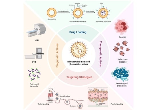
1. Introduction
2. Methodology
3. Fundamental Design Principles of Nanoparticles
3.1. Size and Shape
3.2. Surface Charge, Chemistry, and Surface Modifications
4. Platform Categories of Nanoparticles: A Functional Classification
4.1. Organic Nanoparticle Platforms
4.2. Inorganic Nanoparticle Platforms
4.3. Hybrid and Bioinspired Nanoparticle Platforms
5. Mechanisms of Nanoparticle-Mediated Therapeutic Action
6. Nanoparticles in Therapeutic Applications
6.1. Cancer

| Sr No. | Cancer Type | Nanocarrier Type | Therapeutic Payload | Diagnostic/ Imaging/ Theranostic Component | Study Type | Particle Characteristics | Key Findings | References |
|---|---|---|---|---|---|---|---|---|
| 1 | Breast Cancer | Iron Oxide NPs | DOX | chlorine e6 (Ce6) | In vitro (4T1 cell line) In vivo (Balb/c mice) | - |
| [178] |
| 2 | Breast Cancer | Albumin-modified melanin-silver hybrid NPs | - | HSA | In vitro (HS578T, and MCF10a cell lines) |
|
| [179] |
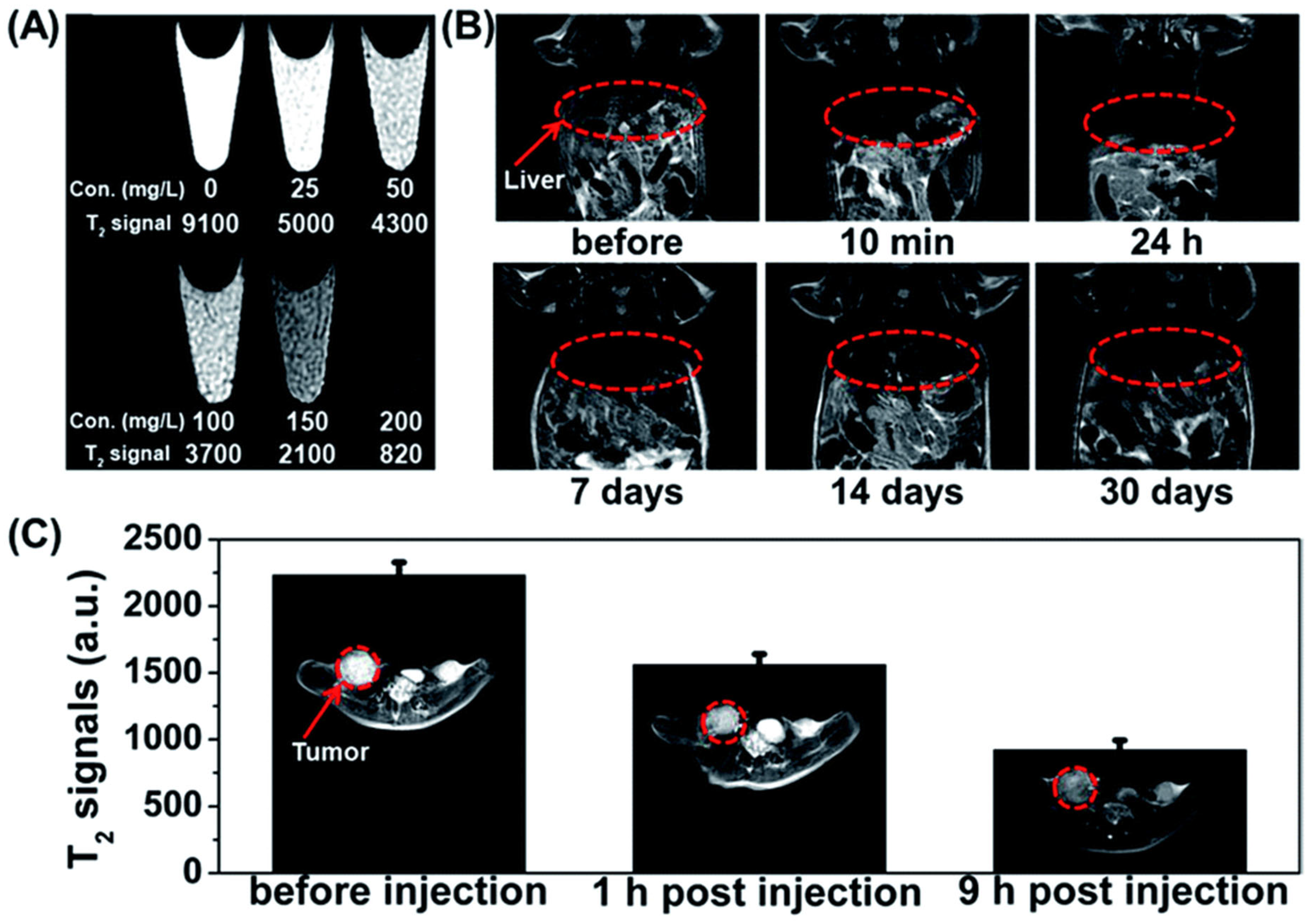
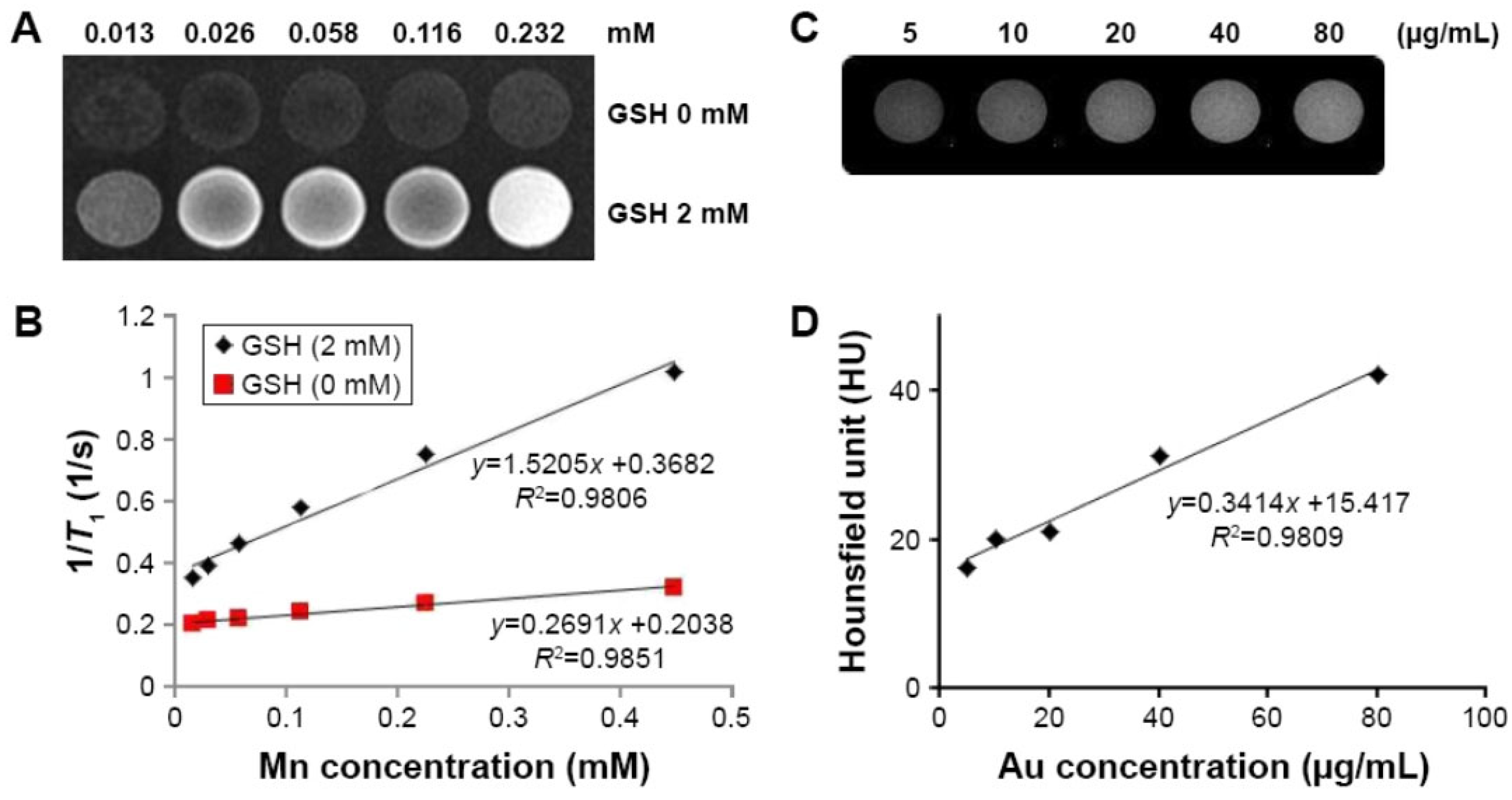
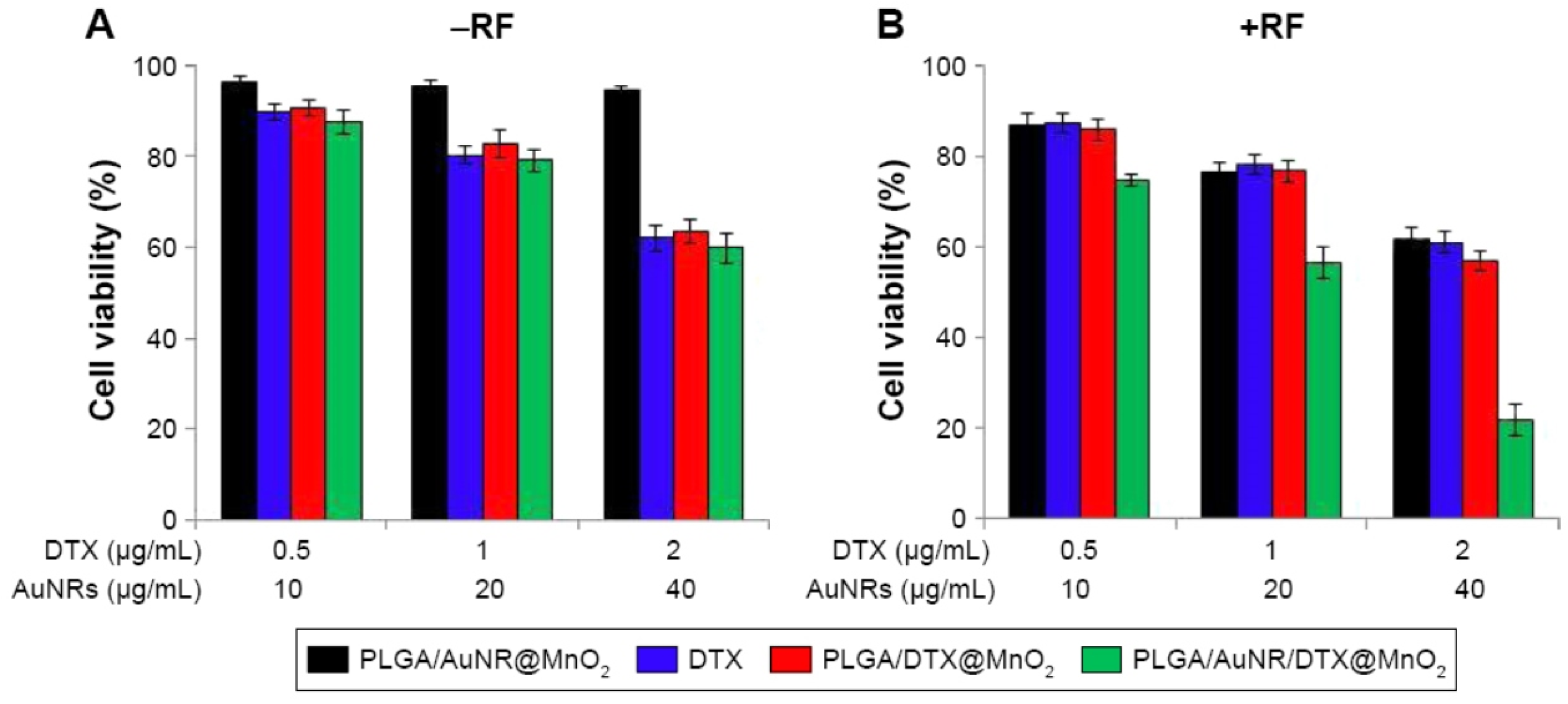
| 3 | Cancer | poly(lactic-co-glycolic acid) (PLGA) NPs loaded with AuNRs | DTX | MnO2 nanosheets | In vitro (MCF-7 human breast cancer cell line) In vivo (female Kunming mice (~18–20 g) |
|
| [177] |
| 4 | Breast Cancer | Hybrid silica/melanin NPs | DOX | HSA | In vitro (HS578T Cells) |
|
| [180] |
| 5 | Breast Cancer | pH-sensitive near-infrared (NIR) croconaine (Croc) dyes-loaded copolymeric PEG−PLGA | - | PPC815 | In vitro (MDA-MB-231 cells) In vivo (BALB/c nude female mice) |
|
| [181] |
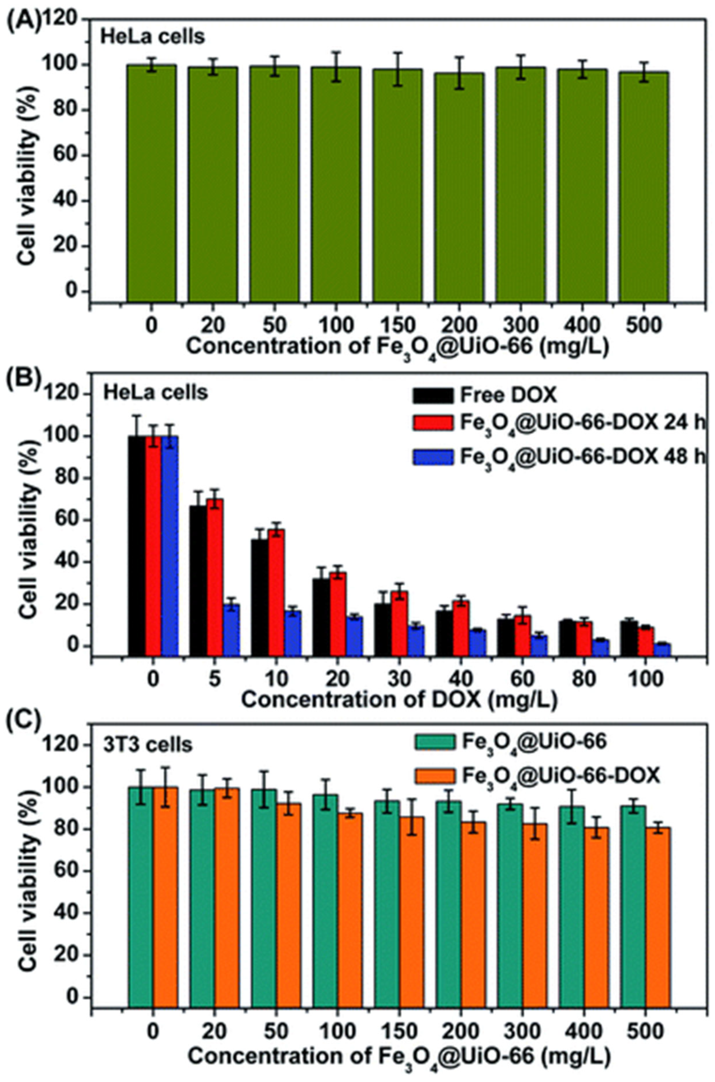
| 6 | Cervical adenocarcinoma | Fe3O4 NPs (Fe3O4@UiO-66 core–shell composites) | DOX | Fe3O4 | In vitro (HeLa cells) In vivo (Kunming mouse) |
|
| [175] |
| 7 | Breast Cancer | Gold-nanoshelled poly (lactic-co-glycolic acid) (PLGA) magnetic hybrid NPs carrying anti-HER-2 antibody | HER-2 antibodies | Her2 functionalized gold-nanoshelled magnetic hybrid NPs (HER2-GPH-NPs) | In vitro (SKBR3 and MDA-MB-231 cells) |
|
| [182] |
| 8 | Metastatic Breast Cancer | Albumin-based NPs (PSN-HSA-PTX-IR780) | PTX | HSA | In vitro (4T1 cells) In vivo (tumor-bearing mice) |
|
| [183] |
| 9 | Breast and prostate cancer | Albumin-Coated Tobacco Mosaic Virus NPs | DOX | TMV | In vitro In vivo | - |
| [184] |
| 10 | Head and neck squamous cell carcinoma | Iron-oxide NPs functionalized with integrin-targeting fibronectin-mimetic peptide (Fmp) | Silicon phthalocyanine 4 (Pc 4) | Iron-oxide NPs | In vitro In vivo |
|
| [185] |
| 11 | Head and neck squamous cell carcinoma | Cholesterol/vitamin E-doped distearoylphosphatidylcholine liposomes | Rhenium-186 (186Re) radionuclide | β-emitting radionuclides | In vivo |
|
| [186] |
| 12 | Cancer | Pluronic F127 polymer micelles | - | TPVTR (aggregation-induced emission fluorogen) dots | In vitro In vivo |
|
| [187] |
| 13 | Bladder cancer | Self-assembled porphyrin-cholic acid-PEG micelles decorated with PLZ4 targeting ligand | DOX and Pyropheophorbide-a | PLZ4-nanoporphyrin | In vitro In vivo |
|
| [188] |
| 14 | Non-small cell lung cancer, bladder cancer, and murine fibroblast | Anisamide-targeted lipid-calcium-phosphate (LCP) NPs | Lutetium-177 (177Lu) radionuclide | Lutetium-177 (177Lu) radionuclide | In vitro In vivo |
|
| [189] |
| 15 | Thyroid cancer | Polyethylene glycol-functionalized copper sulfide (CuS) NPs | Copper-64 (64Cu) radionuclide | CuS photothermal agent | In vivo |
|
| [190] |
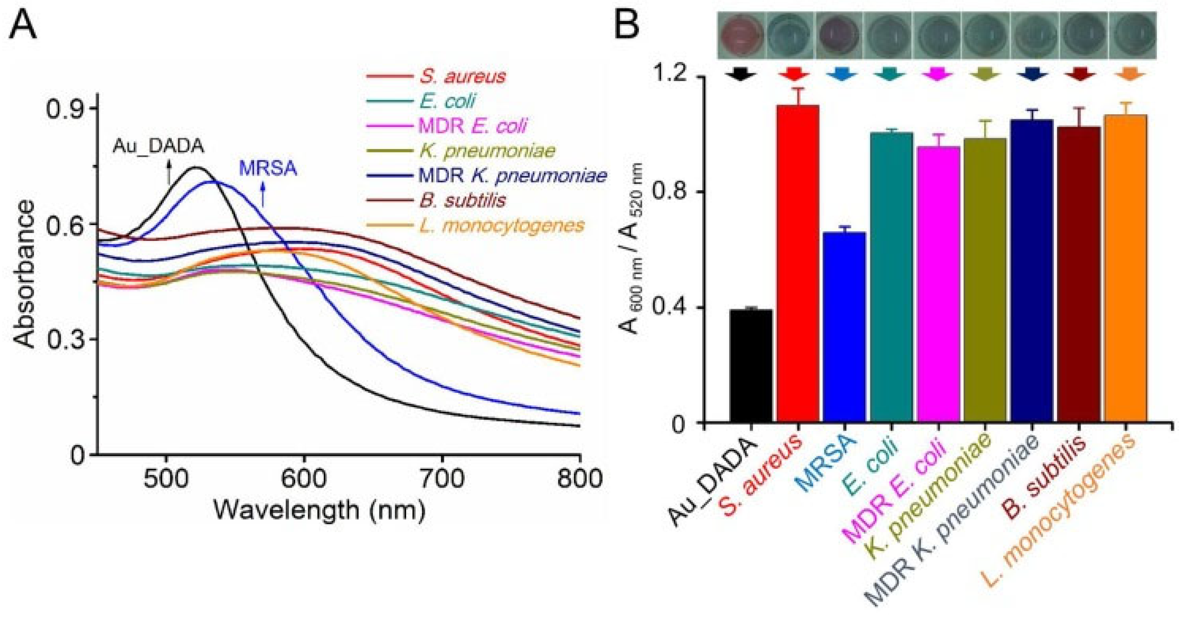
| 16 | Colorectal cancer | Polyethylene glycol-coated gold NPs | Zinc sulfothiolphthalocyanine (ZnPcS4) | ZnPcS4 | In vitro |
|
| [191] |
| 17 | Breast cancer | Rare earth (RE 3+)-doped NPs (RENPs) | Human MSCs | chlorine6 (Ce6) | In vitro |
|
| [192] |
| 18 | RAW264.7murinemacrophage and breast cancer | M1-like macrophage membrane-camouflaged zinc 2-methylimidazole (ZIF-8) metal-organic framework | BN-O (hypoxia-responsive phototheranostic molecule) and Combretastatin-A4 phosphate (CA4P) | BN-O | In vitro In vivo |
|
| [193] |
6.2. Infectious Disease
6.3. Neurological and Cardiovascular Diseases
7. Current Clinical Landscape
8. Challenges in Clinical Translation
9. Conclusions and Future Perspectives
Author Contributions
Funding
Institutional Review Board Statement
Informed Consent Statement
Data Availability Statement
Conflicts of Interest
References
- Tiwary, P.; Oswal, K.; Varghese, R.; Gupta, P. Solid Lipid Nanoparticles in Imaging, Diagnostics and Theranostics: A Review of a decade of Innovations and Clinical Applications. Colloids Surf. B Biointerfaces 2025, 257, 115122. [Google Scholar] [CrossRef] [PubMed]
- Chaturvedi, P.; Sharma, R.; Khare, S.; Singh, T.; Shrivastava, D.; Singh, A.; Sarolia, J. Prodrug-based drug delivery systems: Strategic approaches and emerging challenges in targeted therapeutics. Naunyn. Schmiedebergs Arch. Pharmacol. 2025, 399, 4811–4826. [Google Scholar] [CrossRef]
- Du, W.; Gao, Y.; Liu, L.; Sai, S.; Ding, C. Striking back against fungal infections: The utilization of nanosystems for antifungal strategies. Int. J. Mol. Sci. 2021, 22, 10104. [Google Scholar] [CrossRef] [PubMed]
- Zhang, M.; Zhang, R.; Feng, C.; Jiang, X.; Xu, X.; Wang, J. Ginsenoside compound K-based multifunctional liposomes for the treatment of rheumatoid arthritis. Drug Deliv. 2025, 32, 2464190. [Google Scholar] [CrossRef]
- Yan Chan Edgar, J.; Wang, H. Introduction for design of nanoparticle based drug delivery systems. Curr. Pharm. Des. 2017, 23, 2108–2112. [Google Scholar] [CrossRef]
- Thakur, S.; Riyaz, B.; Patil, A.; Kaur, A.; Kapoor, B.; Mishra, V. Novel drug delivery systems for NSAIDs in management of rheumatoid arthritis: An overview. Biomed. Pharmacother. 2018, 106, 1011–1023. [Google Scholar] [CrossRef]
- Tiwary, P.; Oswal, K.; Varghese, R.; Anchan, H.; Gupta, P. Solid Lipid Nanoparticles in the diagnosis and treatment of Prostate Cancer: A Comprehensive Review of Two Decades of Advancements. Chem. Phys. Lipids 2025, 270, 105510. [Google Scholar] [CrossRef]
- Varghese, R.; Salvi, S.; Sood, P.; Kulkarni, B.; Kumar, D. Cubosomes in cancer drug delivery: A review. Colloid Interface Sci. Commun. 2022, 46, 100561. [Google Scholar] [CrossRef]
- Sarkar, M.; Wang, Y.; Ekpenyong, O.; Liang, D.; Xie, H. Pharmacokinetic behaviors of soft nanoparticulate formulations of chemotherapeutics. Wiley Interdiscip. Rev. Nanomed. Nanobiotechnol. 2023, 15, e1846. [Google Scholar] [CrossRef]
- Singh, P.; Waghambare, P.; Khan, T.A.; Omri, A. Colorectal cancer management: Strategies in drug delivery. Expert Opin. Drug Deliv. 2022, 19, 653–670. [Google Scholar] [CrossRef] [PubMed]
- Kazi, R.N.A.; Hasani, I.W.; Khafaga, D.S.R.; Kabba, S.; Farhan, M.; Aatif, M.; Muteeb, G.; Fahim, Y.A. Nanomedicine: The effective role of nanomaterials in healthcare from diagnosis to therapy. Pharmaceutics 2025, 17, 987. [Google Scholar] [CrossRef] [PubMed]
- Joo, J. Diagnostic and therapeutic nanomedicine. In Advanced Imaging and Bio Techniques for Convergence Science; Springer: Berlin/Heidelberg, Germany, 2021; pp. 401–447. [Google Scholar]
- Mittal, M.; Juneja, S.; Pandey, N.; Mittal, R. Nanoparticle-based drug delivery systems: Current advances and future directions. Curr. Drug Targets 2025, 27, 150–172. [Google Scholar] [CrossRef]
- Bayrak, E.; Bayir, E.; Baysoy, E.; Özcan, A.; Ayan, B.; Saygili, E.; Kaleli-Can, G. Nintedanib loaded iron (III) chelated melanin nanoparticles as an MRI-visible antifibrotic drug delivery system. Colloids Surf. B Biointerfaces 2025, 252, 114652. [Google Scholar] [CrossRef]
- Wathoni, N.; Rusdin, A.; Motoyama, K.; Joni, I.M.; Lesmana, R.; Muchtaridi, M. Nanoparticle drug delivery systems for α-mangostin. Nanotechnol. Sci. Appl. 2020, 13, 23–36. [Google Scholar] [CrossRef]
- Tiwary, P.; Oswal, K.; Varghese, R.; Peri, R.V.; Gupta, P. Solid lipid nanoparticles in cervical cancer: A comprehensive review of a decade of progress and prospects. RSC Pharm. 2025, 2, 930–949. [Google Scholar] [CrossRef]
- Lee, G.Y.; Qian, W.P.; Wang, L.; Wang, Y.A.; Staley, C.A.; Satpathy, M.; Nie, S.; Mao, H.; Yang, L. Theranostic nanoparticles with controlled release of gemcitabine for targeted therapy and MRI of pancreatic cancer. ACS Nano 2013, 7, 2078–2089. [Google Scholar] [CrossRef]
- Yu, X.; Zhang, Q.; Wang, L.; Zhang, Y.; Zhu, L. Engineered nanoparticles for imaging and targeted drug delivery in hepatocellular carcinoma. Exp. Hematol. Oncol. 2025, 14, 62. [Google Scholar] [CrossRef]
- Fattahi, M.R.; Dehghani, M.; Paknahad, S.; Rahiminia, S.; Zareie, D.; Hoseini, B.; Oroomi, T.R.; Motedayyen, H.; Arefnezhad, R. Clinical insights into nanomedicine and biosafety: Advanced therapeutic approaches for common urological cancers. Front. Oncol. 2024, 14, 1438297. [Google Scholar] [CrossRef] [PubMed]
- Fang, Y.; Lin, S.; Yang, F.; Situ, J.; Lin, S.; Luo, Y. Aptamer-Conjugated Multifunctional Polymeric Nanoparticles as Cancer-Targeted, MRI-Ultrasensitive Drug Delivery Systems for Treatment of Castration-Resistant Prostate Cancer. Biomed. Res. Int. 2020, 2020, 9186583. [Google Scholar] [CrossRef]
- Wang, Y.; Wang, J.; Zhu, D.; Wang, Y.; Qing, G.; Zhang, Y.; Liu, X.; Liang, X.-J. Effect of physicochemical properties on in vivo fate of nanoparticle-based cancer immunotherapies. Acta Pharm. Sin. B 2021, 11, 886–902. [Google Scholar] [CrossRef] [PubMed]
- Tiwari, A.K.; Pandey, P.C.; Gupta, M.K.; Narayan, R.J. Nano–bio interaction and antibacterial mechanism of engineered metal nanoparticles: Fundamentals and current understanding. J. Clust. Sci. 2025, 36, 5. [Google Scholar] [CrossRef]
- Powers, K.W.; Palazuelos, M.; Moudgil, B.M.; Roberts, S.M. Characterization of the size, shape, and state of dispersion of nanoparticles for toxicological studies. Nanotoxicology 2007, 1, 42–51. [Google Scholar] [CrossRef]
- Gehr, P.; Zellner, R. Biological Responses to Nanoscale Particles; Springer: Berlin/Heidelberg, Germany, 2019; ISBN 3030124614. [Google Scholar]
- Blanco, E.; Shen, H.; Ferrari, M. Principles of nanoparticle design for overcoming biological barriers to drug delivery. Nat. Biotechnol. 2015, 33, 941–951. [Google Scholar] [CrossRef] [PubMed]
- Foroozandeh, P.; Aziz, A.A. Insight into cellular uptake and intracellular trafficking of nanoparticles. Nanoscale Res. Lett. 2018, 13, 339. [Google Scholar] [CrossRef]
- Makaraci, P.; Kim, K. trans-Golgi network-bound cargo traffic. Eur. J. Cell Biol. 2018, 97, 137–149. [Google Scholar] [CrossRef]
- Wang, X.; Wang, W. Cellular journey of nanomaterials: Theories, trafficking, and kinetics. Aggregate 2023, 4, e372. [Google Scholar] [CrossRef]
- Rennick, J.J.; Johnston, A.P.R.; Parton, R.G. Key principles and methods for studying the endocytosis of biological and nanoparticle therapeutics. Nat. Nanotechnol. 2021, 16, 266–276. [Google Scholar] [CrossRef]
- de Almeida, M.S.; Susnik, E.; Drasler, B.; Taladriz-Blanco, P.; Petri-Fink, A.; Rothen-Rutishauser, B. Understanding nanoparticle endocytosis to improve targeting strategies in nanomedicine. Chem. Soc. Rev. 2021, 50, 5397–5434. [Google Scholar] [CrossRef] [PubMed]
- Cheng, X.; Tian, X.; Wu, A.; Li, J.; Tian, J.; Chong, Y.; Chai, Z.; Zhao, Y.; Chen, C.; Ge, C. Protein corona influences cellular uptake of gold nanoparticles by phagocytic and nonphagocytic cells in a size-dependent manner. ACS Appl. Mater. Interfaces 2015, 7, 20568–20575. [Google Scholar] [CrossRef] [PubMed]
- Ho, Y.T.; Kamm, R.D.; Kah, J.C.Y. Influence of protein corona and caveolae-mediated endocytosis on nanoparticle uptake and transcytosis. Nanoscale 2018, 10, 12386–12397. [Google Scholar] [CrossRef]
- Cheng, J.P.X.; Nichols, B.J. Caveolae: One function or many? Trends Cell Biol. 2016, 26, 177–189. [Google Scholar] [CrossRef]
- Lolo, F.-N.; Walani, N.; Seemann, E.; Zalvidea, D.; Pavón, D.M.; Cojoc, G.; Zamai, M.; Viaris de Lesegno, C.; Martinez de Benito, F.; Sánchez-Álvarez, M. Caveolin-1 dolines form a distinct and rapid caveolae-independent mechanoadaptation system. Nat. Cell Biol. 2023, 25, 120–133. [Google Scholar] [CrossRef]
- Butt, A.M.; Abdullah, N.; Rani, N.N.I.M.; Ahmad, N.; Amin, M.C.I.M. Endosomal escape of bioactives deployed via nanocarriers: Insights into the design of polymeric micelles. Pharm. Res. 2022, 39, 1047–1064. [Google Scholar] [CrossRef]
- Cui, X.; Wan, B.; Yang, Y.; Ren, X.; Guo, L.-H. Length effects on the dynamic process of cellular uptake and exocytosis of single-walled carbon nanotubes in murine macrophage cells. Sci. Rep. 2017, 7, 1518. [Google Scholar] [CrossRef] [PubMed]
- Henna, T.K.; Pramod, K. Graphene quantum dots redefine nanobiomedicine. Mater. Sci. Eng. C 2020, 110, 110651. [Google Scholar] [CrossRef]
- Zheng, N.; Li, J.; Xu, C.; Xu, L.; Li, S.; Xu, L. Mesoporous silica nanorods for improved oral drug absorption. Artif. Cells Nanomed. Biotechnol. 2018, 46, 1132–1140. [Google Scholar] [CrossRef]
- Behzadi, S.; Serpooshan, V.; Tao, W.; Hamaly, M.A.; Alkawareek, M.Y.; Dreaden, E.C.; Brown, D.; Alkilany, A.M.; Farokhzad, O.C.; Mahmoudi, M. Cellular uptake of nanoparticles: Journey inside the cell. Chem. Soc. Rev. 2017, 46, 4218–4244. [Google Scholar] [CrossRef] [PubMed]
- Kaksonen, M.; Roux, A. Mechanisms of clathrin-mediated endocytosis. Nat. Rev. Mol. Cell Biol. 2018, 19, 313–326. [Google Scholar] [CrossRef]
- Yue, J.; Feliciano, T.J.; Li, W.; Lee, A.; Odom, T.W. Gold nanoparticle size and shape effects on cellular uptake and intracellular distribution of siRNA nanoconstructs. Bioconjug. Chem. 2017, 28, 1791–1800. [Google Scholar] [CrossRef] [PubMed]
- Chithrani, B.D.; Chan, W.C.W. Elucidating the mechanism of cellular uptake and removal of protein-coated gold nanoparticles of different sizes and shapes. Nano Lett. 2007, 7, 1542–1550. [Google Scholar] [CrossRef]
- Geiser, M.; Rothen-Rutishauser, B.; Kapp, N.; Schürch, S.; Kreyling, W.; Schulz, H.; Semmler, M.; Im Hof, V.; Heyder, J.; Gehr, P. Ultrafine particles cross cellular membranes by nonphagocytic mechanisms in lungs and in cultured cells. Environ. Health Perspect. 2005, 113, 1555. [Google Scholar] [CrossRef] [PubMed]
- Jin, H.; Heller, D.A.; Sharma, R.; Strano, M.S. Size-dependent cellular uptake and expulsion of single-walled carbon nanotubes: Single particle tracking and a generic uptake model for nanoparticles. ACS Nano 2009, 3, 149–158. [Google Scholar] [CrossRef] [PubMed]
- Lu, F.; Wu, S.H.; Hung, Y.; Mou, C.-Y. Size effect on cell uptake in well-suspended, uniform mesoporous silica nanoparticles. Small 2009, 5, 1408–1413. [Google Scholar] [CrossRef]
- Osaki, F.; Kanamori, T.; Sando, S.; Sera, T.; Aoyama, Y. A quantum dot conjugated sugar ball and its cellular uptake. On the size effects of endocytosis in the subviral region. J. Am. Chem. Soc. 2004, 126, 6520–6521. [Google Scholar] [CrossRef] [PubMed]
- Wang, S.-H.; Lee, C.W.; Chiou, A.; Wei, P.K. Size-dependent endocytosis of gold nanoparticles studied by three-dimensional mapping of plasmonic scattering images. J. Nanobiotechnol. 2010, 8, 33. [Google Scholar] [CrossRef]
- Zhang, C.; Cheng, Y.; Liu, D.; Liu, M.; Cui, H.; Zhang, B.; Mei, Q.; Zhou, S. Mitochondria-targeted cyclosporin A delivery system to treat myocardial ischemia reperfusion injury of rats. J. Nanobiotechnol. 2019, 17, 18. [Google Scholar] [CrossRef]
- Hoshyar, N.; Gray, S.; Han, H.; Bao, G. The effect of nanoparticle size on in vivo pharmacokinetics and cellular interaction. Nanomedicine 2016, 11, 673–692. [Google Scholar] [CrossRef]
- Hocking, K.M.; Evans, B.C.; Komalavilas, P.; Cheung-Flynn, J.; Duvall, C.L.; Brophy, C.M. Nanotechnology enabled modulation of signaling pathways affects physiologic responses in intact vascular tissue. Tissue Eng. Part A 2019, 25, 416–426. [Google Scholar] [CrossRef]
- Shahin, M.; Safaei-Nikouei, N.; Lavasanifar, A. Polymeric micelles for pH-responsive delivery of cisplatin. J. Drug Target. 2014, 22, 629–637. [Google Scholar] [CrossRef]
- Chen, L.Q.; Fang, L.; Ling, J.; Ding, C.Z.; Kang, B.; Huang, C.Z. Nanotoxicity of silver nanoparticles to red blood cells: Size dependent adsorption, uptake, and hemolytic activity. Chem. Res. Toxicol. 2015, 28, 501–509. [Google Scholar] [CrossRef]
- Champion, J.A.; Mitragotri, S. Role of target geometry in phagocytosis. Proc. Natl. Acad. Sci. USA 2006, 103, 4930–4934. [Google Scholar] [CrossRef]
- Yusuf, A.; Almotairy, A.R.Z.; Henidi, H.; Alshehri, O.Y.; Aldughaim, M.S. Nanoparticles as drug delivery systems: A review of the implication of nanoparticles’ physicochemical properties on responses in biological systems. Polymers 2023, 15, 1596. [Google Scholar] [CrossRef]
- Wang, Y.; Gou, K.; Guo, X.; Ke, J.; Li, S.; Li, H. Advances in regulating physicochemical properties of mesoporous silica nanocarriers to overcome biological barriers. Acta Biomater. 2021, 123, 72–92. [Google Scholar] [CrossRef] [PubMed]
- Hadji, H.; Bouchemal, K. Effect of micro-and nanoparticle shape on biological processes. J. Control. Release 2022, 342, 93–110. [Google Scholar] [CrossRef] [PubMed]
- García-Hevia, L.; Soltani, R.; González, J.; Chaloin, O.; Ménard-Moyon, C.; Bianco, A.; Fanarraga, M.L. Carbon nanotubes targeted to the tumor microenvironment inhibit metastasis in a preclinical model of melanoma. Bioact. Mater. 2024, 34, 237–247. [Google Scholar] [CrossRef]
- Rastogi, V.; Yadav, P.; Bhattacharya, S.S.; Mishra, A.K.; Verma, N.; Verma, A.; Pandit, J.K. Carbon nanotubes: An emerging drug carrier for targeting cancer cells. J. Drug Deliv. 2014, 2014, 670815. [Google Scholar] [CrossRef] [PubMed]
- Jahangiri-Manesh, A.; Mousazadeh, M.; Taji, S.; Bahmani, A.; Zarepour, A.; Zarrabi, A.; Sharifi, E.; Azimzadeh, M. Gold nanorods for drug and gene delivery: An overview of recent advancements. Pharmaceutics 2022, 14, 664. [Google Scholar] [CrossRef]
- Raghuram, S.; Mackeyev, Y.; Symons, J.; Zahra, Y.; Gonzalez, V.; Mahadevan, K.K.; Requejo, K.I.; Liopo, A.; Derry, P.; Zubarev, E.; et al. Uncloaking cell-impermeant gold nanorods via tumor microenvironmental cathepsin B facilitates cancer cell penetration and potent radiosensitization. Biomaterials 2022, 291, 121887. [Google Scholar] [CrossRef]
- Zangabad, P.S.; Karimi, M.; Mehdizadeh, F.; Malekzad, H.; Ghasemi, A.; Bahrami, S.; Zare, H.; Moghoofei, M.; Hekmatmanesh, A.; Hamblin, M.R. Nanocaged platforms: Modification, drug delivery and nanotoxicity. Opening synthetic cages to release the tiger. Nanoscale 2017, 9, 1356–1392. [Google Scholar] [CrossRef]
- Bhaskar, S.; Lim, S. Engineering protein nanocages as carriers for biomedical applications. NPG Asia Mater. 2017, 9, e371. [Google Scholar] [CrossRef]
- Elzoghby, A.O.; Samir, O.; Soliman, A.; Solomevich, S.; Yu, M.; Schwendeman, A.; Nasr, M.L. Nanodiscs: Game changer nano-therapeutics and structural biology tools. Nano Today 2023, 53, 102026. [Google Scholar] [CrossRef]
- Pires, I.S.; Hostetler, A.; Covarrubias, G.; Carlo, I.S.; Suggs, J.R.; Kim, B.J.; Pickering, A.J.; Gordon, E.; Irvine, D.J.; Hammond, P.T. Charge-Stabilized Nanodiscs as a New Class of Lipid Nanoparticles. Adv. Mater. 2024, 36, 2408307. [Google Scholar] [CrossRef]
- Shin, D.; Choi, J.; Park, B.; Kim, S.H.; Shim, M.K. Factors Governing Tumor Penetration of Nanomedicines: Intrinsic and Extrinsic Determinants. Nanoscale 2026, 18, 2363–2381. [Google Scholar] [CrossRef]
- Manzanares, D.; Ceña, V. Endocytosis: The nanoparticle and submicron nanocompounds gateway into the cell. Pharmaceutics 2020, 12, 371. [Google Scholar] [CrossRef]
- Mitchell, M.J.; Billingsley, M.M.; Haley, R.M.; Wechsler, M.E.; Peppas, N.A.; Langer, R. Engineering precision nanoparticles for drug delivery. Nat. Rev. Drug Discov. 2021, 20, 101–124. [Google Scholar] [CrossRef] [PubMed]
- Von Roemeling, C.; Jiang, W.; Chan, C.K.; Weissman, I.L.; Kim, B.Y.S. Breaking down the barriers to precision cancer nanomedicine. Trends Biotechnol. 2017, 35, 159–171. [Google Scholar] [CrossRef]
- Choi, C.H.J.; Zuckerman, J.E.; Webster, P.; Davis, M.E. Targeting kidney mesangium by nanoparticles of defined size. Proc. Natl. Acad. Sci. USA 2011, 108, 6656–6661. [Google Scholar] [CrossRef]
- Wilhelm, S.; Tavares, A.J.; Dai, Q.; Ohta, S.; Audet, J.; Dvorak, H.F.; Chan, W.C.W. Analysis of nanoparticle delivery to tumours. Nat. Rev. Mater. 2016, 1, 16014. [Google Scholar] [CrossRef]
- Jain, R.K.; Stylianopoulos, T. Delivering nanomedicine to solid tumors. Nat. Rev. Clin. Oncol. 2010, 7, 653–664. [Google Scholar] [CrossRef] [PubMed]
- Tenzer, S.; Docter, D.; Kuharev, J.; Musyanovych, A.; Fetz, V.; Hecht, R.; Schlenk, F.; Fischer, D.; Kiouptsi, K.; Reinhardt, C.; et al. Rapid formation of plasma protein corona critically affects nanoparticle pathophysiology. In Nano-Enabled Medical Applications; Jenny Stanford Publishing: Singapore, 2020; pp. 251–278. [Google Scholar]
- Cox, A.; Andreozzi, P.; Dal Magro, R.; Fiordaliso, F.; Corbelli, A.; Talamini, L.; Chinello, C.; Raimondo, F.; Magni, F.; Tringali, M.; et al. Evolution of nanoparticle protein corona across the blood–brain barrier. ACS Nano 2018, 12, 7292–7300. [Google Scholar] [CrossRef]
- Simpson, J.D.; Smith, S.A.; Thurecht, K.J.; Such, G. Engineered polymeric materials for biological applications: Overcoming challenges of the bio–nano interface. Polymers 2019, 11, 1441. [Google Scholar] [CrossRef] [PubMed]
- Salvati, A.; Pitek, A.S.; Monopoli, M.P.; Prapainop, K.; Bombelli, F.B.; Hristov, D.R.; Kelly, P.M.; Åberg, C.; Mahon, E.; Dawson, K.A. Transferrin-functionalized nanoparticles lose their targeting capabilities when a biomolecule corona adsorbs on the surface. Nat. Nanotechnol. 2013, 8, 137–143. [Google Scholar] [CrossRef] [PubMed]
- Beach, M.A.; Nayanathara, U.; Gao, Y.; Zhang, C.; Xiong, Y.; Wang, Y.; Such, G.K. Polymeric nanoparticles for drug delivery. Chem. Rev. 2024, 124, 5505–5616. [Google Scholar] [CrossRef]
- Lu, Q.; Yu, H.; Zhao, T.; Zhu, G.; Li, X. Nanoparticles with transformable physicochemical properties for overcoming biological barriers. Nanoscale 2023, 15, 13202–13223. [Google Scholar] [CrossRef]
- Souri, M.; Golzaryan, A.; Soltani, M. Charge-Switchable nanoparticles to enhance tumor penetration and accumulation. Eur. J. Pharm. Biopharm. 2024, 199, 114310. [Google Scholar] [CrossRef]
- Stylianopoulos, T.; Poh, M.-Z.; Insin, N.; Bawendi, M.G.; Fukumura, D.; Munn, L.L.; Jain, R.K. Diffusion of particles in the extracellular matrix: The effect of repulsive electrostatic interactions. Biophys. J. 2010, 99, 1342–1349. [Google Scholar] [CrossRef]
- Schöttler, S.; Landfester, K.; Mailänder, V. Controlling the stealth effect of nanocarriers through understanding the protein corona. Angew. Chem. Int. Ed. 2016, 55, 8806–8815. [Google Scholar] [CrossRef]
- McSweeney, M.D.; Wessler, T.; Price, L.S.L.; Ciociola, E.C.; Herity, L.B.; Piscitelli, J.A.; Zamboni, W.C.; Forest, M.G.; Cao, Y.; Lai, S.K. A minimal physiologically based pharmacokinetic model that predicts anti-PEG IgG-mediated clearance of PEGylated drugs in human and mouse. J. Control. Release 2018, 284, 171–178. [Google Scholar] [CrossRef]
- Wang, X.; Ishida, T.; Kiwada, H. Anti-PEG IgM elicited by injection of liposomes is involved in the enhanced blood clearance of a subsequent dose of PEGylated liposomes. J. Control. Release 2007, 119, 236–244. [Google Scholar] [CrossRef]
- Hu, C.-M.J.; Fang, R.H.; Wang, K.-C.; Luk, B.T.; Thamphiwatana, S.; Dehaini, D.; Nguyen, P.; Angsantikul, P.; Wen, C.H.; Kroll, A.V.; et al. Nanoparticle biointerfacing by platelet membrane cloaking. Nature 2015, 526, 118–121. [Google Scholar] [CrossRef] [PubMed]
- Wibroe, P.P.; Anselmo, A.C.; Nilsson, P.H.; Sarode, A.; Gupta, V.; Urbanics, R.; Szebeni, J.; Hunter, A.C.; Mitragotri, S.; Mollnes, T.E.; et al. Bypassing adverse injection reactions to nanoparticles through shape modification and attachment to erythrocytes. Nat. Nanotechnol. 2017, 12, 589–594. [Google Scholar] [CrossRef]
- Pan, K.; Zhong, Q. Organic nanoparticles in foods: Fabrication, characterization, and utilization. Annu. Rev. Food Sci. Technol. 2016, 7, 245–266. [Google Scholar] [CrossRef] [PubMed]
- Joudeh, N.; Linke, D. Nanoparticle classification, physicochemical properties, characterization, and applications: A comprehensive review for biologists. J. Nanobiotechnol. 2022, 20, 262. [Google Scholar] [CrossRef]
- Mehrdadi, S. Lipid-based nanoparticles as oral drug delivery systems: Overcoming poor gastrointestinal absorption and enhancing bioavailability of peptide and protein therapeutics. Adv. Pharm. Bull. 2023, 14, 48. [Google Scholar] [CrossRef]
- Priya, S.; Desai, V.M.; Singhvi, G. Surface modification of lipid-based nanocarriers: A potential approach to enhance targeted drug delivery. ACS Omega 2022, 8, 74–86. [Google Scholar] [CrossRef] [PubMed]
- Li, Q.; Cai, T.; Huang, Y.; Xia, X.; Cole, S.P.C.; Cai, Y. A review of the structure, preparation, and application of NLCs, PNPs, and PLNs. Nanomaterials 2017, 7, 122. [Google Scholar] [CrossRef] [PubMed]
- Xu, L.; Wang, X.; Liu, Y.; Yang, G.; Falconer, R.J.; Zhao, C.-X. Lipid nanoparticles for drug delivery. Adv. NanoBiomed Res. 2022, 2, 2100109. [Google Scholar] [CrossRef]
- Niculescu, A.-G.; Grumezescu, A.M. Polymer-based nanosystems—A versatile delivery approach. Materials 2021, 14, 6812. [Google Scholar] [CrossRef]
- De, T.; Trieu, V.; Myers, S.; Qazi, S.; Saund, S.; Lee, C. Sub-15 nm Nanoparticles for Drug Delivery: Emerging Frontiers and Therapeutic Potential. Int. J. Mol. Sci. 2025, 26, 10842. [Google Scholar] [CrossRef]
- Sgorla, D.; Bunhak, É.J.; Cavalcanti, O.A.; Fonte, P.; Sarmento, B. Exploitation of lipid-polymeric matrices at nanoscale for drug delivery applications. Expert Opin. Drug Deliv. 2016, 13, 1301–1309. [Google Scholar] [CrossRef]
- Unnikrishnan, G.; Joy, A.; Megha, M.; Kolanthai, E.; Senthilkumar, M. Exploration of inorganic nanoparticles for revolutionary drug delivery applications: A critical review. Discov. Nano 2023, 18, 157. [Google Scholar] [CrossRef]
- Kumar, R.; Mondal, K.; Panda, P.K.; Kaushik, A.; Abolhassani, R.; Ahuja, R.; Rubahn, H.-G.; Mishra, Y.K. Core–shell nanostructures: Perspectives towards drug delivery applications. J. Mater. Chem. B 2020, 8, 8992–9027. [Google Scholar] [CrossRef]
- Ealia, S.A.M.; Saravanakumar, M.P. A review on the classification, characterisation, synthesis of nanoparticles and their application. In Proceedings of the IOP Conference Series: Materials Science and Engineering; IOP Publishing: Bristol, UK, 2017; Volume 263, p. 32019. [Google Scholar]
- Kim, M.; Lee, J.; Nam, J. Plasmonic photothermal nanoparticles for biomedical applications. Adv. Sci. 2019, 6, 1900471. [Google Scholar] [CrossRef] [PubMed]
- Shukla, S.; Phatale, V.; Khairnar, P.; Puri, N.; Sahane, P.; Wagh, S.; Sharma, A.; Srinivasarao, D.A.; Srivastava, S. Emerging Metallic Nanoparticle Modalities in Oral Squamous Cell Carcinoma: A Comprehensive Review on Theranostic Strategies and Future Perspectives. ACS Appl. Bio Mater. 2026, 9, 529–572. [Google Scholar] [CrossRef]
- Liong, M.; Lu, J.; Kovochich, M.; Xia, T.; Ruehm, S.G.; Nel, A.E.; Tamanoi, F.; Zink, J.I. Multifunctional inorganic nanoparticles for imaging, targeting, and drug delivery. ACS Nano 2008, 2, 889–896. [Google Scholar] [CrossRef] [PubMed]
- Naz, S.; Shamoon, M.; Wang, R.; Zhang, L.; Zhou, J.; Chen, J. Advances in therapeutic implications of inorganic drug delivery nano-platforms for cancer. Int. J. Mol. Sci. 2019, 20, 965. [Google Scholar] [CrossRef]
- Janjua, T.I.; Cao, Y.; Yu, C.; Popat, A. Clinical translation of silica nanoparticles. Nat. Rev. Mater. 2021, 6, 1072–1074. [Google Scholar] [CrossRef]
- Tikhonov, A.; Kachanov, A.; Yudaeva, A.; Danilik, O.; Ponomareva, N.; Karandashov, I.; Kostyusheva, A.; Zamyatnin, A.A., Jr.; Parodi, A.; Chulanov, V. Biomimetic nanoparticles for basic drug delivery. Pharmaceutics 2024, 16, 1306. [Google Scholar] [CrossRef]
- Zhang, F.; Wen, C.; Peng, Y.; Hu, Z.; Zheng, S.; Chen, W.; Wen, L. Biomimetic lipid nanoparticles for homologous-targeting and enhanced photodynamic therapy against glioma. Eur. J. Pharm. Sci. 2023, 190, 106574. [Google Scholar] [CrossRef]
- Kang, W.; Xu, Z.; Lu, H.; Liu, S.; Li, J.; Ding, C.; Lu, Y. Advances in biomimetic nanomaterial delivery systems: Harnessing nature’s inspiration for targeted drug delivery. J. Mater. Chem. B 2024, 12, 7001–7019. [Google Scholar] [CrossRef] [PubMed]
- Zhao, X.; Yan, C. Research progress of cell membrane biomimetic nanoparticles for tumor therapy. Nanoscale Res. Lett. 2022, 17, 36. [Google Scholar] [CrossRef]
- Wang, H.; Liu, Y.; He, R.; Xu, D.; Zang, J.; Weeranoppanant, N.; Dong, H.; Li, Y. Cell membrane biomimetic nanoparticles for inflammation and cancer targeting in drug delivery. Biomater. Sci. 2020, 8, 552–568. [Google Scholar] [CrossRef]
- Rafiya, K.; Alam, S.; Nadaf, A.; Hasan, N.; Ahmad, F.J. Cell membrane-engineered nanoparticles: A bionic platform for targeted cancer therapy. Biomater. Adv. 2025, 178, 214471. [Google Scholar] [CrossRef] [PubMed]
- Xie, L.; Gan, F.; Hu, Y.; Zheng, Y.; Lan, J.; Liu, Y.; Zhou, X.; Zheng, J.; Zhou, X.; Lou, J. From blood to therapy: The revolutionary application of platelets in cancer-targeted drug delivery. J. Funct. Biomater. 2025, 16, 15. [Google Scholar] [CrossRef]
- Zeng, M.; Hu, C.; Chen, T.; Zhao, T.; Dai, X. Advancements in Cell Membrane-Derived Biomimetic Nanotherapeutics for Breast Cancer. Int. J. Nanomed. 2025, 20, 6059–6083. [Google Scholar] [CrossRef]
- Oehler, J.B.; Rajapaksha, W.; Albrecht, H. Emerging applications of nanoparticles in the diagnosis and treatment of breast cancer. J. Pers. Med. 2024, 14, 723. [Google Scholar] [CrossRef]
- Saluja, V.; Mishra, Y.; Mishra, V.; Giri, N.; Nayak, P. Dendrimers based cancer nanotheranostics: An overview. Int. J. Pharm. 2021, 600, 120485. [Google Scholar] [CrossRef]
- Zhang, Y.; Zhang, T.; Cui, C.; Bao, Z.; Liu, Y.; Sun, J.; Wang, W. Curcumin-loaded nanotheranostics for cancer therapy: Advances in biosensing, imaging, and multimodal therapy. Ind. Crop. Prod. 2025, 235, 121518. [Google Scholar] [CrossRef]
- Akpe, V.; Cock, I.E. Advances in Cancer Treatment Through Nanotheranostics and Emerging Therapies. J. Nanotheranostics 2025, 6, 29. [Google Scholar] [CrossRef]
- Blasco-Navarro, C.; Alonso-Moreno, C.; Bravo, I. From Traditional Nanoparticles to Cluster-Triggered Emission Polymers for the Generation of Smart Nanotheranostics in Cancer Treatment. J. Nanotheranostics 2025, 6, 3. [Google Scholar] [CrossRef]
- Chen, X. From quantum dots to cancer nanotheranostics. Nano Lett. 2025, 25, 13967–13969. [Google Scholar] [CrossRef] [PubMed]
- Mansur, A.A.P.; Mansur, H.S. Nanozyme-based cancer nanotheranostics: Emerging applications and challenges in brain cancer therapeutics. J. Nanotheranostics 2025, 6, 4. [Google Scholar] [CrossRef]
- Kale, A.; Randhave, N.V.; Patil, D.; Wagh, B.; Setia, A.; Malik, A.K.; Vaishali; Muthu, M.S. Nanotheranostics in Zebrafish Cancer Models: Insights into Targeting, Biodistribution, and Systemic Drug Delivery. Mol. Pharm. 2026, 23, 639–661. [Google Scholar] [CrossRef]
- Omidian, H.; Gill, E.J.; Cubeddu, L.X. Conjugate Nanoparticles in Cancer Theranostics. J. Nanotheranostics 2025, 6, 24. [Google Scholar] [CrossRef]
- Kulkarni, S.; Soman, S.; Pandey, A.; Mutalik, S. Engineered transferrin-conjugated PEGylated multifunctional MOF-74 as precision nanotheranostics for triple-negative breast cancer. Int. J. Biol. Macromol. 2025, 322, 146857. [Google Scholar] [CrossRef]
- Xu, Y.; Moradialvand, M.; Della Sala, F.; Zare, E.N.; Hushmandi, K.; Rahimmanesh, I.; Borzacchiello, A.; Iravani, S.; Zare, I.; Shin, J.; et al. Stimuli-responsive smart nanomaterials for theranostics of urological cancers. Coord. Chem. Rev. 2025, 539, 216745. [Google Scholar] [CrossRef]
- Rai, S.; Nair, A.; Saleem, Z.; Ray, S.K.; Kanwar, J.R.; Mukherjee, S. Advancing nanotheranostics for neuro-immunological disorders: Current status and future prospects. Neuroscience 2025, 585, 233–248. [Google Scholar] [CrossRef]
- Awuah, W.A.; Ahluwalia, A.; Tan, J.K.; Sanker, V.; Roy, S.; Ben-Jaafar, A.; Shah, D.M.; Tenkorang, P.O.; Aderinto, N.; Abdul-Rahman, T.; et al. Theranostics advances in the treatment and diagnosis of neurological and neurosurgical diseases. Arch. Med. Res. 2025, 56, 103085. [Google Scholar] [CrossRef]
- Zheng, C.; Zhao, R.; Wang, J.; Dong, M.; Zhao, Y.; Mu, J. Design and Targeting Strategies in Nanotheranostics for Glioma. ACS Nano Med. 2025, 1, 382–396. [Google Scholar] [CrossRef]
- Setia, A.; Mehata, A.K.; Priya, V.; Pawde, D.M.; Jain, D.; Mahto, S.K.; Muthu, M.S. Current advances in nanotheranostics for molecular imaging and therapy of cardiovascular disorders. Mol. Pharm. 2023, 20, 4922–4941. [Google Scholar] [CrossRef]
- Sahoo, R.K.; Singh, H.; Thakur, K.; Gupta, U.; Goyal, A.K. Theranostic applications of nanomaterials in the field of cardiovascular diseases. Curr. Pharm. Des. 2022, 28, 91–103. [Google Scholar] [CrossRef] [PubMed]
- Ramchandani, M.; Kumari, P.; Goyal, A.K. Aptamers as theranostics in cardiovascular diseases. J. Nanotheranostics 2023, 4, 408–428. [Google Scholar] [CrossRef]
- Verma, N.; Setia, A.; Mehata, A.K.; Randhave, N.; Badgujar, P.; Malik, A.K.; Muthu, M.S. Recent advancement of indocyanine green based nanotheranostics for imaging and therapy of coronary atherosclerosis. Mol. Pharm. 2024, 21, 4804–4826. [Google Scholar] [CrossRef]
- Wu, M.; Lu, L.; Li, J.; Guo, Q.; Wang, D.; Gong, Y.; Wu, X.; Zhao, X.; Zhang, X.; Zhao, Y.; et al. Bioinspired Multiantioxidant-Cooperative Nanotheranostic Platform for Realizing Time-Sensitive Management of Acute Kidney Injury. Adv. Funct. Mater. 2023, 33, 2301664. [Google Scholar] [CrossRef]
- Wu, R.; Wang, K.; Gai, Y.; Li, M.; Wang, J.; Wang, C.; Zhang, Y.; Xiao, Z.; Jiang, D.; Gao, Z.; et al. Nanomedicine for renal cell carcinoma: Imaging, treatment and beyond. J. Nanobiotechnol. 2023, 21, 3. [Google Scholar]
- Hristova-Panusheva, K.; Xenodochidis, C.; Georgieva, M.; Krasteva, N. Nanoparticle-mediated drug delivery systems for precision targeting in oncology. Pharmaceuticals 2024, 17, 677. [Google Scholar] [CrossRef] [PubMed]
- Farokhzad, O.C.; Langer, R. Nanomedicine: Developing smarter therapeutic and diagnostic modalities. Adv. Drug Deliv. Rev. 2006, 58, 1456–1459. [Google Scholar] [CrossRef]
- Von Hoff, D.D.; Mita, M.M.; Ramanathan, R.K.; Weiss, G.J.; Mita, A.C.; LoRusso, P.M.; Burris, H.A., III; Hart, L.L.; Low, S.C.; Parsons, D.M. Phase I study of PSMA-targeted docetaxel-containing nanoparticle BIND-014 in patients with advanced solid tumors. Clin. Cancer Res. 2016, 22, 3157–3163. [Google Scholar] [CrossRef]
- Fan, D.; Cao, Y.; Cao, M.; Wang, Y.; Cao, Y.; Gong, T. Nanomedicine in cancer therapy. Signal Transduct. Target. Ther. 2023, 8, 293. [Google Scholar] [CrossRef]
- Zhao, F.; Wang, J.; Zhang, Y.; Hu, J.; Li, C.; Liu, S.; Li, R.; Du, R. In Vivo fate of targeted drug delivery carriers. Int. J. Nanomed. 2024, 19, 6895–6929. [Google Scholar] [CrossRef]
- Kerbel, R.S. Tumor angiogenesis. N. Engl. J. Med. 2008, 358, 2039–2049. [Google Scholar] [CrossRef]
- Seki, T.; Fang, J.; Maeda, H. Enhanced delivery of macromolecular antitumor drugs to tumors by nitroglycerin application. Cancer Sci. 2009, 100, 2426–2430. [Google Scholar] [CrossRef]
- Maeda, H. The 35th anniversary of the discovery of EPR effect: A new wave of nanomedicines for tumor-targeted drug delivery—Personal remarks and future prospects. J. Pers. Med. 2021, 11, 229. [Google Scholar] [CrossRef]
- Liu, Y.; Kong, L.; Yu, Y.; Zang, J.; Zhang, L.; Guo, R.-B.; Li, S.-T.; Cheng, L.; Li, X.-T.; Chen, Y.-Q. Tumor microenvironment responsive key nanomicelles for effective against invasion and metastasis in ovarian cancer using mice model. Int. J. Nanomed. 2025, 20, 215–238. [Google Scholar] [CrossRef] [PubMed]
- Subhan, M.A.; Yalamarty, S.S.K.; Filipczak, N.; Parveen, F.; Torchilin, V.P. Recent advances in tumor targeting via EPR effect for cancer treatment. J. Pers. Med. 2021, 11, 571. [Google Scholar] [CrossRef]
- Golombek, S.K.; May, J.-N.; Theek, B.; Appold, L.; Drude, N.; Kiessling, F.; Lammers, T. Tumor targeting via EPR: Strategies to enhance patient responses. Adv. Drug Deliv. Rev. 2018, 130, 17–38. [Google Scholar] [CrossRef] [PubMed]
- Vagena, I.-A.; Malapani, C.; Gatou, M.-A.; Lagopati, N.; Pavlatou, E.A. Enhancement of EPR effect for passive tumor targeting: Current status and future perspectives. Appl. Sci. 2025, 15, 3189. [Google Scholar] [CrossRef]
- Kalyane, D.; Raval, N.; Maheshwari, R.; Tambe, V.; Kalia, K.; Tekade, R.K. Employment of enhanced permeability and retention effect (EPR): Nanoparticle-based precision tools for targeting of therapeutic and diagnostic agent in cancer. Mater. Sci. Eng. C 2019, 98, 1252–1276. [Google Scholar] [CrossRef]
- Maeda, H.; Khatami, M. Analyses of repeated failures in cancer therapy for solid tumors: Poor tumor-selective drug delivery, low therapeutic efficacy and unsustainable costs. Clin. Transl. Med. 2018, 7, 11. [Google Scholar] [CrossRef]
- Nagamitsu, A.; Greish, K.; Maeda, H. Elevating blood pressure as a strategy to increase tumor-targeted delivery of macromolecular drug SMANCS: Cases of advanced solid tumors. Jpn. J. Clin. Oncol. 2009, 39, 756–766. [Google Scholar] [CrossRef]
- Bogart, L.K.; Pourroy, G.; Murphy, C.J.; Puntes, V.; Pellegrino, T.; Rosenblum, D.; Peer, D.; Lévy, R. Nanoparticles for imaging, sensing, and therapeutic intervention. ACS Nano 2014, 8, 3107–3122. [Google Scholar] [CrossRef]
- Hoang, C.N.M.; Nguyen, S.H.; Tran, M.T. Nanoparticles in cancer therapy: Strategies to penetrate and modulate the tumor microenvironment–A review. Smart Mater. Med. 2025, 6, 270–284. [Google Scholar]
- Hsu, C.-Y.; Rheima, A.M.; Kadhim, M.M.; Ahmed, N.N.; Mohammed, S.H.; Abbas, F.H.; Abed, Z.T.; Mahdi, Z.M.; Abbas, Z.S.; Hachim, S.K.; et al. An overview of nanoparticles in drug delivery: Properties and applications. S. Afr. J. Chem. Eng. 2023, 46, 233–270. [Google Scholar] [CrossRef]
- Sitia, L.; Sevieri, M.; Signati, L.; Bonizzi, A.; Chesi, A.; Mainini, F.; Corsi, F.; Mazzucchelli, S. HER-2-targeted nanoparticles for breast cancer diagnosis and treatment. Cancers 2022, 14, 2424. [Google Scholar] [CrossRef]
- Fu, Z.; Xiang, J. Aptamer-functionalized nanoparticles in targeted delivery and cancer therapy. Int. J. Mol. Sci. 2020, 21, 9123. [Google Scholar] [CrossRef]
- Kesharwani, P.; Chandra, J.; Karim, S.; Gupta, G.; Karwasra, R.; Sharma, A. αvβ3 integrin targeting RGD peptide-based nanoparticles as an effective strategy for selective drug delivery to tumor microenvironment. J. Drug Deliv. Sci. Technol. 2024, 96, 105663. [Google Scholar] [CrossRef]
- Lee, J.-Y.; Termsarasab, U.; Park, J.-H.; Lee, S.Y.; Ko, S.-H.; Shim, J.-S.; Chung, S.-J.; Cho, H.-J.; Kim, D.-D. Dual CD44 and folate receptor-targeted nanoparticles for cancer diagnosis and anticancer drug delivery. J. Control. Release 2016, 236, 38–46. [Google Scholar] [CrossRef] [PubMed]
- Afzal, O.; Altamimi, A.S.A.; Nadeem, M.S.; Alzarea, S.I.; Almalki, W.H.; Tariq, A.; Mubeen, B.; Murtaza, B.N.; Iftikhar, S.; Riaz, N.; et al. Nanoparticles in drug delivery: From history to therapeutic applications. Nanomaterials 2022, 12, 4494. [Google Scholar] [CrossRef]
- Zhu, W.; Wei, Z.; Han, C.; Weng, X. Nanomaterials as promising theranostic tools in nanomedicine and their applications in clinical disease diagnosis and treatment. Nanomaterials 2021, 11, 3346. [Google Scholar] [CrossRef] [PubMed]
- Pallares, R.M.; Mottaghy, F.M.; Schulz, V.; Kiessling, F.; Lammers, T. Nanoparticle diagnostics and theranostics in the clinic. J. Nucl. Med. 2022, 63, 1802–1808. [Google Scholar] [CrossRef]
- Kolesnikova, O.A.; Shikvin, D.A.; Antonova, A.O.; Iureva, A.M.; Komedchikova, E.N.; Obozina, A.S.; Kachan, V.S.; Svetlakova, A.V.; Kukushkin, I.D.; Shipunova, V.O. Nanoparticle Detection in Biology and Medicine: A Review. Biosensors 2025, 15, 809. [Google Scholar] [CrossRef] [PubMed]
- Chinen, A.B.; Guan, C.M.; Ferrer, J.R.; Barnaby, S.N.; Merkel, T.J.; Mirkin, C.A. Nanoparticle probes for the detection of cancer biomarkers, cells, and tissues by fluorescence. Chem. Rev. 2015, 115, 10530–10574. [Google Scholar] [CrossRef]
- Sivasubramanian, M.; Hsia, Y.; Lo, L.-W. Nanoparticle-facilitated functional and molecular imaging for the early detection of cancer. Front. Mol. Biosci. 2014, 1, 15. [Google Scholar] [CrossRef]
- Shin, S.J.; Beech, J.R.; Kelly, K.A. Targeted nanoparticles in imaging: Paving the way for personalized medicine in the battle against cancer. Integr. Biol. 2013, 5, 29–42. [Google Scholar] [CrossRef]
- Chavda, V.P.; Khadela, A.; Shah, Y.; Postwala, H.; Balar, P.; Vora, L. Current status of Cancer Nanotheranostics: Emerging strategies for cancer management. Nanotheranostics 2023, 7, 368. [Google Scholar] [CrossRef] [PubMed]
- Li, X.; Lovell, J.F.; Yoon, J.; Chen, X. Clinical development and potential of photothermal and photodynamic therapies for cancer. Nat. Rev. Clin. Oncol. 2020, 17, 657–674. [Google Scholar] [CrossRef]
- Ming, L.; Cheng, K.; Chen, Y.; Yang, R.; Chen, D. Enhancement of tumor lethality of ROS in photodynamic therapy. Cancer Med. 2021, 10, 257–268. [Google Scholar] [CrossRef]
- Sai, D.L.; Lee, J.; Nguyen, D.L.; Kim, Y.-P. Tailoring photosensitive ROS for advanced photodynamic therapy. Exp. Mol. Med. 2021, 53, 495–504. [Google Scholar] [CrossRef]
- Jain, N.K.; Chathoth, B.M.; Bhaskar, V.S.; Meena, H.; Prasad, R.; Srivastava, R. Nanoengineered photoactive theranostic agents for cancer. Nanophotonics 2021, 10, 2973–2997. [Google Scholar] [CrossRef]
- Wang, C.; Zhao, P.; Jiang, D.; Yang, G.; Xue, Y.; Tang, Z.; Zhang, M.; Wang, H.; Jiang, X.; Wu, Y.; et al. In situ catalytic reaction for solving the aggregation of hydrophobic photosensitizers in tumor. ACS Appl. Mater. Interfaces 2020, 12, 5624–5632. [Google Scholar] [CrossRef]
- Zhang, Y.; Wang, B.; Zhao, R.; Zhang, Q.; Kong, X. Multifunctional nanoparticles as photosensitizer delivery carriers for enhanced photodynamic cancer therapy. Mater. Sci. Eng. C 2020, 115, 111099. [Google Scholar] [CrossRef]
- Vankayala, R.; Hwang, K.C. Near-infrared-light-activatable nanomaterial-mediated phototheranostic nanomedicines: An emerging paradigm for cancer treatment. Adv. Mater. 2018, 30, 1706320. [Google Scholar] [CrossRef]
- Yu, X.-T.; Sui, S.-Y.; He, Y.-X.; Yu, C.-H.; Peng, Q. Nanomaterials-based photosensitizers and delivery systems for photodynamic cancer therapy. Biomater. Adv. 2022, 135, 212725. [Google Scholar] [CrossRef]
- Loh, K.P.; Ho, D.; Chiu, G.N.C.; Leong, D.T.; Pastorin, G.; Chow, E.K. Clinical applications of carbon nanomaterials in diagnostics and therapy. Adv. Mater. 2018, 30, 1802368. [Google Scholar] [CrossRef] [PubMed]
- Li, G.; Wang, C.; Jin, B.; Sun, T.; Sun, K.; Wang, S.; Fan, Z. Advances in smart nanotechnology-supported photodynamic therapy for cancer. Cell Death Discov. 2024, 10, 466. [Google Scholar] [CrossRef]
- Fernandes, D.A. Review on metal-based theranostic nanoparticles for cancer therapy and imaging. Technol. Cancer Res. Treat. 2023, 22, 15330338231191492. [Google Scholar] [CrossRef]
- Singh, S.; Chauhan, V.; Barik, P. Nano-based theranostics approach in the management of cancer. Pharm. Nanotechnol. 2025, 13, 633–647. [Google Scholar] [CrossRef]
- Ray, S.; Li, Z.; Hsu, C.-H.; Hwang, L.-P.; Lin, Y.-C.; Chou, P.-T.; Lin, Y.-Y. Dendrimer-and copolymer-based nanoparticles for magnetic resonance cancer theranostics. Theranostics 2018, 8, 6322. [Google Scholar] [CrossRef]
- Siddique, S.; Chow, J.C.L. Recent advances in functionalized nanoparticles in cancer theranostics. Nanomaterials 2022, 12, 2826. [Google Scholar] [CrossRef] [PubMed]
- Carrese, B.; Sanita, G.; Lamberti, A. Nanoparticles design for theranostic approach in cancer disease. Cancers 2022, 14, 4654. [Google Scholar] [CrossRef] [PubMed]
- Zhao, H.-X.; Zou, Q.; Sun, S.-K.; Yu, C.; Zhang, X.; Li, R.-J.; Fu, Y.-Y. Theranostic metal–organic framework core–shell composites for magnetic resonance imaging and drug delivery. Chem. Sci. 2016, 7, 5294–5301. [Google Scholar] [CrossRef]
- Zhou, T.; Cheng, Q.; Zhang, L.; Zhang, D.; Li, L.; Jiang, T.; Huang, L.; Xu, H.; Hu, M.; Jing, S. Ferrocene-functionalized core–shell lanthanide-doped upconversion nanoparticles: NIR light promoted chemodynamic therapy and luminescence imaging of solid tumors. Chem. Eng. J. 2022, 438, 135637. [Google Scholar] [CrossRef]
- Wang, L.; Li, D.; Hao, Y.; Niu, M.; Hu, Y.; Zhao, H.; Chang, J.; Zhang, Z.; Zhang, Y. Gold nanorod–based poly (lactic-co-glycolic acid) with manganese dioxide core–shell structured multifunctional nanoplatform for cancer theranostic applications. Int. J. Nanomed. 2017, 12, 3059–3075. [Google Scholar] [CrossRef]
- Wang, C.; Sun, X.; Cheng, L.; Yin, S.; Yang, G.; Li, Y.; Liu, Z. Multifunctional theranostic red blood cells for magnetic-field-enhanced in vivo combination therapy of cancer. Adv. Mater. 2014, 26, 4794–4802. [Google Scholar] [CrossRef]
- Sanità, G.; Armanetti, P.; Silvestri, B.; Carrese, B.; Calì, G.; Pota, G.; Pezzella, A.; d’Ischia, M.; Luciani, G.; Menichetti, L.; et al. Albumin-modified melanin-silica hybrid nanoparticles target breast cancer cells via a SPARC-dependent mechanism. Front. Bioeng. Biotechnol. 2020, 8, 765. [Google Scholar] [CrossRef] [PubMed]
- Carrese, B.; Cavallini, C.; Sanita, G.; Armanetti, P.; Silvestri, B.; Cali, G.; Pota, G.; Luciani, G.; Menichetti, L.; Lamberti, A. Controlled release of doxorubicin for targeted chemo-photothermal therapy in breast cancer HS578T cells using albumin modified hybrid nanocarriers. Int. J. Mol. Sci. 2021, 22, 11228. [Google Scholar] [CrossRef]
- Li, S.; Lui, K.-H.; Li, X.; Fang, X.; Lo, W.-S.; Gu, Y.-J.; Wong, W.-T. pH-triggered poly (ethylene glycol)–poly (lactic acid/glycolic acid)/croconaine nanoparticles-assisted multiplexed photoacoustic imaging and enhanced photothermal cancer therapy. ACS Appl. Bio Mater. 2021, 4, 4152–4164. [Google Scholar] [CrossRef] [PubMed]
- Dong, Q.; Yang, H.; Wan, C.; Zheng, D.; Zhou, Z.; Xie, S.; Xu, L.; Du, J.; Li, F. Her2-functionalized gold-nanoshelled magnetic hybrid nanoparticles: A theranostic agent for dual-modal imaging and photothermal therapy of breast cancer. Nanoscale Res. Lett. 2019, 14, 235. [Google Scholar] [CrossRef]
- Zhao, W.; Li, T.; Long, Y.; Guo, R.; Sheng, Q.; Lu, Z.; Li, M.; Li, J.; Zang, S.; Zhang, Z.; et al. Self-promoted albumin-based nanoparticles for combination therapy against metastatic breast cancer via a hyperthermia-induced “platelet bridge.”. ACS Appl. Mater. Interfaces 2021, 13, 25701–25714. [Google Scholar] [CrossRef]
- Pitek, A.S.; Hu, H.; Shukla, S.; Steinmetz, N.F. Cancer theranostic applications of albumin-coated tobacco mosaic virus nanoparticles. ACS Appl. Mater. Interfaces 2018, 10, 39468–39477. [Google Scholar] [CrossRef] [PubMed]
- Wang, D.; Fei, B.; Halig, L.V.; Qin, X.; Hu, Z.; Xu, H.; Wang, Y.A.; Chen, Z.; Kim, S.; Shin, D.M.; et al. Targeted iron-oxide nanoparticle for photodynamic therapy and imaging of head and neck cancer. ACS Nano 2014, 8, 6620–6632. [Google Scholar] [CrossRef]
- French, J.T.; Goins, B.; Saenz, M.; Li, S.; Garcia-Rojas, X.; Phillips, W.T.; Otto, R.A.; Bao, A. Interventional therapy of head and neck cancer with lipid nanoparticle–carried rhenium 186 radionuclide. J. Vasc. Interv. Radiol. 2010, 21, 1271–1279. [Google Scholar] [CrossRef]
- Xia, Q.; Chen, Z.; Zhou, Y.; Liu, R. Near-infrared organic fluorescent nanoparticles for long-term monitoring and photodynamic therapy of cancer. Nanotheranostics 2019, 3, 156–165. [Google Scholar] [CrossRef]
- Lin, T.-Y.; Li, Y.; Liu, Q.; Chen, J.-L.; Zhang, H.; Lac, D.; Zhang, H.; Ferrara, K.W.; Wachsmann-Hogiu, S.; Li, T.; et al. Novel theranostic nanoporphyrins for photodynamic diagnosis and trimodal therapy for bladder cancer. Biomaterials 2016, 104, 339–351. [Google Scholar] [CrossRef]
- Satterlee, A.B.; Yuan, H.; Huang, L. A radio-theranostic nanoparticle with high specific drug loading for cancer therapy and imaging. J. Control. Release 2015, 217, 170–182. [Google Scholar] [CrossRef] [PubMed]
- Zhou, M.; Chen, Y.; Adachi, M.; Wen, X.; Erwin, B.; Mawlawi, O.; Lai, S.Y.; Li, C. Single agent nanoparticle for radiotherapy and radio-photothermal therapy in anaplastic thyroid cancer. Biomaterials 2015, 57, 41–49. [Google Scholar] [CrossRef] [PubMed]
- Simelane, N.W.N.; Kruger, C.A.; Abrahamse, H. Targeted nanoparticle photodynamic diagnosis and therapy of colorectal cancer. Int. J. Mol. Sci. 2021, 22, 9779. [Google Scholar] [CrossRef]
- Butkiene, G.; Daugelaite, A.M.; Poderys, V.; Marin, R.; Steponkiene, S.; Kazlauske, E.; Uzieliene, I.; Daunoravicius, D.; Jaque, D.; Rotomskis, R.; et al. Synergistic enhancement of photodynamic cancer therapy with mesenchymal stem cells and theranostic nanoparticles. ACS Appl. Mater. Interfaces 2024, 16, 49092–49103. [Google Scholar] [CrossRef]
- Song, J.; Wang, H.; Meng, X.; Li, W.; Qi, J. A hypoxia-activated and microenvironment-remodeling nanoplatform for multifunctional imaging and potentiated immunotherapy of cancer. Nat. Commun. 2024, 15, 10395. [Google Scholar] [CrossRef]
- Huang, Y.; Guo, X.; Wu, Y.; Chen, X.; Feng, L.; Xie, N.; Shen, G. Nanotechnology’s frontier in combatting infectious and inflammatory diseases: Prevention and treatment. Signal Transduct. Target. Ther. 2024, 9, 34. [Google Scholar] [CrossRef] [PubMed]
- Şen Karaman, D.; Pamukçu, A.; Karakaplan, M.B.; Kocaoglu, O.; Rosenholm, J.M. Recent advances in the use of mesoporous silica nanoparticles for the diagnosis of bacterial infections. Int. J. Nanomed. 2021, 16, 6575–6591. [Google Scholar] [CrossRef]
- Hajipour, M.J.; Saei, A.A.; Walker, E.D.; Conley, B.; Omidi, Y.; Lee, K.; Mahmoudi, M. Nanotechnology for targeted detection and removal of bacteria: Opportunities and challenges. Adv. Sci. 2021, 8, 2100556. [Google Scholar] [CrossRef]
- Yang, X.; Dang, Y.; Lou, J.; Shao, H.; Jiang, X. D-alanyl-D-alanine-modified gold nanoparticles form a broad-spectrum sensor for bacteria. Theranostics 2018, 8, 1449–1457. [Google Scholar] [CrossRef]
- Mendez-Pfeiffer, P.; Ballesteros Monrreal, M.G.; Mendez-Encinas, M.A.; Valencia, D.; Ortiz, B.; González-Davis, O.; Cadena-Nava, R.D. Nanoparticles in Antibacterial Therapy: A Systematic Review of Enhanced Efficacy against Intracellular Bacteria. ACS Omega 2025, 10, 17070–17086. [Google Scholar] [CrossRef]
- Brito, C.; Lourenço, C.; Magalhães, J.; Reis, S.; Borges, M. Nanoparticles as a Delivery System of Antigens for the Development of an Effective Vaccine against Toxoplasma gondii. Vaccines 2023, 11, 733. [Google Scholar] [CrossRef] [PubMed]
- Mehrizi, T.Z.; Ardestani, M.S. therapeutic application of nanoparticles in hepatitis diseases: A narrative review (2011–2021). Curr. Pharm. Biotechnol. 2023, 24, 611–632. [Google Scholar] [CrossRef] [PubMed]
- Wang, H.; Li, B.; Sun, Y.; Ma, Q.; Feng, Y.; Jia, Y.; Wang, W.; Su, M.; Liu, X.; Shu, B.; et al. NIR-II AIE luminogen-based erythrocyte-like nanoparticles with granuloma-targeting and self-oxygenation characteristics for combined phototherapy of tuberculosis. Adv. Mater. 2024, 36, 2406143. [Google Scholar] [CrossRef] [PubMed]
- Abo-zeid, Y.; Williams, G.R. The potential anti-infective applications of metal oxide nanoparticles: A systematic review. Wiley Interdiscip. Rev. Nanomed. Nanobiotechnol. 2020, 12, e1592. [Google Scholar] [CrossRef]
- Witzigmann, D.; Uhl, P.; Sieber, S.; Kaufman, C.; Einfalt, T.; Schöneweis, K.; Grossen, P.; Buck, J.; Ni, Y.; Schenk, S.H.; et al. Optimization-by-design of hepatotropic lipid nanoparticles targeting the sodium-taurocholate cotransporting polypeptide. eLife 2019, 8, e42276. [Google Scholar] [CrossRef]
- Gastelum-Leyva, F.; Pena-Jasso, A.; Alvarado-Vera, M.; Plascencia-López, I.; Patron-Romero, L.; Loera-Castaneda, V.; Gándara-Mireles, J.A.; Lares-Asseff, I.; Leal-Ávila, M.Á.; Alvelais-Palacios, J.A.; et al. Evaluation of the efficacy and safety of silver nanoparticles in the treatment of non-neurological and neurological distemper in dogs: A randomized clinical trial. Viruses 2022, 14, 2329. [Google Scholar] [CrossRef]
- Costa-Gouveia, J.; Pancani, E.; Jouny, S.; Machelart, A.; Delorme, V.; Salzano, G.; Iantomasi, R.; Piveteau, C.; Queval, C.J.; Song, O.-R.; et al. Combination therapy for tuberculosis treatment: Pulmonary administration of ethionamide and booster co-loaded nanoparticles. Sci. Rep. 2017, 7, 5390. [Google Scholar] [CrossRef] [PubMed]
- Liu, J.; Budylowski, P.; Samson, R.; Griffin, B.D.; Babuadze, G.; Rathod, B.; Colwill, K.; Abioye, J.A.; Schwartz, J.A.; Law, R.; et al. Preclinical evaluation of a SARS-CoV-2 mRNA vaccine PTX-COVID19-B. Sci. Adv. 2022, 8, eabj9815. [Google Scholar] [CrossRef]
- Hajfathalian, M.; de Vries, C.R.; Hsu, J.C.; Amirshaghaghi, A.; Dong, Y.C.; Ren, Z.; Liu, Y.; Huang, Y.; Li, Y.; Knight, S.A.B.; et al. Theranostic gold-in-gold cage nanoparticles enable photothermal ablation and photoacoustic imaging in biofilm-associated infection models. J. Clin. Investig. 2023, 133, e168485. [Google Scholar] [CrossRef]
- Ducournau, C.; Moiré, N.; Carpentier, R.; Cantin, P.; Herkt, C.; Lantier, I.; Betbeder, D.; Dimier-Poisson, I. Effective nanoparticle-based nasal vaccine against latent and congenital toxoplasmosis in sheep. Front. Immunol. 2020, 11, 2183. [Google Scholar] [CrossRef]
- Morris, D.; Ansar, M.; Speshock, J.; Ivanciuc, T.; Qu, Y.; Casola, A.; Garofalo, R.P. Antiviral and immunomodulatory activity of silver nanoparticles in experimental RSV infection. Viruses 2019, 11, 732. [Google Scholar] [CrossRef] [PubMed]
- Bharathan, S.; Sundaramoorthy, N.S.; Chandrasekaran, H.; Rangappa, G.; ArunKumar, G.; Subramaniyan, S.B.; Veerappan, A.; Nagarajan, S. Sub lethal levels of platinum nanoparticle cures plasmid and in combination with carbapenem, curtails carbapenem resistant Escherichia coli. Sci. Rep. 2019, 9, 5305. [Google Scholar] [CrossRef]
- Zhao, K.; Zhang, Y.; Zhang, X.; Li, W.; Shi, C.; Guo, C.; Dai, C.; Chen, Q.; Jin, Z.; Zhao, Y.; et al. Preparation and efficacy of Newcastle disease virus DNA vaccine encapsulated in chitosan nanoparticles. Int. J. Nanomed. 2014, 9, 389–402. [Google Scholar] [CrossRef]
- Lawitz, E.J.; Shevell, D.E.; Tirucherai, G.S.; Du, S.; Chen, W.; Kavita, U.; Coste, A.; Poordad, F.; Karsdal, M.; Nielsen, M.; et al. BMS-986263 in patients with advanced hepatic fibrosis: 36-week results from a randomized, placebo-controlled phase 2 trial. Hepatology 2022, 75, 912–923. [Google Scholar] [CrossRef]
- Kumar, A.; Chaudhary, R.K.; Singh, R.; Singh, S.P.; Wang, S.-Y.; Hoe, Z.-Y.; Pan, C.-T.; Shiue, Y.-L.; Wei, D.-Q.; Kaushik, A.C.; et al. Nanotheranostic applications for detection and targeting neurodegenerative diseases. Front. Neurosci. 2020, 14, 305. [Google Scholar] [CrossRef] [PubMed]
- Sharma, M.; Dube, T.; Chibh, S.; Kour, A.; Mishra, J.; Panda, J.J. Nanotheranostics, a future remedy of neurological disorders. Expert Opin. Drug Deliv. 2019, 16, 113–128. [Google Scholar] [CrossRef]
- Singh, A.; Patel, A.; Chaudhary, H.; Yadav, K.; Minocha, N. Nanotheranostics: The fabrication of theranostics with nanoparticles and their application to treat the neurological disorders. Recent Pat. Nanotechnol. 2025, 19, 17–34. [Google Scholar] [CrossRef] [PubMed]
- Moscariello, P.; Raabe, M.; Liu, W.; Bernhardt, S.; Qi, H.; Kaiser, U.; Wu, Y.; Weil, T.; Luhmann, H.J.; Hedrich, J. Unraveling in vivo brain transport of protein-coated fluorescent nanodiamonds. Small 2019, 15, 1902992. [Google Scholar] [CrossRef]
- Smith, E.S.; Porterfield, J.E.; Kannan, R.M. Leveraging the interplay of nanotechnology and neuroscience: Designing new avenues for treating central nervous system disorders. Adv. Drug Deliv. Rev. 2019, 148, 181–203. [Google Scholar] [CrossRef]
- Rahman, M. Magnetic resonance imaging and iron-oxide nanoparticles in the era of personalized medicine. Nanotheranostics 2023, 7, 424. [Google Scholar] [CrossRef]
- Kakkar, S.; Jha, R.K.; Hattila, D.; Singh, A.K.; Shukla, P.K.; Yadav, S.P.S.; Meena, A.S. Plant-Derived Natural Product-Based Nanoformulations for Healthcare Application. Nanotheranostics 2025, 9, 262–279. [Google Scholar] [CrossRef] [PubMed]
- Pala, R.; Pattnaik, S.; Busi, S.; Nauli, S.M. Nanomaterials as novel cardiovascular theranostics. Pharmaceutics 2021, 13, 348. [Google Scholar] [CrossRef]
- Spivak, M.Y.; Bubnov, R.V.; Yemets, I.M.; Lazarenko, L.M.; Tymoshok, N.O.; Ulberg, Z.R. Gold nanoparticles-the theranostic challenge for PPPM: Nanocardiology application. EPMA J. 2013, 4, 18. [Google Scholar] [CrossRef] [PubMed]
- Gawne, P.J.; Ferreira, M.; Papaluca, M.; Grimm, J.; Decuzzi, P. New opportunities and old challenges in the clinical translation of nanotheranostics. Nat. Rev. Mater. 2023, 8, 783–798. [Google Scholar] [CrossRef]
- Bejarano, J.; Navarro-Marquez, M.; Morales-Zavala, F.; Morales, J.O.; Garcia-Carvajal, I.; Araya-Fuentes, E.; Flores, Y.; Verdejo, H.E.; Castro, P.F.; Lavandero, S.; et al. Nanoparticles for diagnosis and therapy of atherosclerosis and myocardial infarction: Evolution toward prospective theranostic approaches. Theranostics 2018, 8, 4710. [Google Scholar] [CrossRef]
- Wang, S.; He, H.; Mao, Y.; Zhang, Y.; Gu, N. Advances in atherosclerosis theranostics harnessing iron oxide-based nanoparticles. Adv. Sci. 2024, 11, 2308298. [Google Scholar] [CrossRef]
- Li, X.; Qi, H.; Cui, W.; Wang, Z.; Fu, X.; Li, T.; Ma, H.; Yang, Y.; Yu, T. Recent advances in targeted delivery of non-coding RNA-based therapeutics for atherosclerosis. Mol. Ther. 2022, 30, 3118–3132. [Google Scholar] [CrossRef] [PubMed]
- Rastogi, S.; Rani, K.; Rai, S.; Singh, R.; Bharti, P.S.; Sharma, V.; Sahu, J.; Kapoor, V.; Vishwakarma, P.; Garg, S.; et al. Fluorescence-tagged salivary small extracellular vesicles as a nanotool in early diagnosis of Parkinson’s disease. BMC Med. 2023, 21, 335. [Google Scholar] [CrossRef] [PubMed]
- Gao, C.; Huang, Q.; Liu, C.; Kwong, C.H.T.; Yue, L.; Wan, J.-B.; Lee, S.M.Y.; Wang, R. Treatment of atherosclerosis by macrophage-biomimetic nanoparticles via targeted pharmacotherapy and sequestration of proinflammatory cytokines. Nat. Commun. 2020, 11, 2622. [Google Scholar] [CrossRef] [PubMed]
- Yin, N.; Wang, W.; Pei, F.; Zhao, Y.; Liu, C.; Guo, M.; Zhang, K.; Zhang, Z.; Shi, J.; Zhang, Y.; et al. A neutrophil hijacking nanoplatform reprograming NETosis for targeted microglia polarizing mediated ischemic stroke treatment. Adv. Sci. 2024, 11, 2305877. [Google Scholar] [CrossRef]
- Sun, F.; Wang, G.; Pradhan, A.; Xu, K.; Gomez-Arroyo, J.; Zhang, Y.; Kalin, G.T.; Deng, Z.; Vagnozzi, R.J.; He, H.; et al. Nanoparticle delivery of STAT3 alleviates pulmonary hypertension in a mouse model of alveolar capillary dysplasia. Circulation 2021, 144, 539–555. [Google Scholar] [CrossRef]
- Hadjidemetriou, M.; Rivers-Auty, J.; Papafilippou, L.; Eales, J.; Kellett, K.A.B.; Hooper, N.M.; Lawrence, C.B.; Kostarelos, K. Nanoparticle-enabled enrichment of longitudinal blood proteomic fingerprints in Alzheimer’s disease. ACS Nano 2021, 15, 7357–7369. [Google Scholar] [CrossRef]
- Wang, L.; Zhang, X.; Zhang, H.; Wang, X.; Ren, X.; Bian, W.; Shi, C.; Wang, J.; Li, L.; Zhang, R.; et al. Novel metal-free nanozyme for targeted imaging and inhibition of atherosclerosis via macrophage autophagy activation to prevent vulnerable plaque formation and rupture. ACS Appl. Mater. Interfaces 2024, 16, 51944–51956. [Google Scholar] [CrossRef]
- Li, C.; Zhao, Z.; Luo, Y.; Ning, T.; Liu, P.; Chen, Q.; Chu, Y.; Guo, Q.; Zhang, Y.; Zhou, W.; et al. Macrophage-disguised manganese dioxide nanoparticles for neuroprotection by reducing oxidative stress and modulating inflammatory microenvironment in acute ischemic stroke. Adv. Sci. 2021, 8, 2101526. [Google Scholar] [CrossRef]
- Chi, Y.; Song, C.; Jia, Q.; Zhang, R.; Sun, F.; Li, Z.; Jia, Y.; An, X.; Wang, Z.; Li, J. A metal coordination polymer nanoparticle synergistically re-establishes acidosis and enhances chemodynamic therapy for Glioblastoma. Acta Biomater. 2025, 192, 290–301. [Google Scholar] [CrossRef]
- Wang, G.; Wang, N.; Xia, X.; Ismail, M.; Shi, B. Clinical translation of nanomedicines for brain diseases: Current challenges and future directions. J. Control. Release 2025, 389, 114426. [Google Scholar] [CrossRef]
- Bobo, D.; Robinson, K.; Islam, J.; Thurecht, K.; Corrie, S. Nanoparticle-based medicines: A review of FDA-approved materials and clinical trials to date. Pharm. Res. 2016, 33, 2373–2387. [Google Scholar] [CrossRef]
- Gultepe, E.; Valluru, R.; Brown, N.B.; Sridhar, S. The landscape of nanomedical clinical trials. Nano Today 2026, 66, 102898. [Google Scholar] [CrossRef]
- Desai, N.; Rana, D.; Patel, M.; Bajwa, N.; Prasad, R.; Vora, L.K. Nanoparticle Therapeutics in Clinical Perspective: Classification, Marketed Products, and Regulatory Landscape. Small 2025, 21, 2502315. [Google Scholar] [CrossRef]
- Jia, Y.; Jiang, Y.; He, Y.; Zhang, W.; Zou, J.; Magar, K.T.; Boucetta, H.; Teng, C.; He, W. Approved nanomedicine against diseases. Pharmaceutics 2023, 15, 774. [Google Scholar] [CrossRef] [PubMed]
- Raj, M.; Meena, A.; Seth, R.; Mathur, A.; Luqman, S. An update on nanoformulations with FDA approved drugs for female reproductive cancer. J. Microencapsul. 2025, 42, 266–299. [Google Scholar] [CrossRef] [PubMed]
- Singh, D.; Dilnawaz, F.; Sahoo, S.K. Challenges of moving theranostic nanomedicine into the clinic. Nanomedicine 2020, 15, 111–114. [Google Scholar] [CrossRef]
- Dilnawaz, F.; Acharya, S.; Sahoo, S.K. Recent trends of nanomedicinal approaches in clinics. Int. J. Pharm. 2018, 538, 263–278. [Google Scholar] [CrossRef]
- Jackman, J.A.; Mészáros, T.; Fülöp, T.; Urbanics, R.; Szebeni, J.; Cho, N.-J. Comparison of complement activation-related pseudoallergy in miniature and domestic pigs: Foundation of a validatable immune toxicity model. Nanomed. Nanotechnol. Biol. Med. 2016, 12, 933–943. [Google Scholar] [CrossRef]
- Szebeni, J.; Storm, G. Complement activation as a bioequivalence issue relevant to the development of generic liposomes and other nanoparticulate drugs. Biochem. Biophys. Res. Commun. 2015, 468, 490–497. [Google Scholar] [CrossRef] [PubMed]
- Mahamuni-Badiger, P.; Dhanavade, M.J. Challenges and toxicity assessment of inorganic nanomaterials in biomedical applications: Current status and future roadmaps. J. Drug Deliv. Sci. Technol. 2023, 87, 104806. [Google Scholar] [CrossRef]
- Wang, Y.-L.; Lee, Y.-H.; Chou, C.-L.; Chang, Y.-S.; Liu, W.-C.; Chiu, H.-W. Oxidative stress and potential effects of metal nanoparticles: A review of biocompatibility and toxicity concerns. Environ. Pollut. 2024, 346, 123617. [Google Scholar] [CrossRef]
- Liu, H.; Lai, W.; Liu, X.; Yang, H.; Fang, Y.; Tian, L.; Li, K.; Nie, H.; Zhang, W.; Shi, Y.; et al. Exposure to copper oxide nanoparticles triggers oxidative stress and endoplasmic reticulum (ER)-stress induced toxicology and apoptosis in male rat liver and BRL-3A cell. J. Hazard. Mater. 2021, 401, 123349. [Google Scholar] [CrossRef]
- Ansari, M.A.; Shukla, A.K.; Oves, M.; Khan, H.M. Electron microscopic ultrastructural study on the toxicological effects of AgNPs on the liver, kidney and spleen tissues of albino mice. Environ. Toxicol. Pharmacol. 2016, 44, 30–43. [Google Scholar] [CrossRef] [PubMed]
- Khan, A.M.; Korzeniowska, B.; Gorshkov, V.; Tahir, M.; Schrøder, H.; Skytte, L.; Rasmussen, K.L.; Khandige, S.; Møller-Jensen, J.; Kjeldsen, F. Silver nanoparticle-induced expression of proteins related to oxidative stress and neurodegeneration in an in vitro human blood-brain barrier model. Nanotoxicology 2019, 13, 221–239. [Google Scholar] [CrossRef]
- Wei, W.; Yan, Z.; Liu, X.; Qin, Z.; Tao, X.; Zhu, X.; Song, E.; Chen, C.; Ke, P.C.; Leong, D.T.; et al. Brain accumulation and toxicity profiles of silica nanoparticles: The influence of size and exposure route. Environ. Sci. Technol. 2022, 56, 8319–8325. [Google Scholar] [CrossRef]
- Hare, J.I.; Lammers, T.; Ashford, M.B.; Puri, S.; Storm, G.; Barry, S.T. Challenges and strategies in anti-cancer nanomedicine development: An industry perspective. Adv. Drug Deliv. Rev. 2017, 108, 25–38. [Google Scholar] [CrossRef]
- Hua, S.; De Matos, M.B.C.; Metselaar, J.M.; Storm, G. Current trends and challenges in the clinical translation of nanoparticulate nanomedicines: Pathways for translational development and commercialization. Front. Pharmacol. 2018, 9, 790. [Google Scholar] [CrossRef]
- Kumar Teli, M.; Mutalik, S.; Rajanikant, G.K. Nanotechnology and nanomedicine: Going small means aiming big. Curr. Pharm. Des. 2010, 16, 1882–1892. [Google Scholar] [CrossRef]
- Narang, A.; Chang, R.-K.; Hussain, M.A. Pharmaceutical development and regulatory considerations for nanoparticles and nanoparticulate drug delivery systems. J. Pharm. Sci. 2013, 102, 3867–3882. [Google Scholar] [CrossRef]
- Hafner, A.; Lovrić, J.; Lakoš, G.P.; Pepić, I. Nanotherapeutics in the EU: An overview on current state and future directions. Int. J. Nanomed. 2014, 9, 1005–1023. [Google Scholar] [CrossRef]
- Tinkle, S.; McNeil, S.E.; Mühlebach, S.; Bawa, R.; Borchard, G.; Barenholz, Y.; Tamarkin, L.; Desai, N. Nanomedicines: Addressing the scientific and regulatory gap. Ann. N. Y. Acad. Sci. 2014, 1313, 35–56. [Google Scholar] [CrossRef]
- Sainz, V.; Conniot, J.; Matos, A.I.; Peres, C.; Zupanǒiǒ, E.; Moura, L.; Silva, L.C.; Florindo, H.F.; Gaspar, R.S. Regulatory aspects on nanomedicines. Biochem. Biophys. Res. Commun. 2015, 468, 504–510. [Google Scholar] [CrossRef]


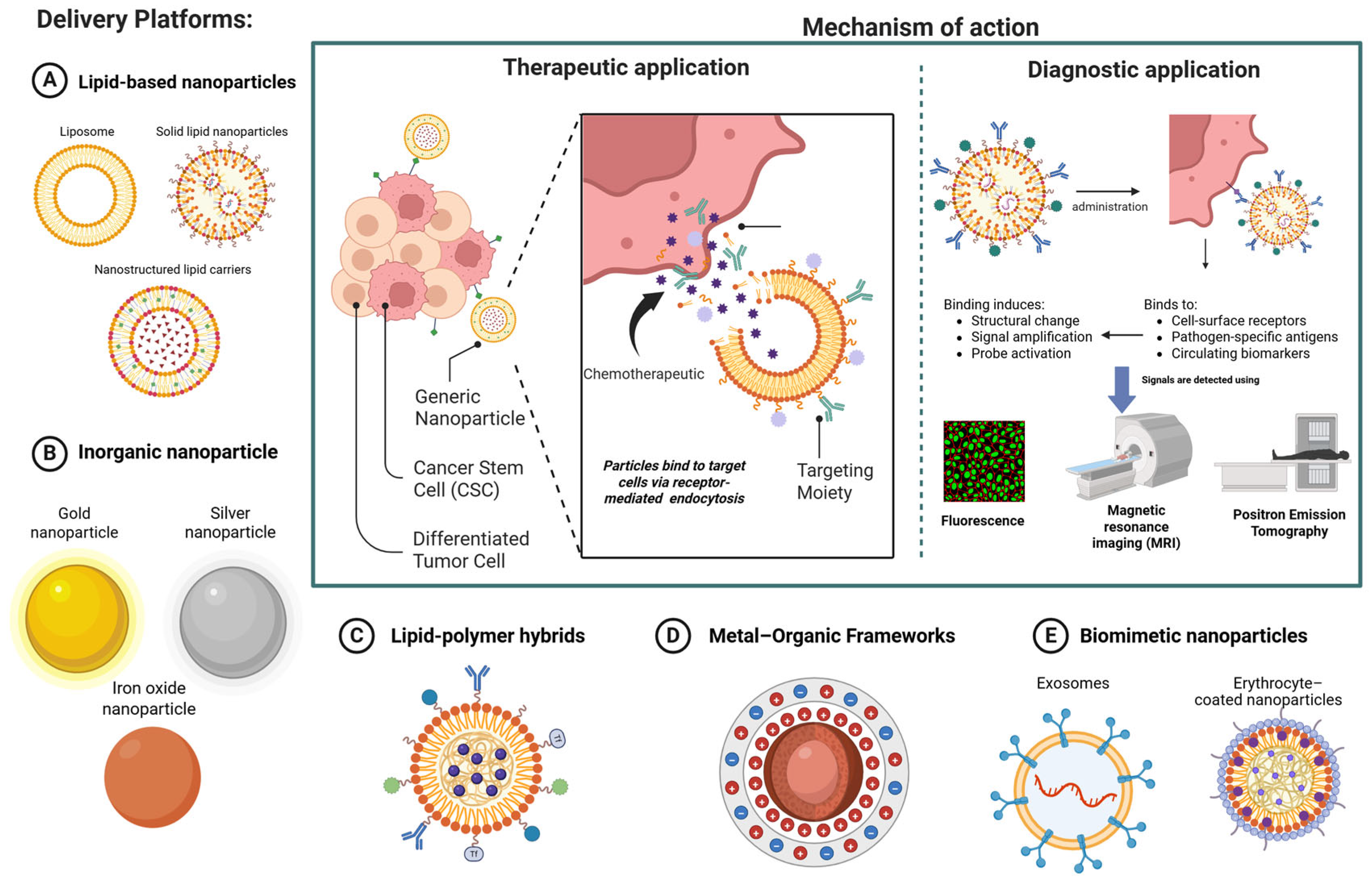


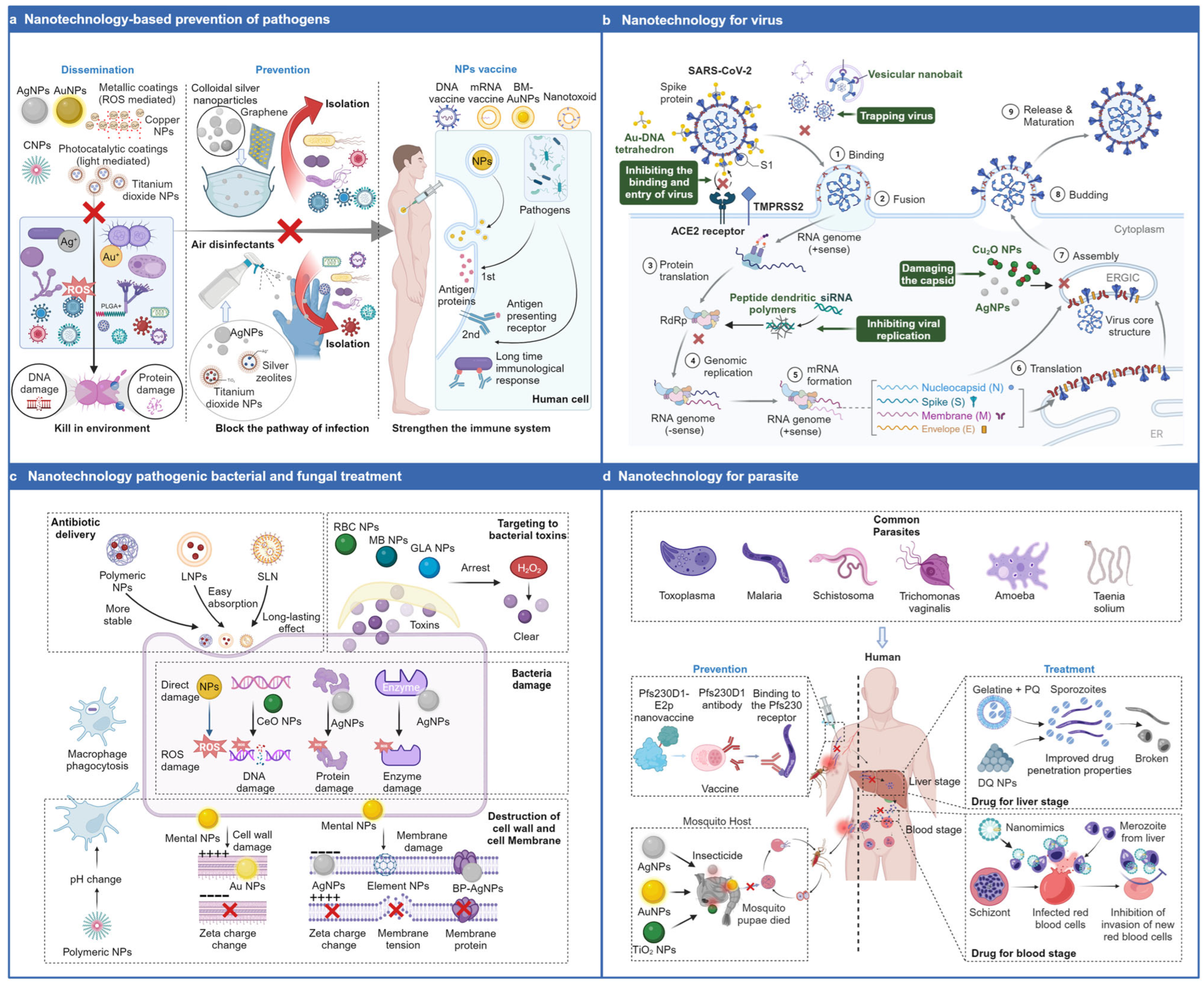

| Nanocarrier Type | Membrane | Nanocarrier | Application | Advantages | Challenges | References |
|---|---|---|---|---|---|---|
| Cell Membrane-Coated Nanoparticles | WBCs, RBCs, cancer cells | Synthetic nanoparticles coated with cell membranes | Targeted drug delivery to tumors and inflammatory sites) | Immune evasion; prolonged circulation time; enhanced targeted delivery; high biocompatibility and low toxicity; mimic natural cell functions | Manufacturing scalability; potential immunogenicity; membrane heterogeneity; regulatory hurdles; limitations of single-cell membrane coatings for complex pathological microenvironments | [104,107] |
| Platelet Membrane-Coated Nanoparticles | Platelets | Nanoparticles shielded by platelet membranes | Cancer therapy, leveraging innate tumor-homing and cancer cell interaction capabilities | Leverages platelets’ innate tumor-homing and cancer cell interaction capabilities for efficient drug delivery in cancer therapy | Potential clotting risks | [108] |
| Exosomes and Engineered Extracellular Vesicles | Various cell types | Natural or engineered extracellular vesicles | Targeted therapy | Specific targeting and improved therapeutic outcomes | Manufacturing scalability; batch-to-batch variation | [104] |
| Cancer Cell Membrane-Derived Biomimetic NPs | Breast cancer cells | Mesoporous silica | TNBC metastasis | Enhanced tumor interaction | Batch-to-batch variation | [109] |
| Application | Nanoparticle Type | Outcome | References |
|---|---|---|---|
| Diagnostics for bacterial infections | Mesoporous silica nanoparticles (MSN) | Suitable for diagnosis due to the tailored structure, large surface area, and high loading capacity | [195] |
| Antimicrobial therapy | Metal oxide nanoparticles (MONPs) | Demonstrate wide-ranging antimicrobial activities against bacteria, viruses, and protozoans, reducing resistance development through multiple mechanisms of action | [202] |
| Treatment of intracellular bacterial infections (e.g., Mycobacterium tuberculosis, MRSA) | Nanoparticle-based systems | Enhance intracellular penetration of antibiotics, allow controlled release, and reduce side effects | [198] |
| Prevention of experimental toxoplasmosis | Nanovaccines | Act as immunostimulatory adjuvants and vehicles for immunogen delivery | [199] |
| Detection sensors for hepatitis | Gold nanoparticles | Effective for designing detection sensors | [200] |
| Drug delivery and vaccination for hepatitis | Chitosan NPs | Promising nanocarriers | [200] |
| Biosensing for hepatitis | Carbon dots | Show potential for biosensing | [200] |
| Combined phototherapy for tuberculosis | Biomimetic erythrocyte-like nanoparticles (with aggregation-induced second near-infrared emission, granuloma-targeting, and self-oxygenation) | Significantly prolonged blood circulation and increased selective accumulation in tuberculosis granulomas in mouse models, inhibiting granuloma and M. tb colony growth | [201] |
| Sr No. | Type of Infection | Nanoparticle | Therapeutic Payload | Diagnostic/ Imaging/ Theranostic Component | Study Type | Particle Characteristics | Key Findings | References |
|---|---|---|---|---|---|---|---|---|
| 1 | Hepatitis B | PEGylated liposomes (Myr-preS2-31 modified) | Carboxyfluorescein, propidium iodide, DOX, FITC-labeled peptide or DNA vector | 111In, carboxyfluorescein | In vitro In vivo |
|
| [203] |
| 2 | Canine distemper virus | Polyvinylpyrrolidone and hydrolyzed collagen-coated NPs | Silver NPs (intrinsic antiviral) | - | In vivo |
|
| [204] |
| 3 | Tuberculosis | PLA NPs | Ethionamide and BDM41906 booster | - | In vitro In vivo |
|
| [205] |
| 4 | COVID-19 (SARS-CoV-2) | LNPs | Full-length S protein mRNA (PTX-COVID19-B) | - | In vitro In vivo | - |
| [206] |
| 5 | Biofilm-associated infections (Streptococcus mutans, Staphylococcus aureus) | Dextran-coated gold-in-gold cage NPs | - | Gold-in-gold cage photo thermal NPs (PTNPs) | In vitro In vivo |
|
| [207] |
| 6 | Ovine toxoplasmosis (Toxoplasma gondii) | Maltodextrin NPs with a lipid core | Total extract of T. gondii proteins | DiR (fluorophore) | In vitro In vivo | - |
| [208] |
| 7 | RSV | Polyvinylpyrrolidone-coated silver NPs | Silver NPs (intrinsic antiviral) | - | In vitro In vivo |
|
| [209] |
| 8 | Carbapenem resistant Escherichia coli | Pectin-capped platinum coated NPs | Platinum NPs (plasmid-curing agent) + Meropenem | - | In vitro In vivo |
|
| [210] |
| 9 | Newcastle disease virus | Chitosan NPs | F gene plasmid DNA | - | In vitro In vivo |
|
| [211] |
| 10 | Hepatitis C virus-induced advanced hepatic fibrosis | Retinoid-conjugated NPs | Heat shock protein 47 siRNA | - | Human | - |
| [212] |
| Sr No. | Disease | Nanoparticle | Therapeutic Payload | Diagnostic/ Imaging/ Theranostic Component | Study Type | Particle Characteristics | Key Findings | References |
|---|---|---|---|---|---|---|---|---|
| 1 | Parkinson’s disease | - | Salivary small extracellular vesicles (sEV) | Fluorescence-tagged | Human | - |
| [226] |
| 2 | Atherosclerosis | Macrophage membrane-coated ROS-responsive NPs | Targeted pharmacotherapy | - | In vivo |
|
| [227] |
| 3 | Ischemic Stroke | Neutrophil-hijacking nanoplatform (APTS) | A151 (telomerase repeat sequence) | - | In vivo | - |
| [228] |
| 4 | Glioblastoma multiforme | Mixed gold and superparamagnetic iron oxide nanoparticle (SPION) micelles | Gold NPs (radiosensitization) | SPIONs (T2-weighted MRI) and Vascular CT contrast | In vivo |
|
| [229] |
| 5 | Alzheimer’s disease | LNPs | - | Proteomic enrichment tool | In vivo |
|
| [230] |
| 6 | Atherosclerosis | Metal-free nanozyme (HCN@DS) | NR (nanozyme acts intrinsically) | Photoacoustic and photothermal imaging contrast | In vivo |
|
| [231] |
| 8 | Acute ischemic stroke | Macrophage-disguised honeycomb manganese dioxide (MnO2) nanospheres | Fingolimod (FTY) | - | In vivo |
|
| [232] |
| 9 | Glioblastoma | Ferric chloride α-cyano-4-hydroxycinnamate (Fe-CHC) NP | Chemodynamic therapy agents | - | In vivo |
|
| [233] |
| S.N. | NCT Number | Nanoparticle Platform | Payload/Active Agent | Indication | Phase | Primary Outcome | Status |
|---|---|---|---|---|---|---|---|
| 1 | NCT06693375 | Iron oxide NPs | NanoEcho Particle-1 (NEP-1) | Rectal Cancer | Phase II | Trace value for all lymph nodes | Recruiting |
| 2 | NCT06791005 | Carbon NPs | N/A | Papillary Thyroid Cancer | N/A | Intra- and post-operative conditions | Recruiting |
| 3 | NCT06146751 | Superparamagnetic Iron Oxide NPs | Ferumoxytol | Heart Aneurysm | N/A | Detection rate of intracardiac thrombus in dynamic chamber and static chamber | Recruiting |
| 4 | NCT07327996 | Mesoporous silica NPs | Calcium Hydroxide | Necrotic Pulp | N/A | Post-operative pain | Active, not recruiting |
| 5 | NCT06271564 | Zinc Oxide Composite NPs | N/A | Oral Potentially Malignant Lesions | Early phase I | Change in the clinical size in low to moderate dysplastic OPLs | Recruiting |
| 6 | NCT04899908 | N/A | Gadolinium | Brain Cancer | Phase II | Local recurrence | Recruiting |
| 7 | NCT06567301 | Chitosan | N/A | Knee Osteoarthritis | N/A | Anatomical improvement | Recruiting |
| 8 | NCT04881032 | N/A | Temozolomide (TMZ) and AGuIX (Polysiloxane Gadolinium-Chelates) | Glioblastoma | Phase I, II | Recommended dose of AGuIX (phase I) along with TMZ and radiotherapy during the radio-chemotherapy period 6-month Progression Free Survival (PFS) rate (phase II) | Active, not recruiting |
| 9 | NCT04484909 | N/A | Hafnium Oxide | Locally Advanced or Borderline-Resectable Pancreatic Cancer | Phase I | Phase II dose (RP2D) of Hafnium Oxide-containing nanoparticles (NBTXR3) | Recruiting |
| 10 | NCT04751786 | Poly(lactic-co-glycolic acid) (PLGA)- based NPs | Threitolceramide-6 | Advanced Solid Tumor | Phase I | Safety of PRECIOUS-01 (PLGA nanoparticle containing the combination of Threitolceramide-6 and and the New York Esophageal Squamous Cell Carcinoma-1 (NY-ESO-1) cancer-testis antigen peptides) | Active, not recruiting |
| 11 | NCT07308470 | SLNs | Etodolac | Knee Osteoarthritis | Phase II | Pain severity | Not yet recruiting |
| 12 | NCT04744506 | Carbon NPs | N/A | Breast Cancer | N/A | Lymph node retrieval rate. Number of sentinel and marked lymph nodes Complication rate | Recruiting |
| 13 | NCT07347080 | LNPs | Exenatide | Type II Diabetes mellitus (T2DM) | Early Phase I | Safety and tolerability of a single dose of Exenatide Circular RNA-Lipid NP Injection (CR059) | Not yet recruiting |
| 14 | NCT05039632 | N/A | Hafnium Oxide and Immunotherapy (anti-PD-1/L-1) | Advanced Solid Malignancies | Phase I, II | Efficacy and safety of NBTXR3 activated by radiation (Abscopal or RadScopal™) in combination with immunotherapy (anti-PD-1/L-1) | Recruiting |
| 15 | NCT05264974 | LNPs | N/A | Melanoma | Phase I | Maximum tolerated dose | Recruiting |
| 16 | NCT06977126 | N/A | Porphysomes | Gynecological Cancers | Phase I | Incidence of Dose Limiting Toxicities (DLTs) | Recruiting |
| 17 | NCT04789486 | N/A | Gadolinium | Non-small Cell Lung Cancer and Pancreatic Cancer | Phase I, II | Maximum tolerated dose (MTD) (Phase 1) Compare Local Control at 12 months of MTD (Phase II) | Recruiting |
| 18 | NCT06271421 | N/A | - | Glioblastoma Multiforme | N/A | Survival following the surgery (units months 1–24) Progression-free survival (units months 1–24) | Recruiting |
| 19 | NCT04167969 | Silica NPs | Copper-64 (64Cu) radiolabeled PSMA-targeting C′ dot tracer (64Cu-NOTA-PSMAi-PEG-Cy5.5-C′ dots) or zirconium-89 (89Zr) radiolabeled PSMA-targeting C′ dot tracer (89Zr-DFO-PSMAi-PEG-Cy5.5-C′ dots) | Prostate cancer | Phase I | Side effects | Recruiting |
| 20 | NCT02106598 | Silica NPs | Fluorescent cRGDY-PEG-Cy5.5-C dots | Head and Neck Melanoma | Phase I, II | Feasibility of conducting pre-operative SLN mapping | Active, not recruiting |
| 21 | NCT05903339 | Ferritin NPs, mRNA LNPs | Alum ACU-026-001-1 | HIV | Phase I | Local and systemic reactogenicity signs Number of SAEs | Active, not recruiting |
| 22 | NCT06908096 | EBV gH/gL/gp42-ferritin NPs | N/A | Epstein-Barr Virus (EBV) infection | Phase I | Local and systemic reactogenicity signs and symptoms during the 7 days after each vaccination Unsolicited AEs | Recruiting |
| 23 | NCT04682847 | SPION | Ferumoxytol injection | Hepatic Cancers | NA | Platform to maintain liver functionality during treatment for liver cancer | Active, not recruiting |
| 24 | NCT01525966 | Paclitaxel albumin-stabilized NP formulation | Carboplatin | TNBC | Phase II | Whether carboplatin + nab-paclitaxel therapy will achieve a promising neoadjuvant pathologic complete response (pCR) rate | Active, not recruiting |
| 25 | NCT00616967 | NA | Carboplatin Vorinostat Paclitaxel albumin-stabilized NP formulation | Breast Cancer | Phase II | To determine pCR rates in patients with HER2-negative primary operable breast cancer | Active, not recruiting |
| 26 | NCT06628076 | Zinc oxide NPs | NA | Acinetobacter baumannii infection | NA | Susceptibility profile | Not yet recruiting |
| 27 | NCT01730833 | NA | Pertuzumab Trastuzumab Paclitaxel albumin-stabilized NP | HER2-positive advanced breast cancer | Phase II | Efficacy of pertuzumab in combination with trastuzumab with nab-paclitaxel in subjects with stage IV in HER-2 overexpressing metastatic breast cancer (MBC) | Active, not recruiting |
| 28 | NCT01463072 | NA | Nab-paclitaxel | Advanced or metastatic breast cancer | Phase II | Tolerability of weekly nab-paclitaxel | Active, not recruiting |
| 29 | NCT00609791 | NA | Nab-paclitaxel | MBC | Phase II | Age-related changes in the pharmacokinetics (pK) and pharmacodynamics of weekly nab-paclitaxel | Active, not recruiting |
| 30 | NCT07034248 | Graphene Quantum dot decorated Gold NPs | NA | Breast Cancer | NA | Sensitivity and selectivity on an electrochemical biosensor | Not yet recruiting |
| 31 | NCT04615013 | Hafnium oxide-containing NPs | Capecitabine, carboplatin, docetaxel, fluorouracil, leucovorin, oxaliplatin, paclitaxel | Esophageal Cancer | Phase I | Recommended phase II dose of NBTXR3 activated by radiotherapy with concurrent chemotherapy | Recruiting |
| 32 | NCT07321301 | Polymer LNPs | CD19/CD20 Dual-Targeting InViVoCAR1920 mRNA | Relapsed/refractory B-cell lymphoma/leukemia | Phase II | Safety and maximum tolerated dose of CAR-T cell immunotherapy mediated by polymer-lipid nanoparticles delivering CD19/CD20 dual-targeting InViVoCAR1920 mRNA | Recruiting |
| 33 | NCT06577532 | mRNA NPs | ABO2102 Toripalimab | KRAS -mutated solid tumors | Early phase I | Incidence and nature of dose-limiting toxicity (DLT) for ABO2102 as monotherapy or in combination with toripalilmab Overall Response Rate (ORR) per RECIST version 1.1. | Recruiting |
| 34 | NCT04645147 | Ferritin NPs | EBV gp350-ferritin vaccine | EBV infection | Phase I | Safety and immunogenicity | Active, not recruiting |
| 35 | NCT05816694 | NA | NAB-Paclitaxel plus Cisplatin Cisplatin plus Epirubicin plus Cyclophosphamide | Thymoma | Phase II | ORR | Not yet recruiting |
| 36 | NCT07183709 | NA | PepGNP-COVID19 | COVID-19 | Phase I | Proportion of participants experiencing solicited and unsolicited AEs | Recruiting |
| 37 | NCT06686654 | LNPs | Investigational RSV + hMPV vaccine | Human metapneumovirus (HMPV)/Respiratory syncytial virus (RSV) | Phase I Phase II | Presence of solicited and unsolicited AEs | Active, not recruiting |
| 38 | NCT02336087 | NA | Gemcitabine HCl, nab-Paclitaxel, metformin HCl | Pancreatic Cancer | Phase I | Compliance, toxicity, and feasibility of administered formulation | Active, not recruiting |
| 39 | NCT07019883 | mRNA-LNP | H5 AC-Anhui RNA Vaccine | Influenza | Phase I | Proportion of participants experiencing AE and SAE | Active, not recruiting |
| 40 | NCT05945485 | mRNA-LNP | DCVC H1 HA mRNA vaccine Quadrivalent Recombinant Seasonal Influenza Vaccine | Influenza | Phase I | Proportion of participants experiencing AE and SAE | Active, not recruiting |
| 41 | NCT03337087 | Liposome | Fluorouracil, irinotecan sucrosofate, leucovorin calcium, and rucaparib | Metastatic pancreatic, colorectal, GI, or biliary Cancer | Phase I Phase II | Number of participants with dose-limiting toxicities (Phase I) | Active, not recruiting |
Disclaimer/Publisher’s Note: The statements, opinions and data contained in all publications are solely those of the individual author(s) and contributor(s) and not of MDPI and/or the editor(s). MDPI and/or the editor(s) disclaim responsibility for any injury to people or property resulting from any ideas, methods, instructions or products referred to in the content. |
© 2026 by the authors. Licensee MDPI, Basel, Switzerland. This article is an open access article distributed under the terms and conditions of the Creative Commons Attribution (CC BY) license.
Share and Cite
Tiwary, P.; Oswal, K.; Varghese, R.; Gupta, P. Nanoparticles in Therapy and Diagnosis: A Comprehensive Review of Mechanisms, Applications, and Translational Challenges. J. Nanotheranostics 2026, 7, 11. https://doi.org/10.3390/jnt7020011
Tiwary P, Oswal K, Varghese R, Gupta P. Nanoparticles in Therapy and Diagnosis: A Comprehensive Review of Mechanisms, Applications, and Translational Challenges. Journal of Nanotheranostics. 2026; 7(2):11. https://doi.org/10.3390/jnt7020011
Chicago/Turabian StyleTiwary, Pooja, Krishil Oswal, Ryan Varghese, and Pardeep Gupta. 2026. "Nanoparticles in Therapy and Diagnosis: A Comprehensive Review of Mechanisms, Applications, and Translational Challenges" Journal of Nanotheranostics 7, no. 2: 11. https://doi.org/10.3390/jnt7020011
APA StyleTiwary, P., Oswal, K., Varghese, R., & Gupta, P. (2026). Nanoparticles in Therapy and Diagnosis: A Comprehensive Review of Mechanisms, Applications, and Translational Challenges. Journal of Nanotheranostics, 7(2), 11. https://doi.org/10.3390/jnt7020011