Review

A Survey on Denoising Techniques of Electroencephalogram Signals Using Wavelet Transform

1
Department of Neurology, University of Wisconsin Madison, Madison, WI 53792, USA
2
Department of Earth Sciences, Sapienza University of Rome, Piazzale Aldo Moro, 5, 00185 Rome, Italy
3
William S Middleton Veterans Hospital, Madison, WI 53705, USA
4
Department of Electrical Engineering, National Institute of Technology Silchar, Silchar 788010, Assam, India
5
School of Computing Science and Engineering, VIT Bhopal University, Kotri Kalan 466114, Madhya Pradesh, India
6
Department of Computer Science & Engineering, National Institute of Technology Silchar, Silchar 788010, Assam, India
*
Author to whom correspondence should be addressed.
Signals 2022, 3(3), 577-586; https://doi.org/10.3390/signals3030035
Submission received: 26 June 2022 / Revised: 28 July 2022 / Accepted: 16 August 2022 / Published: 17 August 2022
(This article belongs to the Special Issue Advancing Signal Processing and Analytics of EEG Signals)

Abstract

:
Electroencephalogram (EEG) artifacts such as eyeblink, eye movement, and muscle movements widely contaminate the EEG signals. Those unwanted artifacts corrupt the information contained in the EEG signals and degrade the performance of qualitative analysis of clinical applications and as well as EEG-based brain–computer interfaces (BCIs). The applications of wavelet transform in denoising EEG signals are increasing day by day due to its capability of handling non-stationary signals. All the reported wavelet denoising techniques for EEG signals are surveyed in this paper in terms of the quality of noise removal and retrieving important information. In order to evaluate the performance of wavelet denoising techniques for EEG signals and to express the quality of reconstruction, the techniques were evaluated based on the results shown in the respective literature. We also compare certain features in the evaluation of the wavelet denoising techniques, such as the requirement of reference channel, automation, online, and performance on a single channel.

1. Introduction

From 1929, electroencephalogram (EEG) saw steady progress, and it was in the 1960s that the computerization of EEG started. It was this computerization of EEG that allowed for the introduction of automated data analysis in EEG, which came in the form of the fast Fourier transform being used as the basis for power spectral analysis [1]. From these years of development, we have come to understand that electroencephalography is the measurement, amplification, and registration of fluctuating electrical fields produced by the brain as a function of time [1]. When neurons inside the brain communicate with each other, they generate electrical pulses or voltage fluctuations. These electrical pulses or voltage fluctuations contain information on the communication between different cortices in the brain, as well as communication to areas such as the peripheral nervous system.
EEG signals have been extensively used to diagnose a variety of brain disorders such as epilepsy, Alzheimer’s disease, brain tumors, etc. [2]. Furthermore, EEG has also been used for evaluating sleep patterns of individuals, as well as understanding learning or attention disorders [2]. EEG is recorded using differential amplifiers and it takes two electrical inputs to display the output as the difference between them [3]. Electrodes consisting of tiny metal discs are placed on the scalp during the signal recording technique [3]. However, EEG has high temporal resolution but lacks spatial resolution due to the different scales that the EEG electrodes and neural networks operate on [3]. The neuronal activity being on the scale of micro-volts means that EEG recordings require a level of neural synchronicity in order to have measurable activity [3]. Even with synchronous activity, the electrodes and EEG recordings are often subject to artifacts from differing sources. The most common artifacts seen result from physiological, environmental, and experimental sources [4].
Artifacts are undesired signals that can cause recordings to change and impact the signal of interest [4]. The most common physiological artifacts found are ocular artifacts, due to eye blinks and eye movement, muscle artifacts due to inherent flexion and relaxation of muscles present on the forehead and scalp, and cardiac artifacts due to electrode placement on or near a blood vessel [5]. Because noise sources are so varied and have so many different properties, most authors concentrate on removing certain types of artifacts. The removal of artifacts plays a key role in EEG signal processing for both clinical applications and as well as brain–machine interfaces.
Several denoising techniques have been developed for the purpose of artifact correction in EEG signals [6,7,8]. Rejecting artifactual portions of the EEGs is the simplest way, which deletes epochs containing artifacts. However, removal of artifactual portions can be a time-consuming process that can lead to significant information loss, which is in turn detrimental for data analysis [6]. Traditionally, regression and linear filtering-based analyses are employed to reject artifactual noises from corrupted signals [8]. Due to the overlap of brain activities and artifactual noises in the spectrum of an EEG signal, filtering them in either the frequency-domain or the time-domain may result in loss or distortion of physiological activity [9]. Regression-based methods rely on an extra one or more regressive channels, which gives rise to a fundamental weakness in that the spectral range of some artifacts overlaps with the spectral range of an EEG signal [10]. In both the temporal and frequency domains, wavelet transform-based analysis has been proven to be more efficient in repairing EEG artifacts while keeping the original EEG signal [11,12].
EEG signals are inherently non-stationary. One of the most widely used approaches for studying non-stationary signals is the wavelet transform. Its efficiency in transforming a time-domain signal into time-frequency-domain offers major advantages in the extraction of multiple components of a signal. A wavelet-based method eliminates the artifacts while retaining the integrity of the EEG signal. Jiang et al. [2] comprehensively addresses techniques found frequently in EEG artifact rejection and how these techniques work as individual tools as well as hybrid techniques such as EMD-BSS, wavelet-BSS, and BSS-Support vector machine. This paper will attempt to serve as a survey focused specifically on using the wavelet transform as an artifact rejection tool in EEG signals and will attempt to hone in on some specific wavelet transforms hybrids that have shown promising performance.

2. Wavelet Denoising

Wavelet transform has been widely used in representing signals in the time-frequency domain. The wavelet transform decomposes a time-domain signal into its wavelet coefficients through a mother wavelet function. These coefficients are obtained by performing shifting and dilation of the mother wavelet as shown in Equation (1):
Ψ a , b ( t ) = Ψ ( t b a )
where a is the scaling parameter and b is the shifting parameter [13,14].
When the noisy version is available, the problem is to restore the information contained in the signal. In traditional wavelet denoising, the wavelet coefficients are modified by taking advantage of their local properties and then inverting the transformation to obtain a clean version of the signal. A basic flow chart of wavelet-based denoising is shown in Figure 1. An eyeblink corrupted EEG signal of 15 sec duration is shown in the top panel of Figure 2. The corrupted EEG signal is then decomposed into wavelet coefficients up to level 5 using Daubechies wavelet with the vanishing moment as 4, and the corresponding detail and approximation coefficients are shown in the Figure 3. After thresholding both the detail and approximation coefficients, the corrected coefficients are used in inverse operation, and an artifact-free EEG signal is shown in the middle panel of Figure 2 in black.
Generally, there are two types of wavelet transform: one is the discrete wavelet transform (DWT) and another is a continuous wavelet transform (CWT) [15]. DWT is regarded as a non-redundant and extremely dynamic wavelet transform for obtaining wavelet representation of signal [16,17]. In DWT, the signal is passed through a half-band high-pass and a half-band low-pass filter, resulting in detail coefficients and approximate coefficients, respectively. The process continues until the expected frequency is obtained. The time-variance of DWT is a severe flaw, which is especially critical in statistical signal processing applications such as EEG [18]. The stationary wavelet transform (SWT) solves the DWT’s translation invariance problem; however, it has redundant information and is sluggish [19]. The filter at each stage is the design difference between DWT and SWT [20]. At each level of decomposition, the approximate and detail sequences are the same length as the original sequence. Wavelet coefficients extracted through SWT are shown in Figure 4, and the corrected EEG signal is illustrated in the bottom panel of Figure 2 in blue.

3. A Survey on Wavelet Transform-based EEG Denoising Techniques

According to the wavelet denoising methods for EEG signals (irrespective of any kind of artifacts) available in the literature, we grouped all the methods into various categories and are discussed in this section.

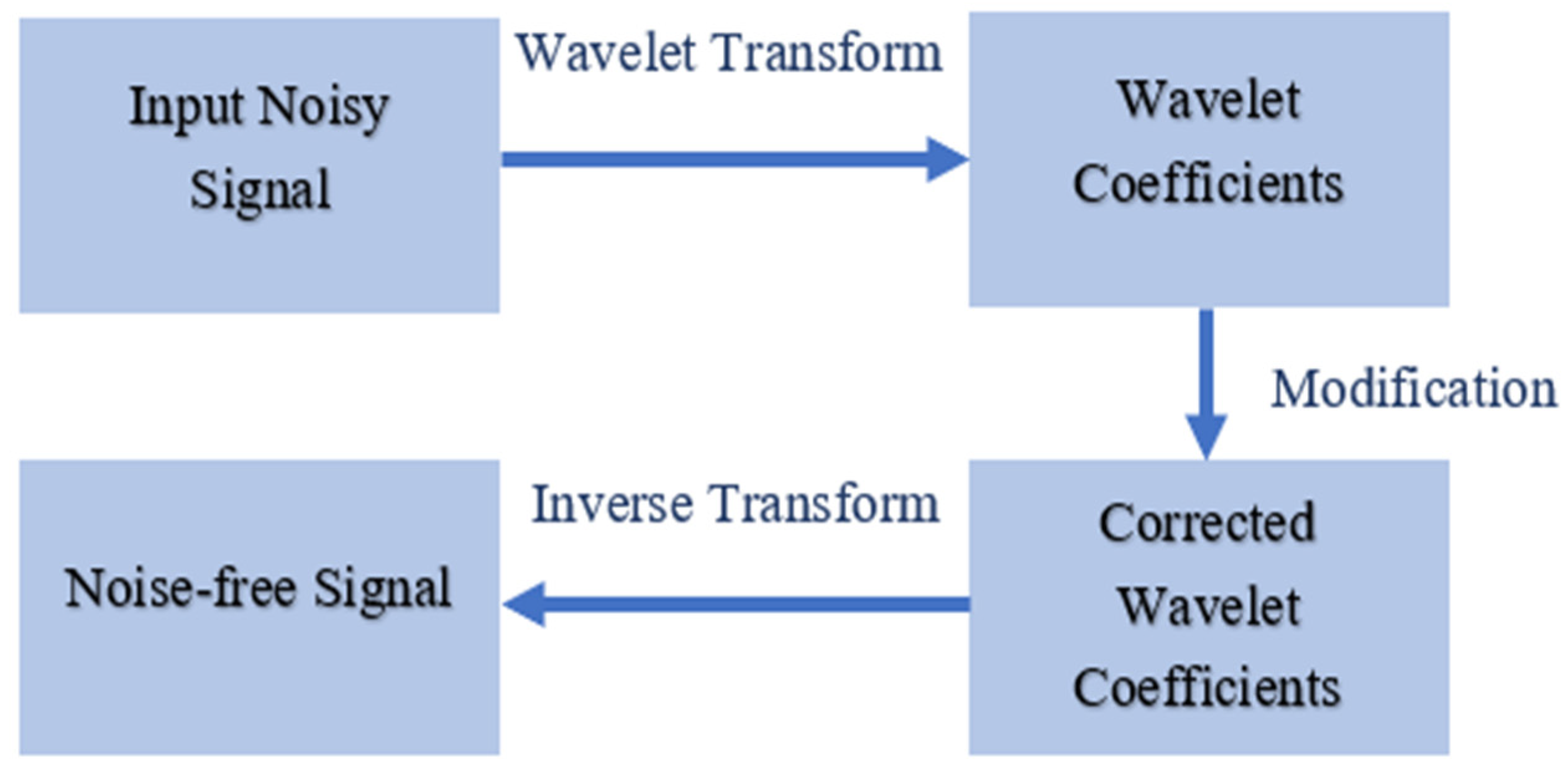
3.1. Wavelet Denoising with Thresholding

The most efficient and widely used wavelet denoising is based on thresholding wavelet coefficients. This process follows three important steps: (i) wavelet decomposition: the input signals are decomposed into wavelet coefficients; (ii) thresholding: the wavelet coefficients are modified according to a threshold; and (iii) reconstruction: modified coefficients are used in inverse transform to obtain the noise-free signal. Several researchers have used thresholding wavelet denoising techniques [21,22,23,24]. The universal threshold and statistical threshold functions are efficiently used in wavelet-based EEG denoising.
Krishnaveni et al. [21] proposed automatic detection and elimination of ocular artifacts (OA) from EEG signals using wavelet transform. They used a DWT with a Haar wavelet as the basis function to identify the OA zone. Next, identified OA zones are decomposed into wavelet domain through SWT with Coiflet wavelet with vanishing moment 3 as a basis function. The identified OAs are removed from EEG using a non-linear time-scale adaptive denoising technique, which is based on the wavelet shrinkage strategy. The optimal thresholds are selected based on Stein’s unbiased risk estimate (SURE) and soft-like thresholding function using gradient-based adaptive algorithm. Instead of thresholding all of the wavelet coefficients, Zikov et al. [22] performed thresholding on lower-frequency bands as OAs have very low-frequency characteristics. They estimated the threshold by simple statistical analysis of baseline EEG. Islam et al. [23] employed a non-negative garrote shrinkage function during denoising due to its nice tradeoff between soft and hard threshold and modified universal threshold as:
t j , l = K α j , l 2 ln N
where N is epoch length and α j , l is the estimated noise variance for the wavelet coefficients at lth level ( w j , l ).
α j , l = m e d i a n   ( | w j , l | ) 0.6745
The K is a new parameter estimated through empirical observations:
K = { K A           ( 0 < K A < 1 )                   K D           ( 1 < K D < 3 )            
Recently, Phadikar et al. [24] proposed an automatic eyeblink artifact correction technique using wavelet transform and metaheuristic algorithms. In their method, the wavelet coefficients are thresholded in a backward manner to modify only the lower frequency bands of the observed EEG signals. Further, to make the system fully automatic, the optimal thresholds are selected through the grey wolf optimizer.

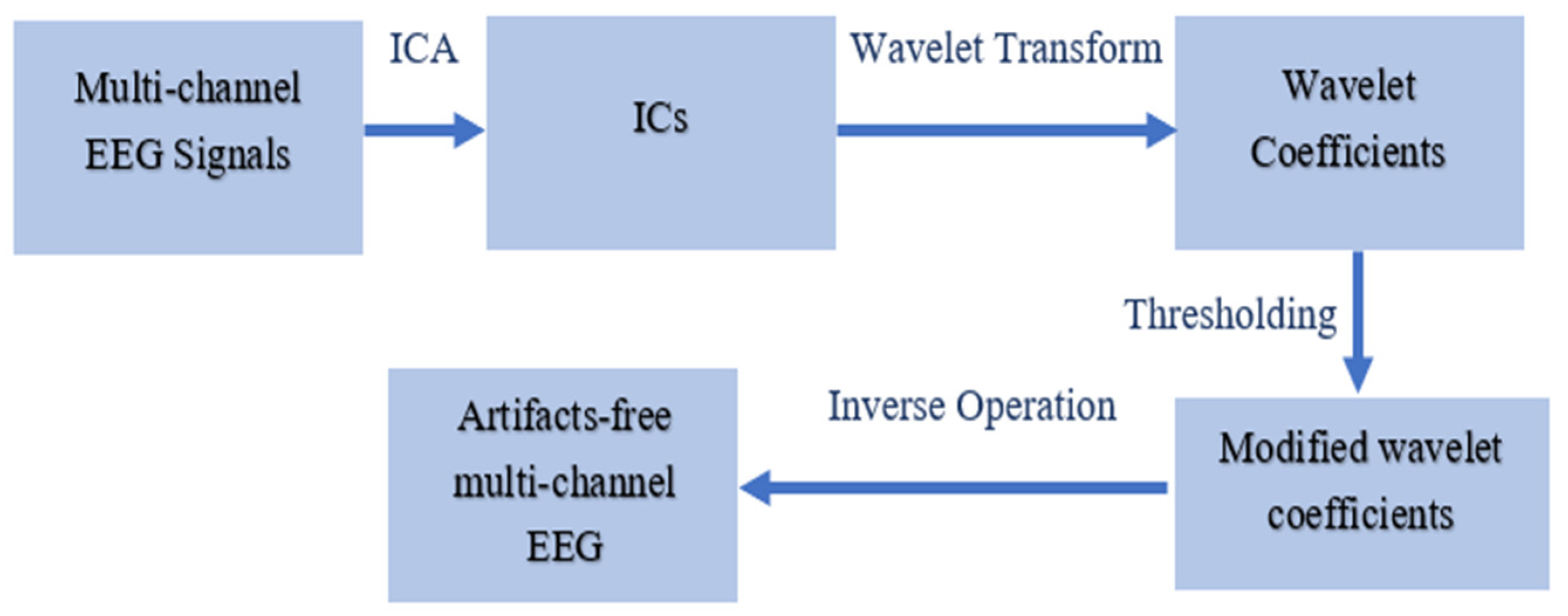
3.2. Hybrid Methods with Wavelet Transform

Recently, researchers have been hybridizing the wavelet transform with other efficient denoising techniques. For handling the EEG artifacts, independent component analysis (ICA) is widely employed with wavelet transform [25,26,27,28]. ICA is a mathematical model that decomposes multivariate signals into their subcomponents [29]. In the wavelet-ICA-based method, the input EEG signals are decomposed into wavelet coefficients. Next, all the coefficients are used in ICA operation to separate the various sources of EEG in the time-frequency domain. Then, the artifacted independent components (ICs) are directly eliminated. Finally, all ICs are used in inverse operation followed by inverse wavelet transform to obtain the noise-free EEG signals. Zhou et al. [25] provide an example of combining wavelet soft-thresholding, whitening method for preprocessing, and ICA for the removal of EMG and ECG artifacts. While Zhou et al. proposed a methodology for a wavelet transform-ICA hybrid, Inuso et al. [26] showed that using the wavelet transform as an integral part in the separation processes with ICA outperforms hybrid methods that used the wavelet transform as a denoising technique either pre- or post-ICA.
However, direct elimination of ICs may result in huge information loss as artifacted ICs also contain cerebral activities. Sai et al. [27] modified the wavelet ICA algorithms and instead of direct elimination of artifacted ICs in wavelet domain, they performed thresholding on artifacted ICs to maintain the cerebral activities in ICs. Yasoda et al. [28] introduced increased automation by implementing a fuzz-kernel support vector machine for identifying artifacts, prior to removal using a wavelet-ICA combination. A basic flow chart of wavelet-ICA is shown in Figure 5. Later on, Sai et al. [30] proposed an unsupervised machine-learning-based method combined with Wavelet-ICA to remove EEG artifacts. They also proposed that the techniques that rely on some arbitrarily defined threshold often fail to accurately identify the signal artifacts in a given dataset.
However, wavelet transform is limited to single-channel EEG signals. It may perform well for multi-channel EEG signals with the cost of higher computational time and higher computational complexity as performing a wavelet transform channel by channel is a laborious process. To overcome the issue with performing the wavelet transform for multi-channel EEG, a new hybrid method, ICA-wavelet, was developed in the literature [31,32]. In the ICA-wavelet-based methods, the multi-channel EEG signals were used in ICA to decompose into ICs. Then, artifacted ICs are identified and decomposed into wavelet coefficients. Wavelet coefficients are then thresholded using a universal threshold or a statistical threshold with a hard or soft thresholding function. Finally, clean multi-channel EEG signals are achieved by performing inverse operation of wavelet transform followed by ICA. A basic flow chart of ICA-wavelet based method is shown in Figure 6.

3.3. Other Wavelet Transform-Based Method

Besides ICA, empirical ensemble mode decomposition (EEMD) and canonical correlation analysis (CCA) can also be combined with wavelet transform for successful removal of artifacts from EEG signals [33,34,35,36]. When SWT and EEMD are studied separately, they both appear to be effective in artifact removal from EEG recordings. One cause could be that the EEMD approach decomposes the signal into frequency and amplitude components [33]. This method does not confine the artifacts to a specific level. As a result, the EEMD’s assumption of artifacts with larger amplitude at a specific decomposition level may not be correct. The CCA algorithms are used to segregate components depending on the EEG sources, such that the artifact component is considered as a single CC with a random distribution that can be removed easily [33,34]. Mowla et al. [33] showed that the combination of CCA-SWT coupled with a second-order blind identification and SWT had significant improved performance in removing EOG and EMG artifacts. A combination of CCA-WT can yield improved results compared to using them separately; however, adding in an initial decomposition using EEMD yields improved performance when coupled with a support vector machine (SVM), leading to EEMD-CCA-DWT [36].
Chen et al. [37] presented an OA removal technique where Kalman filter is combined with DWT. In their work, DWT is applied to the raw EEG in identified OA zones to reconstruct a rough OAs approximation. The Kalman filter is used to optimize the OAs approximations from the previous step. Finally, the optimized OAs are subtracted from raw EEG. Bajaj et al. [38] proposed a tunable algorithm for artifacts removal from EEG signals using wavelet packet decomposition and wavelet filtering. Phadikar et al. [39] proposed a multi-stage EEG denoising method that combines wavelet packet decomposition (WPD) with a modified non-local means (NLM) algorithm for muscle artifact identification and removal. Abdi-Sargezeh et al. [40] introduced two novel methods for the removal of EEG artifacts. In the first method, the common components among EEG channels were extracted and eliminated as artifacts, called common component rejection (CCR). In the second method, wavelet decomposition was employed to decompose the EEG signals, then the CCR method was applied to remove artifacts in the time-frequency domain, referred to as automatic wavelet CCR (AWCCR). Dora et al. [41] developed a flexible technique to remove EEG artifacts in the context with minimal supervision. They proposed a new wavelet-based method that allows to remove artifacts from single-channel EEG based on a data-driven renormalization of the wavelet coefficients. Their method is capable of adaptively attenuating artifacts of a different nature.
There are many other robust wavelet decomposition methods whose performances are yet to be investigated in processing EEG signals. The reader is referred to [14] for more details of some of these methods.

4. Comparative Analysis

The methods mentioned above are among the most popular for removing EEG artifacts. Some of these artifact-reduction techniques limit eye movements and blinking during data collection or exclude artifact-contaminated trials from the analysis. An exhaustive comparison among the above-stated methods is presented in Table 1.
The majority of EEG-based real applications necessitate real-time signal processing and are resistant to artifacts. This necessitated that the artifact removal methods used be both automatic and minimal in computational expense. The term “automated process” refers to a procedure that may automatically identify and eliminate artifact components without the need for human participation. The wavelet-thresholding-based technique is simple and efficient for handling all kind of artifacts in the EEG signals. However, Phadikar et al. [24] showed that the reconstructed clean EEG signals using the universal threshold and statistical threshold are distinctly different from true clean EEG signals, even though the artifacts are removed. If we tune both the threshold calculation, it may reconstruct near the original shape of the EEG. However, tuning them may make the system unable to operate automatically. ICA is a widely used technique for the removal of artifacts from EEG signals. However, it should follow three assumptions: (i) the sources are statistically independent, (ii) each independent component has a non-gaussian distribution, and (iii) the mixing system is determined. Hence, it may not perform well for single-channel EEG signals or fewer channels. The wavelet transform is unable to detect artifacts that overlap with spectral features fully. The disadvantage of mode mixing is also present in EMD. As a result, finding a single solution that is both efficient and accurate enough to satisfy all of the prerequisites is extremely challenging.

5. Conclusions

The cerebral cortex generates EEG signals, which can be distorted by certain external disturbances. EEG is a highly non-stationary signal that is generally contaminated by a variety of artifacts. Despite there being a variety of strategies proposed for eliminating unwanted artifacts, an artifact-removal method that combines high accuracy with algorithmic efficiency has yet to be established. This paper summarizes wavelet-based EEG denoising techniques based on the conclusion made in the published literature. The article mainly focuses on wavelet-based denoising as it efficiently handles non-stationary signals. The advantages and limitations of all the mentioned methods have been highlighted. Although the majority of the removal algorithms perform well, the approaches outlined above have a variety of drawbacks when used in a specific EEG-based application. Few methods require a reference channel to improve the performance of artifact removal, which is not possible in some applications. Wavelet-based methods are quite accurate at removing EEG artifacts; nevertheless, it suffers from higher computational complexity, which may not be appropriate for online applications. As a result, there is no best option for removing all forms of artifacts. Therefore, one of the long-term goals of effective artifact attenuation is to create an application-specific algorithm that is more efficient in terms of time and accuracy.

Funding

This research received no external funding.

Institutional Review Board Statement

Not applicable.

Informed Consent Statement

Not applicable.

Conflicts of Interest

The authors declare no conflict of interest.

References

  1. Niedermeyer, E.; Silva, F.L.D. Electroencephalography: Basic Principles, Clinical Applications, and Related Fields; Lippincott Williams & Wilkins: Philadelphia, PA, USA, 2005. [Google Scholar]
  2. Ahmed, M.Z.I.; Sinha, N.; Phadikar, S.; Ghaderpour, E. Automated Feature Extraction on AsMap for Emotion Classification Using EEG. Sensors 2022, 22, 2346. [Google Scholar] [CrossRef]
  3. Im, C.H. Basics of EEG: Generation, Acquisition, and Applications of EEG. In Computational EEG Analysis; Springer: Singapore, 2018; pp. 3–11. [Google Scholar]
  4. Jiang, X.; Bian, G.B.; Tian, Z. Removal of Artifacts from EEG Signals: A Review. Sensors 2019, 19, 987. [Google Scholar] [CrossRef]
  5. Mumtaz, W.; Rasheed, S.; Irfan, A. Review of challenges associated with the EEG artifact removal methods. Biomed. Signal Process. Control. 2021, 68, 102741. [Google Scholar] [CrossRef]
  6. Croft, R.J.; Barry, R.J. Removal of ocular artifact from the EEG: A Review. Clin. Neurophysiol. 2000, 30, 5–19. [Google Scholar] [CrossRef]
  7. Kandaswamy, A.; Krishnaveni, V.; Jayaraman, S.; Malmurugan, N.; Ramadoss, K. Removal of ocular artifacts from EEG—A Survey. IETE J. Res. 2005, 51, 121–130. [Google Scholar] [CrossRef]
  8. Lai, C.Q.; Ibrahim, H.; Abdullah, M.Z.; Abdullah, J.M.; Suandi, S.A.; Azman, A. Artifacts and noise removal for electroencephalogram (EEG): A literature review. In Proceedings of the International Conference on Computer Applications and Industrial Electronics (ICCAIE), Penang, Malaysia, 28–29 April 2018; IEEE: Manhattan, NY, USA, 2018; pp. 326–332. [Google Scholar]
  9. Pereira, L.F.; Patil, S.A.; Mahadeshwar, C.D.; Mishra, I.; D’Souza, L. Artifact removal from EEG using ANFIS-GA. In Proceedings of the 2016 Online International Conference on Green Engineering and Technologies (IC-GET), Coimbatore, India, 19 November 2016; IEEE: Manhattan, NY, USA, 2016; pp. 1–6. [Google Scholar]
  10. Gratton, G.; Coles, M.G.; Donchin, E. A new method for off-line removal of ocular artifact. Electroencephalogr. Clin. Neurophysiol. 1983, 55, 468–484. [Google Scholar] [CrossRef]
  11. Zhang, J.H.; Janschek, K.; Böhme, J.F.; Zeng, Y.J. Multi-resolution dyadic wavelet denoising approach for extraction of visual evoked potentials in the brain. IEE Proc. Vis. Image Signal Process. 2004, 151, 180–186. [Google Scholar] [CrossRef]
  12. Mamun, M.; Al-Kadi, M.; Marufuzzaman, M. Effectiveness of wavelet denoising on electroencephalogram signals. J. Appl. Res. Technol. 2013, 11, 156–160. [Google Scholar] [CrossRef]
  13. Inuso, G.; La Foresta, F.; Mammone, N.; Morabito, F.C. Brain activity investigation by EEG processing: Wavelet analysis, kurtosis and Renyi’s entropy for artifact detection. In Proceedings of the 2007 International Conference on Information Acquisition, Seogwipo, Korea, 8–11 July 2007; IEEE: Manhattan, NY, USA, 2007; pp. 195–200. [Google Scholar]
  14. Ghaderpour, E.; Pagiatakis, S.D.; Hassan, Q.K. A Survey on Change Detection and Time Series Analysis with Applications. Appl. Sci. 2021, 11, 6141. [Google Scholar] [CrossRef]
  15. Ghaderpour, E. Least-Squares Wavelet Analysis and Its Applications in Geodesy and Geophysics. Ph.D. Thesis, York University, Toronto, ON, Canada, 2018. [Google Scholar]
  16. Mallat, S.G. A theory for multiresolution signal decomposition: The wavelet representation. IEEE Trans. Pattern Anal. Mach. Intell. 1989, 11, 674–693. [Google Scholar] [CrossRef]
  17. Samar, V.J.; Bopardikar, A.; Rao, R.; Swartz, K. Wavelet analysis of neuroelectric waveforms: A conceptual tutorial. Brain Lang. 1999, 66, 7–60. [Google Scholar] [CrossRef]
  18. Pesquet, J.C.; Krim, H.; Carfantan, H. Time-invariant orthonormal wavelet representations. IEEE Trans. Signal Process. 1996, 44, 1964–1970. [Google Scholar] [CrossRef]
  19. Kumar, P.S.; Arumuganathan, R.; Sivakumar, K.; Vimal, C. An adaptive method to remove ocular artifacts from EEG signals using wavelet transform. J. Appl. Sci. Res. 2009, 5, 711–745. [Google Scholar]
  20. Nason, G.P.; Silverman, B.W. The stationary wavelet transform and some statistical applications. In Wavelets and Statistics; Springer: New York, NY, USA, 1995; pp. 281–299. [Google Scholar]
  21. Krishnaveni, V.; Jayaraman, S.; Aravind, S.; Hariharasudhan, V.; Ramadoss, K. Automatic identification and removal of ocular artifacts from EEG using wavelet transform. Meas. Sci. Rev. 2006, 6, 45–57. [Google Scholar]
  22. Zikov, T.; Bibian, S.; Dumont, G.A.; Huzmezan, M.; Ries, C.R. A wavelet based de-noising technique for ocular artifact correction of the electroencephalogram. In Proceedings of the Second Joint 24th Annual Conference and the Annual Fall Meeting of the Biomedical Engineering Society Engineering in Medicine and Biology, Houston, TX, USA, 23–26 October 2002; Volume 1, pp. 98–105. [Google Scholar]
  23. Islam, M.K.; Rastegarnia, A.; Yang, Z. A wavelet-based artifact reduction from scalp EEG for epileptic seizure detection. IEEE J. Biomed. Health Inform. 2015, 20, 1321–1332. [Google Scholar] [CrossRef]
  24. Phadikar, S.; Sinha, N.; Ghosh, R. Automatic eyeblink artifact removal from EEG signal using wavelet transform with heuristically optimized threshold. IEEE J. Biomed. Health Inform. 2020, 25, 475–484. [Google Scholar] [CrossRef]
  25. Zhou, W.; Gotman, J. Removal of EMG and ECG artifacts from EEG based on wavelet transform and ICA. In Proceedings of the 26th Annual International Conference of the IEEE Engineering in Medicine and Biology Society, San Francisco, CA, USA, 1–5 September 2004; Volume 1, pp. 392–395. [Google Scholar]
  26. Inuso, G.; La Foresta, F.; Mammone, N.; Morabito, F.C. Wavelet-ICA methodology for efficient artifact removal from Electroencephalographic recordings. In Proceedings of the 2007 International Joint Conference on Neural Networks, Orlando, FL, USA, 12–17 August 2007; pp. 1524–1529. [Google Scholar]
  27. Sai, C.Y.; Mokhtar, N.; Arof, H.; Cumming, P.; Iwahashi, M. Automated classification and removal of EEG artifacts with SVM and wavelet-ICA. IEEE J. Biomed. Health Inform. 2017, 22, 664–670. [Google Scholar] [CrossRef]
  28. Yasoda, K.; Ponmagal, R.S.; Bhuvaneshwari, K.S.; Venkatachalam, K. Automatic detection and classification of EEG artifacts using fuzzy kernel SVM and wavelet ICA (WICA). Soft Comput. 2020, 24, 16011–16019. [Google Scholar] [CrossRef]
  29. Phadikar, S.; Sinha, N.; Ghosh, R. Automatic EEG eyeblink artefact identification and removal technique using independent component analysis in combination with support vector machines and denoising autoencoder. IET Signal Process. 2020, 14, 396–405. [Google Scholar] [CrossRef]
  30. Sai, C.Y.; Mokhtar, N.; Iwahashi, M.; Cumming, P.; Arof, H. Fully automated unsupervised artefact removal in multichannel electroencephalogram using wavelet-independent component analysis with density-based spatial clustering of application with noise. IET Signal Process. 2021, 15, 535–542. [Google Scholar] [CrossRef]
  31. Jirayucharoensak, S.; Israsena, P. Automatic removal of EEG artifacts using ICA and lifting wavelet transform. In Proceedings of the 2013 International Computer Science and Engineering Conference (ICSEC), Nakhonpathom, Thailand, 4–6 September 2013; pp. 136–139. [Google Scholar]
  32. Bigirimana, A.D.; Siddique, N.; Coyle, D. A hybrid ICA-wavelet transform for automated artefact removal in EEG-based emotion recognition. In Proceedings of the 2016 IEEE International Conference on Systems, Man, and Cybernetics (SMC), Budapest, Hungary, 9–12 October 2016; pp. 004429–004434. [Google Scholar]
  33. Mowla, M.R.; Ng, S.C.; Zilany, M.S.; Paramesran, R. Artifacts-matched blind source separation and wavelet transform for multichannel EEG denoising. Biomed. Signal Process. Control. 2015, 22, 111–118. [Google Scholar] [CrossRef]
  34. Shukla, S.; Roy, V.; Prakash, A. Wavelet based empirical approach to mitigate the effect of motion artifacts from EEG signal. In Proceedings of the 2020 IEEE 9th International Conference on Communication Systems and Network Technologies (CSNT), Gwalior, India, 10–12 April 2020; pp. 323–326. [Google Scholar]
  35. Satpathy, R.B.; Ramesh, G.P. Advance approach for effective EEG artefacts removal. In Recent Trends and Advances in Artificial Intelligence and Internet of Things; Springer: Cham, Switzerland, 2020; pp. 267–278. [Google Scholar]
  36. Stalin, S.; Roy, V.; Shukla, P.K.; Zaguia, A.; Khan, M.M.; Shukla, P.K.; Jain, A. A machine learning-based big EEG data artifact detection and wavelet-based removal: An empirical approach. Math. Probl. Eng. 2021, 2021, 2942808. [Google Scholar] [CrossRef]
  37. Chen, Y.; Zhao, Q.; Hu, B.; Li, J.; Jiang, H.; Lin, W.; Li, Y.; Zhou, S.; Peng, H. A method of removing ocular artifacts from EEG using discrete wavelet transform and Kalman filtering. In Proceedings of the 2016 IEEE International Conference on Bioinformatics and Biomedicine (BIBM), Shenzhen, China, 15–18 December 2016; pp. 1485–1492. [Google Scholar]
  38. Bajaj, N.; Carrión, J.R.; Bellotti, F.; Berta, R.; De Gloria, A. Automatic and tunable algorithm for EEG artifact removal using wavelet decomposition with applications in predictive modeling during auditory tasks. Biomed. Signal Process. Control. 2020, 55, 101624. [Google Scholar] [CrossRef]
  39. Phadikar, S.; Sinha, N.; Ghosh, R.; Ghaderpour, E. Automatic Muscle Artifacts Identification and Removal from Single-Channel EEG Using Wavelet Transform with Meta-Heuristically Optimized Non-Local Means Filter. Sensors 2022, 22, 2948. [Google Scholar] [CrossRef]
  40. Abdi-Sargezeh, B.; Foodeh, R.; Shalchyan, V.; Daliri, M.R. EEG artifact rejection by extracting spatial and spatio-spectral common components. J. Neurosci. Methods 2021, 358, 109182. [Google Scholar] [CrossRef]
  41. Dora, M.; Holcman, D. Adaptive single-channel EEG artifact removal with applications to clinical monitoring. IEEE Trans. Neural Syst. Rehabil. Eng. 2022, 30, 286–295. [Google Scholar] [CrossRef]
Figure 1. Basic flow chart of wavelet-based signal denoising.
Signals 03 00035 g001
Figure 2. Eyeblink corrupted EEG signal in red, corrected EEG signal based on DWT in black and SWT in blue.
Signals 03 00035 g002
Figure 3. Wavelet coefficients up to level 5 of the corrupted EEG shown in the top panel of Figure 2 using Daubechies wavelet with vanishing moment as 4.
Signals 03 00035 g003
Figure 4. SWT coefficients up to level 5 of the corrupted EEG shown in the top panel of Figure 2 using Daubechies wavelet with vanishing moment as 4.
Signals 03 00035 g004
Figure 5. Basic flow chart of wavelet-ICA based EEG denoising.
Signals 03 00035 g005
Figure 6. ICA-wavelet-based EEG denoising.
Signals 03 00035 g006
Table 1. Comparison of wavelet-based EEG denoising techniques.
MethodologyRequirement of Reference ChannelAutomaticOnlineCan Perform on Single Channel
Wavelet ThresholdingNoYesNoYes
Wavelet-ICANoYesNoYes
ICA-waveletNoYesNoNo
EEMD-WaveletNoNoNoNo
EEMD-CCA-WaveletNoNoNoNo
CCR-WaveletNoYesNoNo
Publisher’s Note: MDPI stays neutral with regard to jurisdictional claims in published maps and institutional affiliations.

