Case Report

Case Report: Modulation of Effective Connectivity in Brain Networks after Prosthodontic Tooth Loss Repair

1
Department of Medical Sciences and Public Health, University of Cagliari, 09124 Cagliari, Italy
2
Odontoiatric Outpatient Clinic, 09100 Cagliari, Italy
3
Department of Electric and Electronic Engineering, University of Cagliari, 09124 Cagliari, Italy
4
A.I.A.S. Associazione Italiana Assistenza Spastici, Cagliari, 09126 Cagliari, Italy
*
Author to whom correspondence should be addressed.
Signals 2022, 3(3), 550-558; https://doi.org/10.3390/signals3030033
Submission received: 12 May 2022 / Revised: 29 June 2022 / Accepted: 14 July 2022 / Published: 5 August 2022
(This article belongs to the Special Issue Advancing Signal Processing and Analytics of EEG Signals)

Abstract

:
INTRODUCTION. Recent neuroimaging studies suggest that dental loss replacements induce changes in neuroplasticity as well as in correlated connectivity between brain networks. However, as the typical temporal delay in detecting brain activity by neuroimaging cannot account for the influence one neural system exerts over another in a context of real activation (“effective” connectivity), it seems of interest to approach this dynamic aspect of brain networking in the time frame of milliseconds by exploiting electroencephalographic (EEG) data. MATERIAL AND METHODS. The present study describes one subject who received a new prosthodontic provisional implant in substitution for previous dental repairs. Two EEG sessions led with a portable device were recorded before and after positioning the new dental implant. By following MATLAB-EEGLAB processing supported by the plugins FIELDTRIP and SIFT, the independent component analysis (ICA) derived from EEG raw signals was rendered as current density fields and interpolated with the dipoles generated by each electrode for a dynamic study of the effective connectivity. One more recording session was undertaken six months after the placement of the final implant. RESULTS. Compared to the baseline, the new prosthodontic implant induced a novel modulation of the neuroplasticity in sensory-motor areas which was maintained following the definitive implant after six months, as revealed by changes in the effective connectivity from the basal strong enslavement of a single brain area over the others, to an equilibrate inter-related connectivity evenly distributed along the frontotemporal regions of both hemispheres. CONCLUSIONS. The rapid shift of the effective connectivity after positioning the new prosthodontic implant and its substantial stability after six months suggest the possibility that synaptic modifications, induced by novel sensory motor conditions, modulate the neuroplasticity and reshape the final dynamic frame of the interarea connectivity. Moreover, given the viability of the EEG practice, this approach could be of some interest in assessing the association between oral pathophysiology and neuronal networking.
Keywords:
EEG; prosthodontic; tooth

1. Introduction

Oral health is a worldwide challenge that is reflected by several organized social actions involving adult and senior subjects both in developing and in western countries [1,2]. According to systematic studies which investigate the global burden of diseases (GBD) with the disability-adjusted life-years (DALYs), the conditions of oral disability including tooth loss pay a toll of 224 years per 100,000 population, as severe tooth loss has been associated with a compromised quality of life which encompasses the ability in speaking, chewing, sensory alteration of oral proprioception and pain [3]. Recently, experimental studies led with structural magnetic resonance imaging (sMRI), by comparing sham operation with tooth extraction and their replacement in rodent models, showed a modification of regional and voxel-wise volumes in cortical regions involved in somatosensory and motor integration [4]. An interesting consideration addressed by these studies deals with the correlations between the worsening of oral health conditions in the context of a population aging health and impaired cognitive functions [5,6]. Indeed, though tooth loss might be induced/co-caused by random variables in habits, culture and individual lifestyle, studies in animals and humans show that mastication maintains cognitive function in the hippocampus, a crucial hub for learning and memory, given that altered dental functions seem to impair spatial memory as certain hippocampal neural assemblies deteriorate their pattern in experimental aged animals; moreover, efficient mastication seems to improve cognitive performances along with the neuronal activity in both hippocampal and prefrontal areas which show increased cognitive and spatial learning ability [7,8]. These considerations suggest that, despite several studies addressing the relationships between neuroplasticity, percentage of tooth loss and prosthetic options adopted [9,10], more work is still needed to investigate whether tooth replacement would reflect a new remodeling modality of brain networks. Indeed, the approach to this problem is of relevance since neuroimages, though positively supporting the correlation between changes in neuroplasticity and tooth replacement by prosthetic devices, fall short in explaining the dynamic correlations between the brain areas involved in sensory-motor changing conditions, given the delay between real neuronal signals and image acquisition. Thus, though optimally suited for showing the iconic representation of anatomical and functional inter-area connectivity, these studies are still unable to point at the “effective” connectivity [11], intended as the influence one neural system exerts over another in the context of a rapid activation [12]. Together, the study of the effective connectivity in tooth replacement might represent substantial support in understanding the relationships between dynamic aspects of neuroplasticity with oral impairment and prosthodontic procedures.

2. Material and Methods
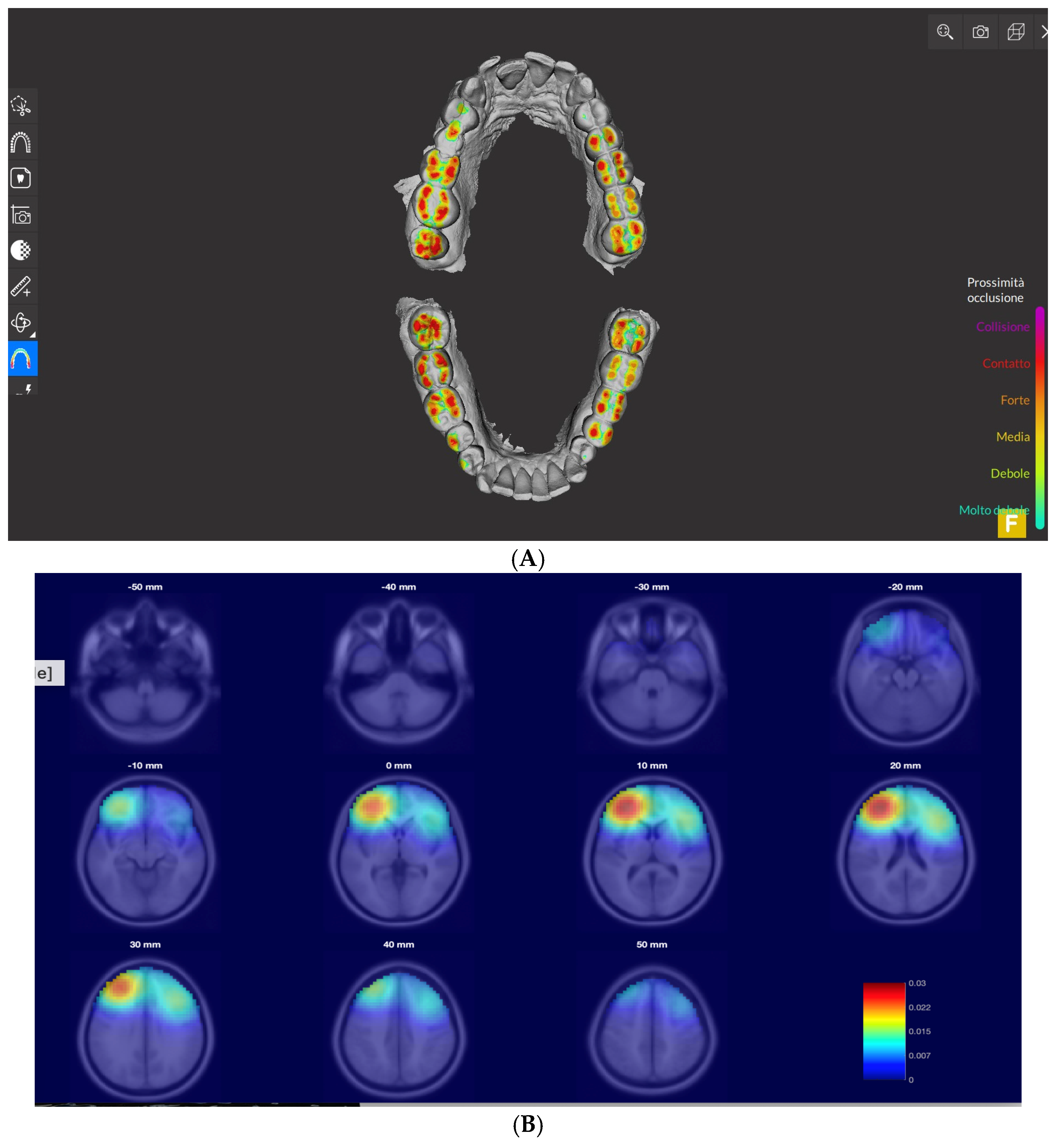
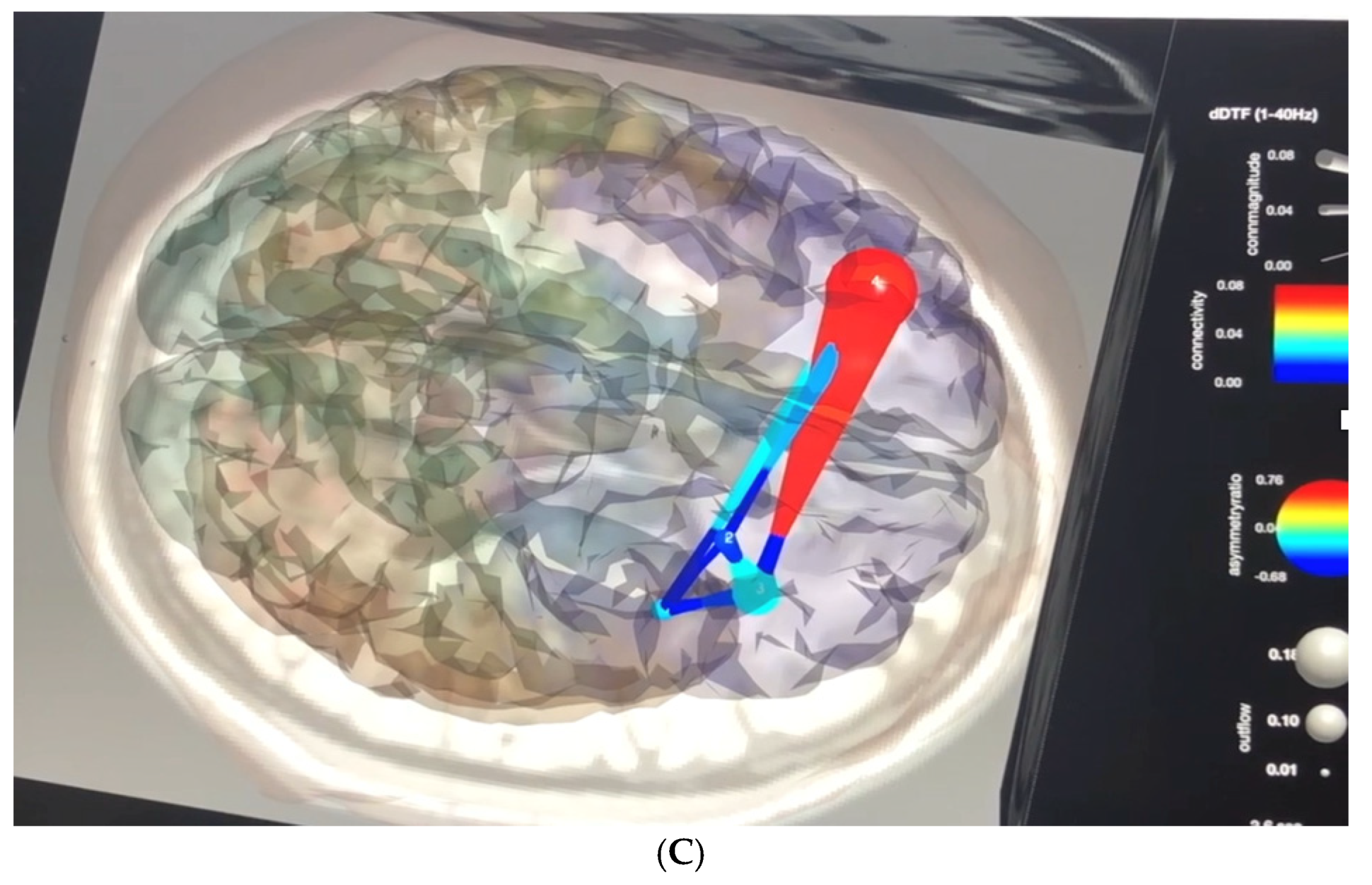
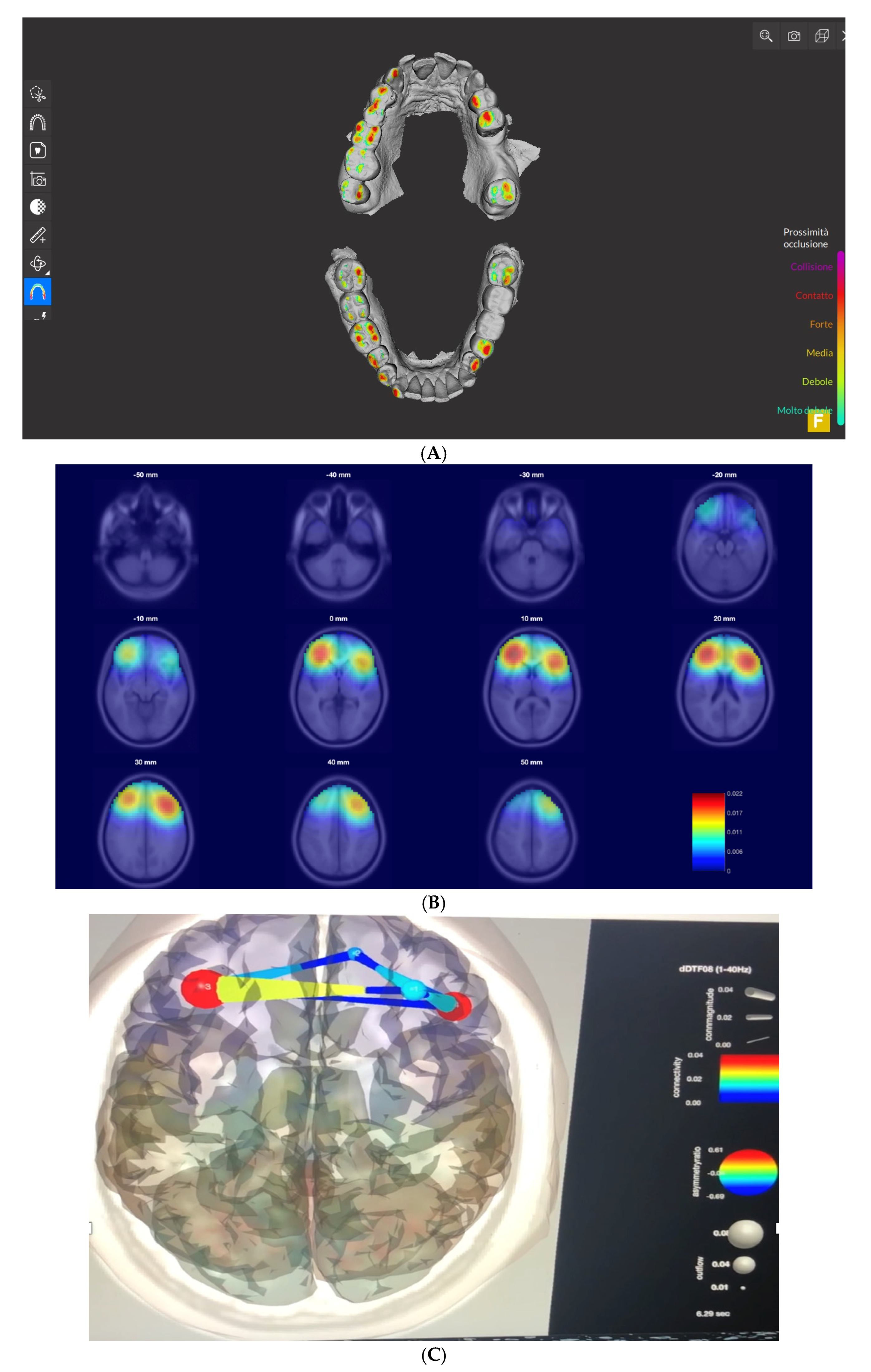
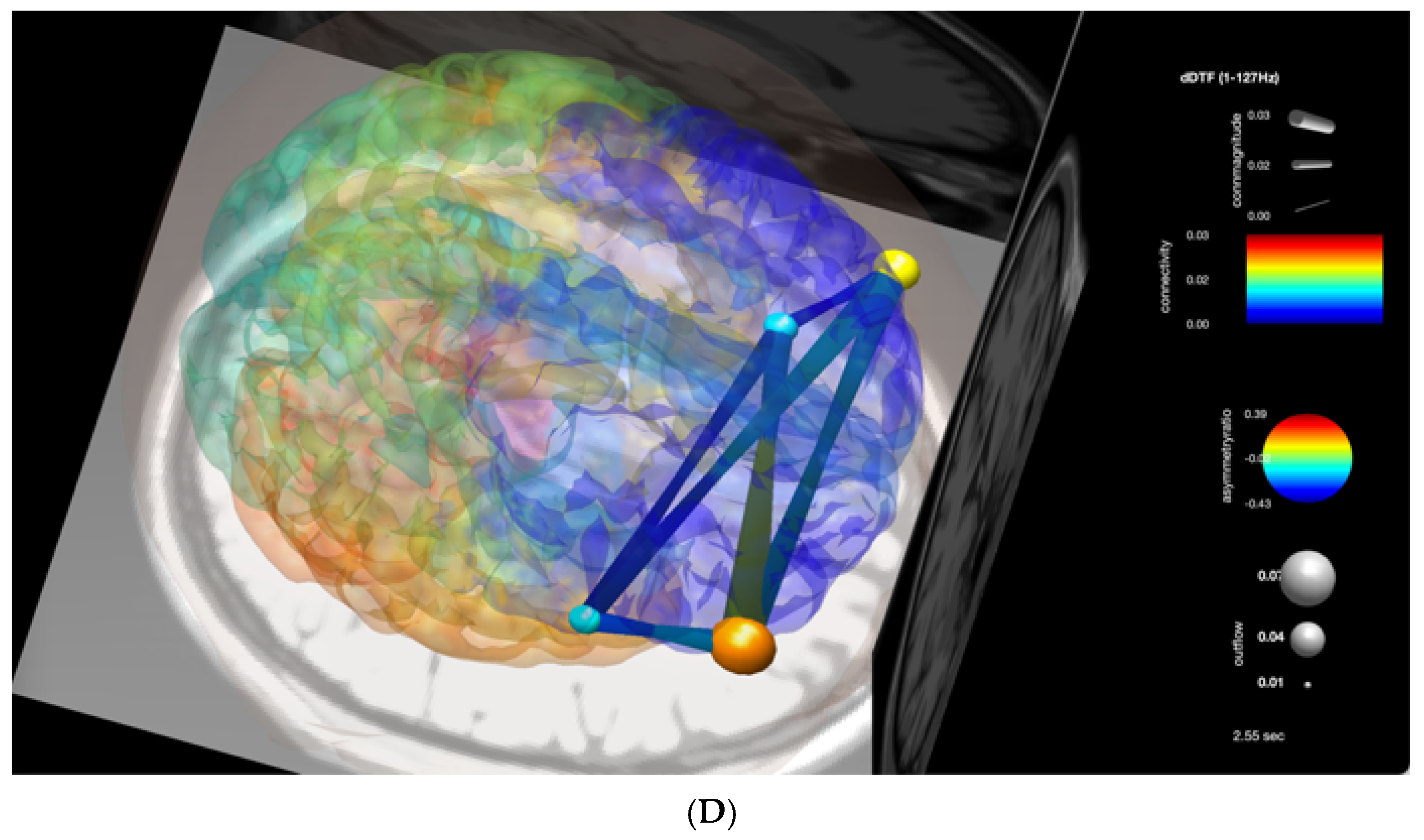
A 45-year-old right-handed male was selected for this study. His family and personal medical history were unremarkable, while his dental condition showed severe tooth loss which has been treated over time with different extemporary repairs. The deteriorating functionality of the prosthodontic repairs suggests a de novo complete replacement of the old implants. The project for a rational replacement was led with a computer-assisted 3D oral scanner (Carestream Dental®, Atlanta, GA, USA) which assessed the dynamics of the maxillary and mandibular pressure between the superior and inferior dental arch (Figure 1A). The dynamic analysis of the inter arch dental pressure allows for planning a new prosthodontic replacement whose architecture was tested by building a provisional methacrylate-based implant according to the recommendations of the International Association for Testing and Materials [13]. In addition to previous reports [4,9,10], in the present study, we intended to investigate the variations of neuroplasticity between the old and the new prosthodontic implants following electroencephalograms (EEG) recorded three hours before and after the provisional implant and assessed after six months when the definitive prosthesis was perfectly adapted. As EEG signals operate at the millisecond time scale, this approach, in comparison with voxel-based neuroimages, would carry a more time strength account in the dynamics correlated with ongoing inter-areas changes induced by new sensory-motor conditions brought about by the updated dental prosthesis. This study was led with the portable wireless MUSE headband EEG system (InterAxon Inc., Toronto, ON, Canada) in order to collect data that would yield quantifiable EEG tracings [14] eventually available for further study by MATLAB The MathWorks, Inc., Natick, Massachusetts, United States (2021 version) and EEGLAB (2019 version) [15]. The recording data were collected with preset AD at a 256 Hz sampling rate (http://developer.choosemuse.com/hardware-firmware/hardware specifications for full technical information, accessed on 1 May 2022). The MUSE EEG system has electrodes located analogous to Fpz, 97 AF7, AF8, TP9, and TP10 with electrode Fpz utilized as the reference electrode. Given that, the inter-electrode distances are now detailed by the referring system which is the 10-10 Electrode Placement System. A thin layer of water was applied to the dry electrodes for the frontal metallic sensors and the conductive silicone rubber mastoid sensors behind the ears to decrease the impedance and increase the quality signal.
The data obtained were streamed from the MUSE EEG system to MATLAB EEGLAB and read by an in-built EEGLAB program. The possible contamination of the signals recorded by noise was assessed by the EEGLab plugin ASR [16]. From about 30 min of eyes closed continuous recording we select for each study a continuous artifact-free part of 8000 ms, a time length sufficient to optimize the estimated functional connectivity [17]. All the data considered in the study pertain to a continuous recording set. In more detail, this simple flowchart about the different stages of data collection has been followed: stage 1 and stage 2 were recorded on the same day, respectively, when the patient was assessed for the functionality of the old dental implant and three hours after positioning the new provisional prosthodontic support; stage 3 is referred to the EEG recording taken after 6 months from the positioning of the definitive implant. Though the choice of these stages has been arbitrarily decided and several intermediate periods of recording could have been investigated, the adopted solution can indicate a clear-cut continuity of results over a time frame during which the stability of the implant has been assessed. An independent component analysis (ICA) of the selected EEG recording was interpolated with the spherical splines which allow for the tri-dimensional location of the dipole generated by each electrode location by using the EEG LAB plugin FIELDTRIP and SIFT; these procedures can analyze several aspects of brain connectivity as the first estimates the current densities of vector field distribution, while the latter exploits the “effective” connectivity intended as the influence one neural system exerts over another [18]. In order to obtain validated data accordingly, the current density field was estimated with the low-resolution brain electrographic tomography algorithm (LORETA) [19], while the normalized directed transfer function (DTF), a variant of the Granger causality algorithm, was used for assessing the effective connectivity which assumes that the information in a cause’s past “must improve the prediction of the effect above and beyond the information contained in the collective past of all other measured variables” [12]. The data obtained are graphically rendered by plotting a series of distributed color-coded source reconstruction inside 2D slices in a standardized stereotactic space scale (for LORETA) and, during the time course selected by the simulated brain visualization of the effective connectivity, by the asymmetry index between areas of maximum activity and pathways of connection (hub and edges, respectively), rendered by a 3D DTF color-coded graph (red maximum, blue minimum) projected in a template brain model.

3. Results

After the replacement of the old implant, the pathologic pressure exerted inside the dental arch is estimated by the computer-assisted 3D virtual rendering oral scanner and calculated as the weight differences in the distribution of opposing forces. The evolution between the basal condition and the positioning of the provisional prosthodontic implant is summarized by the comparison between Figure 1A and Figure 2A. The figures show the substantial modification of the strength exerted by the inter arch forces of occlusion in the conditions above described. Furthermore, these results are exploited in their neurophysiological outcome obtained by challenging the functional changes between the electric neuronal distribution (investigated with the LORETA procedure) between the basal and the provisional prosthodontic implant. As the LORETA method relies on a peculiar visualization of an inverse solution of the source localization, the recording EEG is particularly useful when the correspondent ICA has been founded and settled. In the present case report, this method offers a further advantage, since the 3D distribution of electrical activity reported in Figure 1B and Figure 2B consists of a high and a small number of sensors, a condition which already fits with this study. The theoretical implication is explained and reported in Figure 1B and Figure 2B which show the difference in both orientation and strength between the neural populations in neighboring neuronal assemblies. Furthermore, the differences between Figure 1B and Figure 2B show, in a limited but significant brain area, the changes in the standardized current density as an instantaneous distributed discrete image of the EEG [19] and account for the equalization of the current density between the frontal-parietal regions after positioning the new implant. A further refinement of the complex changes in neuronal dynamics accounted by the study of the current density is represented by the results of the investigation on the dynamic expression of the effective connectivity obtained by the study of the DTF. Figure 1C (basal conditions), compared to Figure 2C,D (3 h after positioning the provisional implant and 6 months after the definitive implant, respectively), show the dynamic interdependence calculated according to an “asymmetry” bioelectrical flow parameter between the areas recorded. It is worth mentioning that the DTF algorithm, similarly to the LORETA procedure, relies upon the transformation of the EEG signal in ICA and that their virtual projection in an MR template can be considered only from a perspective of the best approximation of the eventual localization of a stationary dominant dipole. The evolution in the DFT interdependence between Figure 1C and Figure 2C,D can be thus estimated as the virtual expression of the interhemispheric reshaping in the local dynamics of these neural assemblies.

4. Discussion

The results obtained in the present case study suggest that changes in brain neuroplasticity can be related to rapid improvements in sensory-motor functions induced by the new prosthodontic implant. Indeed, previous studies suggest that under such conditions, the diverse “ossiperception” mediated by periodontal mechanoreceptors and temporo-mandibular joints prompts a new oral kinesthesis which leads to a novel integration of regional neuroplasticity [20]. Moreover, the observation that tactile and discriminative functions improved after tooth loss replacement [9,20], suggests the possibility of a new brain “readout” of different sensory-motor neuroplasticity. Furthermore, in line with, and extending these observations, the present report shows that the dynamic relationships of neuroplasticity expressed by the effective connectivity show more balanced inter-area changes after the new tooth replacement. It is worth noting that our results exploit an as yet scarcely investigated aspect of the dynamic neuroplasticity, which in the pre-implant conditions clearly showed the rupture of the regional modularity with the global hierarchical organization, as reflected by the strong “enslavement” that the left frontal-parietal area engages over the contralateral (Figure 1B,C). The net result of such altered neural dynamic shows that the frontal-parietal circuitry is “forced” to downplay the role of the contralateral region in the process of sensory-motor integration which, instead of interacting with flexible reciprocity [21] and that, in doing so, impairs the modular connectivity between the frontoparietal areas and the chain of the potential motor effectors. Moreover, according to the expected theoretical previsions derived by these studies, after positioning the provisional implant and following new kinesthetic conditions the connectivity between the frontal areas and the distribution of the natural wiring pressure showed more balanced interarea relations (Figure 2B,C). However, the short time interval between removing the old implants and positioning the new provisional prosthesis cannot be explained by parallel clinical benefits [4,9,10]. Thus, though the relationships between positioning the new prosthodontic implant and the deep modulation of effective cortical connectivity appear strongly correlated, an eventual clinical improvement would have been grounded in neuroplasticity after a longer interval [10,22] than the one observed after three hours only. However, dealing with a different hypothesis one should relate the novel neuroplasticity occurring as a rapid modification in the effective connectivity with the fact that the new implant, by redrafting the original conditions of nociceptive and proprioceptive afferents in multiple brain regions, could eventually induce rapid changes in inter-area brain connectivity by a quick reframing of extensive small and micro-scale events. Indeed, rapid changes operated by early genes could account for synaptic changes which eventually reverberate along brain networks. In support of such a hypothesis, seminal studies [23] indicate the early gene c-fos, expressed in several neural populations, as a possible candidate for shifting synaptic conditions in response to significant changes induced by sensory-motor depolarizations. Furthermore, this mechanism could play a crucial role in inducing a remodulation of long-term neuroplasticity [24] in their dynamic expression, here represented by the reshaping of the effective connectivity.

5. Conclusions

The results of this study support previous investigations on tooth replacement and neuroplasticity and add novel features by describing the dynamic correlations of the latter in crucial sensory-motor brain networks.
Recent advancements in the application of the Virtual, Augmented and/or Mixed Realities in dental and prosthodontic practice, such as the holographic projection of internal dental conditions and the use of interactive simulators have opened new vistas in the study of the digital dental models [25,26]. These new technologies can support the assessment of the investigations of the cortical responses and can improve the monitoring of prosthodontic procedures.
Though the present results address new aspects of prosthodontic-related neuroplasticity, it is worth considering some critical aspects of this study as summarized by the following points: (i) the estimated connectivity has been incompletely defined, given the paucity of the electrode arrays used; (ii) the impact of the term “effective connectivity” exploited by the Granger causality could downplay the real time-frame of the bioelectrical activity, as this method cannot take into account the typical vector changing in EEG signals which is instead deduced on the basis of an artifactual stationary algorithm; (iii) though the results cover a short and medium period of observation, long monitoring punctuating more extensive periods, could be potentially useful in supporting eventual continuity or revealing different phases played by the dynamic aspects of effective connectivity.
Aside from these caveats, our report suggests that the study of brain connectivity represents a new approach to testing prosthodontic implants and can be potentially extended in monitoring the treatment of several distressing oral conditions.

Author Contributions

M.F. and F.M. planned the work and wrote the manuscript, D.B. acquired the dental data, A.M., M.M. and G.D. elaborated the electrophysiological data and the images. All authors have read and agreed to the published version of the manuscript.

Funding

This research received no external funding.

Informed Consent Statement

Written informed consent was obtained from the patient for the publication of any potentially identifiable images or data included in this article.

Data Availability Statement

The data presented in this study are available on request.

Acknowledgments

We acknowledge support by the president of the no-profit organization AIAS Cagliari Anna Paola Randazzo for the instrumental and clinical facilities of the institution.

Conflicts of Interest

The authors declare no conflict of interest.

References

  1. Dye, B.A. The Global Burden of Oral Disease: Research and Public Health Significance. J. Dent. Res. 2017, 96, 361–363. [Google Scholar] [CrossRef]
  2. Kassebaum, N.J.; Smith, A.G.C.; Bernabé, E.; Fleming, T.D.; Reynolds, A.E.; Vos, T.; Murray, C.J.L.; Marcenes, W.; GBD 2015 Oral Health Collaborators. Global, regional, and national prevalence, incidence, and disability-adjusted life years for oral conditions for 195 countries, 1990–2015: A systematic analysis for the global burden of diseases, injuries, and risk factors. J. Dent. Res. 2017, 96, 380–387. [Google Scholar] [CrossRef] [PubMed]
  3. Avivi-Arber, L. Neuroplasticity and the edentulous patient- toward a paradigm shift in oral rehabilitation. Int. J. Prosthodont. 2015, 28, 115. [Google Scholar] [CrossRef] [PubMed]
  4. Cerutti-Kopplin, D.; Feine, J.; Padilha, D.M.; De Souza, R.F.; Ahmadi, M.; Rompré, P.; Booij, L.; Emami, E. Tooth loss increases the risk of diminished cognitive function: A systematic review and meta-analysis. JDR Clin. Transl. Res. 2016, 1, 10–19. [Google Scholar] [CrossRef] [PubMed]
  5. Lopez-Jornet, P.; Zamora, C.; Pons-Fuster Lopez, L.E.; Tvarijonaviciute, A. Oral Health Status in Older People with Dementia: A Case-Control Study. J. Clin. Med. 2021, 10, 477. [Google Scholar] [CrossRef]
  6. Avivi-Arber, L.; Lee JCSood, M.; Lakschevitz, F.; Fung, M.; Barashi-Gozal, M.; Giogauer, M.; Sessle, B.J. Long-term neuroplasticity of the face primary motor cortex and adjacent somatosensory cortex induced by tooth loss can be reversed following dental implant replacement in rats. J. Comp. Neurol. 2015, 523, 2372–2389. [Google Scholar] [CrossRef]
  7. Fornito, A.; Zalesky, A.; Bullmore, E. Fundamentals of Brain Network Analysis; Academic Press: Cambridge, MA, USA, 2016; ISBN 9780124079083/9780124081185. [Google Scholar]
  8. Luraschi, J.; Korgaonkar, M.S.; Whittle, T.; Schimmel, M.; Müller, F.; Klineber, I.g. Neuroplasticity in the adaptation to prosthodontic treatment. J. Oral Facial Pain Headache 2013, 27, 206–216. [Google Scholar] [CrossRef]
  9. Avivi-Arber, L.; Lee, J.C.; Sessle, B.J. Dental occlusal changes induce motor cortex neuroplasticity. J. Dent. Res. 2015, 94, 1757–1764. [Google Scholar] [CrossRef]
  10. Friston, K. Functional and effective connectivity: A review. Brain Connect. 2011, 1, 13–36. [Google Scholar] [CrossRef]
  11. Jones, D.W. International dental standard. Br. Dent. J. 2007, 203, 361–369. [Google Scholar] [CrossRef]
  12. Krigolson, O.E.; Williams, C.C.; Norton, A.; Hassall, C.D.; Colino, F.L. Choosing MUSE: Validation of a low-cost, portable EEG system for ERP research. Front. Neurosci. 2017, 11, 109. [Google Scholar] [CrossRef] [PubMed] [Green Version]
  13. Fraschini, M.; Demuru, M.; Crobe, A.; Marrosu, F.; Cornelis, J.; Stam, C.J.; Hillebrand, A. The effect of epoch length on estimated EEG functional connectivity and brain network organization. J. Neural Eng. 2016, 13, 036015. [Google Scholar] [CrossRef] [PubMed]
  14. Delorme, A.; Mullen, T.; Kothe, C.; Akalin Acar, Z.; Bigdely-Shamlo, N.; Vankov, A.; Makeig, S. EEGLAB, SIFT, NFT, BCILAB, and ERICA: New tools for advanced EEG Processing. Comput. Intell. Neurosci. 2011, 2011, 10. [Google Scholar] [CrossRef] [PubMed]
  15. Pascual-Marqui, R.D.; Michel, C.M.; Lehmann, D. Low resolution electromagnetic tomography: A new method for localizing electrical activity in the brain. Int. J. Psychophysiol. 1994, 18, 49–65. [Google Scholar] [CrossRef]
  16. Marek, S.; Dosenbach, N.U.F. The frontoparietal network: Function, electro-physiology, and importance of individual precision mapping. Dialogues Clin. Neurosci. 2018, 20, 133–140. [Google Scholar] [CrossRef]
  17. Chen, Y.; Lin, Y.; Jin, Z.; Li, K.; Liu, G. Neuroplasticity of edentulous patients with implant-supported full dentures. Eur. J. Oral Sci. 2008, 118, 387–393. [Google Scholar] [CrossRef]
  18. Sessle, B.J. Sensation from the face. In Encyclopedia of Neuroscience; Squire, L., Ed.; Academic Press: Oxford, UK, 2009; Volume 8, pp. 585–592. [Google Scholar]
  19. Kaczmarek, L. Expression of c-fos and other genes encoding transcription factors in long-term potentiation. Behav. Neural Biol. 1992, 57, 263–266. [Google Scholar] [CrossRef]
  20. Jaworski, J.; Kalita, K.; Kanapska, E. c-Fos and neuronal plasticity: The aftermath of Kaczmarek’s theory. Acta Neurobiol. Exp. 2018, 78, 287–296. [Google Scholar] [CrossRef] [Green Version]
  21. Ono, Y.; Yamamoto, T.; Kubo, K.; Onozuka, M. Occlusion and brain function: Mastication as a prevention of cognitive dysfunction. J. Oral Rehabil. 2010, 37, 624–640. [Google Scholar] [CrossRef]
  22. Xu, S.; Huang, X.; Gong, Y.; Sun, J. Association between tooth loss rate and risk of mild cognitive impairment in older adults: A population-based longitudinal study. J. Aging 2021, 13, 21599–21609. [Google Scholar] [CrossRef]
  23. Gomaa, N.; Tenenbaum, H.; Glogauer, M.; Quiñonez, C. The Biology of Social Adversity Applied to Oral Health. J. Dent. Res. 2019, 98, 1442–1449. [Google Scholar] [CrossRef] [PubMed]
  24. Chang, C.-Y.; Hsu, S.-H.; Pion-Tonachini, L.; Jung, T.-P. Evaluation of Artifact Subspace Reconstruction for Automatic EEG Artifact Removal. In Proceedings of the 2018 40th Annual International Conference of the IEEE Engineering in Medicine and Biology Society (EMBS), Honolulu, HI, USA, 18–21 July 2018. [Google Scholar]
  25. Moussa, R.; Alghazaly, A.; Althagafi, A.; Eshky, R.; Borzangy, S. Effectiveness of Virtual Reality and Interactive Simulators on Dental Education Outcomes: Systematic Review. Eur. J. Dent. 2021, 16, 8–24. [Google Scholar] [CrossRef] [PubMed]
  26. Dolega-Dolegowski, D.K.; Dolega-Dolegowska, M.; Pregowska, A.; Hajto-Bryk, J.; Trojak, M.; Chmiel, J.; Walecki, P.; Fudalej, P.S. Application of holography and augmented reality based technology to visualize the internal structure of the dental root—A proof of concept. Head Face Med. 2022, 18, 12. [Google Scholar] [CrossRef] [PubMed]
Figure 1. Pre-implant condition. (A) 3D oral scanner study. Points of dental inter arch pressure by color-coded depiction (red as maximum altered dental pressure, green as minimal) show several spots of dental abnormal pressure. (B) Representation of the frontal-parietal current density dipole field estimated by the low-resolution brain electrographic tomography (LORETA). Red color indicates maximum current density while deep blue coded the minimum, shows a strong asymmetry between the distribution field of the left frontal-parietal areas. (C) Granger causality for four component dipoles localized to the right, left frontal gyrus and the temporal–parietal regions. Dimensions of blue circles and thin lines indicates lower causality, while red indicate high causality. Causality image at baseline occurring at 6310 of 8000 ms of EEG recording. The effective connectivity (high causality) was depicted by the color-coded (full red circle) asymmetry index with the right dominant frontal-parietal hub and by the red color of the direction of the edge originating from these areas.
Signals 03 00033 g001aSignals 03 00033 g001b
Figure 2. (A) 3D oral scanner study performed after positioning the provisional new implant. Points of dental inter arch pressure by color-coded depiction (red as maximum altered dental pressure, green as minimal) show the strong reduction of the spots of dental abnormal pressure as a shift of the red points towards green colors. (B) Three hours after positioning the provisional implant: LORETA assessed current density dipole field evenly distributed between the frontal-parietal areas. Red color indicates maximum current density, deep blue the minimum. The distribution of the current fields between the frontal-parietal areas now appears evenly distributed. (C,D) (after provisional and definitive implant, respectively). Granger causality exploited by the normalized directed transfer function (DTF) at 6290 of 8000 ms. of EEG recording. The effective connectivity rendering is represented by the anterior full red circle which correlates with the almost symmetric distribution of causality between the frontal-parietal hubs, represented by the red circles (hubs) and the yellow lines of connections (edges).
Signals 03 00033 g002aSignals 03 00033 g002b
Publisher’s Note: MDPI stays neutral with regard to jurisdictional claims in published maps and institutional affiliations.

