Abstract
Polypyrrole (PPy) is cationic in its conducting form, requiring a charge-balancing counterion, or dopant. The release of bioactive dopants, driven by the reduction of PPy films, offers a route to controlled drug delivery. Thiol-terminated long chain poly (ethylene glycol) (PEG) reacts with a dodecylbenzene sulfonate (DBSA)-doped PPy, forming a dense overlayer and partially liberating DBSA via the chemical reduction of the film. The resulting PEG brush acts as a barrier to dopant diffusion from the film, but proteins have been shown to disrupt this layer, releasing the DBSA. The mechanism by which this disruption occurs has not been thoroughly investigated. In this study, dopant release from PEG-PPy composites was examined via systematic exposure to a variety of chemical stimuli, including macromolecules such as poly (ethylene imine), polyethylene glycol, and poloxamers, as well as small-molecular-weight alcohols, carboxylic acids, and amines. Dopant release was quantified by quartz crystal microbalance. Poly (ethylene imine) efficiently released DBSA, while anionic and uncharged macromolecules did not. All classes of small molecules triggered dopant release, with longer homologues magnifying the response. The mechanisms of dopant removal are dependent on the functional groups of the stimulating agent and include ion exchange and nucleophilic reduction of the polycationic backbone. Tosylate, salicylate, and penicillin dopants showed release behaviors similar to DBSA, demonstrating the generality of the PEG barrier.
1. Introduction
The controlled release of bioactive compounds from a matrix reservoir is an important technology in many fields, including agriculture [1], cosmetics [2], antifouling coatings [3,4], and especially pharmaceuticals [5,6]. Many traditional formulations suffer from an initial burst release of encapsulated compounds, followed by a steady decrease in the release rate. “Extended” or “sustained” release strategies attempt to release an active species at a steady rate that minimizes toxicity or other undesired effects while maximizing desired outcomes. “Smart” or stimulated release strategies have the potential to dramatically enhance the tunability of controlled release systems. This approach takes advantage of specific stimuli, such as pH, temperature, redox reactions, magnetic fields, irradiation, and biological species, to modulate the expulsion of the compound of interest [7]. Stimulated release enables not only heightened control over the concentration of the bioactive species, but also some level of site specificity when the matrix is positioned near a target area.
Intrinsically conductive polymers (ICPs) such as polyaniline (PANI), poly (3,4-ethylenedioxythiophene) (PEDOT), and polypyrrole (PPy) have unique properties that have attracted interest in several areas of biomedicine. Their electrical conductivity and respectable biocompatibility make them useful as biosensors and electrodes for neural implants [8,9,10]. Potential applications in the fields of wound healing and tissue engineering are under investigation as well, since electrical stimulation and actuation can influence cell differentiation and growth [11,12,13]. The area of greatest interest, however, is in designing composites for stimulated drug release [14]. The polymers discussed here are polycationic in their conducting forms, requiring a counterion (usually called a “dopant”) for overall charge balance. Thus, a dopant that displays some bioactivity can be released from the composite upon a backbone reduction, upon a dopant exchange with another anion in solution, or by the simple diffusion of excess dopant that may be entrapped during the polymerization reaction [15]. The electrochemically controlled release approach has received the most attention. For example, Wu and coworkers demonstrated the electrically controlled release of the anti-inflammatory drug dexamethasone (Dex) from a PPy composite to inhibit inflammation [16]. Similarly, Boehler and co-workers investigated different electrical trigger signals on the release of Dex from PEDOT films. They were able to identify electrostatically bound Dex as well as additional “free” Dexentrapped in the polymer matrix. The total drug released by both the passive diffusion and electrically triggered mechanisms, however, represented only a portion of the total calculated loading [17].
The extent of a passive or an anion exchange release depends on the nature of the dopant (size, hydrophilicity), the pH, the temperature, and the morphology of the film [18]. For example, Seike and co-workers observed a more rapid dopant release from PPy films under basic conditions, perhaps due to ion exchange or nucleophilic attack on the backbone by the hydroxide ion [19]. Tiwari and co-workers demonstrated a PPy-based platform that controlled the release of paclitaxel by a combination of pH and near-infrared heating [20]. Organic solvents can also stimulate dopant diffusion. Ethanol has been shown to affect PPy films by opening pores and increasing surface area [21]. This, in turn, increased dopant exchange rates.
Reducing agents such as hydrazine can neutralize the cationic backbone, freeing dopants from electrostatic adhesion and driving release [22]. Neutral nucleophiles can also reduce the ICP backbone. It has long been known that certain gases, particularly ammonia [23], alter the electrical conductivity of ICPs, which has led to their use in gas sensing. Thiols also readily bind to and chemically reduce PANI and PPy, releasing dopants. This is likely through a thiol-ene-type mechanism [24]. In addition to reducing the polymer, some thiols also modify the surface properties. For example, treatment with alkane thiols dramatically reduces the surface energies of ICP thin films [25], while exposure to thiol-terminated polyethylene glycol (PEG-SH) results in a material with significant biofouling resistance [26]. This reaction has also been used to create sensing systems or to surface-modify electrodes for use in electrical devices [27,28].
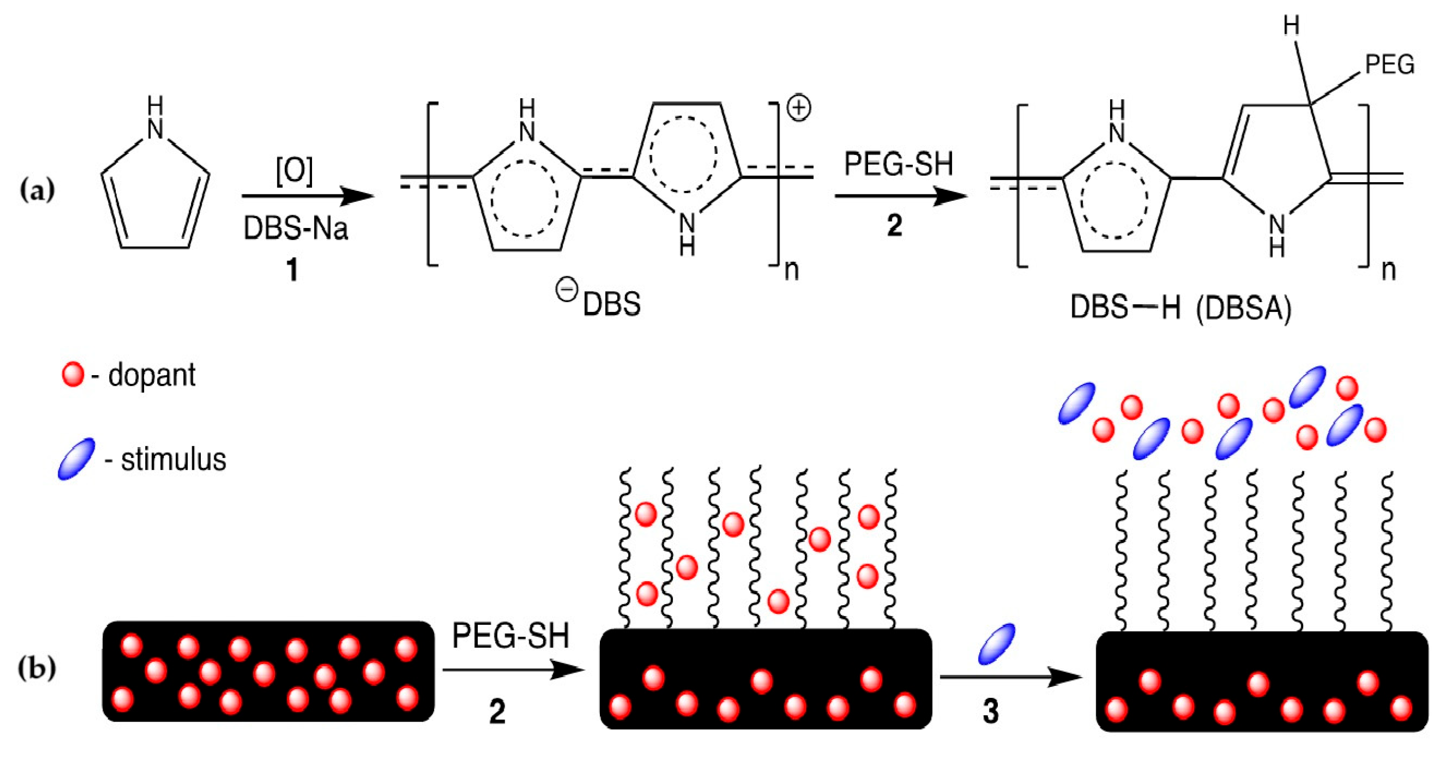
While investigating anti-fouling PPy surfaces, we found that dense assemblies of long-chain PEG thiols on PPy could effectively entrap the dodecylbenzene sulfonic acid (DBSA) dopant that was released from the polymer during the thiol addition reaction (Figure 1a) [29]. Interestingly, the release of DBSA from PEG-modified PPy is triggered selectively upon exposure to certain proteins, suggesting a potential application of these materials for controlled drug delivery (Figure 1b). While the surface modification of PPy is expected to influence the rates of dopant transport to the environment, little is known about what those effects are. In this study, we compared the dopant release behavior of PEG-modified PPy to unmodified PPy in the presence of various small molecules and macromolecules. The PPy films were electrochemically grown on quartz crystal microbalance (QCM) resonators. The frequency of the resonators is directly related to the mass of material on them, and changes in frequency can be quantified using the Sauerbrey relationship [30]. Loss of mass from the films caused an increase in the observed oscillation frequency. Only frequency data are reported here in order to highlight the influence of chemical species on the films.

Figure 1.
(a) A molecular scheme showing the oxidative polymerization of pyrrole in the presence of the dopant sodium dodecylbenzene sulfonate (step 1), followed by a reaction with thiol-terminated polyethylene glycol (step 2). (b) An illustration of the reductive nucleophilic reduction of PPy with PEG-SH (step 2, same as in 1a), followed by exposure to a stimulus that releases the entrapped dopant. In previous work, the dopant was DBSA and the stimulus PEI [29].
2. Materials and Methods
2.1. General
Pyrrole (Py) monomer was purified by distillation and stored at −18 °C. Sodium dodecylbenzene sulfonate (DBS, but generally referred to here as its nominally protonated form DBSA), bovine serum albumin (A-3059), sodium p-toluenesulfonate (Ts), sodium penicillin-G (Pen-G), sodium salicylate (SA), and Pluronic L64 (P184) were purchased from Sigma Aldrich (Burlington, MA, USA) and used as received. Pluronic 17 R4 was purchased from BASF. Branched poly(ethylene imine) with Mn of 600 (PEI-600) and 60,000 (PEI-60,000), respectively, was purchased from Thermo Fisher Scientific (Pittsburgh, PA, USA). Thiol-terminated poly(ethylene glycol) (PEG-SH, 40,000 MW) was purchased from JenKem USA (Dallas, TX, USA). All other reagents were purchased from commercial houses and used as received.
2.2. Electrochemical Polymerization of Py
2.2.1. Electrochemical Synthesis of DBSA-Doped Polypyrrole Films
Polypyrrole (PPy) films were prepared as previously described [29]. Briefly, electrochemical polymerization was performed using a Q-Sense Electrochemistry Module flow cell (QEM 401, Q-SenseAB, Västra, Frölunda, Sweden) with a quartz crystal microbalance with a dissipation system (QCM). A three-electrode system was used, including a platinum counter electrode, a Ag|AgCl reference electrode (DRIREF-5 SH, World Precision Instruments, Sarasota, FL, USA), and the QCM sensor as the working electrode. The piezoelectric sensor was an A−T-cut quartz crystal with a 10 mm diameter gold electrode (QSX301) with a fundamental resonance frequency of 5 MHz (QSX 301, Q-SenseAB, Västra, Frölunda, Sweden). Gold sensors were cleaned in a concentrated bleach solution (7.5% sodium hypochlorite) for 3 min, rinsed thoroughly with distilled (DI) water, and dried under N2 prior to each use. PPy films were polymerized using an eDAQ e-corder 410 recorder (eDAQ Inc. Colorado Springs, CO, USA) and an eDAQ EA163 potentiostat coupled with the Q-Sense QEM 401 electrochemistry module.
An aqueous solution containing DBSA (2 mg/mL, 5.74 mM) and pyrrole (2 mM) was prepared and deoxygenated by nitrogen sparging for 10 min prior to use. The aqueous pyrrole/DBSA solution was passed through the QEM 401 flow module at a rate of 30 μL/min, and once the QCM frequency (f) parameters were stable, a current density of 0.25 mA/cm2 was applied for 3 min for each polymer film growth. After polymerization, the quartz electrodes supporting the polymerized films were removed from the QEM 401 flow cell, rinsed with DI water, and dried under nitrogen gas.
2.2.2. Electrochemical Synthesis of Polypyrrole Films with Other Dopants
Polypyrrole films were grown with alternative dopants using a similar procedure to those grown with DBSA. These included Ts, Pen-G, and SA. The procedure for these syntheses varied only in the concentration of the dopant used and the growth time. Growth time was adjusted in order to obtain films with a similar mass to that of DBSA-doped films, which was quantified by the frequency decrease of the QCM resonance sensor. The dopant concentration was increased in cases where conductivity was insufficient to achieve satisfactory film growth. Pen-G-doped films were grown in 0.5 M Pen-G for 7 min, while SA-doped films were grown in 0.5 M salicylate sodium salt for 6 min. Ts-doped films were grown at 0.5 M Ts for 4.5 min. All other components of the DBSA procedure were identical to those used for the DBSA films.
2.2.3. Surface Modification of Polypyrrole Films with Poly(ethylene glycol)
Each film, regardless of the dopant used, was subjected to the same surface modification procedure. PPy-coated sensors were placed in a glass dish and heated on a hot plate at 60 °C. After equilibrating for five minutes in air, 300 μL of PEG-SH solution (0.2 mM, 40,000 MW) was pipetted onto the sensor. The sensor was kept at an elevated temperature for 45 min, allowing the reaction to proceed until the liquid evaporated, leaving a waxy residue atop the PPy surface. The sensor was then rinsed thoroughly with DI water and dried under N2 in order to remove the residue. The control (unmodified) films were also heated and rinsed in the same way but in the absence of the PEG-SH solution.
2.2.4. Exposure of Polypyrrole Films to Chemical Stimuli
Each PPy film was exposed to a variety of aqueous solutions of small alcohols, amines, carboxylic acids, or macromolecules, referred to as “stimulating solutions” here. The concentrations of the stimulating solutions varied. Aqueous solutions of small molecules (ethanol, 1-propanol, 2-propanol, 1-butanol, 2-butanol, 2-methyl-2-propanol, propanoic acid, butanoic acid, and pentanoic acid) were used at a concentration of 5% v/v. The effect of stimulus concentration was also conducted with 1-butanol at concentrations of 1%, 3%, and 8% solutions in addition to the 5% solution. Macromolecules (60,000 MW PEI, 600 MW PEI, L64 Poloxamer, and 17 R4 Poloxamer) were used at a 0.083 M concentration, which is consistent with the molarity of a 0.5% solution of 60,000 MW PEI.
The general procedure was as follows: PPy-coated sensors were mounted in a Q-Sense Flow Module (QFM 401). The QFM was flushed with DI water at 10 μL/min for 45 min, at which point the system was equilibrated. Next, the system was switched to a stimulus solution. After 1 h, the stimulus solution flow was stopped. The DI water flow was resumed, and the system flushed until the resonance frequency was stabilized. The mass loss associated with dopant release was quantified by the net change in the sensor’s 3rd overtone frequency before and after stimulation, including equilibration times. This resonance gave the most stable and reproducible results.
3. Results and Discussion
Previous work using QCM and UV–vis spectroscopy demonstrated that fetal bovine serum (FBS) decreased the resonance frequency of the quartz sensor of PPy thin films featuring a dodecylbenzene sulfonate dopant (PPy-DBSA) due to adsorption [29]. Conversely, the modification of the films with PEG-SH (PPy-DBSA-PEG), particularly high-MW (40,000) oligomers, not only resisted protein adsorption, but also liberated DBSA entrapped in the PEG brush layer. Exposure of the modified films to alginic acid, which is a polyanionic polysaccharide in aqueous solution, and electrically neutral PEG did not trigger DBSA release. High MW (60,000) poly(ethylene imine) (PEI, Figure 2), which is polycaionic in aqueous solution, did result in the extraction of DBSA. Here, we investigate other macromolecular stimuli.
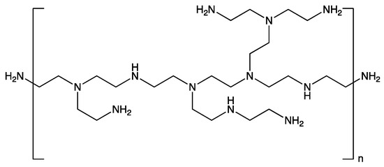
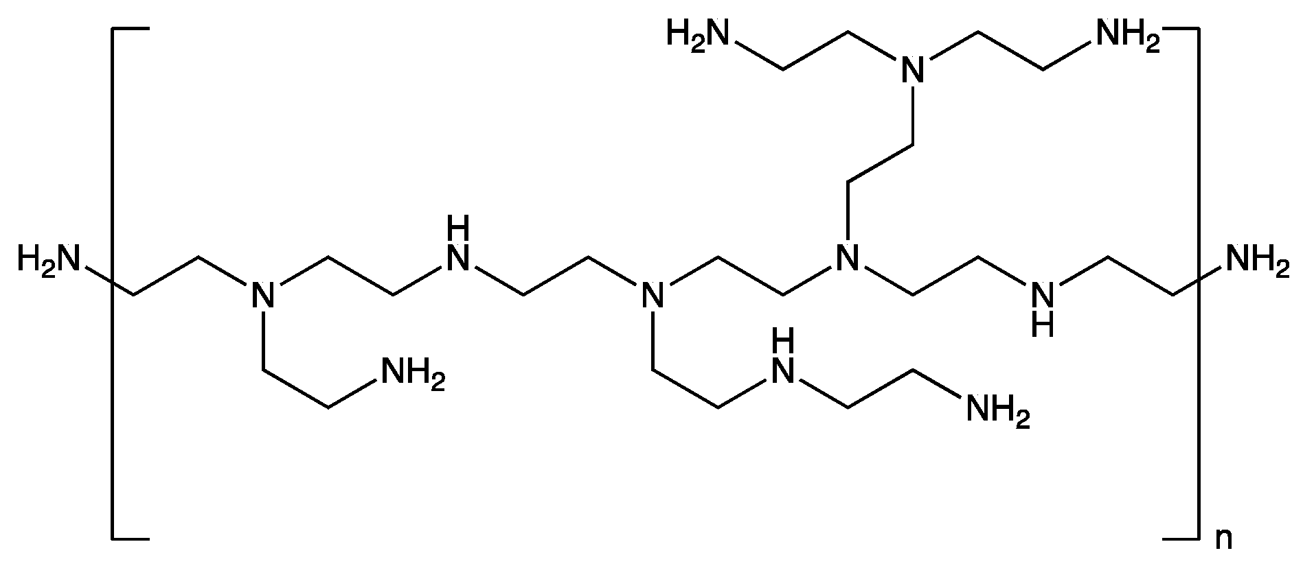
Figure 2.
Structure of branched poly(ethylenimine).
3.1. Dopant Extraction by Macromolecules
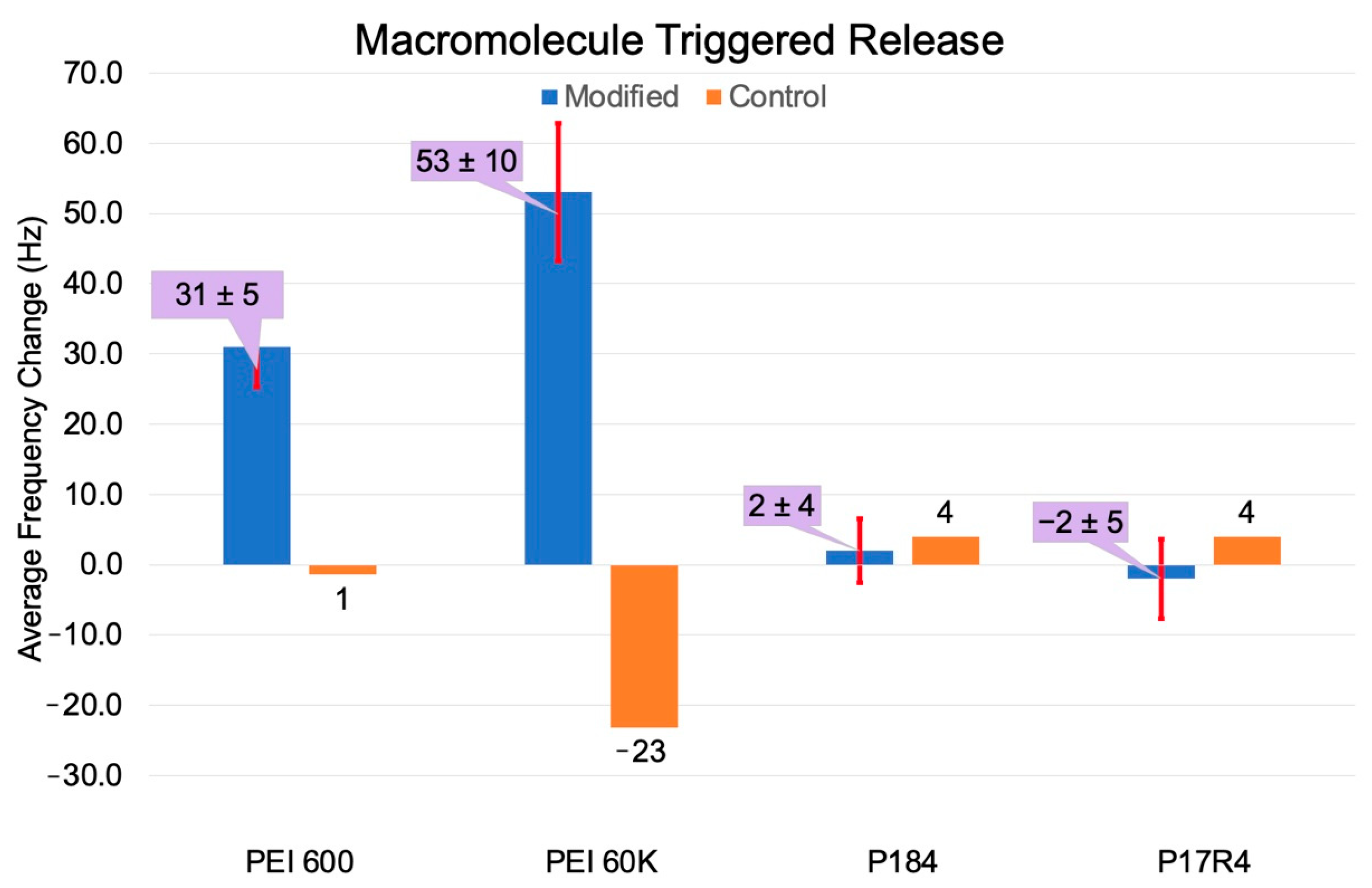
The interaction of PEI with ICPs has important industrial and scientific applications, including the production of low-work-function electronics [31] and improving adhesion between ICPs and metal surfaces [32]. Consistent with this and our previous report, highMW branched PEI deposits on PPy-DBSA films, while PEI with a MW of 600 effectively doesnot (Figure 3). While PEI solutions are basic, there was neither evidence of ion exchange between hydroxide and DBSA, nor did there seem to be significant expulsion of DBSA due to hydroxide attack on the polymer backbone over the timeframe of the experiment [29].

Figure 3.
Comparison of the response of PPy-DBSA and PPy-DBSA-PEG films to 0.083 mM solutions of 600 and 60,000 MW PEI and to poloxamers P184 and P17 R4 in DI water.
When exposed to PEI with a molecular weight of 600 at the same molarity as was used for 60,000 MW PPy-DBSA-PEG, DBSA release was clearly observed (Δf + 31 ± 5 Hz). This, however, was significantly less than that observed during exposure to higher MW PEI (Δf + 53 ± 10 Hz). This suggests that the electrostatic attraction between DBSA and the protonated amine groups is only partially responsible for the dopant release. We propose that the smaller oligomer is better solvated by water and less able to interact strongly with the pristine PPy-DBSA or to disrupt the PEG-modified adlayer.
The mixing of free PEG with the PPy-PEG polymer brush might be expected to result in a significant disruption of the PPy-DBSA-PEG-SH layer. However, no DBSA release has been observed in the presence of free PEG [29]. Additionally, the adsorption of free PEG at the unmodified PPy surface has not been observed, which is likely due to the high thermodynamic barrier associated with desolvating hydrophilic polymer chains.
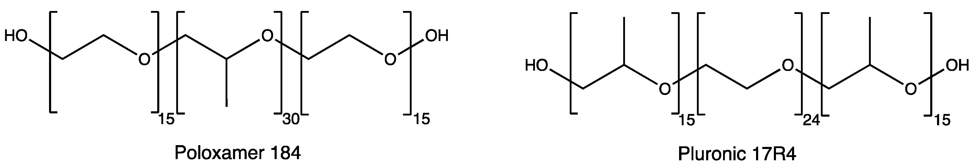
There are many alkylene oxides commercially available with varying degrees of hydrophilicity that could potentially prove better at disrupting the PEG-brush. For example, the poloxamer family of triblock copolymers is generally formed through living ring-opening polymerizations of oxiranes, commonly ethylene oxide and propylene oxide. Poloxamers are very versatile materials, as their hydrophobicity can be manipulated by altering the lengths of their polyethylene oxide (PEG) and more hydrophobic polypropylene oxide (PPO) blocks. They are widely used as surfactants, cleansers, and lubricants and in cosmetics. While generally non-toxic, certain poloxamers have been shown to interact strongly with biological membranes [33].
In order to explore this idea, PPy-DBSA and PPy-DBSA-PEG films were treated with Poloxamer 184 (P184) and Pluronic 17 R4 (Figure 4). P184 is a classic poloxamer with PEG end groups and a large PPO core, while P17 R4 is smaller and has a PEG core with PPO end groups. P184 has an average MW of 3000, while P17 R4 has an average MW of 2650, though their hydrophilic–lipophilic balances are similar. Their morphologies in aqueous solution are expected to be somewhat different, as the PEG blocks will be largely extended chains, while the PPO blocks will be more compact.

Figure 4.
Average structures of P184 and P17 R4.
Like pure PEG, neither poloxamer showed either a tendency to adsorb onto the PPy-DBSA films or to perturb the PEG layer of PPy-DBSA-PEG (Figure 3). The observed frequency changes are too small to be significant in all cases. Despite molecule size and hydrophobicity having an apparent effect on the ability for cationic macromolecules to extract dopants, these factors seem to have little to no effect on the ability for neutral alkylene oxide macromolecules to extract dopants. This suggests that the major contribution to the ability of cationic macromolecules to affect DBSA extraction is their ability to orient electropositive regions to the PEG surface.
3.2. Dopant Extraction by Small Molecules
Organic solvents can affect the morphology of PPy films and facilitate the passive diffusion of dopants [21]. Other mechanisms could influence dopant release as well, including nucleophilic attack on the PPy backbone or counterion exchange. Here, we present the results of PPy-DBSA and PPy-DBSA-PEG films with small, water-soluble alcohols, carboxylic acids, and amines.
3.2.1. Small Alcohol Stimuli
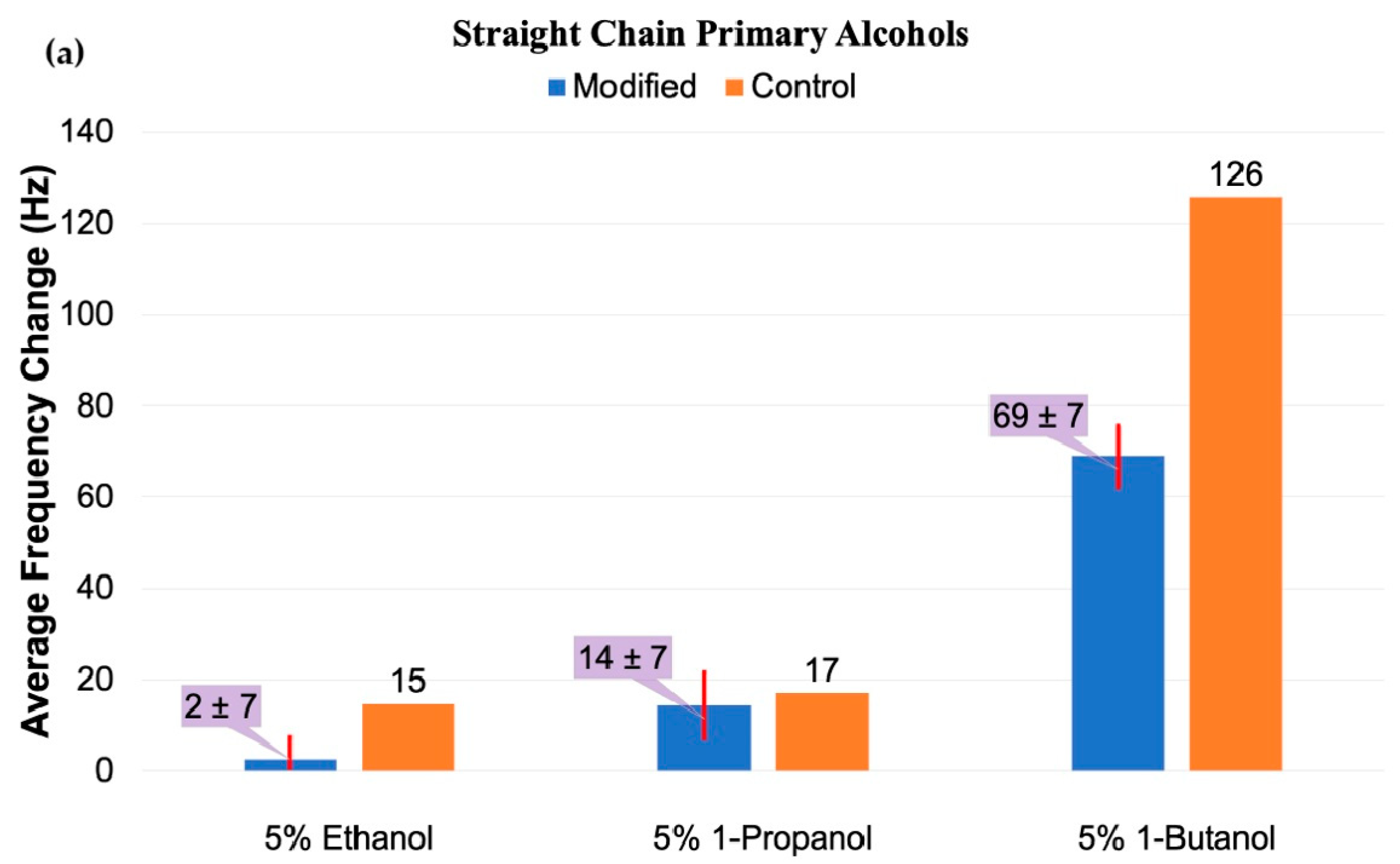
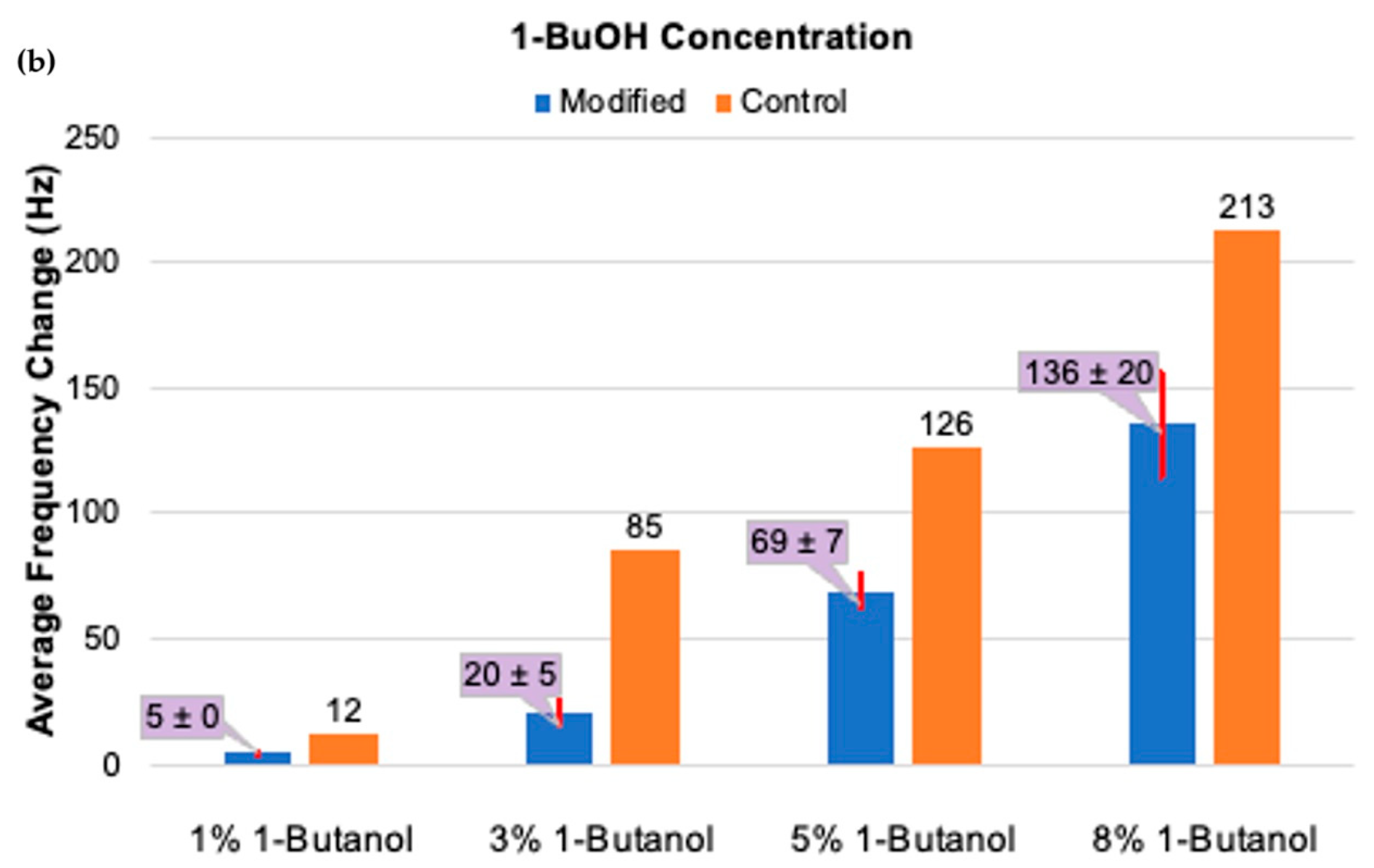
When exposed to 5% solutions of ethanol, PPy-DBSA-PEG films showed little or no response, while unmodified control films showed a modest frequency change of Δf + 15 Hz (Figure 5a). Exposure to 1-propanol resulted in a similar loss of DBSA to that of the control films (Δf + 17 Hz), but the PEG-coated films released more (Δf + 14 ± 7 Hz). A 5% solution of 1-butanol released significantly more DBSA (Δf + 69 ± 7 Hz) than the smaller alcohols and even more than 60,000 PEI under the same conditions. This is similar to that previously seen with fetal bovine serum in PBS after incubating for 1 h [29]. The unmodified control film, however, lost nearly twice as much mass. The effect of 1-butanol on the film was concentration-dependent (Figure 5b), but the control film always released more dopant than the surface modified films. Increasing the concentration from 1% to 8% solution greatly increased the DBSA release in both the modified and control films, though always more from the controls.


Figure 5.
(a) Average frequency change at 95% confidence after exposing DBSA-doped PPy films to 5% v/v solutions of ethanol, 1-propanol, and 1-butanol. (b) Average frequency change at 95% confidence after exposing DBSA-doped PPy films to 1-butanol at various concentrations (1%, 3%, 5%, 8%).
It is evident that the more hydrophobic 1-butanol can significantly disrupt the PPy film, perhaps by swelling it and allowing for deeper penetration into the bulk. Seike and coworkers recently noted the strong physical association of hydrophobic alcohols with PPy, resulting in chemical degradation [34]. They proposed a mechanism by which aqueous solutions of the alcohols could perform a nucleophilic addition to the polymer, similar to that proposed for the reaction with thiols, and likewise releasing DBSA. The PEG layer of the PPy-DBSA-PEG films likely still hinders the transport of DBSA to the surrounding media, reducing the amount of DBSA released as compared to the control.
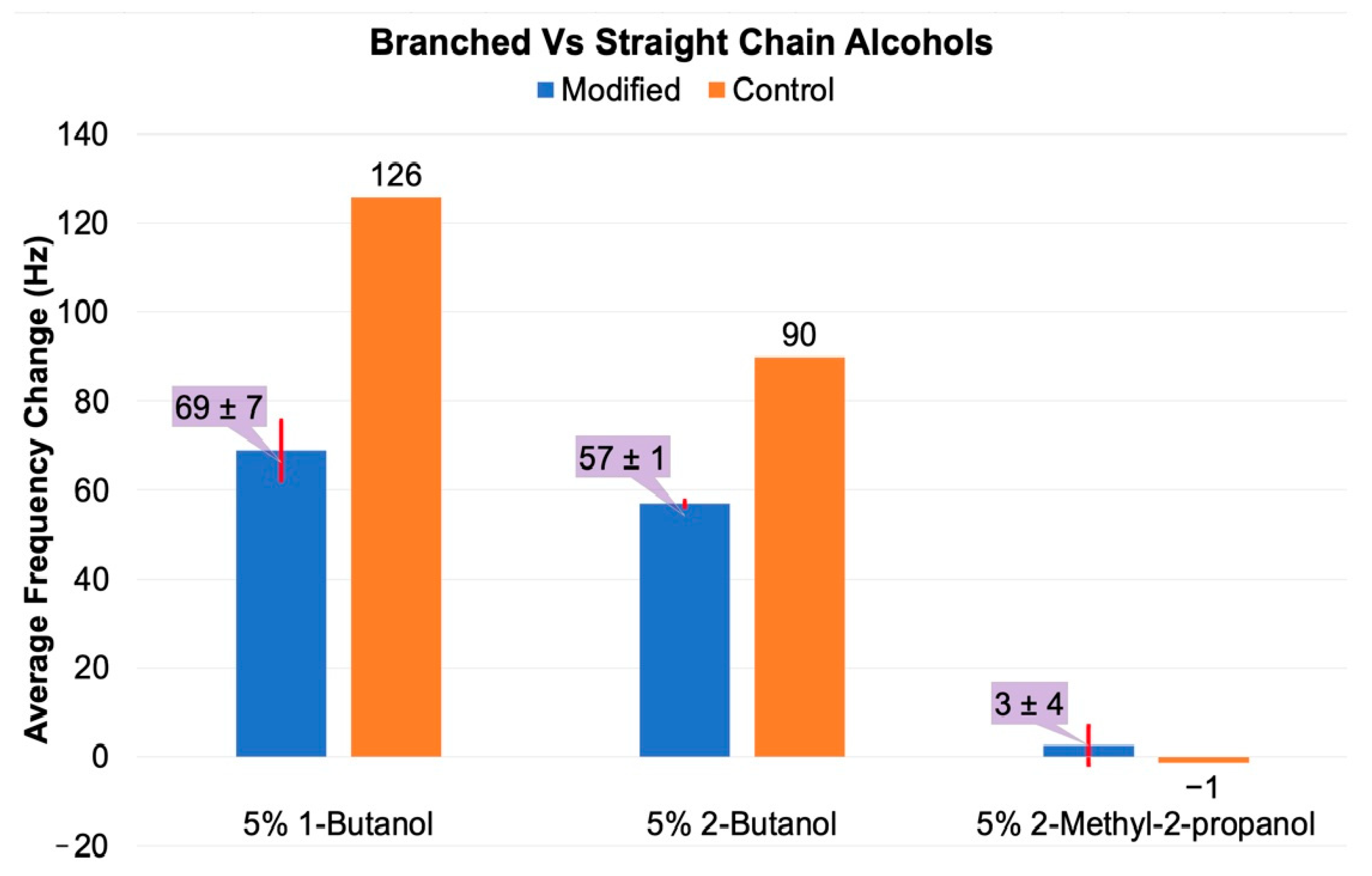
The amount of dopant release from the films was highly dependent on the structure of the alcohol. Upon exposure to a 5% solution of 2-methyl-2-propanol, neither the PPy-DBSA control films nor the surface-modified films showed dopant release beyond the margin of error (Figure 6). This result is drastically different than that of the straight-chain isomer 1-butanol (Δf + 69 ± 7 Hz). Upon exposure to 2-butanol, an intermediate result was observed (Δf + 57 ± 1 Hz). Thus, while more hydrophobic alcohols are able to penetrate into the PPy films, steric bulk may prevent efficient nucleophilic attack on the backbone and the resulting reduction. Not only does the 2-methyl-2-propanol have little effect on the control film, but it is not able to release the dopant entrapped in the PEG layer.
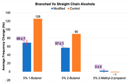
Figure 6.
Average frequency change at 95% confidence after exposing DBSA-doped PPy films to 5% v/v solutions of butanol isomers (1-butanol, 2-butanol, 2-methyl-2-propanol).
3.2.2. Small Carboxylic Acid Stimuli
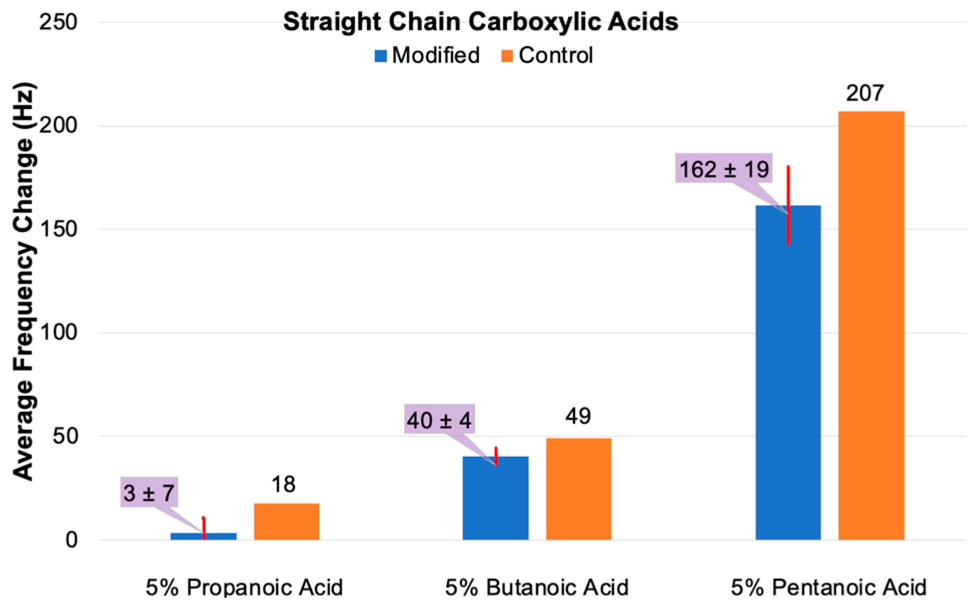
Previous work with the polyanionic macromolecule alginate showed no DBSA release. Small carboxylic acids would, therefore, not be expected to disrupt the PEG layer, nor would they be expected to act as nucleophiles attacking the PPy films. Figure 7 shows the results of the exposure of PPy-DBSA films to 3, 4, and 5 carbon linear carboxylic acids. In all cases, DBSA was released from the unmodified control films. Propionic acid released DBSA at a level (Δf + 18 ± 7 Hz) similar to 1-propanol from the control films, while the release from the PEG-modified films was negligible. Butanoic acid increased the DBSA release to Δf + 49 Hz and Δf + 40 ± 4 Hz for PPy-DBSA and PPy-DBSA-PEG, respectively. This is less than that observed for 1-butanol, and the difference between the PEG-modified films and the control was small. The greater water solubility of the carboxylic acids allowed for the treatment with pentanoic acid. Here, the DBSA loss dramatically increased to Δf + 207 Hz and Δf + 162 ± 19 Hz for PPy-DBSA and PPy-DBSA-PEG, respectively, comparable to an 8% solution of 1-butanol. These results confirm that some degree of hydrophobicity is required to penetrate into the PPy effectively and that the PEG layer is not a significant barrier to these stimuli. However, the mechanism of DBSA release must be different for carboxylic acids than for alcohols as the former are poor nucleophiles. Rather than backbone reduction, ion exchange is likely the primary process. Interestingly, the PEG adlayer seems to have a smaller effect with the carboxylic acids than with the other chemical stimuli investigated.

Figure 7.
Average frequency change at 95% confidence after exposing DBSA-doped PPy films to 5% v/v solutions of propanoic acid, butanoic acid, and pentanoic acid.
3.2.3. Small Amine Stimuli
Unlike other macromolecules, the polyamine PEI effectively displaced DBSA from PPy-DBSA-PEG films while adsorbing onto PPy-DBSA films. In aqueous solution, small amines are in equilibrium between protonated and neutral forms. We attempted to examine dopant release in these films with propylamine, butylamine, and triethylamine, but the frequency changes were very large (~Δf + 2000), and the data were very noisy and not well reproduced. It was observed that the films changed from the normal black color to brown. Like ammonia [23], the amines reacted extensively with the PPy backbone, presumably through a nucleophilic attack much like that observed with thiols.
3.3. The Effect of PEG Adlayers on Alternative Dopants
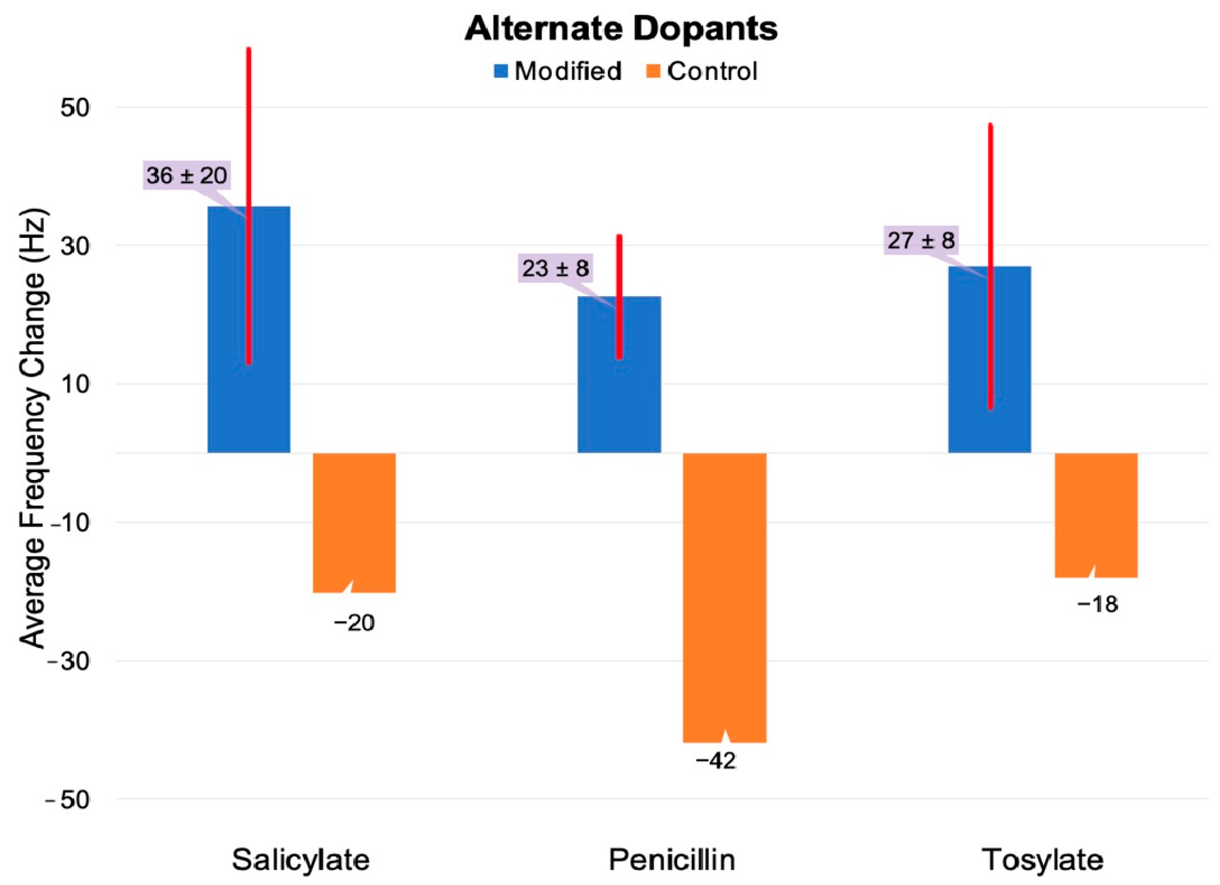
In order to establish the generality of the PEG adlayer effect on dopant release, PPy films were grown with three other dopants. The Ts dopant is a sulfonate closely related to DBSA, minus the hydrophobic alkyl tail. The films were grown in a 5.74 mM solution and showed both film growth and QCM behavior consistent with that seen with the DBSA films. The unmodified film saw a decreased frequency response of Δf −18 Hz when exposed to 0.5% 60,000 MW branched PEI, indicating PEI fouling of the surface (Figure 8). The PEG-modified surfaces showed a Ts release of Δf + 27 ± 18 Hz as the PEG layer was disrupted.
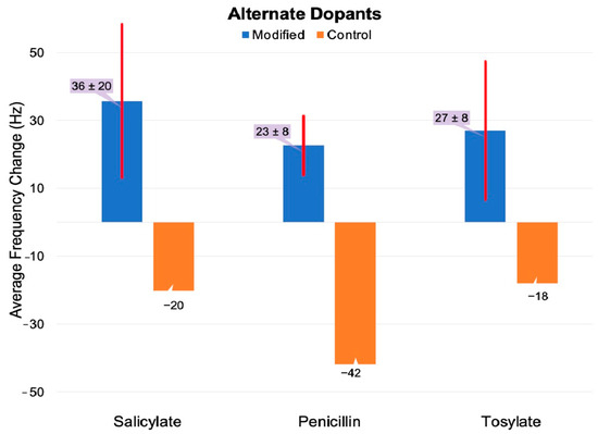
Figure 8.
Average frequency change at 95% confidence upon exposure of salicylate-doped, penicillin-doped, and tosylate-doped films to 0.083 mM 60,000 MW PEI. The salicylate- and penicillin-doped films were grown in a 0.5 M dopant, while the tosylate-doped film was grown in a 5.75 mM dopant.
The primary driver of research on the release of dopants from ICPs is their potential for the controlled release of bioactive compounds. Two simple anionic drug species were prepared using sodium salicylate (anti-inflammatory) and sodium penicillin G (antibiotic) as dopants. After the surface modification of these films, exposure to 0.5% 60,000 MW branched PEI clearly resulted in a stimulated release. Both films displayed PEI adsorption onto the control films and a release of dopants from the PEG-grafted surfaces; Δf + 36 ± 20 Hz for the SA-doped film and Δf + 23 ± 9 Hz for the Pen-G film. Their respective control films registered Δf − 20 Hz and Δf − 45 Hz. PPy film growth rates and morphology at the microscopic level are influenced by the dopant and electrochemical growth conditions [26]. These two compounds are carboxylic acids, which are not as highly ionized in DI water as the sulfonic acids. Since the dopant also serves as the electrolyte during PPy film growth, both a higher concentration of the salts and longer polymerization times are required to achieve films of similar thicknesses. The film density and roughness were different from the DBSA films, and no attempt was made to optimize the electrochemical growth parameters beyond the electrolyte concentration and electrolysis times. Although the frequency changes were not as large as that of the DBSA films, they still demonstrate that the PEI-triggered release is not unique to sulfonate-doped films. It is reasonable to assume that other bioactive anionic compounds will have PEG-mediated release behaviors.
4. Conclusions
The reduction of the polycationic PPy backbone removes the electrostatic requirement for the anionic dopant and allows for it to be released to the surrounding solution. PEG-SH reacts with PPy film surfaces, partially reducing them. This both decreases the ability of macromolecules to be deposited onto the films and inhibits the loss of electrostatically released organic dopants to the surroundings. This behavior was found with the sulfonic acids DBSA and Ts as well as the biologically active carboxylic acid dopants SA and Pen-G. The disruption of the PEG layer and subsequent dopant release is enabled by a wide variety of chemical stimuli, but the impact of small-MW stimuli on the PPy film, regardless of surface modification, cannot be ignored. The findings herein suggest that multiple factors contribute to the ability of a given stimulus to affect dopant release. Among these are molecular weight, hydrophobicity, steric bulk, charge, and, perhaps, others. The complexity of the dopant release behavior in PEG-PPy composites remains an obstacle to our full understanding of these systems. The scope and limitations of this process warrant further exploration, but it does offer a potential route to the controlled release of bioactive dopants triggered by a unique mechanism.
Author Contributions
Conceptualization, P.J.M. and T.W.H.; Methodology, P.J.M.; Formal analysis, G.R.; Investigation, G.R. and A.K.; Resources, T.W.H.; Data curation, G.R.; Writing—original draft preparation, G.R. and T.W.H.; Writing—review and editing, G.R., A.K.; P.J.M. and T.W.H.; Supervision, T.W.H.; Project administration, T.W.H.; Funding acquisition, T.W.H. All authors have read and agreed to the published version of the manuscript.
Funding
This work was supported primarily by the National Science Foundation EPSCoR Program under NSF Award # OIA-1655740. A.K. received a fellowship from the National Institute of General Medical Sciences, National Institutes of Health through award P20GM103499. Any Opinions, findings and conclusions or recommendations expressed in this material are those of the author(s) and do not necessarily reflect those of the National Science Foundation or the National Institutes of Health.
Data Availability Statement
The dataset is available on request from the corresponding author.
Conflicts of Interest
The authors declare no conflict of interest.
References
- Dovzhenko, A.P.; Yapryntseva, O.A.; Sinyashin, K.O.; Doolotkeldievac, T.; Zairov, R.R. Recent progress in the development of encapsulated fertilizers for time-controlled release. Heliyon 2024, 10, e34895. [Google Scholar] [CrossRef] [PubMed]
- Costa, R.; Santos, L. Delivery systems for cosmetics—From manufacturing to the skin of natural antioxidants. Powder Tech. 2017, 322, 402–416. [Google Scholar] [CrossRef]
- Ali, A.; Jamil, M.I.; Jiang, J.; Shoaib, M.; Amin, B.U.; Luo, S.; Zhan, X.; Chen, F.; Zhang, Q. An overview of controlled-biocide-release coating based on polymer resin for marine antifouling applications. J. Polym. Res. 2020, 27, 85. [Google Scholar] [CrossRef]
- Lei, J.; Li, Z.; He, T.; Wang, Z.; Yao, S.; Qiu, H. Lubricant controlled release silicone fouling release coatings based on mesoporous molecular sieves. J. Coat. Technol. Res. 2023, 20, 703–711. [Google Scholar] [CrossRef]
- Adepu, S.; Ramakrishna, S. Controlled drug delivery systems: Current status and future directions. Molecules 2021, 26, 5905. [Google Scholar] [CrossRef]
- Li, W.; Tang, J.; Lee, D.; Rice, T.R.; Schwendeman, S.P.; Prausnitz, M.R. Clinical translation of long-acting drug delivery formulations. Nature Rev. Mater. 2022, 7, 406–420. [Google Scholar] [CrossRef]
- Bruneaua, M.; Bennicia, S.; Brendlea, J.; Dutourniea, P.; Limousy, L.; Pluchon, S. Systems for stimuli-controlled release: Materials and applications. J. Control. Release 2019, 294, 355–371. [Google Scholar] [CrossRef]
- Joy, N.; Gopalan, G.P.; Eldho, J.; Francis, R. Conducting polymers: Biomedical applications. In Biomedical Applications of Polymeric Materials and Composites; Francis, R.D., Kumar, S., Eds.; John Wiley & Sons, Wiley Online Books: Hoboken, NJ, USA, 2016. [Google Scholar] [CrossRef]
- Gupta, R.K. Conducting Polymers Chemistries, Properties and Biomedical Applications; Gupta, R.K., Ed.; Taylor and Francis Online: Abingdon, UK, 2022. [Google Scholar] [CrossRef]
- Paramshetti, S.; Angolkar, M.; Al Fatease, A.; Alshahrani, S.M.; Hani, U.; Garg, A.; Ravi, G.; Osmani, R.A.M. Revolutionizing drug delivery and therapeutics: The biomedical applications of conductive polymers and composites-based systems. Pharmaceutics 2023, 15, 1204. [Google Scholar] [CrossRef]
- Mezhuev, Y.O.; Shtil’man, M.I.; Artyukhov, A.A. The application of polyaniline and polypyrrolein medical and biological fields. Part 2. Tissue engineering, muscle simulation, and systems with controlled release of biologically active substances. Polym. Sci. Ser. D 2021, 14, 427–431. [Google Scholar] [CrossRef]
- Uzieliene, I.; Popov, A.; Vaiciuleviciute, R.; Kirdaite, G.; Bernotiene, E.; Ramanaviciene, A. Polypyrrole-based structures for activation of cellular functions under electrical stimulation. J. Bioelectrochem 2023, 155, 108585. [Google Scholar] [CrossRef]
- Yi, H.; Patel, R.; Patel, K.D.; Bouchard, L.-S.; Jha, A.; Perriman, A.W.; Patel, M. Conducting polymer-based scaffolds for neuronal tissue engineering. J. Mater. Chem. B 2023, 11, 11006–11023. [Google Scholar]
- Alkahtani, M.E.; Elbadawi, M.; Chapman, C.A.; Green, R.A.; Gaisford, S.; Orlu, M.; Basit, A.W. Electroactive polymers for on-demand drug release. Adv. Healthc. Mater. 2024, 13, 2301759. [Google Scholar] [PubMed]
- Puiggalí-Jou, A.; del Valle, L.J.; Alemán, C. Drug delivery systems based on intrinsically conducting polymers. J. Control. Release 2019, 309, 244–264. [Google Scholar] [PubMed]
- Wu, C.; He, X.; Zhu, Y.; Weng, W.; Cheng, K.; Wang, D.; Chen, Z. Electrochemical deposition of Ppy/Dex/ECM coatings and their regulation on cellular responses through electrical controlled drug release. Colloids Surf. B Biointerfaces 2023, 222, 113016. [Google Scholar]
- Boehler, C.; Oberueber, F.; Asplund, M. Tuning drug delivery from conducting polymer films for accurately controlled release of charged molecules. J. Control. Release 2019, 304, 173–180. [Google Scholar] [PubMed]
- Shah, S.A.A.; Firlak, M.; Berrow, S.R.; Halcovich, N.R.; Baldock, S.J.; Yousafzai, B.M.; Hathout, R.M.; Hardy, J.G. Electrochemically enhanced drug delivery using polypyrrole films. Materials 2018, 11, 1123. [Google Scholar] [CrossRef]
- Deike, M.; Asaumi, Y.; Kawashima, H.; Hirai, T.; Nakamura, Y.; Fujii, S. Morphological and chemical stabilities of polypyrrole in aqueous media for 1 year. Polym. J. 2022, 54, 169–178. [Google Scholar]
- Tiwari, A.P.; Hwang, T.I.; Oh, J.-M.; Maharjan, B.; Chun, S.; Kim, B.S.; Joshi, M.K.; Park, C.H.; Kim, C.S. pH/NIR-responsive polypyrrole-functionalized fibrous localized drug-delivery platform for synergistic cancer therapy. ACS Appl. Mater. Interfaces 2018, 10, 20256–20270. [Google Scholar] [CrossRef]
- González-Torres, M.; Cruz-Cruz, G.J.; Sánchez-Mendieta, V.; Gómez-Jiménez, L.M.; González-Salgado, F.; Mondragón-Lozano, R.; Olayo-González, M.G. Dapsone uptake and release from plasma polypyrrole for drug administration. Polym. Bull. 2017, 74, 1761–1773. [Google Scholar]
- Pernaut, J.-M.; Reynolds, J.R. Use of conducting electroactive polymers for drug delivery and sensing of bioactive molecules. A redox chemistry approach. J. Phys. Chem. B 2000, 104, 4080–4090. [Google Scholar]
- Gustafsson, R.; Lundstrom, I. The effect of ammonia on the physical properties of polypyrrole. Synth. Met. 1987, 20, 203–208. [Google Scholar]
- Blomquist, M.; Babacka, J.; Ivaska, A.; Levon, K. Electrochemical and spectroscopic study on thiolation of polyaniline. Electrochim. Acta 2013, 90, 604–614. [Google Scholar]
- Bergman, B.; Hanks, T. Spectroscopic, microscopic, and surface analysis of alkanethiol- and fuoroalkanethiol- modified conducting polymer thin films. Macromolecules 2000, 33, 8035–8042. [Google Scholar]
- Zhang, B.; Nagle, A.; Wallace, G.G.; Hanks, T.W.; Molino, P.J. Functionalised inherently conducting polymers as low biofouling materials. Biofouling 2015, 31, 493–502. [Google Scholar]
- Su, Z.; Liu, Y.; Chen, L.; Zhang, Y.; Meng, Y.; Li, Y.; Fu, Y.; Ma, M.; Yao, S. Preparation of thiolated polymeric nanocomposite for sensitive electroanalysis of dopamine. Biosen. Bioelect. 2012, 36, 154–160. [Google Scholar]
- Yi, J.; Kim, G.; Lee, S.; Ryu, C.; Lee, J.Y. Enzymatically stable, non-cell adhesive, implantable polypyrrole/thiolated hyaluronic acid bioelectrodes for in vivo signal recording. Int. J. Biomacromol. 2024, 276, 133770. [Google Scholar]
- Molino, P.J.; Wetherill, R.E.; Knepper, A.; Trupia, D.; Hayes, P.; Molino, B.Z.; Hanks, T.W. Biofouling resistant electroactive polymer composites for protein triggered counterion delivery. Appl. Polym. Mater. 2021, 3, 6294–6302. [Google Scholar]
- Sauerbrey, G. The use of quarts oscillators for weighing thin layers and for microweighing. Z. Phys. 1959, 155, 206–222. [Google Scholar]
- Zhou, Y.; Fuentes-Hernández, C.; Shim, J.; Meyer, J.; Giordano, A.J.; Li, H.; Winget, P.; Papadopoulos, T.; Cheun, H.; Kim, J.; et al. A universal method to produce low-work function electrodes for organic electronics. Science 2012, 336, 327–332. [Google Scholar]
- Kim, K.-G.; Kim, S.Y. Increase in interfacial adhesion and electrochemical charge storage capacity of polypyrrole on Au electrodes using polyethyleneimine. Sci. Rep. 2019, 9, 2169. [Google Scholar]
- Zarrintaj, P.; Ramsey, J.D.; Samadi, A.; Atoufi, Z.; Yazdi, M.K.; Ganjali, M.R.; Amirabad, L.M.; Zagnene, E.; Farokhi, M.; Formela, K.; et al. Poloxamer: A versatile tri-block copolymer for biomedical applications. Acta Biomater. 2020, 110, 37–67. [Google Scholar] [CrossRef] [PubMed]
- Seike, M.; Hirai, T.; Nakamura, Y.; Fujii, S. Alcohol as hydrophobizer for polypyrrole. Chem. Lett. 2022, 51, 598–600. [Google Scholar] [CrossRef]
Disclaimer/Publisher’s Note: The statements, opinions and data contained in all publications are solely those of the individual author(s) and contributor(s) and not of MDPI and/or the editor(s). MDPI and/or the editor(s) disclaim responsibility for any injury to people or property resulting from any ideas, methods, instructions or products referred to in the content. |
© 2025 by the authors. Licensee MDPI, Basel, Switzerland. This article is an open access article distributed under the terms and conditions of the Creative Commons Attribution (CC BY) license (https://creativecommons.org/licenses/by/4.0/).