1. Introduction
The oxidation of coins represents a concrete and easily observable example of the chemical reactions occurring between a metal and its surroundings. Copper, silver, bronze, steel, and other coins, in fact, are relatively reactive and, when exposed to air, tend to transform slowly through a series of surface reactions with oxygen, humidity, and other substances present in the atmosphere [
1]. The oxidation of metal coins is a natural chemical phenomenon that occurs when metals react with their surroundings. This process falls within redox reactions, in which the metal loses electrons (oxidation) while other chemical species gain them (reduction) [
2].
Coins are generally made from metal alloys such as copper, silver, nickel, zinc, steel, or aluminum-based alloys, chosen for their strength, durability, and low cost. However, no metal is completely immune to the effects of ageing: when exposed to the environment, the surface of coins undergoes chemical changes that lead to the formation of oxides, hydroxides, or other compounds. These reactions cause visible changes, such as blackening, stains, or coloured coatings. For copper-based coins, the most important reactions that can lead to the colour of their surface ageing patina are the following [
3]. When copper reacts with oxygen, cuprous oxide (Cu
2O), red in colour, is formed:
With further oxidation (more oxygen or higher temperatures), cupric oxide (CuO), which is black and responsible for blackening, is formed:
A typical green-blue colour may derive from several corrosion products, especially basic copper carbonates, such as:
However, depending on the environment, basic copper carbonates, sulfates, or chlorides may all contribute. Under alkaline conditions, Cu(II)-bearing compounds may also form, for example:
In transition-metal compounds, colour differences are related to differences in electronic structure, including d-orbital splitting and ligand environment, which influence the energies of electronic transitions [
4]. Accordingly, the colour of copper corrosion products is related to their chemical composition, oxidation state, crystal structure, and local coordination environment. Therefore, different oxides, hydroxides, carbonates, chlorides, and complex corrosion compounds may generate red, black, green, or blue surface hues.
The type of oxidation depends on several factors, including the composition of the alloy, environmental conditions (humidity, temperature, pressure, presence of pollutants or salinity), and exposure time. For example, copper tends to develop green corrosion products under some conditions, while other metals may form different coloured oxides or corrosion layers. In some cases, these transformations represent material deterioration; however, in others, the surface patina can act as a protective barrier that slows down further reactions [
5].
It is important to distinguish between corrosion products that may form relatively stable and protective patinas (“type I”, often referred to as noble patinas) and those associated with active and deleterious corrosion processes (“type II”, or vile patinas). In the case of copper-based alloys, this distinction is well established in the literature and is critical for interpreting both patina formation and laser-cleaning effects [
6,
7].
For instance, compact and adherent layers such as cuprite (Cu2O) and, under certain conditions, stable basic copper carbonates (malachite/azurite) may contribute to a protective behaviour. In contrast, porous or reactive phases, particularly copper chlorides (e.g., nantokite, atacamites, etc.) or sulfates, are typically associated with ongoing corrosion phenomena.
The literature reports that, immediately after exposure to air, copper oxidizes to first form a thin layer of cuprous oxide (Cu
2O). Over time, a second layer of cupric oxide (CuO) grows on the surface. The entire process almost stops spontaneously after a few nanometers, suggesting a natural passivation behaviour due to the reduced diffusion of oxygen through the formed oxides [
8].
Temperature plays a fundamental role in copper oxidation because it influences both the reaction rate and the type of oxides that are formed. As temperature increases, atoms and molecules gain kinetic energy, and the frequency of effective collisions between copper and oxygen increases; consequently, oxidation proceeds more quickly, in accordance with the Arrhenius equation, which shows that even small temperature increases can greatly accelerate the reaction [
9]. In addition, the temperature influences which copper oxide is formed. At lower temperatures, mainly cuprous oxide (Cu
2O), with a reddish-brown colour, is formed, whereas higher temperatures favour the formation of cupric oxide (CuO), with its typical black colour [
10].
The oxidation of copper at high temperatures is governed by two diffusion processes: outward diffusion of copper and inward diffusion of oxygen. The higher the temperature, the higher the diffusivity, and therefore the faster the growth of the oxide layer. At low temperatures, the oxide layer is thin and can be protective (passivating), whereas at high temperatures, the layer grows more rapidly but can become porous, thereby facilitating further oxidation.
The above-described reactions, from Equation (1) to Equation (4), are strongly linked to the nature, colour, and stratigraphy of the patina layers, depending on the temperature and hygrometry, which regulate the diffusion of oxygen into the metal. Such reactions influence the patina composition, colour, and thickness as a function of time. Moreover, copper, oxygen, and other elements involved in the alloy (Sn, Pb, …) may produce different products of corrosion under laser ablation, such as carbonates, nitrates, and oxides, due to the high temperatures involved and to the environment where the ablation occurs (vacuum, oxygen, inert gases, liquids, and others).
The study of the oxidation of metal coins is important not only for understanding the fundamental principles of chemistry but also for the conservation of numismatic assets and for practical applications in the fields of materials science and restoration. Particular attention is paid to copper coins and alloys with a high copper content, such as bronze. In the first stage, metallic copper reacts with oxygen, giving rise to copper oxides, which darken the surface toward brown or black [
10]. Over time, and in the presence of water and carbon dioxide, these oxides can further transform into more complex compounds, such as basic copper carbonates. These compounds generate the typical green patina, often called “verdigris”, which can be observed on ancient objects and artifacts exposed outdoors for a long time [
11].
Oxidation is strongly affected by marine and urban areas, and by nanoparticles from smog or industrial fumes that are rich in reactive species, which promote faster and more intense patina development.
From a practical and historical perspective, however, patina, which can be considered both a sign of degradation and a form of natural protection, is also a document rich in information since the surface layer can contain information and stratifications that, once analyzed, provide evidence of the environments and conditions in which metal coins were preserved.
The study of the oxidation of copper coins, therefore, not only allows us to better understand the fundamental principles of chemistry but also offers interesting insights for the conservation of metallic materials and for artistic and decorative applications [
12]. Patina cleaning, polishing, removal, restoration, and analysis can be carried out using chemical and/or physical techniques [
13]. In this paper, we present cleaning and analysis procedures based on physical processes that use laser irradiations of suitable power to remove small amounts of surface material.
X-ray photoelectron spectroscopy (XPS), Auger electron spectroscopy (AES), Time-of-Flight Secondary Ion Mass Spectrometry (TOF-SIMS), proton-induced X-ray emission (PIXE), ion- and electron-beam techniques, and chemical methods can provide valuable information on the composition and elemental depth profiles of surface layers in many copper-based materials. Some of these techniques may also provide depth-dependent information under suitable experimental conditions. In particular, XPS and AES are highly surface-sensitive techniques, and depth profiling generally requires ion sputtering, which may alter the oxidation state or chemical composition and requires calibration to correlate sputtering time with depth. TOF-SIMS is widely regarded as one of the most powerful methods for surface and depth profiling because of its high sensitivity and depth resolution [
14]. PIXE may also provide depth-dependent information under specific experimental configurations, for example, by changing the proton energy, although it should not be considered a routine depth-profiling technique in all applications.
The concentrations of oxygen and other elements in Cu surface layers, together with their composition and thickness, can be measured by different physical techniques. For instance, the literature reports measurements performed with the LAMQS technique, which employs laser ablation coupled with mass spectrometry. This technique permits measurement of the patina thickness of silver and bronze coins, their composition, the oxygen depth profile, and the lead isotope ratio [
15].
Although in conservation practice, so-called “noble” patinas are generally not removed, as they not only serve a protective function but also constitute an integral part of the historical and material value of the object, the study of patinas, their composition, uniformity, and development, represents a highly relevant aspect of conservation science and the cultural heritage sector. Conservation strategies typically aim to stabilize such patinas rather than remove them [
7]. Within this framework, the analyses and procedures for removing patinas from coins and other artifacts warrant study, both to better understand their chemical and physical characteristics and to adapt them when they may be useful and appropriate, for example, in cases where removal can be used to eliminate unstable or harmful corrosion products, avoiding cases where the patinas should be preserved. Furthermore, the proposed methodology should also be considered for the usefulness of selectively removing aged or degraded protective coatings, preserving stable historical patinas.
In this regard, while the above-mentioned analytical techniques remain the most sensitive and reliable, IR pulsed laser beams were employed here, through three different approaches under vacuum, to introduce alternative approaches that may also be useful and, in some cases, simpler, cheaper, and faster.
The first uses a surface profiler to analyze the interface between the laser-ablated zone and the non-ablated patina. The second combines the laser ablation process with X-ray fluorescence spectroscopy (XRF) to measure the X-ray attenuation in the patina layer and to evaluate its thickness using the absorption coefficient of oxidized copper layers. The third uses laser ablation coupled with mass quadrupole spectrometry to plot the depth profiles of oxygen and copper and evaluate the patina thickness. In all cases, laser ablation must be used in a very controlled manner to remove only the patina layer and not the substrate material, thus avoiding damage to the minting of oxidized coins or valuable objects and maintaining the lowest possible invasiveness, which would make the technique repeatable and reliable, as reported in the literature [
16,
17,
18]. The IR laser during cleaning may induce thermal effects that can produce local thermal modifications at the patina–substrate interfaces with slight changes in composition, hardening, colour, and morphology. This represents a possible drawback of the technique and therefore must be carefully controlled from this point of view, especially using electron microscopy and the associated X-ray microprobe [
19].
2. Materials and Methods
Patina on coin surfaces was investigated in three different groups of bronze coins. This choice was made based on three selected groups of copper-based coins showing different patina conditions, from recently formed surface patinas to thick and irregular archeological patinas. Furthermore, the study aimed to investigate natural and/or artificial patinas by analyzing their composition, thickness, and uniformity, and, where possible, to relate these features to the relative age of the patina.
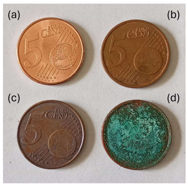
The first group consisted of current 5 euro cent coins, which have a steel core with copper plating (hence the reddish colour): the percentage of steel is 94.64% while that of copper is 5.36%. The electrodeposited copper cladding layer is very thin, typically about 5–10 μm [
20,
21].
Figure 1 shows a set of 5 euro cent coins, with natural patina, found regularly in circulation, with progressively increasing oxidation patina from (a) to (b), (c), and (d). The last green patina is due to both oxidation and chlorination processes, not induced artificially.
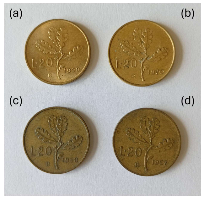
The second group consists of a set of 20 Italian lire coins (so-called “oak coins”) minted between 1957 and 1990, with a simple alloy composition known as Bronzital: copper ~92%, aluminum ~6%, and nickel ~2% [
22]. This alloy is relatively inexpensive, resistant to wear, and golden in colour. Over time, the original golden-yellow colour darkens to brownish or opaque hues due to the oxidation of the copper present in the alloy.
Figure 2 reports a set of such coins with a natural-anthropogenic oxidative patina growing over the years, from 1990 (a) to 1970 (b), 1958 (c), and 1957 (d).
The third group consisted of archeological bronze coins dating to the VI–VII Centuries A.D., from excavations in Alexandria and Antinopolis in Egypt, carried out by the Department of Ancient and Modern Civilization of Messina University (Italy), as previously analyzed for elemental composition and lead-isotope ratios [
23,
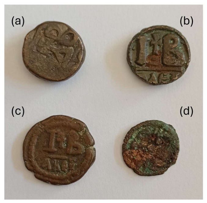
24]. These bronzes have a high Cu content and a thick oxide patina. Their surfaces also contain soil-derived compounds and secondary corrosion products resulting from long burial in the Egyptian environment. In this case, the surface natural-archeological patina is thick, and the coins appear dark brown, and sometimes irregular and porous, as shown in
Figure 3.
The only artificial process adopted was oxidation, applied to two new 5 euro cent coins and to pure Cu calibration samples in air, at 1 atm pressure, in a thermostated oven at 200 °C and 320 °C for different treatment times, namely 15, 30, 60, 90, and 120 min. The 5 euro cent coins were used to evaluate the oxidation behaviour of copper-plated coin surfaces, whereas the pure Cu samples were used as calibration samples for controlled oxide growth and for comparison with the XRF attenuation measurements. The thermally treated pure Cu samples were also used for XRD phase identification.
A Q-switched Nd:YAG laser (TRLi850, Litron Lasers Ltd., Rugby, UK) operating at 1064 nm, with a 3 ns pulse duration, 10–1000 mJ pulse energy, and 3.14 cm2 laser beam output area, was employed for laser ablation and laser cleaning tests. The beam was focused or defocused to obtain spot areas ranging from 0.5 mm2 to 10 cm2, corresponding to fluences from 1 mJ/cm2 to 200 J/cm2. At these fluences, the laser intensity ranges from 3.3 × 10⁵ W/cm2 to 6.67 × 10¹⁰ W/cm2, respectively. Focused irradiation was employed on pure Cu and Cu2O reference targets to determine the ablation yield as a function of laser fluence, and on selected patinated coins to produce micro-ablated craters for patina-thickness measurements. Defocused irradiation was used for cleaning tests on larger areas of 5 euro cent coins. For larger coin surfaces, a step-motor-controlled raster scan of the coin could be employed, while keeping the laser beam fixed.
Laser ablation was carried out both in vacuum, at a pressure of approximately 1 × 10⁻⁶ mbar, and in air at 20 °C, 1 atm pressure and 40% relative humidity. The crater volume generated by focused laser ablation was measured using a surface profiler (Tencor P-10, KLA-Tencor/KLA Corporation, Milpitas, CA, USA), swept across the crater diameter to determine crater shape and depth. The same profiler, with a depth resolution of 1 nm, was also used to estimate the thickness of selected coin patinas from the height difference between the ablated and non-ablated regions. Profilometric measurements were applied to selected naturally patinated coins, including 20 Italian lire coins and archeological bronze coins.
A quadrupole mass spectrometer (Prisma QMS 200, Pfeiffer Vacuum GmbH, Asslar, Germany), with a mass resolution lower than 1 amu, a secondary-electron multiplier (SEM) sensitivity of approximately 0.1 pA, and a detection mass range from 1 to 300 amu, was coupled to laser ablation in vacuum. This configuration was used to monitor Cu and O signals during progressive ablation and to obtain depth-dependent information on thin patina layers. These LAMQS measurements were applied to selected 5 euro cent coins with thin surface oxidation.
X-ray fluorescence (XRF) analysis was performed using an Ametek XRS-FP2 system (Amptek Inc., Bedford, MA, USA) equipped with a 5–50 kV compact X-ray tube (Amptek Inc., Bedford, MA, USA) and a PIN silicon detector (Amptek Inc., Bedford, MA, USA) operating in the 1–20 keV photon-energy range. XRF measurements were performed both in air and in vacuum. In general, XRF analyses on coins and pure Cu calibration samples were carried out using a 10 kV X-ray tube voltage and an 8 mA current. The technique was used to monitor the attenuation of the Cu kα and Cu kβ characteristic lines and to estimate the thickness of oxide layers formed during thermal oxidation. The untreated samples were analyzed before thermal treatment without additional surface polishing or other preparative treatments.
A scanning electron microscope (SEM) equipped with an energy-dispersive X-ray (EDX) system (CrossBeam 540 SEM-EDX, Carl Zeiss Microscopy Deutschland GmbH, Oberkochen, Germany)was employed at an electron beam energy of 20 keV. SEM-EDX analysis was used on selected samples to examine surface morphology and to obtain local compositional information after oxidation or laser treatment.
X-ray diffraction (XRD) measurements were performed on the thermally oxidized pure Cu calibration samples in order to identify the crystalline oxide phases formed under controlled oxidation conditions. XRD patterns were collected using a Bruker Advance diffractometer (D8 Advance, Bruker AXS GmbH, Karlsruhe, Germany) with monochromatic Cu kα radiation in Bragg-Brentano geometry. The analyses were mainly carried out on pure Cu samples oxidized at 200 °C and 320 °C, respectively, to verify the formation of Cu2O and CuO phases inferred from the XRF attenuation measurements.
3. Results and Discussion
The laser ablation of pure Cu and Cu
2O was measured by varying the laser pulse energy (i.e., the laser fluence, in J/cm
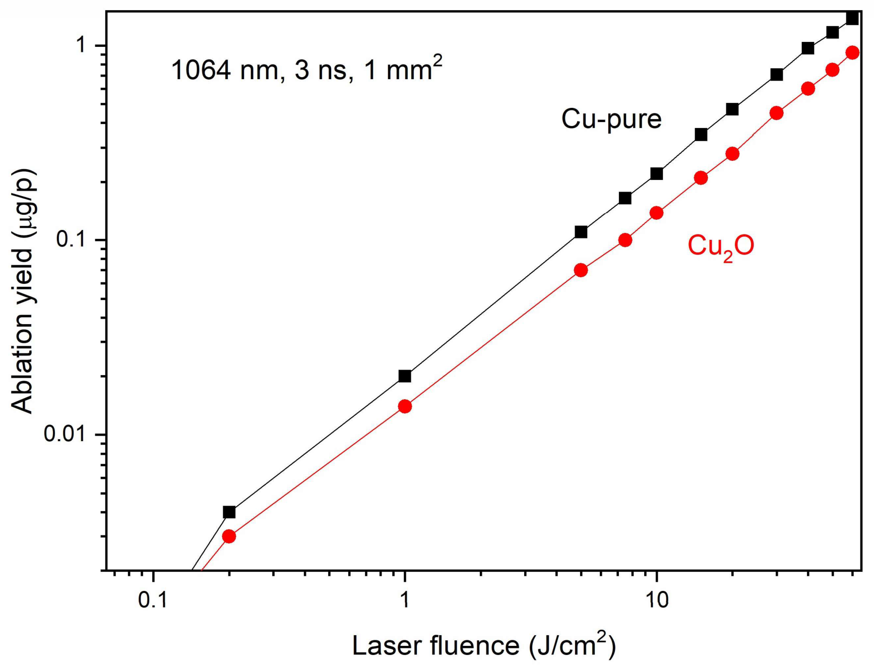
2) and measuring the mass removed in vacuum from the irradiated target surfaces. By changing the laser pulse energy from 50 mJ to 500 mJ and using a 1 mm
2 laser spot, the laser fluence ranges from 5 to 50 J/cm
2. For these values, the experimentally measured ablation yields for pure Cu and Cu
2O reference targets are reported in
Figure 4.
The yield is slightly higher for Cu with respect to Cu
2O. The experimental points are affected by errors of about 10%. The plot shows experimentally measured ablation yield values, whereas the threshold can be estimated from the thermal parameters of Cu and Cu
2O reported in
Table 1 for the pure metal and the oxide [
25,
26]. The threshold is comparable for the two materials and corresponds to about 0.15 J/cm
2. The threshold value is in agreement with the values that can be calculated by the photo-thermal model presented in the literature, mainly based on laser heating, latent heat of evaporation, diffusion length, and surface reflectivity [
26]. In a first approximation, such a fluence threshold can be calculated by the relation:
where
is the latent heat of evaporation,
is the mass density,
K is the thermal conductivity,
is the laser pulse duration, and
cs is the specific heat of the irradiated material.
There is no standard measurable value in the literature for the latent heat of evaporation of CuO because the material decomposes before evaporating as pure CuO.
The ablation yield tends to deviate from linearity at fluences above 50 J/cm
2 due to partial laser absorption in the generated plasma. The thickness of oxidized patinas on copper-based coins may vary widely depending on age, alloy composition, corrosion environment, and preservation conditions [
27,
28]. In the present work, the terms thin, medium, and thick are used only as an empirical descriptive scheme for the analyzed samples. The patina is considered thin when its thickness is approximately between 0.1 and 10 μm, medium for thicknesses of about 10–50 μm, and thick for higher thicknesses, which can reach up to a few hundred micrometres.
Assuming a Cu2O patina thickness of about 1 μm over a coin surface of 1 cm2, the mass to be removed during the laser cleaning process amounts to about 0.6 mg. For example, operating at a fluence of 10 J/cm2, at which the ablation yield is about 0.1 μg/pulse, the ablation yield is minimal, control of patina cleaning is optimal, and, when operating in single-pulse mode or at a low repetition rate, the cleaning procedure can be stopped as soon as necessary by using the minimum number of laser pulses, without damaging the coin surface imprint.
Under these conditions, the 1 μm -thick patina can be removed by about 6000 pulses, at a 10 Hz repetition rate, in a total time of 600 s, or 10 min. This is possible thanks to a focusing or defocusing lens, which generates a spot comparable to the surface of the coin to be cleaned. Of course, the above given value of mass removal is relative to a uniform oxide patina. Real patinas may be heterogeneous, stratified, porous, and composed of multiple corrosion products; this idealized estimation can have significant differences from those expected. Any practical calibration should therefore be regarded as sample-dependent, especially in the case of ancient and porous patinas.
3.1. Patina Layer-Ablated Substrate Interface Measurements
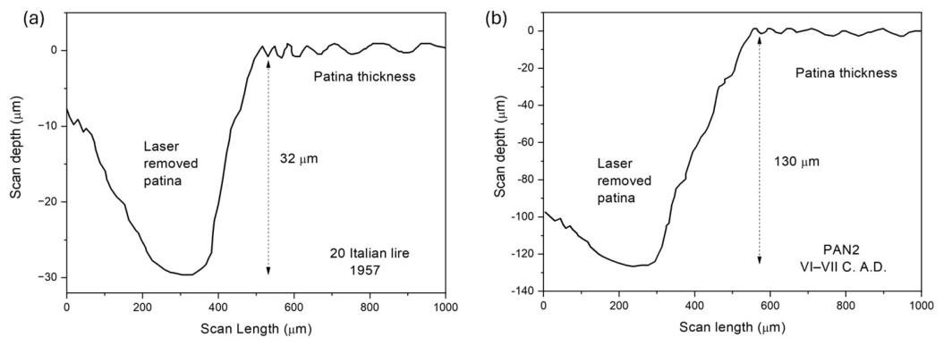
A laser beam ablation focused to only one or two hundred microns in diameter, sufficient to remove the surface patina layer, could be used to perform a subsequent surface profilometric analysis to measure the patina thickness directly from the depth of the produced crater in vacuum.
The patina laser ablation changes in colour when removed, and the substrate surface, if rich in copper, appears reddish in colour. In a vacuum, a CCD camera observes the colour at the bottom of the ablated crater so that the laser pulsed ablation, using a low repetition rate, can be immediately stopped just after the patina colour is changed.
Figure 5a shows an example of patina thickness measurement on a relatively old bronze coin dated 1957, the 20 Italian lire, as shown in
Figure 2a, where a natural patina thickness of about 32 microns was measured.
Figure 5b shows another example of an old bronze coin, namely the archeological one shown in
Figure 3a; it was subjected to a micro-invasive laser crater (about 200 microns in diameter) and subsequently analyzed using surface profilometry to plot the ablated region from the cleaned surface toward the patina surface, indicating a natural patina thickness of about 130 microns. As expected, the patina in the old bronze coin is very thick, while that in relatively recent years is less thick.
3.2. Patina Measurements Using the X-Ray Absorption Method
Using a stabilized ceramic oven in air, calibration samples of pure Cu (4 cm2 surface area and 1 mm thickness) and 5 euro cent coins were subjected to heating at 200 °C and 320 °C for different times, thereby generating surface oxidation and producing red Cu2O at the lower temperature and black CuO at the higher temperature.
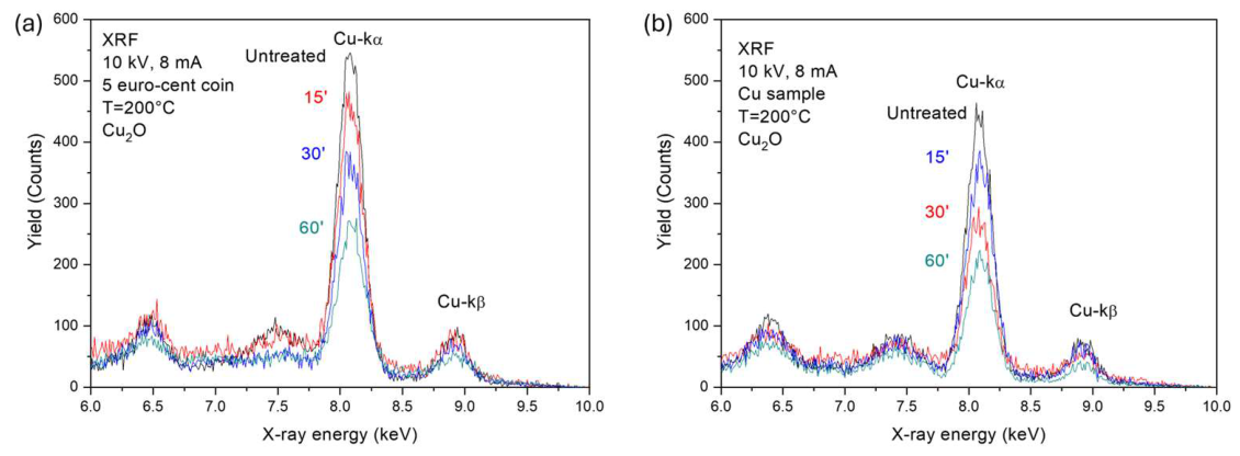
The oxidized copper surfaces were analyzed before and after each thermal treatment by XRF in air, mainly by monitoring the Cu-ka and -kb characteristic line intensities at 8.04 keV and 8.97 keV, respectively. These intensities depend on oxide layer thickness and composition, and decrease with thermal treatment time because the oxide layer becomes thicker. Naturally, the decrease differs at lower temperatures where Cu
2O is involved, and at higher temperatures where CuO is involved.
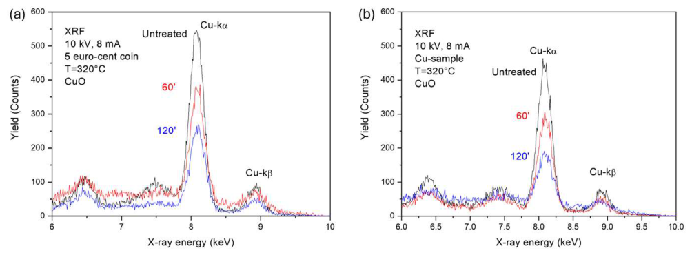
Figure 6 shows XRF spectra of untreated and thermally treated 5 euro cent coins (a) and pure Cu calibration samples (b).
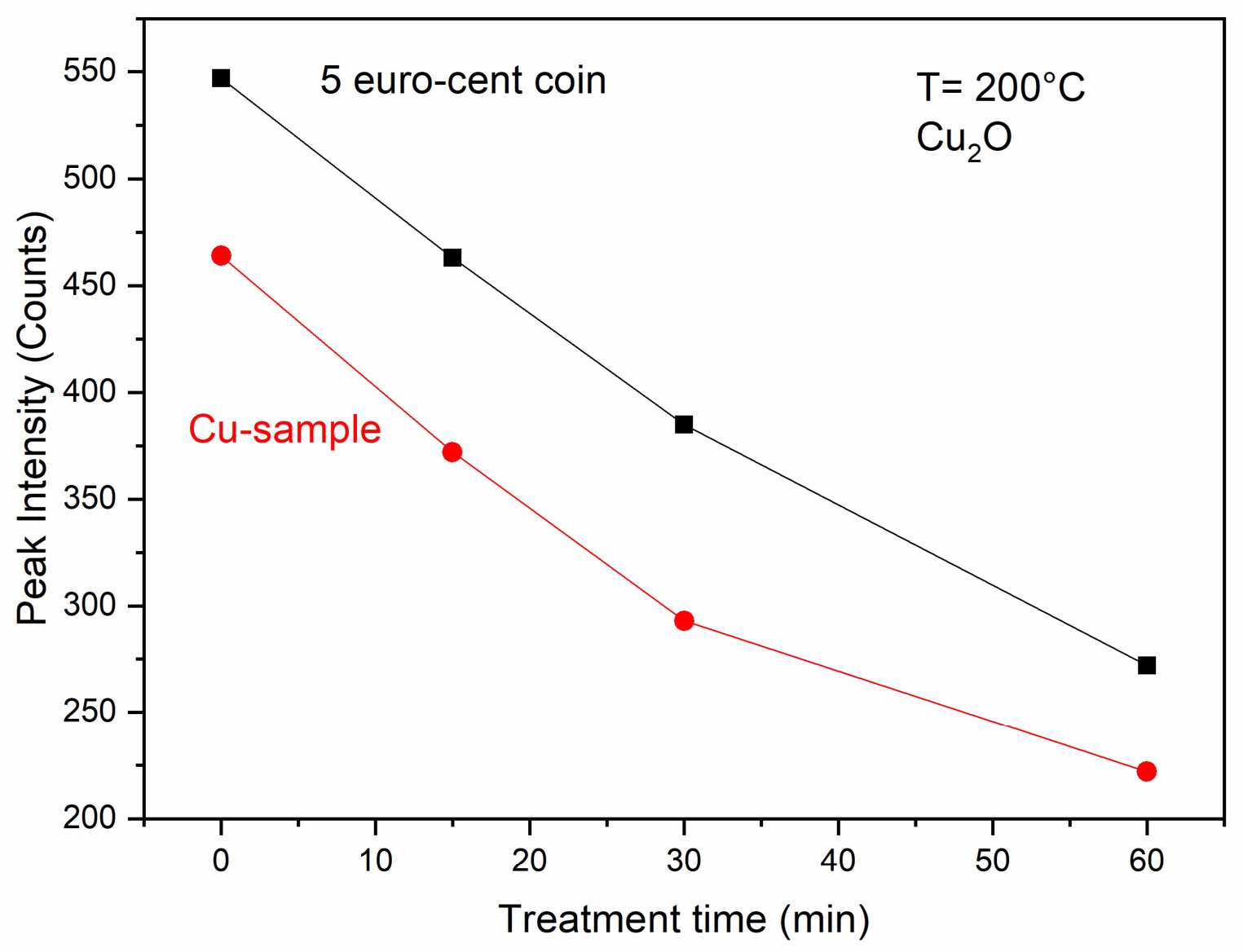
In both cases, the Cu- ka peak intensity decreases with treatment time, indicating that the Cu substrate becomes covered by a thicker Cu
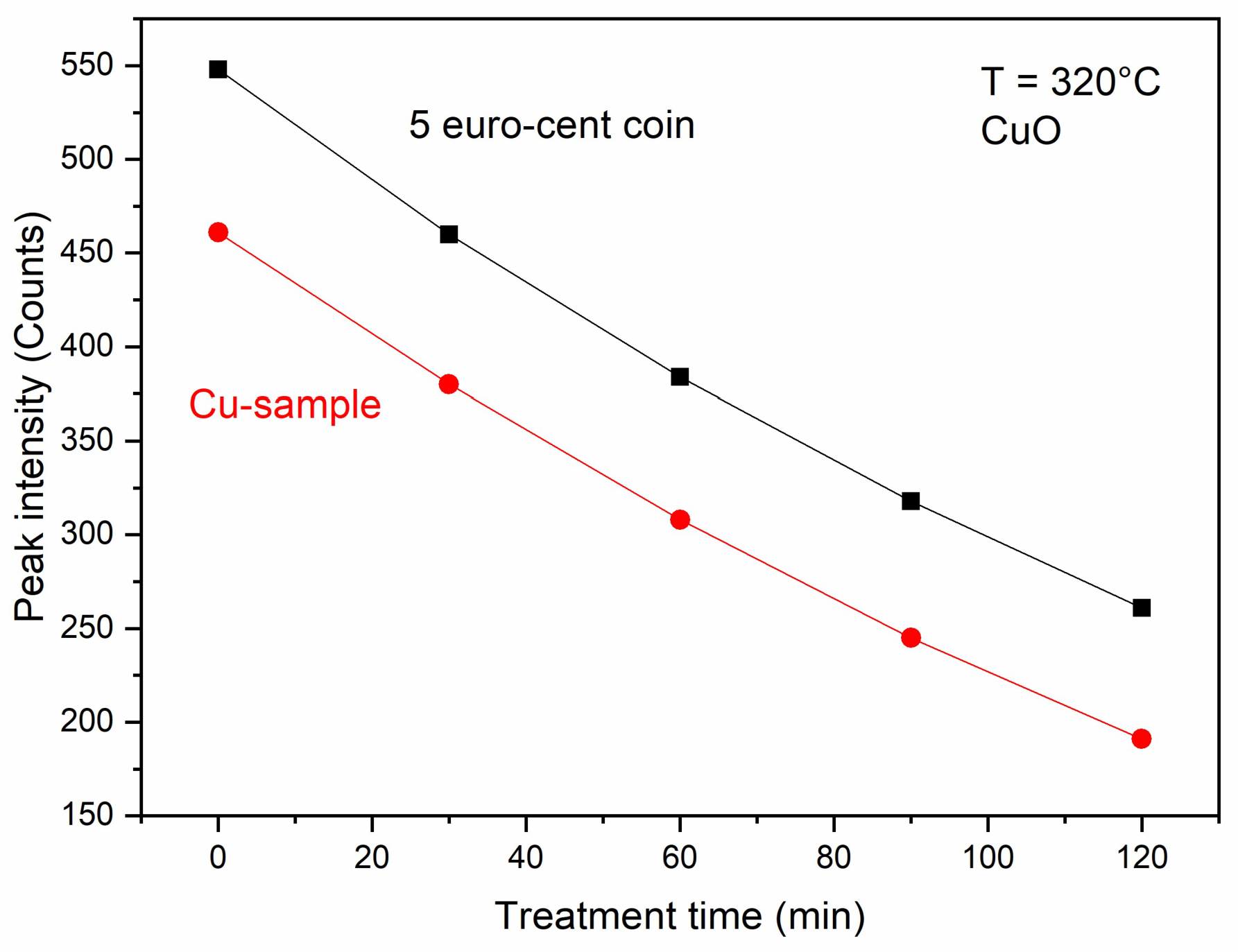
2O layer, and that the Cu X-ray intensity decreases because of the absorption in these layers. The rate of intensity decreases for the coin, and the sample is reported in the plots of
Figure 7.
The reduction in intensity (I
T/I
0) due to thermal treatment at 200 °C for 60 min is 50% for the 5 euro cent coin and 48% for the Cu calibration sample. Starting from the XCOM-NIST database [
29], the mass absorption coefficient for the Cu
2O target (density 6 g/cm
3) at an incident X-ray energy of 8 keV is 46.28 cm
2/g. It corresponds to a linear absorption coefficient μ of 277.7 cm
−1. From this reduction, using the absorption coefficient from the XCOM database, it is possible to calculate the thin Cu
2O thickness, Δx, after 1 h of heating in the two cases:
Thus, the Cu
2O oxide thickness grows with a rate of about 25 μm per hour at 200 °C in air at 1 atm. Therefore, the reduction in the Cu X-ray with respect to the uncovered Cu surface can be used to estimate the patina thickness. Of course, the line attenuation, the absorption coefficient, and the thicknesses of the CuO layers obtained at higher temperatures differ from the previous case reported. Above 300 °C, CuO can be grown on a Cu substrate. This was done in an air oven at 320 °C on 5 euro cent coins and calibration Cu-samples. Also in this case, the XRF analysis was performed on different treated samples before and just after each treatment, as shown in
Figure 8a and
Figure 8b, respectively. Again, the Cu-kα peak decreases with thermal treatment time for both samples because of X-ray absorption in the CuO oxide layer.
The rate of intensity decreases for the coin, and the sample is reported in the plots of
Figure 9.
The reduction in intensity (I
T/I
0) due to thermal treatment at 320 °C in 60 min is about 70% for the 5 euro cent coin and 66.7% for the Cu calibration sample. According to the XCOM-NIST database, the mass absorption coefficient for CuO (density 6.4 g/cm
3) at an incident X-ray energy of 8 keV is 42.76 cm
2/g [
29]. This corresponds to a linear absorption coefficient μ of 273.7 cm
−1. From this reduction, using the absorption coefficient from the XCOM database, it is possible to calculate the CuO thickness, Δx, after 1 h of heating in the two cases:
Thus, the CuO oxide layer grows at a rate of about 14 μm per hour at 320 °C in air at 1 atm. At higher temperatures, oxygen diffusion in the copper substrate increases, promoting the formation of oxides richer in oxygen than those formed at lower temperatures.
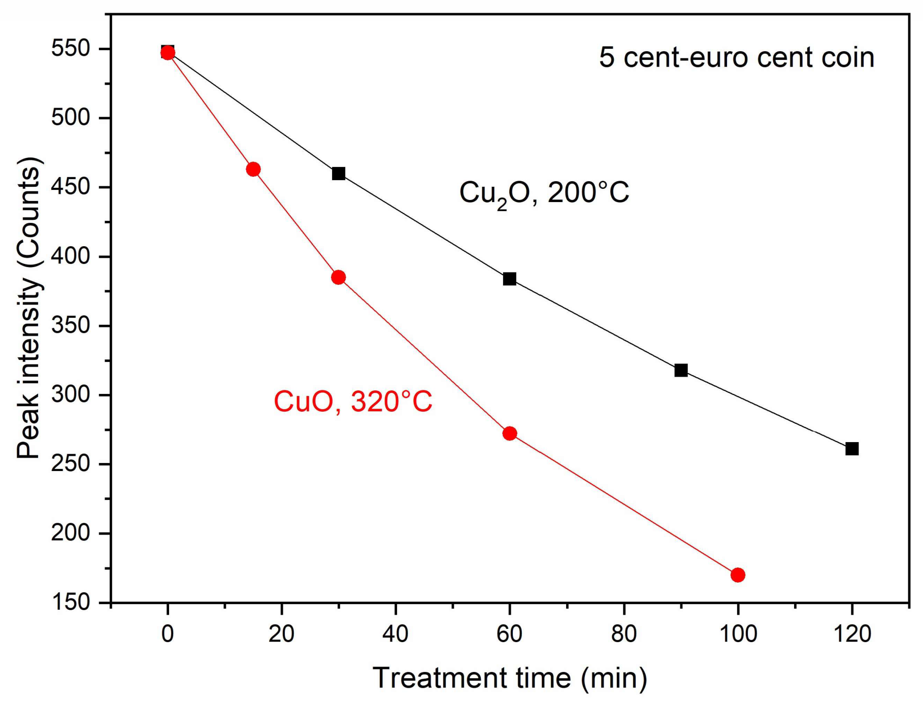
Figure 10 compares the peak intensity reduction as a function of thermal treatment at 200 °C and 320 °C for the formation of the two oxides, Cu
2O and CuO, respectively. The results indicate that the peak intensity decreases exponentially with treatment time; when plotted against the square root of time, it shows an approximately linear trend, consistent with diffusion-controlled kinetics and the expected parabolic time dependence of oxide growth.
Therefore, the reduction in the Cu X-ray intensity with respect to the uncovered Cu surface can be used to measure patina thickness. This thin-film measurement technique, based on X-ray attenuation, is in agreement with the literature data [
30]. However, because the present approach is mainly based on the attenuation of the Cu kα line, the obtained thickness values should be regarded as first-order, semi-quantitative estimates. A more rigorous determination of heterogeneous or multilayer patinas would require additional information, such as Cu kα/kβ fluorescence line ratios and/or Monte Carlo modelling of X-ray transport in stratified corrosion layers.
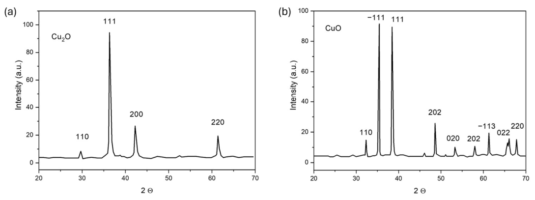
The XRD analysis performed on the thermally oxidized pure Cu calibration samples confirmed the oxide phases inferred from the XRF attenuation measurements. The diffraction pattern obtained after thermal treatment at 200 °C shows features consistent with the formation of Cu
2O (
Figure 11a), whereas the pattern obtained after treatment at 320 °C is consistent with the formation of CuO (
Figure 11b). These assignments agree with literature data on the structural characterization of Cu
2O and CuO phases [
31,
32,
33]. Therefore, the XRD results support the interpretation that the XRF attenuation observed after thermal treatment is associated with the growth of copper oxide layers of different composition, depending on the oxidation temperature.
These results help to clarify the effects of laser cleaning on copper-oxide surfaces. Laser cleaning removes oxide layers and impurities, but it also generates heating and, if prolonged in air, may itself promote oxidation. Therefore, whenever possible, cleaning should be performed in a vacuum or in inert gases.
Cleaning can be applied to small areas, of the order of 1 mm
2 or less, by means of a focusing lens in order to control patina thickness or the composition of the ablated material and to be as minimally invasive as possible, especially for coins of high cultural value. In some cases, cleaning may involve larger areas of coins or artefacts of artistic and/or historical importance. In such cases, the laser beam can be defocused by appropriate lenses, while always aiming to remove layers as thin as possible so as not to alter the morphology of the underlying layers.
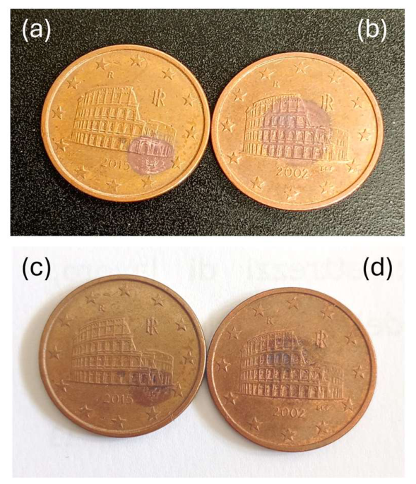
Figure 12 shows photographs of 5 euro cent coins just after laser cleaning in a vacuum over a limited circular area of 5 mm diameter (a) and 8 mm diameter (b). The cleaning process, obtained using a single pulse at 500 mJ, produces Cu
2O, as indicated by the red irradiated areas. This light oxidation is due to laser heating and to the residual oxygen concentration in the vacuum chamber. After exposure to air, the coins undergo renewed oxidation and, after 5 days, they appear as in
Figure 12c,d, showing a dark area, probably due to further oxygen diffusion and formation of a CuO patina. After one month, the contrast between the cleaned and untreated areas nearly disappears.
The results shown in
Figure 12 indicate that laser-cleaned copper surfaces may undergo reoxidation after exposure to air. This aspect is relevant from a conservation perspective because laser cleaning can expose fresh and reactive metallic or oxide-modified surfaces. Therefore, when laser cleaning is considered for copper-based cultural heritage materials, the possible need for post-treatment stabilization should also be evaluated. Protective approaches based on graphene, boron nitride, conductive polymers, or corrosion-inhibiting compounds such as azoles have been proposed in the literature for copper and copper-based cultural heritage materials [
34]. These approaches were not tested in the present work and are mentioned here only as possible future developments following laser cleaning or analytical micro-ablation.
From a conservation perspective, the removal of patina layers should not be considered an automatic objective. Stable and adherent patinas may have protective, historical, and documentary value, whereas unstable or active corrosion products may require selective intervention. The characterization of corrosion layers formed on buried copper-based coins is therefore essential for distinguishing between stable patinas and degradation products that may compromise the conservation of the object [
35]. For this reason, the laser-based procedures investigated here should be regarded primarily as analytical and micro-invasive tools for patina characterization and, only in selected cases, as possible cleaning approaches for the controlled removal of harmful or degraded surface layers.
These considerations also confirm that laser cleaning of copper-based coins should be considered a highly controlled and case-specific procedure rather than a routine conservation treatment. The laser parameters, irradiation environment, number of pulses, and surface response must be carefully evaluated in order to avoid excessive removal, thermal alteration, or modification of historically relevant patina layers. This interpretation is consistent with previous theoretical and experimental studies on laser cleaning of coins [
36].
3.3. Patina Measurements Using the LAMQS Technique
Laser cleaning in high vacuum (1 × 10
−6 mbar) can be coupled with mass quadrupole spectrometry (MQS) in order to measure the masses removed by ablation and establish their concentration as a function of ablation depth [
15,
23,
24]. In this regard, preliminary measurements of Cu and O were considered.
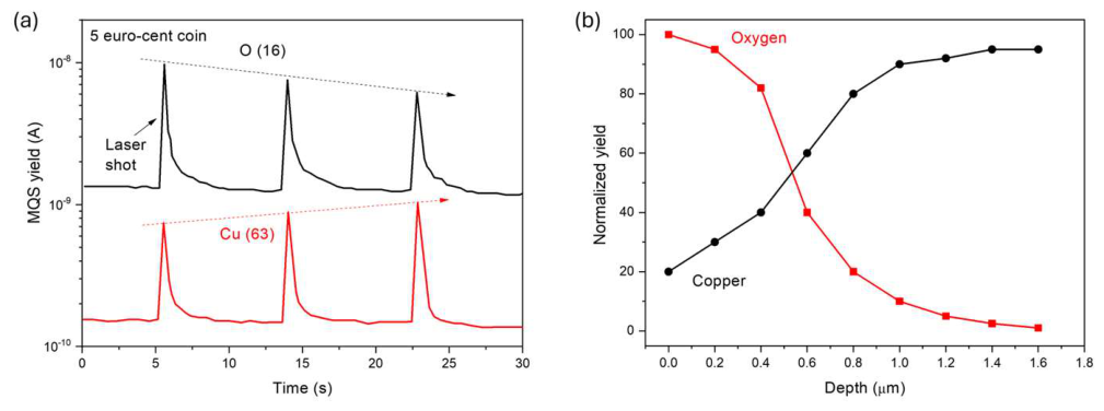
Taking into account the atomic abundance of the stable species, the mass of Cu was taken as 63 amu (abundance 69.15%), while that of O was taken as 16 amu (abundance 99.77%). A single laser shot, 3 ns in duration, at 300 mJ, repeated every 9 s, in vacuum on a new 5 euro cent surface focused over an area of 0.5 mm
2, produces the MQS signals reported in
Figure 13a. It can be observed that the oxygen signal decreases while the Cu signal increases with the number of laser shots. This result indicates that the laser first ablates the superficial CuO layer and then the successive Cu
2O patina layer, approaching the underlying metallic Cu-rich region of the surface layer of the coin, with a clear reduction in oxygen content and an increase in Cu atoms, in agreement with the literature [
8].
For longer ablation under these conditions, with an ablation rate of about 200 nm/pulse, and by irradiating a coin surface with a very thin surface oxide, it was possible to plot the oxygen and copper depth profiles in the coin patina, as shown in
Figure 13b. From the relative O and Cu yields versus patina depth, the results indicate that the thin patina of the new coin has a thickness of about 1.5 μm. This value should be regarded as an indirect estimate derived from the LAMQS depth profiles and the corresponding ablation rate under the adopted experimental conditions. This very low patina thickness derives from the choice of a very new coin, recently minted and circulated for a short time, for which a low patina thickness was expected. No independent cross-sectional SEM/EDX validation was performed for this specific sample.
4. Conclusions
The study investigated the use of laser ablation and complementary analytical methods for the characterization and controlled removal of patina layers on copper-based coins and pure Cu calibration samples. The results show that laser ablation can be used as a micro-invasive approach to evaluate patina thickness when the irradiation conditions are carefully controlled.
Three complementary approaches were considered. Surface profilometry of laser-ablated areas allowed direct estimation of patina thickness from the depth difference between the cleaned crater and the surrounding surface. XRF attenuation of the Cu kα line provided semi-quantitative estimates of oxide-layer thickness on thermally treated 5 euro cent coins and pure Cu calibration samples. LAMQS measurements allowed the evolution of Cu and O signals to be followed as a function of ablation depth, providing additional information on the stratigraphy of thin oxidized layers.
The XRF and XRD results indicate that thermal treatment at 200 °C mainly promotes the formation of Cu2O, whereas treatment at 320 °C favours the formation of CuO. These controlled oxidation experiments provide useful reference conditions for interpreting the response of copper-based surfaces during laser cleaning and patina thickness assessment.
The laser cleaning tests also showed that cleaned copper-based surfaces may undergo reoxidation after exposure to air. Therefore, although laser cleaning can be useful for analytical purposes and for the selective removal of unstable or unwanted surface layers, it should not be considered a routine treatment for cultural heritage objects. Its application should be limited to carefully evaluated, case-specific situations, with particular attention to laser fluence, number of pulses, irradiation environment, and the conservation value of the patina.
Overall, the results suggest that laser ablation, when combined with profilometry, XRF, XRD, and mass spectrometry, can contribute to the characterization of copper-based patinas and to the development of controlled, minimally invasive cleaning strategies for conservation science.
The observed reoxidation of laser-cleaned copper surfaces also indicates that future work should address preservation strategies for oxide-cleaned copper surfaces, including thin protective layers aimed at inhibiting copper corrosion in air. From this perspective, the procedure presented here may be regarded as a first step toward a more complete methodology for the analysis, restoration, and conservation of copper-based materials.