


Abstract
Subadult stress is an important bioarchaeological indicator of the health status of archaeological populations, and its interpretation requires consideration of biological, environmental, and social factors. This paper examines the impact of cribra orbitalia (CO) and linear enamel hypoplasia (LEH) on adult mortality in Late Antique Southern Pannonia. A sample of 400 adult individuals from the sites of Mursa (Osijek), Cibalae (Vinkovci), Certissa (Štrbinci), and Incerum (Tekić) was analyzed. The results show that CO has a statistically significant negative impact on age-at-death in both sexes, whereas LEH shows a non-significant to weak impact with a statistically non-significant trend. The cumulative effect of multiple indicators of subadult stress could particularly negatively affect females, which is associated with reproductive burden and socio-cultural factors. The results confirm the complex interaction between environment and humans—the importance of living conditions and health stressors on the health and mortality of individuals and the entire observed population—at the same time in the analyzed sample.
1. Introduction
Since the second half of the twentieth century, bioarchaeologists and biological anthropologists have shown increasing interest in living conditions and child health at archaeological sites. Bioarchaeological research highlights the importance of health indicators in reconstructing the living conditions of archaeological populations. Lifestyle changes, particularly the transition to sedentary agricultural communities, led to reduced dietary diversity, increased infectious diseases, and increased physiological stress [1,2]. A more detailed examination of subadult stress and its impact on adult mortality shows that it is a complex phenomenon influenced by a range of biological, environmental, and social factors. Living conditions, nutrition, exposure to disease, hygienic standards, sex-related differences in care, and the reproductive role of women affected the health and survival of individuals.
Subadult stress leaves permanent traces on human skeletal remains and dental material, most often manifesting as cribra orbitalia and linear enamel hypoplasia [2,3,4,5,6,7,8].
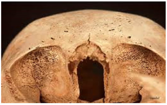
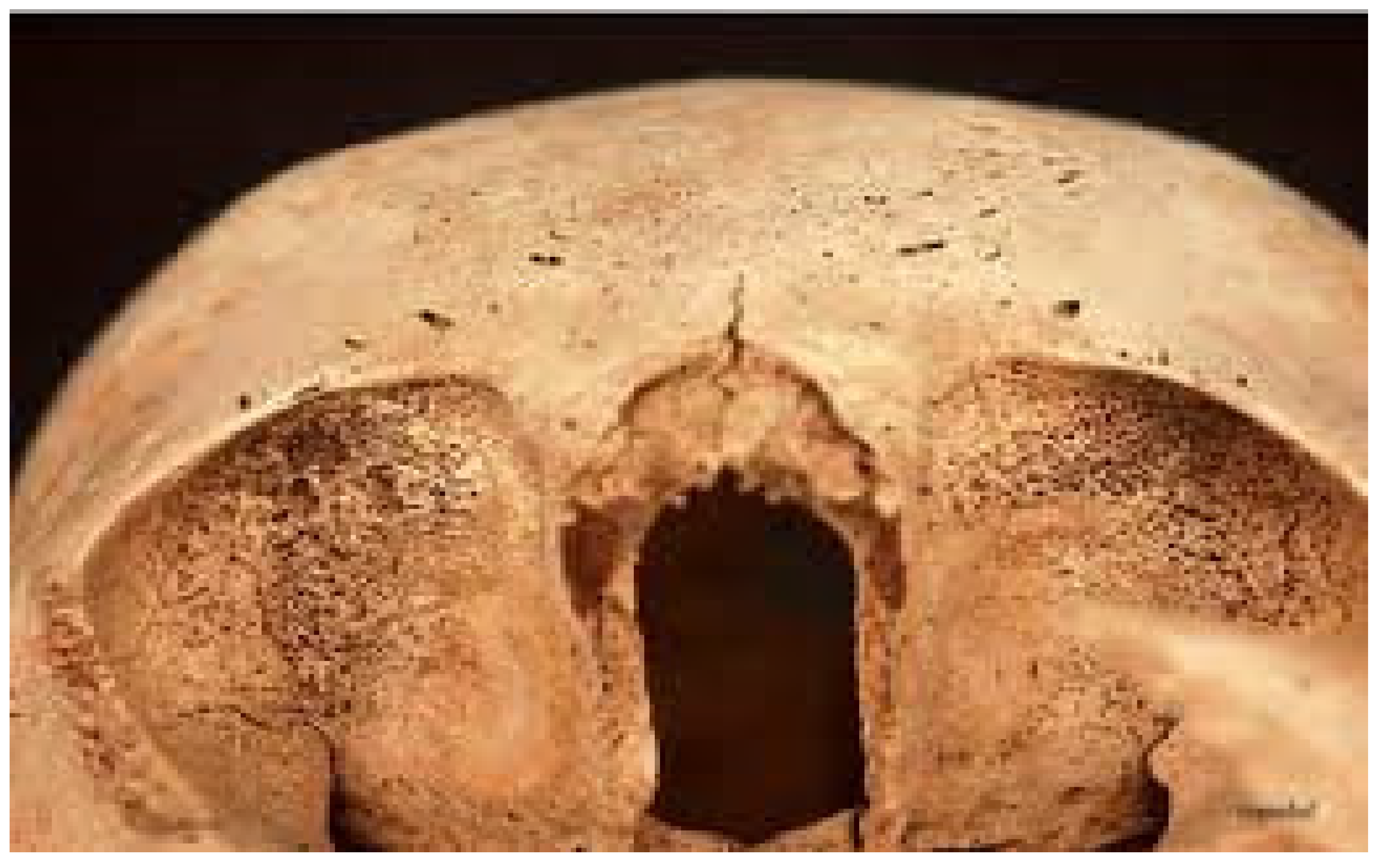
Cribra orbitalia (Figure 1) is associated with anemia and unfavorable living conditions [9,10,11,12,13,14,15,16,17], although its etiology remains debated, including possible links to malaria and environmental factors [18,19,20,21]. Cribra orbitalia develops due to hypertrophy of the diploë (the central porous part of cranial bone), which leads to thinning and destruction of the outer cortical layer and the formation of porous bone in the area of the outer cortex. Macroscopically, CO is characterized by small perforated lesions on the orbital roofs, ranging from less than 1 mm in diameter to larger openings that may partially merge [22]. It may be observed in an active or healed stage. Data collected from human skeletal remains from various archaeological sites show that active CO most often occurs in children, while in adults it is almost always in a healed form [16,23]. Such a distribution of CO is interpreted as a consequence of childhood anemia, whereas healed CO in adults represents exclusively a sign of recovered childhood anemia [16].
Figure 1.
Severe active form of cribra orbitalia [24]. Foto by Vyroubal V. Photograph reproduced with permission of the author.
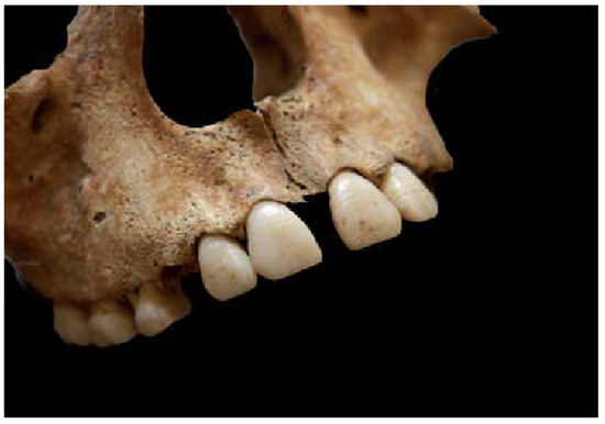
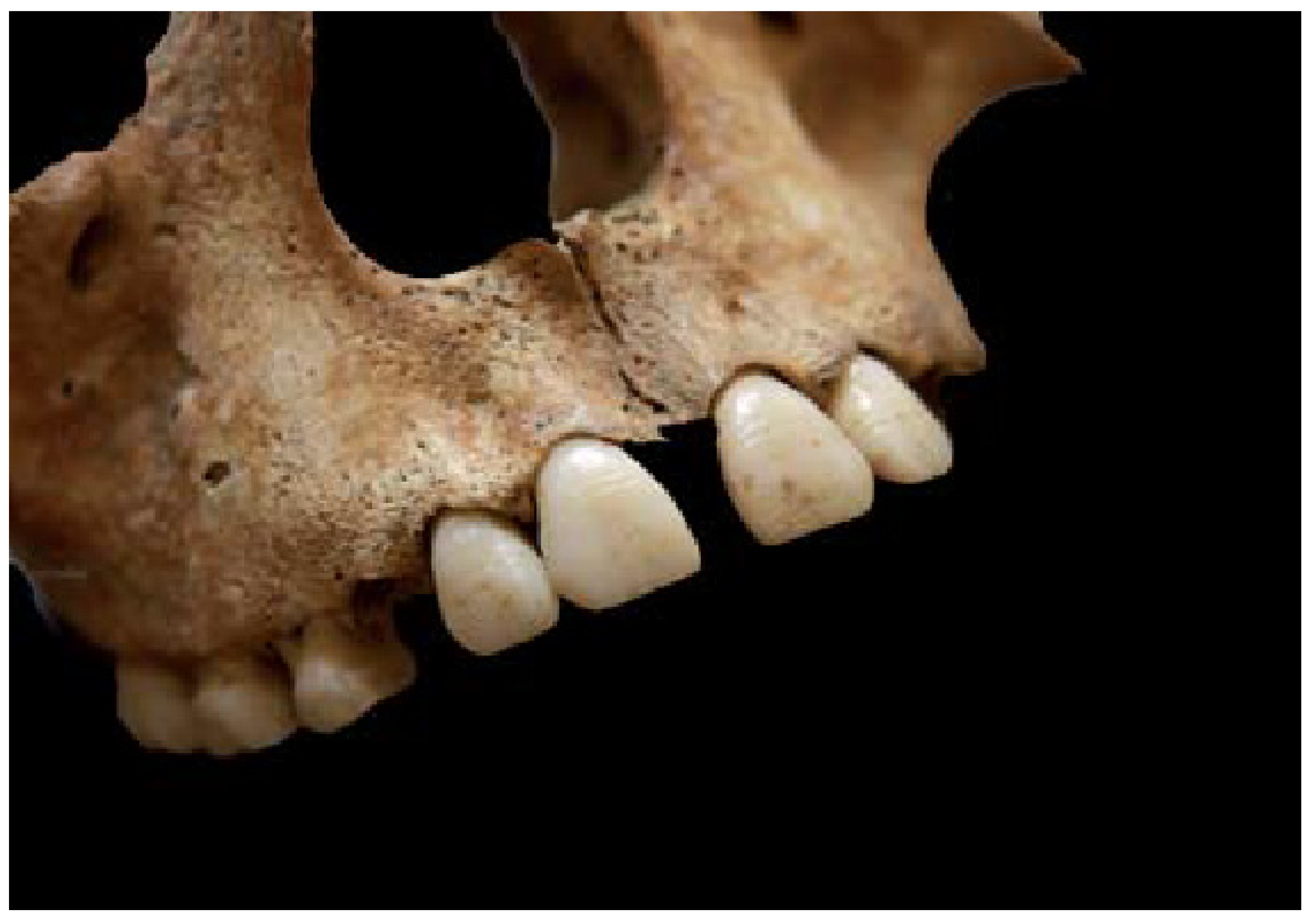
Linear enamel hypoplasia (LEH) (Figure 2) represents a marker of episodic stress during tooth growth and development [25,26,27]. LEH is recognized as a macroscopic defect on the enamel surface [26,27]. This indicator of subadult disturbance arises due to acute, time-limited stress. It is most often associated with starvation, elevated body temperature, deficiency of vitamins A, C, or D, the presence of anemia, and psychological and/or physical trauma [28,29,30,31,32,33].
Figure 2.
Linear enamel hypoplasia (LEH)—horizontal, parallel hypoplastic defects are clearly visible on the crowns of the maxillary incisors and canines. [24] Foto by Vyroubal V. Photograph reproduced with permission of the author.
The main characteristic of LEH is insufficient enamel thickness, and it appears in two forms—most commonly as a series of thin parallel lines on the labial side of the tooth or, more rarely, as shallow pits on the enamel surface. It is a reliable indicator of nonspecific stress during childhood, from birth until approximately age 13, during the period of enamel formation [25]. Enamel begins forming at the crown tip and continues to be deposited evenly until the entire crown is covered, i.e., the crown-root junction. Because enamel, unlike bone, has no remodeling capacity, developmental disturbances remain recorded until the affected crown portion is destroyed by dental wear [25].
For reasons that are still insufficiently clarified, hypoplastic defects most frequently occur on anterior teeth, specifically incisors and canines [25].
During the Roman period, the province of Pannonia encompassed the courses of the Danube, Drava, Mura and Una rivers, that is, western Hungary and Styria to the north; to the west it included Kranjska Gora and the western parts of the territory of present-day Croatia—from Risnjak and Kapela to the Mura and Drava rivers—and to the east the whole of Slavonia, Syrmia and Bosnian Posavina up to the Una River in the south [34,35].
The area of Southern Pannonia during Late Antiquity was characterized by political instability, wars, and changing living conditions [34], which may have negatively affected the population’s health.
Therefore, this study aimed to examine the influence of everyday life on subadult stress and its impact on adult mortality.
Earlier studies similar to this one, conducted in Croatia and abroad, have shown that the presence of one or both manifestations of subadult stress (CO and LEH) clearly correlates with a shorter lifespan in adult individuals within the studied populations. The specific aims of the research are to analyze the frequency and distribution of two indicators of subadult stress—CO and LEH—in adult individuals, to determine whether a relationship exists between them, and to demonstrate that the presence of CO and LEH negatively correlates with age-at-death; in other words, that the lifespan of individuals exhibiting these indicators of subadult stress is significantly shorter than that of individuals who do not show these changes.
2. Materials and Methods
A retrospective cohort bioanthropological study was conducted on human skeletal remains from four Late Antique sites in continental Croatia. The human skeletal remains derive from long-term excavations at several locations in present-day continental Croatia: Osijek (Mursa), Vinkovci (Cibalae), Štrbinci (Certissa), and Tekić (Incrum).
2.1. Osijek (Mursa)
The Roman settlement in the area of present-day Osijek—Mursa—was located in the Donji Grad district, on the right bank of the Drava River, near a former Celtic-Pannonian settlement of the Andizetes tribe. The development of Mursa into an urban center and its further construction are attributed to Emperor Hadrian, who granted it colony status in AD 133, after which it became Colonia Aelia Mursa [36].
The development and urbanization of Mursa continued until AD 378, when the Western Goths devastated that part of the Roman Empire, after which Mursa ceased to develop as an urban center. After the Hun incursions into the Syrmian region in AD 441, that is, into the area between the Drava, Sava, and Danube rivers, Mursa was devastated and never regained its former splendor and status [37]. Because of its position at the crossroads of road routes (Emona, Sirmium, Aquincum) and the proximity to the navigable Danube route, Mursa was one of the more important trade and transport centers of the Roman Empire. For anthropological analysis, 75 skeletons from the Osijek site were available, with preservation ranging from good to excellent.
2.2. Vinkovci (Cibalae)
The Roman settlement in the area of the town of Vinkovci was built along the left bank of the Bosut River and developed around a prehistoric settlement at the former Market area. The development of Cibalae began after the Roman conquest of Pannonia, and it received colony status—Colonia Aurelia Cibalae—during the reign of Emperor Caracalla [38] at the beginning of the 3rd century; until the first half of the 6th century, when the name Cibalae is last mentioned in sources, it was a very important trade and transport center of the Roman Empire (proximity of the water route). South of the settlement of Cibalae lie the remains of a large, well-fortified military camp. The necropolises were located at the exits from the military camp. The best researched is the northern necropolis along the road toward Mursa and Sotin (Cornacum). The most common burial form was elite brick-built tombs with flat roofs or gabled roofs. Several stone sarcophagi were found, while wooden coffins were used for burials in earthen graves, and in several cases, lead coffins were also used [38].
Material from Vinkovci originates from all four necropolises of Cibalae—the northern (the largest number of skeletons), the southern (only four skeletons), the eastern (three skeletons), and one additional area with only one skeleton. However, the earlier literature reports a high density of graves at that location [38]. For anthropological analysis, 186 skeletons from the Vinkovci site were available, whose preservation ranged from poor to excellent.
2.3. Štrbinci (Certissa)
Štrbinci represent a natural unit consisting of two low hills and a plateau [39], approximately 3 km southeast of the town of Đakovo. Štrbinci is a site believed to have hosted a Roman settlement named Certissa. The existence of Certissa is attested in Roman sources dating from the 2nd to the 7th century, although its exact location is not known with certainty.
Excavations were conducted in that area over several decades, yielding large quantities of antique archaeological material, such as various objects made of metal, glass, bone, and ceramics, as well as construction bricks, pottery kilns, plaster, mosaic tesserae, fragments of frescoes, together with parts of civilian and military equipment, jewelry, fibulae, and coins [39]. All burials were skeletal inhumations, and the graves can be divided into brick-built tombs, graves dug directly into the soil, and burials in wooden coffins, as evidenced by numerous nails with wood remains. For anthropological analysis, 120 skeletons from the Štrbinci site were available, with preservation ranging from good to excellent.
2.4. Tekić (Incerum)
Tekić is a settlement located approximately 12 km northeast of Požega. North of Tekić lies Treštanovačka Gradina, an archaeological site inhabited from the 2nd to the 4th century AD [40]. Some authors believe that Incerum, a town mentioned in the Antonine Itinerary, was located there [40].
Treštanovačka Gradina was already known as an archaeological site in the 18th century. Later investigations uncovered 124 graves containing human osteological remains, and it was established that the necropolis belonged to the aforementioned Roman settlement and dated to Late Antiquity.
The human skeletal remains used for analysis originate from archaeological investigations conducted in 1972, 1975, 2004, and 2005. Nineteen skeletons were available for anthropological analysis, whose preservation varied from very poor to excellent. The graves were oriented west–east with the heads facing west. Most graves contained the remains of a single individual, but double burials were also recorded.
2.5. Bioanthropological Analyses
All the data used in this study are part of the laboratory collection of the Anthropological Center at the Croatian Academy of Sciences and Arts. The anthropological analyses used in the laboratory are previously established, simple, generally accepted, and comparable with other databases. After washing and drying, the skeletal remains are recorded on a form containing a schematic representation of the human skeleton in anatomical position. An inventory of each bone, joint, and tooth is recorded on specially designed forms. All present or missing bones are noted, and degenerative, pathological, or trauma-caused changes are also recorded. At the end, all completely preserved bones and skulls are measured.
The analyzed sample included only individuals aged 15 or older with preserved orbital roofs and at least one tooth suitable for analysis of linear enamel hypoplasia. Sex was determined based on basic morphological differences present in the skeletons of males and females. The most important and most evident differences are found in the pelvic girdle [41,42,43,44,45,46] and were used whenever possible. In cases where the pelvis was not preserved, other cranial and postcranial morphological differences were used [47].
The biological age of human skeletal remains is determined based on the growth, development, or degeneration of various parts of the skeleton [43,44,45,46,47]. In adults, the primary and most commonly analyzed features for estimating age include the morphology of the pubic symphysis [44,47,48,49,50], the auricular surface of the ilium [51], and morphological changes at the sternal end of the ribs [52,53]. The degree of fusion of cranial and maxillary sutures [54], degenerative osteoarthritic changes in the joints of long bones [44,47,55], and the degree of wear on the occlusal surfaces of the teeth [44,56] are used as additional, secondary criteria. The amount of cortical bone and the density of trabecular bone are also taken into account [44]. All these changes occur mostly within predictable periods and are analyzed in five-year intervals [44,47].
Cribra orbitalia was recorded in individuals with at least one preserved orbit, by macroscopically determining under strong light the presence or absence of this pathology, its active or healed form, and the degree of severity (mild, moderate, severe) according to the criteria proposed by Mittler and Van Gerven [11].
In the analyzed material, LEH was recorded on the upper jaw incisors and canines of both jaws. This is because central incisors and canines are more susceptible to hypoplastic defects than other teeth [27], canines develop over a relatively long period (from the fourth month to the sixth year of life [56], and incisors and canines have the least amount of calculus (mineralized dental plaque), which can sometimes cover the crown and prevent the determination of hypoplasia. Only macroscopically visible linear hypoplastic defects were considered, and for this study, the presence of LEH was recorded if at least one tooth per individual was affected.
2.6. Statistical Methods
Nonparametric statistical analyses were used to determine the statistical significance of differences in the obtained results. The Kruskal–Wallis test was applied to demonstrate differences in the average age-at-death between males and females, and to demonstrate differences in the frequency of CO and LEH, the χ2 test was used, with Yates’ correction applied when necessary. Statistical significance was set at p < 0.05.
2.7. Previous Research on Subadult Stress in Southern Pannonia
Life in Southern Pannonia during the fourth century was turbulent and demanding due to frequent wars and incursions of barbarian groups into the area of present-day Slavonia [34,35]. An increasing number of studies address subadult stress among adults across different historical periods in Croatia, ranging from the Late Iron Age to the Middle Ages [57,58,59,60,61,62], and from other parts of Europe [18,63,64].
Studies conducted by Mario Šlaus and collaborators sought to confirm historical interpretations of the difficult living conditions in continental Croatia during Late Antiquity. By comparing samples from continental Croatia—Osijek, Vinkovci, Štrbinci and Zmajevac—with a coastal sample from Zadar [59], and with samples from urban settlements in the hinterland of the limes (Osijek, Vinkovci, Štrbinci) and a rural settlement located directly along the Danube section of the limes [58], they obtained very similar results that do not support historical claims of a particularly turbulent and violent period along the frontier during Late Antiquity.
Comparison between the limes sample and the hinterland sample also produced similar results regarding dental diseases and subadult stress. In contrast, physical stress (vertebral osteoarthritis and Schmorl’s defects) was more pronounced among males from Zmajevac, located directly on the limes, than among individuals from urban centers in the hinterland [57].
Although the results from the previously mentioned samples are similar and do not fully confirm historical sources suggesting substantially harsher living conditions in continental Croatia along the limes compared to other parts of Croatia, the period of decline of the Western Roman Empire undoubtedly influenced the quality of life and health status of populations living during Late Antiquity.
Comparison of Late Antique samples from Štrbinci and Zadar yielded similar results, indicating no significant difference in the quality of life among individuals in the observed samples [57].
As previously mentioned, subadult stress and its manifestations on human skeletal remains—cribra orbitalia, linear enamel hypoplasia, and analyzed pathological changes such as periostitis resulting from non-specific infectious diseases—represent clear indicators of difficult living conditions and inadequate nutrition.
Previous research on the necropolis in Tekić [65] showed a high frequency of subadult stress in the analyzed sample, indicating high levels of physiological stress and frequent infectious diseases.
Living conditions and health status of Late Antique populations in Slavonia at the sites discussed in this study have already been extensively investigated by numerous authors. Various diseases [66] and the influence of subadult stress at sites in Osijek and along the limes [57,66], in Štrbinci [24,67,68], Vinkovci [24], and Tekić [65] have been described by many Croatian researchers under the leadership of Mario Šlaus.
3. Results
The overall demographic structure, including 400 adult individuals (Table 1), shows a higher proportion of males (60.3%) than females (39.7%), a pattern common in osteological populations and possibly reflecting differences in bone preservation or funerary practices. The mean age-at-death was 37.38 years for males and 36.46 years for females, indicating relatively similar mortality patterns between the sexes.
Table 1.
Number and age-at-death of all adult individuals in the entire observed sample and at individual sites.
The difference in age-at-death between males and females is not statistically significant (p = 0.199; χ2 = 1.648).
The distribution of mortality in the entire sample, observed by five-year intervals, shows the highest mortality in the aforementioned period, with the largest number of males, 48/191 (21.5%), dying in the five-year interval between 36 and 40 years of age.
If we look at individual sites (Table 2), the average age-at-death for males (35–39) and females (34–40) is between 34 and 41 years, which correlates with the predictable period of higher mortality of males and females in general and in the whole sample.
Table 2.
Demographic composition of the sample per archaeological site.
In the female population, the highest death rate occurs in the reproductive age group, broadly corresponding to the 30–45-year age interval (Table 3).
Table 3.
Distribution by sex and age-at-death in the analyzed sample.
At the sample level, cribra orbitalia (CO) was observed in 54 of 400 adult individuals, while 346 individuals showed no signs of the lesion. Statistical analysis showed that CO presence was significantly associated with age-at-death differences in the total sample.
When analyzed by sex (Table 4), CO was identified in 35 males, while 192 males showed no evidence of the lesion. The difference in age-at-death between males with and without CO was statistically significant (p = 0.001; χ2 = 10.687). In the female population, CO was present in 19 individuals, whereas 134 females did not show signs of the condition. The difference in age-at-death between females with and without CO was also statistically significant (p = 0.034; χ2 = 4.513).
Table 4.
Sex differences and the impact of subadult stress on the mortality of the entire sample.
LEH was recorded in 113 males, while 90 males did not show signs of LEH. In the female population, LEH was identified in 74 individuals, whereas 62 females showed no evidence of the lesion. A comparison of age-at-death between individuals with and without LEH indicates that males with LEH lived, on average, slightly shorter than those without LEH. In contrast, females with LEH lived approximately 3.5 years shorter than females without the lesion. However, these differences were not statistically significant.
The most significant difference between individuals with and without CO was recorded at Certissa. At the sites of Mursa and Cibalae, individuals with CO also show a lower age-at-death compared to those without the lesion, although the differences are smaller. At Incerum, the variability in results is influenced by the small sample size.
The frequency of CO by sex and across all sites ranges between approximately 10% and 33.3%. In the female population of Mursa, although age-at-death differs between individuals with and without CO, the difference is not statistically significant, likely due to the small number of affected individuals.
At Cibalae and Mursa, individuals with LEH lived on average 1–3 years shorter than those without LEH. At Štrbinci, the difference among males was minimal, with a reduction in age-at-death of less than half a year, while among females the difference was approximately two years.
At Incerum, females with LEH lived approximately 2.5 years shorter than those without LEH; however, both groups included two individuals each. In the male population at this site, seven individuals had an average age-at-death of approximately 38 years, while the remaining males lacked teeth suitable for observation of hypoplastic defects.
Across all sites and in both sexes (Table 5), the age-at-death difference between individuals with and without LEH was not statistically significant.
Table 5.
Sex differences and the impact of subadult stress on mortality by site.
4. Discussion
The results obtained regarding the demographic structure of the entire sample (Table 1) are similar to those of domestic research. Novak [69], through research on a Late Antique composite sample from the eastern Adriatic area consisting of rural and devastated urban populations, found that the mean age-at-death of males is 3.7 years higher (41.8 years). In contrast, female age-at-death is identical to that in this study [69]. At the same time, during the analysis of a composite Late Antique sample from continental Croatia, he obtained almost identical age-at-death in males (38.4 years compared to 38.13), while the difference in female age-at-death compared to the result obtained in this study is only one year (37.38 compared to 36.46).
Foreign research, particularly the composite sample from the Late Antique site of Carnuntum in Lower Austria, also yielded similar results [70]. Age-at-death of adult individuals, regardless of sex, at that site amounted to 38.4 years, which is almost equal to that in the observed sample. The concordance of results is not unexpected because both comparative samples, as well as the sample analyzed in this study, belong to the same Late Antique period and were administratively part of the Roman Empire [70]. While the studied area encompasses Southern Pannonia, Lower Austria administratively and geographically belonged to Pannonia Prima. Given the similar spread of technology, comparable living conditions, and close cultural patterns, these regions can be considered a single unit, which explains similarities in dietary habits and population health [69,70].
The period between 30 and 45 years of age is considered the age group with the highest mortality among ancient males at archaeological sites, both during Antiquity in Croatia and in other countries, although the exact reason for this remains unknown. Overall sample analysis (Table 2) shows the same pattern. The distribution of mortality (Table 2) in the entire sample, observed by five-year intervals, shows the highest mortality in the aforementioned period, with the largest number of males, 48/191 (21.5%), dying in the five-year interval between 36 and 40 years of age.
In females (Table 2), the mortality peak corresponds to the reproductive period. Greater female sensitivity to cumulative stress may be associated with the physiological burden of pregnancy and childbirth [71,72]. Increased female mortality during the reproductive period is a common phenomenon in archaeological populations [24,71,72,73]. This pattern is most often associated with complications related to pregnancy and childbirth, as first emphasized by Acsádi and Nemeskéri [71]. Possible causes include toxemia, hemorrhage, blood pressure disorders, premature rupture of membranes, and puerperal sepsis. At the same time, certain studies emphasize puerperal infections related to unhygienic childbirth as the main factor of increased mortality of parturient women in preindustrial populations [74,75]. However, such causes rarely leave osteological traces; death related to childbirth can mostly be assumed based on burial context, for example, double burials of a mother and child. It is important to emphasize that high female mortality is not the result of individual complications but of the cumulative effect of repeated cycles of pregnancy, childbirth, and lactation, which physiologically exhaust the organism and result in increased mortality of women during reproductive years, as confirmed by the results of this research [44].
Sex Differences and the Impact of Subadult Stress on Mortality: The results indicate that sex (Table 4) is a key factor in interpreting the relationship between subadult stress and adult mortality, and that differences between males and females arise from the complex interaction of biological, reproductive, and socio-cultural factors. Such an integrated approach is consistent with other research in bioarchaeology, which emphasizes that osteological markers of disease reflect a long-term process of organismal adaptation to stressful conditions.
4.1. Cribra Orbitalia (Figure 1)
At the sample level (Table 4), CO occurs in 54 of 400 adult individuals. Statistical analysis showed that cribra orbitalia (CO) has a significant negative impact on the age-at-death of adult individuals. At the level of the total sample, individuals with CO lived shorter lives than those without this pathology, and the difference was statistically significant in both sexes.
Cribra orbitalia is present in 35 males, whereas in 192 it is absent. The difference in age-at-death between males with and without CO is statistically significant (p = 0.001, Χ2 = 10.687).
In females, the situation is similar: 19 show signs of CO, while in 134, none are detected. The difference in age-at-death between females with and without CO is also statistically significant (p = 0.034, Χ2 = 4.513).
In males and females at individual sites, it was also observed that both live shorter when CO is present. The average reduction in age-at-death was approximately 5–6 years.
The influence of CO on increased male mortality also varies by site. The distribution of CO by sex and across all sites is expected and ranges from 10% to 33.3%. The results showed that females have a higher incidence of CO, which is consistent with many studies examining CO and its influence on adult mortality [10,76,77,78,79]. This fact is primarily explained as a consequence of the female reproductive role [80], as previously stated in the text.
Analyzing CO by individual sites, the most pronounced negative effect on life expectancy (Table 4) was observed in Certissa, where the age difference among individuals with CO was greater. In Mursa and Cibalae, a similar trend was observed, though with smaller deviations, whereas in Incerum the variability in results is associated with the small sample size.
The fact that the difference in age-at-death in the female population of Mursa (Table 4) is similar, that is, regardless of the large difference in individual age-at-death, that difference is not statistically significant and could be explained by the relatively small number of individuals in whom CO is manifested compared to those in whom this is not the case.
At most ancient sites and observed composite samples, the percentage of CO in the total sample varies from 20 to 30%, for example, the ancient composite samples from Zadar and the composite sample from eastern Adriatic Croatia [69], as well as in the medieval population in Stenjevec [62]. Similar results were obtained outside Croatia as well. Stuart-Macadam reported CO at 30.9% in the sample from Poundbury Camp in England [81]. Salvadei and colleagues in their study of Roman sites from central Italy from the 1st–3rd century and from the 7th century determined a 32% presence of CO in the samples [76], and Jatautis recorded a frequency of 33% at a medieval cemetery in Vilnius in Lithuania [63], while in Kolobrzeg in Poland Kozak and Krenz-Niedbala recorded a CO frequency of 37.2% in the total sample [78]. Almost double the frequency of 60% was obtained by Facchini, Rastelli and Brasili in Italy, and in Ravenna, in a sample from the 1st–4th century [64], as well as Robledo, Trancho and Brothwell who in their research determined a CO frequency of 58% in a Late Antique population sample in England [82], while in Egypt [79], Fairgrieve and Molto, in a composite sample from the pre-Roman and Roman period in the Dakhleh oasis, obtained as much as 67% CO frequency among adult individuals.
In our sample, the percentages for both sexes are below this range (male 15.42%, female 12.42%) compared to both the Croatian and foreign samples. A similar trend is observed across individual sites (all below 20%), except for Incerum, where the results are higher and consistent with the range found in the Croatian samples and most sites in other countries mentioned above.
Large marshes and dense forests [37,83] that surrounded the Late Antique sites examined in this study—Mursa, Cibalae, Certissia, Icerum—represent very suitable habitats for various bacteria and parasites, but also mosquitoes that transmit malaria, which could very easily have led to a more frequent presence of anemia caused by malaria among adult individuals. Similar conclusions were reached by Salvadei et al. [76] and Facchini et al. [64] at ancient sites in Italy, as well as Novak based on composite samples from continental and eastern Adriatic Croatia [69].
4.2. Linear Enamel Hypoplasia (Figure 2)
Analysis of the presence of LEH in the entire sample (Table 4) showed that the number of affected individuals, compared to those without analyzed pathological changes in LEH, is higher. In males, LEH occurs in 113 individuals, whereas in 90 individuals it does not. In females, LEH is observed in 74 individuals, while 62 do not show signs of LEH.
Males with LEH live slightly shorter than those without LEH. At the same time, in females, the difference is somewhat greater: females with LEH live 3.5 years shorter than those without LEH, with neither difference being statistically significant.
The presence of hypoplastic defects on teeth in males and females at individual sites (Table 5) in the observed sample does not impact the expected age-at-death. In Vinkovci and Mursa, male and female life expectancy was shorter on average by 1–3 years in the presence of LEH, while in Štrbinci, hypoplasia slightly reduced age-at-death in the male part of the population by less than half a year. In females, the difference, as at other sites, was approximately two years.
In Tekić, females with hypoplasia lived 2.5 years shorter than females who did not have it. It is important to note that the number of females in both categories is the same: two females in each. In males, the situation is specific: seven of them lived on average 38 years, while the remaining males at the site had no teeth suitable for observation of hypoplastic defects.
At Certissa and Cibalae, a somewhat more pronounced negative trend was observed, while in Mursa and Incerum, the differences were minimal. The frequency of LEH is comparable with findings from Zadar and Stenjevec [59,62], and higher values are often associated with changes in diet and increased exposure to infections during early childhood [2,26,84,85,86].
At all sites and in both sexes, the difference in age-at-death between individuals with and without LEH is not statistically significant.
The presence of hypoplastic defects in tooth enamel is considered a very reliable indicator of strong physiological stress during the phase of enamel deposition, which, depending on the tooth, may extend into the sixth year of life [87]. Strong physiological stress is considered to include systemic malnutrition, infectious diseases, trauma, and metabolic disorders [25].
As already mentioned in the introduction, an important moment in the appearance of subadult stress is the period in history when humans transitioned from a hunter-gatherer to a sedentary way of life. With that crucial change comes an important shift in diet and dietary diversity. A sedentary lifestyle primarily implies a diet based on cereals and, viewed from a social perspective, the accumulation of a larger number of people in one place. An increased number of inhabitants at a single site often results in a lack of dietary resources and poorer hygienic conditions within the community. These two factors alone can lead to diseases that may result in linear enamel hypoplasia in sedentary populations [2,27,56,84,85,86].
The overall frequency of linear enamel hypoplasia at Croatian sites is consistent with values recorded in other samples. Thus, in Zadar LEH was recorded in 61.1% of adults in the total sample, with the frequency being slightly higher in females [59]; in Italy, Facchini and colleagues recorded an LEH frequency of 19% in the Ravenna area and 25% in Rimini [64]; and at the medieval site of Stenjevec in Croatia, Mario Šlaus recorded an LEH frequency of 51% on adult teeth [62].
Studies showing a negative association between LEH and age-at-death confirm that individuals who were exposed to different forms of physiological stress during childhood are, later in life, more susceptible to various diseases, probably as a consequence of becoming partially immunocompromised [26,27,28,88,89,90]. The most common reason for episodes of physiological stress in children between the first and third year of life is cited as the transition of the child from sterile mother’s milk to solid food; it may be a cause of various diseases that can more or less weaken immunity and cause illness later in life [44].
Certain authors [30] argue that, when LEH prevalence differs significantly between males and females, males are generally more affected. However, numerous studies have also shown that LEH is more frequently present in the female population in various parts of the world [30,31,32,72]. Most authors attribute this to systemic neglect of female children, a form of favoring male children, because during historical periods, but also today in certain parts of the world, gender discrimination in favor of males has been present. Due to such cultural–historical circumstances, male children could, from the very beginning, have access to better nutrition and thus be less exposed to hunger and inadequate diets, which are among the causes of hypoplastic defects [44] (Table 1) and are similar to the results of part of the domestic research. Novak [69], through research on a Late Antique composite sample from the eastern Adriatic area comprising rural and devastated urban populations, found that the mean age-at-death of males is 3.7 years higher (41.8 years). In contrast, female age-at-death is identical to that in this study [69]. At the same time, during the analysis of a composite Late Antique sample from continental Croatia, he obtained almost identical age-at-death in males (38.4 years, compared to 38.13), while the difference in female age-at-death compared to the result obtained in this study is only one year (37.38, compared to 36.46).
5. Conclusions
By analyzing a sample of the male and female population from four Late Antique sites—Mursa (SI, necropolis), Cibalae (northern, southern, eastern, and western necropolis), Certissa, and Incerum in continental Croatia, that is, Southern Pannonia—we obtained a not so straightforward and clear picture of the impact of subadult stress on adult mortality as we expected and as it is described in discussion regarding other sites and samples in Croatian and other countries.
The presence of subadult stress and its most common manifestations, cribra orbitalia, has a minimal impact on individuals and the whole sample’s age-at-death; linear enamel hypoplasia has minimal to no impact on age-at-death in adult individuals from the mentioned sites and completely fails to define it on a large scale.
The following conclusions were derived from the conducted research: the presence of cribra orbitalia has a negative effect, although it is smaller than perceived. Hypoplastic defects on the teeth in males and females at the level of the entire sample, as well as individual sites from the observed sample, do not have any negative implications on the expected age-at-death in both males and females. Even if the difference in age-at-death is not significant, we should note that a small number of cases show a difference in age-at-death.
Analysis of the entire sample showed that the presence of cribra orbitalia has a negative effect, but on the other hand, linear enamel hypoplasia does not have a negative impact on age-at-death in females. In cases where signs of cribra orbitalia and/or linear enamel hypoplasia were observed in females, they lived on average shorter than females who did not have any recorded disease.
In addition to the results being unexpected, but mainly consistent with similar studies, in cases of multiple diseases, the cumulative effects have a negative impact on individual health. It was shown that females are more sensitive to the simultaneous presence of two or more diseases than males and that the simultaneous presence of a greater number of diseases in females significantly reduces age-at-death. At the same time, the same does not apply to males.
In males, the difference in age-at-death in the case of one or two diseases compared to healthy males is almost negligible.
By analyzing individual pathological changes in subadult stress—cribra orbitalia and linear enamel hypoplasia—by sex and by site, the basic premises of the research were not fully confirmed in the first place regarding LEH, since its presence had no impact on age-at-death in the observed sample. Even when differences are statistically significant, as in the case of CO, like in our case, we still cannot categorically state that the effect of CO plays such a negative role in age-at-death in the observed sample in South Pannonia.
Finally, it should be noted that commonly used indicators of subadult stress, as well as their presumed negative effects on survival into old age, do not necessarily produce consistent outcomes, even among populations living under similar conditions. Therefore, as previously discussed, cribra orbitalia and dental enamel hypoplasia should be interpreted with caution, critically, and within their broader contextual framework.
Author Contributions
Conceptualization: M.Š. and M.J.; methodology: M.Š. and M.J.; formal analysis: M.Š. and M.J.; writing—original draft preparation: M.J.; writing: M.J.; review and editing: V.K.; visualization: M.J. and V.K.; supervision: M.Š.; funding acquisition: V.K. All authors have read and agreed to the published version of the manuscript.
Funding
This APC was funded by Hrvatsko društvo za bolesti glave i vrata Slavonije i Baranje.
Data Availability Statement
The raw data supporting the conclusions of this article will be made available by the authors on request.
Conflicts of Interest
The authors declare no conflicts of interest.
References
- Goodman, A.; Armelagos, G.; Lallo, J.; Rose, J.C. Health changes at Dickson Mounds, Illinois (950–1300 A.D.). In Paleopathology at the Origins of Agriculture; Cohen, M.N., Armelagos, G.J., Eds.; Academic Press: Orlando, FL, USA, 1984; pp. 271–305. [Google Scholar]
- Cassidy, C. Skeletal evidence for prehistoric subsistence adaptation in the Central. In Paleopathology at the Origins of Agriculture; Cohen, M.N., Armelagos, G.J., Eds.; Academic Press: Orlando, FL, USA, 1984; pp. 307–345. [Google Scholar]
- Larsen, C.S. Health and disease in prehistoric Georgia: The transition to agriculture. In Paleopathology at the Origins of Agriculture; Cohen, M.N., Armelagos, G.J., Eds.; Academic Press: Orlando, FL, USA, 1984; pp. 367–392. [Google Scholar]
- Kennedy, K.A.R. Growth, nutrition, and pathology in changing paleodemographic settings in South Asia. In Paleopathology at the Origins of Agriculture; Cohen, M.N., Armelagos, G.J., Eds.; Academic Press: Orlando, FL, USA, 1984; pp. 169–192. [Google Scholar]
- Goodman, A.H.; Martin, D.L.; Armelagos, G.J.; Clark, G. Indications of Stress from Bone and Teeth. In Paleopathology at the Origins of Agriculture; Cohen, M.N., Armelagos, G.J., Eds.; Academic Press: Orlando, FL, 1984; pp. 13–49. [Google Scholar]
- Palkovich, A.M. Agriculture, marginal environments, and nutritional stress in the prehistoric Southwest. In Paleopathology at the Origins of Agriculture; Cohen, M.N., Armelagos, G.J., Eds.; Academic Press: Orlando, FL, USA, 1984; pp. 425–438. [Google Scholar]
- Allison, M.J. Paleopathology in Peruvian and Chilean populations. In Paleopathology at the Origins of Agriculture; Cohen, M.N., Armelagos, G.J., Eds.; Academic Press: Orlando, FL, USA, 1984; pp. 515–529. [Google Scholar]
- Perzigian, A.J.; Tench, P.A.; Braun, D.J. Prehistoric health in the Ohio River Valley. In Paleopathology at the Origins of Agriculture; Cohen, M.N., Armelagos, G.J., Eds.; Academic Press: Orlando, FL, USA, 1984; pp. 347–366. [Google Scholar]
- Huss-Ashmore, R.; Goodman, A.H.; Armelagos, G.J. Nutritional inference from paleopathology. In Advances in Archaeological Method and Theory; Schiffer, M.B., Ed.; Academic Press: New York, NY, USA, 1982; pp. 395–474. [Google Scholar]
- Martin, D.L.; Goodman, A.H.; Armelagos, G.J. Skeletal pathologies as indicators of quality and quantity of diet. In The Reconstruction of Prehistoric Diets; Gilbert, R., Mielke, J., Eds.; Academic Press: Orlando, FL, USA, 1985; pp. 227–279. [Google Scholar]
- Mittler, D.M.; Van Gerven, D.P. Developmental, diachronic, and demographic analysis of cribra orbitalia in the medieval Christian populations of Kulubnarti. Am. J. Phys. Anthropol. 1994, 93, 287–297. [Google Scholar] [CrossRef] [Scilit]
- Carlson, D.S.; Armelagos, G.J.; Van Gerven, D.P. Factors influencing the etiology of cribra orbitalia in prehistoric Nubia. J. Hum. Evol. 1974, 3, 405–410. [Google Scholar] [CrossRef] [Scilit]
- El-Najjar, M. Maize, malaria, and the anemias in the Pre-Columbian New World. Yearb. Phys. Anthropol. 1974, 20, 329–337. [Google Scholar]
- Lallo, J.W.; Armelagos, G.J.; Mensforth, R.P. The role of diet, disease, and physiology in the origin of porotic hyperostosis. Hum. Biol. 1977, 49, 471–483. [Google Scholar]
- Cybulski, J.S. Cribra orbitalia, a possible sign of anemia in early historic native populations of the British Columbia coast. Am. J. Phys. Anthropol. 1977, 47, 31–39. [Google Scholar] [CrossRef] [Scilit]
- Grauer, A.L. Circulatory, reticuloendothelial, and hematopoietic disorders. In Identification of Pathological Conditions in Human Skeletal Remains; Ortner, D.J., Ed.; Academic Press: San Diego, CA, USA, 2003; pp. 491–529. [Google Scholar]
- Scaffidi, B.K. Spatial paleopathology: A geographic approach to the etiology of cribrotic lesions in the prehistoric Andes. Int. J. Paleopathol. 2020, 29, 102–116. [Google Scholar] [CrossRef] [Scilit] [PubMed]
- Premužić, Z. Bioarheološka Istraživanja Srednjovjekovnih Populacija Hrvatske; Prilozi Instituta za Arheologiju u Zagrebu: Zagreb, Croatia, 2013. [Google Scholar]
- Smith-Guzmán, N.E. Cribra orbitalia in the ancient Nile Valley and its connection to malaria. Int. J. Paleopathol. 2015, 10, 1–12. [Google Scholar] [CrossRef] [Scilit] [PubMed]
- Smith-Guzmán, N.E. The skeletal manifestation of malaria: An epidemiological approach using documented skeletal collections. Am. J. Phys. Anthropol. 2015, 158, 624–635. [Google Scholar] [CrossRef] [Scilit]
- Gowland, R.; Western, A. Morbidity in the marshes: Using spatial epidemiology to investigate skeletal evidence for malaria in Anglo-Saxon England (AD 410–1050). Am. J. Phys. Anthropol. 2012, 147, 301–311. [Google Scholar] [CrossRef] [Scilit]
- Hengen, O. Cribra orbitalia: Pathogenesis and probable etiology. HOMO 1971, 22, 57–75. [Google Scholar]
- Stuart-Macadam, P. Porotic hyperostosis: Representative of a childhood condition. Am. J. Phys. Anthropol. 1985, 66, 391–398. [Google Scholar] [CrossRef] [Scilit] [PubMed]
- Vyroubal, V.; Perić Peručić, J.; Šlaus, M. Biological indicators of subadult stress in the Late Antique population of Cibalae and Štrbinci. Arheol. Rad. Raspr. 2013, 17, 321–337. [Google Scholar]
- Šlaus, M. Bioarheologija: Demografija, Zdravlje, Traume i Prehrana Starohrvatskih Populacija; Školska Knjiga: Zagreb, Croatia, 2006. [Google Scholar]
- Sarnat, B.G.; Schour, I. Enamel hypoplasia (chronologic enamel aplasia) in relation to systemic disease: A chronologic, morphologic and etiologic classification. J. Am. Dent. Assoc. 1942, 29, 67–75. [Google Scholar] [CrossRef] [Scilit]
- Goodman, A.H.; Armelagos, G.J. Factors affecting the distribution of enamel hypoplasias within the human permanent dentition. Am. J. Phys. Anthropol. 1985, 68, 479–493. [Google Scholar] [CrossRef] [Scilit]
- Keenleyside, A. Skeletal evidence of health and disease in pre-contact Alaskan Eskimos and Aleuts. Am. J. Phys. Anthropol. 1998, 107, 51–70. [Google Scholar]
- Stodder, A.L.W. Subadult stress, morbidity, and longevity in Latte Period populations on Guam, Mariana Islands. Am. J. Phys. Anthropol. 1997, 104, 363–380. [Google Scholar] [CrossRef]
- Guatelli-Steinberg, D.; Lukacs, J.R. Interpreting sex differences in enamel hypoplasia in human and non-human primates. Am. J. Phys. Anthropol. 1991, 29, 73–126. [Google Scholar]
- Goodman, A.H.; Allen, L.H.; Hernandez, G.P.; Amador, A.; Arriola, L.V.; Chavez, A.; Pelto, G.H. Prevalence and age at development of enamel hypoplasias in Mexican children. Am. J. Phys. Anthropol. 1987, 72, 7–19. [Google Scholar] [CrossRef] [Scilit]
- Lukacs, J.R. Dental paleopathology and agricultural intensification in South Asia: New evidence from Bronze Age Harappa. Am. J. Phys. Anthropol. 1992, 87, 133–150. [Google Scholar] [CrossRef] [Scilit] [PubMed]
- King, T.; Humphrey, L.T.; Hillson, S. Linear enamel hypoplasias as indicators of systemic physiological stress. Am. J. Phys. Anthropol. 2005, 128, 547–559. [Google Scholar]
- Andrić, S. Južna Panonija u Doba Velike Seobe Narodâ. Scrinia Slavon. 2002, 2, 117–167. [Google Scholar]
- Gračanin, H. Južna Panonija u Kasnoj Antici i Ranom Srednjovjekovlju (od konca 4. do konca 11. stoljeća); Plejada: Zagreb, Croatia, 2011. [Google Scholar]
- Pinterović, D. Mursa; Hrvatska Akademija Znanosti i Umjetnosti: Osijek, Croatia, 2014. [Google Scholar]
- Stotinu Hrvatskih Arheoloških Nalazišta; Durman, A., Ed.; Leksikografski zavod Miroslav Krleža: Zagreb, Croatia, 2006. [Google Scholar]
- Janošić, I. Urbanizacija Cibala i Razvoj Keramičarskih Središta; Hrvatska Akademija Znanosti i Umjetnosti, Centar za Znanstveni Rad: Zagreb/Vinkovci, Croatia, 2001. [Google Scholar]
- Migotti, B.; Štrbinci. Hrvatski Arheološki Godišnjak; Mesić, J., Ed.; Ministarstvo Kulture, Uprava za Zaštitu Kulturne Baštinu: Zagreb, Croatia, 2005; pp. 19–21. [Google Scholar]
- Sokač-Štimac, D. Arheološka iskapanja kasnorimske nekropole na Treštanovačkoj gradini kod Tekića. Muzeologija 1975, 19, 8–15. [Google Scholar]
- Krogman, W.M. The Human Skeleton in Forensic Medicine; Charles C Thomas: Springfield, IL, USA, 1962. [Google Scholar]
- Phenice, T.W. A newly developed visual method of sexing the os pubis. Am. J. Phys. Anthropol. 1969, 30, 297–301. [Google Scholar] [CrossRef] [Scilit]
- Kimura, K. Sex differences of the hip bone among several populations. Okajimas Folia Anat. Jpn. 1982, 58, 265–275. [Google Scholar] [CrossRef] [Scilit] [PubMed]
- Šlaus, M. Vrijedne Kosti; Školska Knjiga: Zagreb, Croatia, 2021. [Google Scholar]
- Weaver, D.S. Sex differences in the ilia of a known sex and age sample of fetal and infant skeletons. Am. J. Phys. Anthropol. 1980, 52, 191–195. [Google Scholar] [CrossRef] [Scilit]
- Sutherland, L.D.; Suchey, J.M. Use of the ventral arc in pubic sex determination. J. Forensic Sci. 1991, 36, 501–511. [Google Scholar] [CrossRef] [Scilit]
- Bass, W.M. Human Osteology: A Laboratory and Field Manual; Missouri Archaeological Society: Columbia, MO, USA, 1987. [Google Scholar]
- Gilbert, B.M.; McKern, T.W. A method for aging the female os pubis. Am. J. Phys. Anthropol. 1973, 38, 31–38. [Google Scholar] [CrossRef] [Scilit]
- Brooks, S.; Suchey, J.M. Skeletal age determination based on the os pubis. Hum. Evol. 1990, 5, 227–238. [Google Scholar] [CrossRef] [Scilit]
- Todd, T.W. Age changes in the pubic bone. I. The male white pubis. Am. J. Phys. Anthropol. 1920, 3, 285–334. [Google Scholar] [CrossRef] [Scilit]
- Lovejoy, C.O.; Meindl, R.S.; Pryzbeck, T.R.; Mensforth, R.P. Chronological metamorphosis of the auricular surface of the ilium. Am. J. Phys. Anthropol. 1985, 68, 15–28. [Google Scholar] [CrossRef] [Scilit]
- Iscan, M.Y.; Loth, S.R.; Wright, R.K. Age estimation from the rib by phase analysis: White males. J. Forensic Sci. 1984, 29, 1094–1104. [Google Scholar] [CrossRef] [Scilit]
- Iscan, M.Y.; Loth, S.R.; Wright, R.K. Age estimation from the rib by phase analysis: White females. J. Forensic Sci. 1985, 30, 853–863. [Google Scholar] [CrossRef] [Scilit]
- Meindl, R.S.; Lovejoy, C.O. Ectocranial suture closure. Am. J. Phys. Anthropol. 1985, 68, 57–66. [Google Scholar]
- Pfeiffer, S. Estimation of age at death. In Snake Hill: An Investigation of a Military Cemetery from the War of 1812; Pfeiffer, S., Williamson, R.E., Williamson, R.F., Eds.; Dundurn: Hamilton, ON, Canada, 1991. [Google Scholar]
- Malville, N.J. Enamel hypoplasia in ancestral Puebloan populations from southwestern Colorado: I. Permanent dentition. Am. J. Phys. Anthropol. 1998, 102, 351–367. [Google Scholar] [CrossRef] [Scilit]
- Šlaus, M. Bone traumas in late antique populations from Croatia. Coll. Antropol. 2010, 34, 239–248. [Google Scholar]
- Šlaus, M.; Pećina-Šlaus, N.; Brkić, H. Life stress on the Roman limes in continental Croatia. HOMO—J. Comp. Hum. Biol. 2004, 54, 240–263. [Google Scholar]
- Šlaus, M.; Novak, M. Health and disease in a Roman walled city: An example of Colonia Iulia Iader. J. Anthropol. Sci. 2010, 88, 189–206. [Google Scholar]
- Šlaus, M.; Novak, M. Analiza spaljenog ljudskog osteološkog materijala. In Zvonimirovo—Veliko Polje. Nalazište Latenske Kulture; Dizdar, M., Ed.; Monographiae Instituti Archaeologici; Institut za Arheologiju: Zagreb, Croatia, 2013; Volume 8, pp. 513–521. [Google Scholar]
- Petra, R.; Ujčić, Ž. Anthropological analysis of the Late Roman/Early Medieval cemetery of Novigrad (Istria). Coll. Antropol. 2003, 27, 803–808. [Google Scholar]
- Šlaus, M. Demography and pathology of the medieval population from Stenjevec. Opvscvla Archaeol. 2002, 26, 257–273. [Google Scholar]
- Jatautis, Š.; Mitokaitė, I.; Rimantas, J. Analysis of cribra orbitalia in the earliest inhabitants of medieval Vilnius. Anthropol. Rev. 2011, 74, 57–68. [Google Scholar] [CrossRef] [Scilit]
- Facchini, F.; Rastelli, E.; Brasili, P. Cribra orbitalia and cribra cranii in Roman skeletal remains from the Ravenna area and Rimini (I–IV century AD). Int. J. Osteoarchaeol. 2004, 14, 126–136. [Google Scholar] [CrossRef] [Scilit]
- Bedić, Ž.; Novak, M.; Šlaus, M. Anthropological analysis of the human skeletal remains from the late antique necropolis (4th century AD) of Tekić—Treštanovačka gradina near Požega. Arheol. Rad. Raspr. 2013, 17, 301–320. [Google Scholar]
- Bedić, Ž.; Novak, M.; Šlaus, M. Dentalna oboljenja s tri kasnoantička nalazišta u sjeveroistočnoj Hrvatskoj. Arheol. Rad. Raspr. 2009, 16, 247–263. [Google Scholar]
- Šlaus, M. Antropološka analiza osteološkog materijala. In Accede ad Certissiam—Antički i Ranokršćanski Horizont Arheološkog Nalazišta Štrbinci kod Đakova; Migotti, B., Ed.; Hrvatska Akademija Znanosti i Umjetnosti: Zagreb, Croatia, 1998; pp. 121–134. [Google Scholar]
- Šlaus, M.; Novak, M.; Kollmann, D. The Štrbinci skeletal series in the context of other Late Antique skeletal series from continental Croatia. Arheol. Rad. Raspr. 2004, 14, 247–292. [Google Scholar]
- Novak, M. Antropološka Analiza Antičke Nekropole Zadar-Relja u kontekstu Antičkih Nekropola Hrvatske. Doktorska Disertacija, University of Zagreb, Zagreb, Croatia, 2008. [Google Scholar]
- Schweder, B.I.M.; Winkler, E.M. Untersuchungen zu den Gräberfeldern in Carnuntum Band II: Die Menschlichen Skelettreste des Römerzeitlichen Gräberfeldes Petronell–Carnuntum Südlich der Zivilstadt; Verlag der Österreichischen Akademie der Wissenschaften: Wien, Austria, 2004. [Google Scholar]
- Acsádi, G.; Nemeskéri, J. History of Human Life Span and Mortality; Akadémiai Kiadó: Budapest, Hungary, 1970. [Google Scholar]
- Šlaus, M. Biocultural analysis of sex differences in mortality profiles and stress levels in the late medieval population from Nova Rača, Croatia. Am. J. Phys. Anthropol. 2000, 111, 193–209. [Google Scholar] [CrossRef]
- Novak, M.; Šlaus, M. Učestalost i distribucija cribrae orbitaliae u kasnosrednjovjekovnoj populaciji iz Dugopolja. Starohrv. Prosvj. 2007, 34, 451–475. [Google Scholar]
- Arriaza, B.; Allison, M.; Gerszten, E. Maternal mortality in pre-Columbian Indians of Arica, Chile. Am. J. Phys. Anthropol. 1988, 77, 35–41. [Google Scholar] [CrossRef] [Scilit]
- Dobbie, B.M. An attempt to estimate the true rate of maternal mortality, sixteenth to eighteenth centuries. Med. Hist. 1982, 26, 79–90. [Google Scholar] [CrossRef] [Scilit] [PubMed]
- Salvadei, L.; Ricci, F.; Manzi, G. Porotic hyperostosis as a marker of health and nutritional conditions during childhood. Am. J. Hum. Biol. 2001, 13, 709–717. [Google Scholar] [CrossRef] [Scilit]
- Walker, P.L. Porotic hyperostosis in a marine-dependent California Indian population. Am. J. Phys. Anthropol. 1986, 69, 345–354. [Google Scholar] [CrossRef] [Scilit]
- Kozak, J.; Krenz-Niedbała, M. The occurrence of cribra orbitalia and its association with enamel hypoplasia in a medieval population from Kołobrzeg, Poland. Var. Evol. 2002, 10, 75–82. [Google Scholar]
- Fairgrieve, S.I.; Molto, J.E. Cribra orbitalia in two temporally disjunct population samples from the Dakhleh Oasis, Egypt. Am. J. Phys. Anthropol. 2000, 111, 319–331. [Google Scholar] [CrossRef] [Scilit]
- Buikstra, J.E. Biocultural dimensions of archaeological study: A regional perspective. In Biocultural Adaptation in Prehistoric America; Blakely, R.L., Ed.; Southern Anthropological Society Proceedings; University of Georgia Press: Athens, Greece, 1977; Volume 11, pp. 67–84. [Google Scholar]
- Stuart-Macadam, P. Anemia in Roman Britain: Poundbury Camp. In Health in Past Societies; Bush, H., Zvelebil, M., Eds.; Archaeopress: Oxford, UK, 1991; pp. 101–113. [Google Scholar]
- Robledo, B.; Trancho, G.J.; Brothwell, D. Cribra orbitalia: Health indicator in the late Roman population of Cannington (Somerset, Great Britain). J. Paleopathol. 1995, 7, 185–193. [Google Scholar]
- Domić-Kunić, A. Bellum Pannonicum (12–11. st. pr. Kr.): Posljednja faza osvajanja južne Panonije. Vjesn. Arheol. Muzeja Zagreb. 2006, 39, 59–164. [Google Scholar]
- Ubelaker, D.J. Enamel hypoplasia in ancient Ecuador. In Recent Contributions to the Study of Enamel Developmental Defects; Goodman, A.H., Capasso, L., Eds.; Edigrafital: Michigan, IN, USA, 1992; pp. 207–217. [Google Scholar]
- Larsen, C.; Ruff, C.; Schoeninger, M.; Hutchinson, D. (Eds.) Population Decline and Extinction in La Florida: Disease and Demography in the Americas; Smithsonian Institution Press: Washington, DC, USA, 1992. [Google Scholar]
- Lanphear, K.M. Frequency and distribution of enamel hypoplasias in a historic skeletal sample. Am. J. Phys. Anthropol. 1990, 81, 35–43. [Google Scholar] [CrossRef] [Scilit] [PubMed]
- Lysell, L.; Magnusson, B.; Thilander, B. Relations between the times of eruption of the primary and permanent teeth: A longitudinal study. Odontol. Rev. 1962, 13, 217–234. [Google Scholar] [CrossRef] [Scilit]
- Šlaus, M. Bioarheološka analiza ljudskog osteološkog materijala s nalazišta Narona—Augusteum. Vjesn. Arheol. Hist. Dalm. 2004, 96, 539–561. [Google Scholar]
- Novak, M.; Šlaus, M.; Pasarić, M. Subadult stress in the Medieval and Early Modern populations of continental Croatia. Pril. Instituta Arheol. Zagreb. 2009, 26, 247–270. [Google Scholar]
- Duray, S.M. Dental indicators of stress and reduced age at death in prehistoric Native Americans. Am. J. Phys. Anthropol. 1996, 99, 275–286. [Google Scholar] [CrossRef]

Disclaimer/Publisher’s Note: The statements, opinions and data contained in all publications are solely those of the individual author(s) and contributor(s) and not of MDPI and/or the editor(s). MDPI and/or the editor(s) disclaim responsibility for any injury to people or property resulting from any ideas, methods, instructions or products referred to in the content. |
© 2026 by the authors. Licensee MDPI, Basel, Switzerland. This article is an open access article distributed under the terms and conditions of the Creative Commons Attribution (CC BY) license.