Abstract
In 1906, Charles T. Currelly participated in excavations at Deir el-Bahri, Egypt, recovering votive offerings from the Temple of Hathor (Dynasty XVIII, reign of Hatshepsut, 1479–1458 BCE). These objects became part of the founding collection of the Royal Ontario Museum, where Currelly served as the first director. Among the offerings are several paintings on linen cloth. During examination of one painted textile, a border fringe with cream (suspected undyed), yellow and blue looped threads was sampled and analysed for dyes using gas chromatography–mass spectrometry. The yellow threads were found to contain a tannin-rich dyestuff, likely derived from Rhus spp., a common dye in ancient Egypt. Unexpectedly, the blue threads yielded brominated-indigoid marker compounds, indicating the use of a Murex-derived dye. While purple shellfish dye is rare due to the high cost of its complex production, blue shellfish dye is even more exceptional and has only been identified a handful of times on archaeological textiles. Calculated values of di-brominated to mono-brominated indigoid compounds suggests the dye originated from an indigotin-rich type of Hexaplex trunculus snail, a Mediterranean species. This finding represents a rare example of blue shellfish dye use in ancient Egypt and provides new insights into the dyeing technologies of Dynasty XVIII and the importance of this sky-blue colour in the worship of the goddess Hathor.
1. Introduction
The Royal Ontario Museum (ROM) owes much of its early development to the first Director, Charles T. Currelly. Before assuming his role at the ROM, Currelly was recruited by the Egypt Exploration Fund (EEF) to work with Edouard Naville, Henry Hall, Edward Aryton, and others in archaeological excavations at Deir el-Bahri, Egypt (1905–1907). Artifacts from these excavations would later become part of the museum’s foundational collection. In his memoir, I Brought the Ages Home, Currelly reflects on his experiences and his efforts to establish a museum within the University of Toronto—an ambition that ultimately culminated in the creation of the ROM [1].
The work by Currelly at Deir el-Bahri focused on excavations at the Mortuary Temple of King Nebhepetre, now known as Mentuhotep II (Dynasty XI, 2060–2009 BCE). However, during excavations in the North Court, unexpected discoveries were made. The team found piles containing large numbers of non-contemporaneous offerings to the goddess Hathor. The origin of the offerings was obvious to the excavation team as the North Court is overlooked by a Hathor Shrine that is part of the Mortuary Temple of Hatshepsut, built during her reign as Pharaoh (Dynasty XVIII, 1479–1458 BCE). The adjacent locations of the Hathor Shrine (Temple of Hatshepsut) and the lower North Court (Temple of Mentuhotep II) are shown in a diagram in Figure 1. In later writings about the excavations, Hall described the discovery of the Hathor offerings:
“An interesting turn of events… It would seem that they are the relics of the innumerable offerings of the common people to the rock-cut shrine of the great goddess of Deir el-Bahri, which, when damaged or broken, or when the shrine became too full of them, were cast out by the sacristans chiefly into the deserted courts of the funerary temple of Mentuhotep below…”[2].
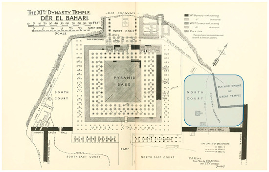
Figure 1.
Map showing the outline of the Dynasty XI Temple of Mentuhotep II at Deir el-Bahri [3], public domain. The discarded votive offerings were excavated from the North Court area of the Temple (shown in blue), which is located below the terraced walls of the Hathor shrine (part of the Dynasty XVIII Mortuary Temple of Hatshepsut).
The discarded objects heaped below the walls of the shrine included stelae, small statues of cats and cows, beaded textiles, vessels, jewellery and paintings made on linen. All were given as offerings to the goddess Hathor, who embodied the sky and was closely associated with the colour blue [4,5]. Indeed, many offerings to the goddess that were found during the excavations at Deir el-Bahri during work funded by the EEF and later groups working in the Hathor Shrine itself [6] were entirely blue or contained motifs that were mainly blue in colour. In 1913, Currelly wrote of the objects they collected and observed: “the special relation of blue colour with the goddess cannot be doubted” [2].
The ROM collection contains six painted linen votives that were acquired by the museum from Deir el-Bahri during the 1905–1907 excavations. The sheer number of Hathor-related iconography, Hall notes, narrows the age of these offerings to the first half of Dynasty XVIII, a flourishing period of worship to the goddess Hathor at both Dier el-Bahri and elsewhere in ancient Egypt [2]. While the textile featured in this study is not found among the set sketched in the published plates, Hall provides a description of a long painted fringed cloth “narrower at one end than at the other,” [2] whose description bears remarkable similarities to the painted textile analysed herein. Currelly further expands on the votive textile finds, highlighting their appearance in a dust and rubbish layer overlaying the Dynasty XI stratigraphy and notes the uniqueness of these painted votives worshipping Hathor to this temple site [2]. These written accounts support the antiquity of this find and its relationship with the site and the excavations conducted between 1905 and 1907 at Dier el-Bahri. In 2023, three of the six paintings were received at the Canadian Conservation Institute (CCI) for photographic examination and scientific analysis. Although all six paintings have at least one fringed edge, only one (accession number 910.16.3) contains dyed threads. The top, looped fringe of votive 910.16.3 contains blue, yellow and cream (suspected undyed) linen threads, and several additional blue threads run the lengths of the side edges of the painted cloth. A photograph of the votive offering is shown in Figure 2, with detailed images of the fringe and the side edges. The threads were not subjected to dye analysis prior to the current study; however, a published examination record from 1993 described the materials used in its construction as “linen cloth with blue dye (woad), tempura paint” [7]. Indeed, it is commonly presumed that in the New Kingdom, blue dyes would have originated from woad (Isatis tinctorum) [8] (pp. 151–152). As for the yellow threads in the fringe, one main yellow dye source in ancient Egypt is generally thought to be safflower (Carthamus tinctorius) [9], and the dried flowers and seeds of safflower have been discovered in several ancient tombs [10,11].
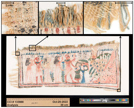
Figure 2.
Normal light photograph of the painted linen votive depicting offerings to the cow goddess Hathor, accession number 910.16.3 (ca. 1400–1350 BCE, Late Dynasty XVIII), Royal Ontario Museum. Details of cropped areas from the side edges show that the cloth has blue threads that run along the length of the side borders. The detail of the fringe shows a mixture of blue, yellow and cream linen loops. Translated inscription: Hathor, Lady of Heaven. Made for/by Sedjem(?), made for/by the Lady of the House, Hemet-netjer. His daughter, his son, his daughter. © Canadian Conservation Institute: 133988-0068, -0181, -0182.
Colourants in woad dye include indigotin (IND) and small relative amounts of its structural isomers, the most abundant of which is indirubin [12] (pp. 338–339). Colourants in yellow safflower include carthamin and anhydrosafflor yellow B [12] (p. 667). Since 2004, dye analysis at the CCI has been undertaken using gas chromatography–mass spectrometry (GC-MS), after extraction and derivatisation using m-(trifluoromethyl)phenyl trimethylammonium hydroxide (TMTFTH) [13,14,15]. The technique is both complementary and consistent with more widely used high-performance liquid chromatography (HPLC) methods for dye analysis and can be used to identify most natural dyes including flavonoids and isoflavonoids, quinones, indigoids (both brominated and non-brominated), tannins, and lichen dyes [14]. The method was modified in 2022 by adding an additional treatment step that was adapted from an extraction methodology used for HPLC dye analysis [16]. Prior to extracting with TMTFTH/toluene, threads are bathed in a 1:1 mixture of pyridine and deionised water (PW) and the resulting solution with thread is dried before continuing with the TMTFTH/toluene extraction. This additional PW treatment technique has been found to be beneficial, particularly for mordanted dyes that are strongly bound to fibres and difficult to extract. For this investigation, the modified two-step technique was employed.
Similarly to GC-MS, as the methodology for HPLC analysis of dyes has developed over the past few decades, extractions have been undertaken using a variety of different solvents and mixtures of solvents. Although extractions for indigoids are generally carried out using warm or hot dimethyl sulfoxide (DMSO), a study has shown that similar peak ratios are obtained using dimethyl formamide (DMF) or pyridine as an extraction solvent [17].
The TMTFTH GC-MS technique allows for quantification based on peak areas and has been utilised for decades in the extraction and identification of other materials used in heritage collections, such as oils and fats, natural resins, and waxes [18,19,20,21]. A key advantage of using GC-MS for dye analysis is that resultant chromatograms of the extraction solutions contain all the soluble organic compounds that are present from a dyed substrate—colourful dye compounds as well as colourless compounds. This is advantageous for several reasons. For one, it means that it may be possible to identify dye degradation products when they remain on the thread even after extreme fading, thus potentially allowing for the original colour to be identified [14]. Moreover, information is often gained about components other than just the dyes (or faded dyes) on the substrate. Compounds present such as oils, sterols, dye bath additives, treatments and coatings can provide further information about and the origin of the thread fibres, including identification and potential degradation, as well as information about the dyeing and textile-processing technologies [22]. Other advantages of GC-MS analysis include the availability of extensive reference libraries of mass spectra, such as those produced by the National Institute for Standards and Technology (NIST). The use of a mass spectrometric detection method also allows for the potential elucidation of compounds not presently described by a published library. While the advantages of TMTFTH GC-MS are many, there are also some drawbacks. Depending on the chemical structure, some dye compounds are methylated by TMTFTH while others are hydrolysed in addition to methylation. These reactions form smaller compounds in addition to sometimes changing the structures of the parent dye compounds. However, most of the methylated and newly formed compounds remain distinct and diagnostic, which allows for accurate identification [14].
2. Materials and Methods
2.1. Examination and Sampling
Photographs of the mounted painted linen votive were taken at the CCI using a full-spectrum Phase One IQ4 150 MP digital camera under normal light, infrared, and ultraviolet illumination (PhaseOne, Frederiksberg, Denmark) (see Supplementary Materials (SM) for more information) and previously employed at the CCI in the analysis of Egyptian polychrome objects [23]. Radiography was also performed at the CCI, using a Lorad LPX 160 X-ray tube (Spellman High Voltage Electronics Corp., Hauppauga, NY, USA) and a CareStream computed radiography system (Carestream Health Inc., Rochester, NY, USA). For photomicrographs of individual threads and fibres, the mounted painted linen votive was examined and photographed using a Leica M205C stereomicroscope interfaced to a DMC 5400 digital camera (Opti-Tech Scientific Inc., Whitby, ON, Canada). All images were collected under normal light illumination. Image processing was undertaken using Leica LASX software (version 3.0.12.21488).
For analysis, one sample each of blue, yellow and cream linen threads (approximately 3–5 mm in length) was collected from the looped top fringe. The threads are very fragile, and the chosen threads were already partially broken from the fringe loops. Care was taken to minimise sampling in keeping with conservation ethical concerns. Based on the woven pathways of the intact loops, the dyed fringe appears to have been constructed using continuous threads that were looped into the selvedge of the cloth as it was woven. It is likely that a sample taken from one loop of the fringe will be made with the same thread as many other loops. For this reason, as well as the overall homogeneity of the coloured threads when viewed under multiple wavelengths of light, the dye analysis results from each singular sample were extrapolated to comprise the entirety of the fringe. For the intact blue weft threads that run along the left and right edge of the cloth, no samples were removed to avoid visibly and structurally damaging the precious cloth.
2.2. Gas Chromatography–Mass Spectrometry
2.2.1. Sample Treatment and Extraction
For each sample (blue, yellow and cream), a thread was placed into a 2 mL clear glass GC–MS vial (cat. no. 5182-0715, Agilent Technologies Inc., Santa Clara, CA, USA). To each of the vials, 40 µL of pyridine (ACS grade, Alfa Aesar, Fisher Scientific Canada, Ottawa, ON, Canada) and 40 µL of deionised water were added. The vials were capped with PTFE/silicon/PTFE septa screw top vials (cat. no. 5175-5862, Agilent Technologies) and placed in a block heater at 60 °C. The vials were heated for 1 h; then, the vial caps were removed, and the vials were completely dried in the warm block heater. After drying and cooling, 10 µL of TMTFTH (5% in methanol) (cat. no. T0961, TCI America, Portland, OR, USA) and 10 µL of toluene (ACS grade, Sigma-Aldrich Canada, Oakville, ON, Canada) were added to the vials and threads, and the capped vials were replaced in the block heater at 60 °C for 1 h. The vials were then removed from the heater and centrifuged at 1500 rpm for 1 min. For every step, the threads remained inside the vial, bathed in the solvents.
2.2.2. Instrumental Conditions
For each analysis, 2 µL of an extract was injected into a glass micro-vial (cat. no. 5190-3187, Agilent Technologies) set in the thermal separation probe (TSP, Agilent Technologies). The TSP was then inserted into a multimode inlet on an Agilent 7890 A GC interfaced to a 5975 C MS (Agilent Technologies Inc., Santa Clara, CA, USA). During analysis, the inlet temperature was increased from 50 °C to 250 °C at a rate of 900 °C/min and held for approximately 38–40 min. Then, at this point in each run, the inlet was cleaned by heating to 450 °C, at a rate of 900 °C/min, and held for 3 min before cooling once again to 250 °C. This built-in pyrolytic cleaning cycle at the end of each run helps to mitigate any sample carry-over from the inlet and produces a chromatographic feature that appears to be a short rise and fall in the baseline at approximately 43 min. For the GC separation, a Phenomenex ZB-5MSi fused silica column (30 m × 0.25 mm i.d., 0.25 µm film thickness with 5 m fused guard column; cat. no. 7HG-G018-11-GGA, Phenomenex Inc., Torrance, CA, USA) was used. Ultra-high purity helium carrier gas was used with a constant flow of 1.2 mL/min. The oven was programmed from 40 °C to 200 °C (at 10 °C/min), and then from 200 °C to 310 °C (at 6 °C/min) with a final hold time of 20 min (54.33 min run time). A solvent delay of 6.4 min was employed to avoid the first solvent peak; the MS was on from 6.4 to 7.5 min then turned off again from 7.5 to 9.7 min to avoid the second large solvent peak. This solvent delay programme allows for a 1.1 min window for compounds to elute during the long solvent removal period of the run. The MS transfer line temperature was held at 280 °C; the temperature of the MS ion source was 230 °C and that of the MS quadrupole was 150 °C. The MS was run in scan mode from 45 to 550 amu (6.4–25 min), 50–700 amu (25–30 min) and 50–800 amu (30 min–end of run). Agilent ChemStation software, v.E.02.02.2.5 and AMDIS v. 2.71 software were used for data processing. Where available, mass spectral comparisons were made against the NIST11 database.
2.3. Elemental Analysis: Scanning Electron Microscopy/Energy Dispersive X-Ray Spectrometry
Elemental analysis was performed on the fringe threads using scanning electron microscopy/energy dispersive X-ray spectrometry (SEM/EDX) to determine the possible presence of iron buff mineral dye. Approximately 2 mm of thread was cut from the cream (suspected undyed), yellow and blue fringe threads and sonicated in deionised water for a few minutes to remove extraneous dirt. Samples were then removed from the water and air-dried.
SEM/EDX analysis was performed on the cleaned thread fragments using a Hitachi S-3500 N VP SEM integrated with an Oxford Inca X-act analytical silicon drift X-ray detector and an AZtec X-ray microanalysis system (AZtec 3.1 SP1). The SEM was operated at an accelerating voltage of 20 kV at a pressure of 60 Pa using a backscattered electron detector. With this technique, elemental analysis of volumes down to a few cubic micrometers can be obtained for elements from boron (B) to uranium (U) in the periodic table at a level of approximately 0.1–1% or greater.
3. Results
3.1. Photographic and Microscopic Examination
Scientific imaging of this textile identified no visible differences in the similarly col-oured areas under different fluorescence and reflected light sources. Photographic detail of the top left corner of painted linen votive 910.16.3 is presented in Figure 3. The knots on the left side edge possibly indicate that the warp runs left to right, and the weft threads run from the top to the bottom. A looped fringe of cream, yellow and blue threads has been woven into the top selvedge edge of the cloth. The main canvas has been woven in an uneven 1:1 plain weave pattern, producing a warp-faced fabric. Thread counts in the centre of the cloth are approximately 10–12 threads/cm for the weft, and 17–20 threads/cm for the warp [7]. As the fabric was woven, it appears to have had more draw-in on the left side than the right, producing a rectangular shape that is thinner on the left than the right (see Figure 2). The cloth measures approximately 48 cm in width, 18 cm in height on the left edge, and 23.5 cm in height on the right edge. The knotted warp ends on the left side form a fringe measuring approximately 5 cm in length, and the looped fringe along the top edge measures 1.5 cm. Doubled warp and tripled weft threads are visible along the top and left edge, respectively. Individual threads are spun in the natural direction of the flax fibres (S-spun) and the fringe threads are Z-plied together to create 2-ply fringe.
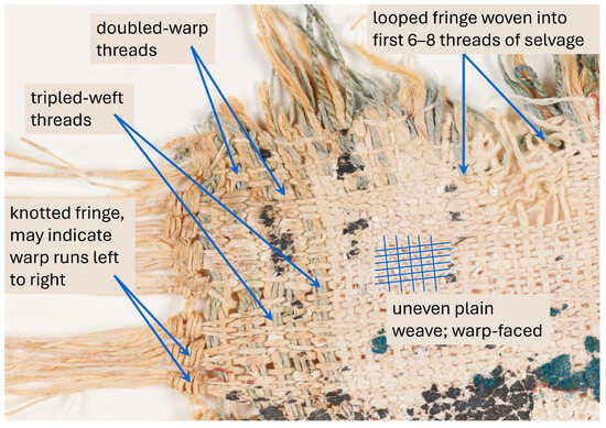
Figure 3.
A detailed photograph of painted linen votive 910.16.3; top left corner. Arrows indicate doubled and tripled threads along the top and side edges, respectively; a knotted fringe which suggests that the warp runs in the left to right direction of the painting; and the weaving of the looped fringe onto a selvedge edge. A hatched diagram drawn onto individual warp and weft threads aids in visualising the warp-faced weave of the cloth.
3.2. Dye Analysis of Looped Fringe
The looped fringe along the top of the painted linen votive consists of a mixture of cream, light yellow, and light blue threads. The colours were likely chosen for the association to the life-giving goddess Hathor and her connection to both the golden sun and the blue of the sky [5].
3.2.1. Yellow Thread
Previous studies of ancient Egyptian linen textiles have identified safflower as a key yellow colourant; other yellow dyes in use during this period include sumac (Rhus spp.) and iron buff [24,25,26,27]. Iron buff is a yellow mineral dye, and its presence can be revealed using a simple staining experiment, first described by Hübner in 1909 [24]. He subjected yellow linen threads from a mummy wrapping to a logwood dye solution. Because iron buff dye is bound to textile fibres in a manner similar to that of an iron mordant, exposing the iron buff thread to logwood dye causes the fibres to become stained black with an iron–haematein complex. The results of the logwood staining on a yellow thread from the looped fringe are presented in the SM. The thread did not become black, and it was thus accepted that the yellow fringe threads had not been dyed using iron buff mineral dye. SEM/EDX analysis identified traces of iron on the cream (suspected undyed), blue and yellow threads. Although the threads had been sonicated in deionised water to remove extraneous dirt, this consistent finding among all the threads likely indicates some lingering contamination of the burial environment. If the yellow thread had been dyed using iron buff, an elevated abundance of iron would be expected with respect to the amounts shown for the cream and blue threads.
Further investigation into the dyes present on the fringe threads involved treatment and extraction using the two-step procedure outlined in Section 2.2.1. This technique is not only useful for investigating dyes but also other organic components bound to the textile fibres. Analysis of the extracts from the yellow, blue and cream fringe threads using GC-MS found that each contained compounds related to hydrolysable tannins and humic substances. Overlaid extracted ion chromatograms (EICs, m/z 221, 223, 224, 226, and 284) are shown in Figure 4 for the cream fringe, the yellow fringe and the blue fringe; peak labels are described in Table 1. Chosen extracted ions correspond to the base ions from the mass spectra of the tannins and humic acid peaks. For the heptadecanoic acid peak, the m/z 284 fragment ion was used as it is diagnostic and also provides a peak of equivalent size with which to compare. The extracted peaks show many of the compounds related to hydrolysable tannins and humic substances that are present on the differently coloured fringe threads, including methylated derivatives of gallic acid (T1), dihydroxy benzenedicarboxylic acids (T2 and T4) and benzenetricarboxylic acids (T5 and T6). All of the benzenecarboxylic acid compounds shown in Figure 4 are typical for textiles that have been exposed to tannin-containing plant material. This exposure can occur through dyeing or mordanting with tannic substances and also through contact with water from a tannin-rich source during cleaning or processing of the cloth or fibres. It is also possible that some of the tannins and humic substances present in these samples may originate from exposure of the fibres to degrading plant material during the fermentation process of retting the flax plants [22]. The EICs shown in Figure 4 have each been normalised based on the peak height of heptadecanoic acid (T7), a compound which occurs in each sample and does not likely originate from any hydrolysable tannins, humic substances or dyes that may be present. In examination of Figure 4, it is noticeable that the tannin and humic substance peaks found in the extract of the yellow fringe thread are present in greater abundances than the cream natural fringe and the blue fringe. It is possible that the tannin and humic substances’ peak abundances occur not only from universal processing, such as retting, but also from exposure to a tannin-rich yellow dye, such as sumac (Rhus spp.). In fact, the yellow fringe contains an unassigned peak of significant relative abundance (T3) that has been previously identified on reference textiles dyed with sumac [16]. The mass spectrum for this unassigned compound is shown in Figure 4. This peak is not present in the cream or blue fringe samples.
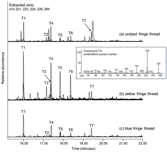
Figure 4.
Overlaid extracted ion chromatograms (EICs) created using m/z 221, 223, 224, 226, and 284 for (a) cream fringe, (b) yellow fringe and (c) blue fringe threads on painted linen votive 910.16.3. The EIC abundances have been normalised to the peak for heptadecanoic acid (T7). Peak labels are described in Table 1. The inset mass spectrum is compound T3, an unidentified marker found in sumac (Rhus sp.). This compound is only present in the extract from the yellow thread.
3.2.2. Cream Thread
Analysis of the extract from the cream colour thread that was presumed to be undyed did not identify any dye markers other than the traces of tannins and humic substances that are shown in Figure 4 and discussed above. Data analysis for dye extracts was performed using AMDIS software and an extensive search library (with retention indices) that has been developed at the CCI using reference materials. This consistent approach allows for automated scanning of chromatograms and the detection of possible dye marker compounds using the same sensitivity and deconvolution parameters for each sample.
3.2.3. Blue Thread
Many of the colourant markers identified in the extract from the blue fringe thread were unanticipated. The reason for this is that there are very few natural dyes that produce a blue colour. And, based on the age and culture of the votive, it was presumed that the blue threads in the fringe were dyed with woad (Isatis tinctorum) [8,28]. However, in an exciting turn of events, it was found that the extract contained not only the expected derivatised compounds from IND but also brominated indigoid compounds—a clear indication of a shellfish source for the dyestuff [29]. The total ion chromatogram (TIC) for the extract is presented with labelled peaks in the SM, and overlaid EICs produced using base ions from the mass spectra of the identified indigoid compounds (m/z 119, 132, 146, 165, 192, 199, 210, 224, 243, 268, 270, 320, 385, and 463) are presented in Figure 5 and described in Table 1. Chosen extracted ions correspond to the base ions from the mass spectra of the indigoid marker peaks. The most abundant marker compounds identified are those eluting prior to 20 min. These compounds form through natural degradation pathways and also through the alkaline TMTFTH extraction and methylation reactions [14,30]. The degradation occurs through reactions involving C2–C2′ hydrolytic cleavage, resulting in halving the whole indigoid molecules. For this reason, compounds 1–10 will be referred to as hemi-indigoids. Other, larger compounds identified at later retention times are whole brominated and non-brominated derivatives of leuco-IND and oxidation products of IND. Except for IND-OX (13) and MBI-OX (14), the whole indigoid compounds are present at low abundances, particularly the peak assigned to the DBI-H (17), which has a signal to noise ratio (S/N) of only 7. This indicates that the DBI-H peak is present just above the limit of detection for the instrument.
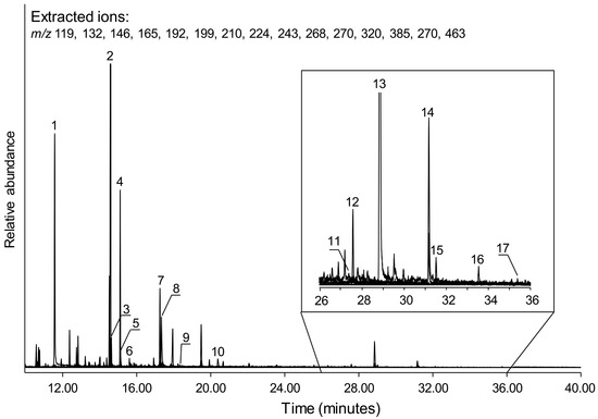
Figure 5.
Overlaid extracted ion chromatograms (EICs), created using m/z 119, 132, 146, 165, 192, 199, 210, 224, 243, 268, 270, 320, 385, and 463 for the blue fringe on painted votive 910.16.3. An expanded section of the EICs from 26 to 36 min is shown to more clearly display the smaller peaks from that region. Peak labels correspond to compounds listed in Table 1.
The mass spectra and chemical structures for the marker compounds identified in the extract from the blue fringe are shown in Figure 6 and Figure 7, labelled in accordance with Figure 5 and Table 1. In Figure 6, rather than presenting the mass spectra in order of elution, the hemi-indigoids are presented in structural pairs: non-brominated markers alongside the brominated equivalents.
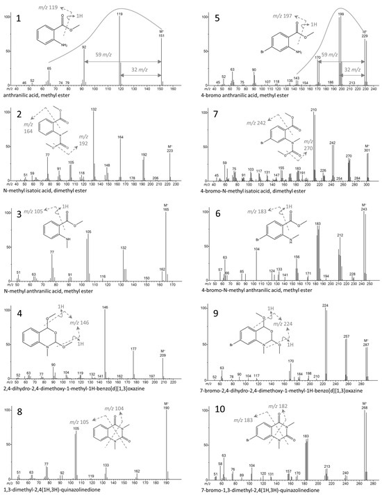
Figure 6.
Hemi-indigoid compounds present in the extract of the blue fringe. Mass spectra are presented in pairs of non-brominated and brominated compounds. Trend lines (shown for compounds 1 and 5) and fragment loss calculations have been added to highlight the similarities in ion fragmentations for the pairs.
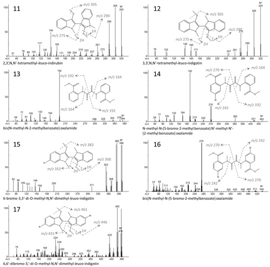
Figure 7.
Mass spectra and chemical structures for main marker compounds derived from whole indigoids: IND, MBI and DBI. The leuco-indigoids include compounds 11, 12, 15 and 17, while the oxidised indigoids include compounds 13, 14, and 16. The compounds are labelled in accordance with Figure 5 and Table 1.

Table 1.
List of compounds corresponding to the labelled peaks in Figure 4, Figure 5, Figure 6 and Figure 7.
| Label | Compound | M+ (% Abund.) | Characteristic Fragment Ions m/z (% Abund.) |
|---|---|---|---|
| 1 | anthranilic acid, methyl ester | 151(68) | 119(100), 92(52), 65(22) |
| 2 | N-methyl isatoic acid, dimethyl ester | 223(40) | 132(100), 164(60), 192(35), 77(34), 105(23), 148(19) |
| 3 | N-methyl anthranilic acid, methyl ester | 165(100) | 105(68), 104(61), 132(45), 162(14) |
| 4 | 2,4-dihydro-2,4-dimethoxy-1-methyl-1H-benzo[d][1,3]oxazine | 209(29) | 146(100), 177(40), 90(17), 104(6), 120(5) |
| 5 | 4-bromo anthranilic acid, methyl ester | 229(67) | 199(100), 197(98), 231(64), 170(37), 63(20), 90(20) |
| 6 | 4-bromo-N-methyl anthranilic acid, methyl ester | 243(100) | 245(95), 183(79), 185(79), 212(62), 104(42), 156(32) |
| 7 | 4-bromo-N-methyl isatoic acid, dimethyl ester * | 301(40) | 210(100), 212(95), 242(52), 244(52), 270(33), 272(32) |
| 8 | 1,3-dimethyl-2,4(1H,3H)-quinazolinedione * | 190(100) | 105(70), 104(65), 77(18), 77(18), 133(17), 162(8) |
| 9 | 7-bromo-2,4-dihydro-2,4-dimethoxy-1-methyl-1H-benzo[d][1,3]oxazine * | 287(45) | 224(100), 226(96), 257(57), 255(56), 289(45), 170(23) |
| 10 | 7-bromo-1,3-dimethyl-2,4(1H,3H)-quinazolinedione * | 268(100) | 270(96), 185(58), 183(58), 184(55), 182(52), 63(31), 104(17), 213(16), 213(15), 240(7) |
| 11 | 2,3′,N,N′-tetramethyl-leuco-indirubin (INR-H) | 320(91) | 305(100), 290(68), 145(50), 261(25) |
| 12 | 3,3′,N,N′-tetramethyl-leuco-indigotin (IND-H) a | 320(100) | 305(89), 275(48), 290(23), 146(22), 160(20), 76(9) |
| 13 | bis(N-methyl-N-2-methylbenzoate) oxalamide (IND-OX) b | 184(2) | 192(100), 133(16), 132(15), 77(15), 164(13), 376(3) |
| 14 | N-methyl-N-(5-bromo-2-methyl benzoate) N′-methyl-N′-(2-methyl benzoate) oxalamide (MBI-OX) * | 462(1) | 192(100), 270(25), 132(18), 164(17), 77(13), 104(12) |
| 15 | 6-bromo-3,3′-di-O-methyl-N,N′-dimethyl-leuco-indigotin (MBI-H) * | 398(95) | 385(100), 383(96), 400(95), 352(51), 355(40), 268(23), |
| 16 | bis(N-methyl-N-(5-bromo-2-methylbenzoate) oxalamide (DBI-OX) * | 540(<1) | 270(100), 272(99), 83(17), 210(15), 212(15), 244(14), 242(13) |
| 17 | 6,6′-dibromo-3,3′-di-O-methyl-N,N′-dimethyl-leuco-indigotin (DBI-H) * | 476(48) | 463(100), 478(97), 433(42), 224(36), 239(27), 448(17) |
| T1 | 3,4,5-tri-O-methyl gallic acid, methyl ester | 226(100) | 211(44), 195(28), 155(27), 151(11), 125(10) |
| T2 | 1,2,4-benzenetricarboxylic acid, trimethyl ester c | 252(10) | 221(100), 103(6), 193(5), 119(5) |
| T3 | unidentified sumac marker | 255(38) | 224(100), 194(26), 165(14), 137(8) |
| T4 | 1,3,5-benezenetricarboxylic acid, trimethyl ester c | 252(27) | 221(100), 193(22), 161(7), 147(7) |
| T5 | 1,2-benzenedicarboxylic acid, 3,4-dimethoxy, dimethyl ester c | 254(40) | 223(100), 191(46), 169(13), 137(11) |
| T6 | 1,2-benzenedicarboxylic acid, 4,5-dimethoxy, dimethyl ester c | 254(51) | 223(100), 155(22), 199(17), 100(12) |
| T7 | heptadecanoic acid, methyl ester | 284(9) | 74(100), 87(80), 143(16), 241(12) |
* Not previously published, tentative identification based on mass spectral fragmentation. a [31]; b [32]; c Structures and order of elution identified from NIST11 library and published study [33].
The mass spectra for the brominated hemi-indigoids (compounds 5, 6, 7, 9, and 10) in Figure 6 are not present in the NIST mass spectral database employed for this study, nor have they been previously published. Identifications were made based on the known mass spectra for analogous hemi-indigoid markers from indigotin (IND), the calculated molecular weights of expected monobrominated hemi-indigoid markers, and close similarities in fragment ion profiles between the mass spectral pairs (see the calculations and ion fragment distribution curves for compounds 1 and 5 in Figure 6 as an example). The three markers in Figure 6 with the highest relative abundances are all non-brominated. These are the methyl derivatives of anthranilic acid (1), isatoic acid (2), and 2,4-dihydro-2,4-dimethoxy-1-methyl-1H-benzo[d][1,3]oxazine (4). Compound 4 is the reduced form of isatoic anhydride, a known oxidative degradation product of IND [30].
Figure 7 shows the proposed structures for the whole indigoid derivatives identified in the extract from the blue fringe. Identifications of compounds 11, 12, 15, and 17 (derivatised leuco indigoids, INR-H, IND-H, MBI-H, and DBI-H) were based on the published mass spectrum of IND after reduction and derivatisation with tetramethyl ammonium hydroxyl (TMAH) [31], calculated molecular weights of expected derivatised leuco markers, and close similarities in fragmentation patterns. Identifications of the oxidised indigoid markers (IND-OX, MBI-OX, and DBI-OX, compounds 13, 14, and 16) were made based on the synthesis and TMTFTH derivatisation of compound 13 by Chris Petersen at Winterthur Museum [32], calculated molecular weights of expected brominated oxidised markers, and close similarities in fragment patterns. Although the chromatogram shows a trace relative abundance of methylated leuco-indirubin (INR-H), there are no detected peaks of monobrominated or dibrominated leuco analogues. The fluctuating levels of indirubin (and in some cases, the absence) in dye extracts from the Mediterranean sea snail species Hexaplex trunculus have been previously discussed by Koren [34]. As the blue fringe dye extract does contain a trace of INR-H, it is quite possible that brominated leuco-indirubin compounds are present at levels below the limit of detection for the instrument. All indirubin colourants, whether brominated or not, are red in colour. The absence of these compounds will have a definite effect on the colour of the dye produced. The blue colouration of the fringe threads may be dependent on several factors, including the high relative abundance of IND, the low relative abundance of DBI, the trace abundance of INR, and the absence of brominated indirubins.
4. Discussion
The complex chemical reactions that take place during the shellfish dyeing process have been previously studied and described in detail [29,35,36] and proceed through a multistep pathway, beginning with the removal of the hypobranchial gland of the animal. The colourless dye precursor fluid is present in very small quantities in the gland, and this needs to be extracted. In his book Historia Naturalis, Pliny the Elder describes a multi-day process of soaking and boiling the glands in salt and water to extract the precursor fluid [37]. Snail harvesting and processing took place at sites dotted along the coastlines of the snails’ habitats, particularly in the Mediterranean region [12] (pp. 553–606). The principal location of shellfish dyeing in the ancient Mediterranean region is considered to be the Phoenician city of Tyre (present day southwestern Lebanon). The beautiful red-violet colourants known to originate from this region have been referred to as Tyrian purple, royal purple, and now more commonly, shellfish purple.
For the blue threads of the votive, the presence of brominated indigoid compounds definitively indicates a shellfish source for the colourant. However, the uniform blue colour of the threads is far less common than the red-violet colour of shellfish purple. Analyses of historic and modern reference samples has shown that the red-violet colour is generally due to a high relative abundance of 6,6′-dibromoindigotin (DBI), with lower relative abundances (or even the complete absences) of the mono-brominated (6-monobromoindigotin, MBI) and the non-brominated (IND) compounds [38,39]. Three shellfish in the Mediterranean region can produce red-violet colourants. These are Hexaplex trunculus, Bolinus brandaris, and Stramonita haemastoma [40] (pp. 285–306); however, the dye precursor content in the latter two species is considerably less than found in H. trunculus [41]. Additionally, there are shellfish species found in other areas of the world that also produce a red-violet colourant. Chief among them is the species Plicopurpura patula, found in Central and South America. With the abundance of existing knowledge on shellfish purple dyes, the principal questions for the blue fringe dye were as follows: what is the distribution of indigoid colourants (IND, MBI and DBI) in shellfish blue dye and from which sea snail species is it obtained?
The investigation of the shellfish dye source for the blue fringe threads began with the identification of all the dye marker compounds, both hemi-indigoids and whole indigoids, and determining their relative abundances. Because the chromatogram (see Figure 5 and Figure S4) contained whole indigoid compounds that appeared to result from both oxidative and reductive reactions, a review of the PW treatment and extraction methodology was also conducted.
4.1. TMTFTH: Extraction, Methylation, and Hydrolysis Reactions
Depending on their molecular structures, some dye compounds are methylated by TMTFTH while others are hydrolysed in addition to methylation [14]. These processes have been previously shown to break apart dye molecules into smaller compounds, and this also occurs for indigoids [14]. However, for IND and its mono- and di-brominated analogues (MBI, and DBI, respectively), there are additional, opposing reactions which occur during each of the two steps. The first step in removing dye compounds from substrates is a treatment in warm pyridine and deionised water. For indigoids, this promotes oxidation, resulting in the opening of the indole rings and the formation of benzoic acid oxalamides (IND-OX, MBI-OX, and DBI-OX; 13, 14, and 16). Next, reduction reactions occur in the second extraction step with the introduction of the alkaline methanolic TMTFTH reagent. This step likely reduces the remaining, intact IND, MBI, and DBI to their leuco forms (IND-H, MBI-H, and DBI-H; 12, 15, and 17). These two pathways are shown in Figure 8 with the structures of the resulting indigoids. For indigoids, the consequence of adding an additional extraction step to increase overall dye concentration has been to increase the relative abundance of the benzoic acid oxalamide derivatives and decrease the relative abundances of the reduced leuco derivatives when compared to past extraction results utilising solely TMTFTH [14].
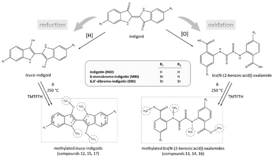
Figure 8.
Reduction and oxidation pathways for IND, MBI and DBI occurring during the PW treatment, extraction and GC-MS analysis processes. This results in the formation of methylated leuco-forms, IND-H (12), MBI-H (15), and DBI-H (17), as well as the formation of methylated bis(N-(2-benzoic acid)) oxalamides, IND-OX (13), MBI-OX (14), and DBI-OX (16).
But is this the correct explanation for what occurred in the extraction of the blue fringe thread? And how can the explanation be tested? A previous study by Katarzyna Witkós et al. determined that IND-OX (13) forms naturally through the oxidative degradation processes of IND [30]. So, could the increased relative abundances of the oxidised indigoids when compared to the reduced leuco indigoids have more to do with the tremendous age of the votive painting, rather than exposure to oxidising reagents? To help shed some light on this, we observed the effect of the PW treatment and TMTFTH extraction steps on a contemporary reference thread. Due to the scarcity of shellfish dyestuff, the sample that was used for this purpose was one that had been generously given to CCI for the reference dye collection. The thread was coloured using dye sustainably obtained from Mexican sea snails (Plicopurpura patula) in 2018. Peak areas compared side-by-side in Figure 9 show that the relatively fresh P. patula dye extract also contains an overall higher relative abundance of the oxidised indigoids in comparison to the reduced leuco indigoids, just like the blue fringe sample. Although the painted linen votive is ancient and has been exposed to millennia of environmental and burial conditions, the greater abundances of oxidised indigoids are not likely driven by natural oxidative degradation processes, but rather by the PW treatment and TMTFTH extraction conditions. Although further work will be carried out to more fully understand this trend, at this point it appears to be a feature of the procedure. Figure 9 clearly shows that the reduced leuco indigoid compounds (IND-H, MBI-H, and DBI-H) are only present in trace abundances for both the blue fringe thread and the P. patula reference thread. Therefore, ratios calculated on these abundances will not be as accurate as those calculated from the oxidised whole indigoids (IND-OX, MBI-OX, and DBI-OX).
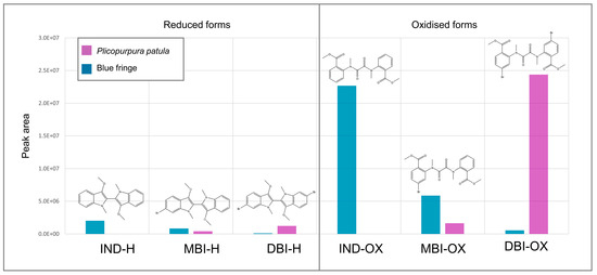
Figure 9.
Peak areas of whole indigoid compounds identified in the extracts from the blue fringe thread and a 2018 thread dyed with dyestuff from Plicopurpura patula. The peaks are divided into those for the reduced indigoids IND-H, MBI-H and DBI-H on the left, and the oxidised indigoids IND-OX, MBI-OX, and DBI-OX peaks on the right.
In Figure 9, the distribution of IND:MBI:DBI peaks from the blue fringe sample and the red-violet P. patula reference thread appear to have opposing trendlines. For the blue fringe, the most abundant peaks are those that result from IND, while for the P. patula samples, the most abundant peaks are those resulting from DBI. The calculated ratios of the IND:MBI:DBI derivatives for the extract of the blue fringe thread are 78:20:2 (oxidised), while the calculated ratios for the extract of the P. patula sample are 0:6:94 (oxidised). It should be noted that the ratios for the P. patula sample are consistent with those that have been previously published for P. patula dyestuff using HPLC methodologies. For instance, a study from Withnall et al. performed using HPLC-PDA after extraction in hot DMF calculated a ratio of 0:9:90 [32], while Clementi et al. used fast-HT-HPLC-PDA after extraction in room temperature DMSO to detect ratios of approximately 0:2:98 and 1:12:87 [42]. The similarity of the P. patula ratios obtained in this study to those of published data is promising for the identification of dyestuff on the blue fringe sample.
4.2. Di-Mono Index (DMI)
As secretive as the ancient manufacturing practices were for the red-violet shellfish dye, the existence of a blue shellfish colourant emphasises another level of mystery altogether. Indeed, there were long centuries of time during which the knowledge and skills required to produce the blue dye were completely lost [43].
Recent experiments have shown that DBI can undergo some level of debromination when exposed to heat, thus changing colour from red-violet to bluish [44,45]. It has also been shown that light exposure can cause degradation of IND, MBI and DBI. Fibres dyed with H. trunculus and exposed to harsh UV light showed decreases in concentration for IND, MBI, and DBI [46]. Although the initial rate of degradation for DBI was determined to be faster than seen for IND and MBI, after prolonged exposure the relative percentages of the three compounds stabilised and were effectively the same as initially measured [46]. Another study found that light exposure during the vat dyeing process can influence the final colour of the dyed textile [47]. Exposure of H. trunculus dye vats to UV light caused debromination of the leuco form of MBI to IND and, to a lesser extent, debromination of leuco-DBI [47]. The same study also concluded that observed degradation due to UV exposure after dyeing was minor in comparison to degradation observed in the dye vat when the colourants are in the leuco forms [47]. Experimental archaeological research has found that the final colour of dyed textiles can be influenced by dyeing conditions, such as exposure to sunlight and salt concentrations. These can act to increase the concentration of IND, while ratios of MBI and DBI remain constant, thus shifting the dye bath colour to a more bluish hue [29].
All of these degradative reactions and dye processing conditions can shift the colour of a shellfish dyebath. However, there may be a simpler explanation to obtaining blue shellfish dye in the Mediterranean region in the past. Researchers have determined that the modern H. trunculus snails can produce blue dye in addition to the more common red-violet dye [17,48,49,50]; no other sea snail species in the world has shown this ability. What circumstances or conditions are required to produce the blue colour from H. trunculus, rather than the red-violet? Shellfish dye researcher Zvi Koren has been at the forefront of the work to bring this mystery to light [39,41,51]. Koren has hypothesised that there exist two forms of H. trunculus snails, with those that are capable of producing dyes having a greater relative abundance of DBI (DBI-rich H. trunculus) and those that produce dyes having a greater relative abundance of IND (IND-rich H. trunculus) [39,41,51]. This may also link to the fact that some non-brominated precursor compounds found in H. trunculus can vary in abundance based on the time of year that they were harvested, producing increased or decreased amounts of IND when processed [52]. Although it has also been theorised that the resulting dye colour (red-violet or blue) may corelate to the sex of the H. trunculus snails, this theory is complicated by the pseudohermaphroditic nature of male H. trunculus snails and has not yet been statistically proven [53].
Working through the hypothesis that some H. trunculus are able to produce a blue IND-rich dyestuff, while others are able to produce a red-violet DBI-rich dyestuff [39], Koren has developed a simple calculation that standardises the application of these two labels. The Di-Mono Index (DMI) is based on instrumental and colorimetric analyses of H. trunculus dye from both historical sources and modern reference materials [39,41,51], focussed on measured abundances of both MBI and DBI. For the calculation, although a significant relative abundance of IND is important in any consideration of whether the sample is IND-rich, the DMI does not take IND peak areas into account because the compound is not unique to H. trunculus snails; it is also present in indigo (Indigofera spp.) and woad (Isatis tinctoria) plants. Therefore, if these plant materials were present in a dye mixture, the IND levels that they contribute would adjust the colour towards a bluer hue but not affect the calculated DMI and the characterisation of the shellfish dye. Koren has determined that the maximum DMI for IND-rich H. trunculus dyes is 0.6, meaning that all calculated ratios less than 0.6 should be considered IND-rich, and ratios above that calculation should be considered DBI-rich [39,41,49]. The DMI can also be used for other red-violet-producing snail species as well, such as B. brandaris and S. haemastoma. Extensive research has shown that these two Mediterranean species contain very low to negligible quantities of MBI, giving DMIs over 0.6 and having good correlation with DBI-rich H. trunculus snails [39]. These DMI calculations highlight one fact in particular—H. trunculus snails are the only snails that are capable of producing significant quantities of MBI [39], and that is why this compound plays such an important role in the characterisation of shellfish dyestuff.
For the blue fringe sample, the DMI was calculated using the oxidised forms of DBI and MBI, as obtained using TMTFTH GC-MS analysis. These were calculated as DMIOX = (DBI-OX/MBI-OX) = 0.10. These results are also consistent with published peak area ratios and the calculated DMIs from both modern reference samples and archaeological samples dyed with IND-rich H. trunculus shellfish and analysed using HPLC-PDA [33]. Based on these results, the comparisons to known samples, and the unmistakable blue colour, the dye on the fringe threads also appears to originate from IND-rich H. trunculus snails. Science, however, is cautious. The study set of archaeological samples dyed using IND-rich H. trunculus remains very small, and there are known thermal- and photo-degradative reactions that can cause debromination during dyeing processes, which can skew IND:MBI:DBI ratios calculated from both HPLC and GC-MS techniques. Although the current evidence from this study likely indicates that the blue fringe was dyed using IND-rich H. trunculus, future work on further archaeological samples will help to strengthen that assertion.
4.3. Nomenclature
Historically, shellfish dye production in the Mediterranean region left behind unmistakable evidence as thousands of snails were needed to produce just sub-gram quantities of dye [54]. The earliest known archaeological traces include massive heaps of discarded snail shells and clay dye vats along the shores of Crete, dating to approximately 1800–1900 BCE [54,55]. By 1600 BCE, the ancient city of Tyre had become the major centre for shellfish dye production, giving rise to one of the most well-known names for the red-violet dye—Tyrian purple. However, as dyes with similar chemical compositions have been identified on historical textiles and from shellfish species worldwide, the colour is now more inclusively referred to as shellfish purple.
The blue IND-rich form of shellfish dye from the Mediterranean region currently lacks a standardised name. The ancient Hebrews referred to the colour as Tekhelet [56] and, given the mention of the dye in the Old Testament, it is also sometimes called Biblical blue. However, the significance of the dye to more than one religion and cultural group, as is highlighted by its further appearance in a temple to Hathor and the numerous shellfish dye production centres in the Mediterranean region, may indicate a need for a shift in nomenclature to a more generalist term, as seen with shellfish purple. It may be more inclusive to adopt shellfish blue as a more universally applicable term, acknowledging both its biological origins and its widespread geographical significance.
4.4. The Rarest Natural Dyestuff
Direct archaeological evidence of shellfish blue dye on ancient textiles is limited; it has only been identified on historical textiles a handful of times. Most of the identifications to date have been on textile fragments found in Israel and Egypt, dating from the Roman Period approximately 2000 years ago [57,58]. To the authors’ knowledge, only two other textiles older than 3000 years have been analysed and reported to have been dyed with shellfish blue [59]. Both of these Egyptian textiles are fragments of loosely woven linen, having a few stripes made using dark blue supplemental woollen weft threads, and they date to the New Kingdom (ca. 1686–1069 BCE) (Louvre Museum, accession numbers E 27366 and E 27377) [59,60,61]. Fragment E 27366 has a looped weft fringe of undyed linen, and fragment E 27377 has decorative turquoise beads (some loose and some attached). The two fragments were found associated with terracotta fertility figurines in Gebel el-Zeit, Egypt. The associations of fertility and the colour blue may indicate that the figurines were produced to honour the goddess Hathor, who was worshipped at Gebel el-Zeit during this period [62].
At approximately 3400 years old, the ROM painted votive textile with its remarkable blue and yellow fringe is the first large and intact ancient textile object with shellfish blue dyestuff that has been discovered. It is a marvel, not only due to the precious dye, but for the information that can be gained concerning the religion, technology, artistry, and trade in Egypt during this period.
5. Conclusions
During the 1905–1907 excavations of the EEF at Deir el-Bahri, Egypt, archaeologists discovered a discarded votive offering in the rubbish heap alongside and below the Dynasty XVIII (1479–1458 BCE) Temple of Hathor. The votive consists of a painted linen canvas, depicting a family making offerings to the goddess Hathor. The top of the canvas is edged with a mostly intact looped fringe created using cream (suspected undyed), yellow and blue threads. Analysis by GC-MS identified a tannin-based dye, probably from a sumac source (Rhus spp.), on the yellow threads and shellfish blue (H. trunculus) on the blue threads.
The extraction and analysis methodologies produced dye marker compounds that are different from those markers found through dye studies involving more common liquid chromatography–mass spectrometric methods. These include oxidised, reduced and hydrolytically cleaved species which are, nonetheless, unique to brominated indigoid dyes from a Murex source. This study has shown that the GC-MS methodology is a practical and complementary choice for dye analysis using a mass spectrometric method. Integrated abundances of the indigoid-derived compounds were used in a calculation (DMI) that was designed for data obtained using HPLC analysis. The results of the calculations were found to lie within the limits that were determined to represent shellfish blue dyestuff. The current work, however, is a small study involving just two samples of shellfish dyestuff. Further work involving other species of snails, especially those from the Mediterranean region, is warranted to accumulate colourant ratios, study the effects of ageing and photodegradation reactions, and possibly elucidate further chemical markers, including precursors using GC-MS.
As with most evolving instrumental investigative methods, improvements are continuously being sought. For GC-MS dye analysis, future work consisting of controlled studies for different extraction conditions will continue to improve this dye analysis method for all dyes. Having a universal method that can be used to accurately identify multiple classes of dyes in an unknown mixture is important to minimise sample destruction for precious heritage objects, while maximising the information that is gained. When undertaking scientific investigations in heritage science, samples are only removed from an object if the information that can be gained outweighs the damage that will be caused by sampling. Future work on this remarkable ancient object may also involve non-destructive analysis techniques such as hyperspectral imaging, or if the ROM decides that removing another blue fringe sample is warranted, work may include analysis of both the yellow and blue fringe using an additional dye analysis technique, such as HPLC.
This finding represents an extremely rare example of blue shellfish dye use in Ancient Egypt and provides new insights into the dyeing technologies of Dynasty XVIII and the importance of this sky-blue colour in the worship of the goddess Hathor. With an age of approximately 3400 years, the beautiful and precious votive offering in the collection of the ROM marks one of the earliest, and the most intact, textile objects that has been recovered with shellfish blue dye. This discovery provides insight into ancient dyeing practices and suggests the use of extraordinary resources to achieve the blue hue. In addition to the sense of wonder that the painted votive stirs in researchers today, it also represents a strong ancestral connection. The votive was a precious gift from a family who, millennia ago, worshipped together in Deir el-Bahri. Hathor held important roles in Egyptian culture. The symbolism of the colour blue on this object and the other votive offerings discovered at Deir el-Bahri is strongly tied to Hathor, and as Currelly wrote in 1913, “the special relation of blue colour with the goddess cannot be doubted” [2].
Supplementary Materials
The following supporting information can be downloaded at: https://www.mdpi.com/article/10.3390/heritage8070257/s1, Figure S1: Images showing a woollen thread in the three stages of creating iron buff dyestuff; Figure S2: Structural drawing of iron buff dye bound to a textile substrate and iron buff dye on a textile after staining using an aqueous logwood extract; Figure S3: Results from the exposure of yellow threads to logwood staining; Figure S4. Total ion chromatogram (8 to 40 min) from the extraction of a woollen thread dyed using sumac (Rhus spp.) from the CCI dye reference collection (dyed at CCI ca. 1980s). Peaks are labelled in accordance with Table S2; Figure S5. Total ion chromatogram (9 to 40 min) from the extraction of a thread from the blue fringe. Peaks are labelled in accordance with Table S1. Blue coloured labels indicate compounds originating from shellfish blue colourant. Peaks marked with an Asterix (*) originate from the TMTFTH reagent; Figure S6. Total ion chromatogram (9 to 40 min) from the extraction of a reference thread dyed using colourant obtained from Plicopurpura patula (2018). Peaks are labelled in accordance with Table S1. Purple coloured labels indicate compounds originating from shellfish purple colourant. Peaks marked with an Asterix (*) originate from the TMTFTH reagent; Figure S7. Overlaid extracted ion chromatograms from an extracted reference thread dyed with P. patula colourant. Peaks labeled in accordance with Table S1. Mass spectra provid-ed for peaks P4, P6, P7, and P9; Table S1. Results of the elemental analysis by SEM/EDX of the coloured fringe of 910.16.3; Table S2. Compounds corresponding to the labelled peaks in Figures S4–S6.
Author Contributions
Conceptualization, J.P.; methodology, J.P.; formal analysis, J.P. and M.-A.V.; investigation, C.P., M.-A.V. and J.P.; resources, C.P., M.-A.V. and J.P.; data curation, J.P.; writing—original draft preparation, J.P.; writing—review and editing, M.-A.V. and C.P.; visualisation, J.P.; project administration, M.-A.V. and J.P. All authors have read and agreed to the published version of the manuscript.
Funding
This research received no external funding.
Data Availability Statement
Please contact the authors to obtain copies of data files.
Acknowledgments
We would like to thank the anonymous reviewers for their insight and helpfulcomments, which has led to improvements in this manuscript. We would also like to acknowledge the work and collaboration of the Royal Ontario Museum, especially Laura Fox and Cheryl Nairn, Collection Technicians for the Egypt Collection, and Anne Marie Guchardi, Senior Textile Conservator. We thank Aurora Hall and Nicholas Cadiac for making themselves available to pack and transport the votive from CCI to the ROM. We also extend our thanks to Chris Petersen at the Winterthur Museum, who enthusiastically provided his research in the synthesis of the oxidised indigotin marker compound which allowed us to identify unpublished mass spectra. Obtaining reference samples of shellfish dyes is somewhat difficult. We are indebted to Susan Heald from the Smithsonian Museum for her generosity in providing the sample of Plicopurpura patula dyestuff. Without the support from our colleagues at the CCI, this research could not be possible. We would particularly like to thank Germain Wiseman for his great care and expertise in scientific imaging, Kamila Bladek for the elemental analyses, and Eric J. Henderson for his unwavering enthusiasm, support and careful review.
Conflicts of Interest
The authors declare no conflicts of interest.
Abbreviations
The following abbreviations are used in this manuscript:
| BCE | before common era |
| CCI | Canadian Conservation Institute |
| CE | common era |
| DBI | dibromoindigotin |
| DBI-H | dibromoindigotin, reduced leuco form |
| DBI-OX | dibromoindigotin, oxidised oxalamide form |
| DMF | dimethyl formamide |
| DMSO | dimethyl sulfoxide |
| EEF | Egypt Exploration Fund |
| EIC | extracted ion chromatogram |
| GC-MS | gas chromatography–mass spectrometry |
| HPLC | high-performance liquid chromatography |
| HT | high temperature |
| i.d. | internal diameter |
| IND | indigotin |
| IND-H | indigotin, reduced leuco form |
| IND-OX | indigotin, oxidised oxalamide form |
| INR | indirubin |
| INR-H | indirubin, reduced leuco form |
| MBI | monobromoindigotin |
| MBI-H | monobromoindigotin, reduced leuco form |
| MBI-OX | monobromoindigotin, oxidised oxalamide form |
| NIST | National Institute for Standards and Testing |
| PDA | photodiode array detection |
| PW | pyridine: deionised water (1:1) |
| ROM | Royal Ontario Museum |
| SM | Supplementary Materials |
| TIC | total ion chromatogram |
| TMTFTH | m-(trifluoromethyl)phenyl trimethylammonium hydroxide |
| TSP | thermal separation probe |
References
- Currelly, C.T. I Brought the Ages Home; Ryerson Press: Toronto, ON, Canada, 1956. [Google Scholar]
- Naville, E.; Hall, H.R.; Currelly, C.T. The XIth Dynasty Temple at Deir El-Bahari, Part III; Egypt Exploration Fund: London, UK, 1913; Volume 32. [Google Scholar]
- Naville, E. Excavations at Deir El-Bahari. Archaeol. Rep. Egypt Explor. Fund 1906, 1–7. Available online: http://www.jstor.org/stable/41932242 (accessed on 24 June 2025).
- Pinch, G. Offerings to Hathor. Folklore 1985, 93, 138–150. [Google Scholar] [CrossRef]
- Basson, D. The Goddess Hathor and the Women of Ancient Egypt. Master’s Thesis, University of Stellenbosch, Stellenbosch, South Africa, 2012. [Google Scholar]
- Polish Centre of Mediterranean Archaeology. Deir el-Bahari: Extraordinary Discovery Under the Temple of Hatshepsut. Available online: https://pcma.uw.edu.pl/wp-content/uploads/2021/11/EN-Deir-el-Bahari-extraordinary-discovery-under-the-Hathor-Chapel.pdf (accessed on 6 January 2025).
- Holder, T. Eighteenth Dynasty Painted Votive Textiles from Deir el-Bahri, Egypt. Master’s Thesis, University of Toronto, Toronto, ON, Canada, 1993. [Google Scholar]
- Lucas, A.; Harris, J.R. Ancient Egyptian Materials and Industries; Histories & Mysteries of Man Ltd.: London, UK, 1989; pp. 151–152. [Google Scholar]
- Vogelsang-Eastwood, G. Textiles. In Ancient Egyptian Materials and Technology, 5th ed.; Nicholson, P.T., Shaw, S., Eds.; Cambridge University Press: Cambridge, UK, 2000; pp. 268–298. [Google Scholar]
- Hamdy, R.; Fahmy, A.G. Study of plant remains from the embalming cache KV63 at Luxor, Egypt: Progress in African archaeobotany. In Plants and People in the African Past: Progress in African Archaeobotany; Mercuri, A.M., D’Andrea, A.C., Fornaciari, R., Höhn, A., Eds.; Springer International Publishing: Cham, Switzerland, 2018; pp. 40–56. [Google Scholar] [CrossRef]
- Hamza, N.M. Study and Investigations of Archaeobotanical Remains from Tutankhamun Tomb. Master’s Thesis, Universidade de Évora, Évora, Portugal, 2020. [Google Scholar]
- Cardon, D. Natural Dyes: Sources, Traditions, Technology and Science; Archetype Publications: London, UK, 2007; p. 667. [Google Scholar]
- Poulin, J. Identification of indigo and its degradation products on a silk textile fragment using gas chromatography-mass spectrometry. J. Can. Assoc. Conserv. 2007, 32, 48–56. [Google Scholar]
- Poulin, J. A new methodology for the characterisation of natural dyes on museum objects using gas chromatography–mass spectrometry. Stud. Conserv. 2018, 63, 36–61. [Google Scholar] [CrossRef]
- Peggie, D.A.; Kirby, J.; Poulin, J.; Genuit, W.; Romanuka, J.; Wills, D.F.; De Simone, A.; Hulme, A.N. Historical mystery solved: A multi-analytical approach to the identification of a key marker for the historical use of brazilwood (Caesalpinia spp.) in paintings and textiles. Anal. Methods 2018, 10, 617–623. [Google Scholar] [CrossRef]
- Mouri, C.; Laursen, R. Identification of anthraquinone markers for distinguishing Rubia species in madder-dyed textiles by HPLC. Microchim. Acta 2012, 179, 105–113. [Google Scholar] [CrossRef]
- Karapanagiotis, I.; Mantzouris, D.; Cooksey, C.; Mubarak, M.S.; Tsiamyrtzis, P. An improved HPLC method coupled to PCA for the identification of Tyrian Purple in archaeological and historical samples. Microchem. J. 2013, 110, 70–80. [Google Scholar] [CrossRef]
- Sutherland, K.; del Río, J.C. Characterisation and discrimination of various types of lac resin using gas chromatography mass spectrometry techniques with quaternary ammonium reagents. J. Chromatogr. A 2014, 1338, 149–163. [Google Scholar] [CrossRef]
- Sutherland, K. Gas chromatography/mass spectrometry techniques for the characterisation of organic materials in works of art. Phys. Sci. Rev. 2019, 4, 20180010. [Google Scholar] [CrossRef]
- Watts, S.; de la Rie, E.R. GCMS analysis of triterpenoid resins: In situ derivatization procedures using quaternary ammonium hydroxides. Stud. Conserv. 2002, 47, 257–272. [Google Scholar] [CrossRef]
- White, R.; Pilc, J. Analyses of paint media. Natl. Gallery Tech. Bull. 1996, 17, 91–103. [Google Scholar]
- Poulin, J.; Paulocik, C.; Veall, M.-A. Found in the folds: A rediscovery of ancient Egyptian pleated textiles and the analysis of carbohydrate coatings. Molecules 2022, 27, 4103. [Google Scholar] [CrossRef] [PubMed]
- Wiseman, G.; Barnes, S.; Helwig, K. Investigation of Egyptian Blue on a fragmentary Egyptian head using ER-FTIR spectroscopy and VIL imaging. Heritage 2023, 6, 993–1006. [Google Scholar] [CrossRef]
- Hübner, J. The analysis of some ancient Egyptian fabrics. J. Soc. Dye. Colour. 1909, 25, 223–227. [Google Scholar] [CrossRef]
- Wouters, J.; Maes, L.; Germer, R. The identification of haematite as a red colorant on an Egyptian textile from the second millennium B.C. Stud. Conserv. 1990, 35, 89–92. [Google Scholar] [CrossRef]
- Tamburini, D.; Dyer, J.; Vandenbeusch, M.; Borla, M.; Angelici, D.; Aceto, M.; Oliva, C.; Facchetti, F.; Aicardi, S.; Davit, P. A multi-scalar investigation of the colouring materials used in textile wrappings of Egyptian votive animal mummies. Herit. Sci. 2021, 9, 1–26. [Google Scholar] [CrossRef]
- Poulin, J.; Moriarty, M. The identification of yellow iron buff dye on Egyptian textiles. Presented at the Dyes in History and Archaeology 37, Universidade Nova de Lisboa, Lisbon, Portugal, 25–26 October 2018; Available online: https://eventos.fct.unl.pt/dha37/home (accessed on 20 January 2025).
- Abdel-Kareem, O. History of dyes used in different historical periods of Egypt. Res. J. Text. Appar. 2012, 16, 79–92. [Google Scholar] [CrossRef]
- Karapanagiotis, I. A review on the archaeological chemistry of shellfish purple. Sustainability 2019, 11, 3595. [Google Scholar] [CrossRef]
- Witkoś, K.; Lech, K.; Jarosz, M. Identification of degradation products of indigoids by tandem mass spectrometry. J. Mass Spectrom. 2015, 50, 1245–1251. [Google Scholar] [CrossRef]
- Prati, S.; Smith, S.; Chiavari, G. Characterisation of siccative oils, resins and pigments in art works by thermochemolysis coupled to thermal desorption and pyrolysis GC and GC-MS. Chromatographia 2004, 59, 227–231. [Google Scholar] [CrossRef]
- Petersen, C. (Winterthur Museum, Winterthur, DE, USA). Personal communication, 2019.
- del Río, J.C.; González-Vila, F.J.; Martín, F.; Verdejo, T. Characterization of humic acids from low-rank coals by 13C-NMR and pyrolysis-methylation. Formation of benzenecarboxylic acid moieties during the coalification process. Org. Geochem. 1994, 22, 885–891. [Google Scholar] [CrossRef][Green Version]
- Koren, Z.C. HPLC-PDA analysis of brominated indirubinoid, indigoid, and isatinoid dyes. In Indirubin, the Red Shade of Indigo; Meijer, L., Guyard, N., Skaltsounis, L., Eisenbrand, G., Eds.; Life in Progress Editions: Roscoff, France, 2006; pp. 45–53. [Google Scholar]
- McGovern, P.E.; Michel, R.H. Royal purple dye: The chemical reconstruction of the ancient mediterranean industry. Acc. Chem. Res. 1990, 23, 152–158. [Google Scholar] [CrossRef]
- Cooksey, C.J. Tyrian purple: 6,6′-dibromoindigo and related compounds. Molecules 2001, 6, 736–739. [Google Scholar] [CrossRef]
- Pliny the Elder. Chapter 3. How wools are dyed with the juices of the purple. In Book IX. The Natural History of Fishes; Bostock, J., Riley, H.T., Eds.; Taylor and Francis: London, UK, 1855. [Google Scholar]
- Withnall, R.; Patel, D.; Cooksey, C.; Naegel, L. Chemical studies of the purple dye of Purpura pansa. Dyes Hist. Archaeol. 2003, 19, 109–117. [Google Scholar]
- Koren, Z.C. Chromatographic characterization of archaeological molluskan colorants via the di-mono index and ternary diagram. Heritage 2023, 6, 2186–2201. [Google Scholar] [CrossRef]
- Heller, J. Sea Snails: A Natural History, 1st ed.; Springer International Publishing: Cham, Switzerland, 2015; pp. 285–306. [Google Scholar] [CrossRef]
- Koren, Z.C. Monobromoindigo: The singular chromatic biomarker for the identification of the malacological provenance of archaeological purple pigments from Hexaplex trunculus species. In Ancient Textile Production from an Interdisciplinary Perspective: Humanities and Natural Sciences Interwoven for our Understanding of Textiles; Ulanowska, A., Grömer, K., Vanden Berghe, I., Öhrman, M., Eds.; Springer International Publishing: Cham, Switzerland, 2022; pp. 39–52. [Google Scholar] [CrossRef]
- Clementi, C.; Nowik, W.; Romani, A.; Cardon, D.; Trojanowicz, M.; Davantès, A.; Chaminade, P. Towards a semiquantitative non invasive characterisation of Tyrian purple dye composition: Convergence of UV–visible reflectance spectroscopy and fast-high temperature-high performance liquid chromatography with photodiode array detection. Anal. Chim. Acta 2016, 926, 17–27. [Google Scholar] [CrossRef]
- Hoffmann, R. Blue as the sea. Am. Sci. 1990, 78, 308–309. [Google Scholar]
- Lavinda, O.; Mironova, I.; Karimi, S.; Pozzi, F.; Samson, J.; Ajiki, H.; Massa, L.; Ramig, K. Singular thermochromic effects in dyeings with indigo, 6-bromoindigo, and 6,6′-dibromoindigo. Dyes Pigments 2013, 96, 581–589. [Google Scholar] [CrossRef]
- Ramig, K.; Lavinda, O.; Szalda, D.J.; Mironova, I.; Karimi, S.; Pozzi, F.; Shah, N.; Samson, J.; Ajiki, H.; Massa, L.; et al. The nature of thermochromic effects in dyeings with indigo, 6-bromoindigo, and 6,6′-dibromoindigo, components of Tyrian purple. Dyes Pigments 2015, 117, 38–48. [Google Scholar] [CrossRef]
- Vasileiadou, A. UV-Induced degradation of wool and silk dyed with shellfish purple. Dyes Pigments 2019, 168, 317–326. [Google Scholar] [CrossRef]
- Ramig, K.; Islamova, A.; Scalise, J.; Karimi, S.; Lavinda, O.; Cooksey, C.; Vasileiadou, A.; Karapanagiotis, I. The effect of light and dye composition on the color of dyeings with indigo, 6-bromoindigo, and 6,6′-dibromoindigo, components of Tyrian purple. Struct. Chem. 2017, 28, 1553–1561. [Google Scholar] [CrossRef]
- Karapanagiotis, I.; Chryssoulakis, Y. Investigation of red natural dyes used in historical objects by HPLC-DAD-MS. Ann. Chim. 2006, 96, 75–84. [Google Scholar] [CrossRef]
- Koren, Z.C. Archaeo-chemical analysis of royal purple on a Darius I stone jar. Microchim. Acta 2008, 162, 381–392. [Google Scholar] [CrossRef]
- Wouters, J.; Verhecken, A. High-performance liquid chromatography of blue and purple indigoid natural dyes. J. Soc. Dye. Colour. 1991, 107, 266–269. [Google Scholar] [CrossRef]
- Koren, Z.C. Chromatographic and colorimetric characterizations of brominated indigoid dyeings. Dyes Pigments 2012, 95, 491–501. [Google Scholar] [CrossRef]
- Cooksey, C. Fickle Tyrian purple is sometimes blue. R. Soc. Chem. Hist. Group Newsl. 2024, 86, 23–29. [Google Scholar]
- Michel, R.H.; Lazar, J.; McGovern, P. The chemical composition of the indigoid dyes derived from the hypobranchial glandular secretions of Murex molluscs. J. Soc. Dye. Colour. 1992, 108, 145–150. [Google Scholar] [CrossRef]
- Cooksey, C. Tyrian purple: The first four thousand years. Sci. Prog. 2013, 96, 171–186. [Google Scholar] [CrossRef]
- Alberti, M.E. Murex shells as raw material: The purple-dye industry and its by-products: Interpreting the archaeological record. Kaskal Riv. Storia Ambiente E Cult. Vicino Oriente Antico 2008, 5, 73–90. [Google Scholar]
- Ptil Tekhelet. Available online: https://www.tekhelet.com/ (accessed on 18 February 2025).
- Sukenik, N.; Varvak, A.; Amar, Z.; Iluz, D. Chemical analysis of Murex-dyed textiles from Wadi Murabba’at, Israel. J. Archaeol. Sci. Rep. 2015, 3, 565–570. [Google Scholar] [CrossRef]
- Koren, Z.C.; Verhecken-Lammens, C. Microscopic and chromatograpic analyses of molluskan purple yarns in a Late Roman Period textile. e-Preserv. Sci. 2013, 10, 27–34. [Google Scholar]
- Cortopassi, R.; Dallel, M. Mini-Weavings from Gebel Zeit. Explorers First Collectors and Traders of Textiles: From Egypt of the 1st Millennium; Hannibal: Veurne, Belgium, 2021; pp. 144–151. [Google Scholar]
- Département des Antiquités Égyptiennes. Louvre Collection E 27366. Louvre Museum. Available online: https://collections.louvre.fr/en/ark:/53355/cl010030416 (accessed on 15 March 2025).
- Département des Antiquités Égyptiennes. Louvre Collection E 27377. Louvre Museum. Available online: https://collections.louvre.fr/en/ark:/53355/cl010029834 (accessed on 15 March 2025).
- Marée, M. The 12th–17th Dynasties at Gebel El-Zeit: A closer look at the inscribed royal material. Bibl. Orient. 2009, 66, 147–162. [Google Scholar] [CrossRef]
Disclaimer/Publisher’s Note: The statements, opinions and data contained in all publications are solely those of the individual author(s) and contributor(s) and not of MDPI and/or the editor(s). MDPI and/or the editor(s) disclaim responsibility for any injury to people or property resulting from any ideas, methods, instructions or products referred to in the content. |
© 2025 by the authors. Licensee MDPI, Basel, Switzerland. This article is an open access article distributed under the terms and conditions of the Creative Commons Attribution (CC BY) license (https://creativecommons.org/licenses/by/4.0/).