Article

A Preliminary Study on the Efficacy of Essential Oils Against Trichoderma longibrachiatum Isolated from an Archival Document in Italy

by
Benedetta Paolino
1,*,†,
Maria Cristina Sorrentino
2,†,
Severina Pacifico
1,*,
Maria Carmen Garrigos
3,
Marita Georgia Riccardi
4,
Rubina Paradiso
4,
Ernesto Lahoz
2 and
Giorgia Borriello
4
1
Department of Environmental, Biological and Pharmaceutical Sciences and Technologies, University of Campania ‘Luigi Vanvitelli’, Via Vivaldi 43, 81100 Caserta, Italy
2
CREA-Council for Agricultural Research and Economics-Research Centre for Cereal and Industrial Crops (CREA-CI), Via Torrino 3, 81100 Caserta, Italy
3
Department of Analytical Chemistry, Nutrition & Food Sciences, University of Alicante, 03080 Alicante, Spain
4
Istituto Zooprofilattico Sperimentale del Mezzogiorno, Via Salute 2, 80055 Portici, Italy
*
Authors to whom correspondence should be addressed.
These authors contributed equally to this work.
Heritage 2025, 8(6), 187; https://doi.org/10.3390/heritage8060187
Submission received: 14 April 2025 / Revised: 15 May 2025 / Accepted: 20 May 2025 / Published: 24 May 2025

Abstract

:
In this study, a historically significant journal subject to fungal colonization was used as a case study for experimenting with a fumigation treatment using essential oils. The experiments were carried out both in vitro and in vivo directly on the artifact. Post-treatment monitoring showed that the succession of two fumigation treatments (alternately using rosemary and lavender oil) resulted in the complete disinfection of the first and second populations detected on the substrate. The latter was identified as Trichoderma longibrachiatum, a human pathogenic species, which was found to be sensitive to various concentrations of rosemary essential oil (1.2% v/v) and lavender essential oil (0.4% v/v), while it was not contained by the standard biocide based on benzalkonium chloride. The results obtained allowed the proposal of an application protocol for the fumigation of paper items that need to undergo biocidal treatment, which consists of alternating essential oils to increase the action spectrum of the natural substances and implementing a rotation principle to prevent the development of bio-resistances.

1. Introduction

Cultural heritage paper items encompass a variety of artifacts, such as prints, books, manuscripts, miniatures, archival documents, notes, and drawings of historical, artistic, or archival interest. Due to the quantity of objects and the multitude of compositional techniques that make up archives and libraries, such collections often pose a challenge for conservators who must provide for their preservation, as they may be large and heterogeneous from a chronological and compositional point of view. In these cases, the best mode of indirect intervention consists of identifying the environmental conditions suitable for the preservation of the collection in order to avoid problems related to biodegradation, to which paper supports are particularly susceptible [1]. Paper-based materials are indeed an excellent source of nutrients for heterotrophic and saprophagous fungi due to the presence of cellulosic components (cellulose and hemicellulose), but also of other organic components that can constitute such artifacts, as in the case of animal and vegetable glues (rabbit glue, starch glue, etc.), and painting media both protein- and saccharide-based (egg, gum arabic, etc.) [2]. These substrates, in the presence of relative humidity above 65%, represent an environment particularly favorable for the colonization of species with both cellulolytic and non-specific action [3]. Among the microorganisms affecting artworks, Trichoderma species are often isolated from paper or in the air from historical archives, due to their well-known production of large amounts of cellulases and hemicellulases [4]. This genus has gained the interest of the scientific community for being a double-edged sword from the perspective of biodeterioration [5]. It is a genus of fungi that has long been recognized for its properties as a biocontrol agent in sustainable agriculture [6]. However, besides its well-known benefits in agriculture, Trichoderma reveals another aspect that is causing concern, as certain Trichoderma species are opportunistic pathogens involved in infections in immunocompromised individuals [7].
In libraries and facilities suitable for the consultation of these artifacts, the thermo-hygrometric parameters generally adhere to guidelines aimed at ensuring the comfort of both the stored objects and those who use them (workers, consultants). However, this is not always possible, especially in certain environments such as storage areas, where environmental monitoring of thermo-hygrometric conditions, as well as microbiological and chemical monitoring, is not always conducted. As a result, it often becomes necessary to carry out disinfection treatments to eradicate fungal colonization and prevent its recurrence as much as possible.
To date, some studies have already tested the antifungal efficacy of essential oils through in vitro fumigation against species isolated from paper supports [8,9,10,11]. In the cited literature, the following essential oils have been used with inconsistent results: basil, fennel, thyme, clove, oregano, eucalyptus, lavender, and tea tree oils showed a species-specific behavior and a dose-dependent trend.
Few limited examples of in vivo applications are available in the literature where a small sample was taken from a paper support, and the physical-chemical impact of the treatment was investigated [12]. Regarding in vivo intervention, some issues have been highlighted, such as the visible yellowing of paper caused by thymol following exposure to the vapor phase [13]. Additionally, there is a documented case in the literature of an intervention on a book using thyme essential oil applied on the support [14]. However, it does not address the problematic aspects of direct application of the oil on a historical-artistic object. In this perspective, this study aims to evaluate both in vitro and in vivo the effectiveness of two essential oils, from rosemary and lavender, in inhibiting microorganisms that degrade paper-based artworks. The selection of these two essential oils was based on recent experimental studies. Recent research has demonstrated the antifungal activity of lavender essential oil in the field of cultural heritage conservation, through indirect application via fumigation, thus avoiding direct contact with the artistic substrate [15]. As for rosemary oil, the previously cited literature has repeatedly considered its experimental use; it has also been investigated in other fields for its marked biological properties related to its characteristic bio-active compounds [16,17].
The investigation explores the potential benefits of using these essential oils to enhance their antimicrobial spectrum and prevent the development of bio-resistance. Therefore, this research represents the first case study of a paper artwork treated in vivo with essential oils through fumigation followed by ongoing monitoring to assess the treatment’s effectiveness over time. This approach ultimately seeks to optimize and improve preservation practices for delicate artworks.

2. Materials and Methods

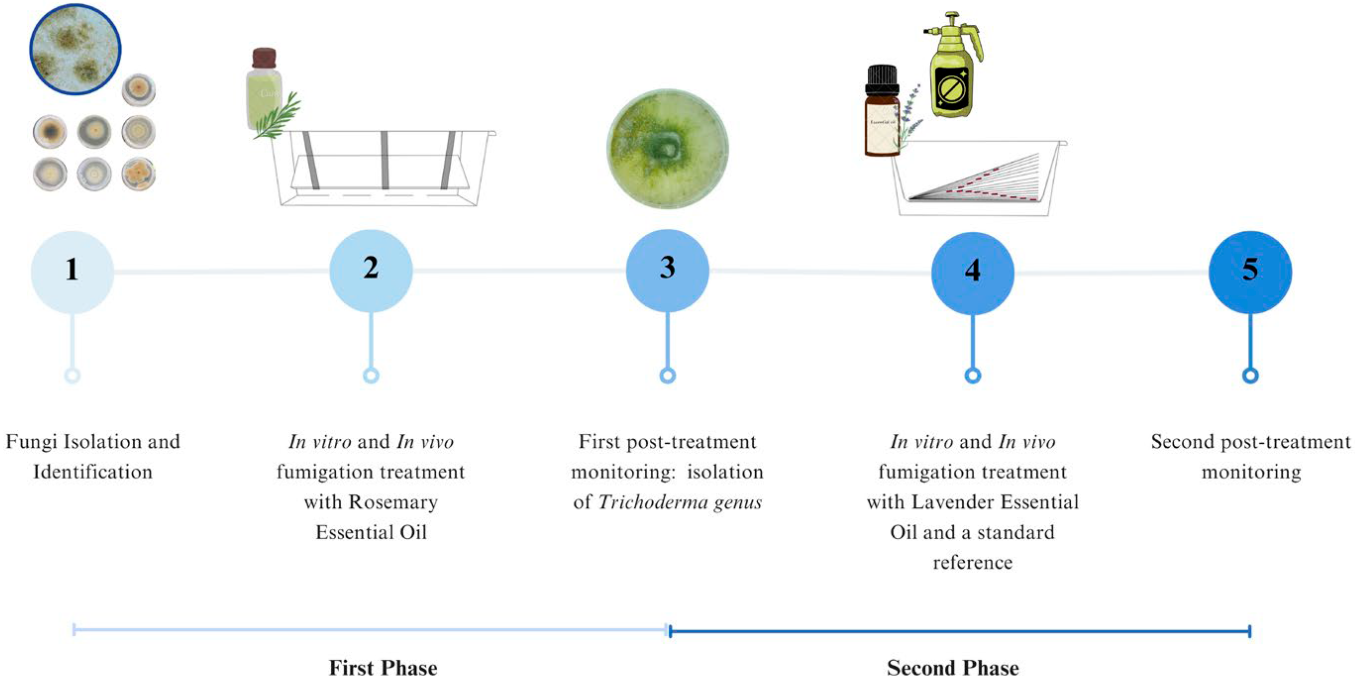
2.1. Experimental Timeline

The present study addresses the disinfection of an active fungal colonization on a historical paper-based document, which is the subject of the case study, through the use of essential oils by exposure to the vapor phase without direct contact with the artifact. The study is divided into two phases (Figure 1): the first phase involved the isolation and identification of fungal species and the evaluation of the biocidal activity of rosemary essential oil both in vitro and in vivo. One month after the treatment, during microbiological monitoring, a new sampling was carried out to assess the long-term effects of the treatment. The second phase involved the identification of a single new colonizer, belonging to the Trichoderma genus. This latter, which proved to be resistant to the activity of rosemary essential oil at the used dose level, underwent new tests to assess its sensitivity to lavender essential oil and a standard reference biocide. This phase also included an in vitro and an application study, as well as post-treatment monitoring to evaluate the durability of the second treatment over time.

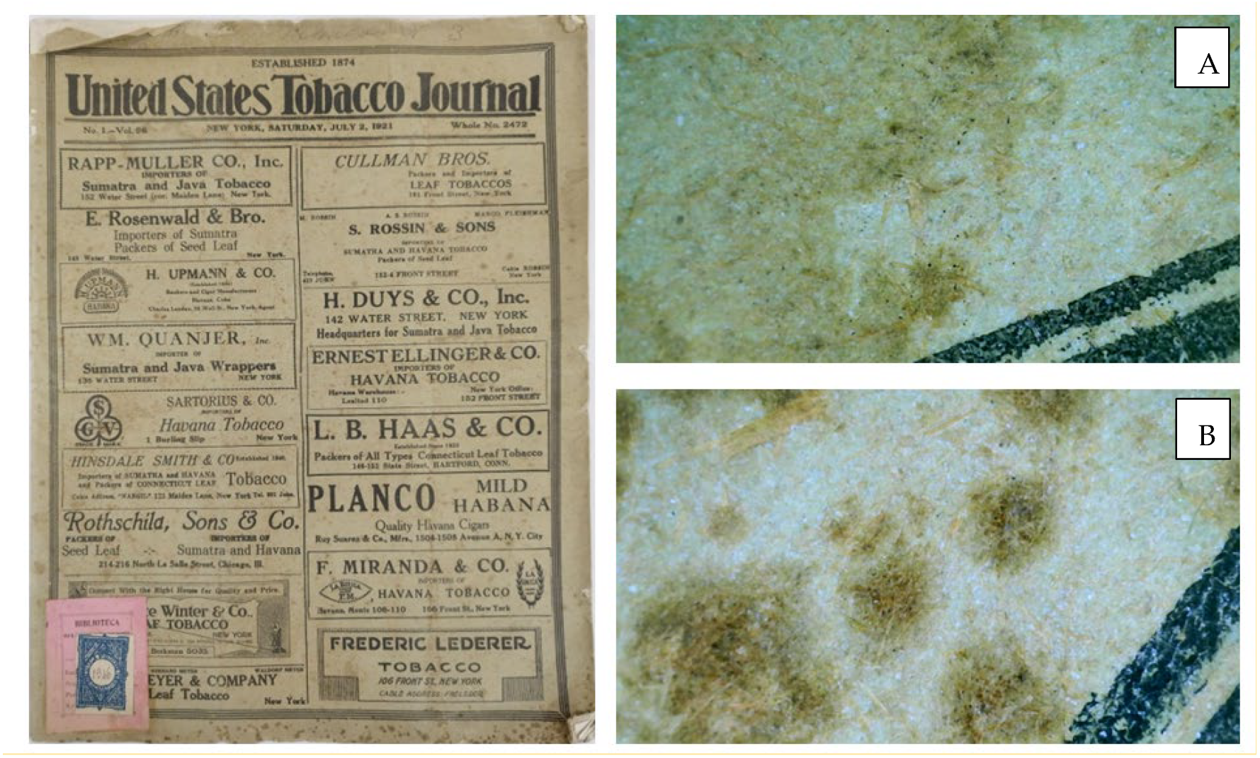
2.2. Case Study: The Archival Journal

The case study for this experiment is a historical journal, the “United States Tobacco Journal”, edited and published in New York in July 1921 (No. 1 Vol. 96). The journal is part of the historical archive collection at the CREA Research Center in Caserta. The journal has become noticeably yellowed due to the oxidation of the cellulose-based support, which resulted in fragility and some tears from wear. Indeed, the most significant preservation issue was the fungal colonization that has led to “foxing”, a type of paper degradation characterized by reddish-brown circular stains (Figure 2).

2.3. Fungal Sample Processing and Identification

To isolate fungi from the journal, sterile swabs were used, which were rubbed on the surface at different points. The portion of cotton was then inserted into a sterile 0.9% saline solution in an Eppendorf tube. Thus, 500 µL of the saline solution was plated onto Petri dishes (90 mm diameter) containing sterilized Potato Dextrose Agar (PDA; Conlab®) as the growth medium. The plates were incubated in the dark for 7 days at 22  ±  2 °C. A total of 7 fungi were isolated, whose molecular identification was carried out according to [18] for DNA extraction and DNA amplification by polymerase chain reaction (PCR) using ITS1 (TCCGTAGGTGAACCTGCGG) and ITS4 (TCCTCCGCTTATTGATATGC) primers. DNA was quantified using a Thermo ScientificTM NanoDropTM One UV-Vis spectrophotometer (Thermo Fisher Scientific Inc., Waltham, MA, USA), followed by sequencing performed by BMR Genomics in Padua. The obtained sequences were processed using Chromas Lite software 2.6.6 (Technelysium Pty Ltd., South Brisbane, Australia) for data cleaning. Finally, they were compared with the BLAST Nucleotide database to identify fungal species based on sequence similarity percentage (minimum threshold for percentage correspondence = 85%).

2.4. In Vitro Antifungal Activity of Rosemary Oil

The isolated and identified species were subjected to antifungal activity tests using a commercial rosemary essential oil (REO) from Rosmarinus officinalis (EUQEE®) through fumigation. Petri plates (90 mm diameter) were filled with 10 mL of PDA culture medium each, and a 0.4 mm diameter mycelial plug was inoculated for each tested fungus. Increasing volumes of the essential oil (20 μL, 40 μL, 60 μL, 80 μL, 100 μL) were tested. The oil was applied on the lid of the inverted Petri plate, which was then sealed with parafilm and incubated in the dark at 23 °C. The antifungal screening was conducted using a previously established method [18] designed to assess the activity of essential oil volatiles under in vitro conditions. Petri dishes were inverted to prevent direct contact between the essential oil and the fungal colonies, ensuring that observed effects were due solely to vapor-phase exposure. This setup simulates non-invasive fumigation conditions relevant to cultural heritage conservation.
The activity was monitored for up to 9 days. The experiment was conducted in triplicate. Inhibition percentages were calculated based on the mean values of treated fungi radial growth compared to untreated controls.

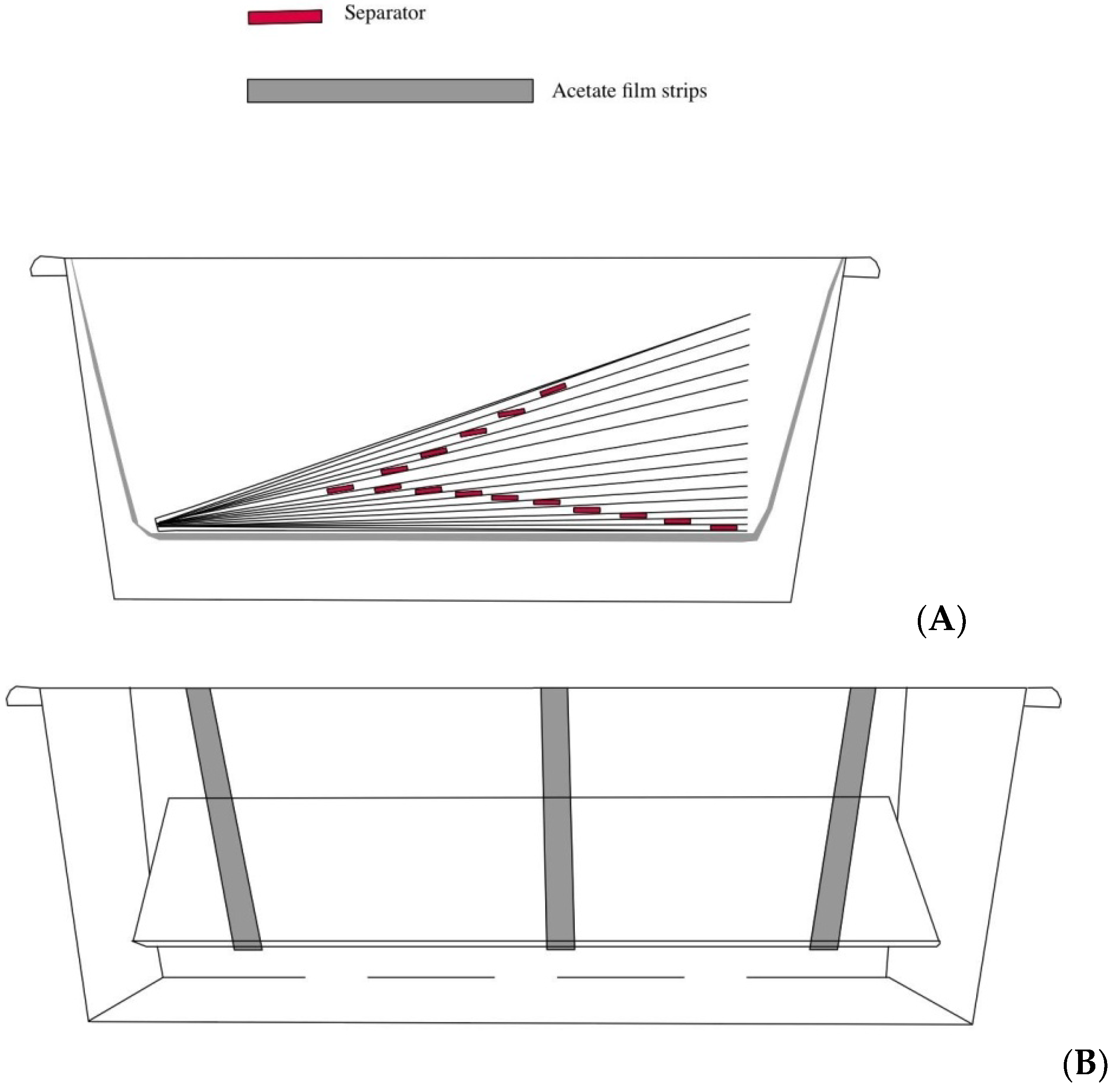
2.5. Evaluation of In Vivo Antifungal Properties of Rosemary Oil

Once the effective concentration (v/v) of essential oil to be used was determined, the experiment was performed in vivo by creating a “clean chamber” using a PET container (Figure 3). The clean chamber has a total volume of 0.06 m3 (75 cm × 40 cm × 20 cm). The journal was suspended inside the chamber using acetate film strips as supports (Figure 3A). Dividers in foam rubber (approximately 0.5 cm high) were inserted between the pages to separate each sheet and allow for uniform fumigation of all the surfaces (Figure 3B). The period of exposure to the rosemary oil vapor phase was 4 weeks. The pure oil was poured at the base of the clean chamber, beneath the suspended journal, ensuring no direct contact. Each concentration tested in vitro represents a percentage concentration relative to the total volume to be treated (20 μL = 0.2%, 40 μL = 0.4%, 60 μL = 0.6%, 80 μL = 0.8%, 100 μL = 1%). The concentration found to be the most effective in the in vitro assay (1%) was selected for the in vivo experiment: based on the volume of the chamber, a total of 60 mL of essential oil was used.

2.6. Post-Treatment Isolation and Morphological Identification

After the fumigation treatment, the “clean chamber” was opened, and the journal was relocated to the repository of the relevant archive. Resampling to monitor for efficacy and post-treatment colonization of the archive was carried out, as previously described in Section 2.3. Following a 7-day incubation period on Petri dishes, a single microorganism was isolated, morphologically similar to a fungus belonging to the genus Trichoderma. Morphological characterization was performed by observing macroscopic characteristics such as color and growth rate of fungal colonies isolated on PDA. Additionally, under an optical microscope (Olympus BH-2; Olympus Corporation, Tokyo, Japan), the fruiting bodies and conidia produced by 10–14-day-old fungal cultures grown on PDA plates were observed, using water as the mounting medium. Cultivation and each experiment were conducted in triplicate. Given the slow growth rate observed at a temperature of +22 °C [19], a comparison between growth rates of this fungus at 22 and 37 °C on PDA using 9 cm Petri dishes was made. In order to evaluate if this isolate had the characteristics of a human pathogen, the growth temperature was increased up to +37 °C. Upon observing improved and faster growth on the plates, it was hypothesized that the microorganism might be a potential opportunistic human pathogen. DNA extraction and sequencing for species identification were carried out at the Zooprophylactic Institute (Portici, Naples).

2.7. DNA Extraction, Sequencing, and Phylogenetic Analysis

DNA extraction and PCR were performed according to the protocol described in the literature [18]. Specifically, to identify the isolated fungus, the ITS1, ITS2 (GCTGCGTTCTTCATCGATGC), and TEF1A forward and reverse (CATCGAGAAGTTCGAGAAGG and AACTTGCAGGCAATGTGG) primers were utilized. The annealing temperatures used were 58 °C for the ITS primers and 55 °C for the TEF1A primers. PCR products were purified with ExoSAP-IT PCR Product Cleanup Reagent (Applied Biosystems, Foster City, CA, USA) and sequenced in both directions using the Big Dye Terminator v 1.1 Cycle Sequencing Kit, according to the manufacturer’s instructions. Capillary electrophoresis was performed on an ABI-Prism 3500 Genetic Analyzer (Applied Biosystems, Foster City, CA, USA). The sequences obtained were first analyzed using the SeqScanner 2.0 software (Applied Biosystems, Waltham, MA, USA) for quality check and sequence determination. DNA sequences were then compared with those available in the GenBank database by Basic Local Alignment Search Tool (BLAST; http://blast.ncbi.nlm.nih.gov/Blast.cgi; accessed on 3 March 2024) for taxonomic identification.
For the phylogenetic analysis, sequences of Trichoderma longibrachiatum (n = 26), isolated from human and soil samples, were obtained from the NCBI database. Multiple alignments of the ITS1, ITS2, and TEF1a sequences were performed using ClustalW in MEGA version 11.0.13 and manually adjusted. Phylogenetic analysis was conducted using the Maximum Likelihood (ML) method, assessing the topological confidence of the tree by maximum parsimony bootstrap proportion (MPBP) with 1000 replicates. The Hasegawa–Kishino–Yano model was identified as the best model for the combined sequences.

2.8. New Antifungal Treatment: The Essential Oils and the Commercial Standard

Following the post-treatment isolation, a colonization by a new fungal species was detected. Thus, to achieve complete disinfection of the surface, an in vitro comparison was made using the fumigation protocol with the previously used rosemary essential oil and a commercial lavender essential oil (La Saponaria®) at concentrations of 20 μL, 40 μL, 60 μL, 80 μL, 100 μL, and 120 μL. The aim was to assess whether the new isolated Trichoderma specie was sensitive to lavender oil (LEO) or to higher concentrations of REO (120 μL). Additionally, we tested a standard reference biocide based on benzalkonium chloride (Biotin T®, C.T.S. Italia), which is commonly used for the disinfection of artworks on organic supports [20]. As for the previous fumigation tests, 90 mm Petri dishes and PDA culture medium were used. The Biotin T was applied according to the guidelines suggested in the product technical data sheet: it was diluted at 3% in water and spread on the surface with a sterile spatula. Subsequently, a 4 mm diameter mycelial plug was inoculated. Based on data acquired, a new in vivo fumigation treatment was carried out as previously described, and, one month after the treatment, a new microbiological sampling followed in order to monitor the risk of a new colonization.

2.9. Colorimetric Analysis

In order to verify that the fumigation treatment does not cause any chromatic changes on the surface of the paper substrate, a colorimetric analysis was conducted before and after exposure to the treatment, measuring six sampling points on the front and six on the back of the journal. Each measurement was repeated three times. Colorimetric analysis was conducted using a Konica CM-3600d COLORFLEX-DIFF2 colorimeter (Reston, VA, USA), employing CIELab color parameters. The instrument was calibrated with a white standard tile. Changes in L* (lightness), a* (red–green coordinate), and b* (yellow–blue coordinate) were determined from the average values of each measurement. Total color variation (ΔE) was calculated using the following formula:
Δ E = L 1 L 2 2 + a 1 a 2 2   b 1 b 2 2
where L 1 , a 1   b 1 represent the control parameters [21]. The color variations were assessed following established procedures [22].

3. Results and Discussion

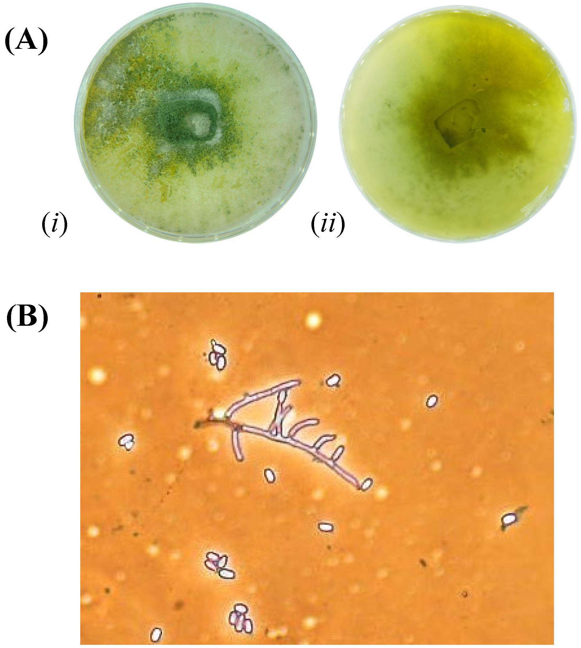
3.1. Fungi Isolation and Identification

Following the isolation process, seven fungi were identified, and their sequences were submitted to GenBank with their accession numbers: Alternaria alternata (PP989965-PP989966); Aspergillus niger (PP989960-PP989961); Aspergillus persii (PP989947-PP989948); Bjerkandera adusta (PP989945-PP989946); Coprinellus sp. (PP989939-PP989940); Hyphodermella rosae (PP989943-PP989944); and Penicillium chrysogenum (PP989950-PP989951) (Figure 4).
These genera, with the exception of Hyphodermella rosae, have been commonly found on paper (laid paper, wood pulp paper) [23] and cellulose textiles (cotton, linen) [24,25]. Species belonging to the genera Aspergillus sp., Alternaria sp., and Penicillium sp. are commonly isolated from artistic artifacts on organic supports, as they are environmental and airborne xerophilic fungi particularly adaptable to varying temperature and relative humidity conditions [1,26]. The literature reports that species belonging to these genera, specifically Aspergillus niger and Alternaria alternata, are associated with the presence of foxing, one of the main types of chemical and aesthetic degradation also observed in the present case study [27,28]. Penicillium chrysogenum has been previously associated with yellow-green stains [29], while Bjerkandera adusta is linked to dark brown foxing [30]. The mentioned species are known to release pigmented substances: the colorants often play biologically important roles in fungi, such as anti-bacterial activity, and they are also light-absorbing molecules that can protect the organism from UV damage, including melanins, polyketides, and carotenoids [31]. In the case of Coprinellus sp. and Hyphodermella rosae, these are known ligninolytic fungi often found in open environments as colonizers of tree species and lower plants [32,33].

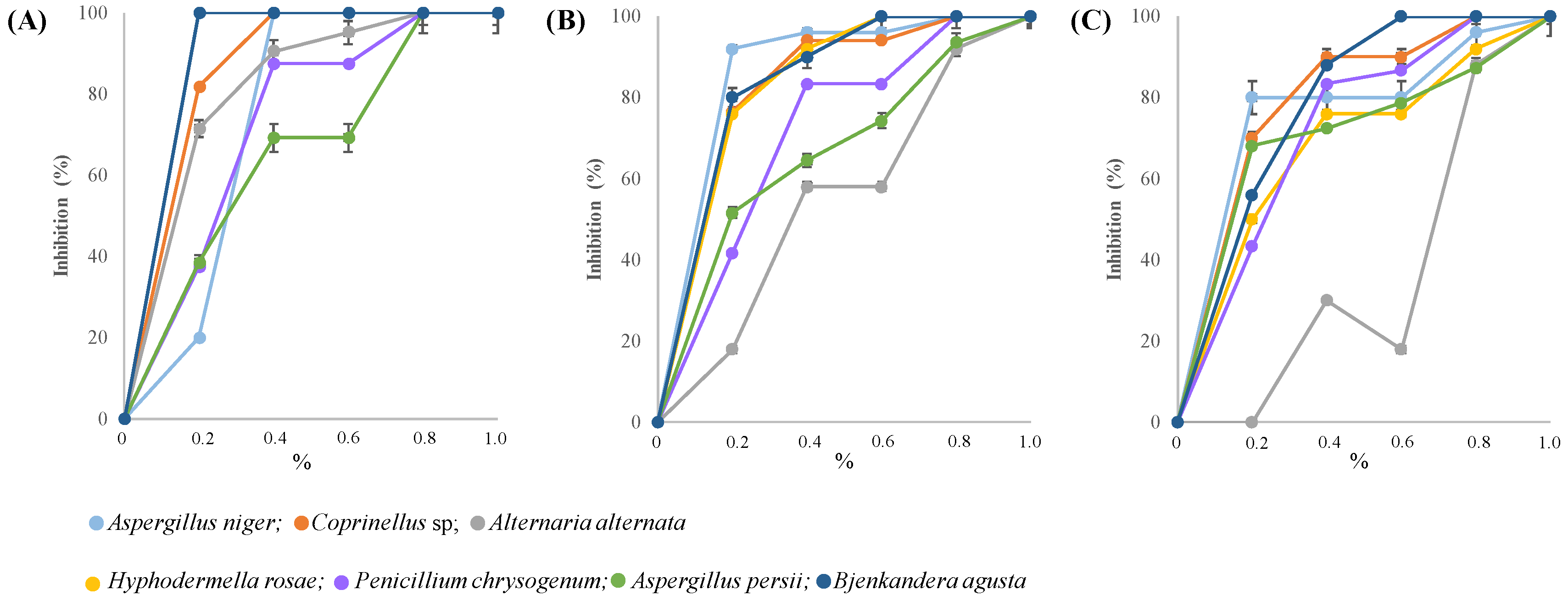
3.2. In Vitro and In Vivo Antifungal Activity of Rosemary Oil

The results of the antifungal activity on the seven identified fungi are presented in Figure 5 (panels A–C)) for the three monitoring periods after 3, 6, and 9 days of observation. All observed fungi show sensitivity to rosemary essential oil, demonstrating good inhibition within the first 3 days even at concentrations of 0.4–0.6% v/v. However, as previously highlighted in the literature, the antifungal trend of essential oils is species-specific, dose-, and time-dependent. After 9 days, differences in sensitivity could be observed, as in the case of Aspergillus niger, which was inhibited by 80% with the 0.2% concentration, while the same concentration was not sufficient to halve radial growth in Alternaria alternata, Hyphodermella rosae, and Penicillium chrysogenum. A concentration of 0.6% was sufficient to inhibit by 50% the growth of all fungi except Alternaria alternata, which remained the most resistant species among those tested. Fumigation with a 0.8% concentration induced a 90% growth inhibition in all species. However, only a 1% concentration guaranteed a biocidal action, resulting in complete inhibition over time. For some of the species isolated in this study, there is already evidence in the literature of their sensitivity to essential oils: in other applications, such as food preservatives, these oils have been used against Alternaria alternata, Penicillium chrysogenum, and Aspergillus niger [34,35]. In fact, researchers have found that rosemary essential oil (REO) exhibits a remarkable ability to disrupt fungal growth by inhibiting ergosterol synthesis in organisms, damaging fungal cell walls and membranes, and inhibiting DNA and protein synthesis. Some examples in the literature show good antifungal activity at concentrations much higher than those reported in this study: while it is reported that concentrations between 25 and 100 μL/mL inhibit growth, this study demonstrates that concentrations of 2–8 μL/mL already ensure growth reduction, and a concentration of 10 μL/mL provides biocidal activity [17].
However, it is important to consider that variability in antifungal effectiveness results from multiple factors: the specific chemical composition of the oil in terms of bioactive compounds, which in turn varies depending on the extraction method, the plant species, the time and place of harvest [36,37], and the specific sensitivity of the fungus to monoterpenes and monoterpenoids, such as α- and β-pinene, 1,8-cineole, camphene, and camphor [16,38]. According to the literature, these compounds appear to be more effective when used together rather than individually, as they act synergistically, triggering multiple mechanisms of fungal growth deactivation and broadening their effectiveness spectrum [38].
Following the in vitro results that demonstrated complete inhibition of fungal growth for the tested species at an oil concentration of 10 μL/mL, the artifact was subjected to a fumigation treatment for 4 weeks as described, using a 1% oil concentration relative to the treated volume, for a total of 60 mL.

3.3. Trichoderma longibrachiatum Identification and Phylogenetic Analysis

After 4 weeks of treatment, the journal was returned to its storage location. A follow-up sampling was conducted 1 month post-treatment to assess the efficacy of REO against the fungi and to determine whether new colonization by other species had occurred. Microbiological sampling led to the isolation of a single new species that was morphologically identified as Trichoderma. Observation of the plates revealed colonies with shades of green ranging from very bright to dark, while the reverse side was light yellow (Figure 6A). Under the microscope, the conidia exhibited a subglobose ellipsoidal shape, measuring 4.5–6 × 2.2–3.3 µm. The conidiophores were characterized by long primary branches, which rarely re-branched (Figure 6B). In addition, the growth rate of this fungus was higher at 37 °C (after 2 days, the fungus completely colonized the 9 cm Petri dish), than at 22 °C (there was less than 30 percent colonization).
All observed morphological features and growth characteristics are typical of some species of the genus Trichoderma and particularly with the species longibrachiatum [39,40]. This species exhibits a dual nature as both a biocontrol agent and a potential human pathogen. Furthermore, it is recognized for its high production of hydrolytic enzymes such as cellulase and chitinase, which are involved in the decomposition of organic materials and in mechanisms for responding to fungal phytopathogens [41]. T. longibrachiatum infections include invasive sinusitis, pulmonary infections, and peritonitis, highlighting its potential as an opportunistic pathogen [42]. Since Trichoderma is both a pathogenic fungus for paper-based materials and an opportunistic pathogen for humans, it poses a potential threat not only to artistic artifacts but also to those who come in contact with them. These different implications for human health necessitate the accurate identification of the Trichoderma species. In fact, the Trichoderma genus is taxonomically complex, often requiring more than standard ITS sequencing for accurate species-level identification [43]. For this reason, to definitively confirm the identity of the isolated species, molecular investigations were conducted through sequencing, using ITS1 and ITS2 primers, as well as TEF1a, a primer specific for species identification within the Trichoderma genus. The results showed a 100% sequence identity with Trichoderma longibrachiatum for the ITS1 and ITS2 primers (PQ107527) and for TEF1a primers (PQ118398.1).
The presence of T. longibrachiatum on paper items has also been documented in previous studies [4]. As previously introduced, this species of Trichoderma is rich in ligninolytic and cellulolytic enzymes, which make cultural heritage plant-based items (such as wood, paper, and canvas) particularly susceptible to this type of microorganism. In fact, many species belonging to the Trichoderma genus produce cellulases in significant quantity, enough to be extracted and used for industrial manufacturing [44]. In Figure 7, the phylogenetic tree is presented to illustrate the position of the T. longibrachiatum isolate obtained from the journal of the present study, in comparison with other sequences of T. longibrachiatum isolated from both human sources and soil. As can be observed, our isolate is closely related to the one [19] derived from foot skin and to an isolate from fungal keratitis of the cornea in an Indian patient [45].
This finding confirms the clinical relevance of the sequence isolated from the journal. Having found a correlation specifically with keratosis, an operator who comes into contact with the contaminated material can easily trigger an inflammatory process or a more severe pathology, particularly if there is a pre-existing immunocompromised condition of the visual system. Molecular phylogenetic studies have revealed that, in most cases, members of this clade can be characterized by their ability to grow at 37 °C, i.e., body temperature, which is a prerequisite for a successful deep infection in humans [19]. In addition to the ability to grow at body temperature, further possible virulence factors of Trichoderma during human infections include the production of extracellular proteolytic enzymes and secondary metabolites toxic to mammalian cells. Antifungal susceptibility studies performed on case isolates or sets of clinical Trichoderma strains have revealed that several isolates are resistant to various antifungal drugs used in clinical practice. Additionally, the risk of bio-resistance increases considering that these species, isolated from plants and soil, are constantly exposed to fungicides used in agriculture, which are often chemically similar to those used in clinical settings [45].

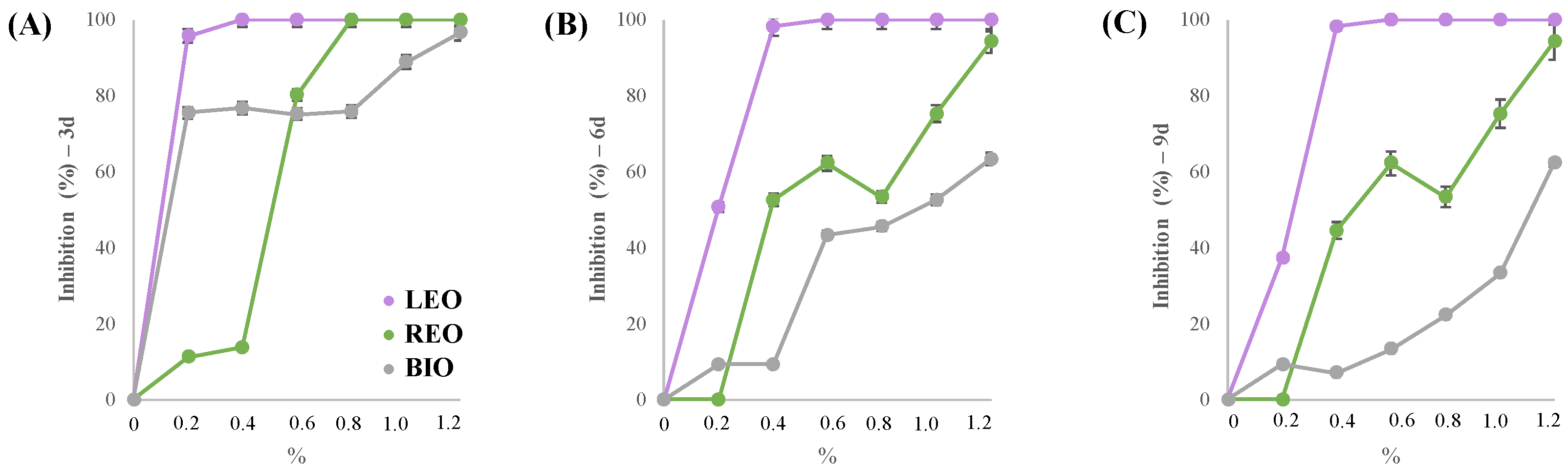
3.4. In Vitro and In Vivo Assessment of Trichoderma longibrachiatum Sensitivity

The results of the in vitro antifungal tests on Trichoderma longibrachiatum treated with commercial rosemary oil, lavender oil, and a standard biocide based on benzalkonium chloride are shown in the graphs (Figure 8A–C). The inhibitory activity of rosemary essential oil is reported: within the first 3 days, it appears to guarantee 100% inhibition at a concentration of 8 μL/mL. However, when observing the other two monitoring times, the efficacy percentage significantly decreases by the sixth day, when total inhibition of growth is no longer guaranteed. At the last monitoring time, it is evident that the highest tested concentration is sufficient to achieve about 90% inhibition. This result explains why the journal was colonized by Trichoderma after the first treatment: the species identified during the initial isolation were treated with a 1% oil concentration, to which they responded with total inhibition in the in vitro test. However, in this case, we see that the 1% concentration (10 μL/mL) induced a 75% reduction in growth: evidently, the treatment was effective against the initial colonizers, but it was not enough to prevent a new colonization by Trichoderma, which proved to be more resistant to rosemary oil. On the other hand, the 1.2% concentration would have ensured 94% inhibition, still not sufficient to completely stop the growth of Trichoderma. It is important to consider that Trichoderma longibrachiatum produces a large number of conidia, as observed in optical microscopy images, so even a growth inhibition percentage of over 90% may not be sufficient over time to prevent colonization of the substrate. As for lavender essential oil, a different response can be observed: the antifungal activity was quite stable even at concentrations of 0.4%, ensuring growth inhibition of over 98%. Studies on the sensitivity of Trichoderma species to essential oils are quite rare in the literature and are mostly related to other species such as T. harzianum [46] and T. viride [47]. In the first case, it is evident that lavender essential oil can reduce Trichoderma sporulation but not radial growth. In the second study, lavender oil, rosemary oil, and BAC were used against T. viride, as in the present study, with interesting results: the fumigation treatment with LEO and REO was generally less effective than the treatment with benzalkonium chloride. Specifically, in the case of REO, the MIC was higher than that obtained in the present study (100 μL/mL vs. 10 μL/mL), and for LEO, the inhibiting concentration was 80 μL/mL compared to the 4 μL/mL presented here. The most effective in the cited study was BAC, with inhibitory activity at concentrations of 0.25–2 μL/mL, which was significantly lower than those obtained in this study. However, as previously mentioned, in this study, the BAC-based mixture was used at 3% compared to the 10% used in the literature, which may explain the substantial difference in inhibition values for the tested concentrations. In the graphs, it is shown that, at the same tested volume, BAC by contact is much less effective than essential oils by fumigation, particularly when compared to lavender oil.
Other studies on T. aggressivum [48] and T. orientale [49] have demonstrated a dose-dependent and species-specific sensitivity of different essential oils toward species within the same genus. In only one study, T. longibrachiatum was isolated from archeological mummified skin and exposed to oregano essential oil in its vapor phase, showing a growth inhibition at 200 μL (about 33 μL/mL).
Following the obtained results, the artifact underwent a four-week fumigation treatment with lavender essential oil at a concentration of 0.4%. After a month from the end of the treatment, further microbiological samplings were carried out to evaluate the long-term effectiveness of the second intervention

3.5. Microbiological Monitoring

The second isolation was conducted to evaluate the performance of the lavender essential oil treatment and its durability 1 month after the intervention. The third sampling, finally, did not show any presence of colonization, proving the effectiveness of LEO against Trichoderma. This result demonstrates that Trichoderma exhibits high sensitivity to the volatile components of lavender oil at a concentration of 0.4% v/v and less sensitivity toward the components of rosemary oil, which requires at least 1.2% v/v. As highlighted in other studies, the synergistic use of multiple essential oils (and more specifically, the combination of their characteristic volatile components) can optimize fumigant treatment and broaden its spectrum of action. As already illustrated for rosemary essential oil, in the case of lavender oil as well, the main bioactive components consist of monoterpenes and monoterpenoids, some of which are shared with other aromatic plant species belonging to the Lamiaceae family, including Rosmarinus officinalis. The most common monoterpenoids of lavender EOs are alcohols (linalool, terpinen-4-ol, α-terpineol, borneol, lavandulol), esters (linalyl acetate, lavandulyl acetate), ketones (camphor, fenchone), and oxides (1,8-cineole) [50]. Although the variability in the antifungal efficacy of essential oils is well known [51], they currently represent a valid alternative to traditional biocides used in the restoration of organic-media artistic objects, as evidenced by recent studies [10,11,12,14,52]. The literature shows examples where the mixture of multiple essential oils is more effective than its individual components [53], allowing for the complete disinfestation of the treated surface. The experiment replicated in our case study, subjected to double fumigation with REO and LEO, demonstrates the potential of alternating treatments based on different essential oils: in addition to broadening the spectrum of efficacy, this approach avoids the periodic reuse of the same active ingredient and, consequently, the risk of bio-resistance development. The principle of rotation in the use of biocidal agents is strategically applied to limit the target microorganisms from developing an adaptive response, rendering the substance ineffective over time [54].

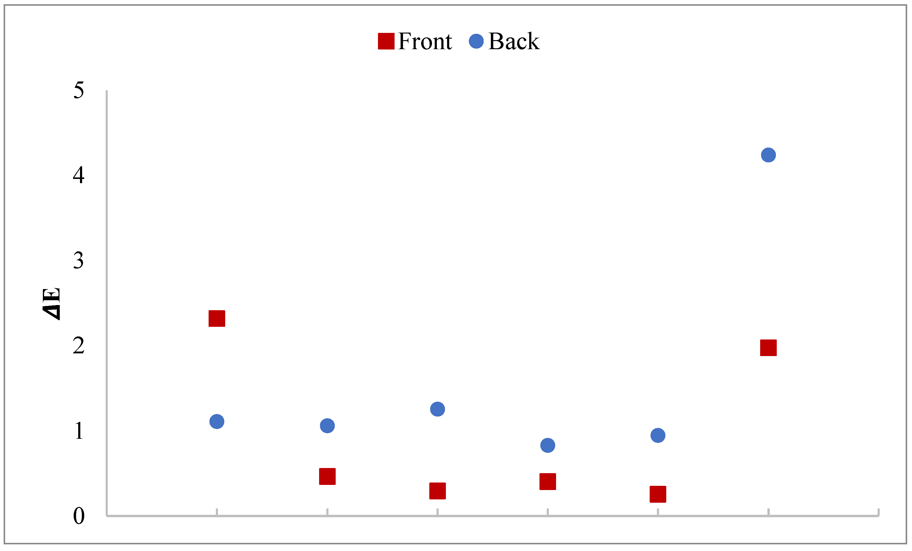
3.6. Data from Colorimetric Measurements

The graph shows the results of the colorimetric analysis of six measurement points on the front and back cover of the journal, expressed as total color variation (ΔE) obtained by comparing the values measured before and after the fumigation treatments (Figure 9). The measurements show mostly ΔE values in the range between 0 and 2.5, which are considered negligible since they are barely noticeable [21]. Nonetheless, even a slight chromatic variation may be considered an acceptable compromise when weighed against the colorimetric alterations resulting from microbiological degradation.
Therefore, it can be concluded that the two fumigation treatments did not induce any chromatic changes on the surface, and this protocol can be regarded as safe for the preservation of paper-based materials.

4. Conclusions

It is well known that essential oils exhibit biocidal activity with species-specific behavior, which can limit their effectiveness. By combining their use in the modality proposed in this study, it is possible to ensure the effectiveness of the intervention, even against potential new colonization. As demonstrated in previous studies, the combined or alternating use of multiple essential oils may enhance the breadth of antifungal efficacy across a wider array of microorganisms, owing to distinct mechanisms of action that vary according to the specific target organism. A similar approach is employed in agricultural practices, where pest management strategies are based on the rotational application of biocidal agents. This is done not only to expand the spectrum of activity but also to mitigate the risk of adaptive responses and, consequently, the emergence of bio-resistance [55]. In fact, the effect achieved by rosemary oil against the first fungal consortium persisted over time, as the fungal species had not been re-isolated after the first treatment. However, the Trichoderma that subsequently colonized the journal was not sensitive to the tested concentrations of REO. The good sensitivity observed toward lavender oil allowed for the eradication of the second colonizer, and, as shown by the results of the third and final sampling 1 month after, the in vivo treatment maintained good durability. The importance of performing biocidal treatments in this case is crucial to ensure the safety of both the artwork and those who interact closely with it. As highlighted in previous studies [56], the conservation environment of historic and artistic artifacts—whether a library, archive, museum, or other type of heritage repository—must ensure not only the preservation of the objects it houses but also the health and safety of those who work in or visit these spaces. Therefore, in order to guarantee appropriate safety conditions, it is essential to conduct regular environmental and microbiological monitoring to assess and control air quality. The use of essential oils in this context has proven to be a viable and safe substitute for traditional biocides based on BAC (benzalkonium chloride) mixtures, which have shown certain limitations. The first aspect to consider is that Trichoderma was not sensitive to the tested concentrations of commercial biocide, which would therefore require much larger quantities for treatment compared to the essential oils. Secondly, commercial products based on quaternary ammonium salts have recently been investigated for their safety profiles and ecotoxicity, as well as for the growing development of bio-resistance associated with their intensive use. The data obtained from the experiments confirmed the validity of the in vitro results, allowing fumigation treatment to be recognized as a real alternative to traditional treatments. The limitations emerging from the present study, as also reflected in the cited literature, concern the fact that, at this stage, intervention strategies have primarily focused on localized actions targeting individual artifacts. However, it would be advisable to expand the scope of research with the aim of developing a standardized protocol for the systematic disinfection of entire collections. Although the artifact examined in this study, once reintroduced into its original storage context, demonstrated, after 1 month, a satisfactory resistance to colonization and recolonization by previously identified microbial species, it remains evident that interventions should preferably target the conservation environment as a whole rather than isolated objects. Indeed, from a microbiological standpoint, among others, an artifact cannot be considered independently from its conservation environment. Therefore, further research is necessary to explore comprehensive rather than case-specific solutions.
Finally, in the frame of the study conducted, personal protective equipment (PPE) must also be considered crucial when coming into contact with colonized artworks. As demonstrated in the presented study, the fungal consortium could contain potential human pathogens and therefore, to ensure the safety of workers and consultants, it will be essential to equip oneself with protective gloves and masks to minimize the risk of contact as much as possible.

Author Contributions

B.P. carried out the in vitro and in vivo antifungal assays, curated the data and visualizations, performed the morphological analysis on Trichoderma, and drafted the manuscript; M.C.S. carried out fungal isolation and identification and contributed to the drafting of the related section; G.B., M.G.R., and R.P. carried out the phylogenetic analysis and sequencing of Trichoderma and drafted the related section; M.C.G. supervised the colorimetric analysis; E.L. and S.P. supervised the work and revised the manuscript. All authors have read and agreed to the published version of the manuscript.

Funding

This research received no external funding.

Institutional Review Board Statement

Not applicable.

Data Availability Statement

The original contributions presented in this study are included in the article. Further inquiries can be directed to the corresponding author(s).

Conflicts of Interest

The authors declare that they have no competing interests.

Abbreviations

BACBenzalkonium chloride
LEOLavender essential oil
REORosemary essential oil

References

  1. Branysova, T.; Demnerova, K.; Durovic, M.; Stiborova, H. Microbial biodeterioration of cultural heritage and identification of the active agents over the last two decades. J. Cult. Herit. 2022, 55, 245–260. [Google Scholar] [CrossRef]
  2. Pinheiro, A.C.; Sequeira, S. Mycological Studies in Cultural Heritage. In Encyclopedia of Mycology; Elsevier: Amsterdam, The Netherlands, 2021; pp. 27–39. [Google Scholar] [CrossRef]
  3. Tiano, P. Biodegradation of Cultural Heritage: Decay Mechanisms and Control Methods. ARIADNE 9 Work. Hist. Mater. Their Diagn. 2009, 2, 7–12. [Google Scholar]
  4. Pinheiro, A.C.; Sequeira, S.O.; Macedo, M.F. Fungi in archives, libraries, and museums: A review on paper conservation and human health. Crit. Rev. Microbiol. 2019, 45, 686–700. [Google Scholar] [CrossRef] [PubMed]
  5. Kredics, L.; Naeimi, S.; Hatvani, L.; Vágvölgyi, C.; Cai, F.; Druzhinina, I.S.; Manczinger, L. “The Good, the Bad and the Ugly” in the shades of green: The genus Trichoderma in the spotlight. Indian Phytopathol. 2021, 74, 403–411. [Google Scholar] [CrossRef]
  6. Woo, S.L.; Hermosa, R.; Lorito, M.; Monte, E. Trichoderma: A multipurpose, plant-beneficial microorganism for eco-sustainable agriculture. Nat. Rev. Microbiol. 2023, 21, 312–326. [Google Scholar] [CrossRef]
  7. Kuhls, K.; Lieckfeldt, E.; Samuels, G.J.; Kovacs, W.; Meyer, W.; Petrini, O.; Gams, W.; Börner, T.; Kubicek, C.P. Molecular evidence that the asexual industrial fungus Trichoderma reesei is a clonal derivative of the ascomycete Hypocrea jecorina. Proc. Natl. Acad. Sci. USA 1996, 93, 7755–7760. [Google Scholar] [CrossRef]
  8. Rakotonirainy, M.S.; Lavédrine, B. Screening for antifungal activity of essential oils and related compounds to control the biocontamination in libraries and archives storage areas. Int. Biodeterior. Biodegrad. 2005, 55, 141–147. [Google Scholar] [CrossRef]
  9. Borrego, S.; Valdés, O.; Vivar, I.; Lavin, P.; Guiamet, P.; Battistoni, P.; ’Omez De Saravia, S.G.; Borges, P.; Fung, D.Y.C.; Velge, P. Essential Oils of Plants as Biocides against Microorganisms Isolated from Cuban and Argentine Documentary Heritage. ISRN Microbiol. 2012, 2012, 826786. [Google Scholar] [CrossRef]
  10. Čabalová, I.; Češek, B.; Mikala, O.; Gojný, J.; Kačík, F.; Tribulová, T. The influence of selected efficient compounds of essential oils for paper protection. J. Cult. Herit. 2019, 37, 148–154. [Google Scholar] [CrossRef]
  11. Ventorino, V.; La Storia, A.; Robertiello, A.; Corsi, S.; Romano, I.; Sannino, L.; Pepe, O. Fungal Biodeterioration and Preservation of Miniature Artworks. J. Fungi 2023, 9, 1054. [Google Scholar] [CrossRef]
  12. Tomić, A.; Šovljanski, O.; Nikolić, V.; Pezo, L.; Aćimović, M.; Cvetković, M.; Stanojev, J.; Kuzmanović, N.; Markov, S. Screening of Antifungal Activity of Essential Oils in Controlling Biocontamination of Historical Papers in Archives. Antibiotics 2023, 12, 103. [Google Scholar] [CrossRef] [PubMed]
  13. Menicucci, F.; Palagano, E.; Michelozzi, M.; Ienco, A. Essential Oils for the Conservation of Paper Items. Molecules 2023, 28, 5003. [Google Scholar] [CrossRef] [PubMed]
  14. Bosco, F.; Mollea, C.; Demichela, M.; Fissore, D. Application of Essential Oils to Control the Biodeteriogenic Microorganisms in Archives and Libraries. Heritage 2022, 5, 2181–2195. [Google Scholar] [CrossRef]
  15. Paolino, B.; Sorrentino, M.C.; Macchia, A.; Troisi, J.; Zaratti, C.; Hansen, A.; Ilardi, S.; Russo, G.; Lahoz, E.; Pacifico, S. Lavender essential oil for a contactless application for contemporary art conservation: A case study. Npj Herit. Sci. 2025, 13, 8. [Google Scholar] [CrossRef]
  16. Jiang, Y.; Wu, N.; Fu, Y.-J.; Wang, W.; Luo, M.; Zhao, C.-J.; Zu, Y.-G.; Liu, X.-L. Chemical composition and antimicrobial activity of the essential oil of Rosemary. Environ. Toxicol. Pharmacol. 2011, 32, 63–68. [Google Scholar] [CrossRef]
  17. Yang, J.; Goksen, G.; Zhang, W. Rosemary essential oil: Chemical and biological properties, with emphasis on its delivery systems for food preservation. Food Control 2023, 154, 110003. [Google Scholar] [CrossRef]
  18. Paolino, B.; Sorrentino, M.C.; Troisi, J.; Carri, M.D.; Kiselev, P.; Raimondo, R.; Lahoz, E.; Pacifico, S. Lavandula angustifolia mill. for a suitable non-invasive treatment against fungal colonization on organic-media cultural heritage. Herit. Sci. 2024, 12, 53. [Google Scholar] [CrossRef]
  19. Hatvani, L.; Manczinger, L.; Vágvölgyi, C.; Kredics, L. Trichoderma as a human pathogen. In Trichoderma: Biology and Applications; CABI: Wallingford, UK, 2013; pp. 292–313. [Google Scholar] [CrossRef]
  20. Paolino, B.; Sorrentino, M.C.; Pacifico, S. Greener solutions for biodeterioration of organic-media cultural heritage: Where are we? Herit. Sci. 2024, 12, 334. [Google Scholar] [CrossRef]
  21. Karbowska-Berent, J.; Górniak, B.; Czajkowska-Wagner, L.; Rafalska, K.; Jarmiłko, J.; Kozielec, T. The initial disinfection of paper-based historic items-Observations on some simple suggested methods. Int. Biodeterior. Biodegrad. 2018, 131, 60–66. [Google Scholar] [CrossRef]
  22. UNI 8941; Superfici Colorate-Metodi di Misura e Valutazione delle Variazioni di Colore. Milano UNI., Ente Nazionale Italiano di Normazione (UNI): Via Sannio, Italy, 1986.
  23. Savković, Ž.; Stupar, M.; Unković, N.; Knežević, A.; Vukojević, J.; Grbić, M.L. Fungal Deterioration of Cultural Heritage Objects. In Biodegradation Technology of Organic and Inorganic Pollutants; IntechOpen: London, UK, 2022. [Google Scholar] [CrossRef]
  24. Sterflinger, K. Fungi: Their role in deterioration of cultural heritage. Fungal Biol. Rev. 2010, 24, 47–55. [Google Scholar] [CrossRef]
  25. Skóra, J.; Gutarowska, B.; Pielech-Przybylska, K.; Stępień, Ł.; Pietrzak, K.; Piotrowska, M.; Pietrowski, P. Assessment of microbiological contamination in the work environments of museums, archives and libraries. Aerobiologia 2015, 31, 389–401. [Google Scholar] [CrossRef] [PubMed]
  26. Bastholm, C.J.; Madsen, A.M.; Andersen, B.; Frisvad, J.C.; Richter, J. The mysterious mould outbreak—A comprehensive fungal colonisation in a climate-controlled museum repository challenges the environmental guidelines for heritage collections. J. Cult. Herit. 2022, 55, 78–87. [Google Scholar] [CrossRef]
  27. Nol, L.; Henis, Y.; Kenneth, R.G. Biological factors of foxing in postage stamp paper. Int. Biodeterior. Biodegrad. 2001, 48, 98–104. [Google Scholar] [CrossRef]
  28. Borrego, S.; Lavin, P.; Perdomo, I.; de Saravia, S.G.; Guiamet, P. Determination of Indoor Air quality in archives and biodeterioration of the documentary heritage. ISRN Microbiol. 2012, 2012, 680598. [Google Scholar] [CrossRef]
  29. Szczepanowska, H.; Lovett, C.M. A Study of the Removal and Prevention of Fungal Stains on Paper. J. Am. Inst. Conserv. 1992, 31, 147–160. [Google Scholar] [CrossRef]
  30. Rakotonirainy, M.S.; Heude, E.; Lavédrine, B. Isolation and attempts of biomolecular characterization of fungal strains associated to foxing on a 19th century book. J. Cult. Herit. 2007, 8, 126–133. [Google Scholar] [CrossRef]
  31. Stickings, C.E.; Raistrick, H. Chemistry of the Fungi. Annu. Rev. Biochem. 1956, 25, 225–256. [Google Scholar] [CrossRef]
  32. Ainsworth, A.M.; Liimatainen, K. Hyphodermella rosae: A “twig-welding” corticioid new to Britain. Field Mycol. 2019, 20, 43–46. [Google Scholar] [CrossRef]
  33. Schilling, M.; Schilling, M.; Farine, S.; Péros, J.-P.; Bertsch, C.; Gelhaye, E. Wood degradation in grapevine diseases. Adv. Bot. Res. 2021, 99, 175–207. [Google Scholar] [CrossRef]
  34. Phillips, C.A.; Laird, K.; Allen, S.C. The use of Citri-VTM®—An antimicrobial citrus essential oil vapour for the control of Penicillium chrysogenum, Aspergillus niger and Alternaria alternata in vitro and on food. Food Res. Int. 2012, 47, 310–314. [Google Scholar] [CrossRef]
  35. Krisch, J. Activity of essential oils in vapor phase against bread spoilage fung. Acta Biol. Szeged. 2013, 57, 9–12. [Google Scholar]
  36. Melito, S.; Petretto, G.L.; Chahine, S.; Pintore, G.; Chessa, M. Seasonal Variation of Essential Oil in Rosmarinus officinalis Leaves in Sardinia. Nat. Prod. Commun. 2019, 14, 1934578X1986400. [Google Scholar] [CrossRef]
  37. Rathore, S.; Mukhia, S.; Kapoor, S.; Bhatt, V.; Kumar, R.; Kumar, R. Seasonal variability in essential oil composition and biological activity of Rosmarinus officinalis L. accessions in the western Himalaya. Sci. Rep. 2022, 12, 3305. [Google Scholar] [CrossRef]
  38. Micić, D.; Đurović, S.; Riabov, P.; Tomić, A.; Šovljanski, O.; Filip, S.; Tosti, T.; Dojčinović, B.; Božović, R.; Jovanović, D.; et al. Rosemary Essential Oils as a Promising Source of Bioactive Compounds: Chemical Composition, Thermal Properties, Biological Activity, and Gastronomical Perspectives. Foods 2021, 10, 2734. [Google Scholar] [CrossRef]
  39. Yabuki, T.; Miyazaki, K.; Okuda, T. Japanese species of the Longibrachiatum Clade of Trichoderma. Mycoscience 2014, 55, 196–212. [Google Scholar] [CrossRef]
  40. Haouhach, S.; Karkachi, N.; Oguiba, B.; Sidaoui, A.; Chamorro, I.; Kihal, M.; Monte, E. Three New Reports of Trichoderma in Algeria: T. atrobrunneum, (South) T. longibrachiatum (South), and T. afroharzianum (Northwest). Microorganisms 2020, 8, 1455. [Google Scholar] [CrossRef]
  41. Kubicek, C.P.; Bölzlbauer, U.M.; Kovacsa, W.; Mach, R.L.; Kuhlsb, K.; Lieckfeldtb, E.; Börnerb, T.; Samuels, G.J. Cellulase production by species of Trichoderma sect. Longibrachiatum and of Hypocrea species with anamorphs referable to Trichoderma sect. Longibrachiatum. Fungal Genet. Biol. 1996, 20, 105–114. [Google Scholar] [CrossRef]
  42. Sautour, M.; Chrétien, M.; Valot, S.; Lafon, I.; Basmaciyan, L.; Legouge, C.; Verrier, T.; Gonssaud, B.; Abou-Hanna, H.; Dalle, F.; et al. First case of proven invasive pulmonary infection due to Trichoderma longibrachiatum in a neutropenic patient with acute leukemia. J. Mycol. Médicale 2018, 28, 659–662. [Google Scholar] [CrossRef]
  43. Druzhinina, I.; Kubicek, C.P. Species concepts and biodiversity in Trichoderma and Hypocrea: From aggregate species to species clusters? J. Zhejiang Univ.-Sci. B 2005, 6, 100–112. [Google Scholar] [CrossRef]
  44. Gordillo-Fuenzalida, F.; Echeverria-Vega, A.; Cuadros-Orellana, S.; Faundez, C.; Kähne, T.; Morales-Vera, R. Cellulases Production by a Trichoderma sp. Using Food Manufacturing Wastes. Appl. Sci. 2019, 9, 4419. [Google Scholar] [CrossRef]
  45. Hatvani, L.; Homa, M.; Chenthamara, K.; Cai, F.; Kocsubé, S.; Atanasova, L.; Mlinaric-Missoni, E.; Manikandan, P.; Revathi, R.; Dóczi, I.; et al. Agricultural systems as potential sources of emerging human mycoses caused by Trichoderma: A successful, common phylotype of Trichoderma longibrachiatum in the frontline. FEMS Microbiol. Lett. 2019, 366, fnz246. [Google Scholar] [CrossRef] [PubMed]
  46. Gorski, R.; Krzysztof, S.; Siwulski, M.; Katarzyna, G. Effect of selected natural essential oils on in vitro development of fungus Trichoderma harzianum found in common mushroom (Agaricus bisporus) cultivation. Ecol. Chem. Eng. 2010, 17, 177–185. [Google Scholar]
  47. Stupar, M.; Grbić, M.L.; Džamić, A.; Unković, N.; Ristić, M.; Jelikić, A.; Vukojević, J. Antifungal activity of selected essential oils and biocide benzalkonium chloride against the fungi isolated from cultural heritage objects. S. Afr. J. Bot. 2014, 93, 118–124. [Google Scholar] [CrossRef]
  48. Diánez, F.; Santos, M.; Parra, C.; Navarro, M.; Blanco, R.; Gea, F. Screening of antifungal activity of 12 essential oils against eight pathogenic fungi of vegetables and mushroom. Lett. Appl. Microbiol. 2018, 67, 400–410. [Google Scholar] [CrossRef]
  49. Menicucci, F.; Palagano, E.; Michelozzi, M.; Cencetti, G.; Raio, A.; Bacchi, A.; Mazzeo, P.P.; Cuzman, O.A.; Sidoti, A.; Guarino, S.; et al. Effects of trapped-into-solids volatile organic compounds on paper biodeteriogens. Int. Biodeterior. Biodegrad. 2022, 174, 105469. [Google Scholar] [CrossRef]
  50. Aprotosoaie, A.C.; Gille, E.; Trifan, A.; Luca, V.S.; Miron, A. Essential oils of Lavandula genus: A systematic review of their chemistry. Phytochem. Rev. 2017, 16, 761–799. [Google Scholar] [CrossRef]
  51. Wińska, K.; Mączka, W.; Łyczko, J.; Grabarczyk, M.; Czubaszek, A.; Szumny, A. Essential Oils as Antimicrobial Agents—Myth or Real Alternative? Molecules 2019, 24, 2130. [Google Scholar] [CrossRef]
  52. Savković, Ž.D.; Stupar, M.Č.; Grbić, M.V.L.; Vukojević, J.B. Comparison of anti-Aspergillus activity of Origanum vulgare L. essential oil and commercial biocide based on silver ions and hydrogen peroxide. Acta Bot. Croat. 2016, 75, 121–128. [Google Scholar] [CrossRef]
  53. Macchia, A.; Aureli, H.; Prestileo, F.; Ortenzi, F.; Sellathurai, S.; Docci, A.; Cerafogli, E.; Colasanti, I.A.; Ricca, M.; La Russa, M.F. In-Situ Comparative Study of Eucalyptus, Basil, Cloves, Thyme, Pine Tree, and Tea Tree Essential Oil Biocide Efficacy. Methods Protocols 2022, 5, 37. [Google Scholar] [CrossRef]
  54. Isola, D.; Bartoli, F.; Meloni, P.; Caneva, G.; Zucconi, L. Black Fungi and Stone Heritage Conservation: Ecological and Metabolic Assays for Evaluating Colonization Potential and Responses to Traditional Biocides. Appl. Sci. 2022, 12, 2038. [Google Scholar] [CrossRef]
  55. Tanasă, F.; Nechifor, M.; Teacă, C.-A. Essential Oils as Alternative Green Broad-Spectrum Biocides. Plants 2024, 13, 3442. [Google Scholar] [CrossRef]
  56. Ilieș, D.C.; Safarov, B.; Caciora, T.; Ilieș, A.; Grama, V.; Ilies, G.; Huniadi, A.; Zharas, B.; Hodor, N.; Sandor, M.; et al. Museal Indoor Air Quality and Public Health: An Integrated Approach for Exhibits Preservation and Ensuring Human Health. Sustainability 2022, 14, 2462. [Google Scholar] [CrossRef]
Figure 1. Experimental timeline.
Heritage 08 00187 g001
Figure 2. The historical Journal “United States Tobacco Journal” (1921) from the historical archive of Crea Research Centre (Caserta, Italy). On the right (A,B): macro-images of “foxing” degradation obtained through Dinolite Electronic Microscope (scale: 1 cm; ×10 magnification).
Heritage 08 00187 g002
Figure 3. (A) Frontal section of the clean chamber where the journal was treated: the red supports have been used to separate the pages from each other. (B) Side view of the clean chamber: Acetate film strips have been used to support the journal.
Heritage 08 00187 g003
Figure 4. Fungi isolated from the journal on PDA culture media (90 mm Petri plates).
Heritage 08 00187 g004
Figure 5. Growth inhibition (%) of rosemary essential oil vs. Aspergillus niger, Coprinellus sp. Alternaria alternata, Hyphodermella rosae, Penicillium chrysogenum, Aspergillus persii, and Bjerkandera adusta. Values (±SD) are by monitoring after (A) 3 days, (B) 6 days, and (C) 9 days.
Heritage 08 00187 g005
Figure 6. (A) Top (i) and bottom (ii) views of Trichoderma longibrachiatum in PDA Petri plates (90 mm). (B) Images of the fruiting bodies and conidia produced by 10–14-day-old fungal cultures on PDA with optical microscope (Olympus BH-2).
Heritage 08 00187 g006
Figure 7. Trichoderma longibrachiatum phylogenetic tree obtained using ITS and TEF1 sequences from NCBI database. DNA sequences were aligned using the multiple sequence alignment program ClustalW. Trichoderma vadicola was used as out-group taxon. Strain under study is labeled in red, and those isolated from soil in green. The others have been isolated from human sources (black).
Heritage 08 00187 g007
Figure 8. Results for growth inhibition percentage of Tricoderma longibrachiatum against rosemary essential oil (REO), lavender essential oil (LEO) and Benzalkonium chloride-based biocide (BIO). Values (±SD) are shown by monitoring after (A) 3 days, (B) 6 days, and (C) 9 days.
Heritage 08 00187 g008
Figure 9. Total colorimetric variation (ΔE) of 12 measurement points (Red = Front; Blue = Back) on the journal before and after the fumigation treatments.
Heritage 08 00187 g009
Disclaimer/Publisher’s Note: The statements, opinions and data contained in all publications are solely those of the individual author(s) and contributor(s) and not of MDPI and/or the editor(s). MDPI and/or the editor(s) disclaim responsibility for any injury to people or property resulting from any ideas, methods, instructions or products referred to in the content.

