1. Introduction
Three-dimensional (3D) technologies have recently entered the mainstream Natural Science and Heritage Studies in the form of surface-based techniques (e.g., laser scanning, structured-light scanning, and digital photogrammetry) and slice-based techniques (e.g., computed tomography) [
1,
2,
3]. Overall, they allow for high efficiency during the collection of morphological and morphometric data while ensuring the possibility of reconstructing and archiving 3D digital models of the studied materials, thus providing reconstructions suitable for a plethora of purposes, including online sharing and 3D printing.
One of the first branches of natural science to have become deeply involved in this technological turnover is Palaeontology, which has transformed in recent years due to the development of 3D methodologies [
4]. Starting from the early 1980s, pioneering studies involving 3D techniques were conducted by Tate and Cann [
5] and Conroy and Vannier [
6], based on the application of computed tomography to fossil bones [
7]. Since then, the number of research studies that integrate traditional studies with innovative tools has increased steadily, with a significant acceleration being observed in the last twenty years [
3,
7,
8,
9,
10,
11,
12,
13,
14,
15,
16,
17,
18,
19,
20,
21,
22]. Other than Palaeontology, 3D technologies are now often applied to Landscape Science and Geology [
23,
24,
25,
26,
27], Plant Science [
28,
29,
30,
31,
32], and Zoology [
33,
34,
35,
36,
37,
38], among other Natural Science disciplines. The 3D technologies have also been applied similarly to Humanities, including Archeology [
39,
40,
41,
42,
43,
44].
The surface digitization of natural and cultural heritage objects also represents an important objective that is currently being pursued by several museums worldwide. Among the many advantages of 3D digitization are the high shareability of virtual 3D objects; their manifold applications for research, teaching, and dissemination purposes; and the way in which they limit the need to move the original specimens, which in turn results in lowering the risk of damaging or losing irreplaceable pieces of heritage. Three-dimensional modeling is also useful for preserving a digital memory of all those natural objects and cultural artifacts that are subject to deterioration and even destruction over time due to their fragility and/or because they are stored in places that cannot guarantee adequate conservation [
45]. This is relevant for Natural History objects such as the holotype specimens [
22] and some unique historical artifacts [
46].
Three-dimensional models can also be used for the creation of virtual exhibits aiming, for example, at valorizing unexhibited specimens in store [
47,
48,
49]. Virtual exhibits enable different audiences, including people that cannot access museums physically, to interact with vast numbers of objects in an engaging way.
Furthermore, 3D models allow for creating solid copies of museum objects using 3D printers, with many advantages over traditional duplication methods that require the more or less risky creation of a mold—an activity that is somewhat restricted at present, not least by the Italian legislation (“Decreto Ministeriale per i Beni e le Attività Culturali” of 20 April 2005). Furthermore, 3D printing allows for reproducing an unlimited number of replicas without quality loss, as well as for making copies at different scales. Not least, printing modified models (e.g., some that have their missing parts virtually reconstructed, or their deformed portions retro-deformed virtually) is also possible. Furthermore, solid 3D models can also be used to create tactile paths and exhibits that leave the general public enthusiast, and can be profitably enjoyed by persons with special needs such as the visually impaired and the blind [
50,
51,
52].
The recent development of 3D technologies and their multifarious applications in a broad spectrum of scientific and museological issues, have led to the origin of several digital archives for the online storage of 3D models, thus creating open-access, digital collections that can be explored by “insiders” as well as by people outside academia worldwide [
53,
54,
55]. Here, we report on a project led by the Natural History Museum of the University of Pisa (= Museo di Storia Naturale dell’Università di Pisa; hereinafter, MSNUP), which resulted in the digitization of several skeletons of cetaceans, including specimens of extant as well as fossil taxa, and the creation of an online archive to make the resulting 3D models accessible and shareable with the broadest audience possible through social media profiles and internet browsers.
2. Materials
The MSNUP is one of the oldest Natural History museums worldwide. The first nucleus of its collection was established in 1591, when Ferdinando I de’ Medici entrusted Father Francesco Malocchi with the task of arranging the naturalistic specimens in the small building annexed to the “Garden of Simples”, as the botanic garden was then called [
56]. Among the Italian museums of Natural History, the MSNUP hosts the most important zoological collection of cetaceans, both considering the high number of taxa and in terms of scientific relevance [
57,
58], as well as a conspicuous paleontological collection that mostly consists of specimens from Italian Miocene and Pliocene deposits.
The earliest evidence for the presence of cetaceans in the museum’s zoological collection can be found in the “Inventario della Galleria e del Giardino de’ Semplici di Sua Altezza Serenissima in Pisa”, which was drafted by Fra’ Matteo Pandolfini on 16 July 1626 [
59]. Therein, remains of at least two cetacean specimens are mentioned, namely, a toothed whale and a baleen whale that are no longer present in the MSNUP collection. The oldest cetaceans among those that are still preserved at the MSNUP consist of an incomplete skeleton of a fin whale (
Balaenoptera physalus), dating back to 1713, and the fused mandibles of a sperm whale (
Physeter macrocephalus), dating back to 1714 [
57].
We owe to Sebastiano Richiardi, who acted as director of the MSNUP from 1871 to 1904, the great merit of having increased the collection to its present state. That was the golden age of the whaling industry, and Richiardi aimed at creating a collection that brought together at least one specimen of every living cetacean species. No relevant additions to the collection were made until 2017, when the MSNUP acquired a male beaked whale that was stranded at Castagneto Carducci (Leghorn Province), which in turn was followed in 2021 by a female Risso’s dolphin that beached at Giannella (Grosseto Province).
Nowadays, the MSNUP collection of modern cetaceans includes 124 specimens, among which are 75 osteological preparations, 31 specimens in the comparative anatomy gallery, 16 liquid-preserved samples, and 2 taxidermized specimens. Highlights of this collection include its high taxonomic diversity (the musealized specimens belong to 27 species in 23 genera and 9 families of both Mysticeti and Odontoceti) and the abundant presence of “exotic” taxa; the latter include the subarctic Monodontidae (Delphinapterus leucas and Monodon monoceros), two river dolphin species (Platanista gangetica and Pontoporia blainvillei, belonging to Platanistidae and Pontoporiidae, respectively), and the overly rare Andrews’ beaked whale, Mesoplodon bowdoini (Ziphiidae). What is also remarkable is that the MSNUP preserves and exhibits complete skeletons of some of the largest living cetaceans (i.e., the Balaenopteridae Balaenoptera borealis, Balaenoptera musculus, Balaenoptera physalus and Megaptera novaeangliae, plus the Balaenidae Eubalaena glacialis and the Physeteridae Physeter macrocephalus).
The historic core of the MSNUP collection of fossil cetaceans was donated in the second half of the XIX century by Roberto Lawley, an important early naturalist [
60] whose private collection was mostly comprised of fossil marine vertebrates from the Tuscan Pliocene. That said, the first cetacean fossil to ever enter the MSNUP was actually the holotype of
Balaena montalionis, which was acquired for the Museum by Charles Immanuel Forsyth Major in 1874 and described by Giovanni Capellini in 1904 [
58]. The MSNUP collection of fossil cetaceans consists mainly of remains of Neoceti, with the significant exception of the holotype of
Aegyptocetus tarfa, which represents the most complete archaeocete skeleton to be found in the Italian museums [
61].
Among the most remarkable specimens are the aforementioned holotypes of
A. tarfa (Protocetidae) and
B. montalionis (Balaenidae), as well as the holotypes of
Balaenula astensis (Balaenidae),
Angelocetus cursiensis (stem Physeteroidea),
Pliokogia apenninica (Kogiidae), and
Casatia thermophila (Monodontidae) [
61,
62,
63,
64,
65,
66]. The MSNUP fossil cetacean collection also includes other significant specimens belonging to
Messapicetus longirostris (Ziphiidae),
C. thermophila, and an undescribed
Globicephala-like member of Delphinidae [
64,
67]. In addition, casts of the almost entire skeleton of the holotype of
Ambulocetus natans (Ambulocetidae), as well as of the skulls of the holotypes of
Zygophyseter varolai (stem Physeteroidea)
, Hemisyntrachelus cortesii,
Hemisyntrachelus pisanus (both belonging to Delphinidae), and
M. longirostris are also present [
68,
69]. Finally, the MSNUP collection also includes the casts of some teeth of the holotype of
Livyatan melvillei [
70] as well as a full-size reconstruction of the skull of this giant physeteroid.
The most important cetacean specimens pertaining to both the zoological and paleontological collections of the MSNUP are exhibited in the so-called “Cetacean Gallery” [
58].
3. Methods
3.1. Structured-Light Scanning
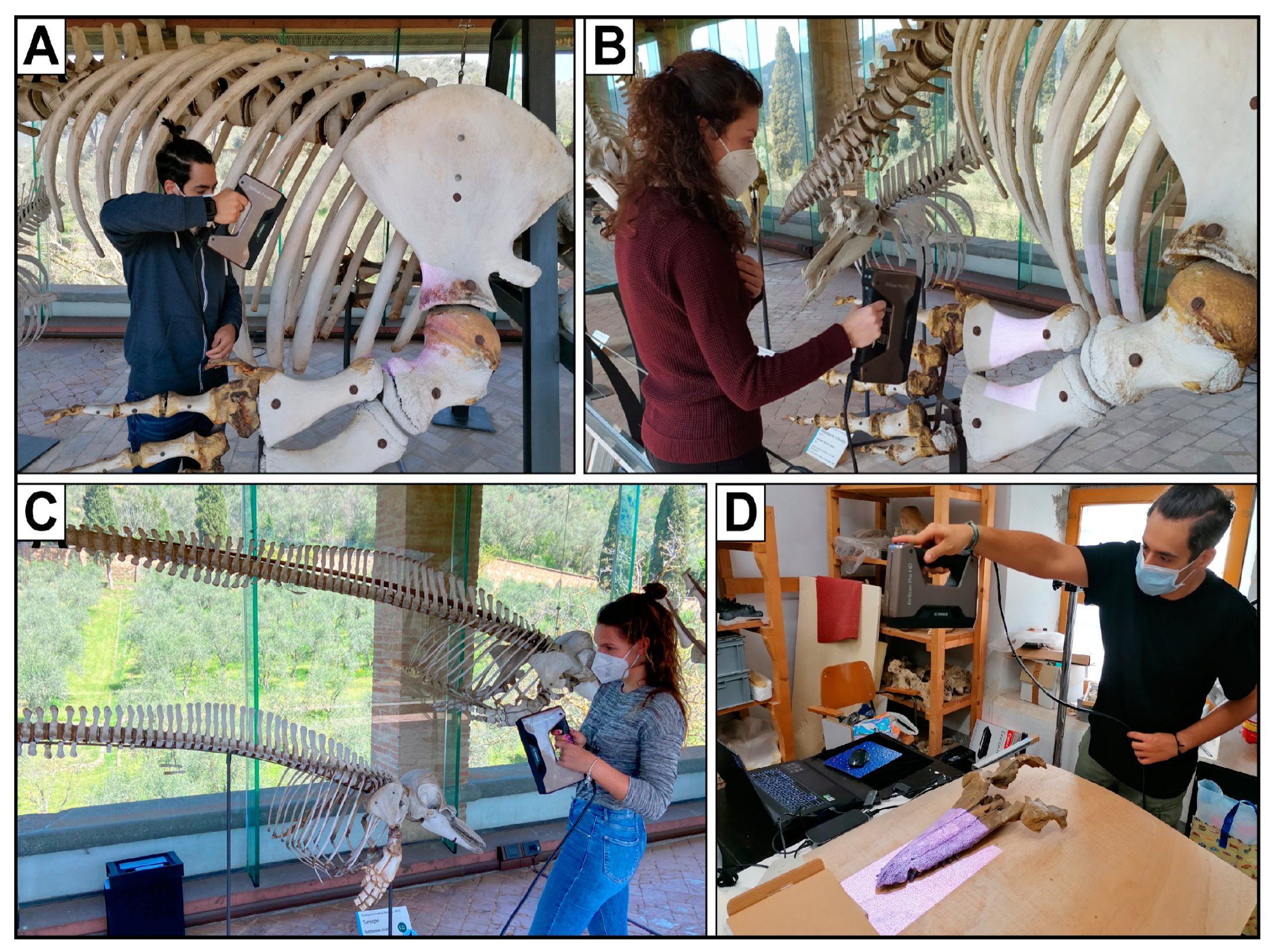
All the digitized specimens were scanned using an EinScan Pro HD structured-light scanner produced by the SHINING 3D Tech. Co., Ltd. (Hangzhou, China). This instrument allows for scanning objects up to a resolution of 0.2 mm, while the accuracy ranges between 0.045 and 0.1 mm. Furthermore, the SHINING 3D EinScan Pro HD scanner is equipped with a texture camera to generate texture models, which means that the scanner also captures the color of the specimen, which is computed as a layer that can be applied directly onto the mesh [
55]. These specifications make this scanner suitable for creating good 3D models of most specimens from the MSNUP collections of extant (
Figure 1A–C) and fossil (
Figure 1D) cetaceans. However, some problems occurred with the scanning of small and/or thin bones and skeletal portions (e.g., teeth, phalanges, and ribs) of the smallest odontocete specimens. These issues were partly resolved during the post-scan processing (see below).
Other problems arose during the scanning of the mounted skeletons of baleen whales in the Cetacean Gallery. Their enormous size made the manually scanning of the whole skeletons extremely difficult; thus, only the well-accessible forelimbs were scanned. Our aim is to acquire complete models of all skeletons in the near future, possibly by means of photogrammetry, using a drone to take multiple photos of even the highest and least-accessible parts of the skeletons [
71].
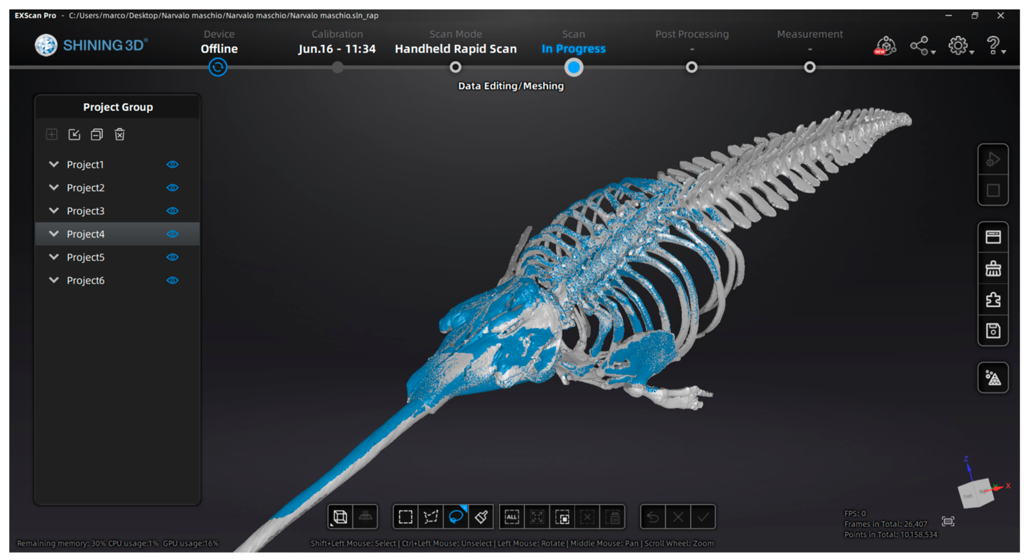
Before starting the scanning operations, the scanner was calibrated for vertical motion (thus allowing for recording the actual dimensions of the scanned specimens), accuracy, and white balancing. The software used to perform the 3D scans was ExScan Pro HD (v. 3.7.3.0), which is included in the scanning package. Most of the specimens were texture-scanned; some were scanned without texture because of shining light in the Cetacean Gallery, which hampered a good-quality texture-scan. These colorless 3D models were painted during the post-production phase. Most of the specimens were acquired with a minimum of two surface scans, with a special focus on the correct overlap of the scans, which was also pursued by applying an automatic alignment (
Figure 2) set by shared features or texture (whenever the object was scanned in texture mode).
After the removal of all the scanned points surrounding the specimen, the aligned partial scans were implemented to create a dense point cloud. Once we set a “watertight” hole-filling reconstruction mode, as well as several options for “mesh optimization” in terms of quality, number of triangles, and smoothing, the dense cloud was processed to generate the 3D mesh. Once the latter was created, texture remapping and optimization were performed to re-homogenize the texture all over the model’s surface.
All the meshes were exported in OBJ format, thus producing three files in a single folder: an .obj file, representing the mesh; an .mtl file, representing a text file containing information on the material associated with the mesh; and a .png texture file.
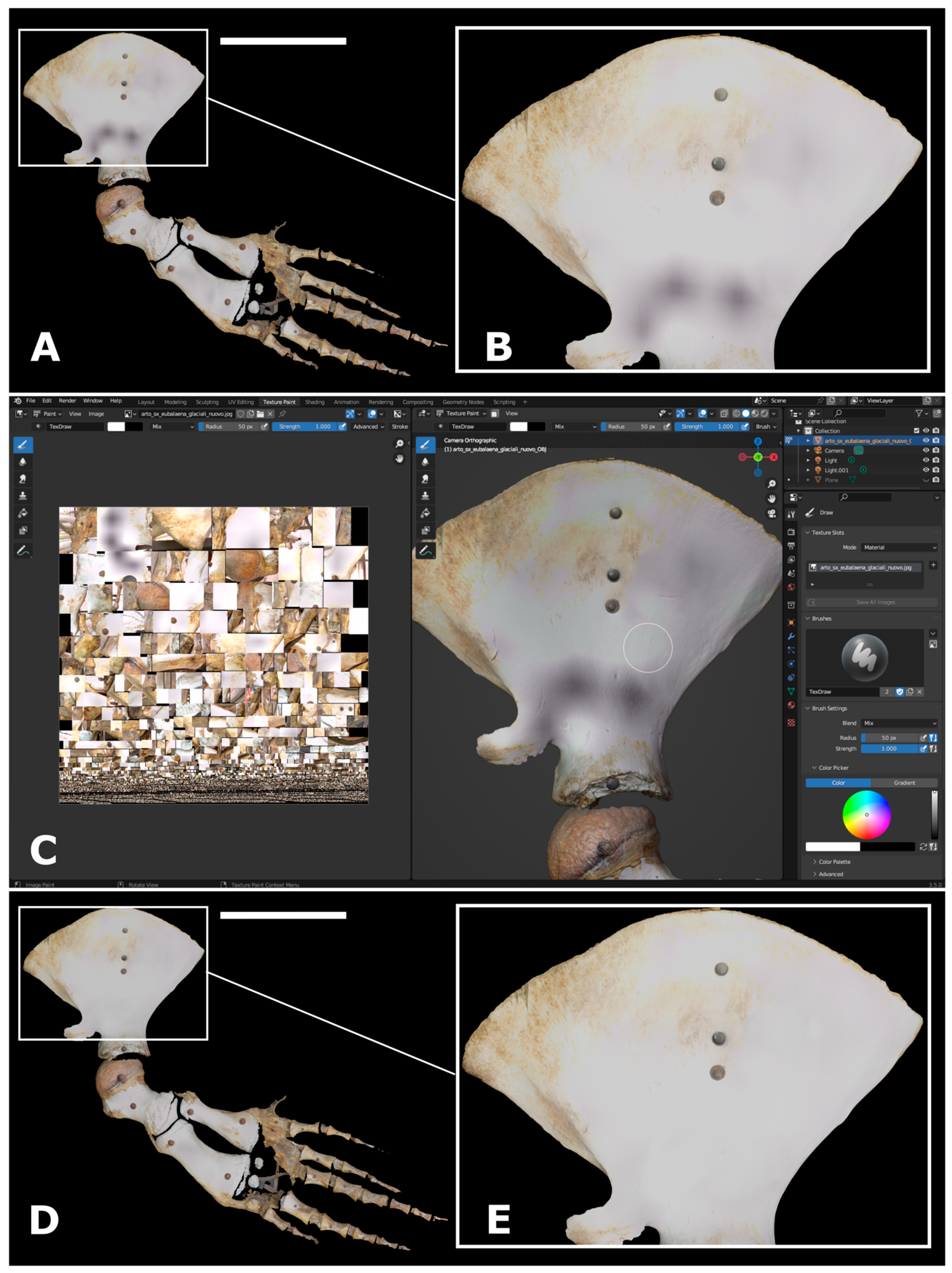
Several issues arose during and after the scanning operations. The Cetacean Gallery, where the digitized specimens are exposed, is characterized by clear glass walls running all along its extension. Here, the sunshine exposure and the green reflection of the surrounding vegetated environment negatively affected the scanning of some specimens, thus creating several artifacts such as green or black stains, holes, and multiple overlaps (
Figure 3A,B). The resolution of these problems required the use of blackout panels to avoid direct exposure to sunlight. Furthermore, a texture modification was also necessary. After importing the 3D model in Blender (v. 3.6) (
Figure 3C), the texture was modified by changing the UV-map and exporting a new .png image texture file (
Figure 3D–E).
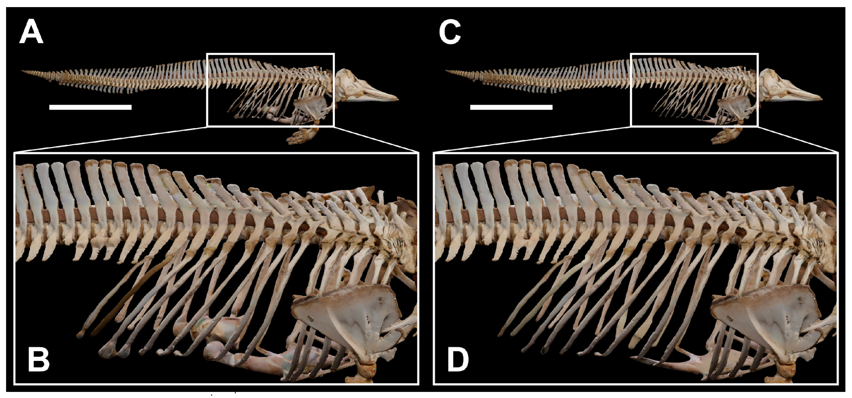
Another frequent issue was linked to the small thickness of several parts of the scanned skeletons, such as the distal portions of the ribs and the caudalmost vertebrae. Due to the technical specifications of the EinScan Pro HD scanner, the reconstruction of these bones (or bone parts) resulted in artifacts such as holes. The “watertight”, hole-filling option provided a partial solution to this problem, but occasional mesh bulges appeared in the occurrence of huge holes (
Figure 4A,B). To fix these artifacts, the models were imported in Blender and re-sculpted to obtain the correct bone shapes (
Figure 4C,D). The re-sculpting was performed through the “sculpting interface” of Blender by using different brushes (e.g., the “clay”, “smooth”, “flat”, “inflate”, and “grab” tools) and setting several parameters such as the radius and strength, depending on the circumstances.
It should be noted that the scanned skeletons are mounted on metal supports to maintain an anatomical arrangement of the vertebral column, whereas some bones (e.g., the forelimbs) are fixed to each other with huge iron nails. During the acquisition, all these supporting structures represented an obstacle for the scanning of the whole skeleton’s surface. In order to avoid unfillable holes in the 3D reconstructions, we decided to include these metallic elements in the final models whenever necessary.
3.2. Online Archiving and Social Channels
Sketchfab has been chosen as the online platform to store and share the 3D models as it represents a user-friendly web repository supporting natural and cultural heritage institutions worldwide. Sketchfab is also used by many other museums, including the British Museum of London (sketchfab.com/britishmuseum/models, accessed on 6 October 2023), the Natural History Museum of London (sketchfab.com/NHM_Imaging, accessed on 6 October 2023), the Natural History Museum of Wien (sketchfab.com/NHMWien, accessed on 6 October 2023), and the Smithsonian Museums of Washington (sketchfab.com/Smithsonian, accessed on 6 October 2023). Regarding the Italian museums, Sketchfab is used as an online repository by the Egyptian Museum of Turin (sketchfab.com/Museoegizio/models, accessed on 6 October 2023), the National Archaeological Museum of Naples (sketchfab.com/MANN/models, accessed on 6 October 2023), the Archaeological Museum of Populonia (sketchfab.com/museopopulonia, accessed on 6 October 2023), the Italian National Antarctic Museum (sketchfab.com/MNA, accessed on 6 October 2023), the University Museum System of Genoa University (sketchfab.com/sma-unige, accessed on 6 October 2023), and the Museum System of Leghorn (sketchfab.com/sistemamuseilivorno, accessed on 6 October 2023). These last three institutions host digitized models of several naturalistic specimens in their online archive. Other Italian museums have similar Sketchfab profiles, including for example, the MUSE of Trento (sketchfab.com/MUSE-Museo_delle_Scienze, accessed on 6 October 2023), even though no models appear to have been uploaded therein at present.
The MSNUP registered on Sketchfab with a “BASIC” account, which differs from the “PRO” and “PREMIUM” account types by being free rather than fee-based. The MSNUP account is named “Natural History Museum, University of Pisa” and includes institutional and historical information.
Each scanned model was stored in a .zip folder and subsequently dragged and dropped in Sketchfab to complete the upload. Three 3D models that had been acquired prior to the beginning of our scanning campaign were also uploaded following the same protocol. These additional models were obtained using a laser scanner and subsequently exported without texture. Once uploaded, several parameters were adjusted for each model through the “3D setting” toolbar of Sketchfab. These included: changing the model orientation and the field of view to 1° (thus avoiding view distortions); setting a homogenous, light grey background; customizing lights, shadows, and emission; modifying material properties (if necessary); and applying post-processing filters regarding for instance, the grain and sharpness. Lastly, annotations were included in each model, thus presenting conspicuous information about the specimen itself. All the 3D models that we made accessible through the MSNUP Sketchfab profile are available for free visualization, as well as for sharing under Creative Commons license after formal request.
To increase the visibility and promote the dissemination of our digital collection of cetacean skeletons, the Sketchfab account of the MSNUP was linked with the museum’s official website, as well as with its official social media profiles on Facebook and Instagram, which at present number more than 17,000 and more than 5600 followers, respectively.
4. Results
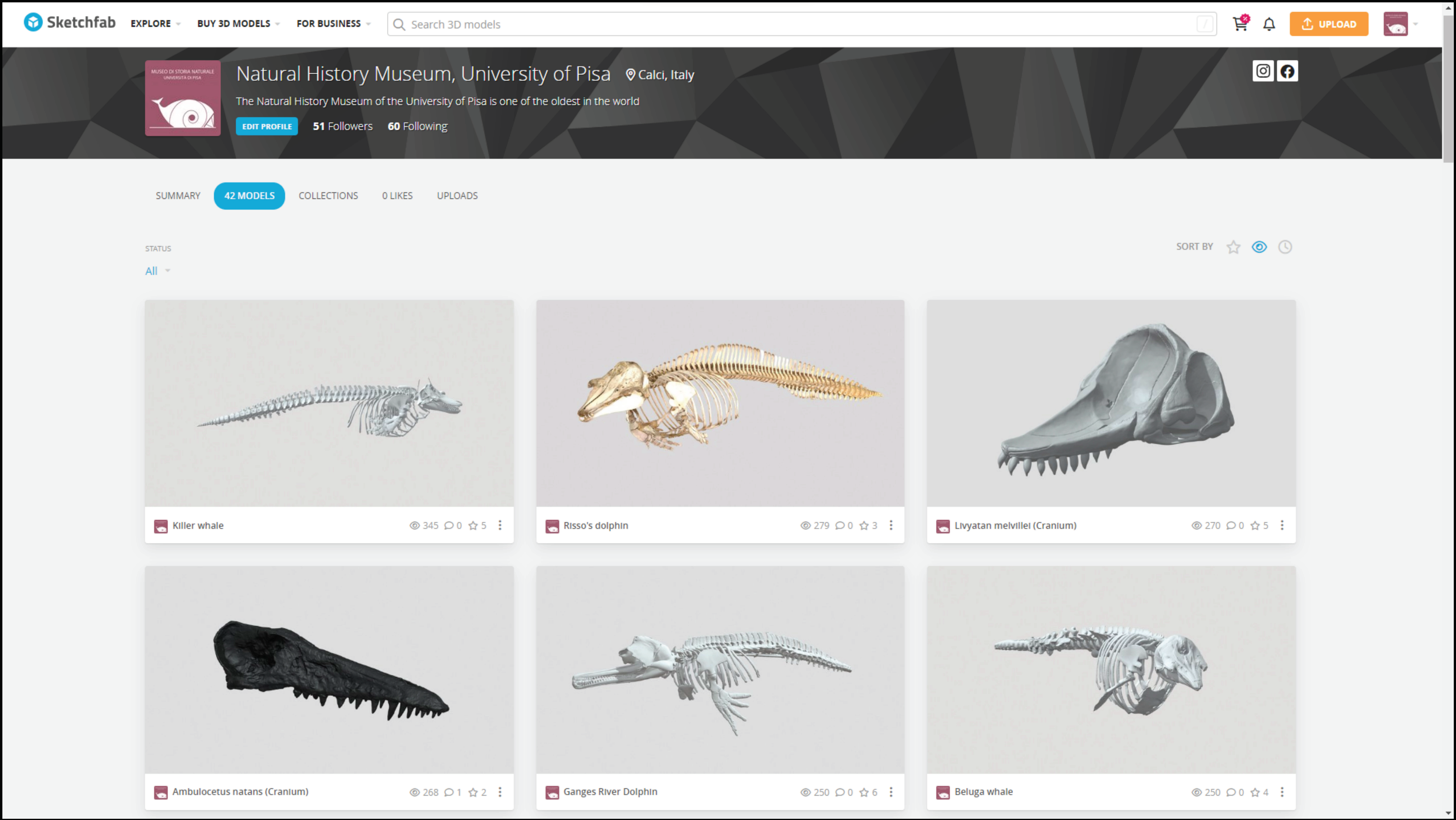
The Sketchfab account of the MSNUP can be visited at: sketchfab.com/MuseoStoriaNaturaleUnipi (accessed on 13 October 2023). Once clicked on, the landing page shows thumbnails of the most recently uploaded 3D models, all of which are accessible through the “Models” sub-page (
Figure 5). Each model’s webpage includes information on the specimen (concerning things such as its provenance, taxonomic identification, and biology) and quantitative data on the 3D model itself (such as the number of triangles and vertices).
Although the MSNUP Sketchfab account was born only recently (it was inaugurated in June 2022), it already includes 35 models of modern and fossil cetaceans—a representative fraction of the collection preserved in the museum. The uploaded models include: (1) thirteen complete odontocete skeletons, namely, a Gange river dolphin (Platanista gangetica, MSNUP C-272), an Andrew’s beaked whale (Mesoplodon bowdoini, MSNUP C-269), a Cuvier’s beaked whale (Ziphius cavirostris, MSNUP C-270), a franciscana (Pontoporia blainvillei, MSNUP C-273), a beluga whale (Delphinapterus leucas, MSNUP C-277), a narwhal (Monodon monoceros, MSNUP C-274), an Indopacific finless porpoise (Neophocaena phocaenoides, MSNUP C-279), a harbor porpoise (Phocoena phocoena, MSNUP C-278), a killer whale (Orcinus orca, MSNUP C-301), an Irrawaddy dolphin (Orcaella brevirostris, MSNUP C-293), a Risso’s dolphin (Grampus griseus, MSNUP C-295), an Atlantic white-sided dolphin (Lagenorhynchus acutus, MSNUP C-290), and a common bottlenose dolphin (Tursiops truncatus, MSNUP C-281); (2) six cranial elements (among which are the skull and mandibles) of a north Atlantic right whale (Eubalaena glacialis, MSNUP C-264), a Risso’s dolphin (Grampus griseus, MSNUP C-3257), and a Cuvier’s beaked whale (Ziphius cavirostris, MSNUP C-3112); (3) forelimbs of six mysticete whales, namely, a fin whale (Balaenoptera physalus, MSNUP C-251), a sei whale (Balaenoptera borealis, MSNUP C-262), a common minke whale (Balaenoptera acutorostrata, MSNUP C-260), a north Atlantic right whale (Eubalaena glacialis, MSNUP C-264), a blue whale (Balaenoptera musculus, MSNUP C-250), and a humpback whale (Megaptera noveangliae, MSNUP C-263); and (4) ten specimens of extinct cetaceans, namely, the original crania of the two known specimens of Casatia thermophila (MSNUP I-17602, MSNUP I-16153) and the holotype of Balaena montalionis (MSNUP I-12357), the casts of the crania of Aegyptocetus tarfa (MSNUP I-15459), Ambulocetus natans (MSNUP I-16826), Messapicetus longirostris (MSNUP I-16832) and Hemisyntrachelus pisanus (MSNUP I-16837), the reconstructed crania and mandibles of Livyatan melvillei (MSNUP I-17140), and the reconstructed mandibles of Messapicetus longirostris (MSNUP I-16832).
The size and geometry of the models vary from a minimum of 7 MB/96.5 K triangles/48.2 K vertices to a maximum of 99 MB/2.7 M triangles/1.4 M vertices. At present, the MSNUP Sketchfab account counts more than 4200 views from different countries.
Coinciding with the launch of the museum’s Sketchfab profile, 11 animations of skeletons of modern cetaceans were made available based on the 3D models uploaded on Sketchfab, and were published weekly on the museum’s Facebook and Instagram profiles in February–May 2023. Each animation, lasting about 11 s, contained the 3D model and basic information such as the scientific name of the species, its Italian vernacular name, and the museum logo, as well as a caption containing a short account on the species and a link to the MSNUP Sketchfab profile. The choice of using Italian as the language for captions and metadata was dictated by the fact that the audience of the museum’s social media channels is overwhelmingly Italian, and the contents are invariably created in that language. Overall, the posts obtained a good number of views and interactions from users (
Table 1).
It is also interesting to underline how on Instagram, thanks to the use of hashtags, a significant percentage of the users who viewed the posts are not followers of the museum’s profile (
Table 2). This datum is remarkable as it indicates that posting on Instagram allows for reaching a wider audience than followers alone.
The choice of using generalist rather than academic social media for spreading the MSNUP Sketchfab repository was dictated by the aim of reaching the broadest audience possible. In a context made of “reels”, “posts”, and “stories”, the understanding of scientific contents is facilitated by relying on multimedia such as gifs, videos, and animations, which now provide relevant support to the museums’ outreach mission.
5. Discussion and Perspectives
In addition to resident and visiting scholars, general visitors are crucial stakeholders for all museums. That being said, the traditional concept of “visitor” has been evolving recently [
54], especially during and after the COVID-19 pandemic. Due to the widespread inaccessibility of museum exhibits, the implementation of virtual tools has exponentially increased in the last few years through the creation of 3D repositories and virtual museums [
49,
72,
73,
74].
The creation of a digital archive for the modern and fossil cetacean skeletons kept at the MSNUP is fully aligned with the common objectives promoted by the European Union, whose 27 country members, including Italy, signed the “Declaration of cooperation on advancing digitization of cultural heritage” in 2019 (digital-strategy.ec.europa.eu/en/news/eu-member-states-sign-cooperate-digitising-cultural-heritage). In addition to digitizing Europe’s cultural heritage, the goals of this declaration include the re-use of digitized cultural resources to foster citizen engagement as well as to enhance cross-sector, cross-border cooperation, and capacity building in the field of digitized cultural heritage [
75]. As a matter of fact, the MSNUP 3D models of cetaceans have already been used by bachelor’s and master’s thesis students, as well as by biologists and paleontologists dealing with, for example, functional morphology [
76]. When the 3D models were further shared through social media such as Instagram and Facebook, an even broader public was reached. We contend that our effort represents an important tool for disseminating knowledge on specimens of high historical and scientific importance from two of the core collections of the MSNUP as widely and effectively as possible.
When evaluating the didactic value of our project, the substantial involvement of students and teachers of the University of Pisa in the creation of the 3D models should also be considered. In doing so, the involved students—all of which are co-authors of the present paper—were able to observe closely the specimens they were scanning while learning how to use modern techniques of 3D acquisition.
The 3D scanning of the osteological collection of modern and fossil cetaceans stored at the MSNUP represents the starting point of a more ambitious project that aims to create 3D scans of the most important specimens of the whole osteological collection of mammals. Our digitization campaign has recently continued with the scanning of the complete skeletons of two sirenians, i.e., a dugong (Dugong dugon, MSNUP C-527) and an American manatee (Trichecus manatus, MSNUP C-1442), as well as those of two marine representatives of Carnivora, namely, a monk seal (Monachus monachus, MSNUP C-1441) and a harp seal (Pagophilus groenlandicus, MSNUP C-1401). The skulls of a walrus (Odobenus rosmarus, MSNUP C-1215) and a lion (Panthera leo, MSNUP C-1053) have also been digitized, such that 42 models are found in the museum’s Sketchfab profile at present.
Furthermore, given the good results obtained by our preliminary scanning tests, we plan to carry out 3D scans of the most important taxidermized specimens, starting with the extinct species and specimens of outstanding scientific and historical relevance. Indeed, the MSNUP is home to a rich collection of taxidermized mammals and birds that could be scanned by using the same protocol as the osteological specimens. Further tests using the structured-light scanner will also be attempted with specimens belonging to the comparative anatomy and liquid-preserved collections. In some cases, different digitizing approaches such as digital photogrammetry could be applied, e.g., when dealing with long-haired specimens, whose external surface may be too heterogeneous for the structured-light scanning to work properly. Photogrammetry has been recently applied successfully to fossil traces belonging to the MSNUP collection, resulting in the production of accurate depth maps [
77].
6. Conclusions
A 3D digitization technique based on structured-light scanning was applied to a conspicuous part of the modern and fossil cetological collections of the MSNUP, one of the most important collections all over Europe. The 3D models of the scanned specimens were exported either with texture or colorless. Further modifications, including painting and sculpting, were applied in some cases during the post-production phase by using the 3D software Blender. In order to store the resulting models and to share them via the internet, a Sketchfab account was created and further disseminated through Facebook and Instagram to reach the broadest public. The preliminary results of such an effort are encouraging in terms of views and online interactions. The digitization and online archiving of the MSNUP specimens of extant and extinct cetaceans is the starting point of a more ambitious project that will hopefully extend to other collections of vertebrates, including both marine and terrestrial forms. The application of 3D technologies and the valorization of museological goods by means of digital archiving in open-access online repositories make the MSNUP closer and closer to the largest and most important natural history museums worldwide.
Author Contributions
Conceptualization, M.M., S.F., A.C., and G.B. (Giovanni Bianucci); Data curation, M.M. and P.S.; Formal analysis, S.F., A.C., and G.B. (Giovanni Bianucci); Funding acquisition, S.F. and A.C.; Investigation, S.F., A.C., and G.B. (Giovanni Bianucci); Methodology, M.M., G.C., G.B. (Giada Bernardini), and A.P.; Project administration, A.C. and G.B. (Giovanni Bianucci); Resources, M.M., S.F., P.S., A.C., and G.B. (Giovanni Bianucci); Software, M.M., G.C., G.B. (Giada Bernardini), and A.P.; Supervision, A.C. and G.B. (Giovanni Bianucci); Validation, M.M., S.F., A.C., and G.B. (Giovanni Bianucci); Visualization, M.M. and G.B. (Giovanni Bianucci); Writing—original draft, M.M. and S.F.; Writing—review and editing, P.S., A.C., and G.B. (Giovanni Bianucci). All authors have read and agreed to the published version of the manuscript.
Funding
Acquisition of the scanner used for creating the 3D models was possible thanks to a funding grant from University of Pisa (PRA_2020_25 to A.C.). This research was supported by a grant from the Italian Ministero dell’Università e della Ricerca (PRIN Project 2022MAM9ZB to A.C.).
Data Availability Statement
Acknowledgments
First and foremost, our gratitude goes to Chiara Sorbini (Natural History Museum of the University of Pisa) for supporting our scanning activity and granting access to the paleontological materials under her care, as well as for many fruitful discussions. We are also very grateful to Ellen Coombs (Smithsonian National Museum of Natural History, Washington, DC) for allowing us to share on the MSNUP Sketchfab profile her 3D models of Aegyptoceus tarfa, Ambulocetus natans, and Eubalaena glacialis. We also thank Damiano Marchi and Elena Bonaccorsi (former and present directors of the Natural History Museum of the University of Pisa, respectively) for encouraging this project. Not least, we are grateful to two anonymous reviewers for sharing with us their constructive comments and suggestions.
Conflicts of Interest
The authors declare no conflict of interest.
References
- Rahman, I.A.; Adcock, K.; Garwood, R.J. Virtual fossils: A new resource for science communication in palaeontology. Evol. Educ. Outreach 2012, 5, 635–641. [Google Scholar] [CrossRef][Green Version]
- Rahman, I.A.; Zamora, S.; Falkingham, P.L.; Phillips, J.C. Cambrian cinctan echinoderms shed light on feeding in the ancestral deuterostome. Proc. R. Soc. 2015, 282, 20151964. [Google Scholar] [CrossRef] [PubMed]
- Sutton, M.; Rahman, I.; Garwood, R. Virtual palaeontology—An overview. Pal. Soc. Pap. 2016, 22, 1–20. [Google Scholar] [CrossRef]
- Das, A.J.; Murmann, D.C.; Cohrn, K.; Raskar, R. A method for rapid 3D scanning and replication of large paleontological specimens. PLoS ONE 2017, 12, e0179264. [Google Scholar] [CrossRef]
- Tate, J.R.; Cann, C.E. High-resolution computed tomography for the comparative study of fossil and extant bone. Am. J. Phys. Anthropol. 1982, 58, 67–73. [Google Scholar] [CrossRef]
- Conroy, G.C.; Vannier, M.W. Noninvasive three-dimensional computer imaging of matrix-filled fossil skulls by high-resolution computed tomography. Science 1984, 226, 456–458. [Google Scholar] [CrossRef]
- Pandolfi, L.; Raia, P.; Fortuny, J.; Rook, L. Evolving virtual and computational palaeontology. Front. Earth Sci. 2020, 8, 591813. [Google Scholar] [CrossRef]
- Lyons, P.D.; Rioux, M.; Patterson, R.T. Application of a three-dimensional color laser scanner to palaeontology: An interactive model of a juvenile Tylosaurus sp. basisphenoid-basioccipital. Palaeontol. Electron. 2000, 3, 4A. [Google Scholar]
- Breithaupt, B.H.; Matthews, N.A. Preserving paleontological resources using photogrammetry and geographic information systems. In Crossing Boundaries in Park Management; The George Wright Society: Hancock, MI, USA, 2001; pp. 62–70. [Google Scholar]
- Zollikofer, C.P.; Ponce de Leon, M. Virtual Reconstruction: A Primer in Computer-Assisted Palaeontology and Biomedicine; John Wiley and Sons: Hoboken, NJ, USA, 2005; 333p. [Google Scholar]
- Falkingham, P.L. Acquisition of high resolution three-dimensional models using free, open-source, photogrammetric software. Palaeontol. Electron. 2012, 15, 1–15. [Google Scholar] [CrossRef]
- Falkingham, P.L. Low cost 3D scanning using off-the-shelf video gaming peripherals. J. Paleontol. Tech. 2013, 11, 1–9. [Google Scholar]
- Falkingham, P.L. Interpreting ecology and behaviour from the vertebrate fossil track record. J. Zool. 2014, 292, 222–228. [Google Scholar] [CrossRef]
- Falkingham, P.L.; Bates, K.T.; Avanzini, M.; Bennett, M.; Bordy, E.M.; Breithaupt, B.H.; Castanera, D.; Citton, P.; Dìaz-Martìnez, I.; Farlow, J.O.; et al. A standard protocol for documenting modern and fossil ichnological data. Palaeontology 2018, 61, 469–480. [Google Scholar] [CrossRef]
- Cardini, A.; Loy, A. Virtual morphology and evolutionary morphometrics in the new millenium. Hystrix 2013, 24, 1–5. [Google Scholar]
- Cunningham, J.A.; Rahman, I.A.; Lautenschlager, S.; Rayfield, E.J.; Donoghue, P.C. A virtual world of palaeontology. Trends Ecol. Evol. 2014, 29, 347–357. [Google Scholar] [CrossRef]
- Fahlke, J.M.; Hampe, O. Cranial symmetry in baleen whales (Cetacea, Mysticeti) and the occurrence of cranial asymmetry throughout cetacean evolution. Sci. Nat. 2015, 102, 58. [Google Scholar] [CrossRef]
- Fau, M.; Cornette, R.; Houssaye, A. Photogrammetry for 3D digitizing bones of mounted skeletons: Potential and limits. Comptes. Rendus. Palevol. 2016, 15, 968–977. [Google Scholar] [CrossRef]
- Bartolini-Lucenti, S.; Bukhsianidze, M.; Martínez-Navarro, B.; Lordkipanidze, D. The wolf from Dmanisi and augmented reality: Review, implications, and opportunities. Front. Earth Sci. 2020, 8, 131. [Google Scholar] [CrossRef]
- Bartolini-Lucenti, S.; Madurell-Malapeira, J.; Martínez-Navarro, B.; Palmqvist, P.; Lordkipanidze, D.; Rook, L. The early hunting dog from Dmanisi with comments on the social behaviour in Canidae and hominins. Sci. Rep. 2021, 11, 13501. [Google Scholar] [CrossRef]
- Cirilli, O.; Melchionna, M.; Serio, C.; Bernor, R.L.; Bukhsianidze, M.; Lordkipanidze, D.; Rook, L.; Profico, A.; Raia, P. Target deformation of the Equus stenonis holotype skull: A virtual reconstruction. Front. Earth Sci. 2020, 8, 247. [Google Scholar] [CrossRef]
- Bartolini-Lucenti, S.; Rook, L. Nurturing Italian Geo-palaeontological Heritage with Virtual Palaeontology: Preliminary Report of Its Application in Two Natural History Museums. Geoheritage 2023, 15, 40. [Google Scholar] [CrossRef]
- Bitelli, G.; Dubbini, M.; Zanutta, A. Terrestrial laser scanning and digital photogrammetry techniques to monitor landslide bodies. ISPRS Arch. 2004, 35, 246–251. [Google Scholar]
- Westoby, M.J.; Brasington, J.; Glasser, N.F.; Hambrey, M.J.; Reynolds, J.M. ‘Structure-from Motion’ photogrammetry: A low-cost, effective tool for geoscience applications. Geomorphology 2012, 179, 300–314. [Google Scholar] [CrossRef]
- Wang, G.; Li, R.; Carranza, E.J.M.; Zhang, S.; Yan, C.; Zhu, Y.; Qu, J.; Hong, D.; Song, Y.; Han, J.; et al. 3D geological modeling for prediction of subsurface Mo targets in the Luanchuan district, China. Ore Geol. Rev. 2015, 71, 592–610. [Google Scholar] [CrossRef]
- Zhang, Q.; Zhu, H. Collaborative 3D geological modeling analysis based on multi-source data standard. Eng. Geol. 2018, 246, 233–244. [Google Scholar] [CrossRef]
- Andersen, T.R.; Poulsen, S.E.; Pagola, M.A.; Medhus, A.B. Geophysical mapping and 3D geological modelling to support urban planning: A case study from Vejle, Denmark. J. Appl. Geophys. 2020, 180, 104–130. [Google Scholar] [CrossRef]
- Stuppy, W.H.; Maisano, J.A.; Colbert, M.W.; Rudall, P.J.; Rowe, T.B. Three-dimensional analysis of plant structure using high-resolution x-ray computed tomography. Trends Plant Sci. 2003, 8, 2–6. [Google Scholar] [CrossRef]
- Brereton, N.J.B.; Ahmed, F.; Sykes, D.; Ray, M.J.; Shield, I.; Karp, A.; Murphy, R.J. X-ray micro-computed tomography in willow reveals tissue patterning of reaction wood and delay in programmed cell death. BMC Plant Bio. 2015, 15, 83. [Google Scholar] [CrossRef]
- Wang, H.L.; Wong, T.H.; Chan, Y.M.; Cheng, Y.S.; Lau, D.T.W. Photogrammetric reconstruction of 3D carpological collection in high resolution for plants authentication and species discovery. PLoS ONE 2022, 17, e0270199. [Google Scholar] [CrossRef]
- Leménager, M.; Burkiewicz, J.; Schoen, D.J.; Joly, S. Studying flowers in 3D using photogrammetry. New Phytol. 2023, 237, 1922–1933. [Google Scholar] [CrossRef]
- Redweik, P.; Reis, S.; Duarte, M.C. A digital botanical garden: Using interactive 3D models for visitor experience enhancement and collection management. Vir. Archaeol. Rev. 2023, 14, 65–80. [Google Scholar] [CrossRef]
- Klaus, A.V.; Kulasekera, V.L.; Schawaroch, V. Three-dimensional visualization of insect morphology using confocal laser scanning microscopy. J. Microsc. 2003, 212, 107–121. [Google Scholar] [CrossRef]
- Nguyen, C.V.; Lovell, D.R.; Adcock, M.; La Salle, J. Capturing natural-colour 3D models of insects for species discovery and diagnostics. PLoS ONE 2014, 9, e94346. [Google Scholar] [CrossRef] [PubMed]
- Postma, M.; Tordiffe, A.S.W.; Hofmeyr, M.S.; Reisinger, R.R.; Bester, L.C.; Buss, P.E.; De Bruyn, P.J.N. Terrestrial mammal three-dimensional photogrammetry multispecies mass estimation. Ecosphere 2015, 6, 1–16. [Google Scholar] [CrossRef]
- Bright, J.A.; Maruga’n-Lobo´n, J.; Rayfield, E.J.; Cobb, S.N. The multifactorial nature of beak and skull shape evolution in parrots and cockatoos (Psittaciformes). BMC Evol. Bio. 2019, 19, 104. [Google Scholar] [CrossRef] [PubMed]
- Medina, J.J.; Maley, J.M.; Sannapareddy, S.; Medina, N.N.; Gilman, C.M.; McCormack, J.E. A rapid and cost-effective pipeline for digitization of museum specimens with 3D photogrammetry. PLoS ONE 2020, 15, e0236417. [Google Scholar] [CrossRef]
- Cilli, E.; Fontani, F.; Ciucani, M.M.; Pizzuto, M.; Di Benedetto, P.; De Fanti, S.; Mignani, S.; Bini, C.; Iacovera, R.; Pelotti, S.; et al. Museomics provides insights into conservation and education: The instance of an African lion specimen from the Museum of Zoology “Pietro Doderlein”. Diversity 2023, 15, 87. [Google Scholar] [CrossRef]
- Guidi, G.; Russo, M.; Angheleddu, D. 3D survey and virtual reconstruction of archeological sites. Digit. Appl. Archaeol. Cult. Herit. 2014, 1, 55–69. [Google Scholar] [CrossRef]
- Pires, H.; Martínez Rubio, J.; Elorza Arana, A. Techniques for revealing 3D hidden archeological features: Morphological residual models as virtual-polynomial texture maps. Int. Arch. Photogramm. Remote Sens. Spat. Inf. Sci. 2015, 40, 415–421. [Google Scholar] [CrossRef]
- Santos, P.; Ritz, M.; Fuhrmann, C.; Fellner, D. 3D mass digitization: A milestone for archeological documentation. Vir. Archaeol. Rev. 2017, 8, 1–11. [Google Scholar] [CrossRef]
- Terlikowski, W.; Gregoriou-Szczepaniak, M.; Sobczyńska, E.; Wasilewski, K. Advantages of using 3d scanning in the survey of architectural monuments on example of archeological sites in Egypt and Russia. Arch. Civ. Eng. 2021, 67, 189–201. [Google Scholar]
- Fiz, J.I.; Martín, P.M.; Cuesta, R.; Subías, E.; Codina, D.; Cartes, A. Examples and results of aerial photogrammetry in archeology with UAV: Geometric documentation, high resolution multispectral analysis, models and 3D printing. Drones 2022, 6, 59. [Google Scholar] [CrossRef]
- Sundell, L. Cyber Archeology: Reconstructing Buildings with 3D Graphics and VR Technology; Luleå University of Technology: Luleå, Sweden, 2022. [Google Scholar]
- Kuzminsky, S.C.; Gardiner, M.S. Three-dimensional laser scanning: Potential uses for museum conservation and scientific research. J. Archaeol. Sci. 2012, 39, 2744–2751. [Google Scholar] [CrossRef]
- Atik, M.E.; Duran, Z.; Yanalak, M.; Seker, D.Z.; Ak, A. 3D modeling of historical measurement instruments using photogrammetric and laser scanning techniques. Digit. Appl. Archaeol. Cult. Herit. 2023, 30, e00286. [Google Scholar] [CrossRef]
- Bowen, J. The virtual museum. Mus. Int. 2000, 52, 4–7. [Google Scholar] [CrossRef]
- Carvajal Loaiza, M.J.; Gónzalez Díaz, P.; Mejía Blandón, C.A.; Bustamante Góez, L.M.; Villarraga Ossa, J.A. Influencia de la posición de impresión y la densidad de relleno en las propiedades mecánicas de probetas fabricadas en ABS. Rev. Ing. Univ. Medellin 2020, 19, 179–193. [Google Scholar] [CrossRef]
- Dasgupta, A.; Williams, S.; Nelson, G.; Manuel, M.; Dasgupta, S.; Gračanin, D. Redefining the digital paradigm for virtual museums: Towards interactive and engaging experiences in the post-pandemic era. In Culture and Computing. Interactive Cultural Heritage and Arts; Lecture Notes in Computer Science; Springer: Cham, Switzerland, 2021; Volume 12794, pp. 357–373. [Google Scholar]
- Short, D.B. Use of 3D printing by museums: Educational exhibits, artifact education, and artifact restoration. 3d Print. Addit. Manuf. 2015, 2, 209–215. [Google Scholar] [CrossRef]
- Wilson, P.F.; Stott, J.; Warnett, J.M.; Attridge, A.; Smith, M.P.; Williams, M.A. Evaluation of Touchable 3D-Printed Replicas in Museums. Curator. Mus. J. 2017, 60, 445–465. [Google Scholar] [CrossRef]
- Montusiewicz, J.; Barszcz, M.; Korga, S. Preparation of 3D models of cultural heritage objects to be recognised by touch by the blind-case studies. Appl. Sci. 2022, 12, 11910. [Google Scholar] [CrossRef]
- Boyer, D.M.; Gunnell, G.F.; Kaufman, S.; McGeary, T.M. MorphoSource: Archiving and sharing 3-D digital specimen data. Paleontol. Soc. Pap. 2016, 22, 157–181. [Google Scholar] [CrossRef]
- Erolin, C.; Jarron, M.; Csetenyi, L.J. Zoology 3D: Creating a digital collection of specimens from the D′ Arcy Thompson Zoology Museum. Digit. Appl. Archaeol. Cult. Herit. 2017, 7, 51–55. [Google Scholar] [CrossRef]
- Ziegler, M.J.; Perez, V.J.; Pirlo, J.; Narducci, R.E.; Moran, S.M.; Selba, M.C.; Hastings, A.K.; Vargas-Vargara, C.; Antonenko, P.D.; MacFadden, B.J. Applications of 3D paleontological data at the Florida Museum of Natural History. Front. Earth Sci. 2020, 8, 600696. [Google Scholar] [CrossRef]
- Repetti, U. Il Museo Pisano di Storia Naturale. Ann. Univ. Toscana. Sez. Sci. Med. Fis. Mat. Nat. Nuova Ser. 1925, 10, 171–180. [Google Scholar]
- Braschi, S.; Cagnolaro, L.; Nicolosi, P. Catalogo dei Cetacei attuali del Museo di Storia Naturale e del Territorio dell’Università di Pisa, alla Certosa di Calci. Note osteometriche e ricerca storica. Atti Soc. Tosc. Sci. Nat. Mem. 2007, 114, 1–22. [Google Scholar]
- Bianucci, G.; Sorbini, C. Le collezioni a cetacei fossili del Museo di Storia Naturale dell’Università di Pisa. Mus. Sci. Mem. 2014, 13, 93–102. [Google Scholar]
- Garbari, F.; Tongiorgi Tomasi, L.; Tosi, A. Il Giardino dei Semplici—L’orto Botanico di Pisa dal XVI al XX Secolo; Pacini: Pisa, Italy, 1991; 397p. [Google Scholar]
- Manganelli, G.; Benocci, A.; Spadini, V. The scientific bibliography of Roberto Lawley (1818–1881) and his contribution to the study of fossil sharks. Arch. Nat. Hist. 2003, 33, 267–281. [Google Scholar] [CrossRef]
- Bianucci, G.; Gingerich, P.D. Aegyptocetus tarfa, n. gen. et sp. (Mammalia, Cetacea), from the middle Eocene of Egypt: Clinorhynchy, olfaction, and hearing in a protocetid whale. J. Vertebr. Paleontol. 2011, 31, 1173–1188. [Google Scholar] [CrossRef]
- Capellini, G. Balene fossili toscane: II Balaena montalionis. Mem. R. Accad. Sci. Ist. Bologna 1904, 6, 45–57. [Google Scholar]
- Trevisan, L. Una nuova specie di Balaenula pliocenica. Palaeontogr. Ital. 1941, 40, 1–13. [Google Scholar]
- Bianucci, G.; Pesci, F.; Collareta, A.; Tinelli, C. A new Monodontidae (Cetacea, Delphinoidea) from the lower Pliocene of Italy supports a warm-water origin for narwhals and white whales. J. Vert. Pal. 2019, 39, e1645148. [Google Scholar] [CrossRef]
- Collareta, A.; Cigala Fulgosi, F.; Bianucci, G. A new kogiid sperm whale from northern Italy supports psychrospheric conditions in the early Pliocene Mediterranean Sea. Acta Palaeontol. Pol. 2019, 64, 609–626. [Google Scholar] [CrossRef]
- Peri, E.; Falkingham, P.L.; Collareta, A.; Bianucci, G. Biting in the Miocene seas: Estimation of the bite force of the macroraptorial sperm whale Zygophyseter varolai using finite element analysis. Hist. Biol. 2022, 34, 1916–1927. [Google Scholar] [CrossRef]
- Merella, M.; Collareta, A.; Granata, V.; Casati, S.; Bianucci, G. New remains of Casatia thermophila (Cetacea, Monodontidae) from the lower Pliocene marine vertebrate-bearing locality of Arcille (Tuscany, Italy). Riv. Ital. Paleont. Strat. 2022, 128, 229–240. [Google Scholar] [CrossRef] [PubMed]
- Thewissen, J.G.M.; Madar, S.I.; Hussain, S.T. Ambulocetus natans, an Eocene cetacean (Mammalia) from Pakistan. Cour. Forsch.-Inst. Senckenberg. 1996, 191, 1–86. [Google Scholar]
- Bianucci, G.; Landini, W. Killer sperm whale: A new basal physeteroid (Mammalia, Cetacea) from the Late Miocene of Italy. Zool. J. Linn. Soc. 2006, 148, 103–131. [Google Scholar] [CrossRef]
- Lambert, O.; Bianucci, G.; Post, K.; De Muizon, C.; Salas-Gismondi, R.; Urbina, M.; Reumer, J. The giant bite of a new raptorial sperm whale from the Miocene epoch of Peru. Nature 2010, 466, 105–108. [Google Scholar] [CrossRef] [PubMed]
- Romano, M.; Antonelli, M.; Palombo, M.R.; Rossi, M.A.; Agostini, S. Drone testing for 3D reconstruction of massive mounted skeletons in museums: The case of Mammuthus meridionalis (Nesti 1825) from Madonna della Strada (Scoppito, L’Aquila, Italy). Hist. Biol. 2022, 34, 1305–1314. [Google Scholar] [CrossRef]
- Fitriyani, F.; Khairani, D.; Saepudin, D.; Asnawi, T.B.; Jabali, F. The Impact of Pandemic COVID-19 on Museum Existence. In Proceedings of the 3rd International Colloquium on Interdisciplinary Islamic Studies, ICIIS 2020, Jakarta, Indonesia, 20–21 October 2020. [Google Scholar]
- Varriale, L.; Volpe, T.; Noviello, V. Enhancing cultural heritage at the time of the COVID-19 outbreak: An overview of the ICT strategies adopted by museums in the campania region of italy. In Tourism Destination Management in a Post-Pandemic Context (Tourism Security-Safety and Post Conflict Destinations); Gowreesunkar, V.G.B., Maingi, S.W., Roy, H., Micera, R., Eds.; Emerald Publishing Limited: Bingley, UK, 2021; pp. 201–218. [Google Scholar]
- Milic, N. Digitalising the Museum. In Handbook of Research on Museum Management in the Digital Era; Bifulco, F., Tregua, M., Eds.; IGI Global: Hershey, PA, USA, 2022; pp. 138–154. [Google Scholar]
- Bellucci, L.; Bartolini-Lucenti, S.; Dominici, S.; Rook, L.; Cioppi, E. Digitalizzazione 3D delle collezioni paleontologiche del Museo di Geologia e Paleontologia di Firenze. Mus. Sci. Mem. 2022, 22, 142–147. [Google Scholar]
- Laeta, M.; Oliveira, L.A.; Siciliano, S.; Lambert, O.; Jensen, F.H.; Galatius, A. Cranial asymmetry in odontocetes: A facilitator of sonic exploration? Zoology 2023, 160, 126108. [Google Scholar] [CrossRef]
- Marchetti, L.; Collareta, A.; Belvedere, M.; Leonardi, G. Ichnotaxonomy, biostratigraphy and palaeoecology of the Monti Pisani tetrapod ichnoassociation (Tuscany, Italy) and new insights on Middle Triassic Dinosauromorpha. Palaeogeogr. Palaeoclimatol. Palaeoecol. 2021, 567, 110235. [Google Scholar] [CrossRef]
| Disclaimer/Publisher’s Note: The statements, opinions and data contained in all publications are solely those of the individual author(s) and contributor(s) and not of MDPI and/or the editor(s). MDPI and/or the editor(s) disclaim responsibility for any injury to people or property resulting from any ideas, methods, instructions or products referred to in the content. |
© 2023 by the authors. Licensee MDPI, Basel, Switzerland. This article is an open access article distributed under the terms and conditions of the Creative Commons Attribution (CC BY) license (https://creativecommons.org/licenses/by/4.0/).


 ,
, 


