Article

Human, All Too Human: Differentiating Non-Human from Human Bones in Protohistoric Cremation Contexts from Northern Italy

Centre of Research in Osteoarchaeology and Paleopathology, Department of Biotechnologies and Life Sciences, University of Insubria, 21100 Varese, Italy
Heritage 2023, 6(1), 647-661; https://doi.org/10.3390/heritage6010034
Submission received: 1 December 2022 / Revised: 9 January 2023 / Accepted: 11 January 2023 / Published: 14 January 2023
(This article belongs to the Special Issue Zooarchaeology)

Abstract

:
Differentiating cremated non-human bones from human ones in archaeological contexts is a challenging task. This analysis aims at proposing a rather solid criterion based on an osteoarchaeological sample. In this work, the main issues of taxonomic identification of cremated remains are analysed and a research methodology tested on an Italian protohistoric sample is proposed. The 314 subjects composing the sample come from 298 tombs of the Golasecca Civilization (1st millennium BC in north-eastern Italy). On a morphological basis, 246 bone fragments were selected from which as many thin sections were obtained for a histomorphological evaluation. From the analyses, we identified the presence of animals in burials, whereas a mere morphometric analysis was not able to recognize them. Furthermore, the taxonomic identification has allowed us to propose new hypotheses on the funerary rite of Golasecca linked to the zooarchaeological remains.

1. Introduction

1.1. Cremation in Northern Italy during Protohistory

In northern Italy, the first findings of bone remains that have undergone a process of combustion or heating date back to the Early and Middle Neolithic VBQ culture. They are fragments coming from Po Valley contexts, whose ritual interpretation is often controversial [1,2]. For example, regarding these debated contexts, five pits containing burnt remains were excavated between 2003 and 2005 in the Early Neolithic settlement of “Lugo di Grezzana.” Many bones mixed with other materials come from pit ES 541/03. Most of the bones were non-human; however, some fragments showed morphological human features. The histomorphological analysis confirmed the identification of these fragments as human bones [1]. Sporadic contexts with cremated remains are also attested during the Copper Age in north-eastern Italy, such as at Vela IX, Velturno and Varna in Trentino-Alto Adige, and a cremation tomb datable to the early Bronze Age (Tomb 22 of the site of Arano, Verona) comes from the same area [3].
Testimonies of cremated remains increase during the Middle Bronze Age and the Final Bronze Age. The cremation rite starts from the Scamozzina facies in southern Piedmont, and the oldest burial of this type comes from the Alba necropolis [4]. An equally ancient attestation could be that of the tumulus of Stenico (Trento, Italy), whose data, coming from the excavation report [5] and from the first archaeological considerations and anthropological analyses [6], have been recently studied; the archaeological area extends to over 7000 m2, and has over 220 circular combustion pits grouped in defined nuclei containing cremated human remains [7,8].
During the Late Bronze Age, in the 13th century BC, in the territories north of the Po, in eastern Piedmont and western Lombardy, the facies of Canegrate developed in which cremation was exclusively adopted as a ritual of body treatment [4]. Even in the eastern area, cremation is a widely attested rite [9].
The Proto-Villanovan and Proto-Venetian facies developed in north-eastern Italy during the final Bronze Age, while the Proto-Golasecchian facies developed in the north-west in direct continuity with the previous one. In north-western Italy, the transition between the Final Bronze Age (12th–10th century BC) and the Early Iron Age does not present any break between cultural groups [10].
From the 9th century BC, the Civilization of the Ancient Veneti developed in north-eastern Italy and that of Golasecca in the north-western regions. In the same period, the Villanovan culture of Bologna and Romagna emerged in the central-eastern Emilian and Romagna area [11] and the area between the Po and the Tyrrhenian Sea the Ligurian one [4,12].

1.2. An Osteoarchaeological Problem

The differentiation between animal and human bones is an analysis of primary importance in archaeological contexts [13,14]. Indeed, animal bones can give us not only information on the ritual adopted, but also on the economy, the subsistence strategies, and the breeding and hunting practices [15,16,17]. By means of laboratory analysis, such as isotope analysis, they can also give insights on the diet and the mobility of human and animal groups [18,19,20,21,22]. Animal bones are found in archaeological cremations, often mixed with human remains [23]. In recent years, the study of archaeological cremations has seen a significant increase in the number of contexts published and in the reliability of the collected data. This was mainly due to the development of new interdisciplinary methods used in archaeology [24,25].
As a rule of thumb, the analysis on animal bones is more complicated than that of human remains. This is due to the characteristics of the remains, often deformed, highly fragmented, and difficult to be recognized anatomically because of the effects of the combustion process [26]. Moreover, for human remains there are methods of diagnosis available from the forensic discipline, which, in turn, are absent for animal remains [27,28].
Therefore, a mere macroscopic analysis proves often inadequate for the discrimination of the human and non-human fragments and for a proper taxonomic identification (from now TI). As a result, the presence of animals in archaeological cremations is often underestimated.
In this work, we try to overcome this gap, proposing a methodological procedure based on a histomorphological approach for the identification of animal bones in samples of cremated fragments. The sample studied comes from protohistoric cremations in north-western Italy

1.3. A Methodological Problem

Recognizing the human or non-human nature of cremated archaeological remains and their TI is a challenging analysis based on the sole morphological criterion [29]. Indeed, the fragmentation and the modifications induced by the cremation process limit the available diagnostic criteria [30]. This applies to the distinction of animal fragments from human ones and above all to TI [31].
In the distinction between man and animal in archaeological contexts, we must consider the possibility of finding bones of animals belonging to fish, reptiles, amphibians, birds, small and large mammals, wild and/or domesticated ones [32]. Each of these categories has features that can be identified macroscopically and that can be clearly helpful for a preliminary analysis [33]. Due to the morphological and structural characteristics of their bones, fish, reptiles and amphibians are easily distinguishable from those of other taxa, and therefore, are not treated in detail in this paper. Birds, for example, are distinguished by light bones, with reduced cortical thicknesses and internal cortical surfaces with rarefied and thin trabecular tissue [34]. This also applies to small mammals for which, however, cortical thickness is not always a fully reliable variable for their identification. In the case of larger mammals, the cortex is generally thicker than in humans and its higher density results in a more compact appearance [35]. However, even within the same species, each element has cortices and bone tissue with variable characteristics. This leads to uncertainties of determination, especially within archaeological cremations where there may be human individuals with different skeletal development and various animal species, all reduced to small fragments. Therefore, the morphological criterion alone is often not sufficient for a correct determination.

1.4. An Analysis Problem

In the analysis of cremated osteoarchaeological remains, the goal is to distinguish between human and non-human fragments and, when possible, to perform TI. In doing that, the researcher may apply different diagnostic techniques, which are briefly discussed below.
In general, molecular analysis of DNA could be a valuable method as TI is a common task in genetics [36]. However, DNA extraction from burnt bones is an expensive, destructive, time-consuming, and complex process that often does not lead to satisfactory results [37], especially as regards osteoarchaeological material [38]. ZooMS (Zooarchaeology by Mass Spectrometry) is affected by the denaturation and destruction of proteins caused by fire. ZooMS is a proteomics-based method used for TI, even with recent non-destructive sampling methods [39]. These methods must be taken into consideration because sometimes the temperatures reached by the fire are not sufficient to denature and destroy the proteins and because sometimes among the burnt remains there are also unburned animal fragments that were part, for example, of the funeral banquet.
Having ruled out the aforementioned analyses, the researcher often has to rely on the morphometric parameters.
Computed microtomography imaging is a useful method to evaluate compact bone morphometry for the TI [31]. It is a rapid protocol, in itself cheap and non-destructive. However, the machinery that allows this type of analysis is expensive and not always available for research groups. Furthermore, the TC images seem to be affected by significant measurement errors, whose entity is still debated today [40]. ESEM (environmental scanning electron microscope) is a widespread technique for the investigation of bone structure [41]. It offers a rapid analysis with reduced costs and, in a non-destructive manner, it exploits an anterior fracture transverse to the cortex, an operation which, however, involves partial readability of the matrix. To overcome this, it is advisable to abrade the bone on a transversal plane to the cortex to obtain a smooth surface, which allows a better evaluation of the osteonic structure. This procedure, despite being cheap and fast, is destructive and does not allow the verification of morphometric parameters useful for TI, which, on the other hand, are easily distinguishable in normal or transmitted polarized light microscopy. TS (Thin Section) analysis of bone tissue, though destructive, is fast, inexpensive, and feasible with machines that are available in most laboratories. Furthermore, it responds to both morphological and metric reading needs useful for statistical analyses [31].

2. Materials and Methods

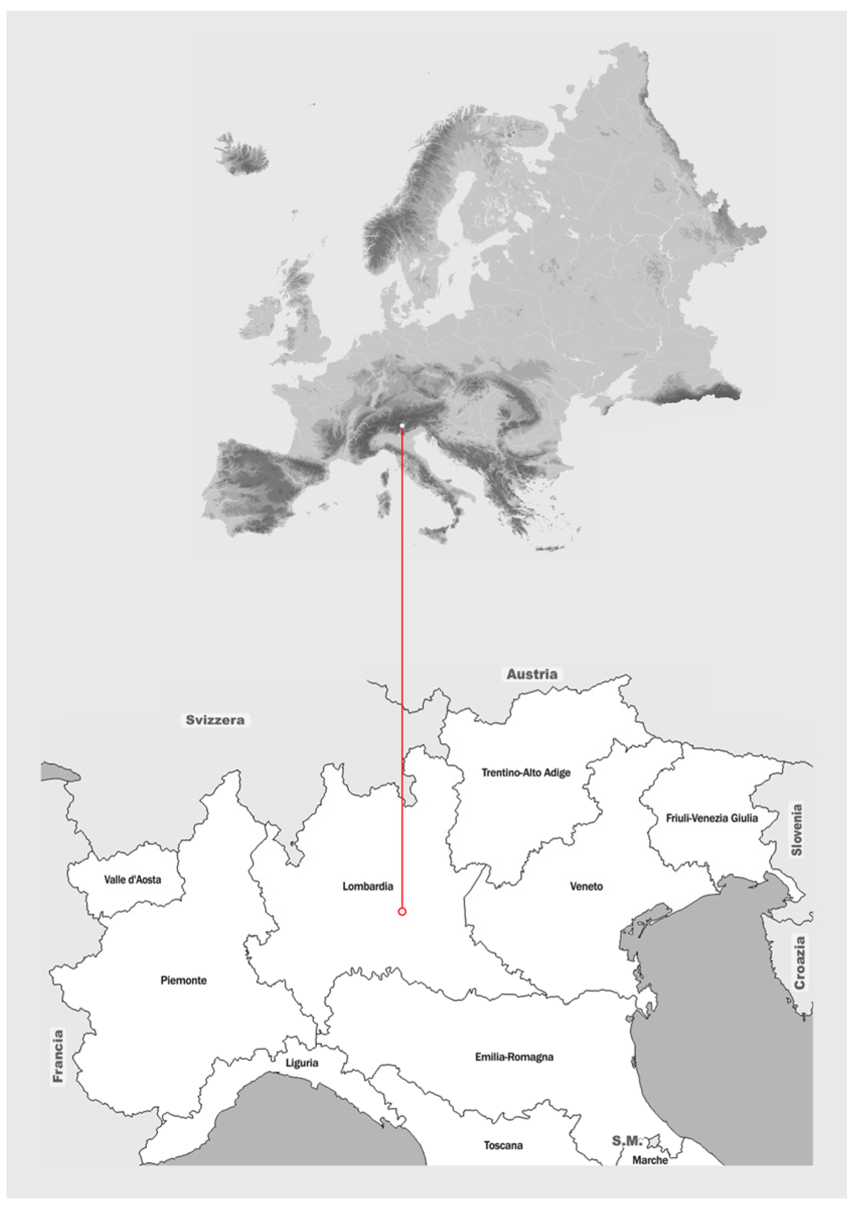
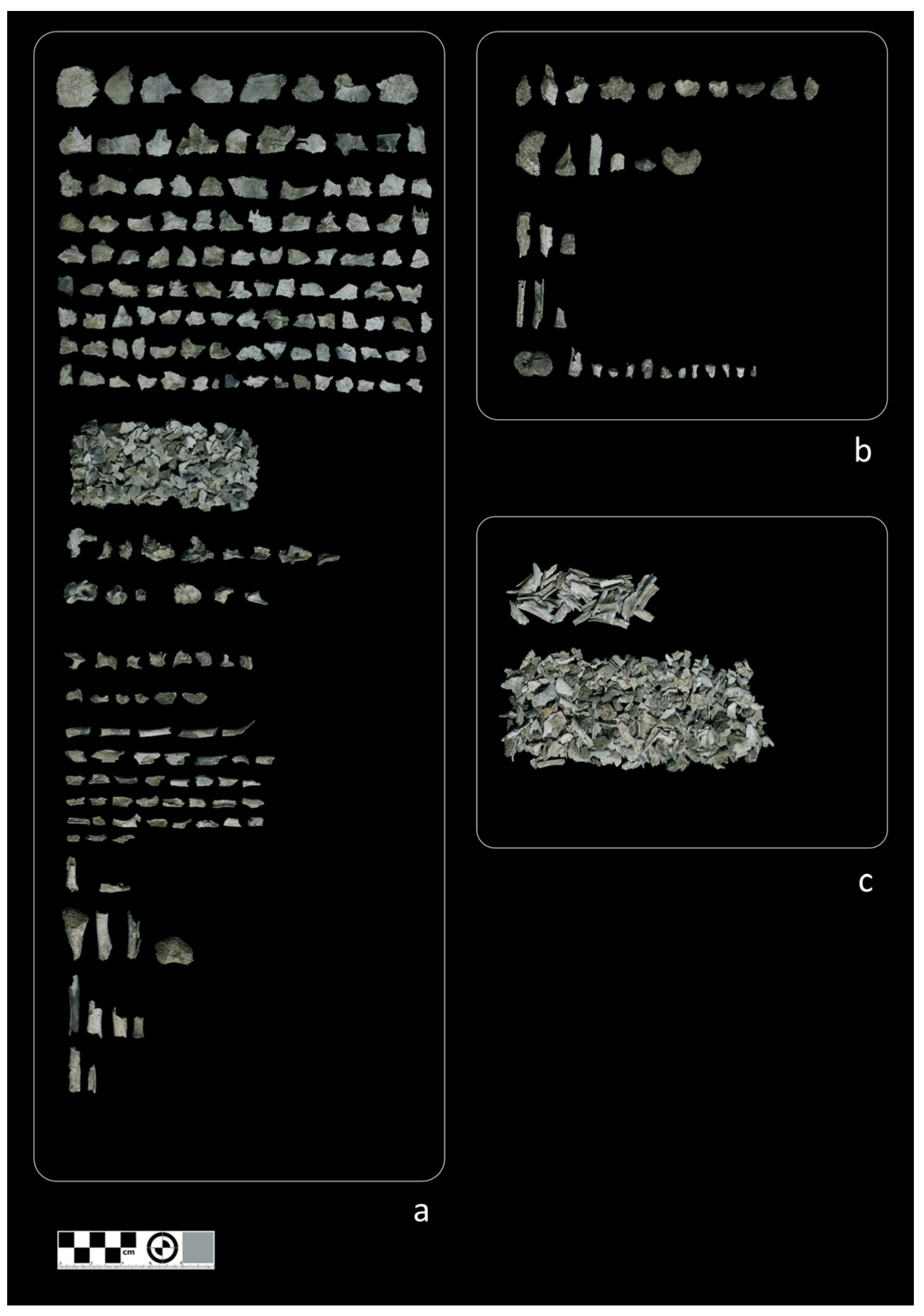
2.1. Materials—The Archaeological Sample

The sample consists of 298 tombs containing 314 cremated subjects of the Golasecca Civilization (7th–4th century BC north-eastern Italy) (Figure 1; Table 1).
The anthropological analysis made it possible to select 82 graves based on the probable or possible animal presence. The tombs have been divided according to the region and province in which they were found and belong to a chronological range that covers the entire Golasecca Civilization, which can be divided into the three main periods of the development of the Civilization in G I (900-625 BC), G II (625-475 BC), G III (475-375 BC). A total of 44 graves are distributed in G I, 36 in G II, and 2 in G III.

2.2. Methods

The first operation is the dry cleaning of the remains, followed by a double-blind morphological analysis to distinguish between non-humans and humans. The goal is to determine the human pieces, those that are non-human, and those that have an uncertain identification (Figure 2). Thus, a list of tombs with only human remains, with human and animal remains, and with human remains and possible animal remains is obtained. Subsequently, for all the graves in which the presence of animal fragments, even on a possible level, was verified, a sampling of 3 fragments, probably attributable to diaphysis, was carried out. TSs are prepared from these according to the protocol presented below (see Section 2.3 Thin section protocol) to verify the presence of animals and, if necessary, to identify them taxonomically. Sampling and TS analysis of the remains attributable to diaphysis were also performed for the individual burials that had weights higher than the estimated average for the Golasecca cremations. In fact, these tombs are more likely to contain other remains than just human ones.
The histological criteria adopted in this study to distinguish human from non-human remains are divided into morphological and morphometric ones. A first distinction can be made on the presence of plexiform tissue that characterizes non-human bones [31]. Subsequently, morphometric parameters related to the Haversian system, which are known to be helpful for TI, were considered: the maximum diameter of the osteon (DOMax), the minimum diameter of the osteon (DOMin), the maximum diameter of the Haversian canal (DCMax), the minimum diameter of the canal (DCMin), the perimeter of the osteon (PerimO), the perimeter of the Haversian canal (PerimC), the area of the osteon (AreaO), and the area of the Haversian canal (AreaO) [31,42]. In particular, following the work of Hillier and Bell [31], the ranges of Haversian canal and system diameter have been shown to be useful for TI (Table 2 and Table 3). Small mammals have a Haversian canal diameter < 20 µm, large mammals between 35 and 70 µm, while humans can be recognized with certainty above 120 µm. Taking into consideration the diameter of the Haversian system, small mammals fall within measurements < 100 µm, with the exception of the cat and dog taxa that share the mean values. Above 190 µm, it is possible to determine a large mammal for which, however, the distinction between human and non-human must be accompanied by the evaluation of any plexiform tissue (Table 2 and Table 3). Given the greater simplicity of the macroscopic morphological TI for fish, reptiles, amphibians, and birds, the study by Hillier and Bell [31] focused on the taxa of large and small mammals most frequent in the European archaeological record.
TS for which it was possible to perform TI, had a clear and legible osteonic structure. A change of structure takes place over 600 °C and also involves changes in the size and volume of the bone. However, since the bone structure is clearly legible, it is conceivable that the changes in volume are not statistically significant [43].

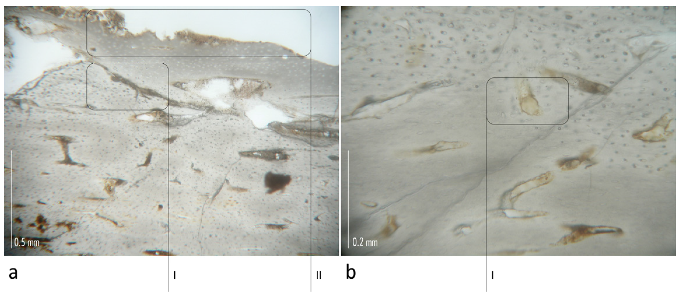
2.3. Thin Section Protocol
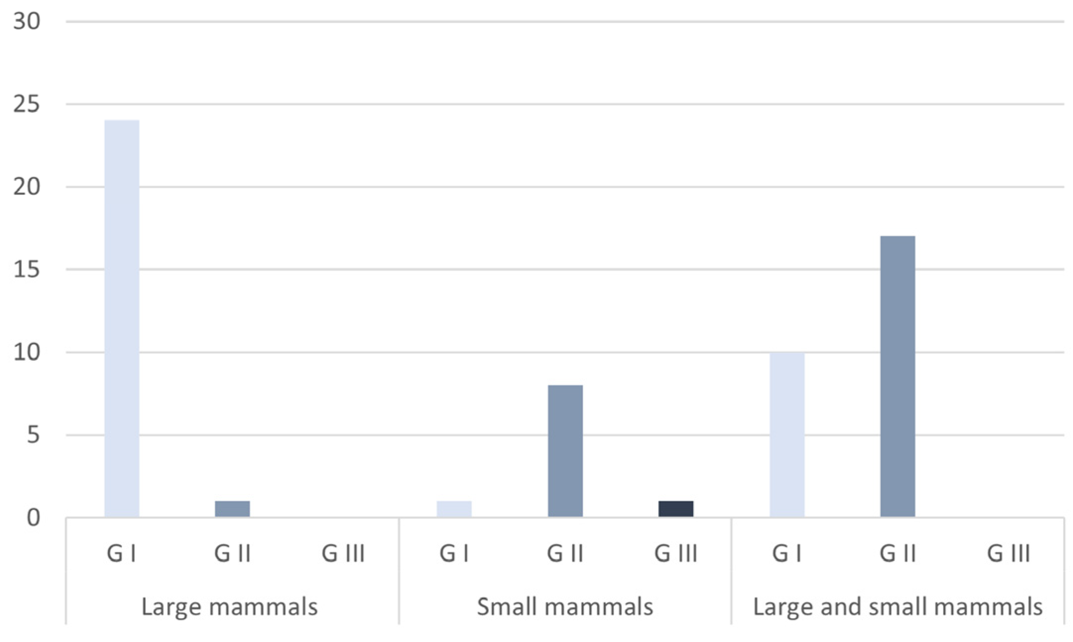
After selection (cfr. 2. Methods), the sample was dry cleaned with soft bristle brushes and then put in demineralized water in an ultrasonic cleaner. Once dried, it was necessary to properly dehydrate the fragments; the bones were put for 10 days in solutions with increasing concentration of alcohol, up to a 100%, so as to ensure the best penetration of the resin, which will be used later. After that, we placed them for 5 days in a solution composed of 50% absolute alcohol and 50% light-curing resin based on methacrylate, and finally, in pure resin.
The resin-impregnated sample is then placed into a resin-filled plastic mould and treated with an Exakt polymerizer to solidify the resin. After solidification, the block containing the sample is detached from the mould and glued to a plastic slide and abraded using sandpaper discs mounted on an Exak lapping machine to obtain a smooth surface. A second coverslip is glued to this surface, creating a sort of sandwich.
Using the Exakt 300 CP cutting unit, a TS is obtained near the second slide; the section is made thinner and vitrification, using abrasive discs until a perfectly smooth section with a thickness of about 100 µm, is obtained. The remaining portion of inclusion adhered to the first slide will remain in the archive in case one wants to prepare other samples. For TS evaluation was used a Leica DM4 P polarizing microscope.

3. Results

Out of the 298 tombs analysed morphologically: 250 had remains attributable only to humans (group A); 216 of these (subgroup A I) were excluded from the analysis, while 34 were selected because they weighed more than the average of the Golasecca cremations (subgroup AII); 26 had remains of possible non-human origin (group B); 22 had remains of probable non-human origin (group C) (Figure 3). For the results reported and subsequently discussed, please refer to Table 4.
Analysis of Subgroup A II (102 TS) showed plexiform tissue in 38 TSs, that of Group B (78 TS) in 55, and that of Group C (66 TS) in 58 for the 150 preparations in which plexiform tissue was found. Referring to the data on the graves, in Subgroup A II there are 19 graves with animal remains, in Group B 21 graves and Group C a total of 22 graves.
From the TI point of view, it was possible to find the presence of human tissue in 13 sections (11 graves), macro mammal in 84 TS (53 graves), of small mammals or deer in 47 TS (36 graves). For 17 TS, it was possible to recognise the plexiform tissue but not to proceed with the TI due to poor legibility of the tissue (17 burials), for 31 to exclude the presence of plexiform tissue but not to proceed with the TI (25 burials), while for 54 the reading of the preparation gave no indications either on the presence of plexiform or on the TI (39 tombs) (Figure 4 and Figure 5).
Regarding the TI concerning chronology, only macro mammals are counted in 25 preparations (G I = 24; G II = 1; G III = 0), only small mammals in 10 (G I = 1; G II = 8; G III = 1), and both macro and small in 27 (G I = 10; G II = 17; G III = 0) (Figure 6).

4. Discussion

The histomorphological analysis of the sample confirmed the efficacy of the non-human determinations for the whole group C; as regards group B, the presence of non-humans was verified in 21 out of 26 graves (81%); for this group, the impossibility of reading some TS, which belong to the remaining 29%, due to the action of fire does not allow excluding non-human TI. As far as the AII subgroup is concerned, the histomorphometric analysis allowed us to identify the presence of animal remains in 19 out of 34 graves (56%), confirming the usefulness of the histological method in the face of the limitations of the morphological one. Furthermore, the histomorphological analysis made it possible to identify in 61 cases the belonging of the animal remains to specific groups of taxa, a fact that cannot be deduced from the morphological analysis alone. However, it should be noted that identifying taxa with certainty is very difficult. The literature proposes more or less complex histomorphometric methods [31,42,44,45], which rarely allow TI because some animals share the size ranges of the analysed variables. However, it is possible to group these animals according to the average size values of the variables, dividing them into small and large mammals. Although these determinations do not fully satisfy the identification of specific taxa, they allow us to acquire valuable information to better understand the presence of animals in archaeological contexts.
From the point of view of the funerary rite, we note the greater presence of tombs containing exclusively macro mammals during the first phase of the Golasecca Civilization, while in the G II the number of tombs containing exclusively the remains of small mammals or deer increases. The latter taxon should perhaps be excluded, as the consistent presence of this animal in archaeological contexts is to be referred to earlier periods. Similarly, it is possible to exclude the presence of rats, as the bone fragments analysed were, in terms of size, all outside the intraspecific variability. Furthermore, the increase in small mammals during the G II could also be verified in graves that contained remains of both macro and small mammals. What emerges, therefore, seems to be the practice of burning large mammals in the first period and small mammals in the second period; this may suggest that animals were buried together with the deceased in the form of a food offering or ritual offering. The archaeological data show how during the GI Castelletto Ticino, Sesto Calende, Golasecca, and Como began to acquire importance as main centres of the Golasecca Civilization [46]. The qualitative and quantitative differences between the grave goods make it possible to identify forms of top-down social stratification, which could be reflected in the choice of animals used during the ritual.
This study allowed us to verify the efficacy of the histomorphological approach in the analysis of the TI of cremated archaeological remains. This has allowed the verification of the morphological attributions, the recognition of non-human fragments, where the morphological approach had not found any, and the TI in most cases. This type of analysis is of fundamental importance in the study of protohistoric cremations, as it has demonstrated how the morphological approach alone leads to an underestimation of the non-human sample compared to the human one to which the remains are usually referred in the absence of more in-depth analyses.

Funding

This research received no external funding.

Institutional Review Board Statement

Not applicable.

Informed Consent Statement

Not applicable.

Data Availability Statement

The data presented in this study are available on request from the corresponding author.

Acknowledgments

I want to thank for the study authorization of the Golasecca’s human cremated sample Barbara Grassi and Daniela Patrizia Locatelli of the Soprintendenza Archeologia Belle Arti e Paesaggio per le province di Como, Lecco, Sondrio e Varese; Cristina Longhi and Serena Solano of the Soprintendenza Archeologia, Belle Arti e Paesaggio per le province di Bergamo e Brescia; Lucia Mordeglia of the Soprintendenza Archeologia Belle Arti e Paesaggio per province di Biella, Novara, Verbano Cusio Ossola e Vercelli. I also want to thank Barbara Cermesoni of the Archaeological Museum of Villa Mirabello (VA) and Mauro Squarzanti of the Archaeological Museum of Sesto Calende (VA) for the study authorization of the material of civic propriety. Finally, I thank Monica Campagnolo of the Insubria University, for the invaluable work conducted on the thin sections.

Conflicts of Interest

The author declares no conflict of interest.

References

  1. Larentis, O.; Maccarinelli, A.; Marconi, S.; Pedrotti, A. The burned remains from Lugo di Grezzana (VR, Italy), 5400–5000 B.C. cal. Potential and limits of the anthropological analysis. In Atti della LVI Riunione Scientifica dell’Istituto Italiano di Preistoria e Protostoria—Le Scienze Della Preistoria e Protostoria: Paleoecologia, Archeobiologia, Applicazioni Digitali e Archeometria; Istituto Italiano di preistoria e Protostoria: Ferrara, Italy, 2022. in press. (In Italian) [Google Scholar]
  2. Bernabò Brea, M.; Maffi, M.; Mazzieri, P.; Salvadei, L. Testimonianze Funerarie Della Gente dei Vasi a Bocca Quadrata in Emilia Occidentale: Archeologia e Antropologia, in Rivista di Scienze Preistoriche: LX, 2010; Istituto Italiano di Preistoria e Protostoria: Florence, Italy, 2010. (In Italian) [Google Scholar]
  3. Salzani, P.; Salzani, L.; Dori, I.; Bortoluzzi, S.; Boccone, S. La necropoli del Bronzo antico di loc. Arano, Cellore di Illasi, Verona. In Preistoria e Protostoria del Veneto, (Studi di Preistoria e Protostoria 2); Istituto Italiano di Preistoria e Protostoria: Florence-Padua, Italy, 2015. (In Italian) [Google Scholar]
  4. Del Lucchese, A.; Gambari, F.M. L’area alpina sud—Occidentale e il mondo ligure. In Celtes et Gaulois, l’Archéologie face à l’Histoire; Vitali, D., Ed.; Atti della Tavola Rotonda di Bologna, Bologna-Monterenzio, 28–29 May 2005; Bibracte, Centre Archéologique: Bibracte, France, 2006. [Google Scholar]
  5. Perini, R. Tomba a Tumulo dell’età del Bronzo ai Calferi di Stenico (Giudicarie Esteriori); Studi Trentini: Trento, Italy, 1979. (In Italian) [Google Scholar]
  6. Perini, R.; Corrain, C.; Capitanio, M. La necropoli a tumulo di Stenico-Calferi (Trento): Notizie archeologiche e studio antropologico. Arch. Per L’antropologia E L’etnologia 1991, 121, 45–75. (In Italian) [Google Scholar]
  7. Marzatico, F. Stenico, località Calferi (Giudicarie Es-teriori, Trentino). In Kult der Vorzeit in den Alpen. Opfergaben—Opferplatze—Opferbrauchtum; Zemmer Planck, L., Ed.; Athesia: Bolzano, Italy, 2002. (In Italian) [Google Scholar]
  8. Marzatico, F.; Degasperi, N.; Endrizzi, N. Fosse di combustione rituali nel rituale del santuario di Cles Campi Neri (Valle di Non-Trentino). In Atti del VI Incontro Annuale di Preistoria e Protostoria, Focolari, Forni e Fornaci tra Neolitico ed età del Ferro (Bologna, 29 March 2019); Dipartimento di Storia Culture Civiltà–DISCI: Bologna, Italy, 2019. (In Italian) [Google Scholar]
  9. Cavazzuti, C.; Salvadei, L.; Salzani, L. Analisi antropologiche sui resti cremati della necropoli del Bronzo medio e recente di Scalvinetto di Legnago (Verona). In Preistoria e Protostoria del Veneto, (Studi di Preistoria e Protostoria 2); Istituto Italiano di Preistoria e Protostoria: Florence-Padua, Italy, 2015. (In Italian) [Google Scholar]
  10. Rubat Borel, F.; Cupitò, M.; Delpino, C.; Guidi, A.; Miari, M. (Eds.) Atti del Secondo Incontro Annuale di Preistoria e Protostoria, Second Annual Meeting of Prehistory and Protohistory, The Bronze and Iron Age in Italy: Protohistoric Contexts in Urban Excavations; Museo Nazionale Preistorico Etnografico “Luigi Pigorini”—Piazza G. Marconi, 14—Roma EUR (Italy)—27th January 2017; Istituto Italiano di Preistoria e Protostoria: Florence, Italy, 2016. (In Italian) [Google Scholar]
  11. Von Eles, P.; Pacciarelli, M. La Romagna dal Bronzo Finale alla Prima età del Ferro. In Studi di Preistoria e Protostoria, 3, Preistoria e Protostoria dell’Emilia Romagna, II; Istituto Italiano di Preistoria e Protostoria: Florence, Italy, 2018. (In Italian) [Google Scholar]
  12. Arslan, E.A. Culture celto–liguri e celto–golasecchiane nel Pavese e nell’Alessandrino. Ziχu Studi Sulla Cult. Di Golasecca 2019, 3, 11–28. [Google Scholar]
  13. Steele, T.E. The contributions of animal bones from archaeological sites: The past and future of zooarchaeology. J. Archaeol. Sci. 2015, 56, 168–176. [Google Scholar] [CrossRef]
  14. Meadow, R.H. Animal bones: Problems for the archaeologists together with some possible solutions. Paléorient 1980, 6, 66–77. [Google Scholar] [CrossRef]
  15. Hukantaival, S.; Bläuer, A. Ritual deposition of animals in late Iron age Finland: A case-study of the Mulli settlement site in Raisio. Estonian. J. Archaeol. 2017, 21, 161–185. [Google Scholar] [CrossRef] [Green Version]
  16. Macheridis, S. Symbolic connotations of animals at early Middle Helladic Asine. A comparative study of the animal bones from the settlement and its graves. Opusc. Annu. Swed. Inst. Athens Rome 2017, 10, 128–152. [Google Scholar] [CrossRef] [Green Version]
  17. Gaastra, J.S. Animal remains from ritual sites: A cautionary tale from the eastern Adriatic. Int. J. Osteoarchaeol. 2017, 28, 18–30. [Google Scholar] [CrossRef]
  18. Stephan, E. Oxygen Isotope Analysis of Animlal Bone Phosphate: Method Refinement, Influence of Consolidants, and Reconstruction of Palaeotemperatures for Holocene Sites. J. Archaeol. Sci. 2000, 27, 523–535. [Google Scholar] [CrossRef]
  19. Lahtinen, M.; Arppe, L.; Nowell, G. Source of strontium in archaeological mobility studies—Marine diet contribution to the isotopic composition. Archaeol. Anthropol. Sci. 2021, 13, 1. [Google Scholar] [CrossRef]
  20. Groot, M.; Evans, J.; Albarella, U. Mobility of cattle in the iron age and Roman Netherlands. J. Archaeol. Sci. Rep. 2020, 32, 102416. [Google Scholar] [CrossRef]
  21. Hopkins, J.B.; Ferguson, J.M. Correction: Estimating the diets of animals using stable isotopes and a comprehensive Bayesian mixing model. PLoS ONE 2012, 7, e28478. [Google Scholar] [CrossRef]
  22. Snoeck, C.; Cheung, C.; Griffith, J.I.; James, H.F.; Salessec, K. Strontium isotope analyses of archaeological cremated remains—New data and perspectives. Data Brief 2020, 42, 108115. [Google Scholar] [CrossRef] [PubMed]
  23. Bond, J.M. Burnt Offerings: Animal Bone in Anglo-Saxon Cremations. World Archaeol. 1996, 28, 76–88. Available online: http://www.jstor.org/stable/124975 (accessed on 8 January 2023). [CrossRef]
  24. Cerezo-Roman, J.; Williams, H. Future directions for the archaeology of cremation. In Transformation by Fire: The Archaeology of Cremation in Cultural Context; Kuijt, I., Quinn, C.P., Cooney, G., Eds.; University of Arizona Press: Tucson, AZ, USA, 2014. [Google Scholar]
  25. Williams, H. Towards an Archaeology of Cremation. In The Analysis of Burned Human Remains; Academic Press: Cambridge, MA, USA, 2015. [Google Scholar]
  26. Gonçalves, D.; Thompson, T.J.U.; Cunha, E. Implications of heat-induced changes in bone on the interpretation of funerary behaviour and practice. J. Archaeol. Sci. 2011, 38, 1308–1313. [Google Scholar] [CrossRef]
  27. Imaizumi, K. Forensic investigation of burnt human remains. Res. Rep. Forensic. Med. Sci. 2015, 5, 67. [Google Scholar] [CrossRef] [Green Version]
  28. Ubelaker, D.H. The forensic evaluation of burned skeletal remains: A synthesis. Forensic. Sci. Int. 2009, 183, 1–5. [Google Scholar] [CrossRef]
  29. Mulhern, D.M. Differentiating Human from Nonhuman Skeletal Remains. In Handbook of Forensic Anthropology and Archaeology, 2nd ed.; Blau, S., Ubelaler, D.H., Eds.; Routledge: London, UK, 2016. [Google Scholar]
  30. Whyte, T.R. Distinguishing Remains of Human Cremations from Burned Animal Bones. J. Field. Archaeol. 2001, 28, 437–448. [Google Scholar] [CrossRef]
  31. Hillier, M.L.; Bell, L.S. Differentiating human bone from animal bone: A review of histological methods. J. Forensic. Sci. 2007, 52, 249–263. [Google Scholar] [CrossRef]
  32. Vigne, J.D. The origins of animal domestication and husbandry: A major change in the history of humanity and the biosphere. Comptes Rendus Biol. 2011, 334, 171–181. [Google Scholar] [CrossRef]
  33. McClelland, W. Distinguishing Human from Non-Human Animal Bone 2018; Arizona State Museum: Tucson, Arizona, 2018. [Google Scholar]
  34. Dumont, E.R. Bone density and the lightweight skeletons of birds. Proc. Biol. Sci. 2010, 277, 2193–2198. [Google Scholar] [CrossRef] [Green Version]
  35. Hillson, S. Mammal Bones and Teeth An Introductory Guide to Methods of Identification; Routledge: New York, NY, USA, 1992. [Google Scholar]
  36. Mock, F.; Kretschmer, F.; Kriese, S.; Böker, S.; Marz, M. Taxonomic classification of DNA sequences beyond sequence similarity using deep neural networks. Proc. Natl. Acad. Sci. USA 2022, 119, e2122636119. [Google Scholar] [CrossRef] [PubMed]
  37. Oh, Y.N.; Park, J. Analysis of an ancestry using cremated old human remains from the Korean War victims. Forensic. Sci. Int. Sci. Genet. Suppl. Ser. 2022, 8, 140–142. [Google Scholar] [CrossRef]
  38. Dulias, K.; Foody, M.G.B.; Justeau, P.; Silva, M.; Martiniano, R.; Oteo-García, G.; Fichera, A.; Rodrigues, S.; Gandini, F.; Meynert, A.; et al. Ancient DNA at the edge of the world: Continental immigration and the persistence of Neolithic male lineages in Bronze Age Orkney. Proc. Natl. Acad. Sci. USA 2022, 119, e2108001119. [Google Scholar] [CrossRef]
  39. Fiddyment, S.; Holsinger, B.; Ruzzier, C.; Devine, A.; Binois, A.; Albarella, A.; Fischer, R.; Nichols, E.; Curtis, A.; Cheese, E.; et al. Animal origin of 13th-century uterine vellum revealed using non invasive peptide fingerprinting. Proc. Natl. Acad. Sci. USA 2015, 112, 15066–15071. [Google Scholar] [CrossRef] [Green Version]
  40. Gaêta-Araujo, H.; Oliveira-Santos, N.; Brasil, D.M.; do Nascimento, E.H.L.; Madlum, D.V.; Haiter-Neto, F.; Oliveira-Santos, C. Effect of micro-computed tomography reconstruction protocols on bone fractal dimension analysis. Dentomaxillofac. Radiol. 2019, 48, 20190235. [Google Scholar] [CrossRef]
  41. Basillais, A.; Bensamoun, S.; Chappard, C.; Brunet-Imbault, B.; Lemineur, G.; Ilharreborde, B.; Ho Ba Tho, M.C.; Benhamou, C.L. Three-dimensional characterization of cortical bone microstructure by microcomputed tomography: Validation with ultrasonic and microscopic measurements. J. Orthopaedic. Sci. 2007, 12, 141–148. [Google Scholar] [CrossRef]
  42. Cattaneo, C.; de Martino, S.; Scali, S.; Craig, O.E.; Grandi, M.; Sokol, R.J. Determining the human origin of fragments of burnt bone: A comparative study of histological, immunological and DNA techniques. Forensic. Sci. Int. 1999, 102, 181–191. [Google Scholar] [CrossRef]
  43. Castillo, R.F.; Ubelaker, D.H.; Acosta, J.A.L.; de la Fuente, G.A.C. Effects of temperature on bone tissue. Histological study of the changes in the bone matrix. Forensic. Sci. Int. 2013, 226, 33–37. [Google Scholar] [CrossRef] [PubMed]
  44. Martiniaková, M.; Grosskopf, B.; Omelka, R.; Vondráková, M.; Bauerová, M. Differences Among Species in Compact Bone Tissue Microstructure of Mammalian Skeleton: Use of a Discriminant Function Analysis for Species Identification. J. Forensic. Sci. 2006, 51, 1235–1239. [Google Scholar] [CrossRef] [PubMed]
  45. Martiniaková, M.; Grosskopf, B.; Omelka, R.; Dammers, K.; Vondráková, M.; Bauerová, M. Histological study of compact bone tissue in some mammals: A method for species determination. Int. J. Osteoarchaeol. 2007, 17, 82–90. [Google Scholar] [CrossRef]
  46. Gambari, F.M. Da Castelletto Ticino a Novaria: L’Ovest Ticino in età preromana. In La Birra e il Fiume. Pombia e le vie dell’Ovest Ticino tra VI e V Secolo a.C; Gambari, F.M., Ed.; CELID: Torino, Italy, 2001. (In Italian) [Google Scholar]
Figure 1. Location of the area under analysis. In the upper part, a map of Europe shows the territories of northern Italy in the lower part of the figure.
Heritage 06 00034 g001
Figure 2. Protohistoric cremation of northern Italy (Golasecca Civilization). (a) Anatomically ordered human bone fragments from the skull (top) to feet (bottom); (b) Animal bone fragments; (c) Taxonomically unidentified fragments. Data were acquired through macroscopic morphological identification with double-blind analysis.
Heritage 06 00034 g002
Figure 3. Pie chart with groups and subgroups analysed in this study; the identification was carried out macroscopically and morphologically. Group A = cremation with only human bone remains; Subgroup A I = cremation with only human bone remains with weights more equal or lower than the average of the other Golasecca cremations; Subgroup A II = cremation with only human bone remains with weights higher than the average of the other Golasecca cremations; Group B = cremation with any remains of non-human origin; Group C = cremation with probable remains of non-human origin.
Heritage 06 00034 g003
Figure 4. Thin section of cremated bone, transmitted by light microscopy. (a) 10× magnification. Assessment of bone tissue is made difficult by the burning process. However, it is possible to identify fractures probably related to alterations induced by cremation or by taphonomic processes (I). In the section, a brown-coloured matrix can be seen, which fills the fracture, probably deposition of soil that has infiltrated the void of the fracture. The same soil is visible adhering to the Haversian canals and on the external bark, which also presents diagenetic phenomena, which have eroded the surface (II). (b) Magnification 25×. In the lower portion of the section, it is possible to note the loss of the osteonic structure; however, some diagnostic morphological traits for TI, such as the Haversian canal of the osteon, are still clearly visible.
Heritage 06 00034 g004
Figure 5. Thin section of cremated bone, polarized light microscopy. (a) 10× magnification. The polarized light allows a better observation of the osteonic structure (I), permitting, in some cases, to identify the presence of plexiform tissue. Even in areas where the evaluation is particularly compromised (III) the morphology of the Haversian canals is visible (II). (b) Magnification 25×. The observation of the cortical tissue benefits from the use of polarized light, allowing us to identify with certainty the osteonic system (I, III). It is worth noticing that reddish areas in which there are deposits of earth infiltrated into the bone (II) can be recognized in some fractures of the bone structure.
Heritage 06 00034 g005
Figure 6. TI distribution in thin section (according to histomorphological data: large mammals = goat; cow, horse, pig, dog; small mammals = cat, deer, rabbit, rat; large and small mammals = at least one non-human fragment reconstituted to both previous categories). The TIs have been divided into the three main periods of the Golasecca Civilization G I (900-625 BC), G II (625-475 BC), and G III (475-375 BC).
Heritage 06 00034 g006
Table 1. Human sample from the Golasecca Civilization, subdivided by region, province, and site location. For each archaeological site, the number of subjects analysed is reported.
RegionProvinceLocationn Subjects
LombardyVareseGolasecca (1954)1
LombardyVareseGolasecca, collezione Cesare da Sesto 2
LombardyVareseGolasecca, Monsorino (1985–1986)6
LombardyVareseSesto Calende, Cascina Bassoni (1958)1
LombardyVareseSesto Calende, Cascina Stallazzo1
LombardyVareseSesto Calende, collezione Bellini13
LombardyVareseSesto Calende, collezione Mattana2
LombardyVareseSesto Calende, loc. Mambrino (1986)2
LombardyVareseSesto Calende, loc. Mulini Bellaria (1977–1981, 1995, 2004)21
LombardyVareseSesto Calende, loc. Mulini, via Sempione (2005)2
LombardyVareseSesto Calende, loc. Mulini, via Beltrami (1983)1
LombardyVareseSesto Calende, loc. Presualdo (1983–1986, 1994, 1997)38
LombardyVareseSesto Calende, via Bellaria–via Marconi (1989)1
LombardyVareseSesto Calende, via Moncenisio (1995–1996)16
LombardyVareseSesto Calende, via Montrucco (1995, 2003)9
LombardyVareseSesto Calende, via Motte (1999)1
LombardyBergamoBrembate Sotto, Strada Provinciale Osio-Trezzo (1888)1
LombardyBergamoCaravaggio, Tangenziale Ovest, via Einaudi (2013)1
LombardyComoComo, Ca’ Morta (fino al 1981)54
LombardyComoComo, Nuovo Ospedale Sant’Anna (2007)2
LombardyComoComo, via Tito Livio (1996)7
LombardyBresciaUrago d’Oglio, loc. Cascina Giardina (2009)4
LombardyPaviaGarlasco, Madonna delle Bozzole (1994) 3
PiedmontNovaraCastelletto Ticino (1959)16
PiedmontNovaraCastelletto Ticino, loc. Motto d’Egro (1984)1
PiedmontNovaraCastelletto Ticino, collezione Cesare da Sesto1
PiedmontNovaraDorbié Superiore, loc. Cascina Riviera (1987)5
PiedmontNovaraCastelletto Ticino, loc. Cascina Brua, via Ardeatine (2009)25
PiedmontNovaraCastelletto Ticino, loc. Forcetto (1986)2
PiedmontNovaraCastelletto Ticino, via Valloni (2005–2006)3
PiedmontNovaraCastelletto Ticino, proprietà Guenzi, via Beati (2001)1
PiedmontNovaraCastelletto Ticino, proprietà Iacomella, via Valsesia (2002)5
PiedmontNovaraCastelletto Ticino, via Repubblica (2002)3
PiedmontNovaraCastelletto Ticino, via Aronco (1986, 1988–1989)12
PiedmontNovaraCastelletto Ticino, via del Maneggio (2001–2003)32
PiedmontNovaraCastelletto Ticino, via Fermi (2014)1
PiedmontNovaraCastelletto Ticino, via Ramacci (1998–2000)3
PiedmontNovaraPombia, Cimitero (1987)2
PiedmontNovaraPombia, loc. Planca, via Veneto (1993)5
PiedmontNovaraPombia, proprietà Baù, loc. Quara (1994–1995)8
Total subjects314
Table 2. Ranges of Haversian canal diameter of several small and large mammals [31].
Ranges of Haversian Canal Diameter (µm)
TaxonMinMaxMean
Rat/ <10
Deer//<10
Rabbit//10–20
Cat//20
Dog155035
Pig//35
Cow155537
Goat187042
Sheep1912070
Human30175105
Table 3. Ranges of Haversian system diameter of several small and large mammals. Data are not available for pig and deer [31].
Ranges of Haversian System Diameter (µm)
TaxonMinMaxMean
Rat//70
Deer///
Rabbit//100
Cat//160
Dog125175155
Pig///
Cow155250210
Goat55320190
Sheep80360220
Human180320260
Table 4. Taxa taken into consideration: humans, sheep, goats, cows, horses, pigs, dogs, cats, deer, rabbits, and rats; the taxa are listed in order of size of the osteonic and Haversian systems, from largest to smallest [31]. Given the recognisability of human tissue and the overlapping of some dimensional ranges of non-human bone tissue, three main categories have been proposed: (a) Human; (b) Large mammal species (sheep, goats, cows, horses, pigs and dogs); (c) Species of small mammals (cats, deer, rabbits, and rats); unfortunately, the histomorphometric method does not allow for the distinction of deer from small mammals.
Group/SubgroupGraveTS 1TS 2TS 3Period
PlexiformTIPlexiformTIPlexiformTI
A II1YesbYesbYesbG I
A II2NoNDYesc//G III
A II3//YesbYesbG I
A II4NoND////G I
A II5YesbYesbYescG II
A II6YesbNoND//G II
A II7NoaNoa//G II
A II8NoaNoNDNoaG I
A II9YescYes/YesbG II
A II10NoNDNoND//G I
A II11YesbYesbYesbG I
A II12//////G II
A II13NoND////G II
A II14Yesc//YescG II
A II15NoNDYesbYescG I
A II16YesbYes///G I
A II17NoNDNoNDNoNDG I
A II18YescYesbYescG I
A II19NoND//NoaG I
A II20//NoND//G II
A II21Yes/YesbNoaG I
A II22Yesb//YesbG I
A II23NoNDNoND//G II
A II24////YescG II
A II25NoNDYesb//G I
A II26NoNDNoaYesbG II
A II27NoNDNoND//G I
A II28////NoNDG I
A II29YescYes/YesbG II
A II30NoNDNoa//G II
A II31YesbYesb/ G I
A II32//NoNDYesbG I
A II33NoNDNoNDNoaG II
A II34////NoNDG I
B1YescYescYesbG II
B2NoaYescYescG II
B3////NoNDG I
B4YesbYescYesbG II
B5YesbYes/YesbG I
B6YesbYescYesbG I
B7YescYescYesbG II
B8NoNDNoa//G II
B9Yes/Yesb//G I
B10YesbYescYesbG I
B11////YesbG I
B12YesbYesbYescG I
B13YesbYescYescG II
B14 //Noa//G II
B15YesbYesbYesbG I
B16//YescYesbG I
B17Yesc//YescG II
B18YesbYescYes/G II
B19YesbYes/YesbG I
B20YesbYescNoaG I
B21YescYes/YesbG II
B22YesbYesb//G I
B23//////G I
B24YesbYescYes/G II
B25NoND////G II
B26YescYesbYesbG I
C1Yes///YescG II
C2YesbYescYesbG II
C3YesbYesc//G II
C4Yes/YesbYescG I
C5NoNDYescYescG II
C6YesbYes/YesbG I
C7//YescYesbG II
C8Yes/YescYesbG II
C9YesbYesbYesbG I
C10YesbYesbYes/G I
C11Yesb//YesbG I
C12YesbYesbYescG I
C13YesbYesbYesbG I
C14Yesb//YesbG I
C15//YesbYescG II
C16YesbYescYesbG II
C17YescYes/YesbG II
C18YesbNoNDYes/G I
C19YesbYesb//G I
C20NoNDYescYescG II
C21YescYescYescG III
C22Yesb//YesbG I
Disclaimer/Publisher’s Note: The statements, opinions and data contained in all publications are solely those of the individual author(s) and contributor(s) and not of MDPI and/or the editor(s). MDPI and/or the editor(s) disclaim responsibility for any injury to people or property resulting from any ideas, methods, instructions or products referred to in the content.

