Abstract
Skeletal evidence dating back to the Mesolithic period is scarce and should be studied under a multidisciplinary perspective. The primary objective of the study was to carefully assess the skeleton of a young woman from this era, named “Avgi,” to compile its bioarchaeological profile, analyze its paleopathology and dental pathology, and deploy a 3D reconstruction and modeling method in order to reveal her face. Both demographic and pathological information were drawn from macroscopically observing the bones, long bone X-rays, skull CT and X-rays, 3D modeling and printing of the skull, and panoramic dental X-rays. The Manchester method was used for the 3D facial reconstruction. On analysis, we determined that Avgi was a female adolescent, aged around 17–19 years at death, and likely suffering from iron deficiency anemia and Class III dental malocclusion. Notably, Harris lines and a hair-on-end pattern were identified in the long bones and skull radiographs, respectively. Various less significant skeletal lesions reflected potential minor pathologies. Our findings suggest that multidisciplinary collaborative approaches should be followed in the modern study of lesser-known past eras. Multiple scientific perspectives, as well as social structures, geographical aspects, settlements, population movements, and social networks should all be taken into account when assessing lifestyle characteristics and paleopathological signs in skeletal remains.
Keywords:
3D modelling; 3D reconstruction; dental pathology; anemia; anthropology; paleopathology; skeleton 1. Introduction
The Mesolithic period in Europe began with a worldwide environmental change to a warmer climate following the Last Glacial Maximum, sometime in the 10th millennium BC. This period does not have the same duration or the same cultural features across space and time [1]. Recent relevant discoveries include sites from León in Spain [2], Dinant in Belgium [3], and Combe-Capelle in France [4]. Lazarides et al. [5] sequenced and analyzed the genomes from two Mesolithic skeletons to show the connection with present day Europeans. Findings from Greece, including the Theopetra Cave, have aided in exploring the evolution of diet and human occupations [6] and in demonstrating a genetic link across populations in Europe [7]. However, human remains from that period are scarce, and ancient DNA research retains inherent methodological difficulties [2]. Thus, skeletal evidence dating back to the Mesolithic period should be studied under a multidisciplinary perspective.
In Greece, the Mesolithic period lasted for approximately two thousand years (9000–7000 BC) and, until the 1970s, relevant archaeological research was limited [8]. Excavations in Franchthi Cave in the Argolid in the late 1960s and early 1970s brought to light Mesolithic deposits and opened new opportunities in the study of the era [9,10]. Since then, and especially during the past twenty years, the archaeological data have been enriched considerably, thanks to the identification of new Mesolithic sites and their subsequent systematic excavations [8,11,12,13,14]. Surface surveys aiming specifically at the identification of Mesolithic sites have also been conducted in Epirus, the Argolid, Northern Sporades, on Meganisi near Leukas and at Plakias in southern Crete [8,15]. More recently, Mesolithic deposits have been also found in the Negros Cave at the island of Astypalaia [16]. In the last few years, the intensification of Mesolithic investigation has offered great insights into our knowledge of this period in Greece.
Despite this recent progress, however, the number of known Mesolithic sites (caves and open-air sites) remains small (ca. 12), and their geographical distribution testifies to sporadic human activity throughout Greece. The scarcity of Mesolithic sites may not only be due to limited archaeological research, but also to taphonomic conditions and limitations. Thus, it has been suggested that the Mesolithic people led a nomadic life focused on the seasonal exploitation of the available natural resources, e.g., hunting large-scale preys, such as Cervus, and fishing open-sea species, such as tuna, or collecting seafood, such as oysters. Indeed, Mesolithic sites in Greece have yielded finds such as seashells, fishbones, and bone fishhooks. These attest to maritime activities and reflect a certain level of familiarity with the sea and the exploitation of marine resources [8,15,17]. Theopetra Cave, however, is located in inland Thessaly and far from the sea, canceling the older model about localizing the Mesolithic only by the sea. The presence of tools made of Melian obsidian in archaeological sites in the Greek mainland, such as the Cave at Franchthi in the Argolid (as early as the Upper Palaeolithic era, some 13,000 years ago) and on island sites, such as Maroulas on Kythnos, reveals the intensification of sea crossings during this period. These represent the earliest known maritime activities around the Mediterranean [8].
The number of uncovered Mesolithic human burials remains small, and are found at Franchthi, Theopetra, and Maroulas [8,11,12,17] (Figure 1). The burial sites seem to be unfurnished, although at Astypalaia, this seems to change. Along with the extant architectural remains (e.g., stone foundations of houses at Maroulas and Sidari), these burial sites could be considered indicators of a semi-permanent habitation, first observed during the Mesolithic period.
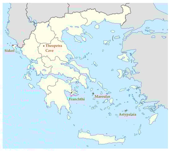
Figure 1.
Sites of Mesolithic excavations in Greece.
Contrary to the central European regions, the geo-morphology of the Greek peninsula provided the hunter-gatherer groups of that time with a number of different environmental niches and habitats. Adapting to a wide range of habitats eventually brought about technological variation, as well as an array of region-specific survival techniques and strategies. In fact, there is indeed not much found in common between the island communities of Youra and Kythnos and those of Theopetra, let alone to recommend studying them collectively under the narrow typo-chronological limits of the term Mesolithic.
Our reading of available evidence from the Greek Peninsula and the Aegean area attributed to the Mesolithic period would suggest a cultural process that took place from the Palaeolithic to the Neolithic eras. This process is perceived as a component of the European Mesolithic era, yet with discrete spatiotemporal traits that could be better understood through cultural continuity. The Greek Mesolithic period is viewed here as the natural outcome of processes started during the Palaeolithic era, which gradually led to the practices of the Neolithic period. From this perspective, many researchers have argued that we should look deeper into the nature of the Greek Mesolithic era as a preparatory stage that could explain differences observed between the south-eastern and the northern European Neolithic cultures [18,19,20]
The skeletal remains uncovered from the excavation of Theopetra Cave, conducted under the direction of Dr. Nina Kyparissi-Apostolika, have been identified as a case study on specific analyses of biological and cultural variables, determinant of the microevolutionary processes of the ancient populations in the Greek Peninsula. The Cave of Theopetra is located on the westernmost edge of the Thessalian plain, very close to the Pindos Mountains and about 100 km from the Aegean coast (Figure 1). The Mesolithic era is represented by a distinct deposit between the Upper Paleolithic and the Neolithic periods.
In this deposit, a human burial was found intact in 1993. The body was buried in a filled shallow pit with the skull on a higher level looking straight ahead, while the rest of the skeleton was in a semiflexed position, turned to the right, looking towards the entrance of the cave (Figure 2). A flint implement was found beside the knees and another beside the chest [12,20], but they are appreciated as findings from the fill, not corresponding to the burial itself. The reason for this is that carbons found in the same fill of the pit were dated between 8290–8720 BC and 9090–8300 BC (DEM 315 and 316, respectively), while dating of the bones yielded different ages: 8070 BC ± 60 years (7280–6830 BC—CAMS 21773) [21]. This chronological gap of about 1500 years could only be explained on the basis that the shallow pit was filled with deposits dug in deeper layers.
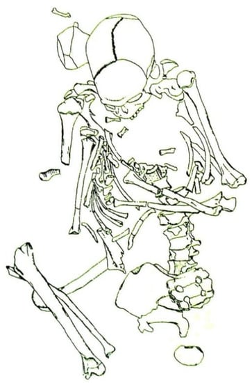
Figure 2.
Schematic representation of the skeleton as found.
In the following years, two or three more Mesolithic burials were uncovered around the same area of the cave, close to the entrance [22]. One of them was not complete, and the scattered human bones found at its vicinity were originally thought to represent a different burial, but it is more likely that they come from the same skeleton, as the dates from the burial and the scattered bones are very close, 8450 ± 45 ΒΡ; 7586–7384 cal BC (OxA 17352) on a bone from the burial and 8549 ± 40 BP; 7605–7529 cal BC (OxA 26680) on a bone from the scatter in the same square [7]. They were probably disturbed due to the activity of small animals [14,22].
In this study, we attempted a multidisciplinary, integrated research of human remains from the Greek Mesolithic period. The primary objective of the study was to carefully assess the skeleton, compile its bioarchaeological profile, analyze its paleopathology and dental pathology, and deploy a 3D reconstruction and modeling method in order to reveal a Mesolithic face. This is the first 3D face reconstruction ever to be attempted on skeletal remains of such antiquity from Greece, and one of the very few in the European Mesolithic period. We conclude by commenting on the implications of studying this era and the challenges and limitations of such research. The study is expected, not only to fill some important gaps in our current knowledge about the activity and nature of the Mesolithic populations in Southeast Europe, but also to promote cultural heritage by ‘giving a face’ to the Greek Mesolithic period, a heuristic for making the archaeological information tangible and accessible to wider audiences.
2. Materials and Methods
Gender and age estimation is an essential part in the process of extracting information from skeletal material. In this case, the estimation of the sex was based on morphological and metrical criteria of the skull [23] by visual assessments of: (i) the prominence and texture of the supraorbital arc, (ii) the sharpness of the supraorbital margin, (iii) the relative size of the zygomatic arc, (iv) the size of the mastoid process, (v) the prominence of the external occipital crest, (vi) the mental eminence, and (vii) the size and shape of the lower jaw and its depression. The estimation of the age was primarily based on dental markers and additionally, on skull and palatal sutures and on teeth development and occlusion through the use of panoramic radiographs [24]. The third molar in particular was a key factor in estimating the age at death [25]. The assessment of the person’s height was done through measuring all bones constituting the components of stature, summing those measurements, and correcting for the missing soft tissue [26].
The estimation of demographic parameters was based on the framework established by a previous work, which applied novel methodological techniques [27] to minimize inherent limitations in the study of ancient skeletons, such as inter-observer error and biased estimates, and strengthen the reliability of the results by testing alternative protocols. It is worth noting that the assessment of demographic parameters is a debatable field in anthropological research, since it applies formulas and reference samples from living populations and mainly from different countries. Greece lacks a relevant database (with the exception of a recently published atlas of craniofacial data by the same team) [28]; therefore, the range of the variance in age intervals, in sexual dimorphism, and stature variants of the population, may be biased [27].
The whole skeleton was examined macroscopically for any pathological lesions. Multiple X-rays of the skeleton and lateral, anteroposterior, and basilar X-rays of the skull provided enough evidence for a differential diagnosis. Radiologic findings were evaluated to facilitate interpretation(s) on clinical condition. Radiography, as a non-destructive technique, has been shown to be an appropriate method for examining anthropological material and assessing paleopathological findings of skeletal remains [29]. A complete medical, as well as dental and orthodontic, diagnosis of the Theopetra skeleton was conducted based on clinical examination, occlusal views, and panoramic radiograph.
In the next stages, for the facial reconstruction, a non-invasive approach was followed, so that the original skull would not be damaged. Reverse engineering (RE) and rapid prototyping (RP) techniques were used. The standard procedure includes computed tomography (CT) scan data acquisition, 3D reconstruction and segmentation of CT data, and 3D visualization and printing of the produced model [30,31,32,33]. The first case of applying CT scans for forensic imaging was that of the 5300-year-old mummy that was found in the Tyrolean Alps [34]. Since then, the latest generation of CT scanners have drastically enhanced the accuracy and quality of the produced 3D medical models for scull reconstruction [35,36,37,38]. The scanner used in this study was a GE Medical Systems Lightspeed VCT. A total of 1040 slices, with a slice increment 0.3 mm, 0.414 pixel size, and 512 × 512 image matrix size, were acquired. A 3D visualization was performed by means of triangulation of a segmented 3D area. The final step of the procedure was the 3D printing, which is a rapid prototyping method widely used in maxillofacial, spinal, and orthopedic medical cases [39]. For this reason, the 3D model reconstructed from the CT images was converted into an STL (stereolithography) 3D print-ready model. The physical model was then generated with a Stratasys dimension soluble support technology (SST) 3D printer, which is based on depositing a fused acrylonitrile butadiene styrene ABS jet with a layer thickness of 0.245 mm.
3. Results
3.1. Demographic Details
Female-specific characteristics were identified by the visual examination of the relevant features. All the sutures were open and an obliteration of the maxillary sutures was not observed. The fully open incisal suture (IN) in particular seems to exist in young individuals around 17–18 years of age [40]. Finally, although the lower wisdom teeth were in complete emergence in the occlusal plane (not the upper), all the root apices remained open. According to the dental attrition system of age estimation—usually inappropriate due to inherent variability—this skeleton would appear to be an adult of 25–30 years old [41,42,43]. However, this would be a false result, taking into account the consumption of hard foods and perhaps leather preparation in Mesolithic populations [44]. In any case, in the center of the acetabulum, a trace of the triradiate cartilage was identified, not yet ossified, which indicated the age of the subject to be around 15 to 20 years old. Macroscopic observation of the long bones showed incomplete fusion of long-bone epiphyses, suggesting an age of around 15; it is not infrequent that estimations based on long bone observations suggest ages younger than observations based on the panoramic X-ray. All in all, it can be reasonably estimated that this female was between 17 and 19 years old at the time of death. Her height was estimated at 1.57 m (p < 0.0001). The research team gave her the name “Avgi”, meaning “dawn” in Greek, signaling the dawn of the Holocene era, in our case.
3.2. Macroscopical Observations
There is no doubt that a typical Homo sapiens individual (with no physical disabilities) who lived at about 7000 BC would have all the anatomical prerequisites to be able to speak (the Broca’s area, inferior frontal gyrus, sufficient brain volume, and sufficient descent of larynx are all elements identified well before this era) [45]. Indeed, the skull in our case exhibits all the aforementioned characteristics (Figure 3). However, due to bone deficits in the posterior cranial fossa, lack of foramen magnum, and a partial lack in the left temporal bone, any statistical measurement of the cranial volume would be subject to significant error. In addition, the aspect of the palate cavity showed that the larynx had descended in the neck, as in the adult human, thus allowing phonation. Therefore, all anatomical premises for speech exist in this case. In addition, a depression on the side of the forehead is related to a premature closure of the suture of the area.
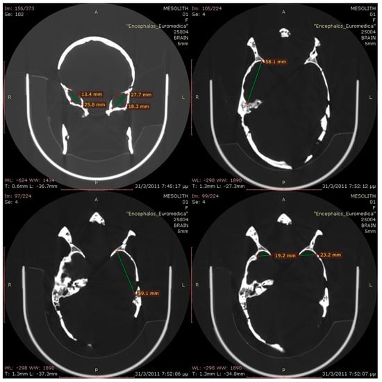
Figure 3.
Computerized tomography views of the skull.
By studying the pelvis macroscopically, the curved ilium and curved ischial tuberosity were recognized. A thin ala was surrounded by thick cortical bone (sciatic buttress below and iliac crest on the top). There was a thick column from the posterior superior spine of the ilium to the tuberosity of the ischium (proving the mode of loading by bipedal locomotion). There was also a thick bony area in the sacroiliac joint. There was no normal curvature of the iliac crest of the fully evolved pelvis. The shorter distance between the iliac crest to the corresponding greater trochanter (opposite to the acetabulum) created a short lever arm of the abductor muscles (gluteus medius), with implications for the lateral stability of the body. The acetabulum was fully developed, as the floor of the acetabulum was sitting on the ilioischial line, and it was not dysplastic, as the Wiberg angle (central edge angle) was normal.
Macroscopical observation of the tibia and femur showed strong cortices in both bones, which revealed the mode of loading. This implied possibly a strong musculature of the studied specimen. Moreover, hypoplastic eminences in the tibia and possibly subsequent hypoplasia of the cruciate ligaments were visible.
3.3. Paleopathology
No pathological lesions were recorded, macroscopically, except for a slight porosis in the bregmatic area. The findings from the endoscopic analysis did not show any intra vitam lesions, but severe diagenesis was diagnosed in almost all areas of the endocranium and the orbital laminae. However, two noteworthy findings were remarked in the X-rays. In the X-ray of the long bones, Harris lines, or growth arrest lines, were visible (Figure 4). Furthermore, in the skull X-ray, a hair-on-end pattern was noted, especially around the lambda point, along with a widening of the diploic space (Figure 5). Signs of pitting edema were suggested around the bregmatic areas of the skull.
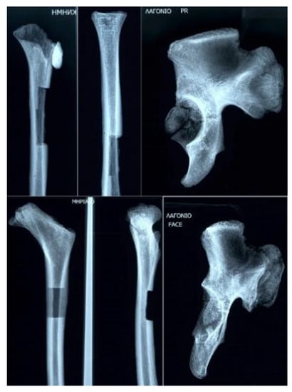
Figure 4.
X-ray of the long and pelvic bones.
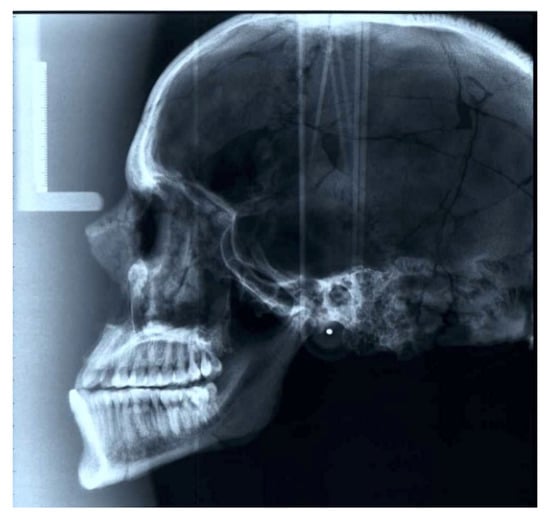
Figure 5.
Lateral cephalometric radiograph of the skull.
3.4. Dental Pathology
A thorough examination of the dental arches did not evidence signs of major dental pathology (pertaining to caries and periodontal disease, as evidenced by alveolar bone loss), with the exception of a noteworthy abrasion of moderate degree on the lower incisors and first molars (Figure 6). From an orthodontic point of view, class III malocclusion was evidenced by the characteristic Class III molar relationship, anterior crossbite, and relative prognathism of the lower jaw. The macroscopic investigation of the palate identified an island of intra vitam porotic lesions in a mixed pattern of perforations of different size and shape overlapping with areas of diagenetic lesions.
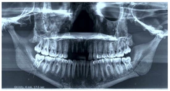
Figure 6.
Panoramic radiograph.
3.5. 3D Modeling
Since the series of CT images covering the head were closely spaced (0.3 mm slice spacing), the 3D-reconstructed model of the skull reproduced very fine details of the original (Figure 7). The bounding box dimensions of the skull were 217 mm height, 134 mm width, and 186 mm depth. Interestingly, a substantial asymmetry between the left and right temporal cavity (in favor of the left) was observed and noted. The entire process of 3D printing took 66 h to be completed, and 479 cc3 of ABS and 236 cc3 of supported material were used. The skull was printed with a layer thickness of 0.245 mm, which corresponds to the highest level of resolution analysis of the Dimension SST 778 3D printer used.
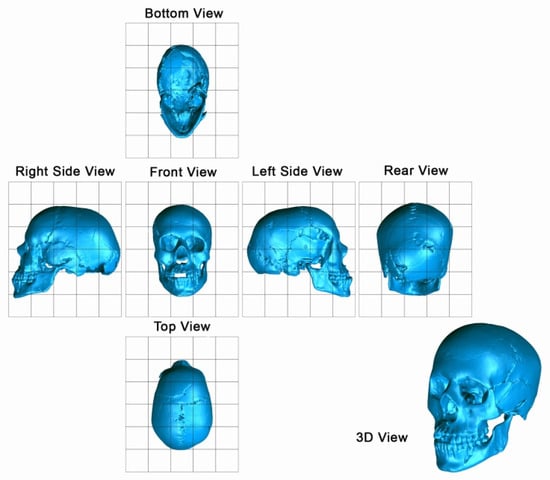
Figure 7.
Three-dimensional digital views of the skull.
3.6. 3D Face Reconstruction
The Manchester method [46] was deployed for the facial reconstruction of Avgi. Measuring particular underlying recipient sites of the skull provided an estimate of the shape and size of her eyes, nose, and mouth, respectively. Tissue thickness on thirty specific points of the skull, marked on 3-mm wooden pegs, was obtained from standard tables [47], taking into consideration the gender, age, race, and nutritional condition parameters. Artificial prosthetic eye bulbs were placed into the orbits, whose shape, surface, and depth indicated how deep the eyes should be set [48]. The color was arbitrarily set to dark brown, taking into account Avgi’s origin. The face was constructed muscle after muscle in a specific order [36], using plasticine clay, numbering twenty muscles in total, carefully considering each muscle’s shape, size, and function. The temporalis and the masseter muscles were first reconstructed, followed by the muscles around the mouth. The pattern of the dentition and the skeletal craniofacial attributes of the mouth area were used to estimate its width and the thickness of the lips [48]. The eyes were covered by flat and circular sculpted orbicularis oculi muscles, and the shape and direction of the eyelids and eyebrows were obtained by studying the shape of the orbits. The size and width of the nose was dictated by the shape of the nasal aperture [49]. The muscles spanning in-between the nose and the mouth, accompanied by the zygomaticus muscles, the occipito-frontalis muscle covering the forehead, and the most prominent sternocleidomastoid and the trapezius major muscles of the neck were the next to be shaped, along with the parotid gland, which was also built at this stage. Although lacking an osseous base, the size, position, and angle of the auricle of the ears were determined following the proportions of the underlying skeletal tissue and the jaw’s angle, respectively. The skin layer was constructed using strips of clay that were rolled out and placed over the muscle structure. Skin texture and details were further developed to provide a realistic impression, considering that Avgi was suffering from iron deficiency anemia, possibly due to iron malabsorption and/or malnutrition, and Class III dental malocclusion. Avgi’s hairstyle was decided arbitrarily, as there are no relevant indications, e.g., drawings, of young women, aged seventeen to nineteen years old, contemporary to Avgi. The pigmented silicon casting of the reconstruction with inserted hair provides for Avgi’s overall vivid look, shown on the rightmost part of Figure 8.
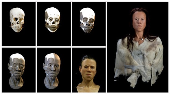
Figure 8.
Three-dimensional facial reconstruction process.
4. Discussion
In this article, we report a Mesolithic case study which, given the scarcity of extant findings, enhances our understanding of the time period studied in context. The skeleton of the woman in Theopetra Cave was found intact in the Mesolithic layer, buried in a semiflexed position. This case is just one of the three Mesolithic human burials in Theopetra Cave in a total of four Mesolithic burials in the Greek territory, with the fourth found in Franchthi Cave, as mentioned before, and a potentially fifth recent find at Negros Cave in Astypalaia Island, which has not yet been published. In fact, the skeleton’s position resembles, schematically, the one found in Franchthi Cave [50]. Interpreted with caution, Avgi’s height is comparable to the range calculated for female remains at Franchthi, that is, 1.37–1.52 m [51], while Avgi is shorter than the female average for the European Mesolithic era [26]. Regarding the burial practice, besides the three primary burials in Theopetra Cave and the one in Franchthi Cave exhibiting similar burial-behavioral patterns [12], there are a number of traits akin to other Mesolithic burials, including Natufian burials in Nahal Oren and Hatoula in Israel [52,53], Muge in Portugal [54], and the western parts of the Russian plain [55].
Regarding radiographic findings, the diploic hair-on-end sign seen in the skull X-ray is usually due to the overactivity of the red bone marrow in response to anemia and blood disorders [56]. Skull abnormalities in patients with anemia are produced by cellular hyperplasia, circulatory factors, or a combination of both. The lesions caused by red marrow hyperplasia, such as the expansion of the diploe, thinning of the outer table, and vertical trabeculations, have been described by investigators in patients with thalassemia major, iron deficiency anemia, sickle cell disease, and spherocytosis [56]. The mild porotic hyperostosis that was noted also implies anemia, though recent findings have also suggested potential hemolytic and megaloblastic anemias [57]. Furthermore, Harris lines, as noticed on the long bones, are commonly a result of malnutrition, disease, or trauma [58]. All these signs seem to complete the puzzle of the diagnosis, so that we can reasonably assume that Avgi suffered from iron deficiency anemia due to iron malabsorption or malnutrition.
No significant dental pathology was reported pertaining to the permanent teeth of the skeleton under study. Apparently, cultural and environmental variations among Mesolithic communities impact on dental health. Τhe class III dental malocclusion that was observed [59] is in most cases a multifactorially generated disorder resulting from an interaction of susceptible genes and environmental factors [60]. Interestingly, the relative prognathism of the mandible is a condition commonly found in Mesolithic skeletons, and also existent in Franchthi cases [51].
It should be noted that the clinical morphotype, categorization, and etiological association of porosis and porotic hyperostosis with anemia are fields of ongoing controversy [61]. The structure and degree of architectural changes and severity in the diploic table, in relation to the outer and inner cranial tables, and in association with the manifestation of clinical features in the postcranial skeleton, is the key variable in the diagnosis on ancient skeletal remains [23,62,63,64]. Recent advances in evidence-based medicine and the histological study of ancient bone disease introduce a new interpretative approach, marking a shift in paleopathological research.
In the X-ray profile of the Theopetra skull, the feature of the hair-on-end may not actually present the outgrowth of the diploic trabeculae at the expense of the disintegration of the external lamina, but rather a localized diagenetic effect of the sagittal and lambdoid sutures, due to the severe diagenesis diagnosed in the endoscopic investigation [64]. However, it is imperative to attempt a differential diagnosis, since the reliability of findings in paleopathological studies of ancient bones demands complementary levels of analysis. It should be noted that the hyperplasia of marrow may also be a sign of lead poisoning and cyanotic heart disease. In this perspective, the stage at which iron deficiency is manifested in the skeleton and the pathway to iron deficiency anemia is not traceable with high credibility in the ancient bones and depends on dietary factors. Furthermore, the association of iron deficiency anemia with infectious factors is documented, involving the protein hepcidin [65] or the bacterium Clostridium botulinum found in honey [66]. Relatively recent data complicate the diagnosis even more, with studies documenting a genetic predisposition for iron deficiency anemia in some groups with high iron intake and uptake [67].
The mild porotic lesions diagnosed as edema is a relatively frequent phenomenon in Greek prehistoric specimens, indicative of vestiges of periostitis of the skull, atypical or caused by inflammatory events in bone and/or spread from soft cranial and/or facial tissues [62]. This finding, in combination with the morphology of the outer and diploic tables and the lesions in the palate, could imply signs of inflammatory processes in co-occurrence with mild anemia. Regarding the possible diagnosis of megaloblastic anemia as a nutritional disease, current studies reveal that mutations in specific gene loci hinder the intake and absorbability of cobalamin—vitamin B12 [68]—even in a sufficient diet; in addition, some individuals manifest megaloblastic anemia as an autoimmune syndrome [69], and others due to nutritional deficiencies [70]. New data indicate that inflammatory processes in the scalp can produce a porotic external surface of the skull, usually thickened, after a healing event [71]. Conclusive evidence documents that porotic hyperostosis is not a disease, but a morphological feature of multi-factorial etiology.
Therefore, under the evidence presented above and the vague clinical features of the skeleton, the differential diagnosis of iron deficiency anemia, with or without anemia of inflammation, is tentative, but possible. However, the lesions seem to occur in a mild or a beginning stage, with signs of a healing process, as it is evidenced in the palate and the porotic pattern, as well as in advanced diagenetic features. Histological investigation will provide more reliable evidence for differential diagnosis.
The paleopathological study of the Theopetra skeleton is part of a project that is essentially a work in progress, including a histological study, biochemical analysis of non-collagenous proteins, and molecular research on the mapping of haplotypes to trace the migratory patterns of prehistoric people [72], all critical variables to be integrated in the database and build up the biogeography of disease syndromes in Greek prehistoric societies. Spectroscopic methods on the quantification of porosis are also in progress in order to introduce data to the diagenetic study, a significant but neglected field in paleopathology.
All in all, it is evident that the reliability of the diagnosis is dependent on the limitations posed by the paleopathological research per se. This is more pronounced with the Theopetra specimen, due to the lack of statistically significant evidence from human samples from the Mesolithic era, the absence of the soft tissues needed for the evaluation of the distribution pattern of the lesions and evolution of disease, and the inherent methodological bias in paleopathology. This notion is a limiting factor, granted that paleopathological research is essentially based on evidence from modern medical data—frequently exhibiting atypical lesions—while the profile, manifestations, and development (or not) of ancient systemic diseases are still under investigation.
5. Conclusions
Our study of the skeletal remains of a Mesolithic female adolescent from Theopetra Cave demonstrates iron deficiency anemia and Class III dental malocclusion. This case-study, when examined in context, provides extremely useful insights into the Mesolithic period, and enhances our understanding of human evolution and behavior during that time period. Multiple scientific perspectives, along with social, geographical, and behavioral parameters—like settlement patterns, population movements, and social networks—should be taken into account when assessing lifestyle characteristics and paleopathological features through skeletal remains. This study is the product of a multidisciplinary project, bringing together physicians of various specialties, dentists, and orthodontists with paleopathologists and medical historians, archeologists, anthropologists, and engineers. We aspire to make this collaborative work part of the modern paradigm for conducting research in lesser-known territories by using cutting-edge research techniques promoting cultural heritage and making the archaeological information tangible and accessible to wider audiences.
Author Contributions
Conceptualization, M.J.P.; methodology, M.J.P. and G.P.C.; software, E.M. and A.K.; validation, N.K.-A., E.S. and O.A.; formal analysis P.T. (Panagiotis Toulas), C.P., T.P., M.M., P.N.S., M.G.T. and G.P.C.; investigation, N.K.-A., E.S., O.A., P.N.S. and P.T. (Peny Tsakanikou); resources, M.J.P. and E.M.; data curation, E.M.; writing—original draft preparation, M.J.P., A.A.K., E.M. and A.K.; writing—review and editing, M.J.P., E.M. and A.K.; visualization, E.M.; supervision, M.J.P. All authors have read and agreed to the published version of the manuscript.
Funding
This research received no external funding.
Informed Consent Statement
Patient consent was waived due to Mesolithic origin.
Data Availability Statement
Not applicable.
Acknowledgments
The authors would like to thank the sculptor and archaeologist Oscar D. Nilsson for the exemplary work conducted in reconstructing the face of Avgi, Ioannis Metzikof and Alexia Pentheroudaki for Avgi’s clothing and jewelry, respectively, and the Museum of Acropolis for Avgi’s exhibition.
Conflicts of Interest
The authors declare no conflict of interest.
References
- Spikins, P. Mesolithic Europe: Glimpses of another world. In Mesolithic Europe; Bailey, G., Spikins, P., Eds.; Cambridge University Press: Cambridge, UK, 2007; pp. 1–17. [Google Scholar]
- Sánchez-Quinto, F.; Schroeder, H.; Ramirez, O.; Avila-Arcos, M.C.; Pybus, M.; Olalde, I.; Velazquez, A.M.; Marcos, M.E.; Encinas, J.M.; Bertranpetit, J.; et al. Genomic affinities of two 7,000-year-old Iberian hunter-gatherers. Curr. Biol. 2012, 22, 1494–1499. [Google Scholar] [CrossRef] [PubMed]
- Toussaint, M. Intentional cutmarks on an early mesolithic human calvaria from Margaux Cave (Dinant, Belgium). Am. J. Phys. Anthropol. 2011, 144, 100–107. [Google Scholar] [CrossRef] [PubMed]
- Hoffmann, A.; Hublin, J.J.; Hüls, M.; Terberger, T. The Homo aurignaciensis hauseri from Combe-Capelle: A Mesolithic burial. J. Hum. Evol. 2011, 61, 211–214. [Google Scholar] [CrossRef]
- Lazaridis, I.; Patterson, N.; Mittnik, A.; Renaud, G.; Mallick, S.; Kirsanow, K.; Sudmant, P.H.; Schraiber, J.G.; Castellano, S.; Lipson, M.; et al. Ancient human genomes suggest three ancestral populations for present-day Europeans. Nature 2014, 513, 409–413. [Google Scholar] [CrossRef] [PubMed]
- Stiner, M.C.; Munro, N.D. On the evolution of diet and landscape during the Upper Paleolithic through Mesolithic at Franchthi Cave (Peloponnese, Greece). J. Hum. Evol. 2011, 60, 618–636. [Google Scholar] [CrossRef] [PubMed]
- Hofmanová, Z.; Kreutzer, S.; Hellenthal, G.; Sell, C.; Diekmann, Y.; Díez-del-Molino, D.; van Dorp, L.; López, S.; Kousathanas, A.; Link, V.; et al. Early farmers from across Europe directly descended from Neolithic Aegeans. Proc. Natl. Acad. Sci. USA 2016, 113, 6886–6891. [Google Scholar] [CrossRef] [PubMed]
- Galanidou, N.; Perlès, C. The Greek Mesolithic: Problems and Perspectives; British School of Athens Studies: London, UK, 2003. [Google Scholar]
- Jacobsen, T. 17,000 Years of Greek Prehistory. Sci. Am. 1976, 234, 76–87. [Google Scholar] [CrossRef]
- Jacobsen, T. Franchthi Cave and the Beginning of Settled Village Life in Greece. Hesperia 1981, 50, 303–319. [Google Scholar] [CrossRef]
- Kyparissi-Apostolika, N. Theopetra Cave, Twelve Years of Excavation and Research 1987–1998; Institute for Aegean Prehistory: Athens, Greece, 2000. [Google Scholar]
- Kyparissi-Apostolika, N. The Mesolithic in Theopetra Cave: New data on a debated period of Greek prehistory. In The Greek Mesolithic: Problems and Perspectives; British School of Athens Studies: London, UK, 2003; pp. 189–198. [Google Scholar]
- Sampson, A. The Cave of the Cyclops: Mesolithic and Neolithic Networks in the Northern Aegean, Greece. Vol II, Bone Tool Industries, Dietary Resources and the Paleoenvironment, and the Archaeological Studies; INSTAP Academic Press: Philadelphia, PA, USA, 2011. [Google Scholar]
- Kyparissi-Apostolika, N. The Thessalian Mesolithic: Evidence from Theopetra Cave. J. Greek Archaeol. 2021, 6, 25–42. [Google Scholar] [CrossRef]
- Strasser, T.F.; Panagopoulou, E.; Runnels, C.N.; Murray, P.M.; Thompson, N.; Karkanas, P.; McCoy, F.W.; Wegmann, K.W. Stone Age Seafaring in the Mediterranean: Evidence from the Plakias Region for Lower Palaeolithic and Mesolithic Habitation of Crete. Hesperia 2010, 79, 145–190. [Google Scholar] [CrossRef]
- Efstathiou, J. Archaeological Research in Caves at Astypalaia: The Negros Cave in Vatses; Vlachopoulos, A., Vathy Astypalaias, I., Eds.; Melissa Publications: Athens, Greece, 2022. [Google Scholar]
- Perlès, C. The Early Neolithic in Greece. The First Farming Communities in Europe; Cambridge University Press: Cambridge, UK, 2001. [Google Scholar]
- Kotsakis, K. From the Neolithic side: The Mesolithic/Neolithic interface in Greece. In The Greek Mesolithic; Galanidou, N., Perlès, C., Eds.; British School at Athens Studies: Athens, Greece, 2003; pp. 217–221. [Google Scholar]
- Sampson, A. The Cave of the Cyclops: Mesolithic and Neolithic Networks in the Northern Aegean, Greece. Vol. I, Intra-Site Analysis, Local Industries, and Regional Site Distribution; INSTAP Academic Press: Philadelphia, PA, USA, 2008. [Google Scholar]
- Stravopodi, E.; Manolis, S. The Bioarchaeological Profile of the Anthropological finds of Theopetra Cave: A pilot study in Greek Peninsula. In Proceedings of the International Conference THEOPETRA CAVE: Twelve Years of Excavation and Research 1987–1998; Kyparissi-Apostolika, N., Ed.; Edition of Ministry of Culture: Athens, Greece, 1998; pp. 95–108. [Google Scholar]
- Facorellis, Υ. Radiocarbon dating the Greek Mesolithic. In The Greek Mesolithic. British School at Athens Studies: Athens, Greece; Galanidou, N., Perlès, C., Eds.; British School at Athens Studies: Athens, Greece, 2003; pp. 51–68. [Google Scholar]
- Kyparissi-Apostolika, N. Prehistoric burials at Theopetra cave. In Anthropological Pathways, Festschrift for Professor N.I. Xirotiris; Simitopoulou, K., Kaufmann, B., Zafeiris, K., Theodorou, T., Papageorgopoulou, C., Eds.; Μystis Publications: Komotini, Greece, 2014; pp. 269–284, (In Greek with English abstract). [Google Scholar]
- Buikstra, J.E.; Ubelaker, D. Standards for Data Collection from Human Skeletal Remains. Ark. Archaeol. Surv. Res. Ser. 1994, 44, 1–272. [Google Scholar]
- Tuteja, M.; Bahirwani, S.; Balaji, P. An evaluation of third molar eruption for assessment of chronologic age: A panoramic study. J. Forensic Dent. Sci. 2012, 4, 13–18. [Google Scholar] [CrossRef] [PubMed][Green Version]
- Suma, G.N.; Rao, B.B.; Annigeri, R.G.; Rao, D.J.K.; Goel, S. Radiographic correlation of dental and skeletal age: Third molar, an age indicator. J. Forensic. Dent. Sci. 2011, 3, 14–18. [Google Scholar] [CrossRef] [PubMed][Green Version]
- Bass, W.M. Human Osteology: A Laboratory and Field Manual; MK: Trimble, MO, USA, 1987. [Google Scholar]
- Manolis, S.; Stravopodi, E. An assessment of the human skeletal remains in the Mesolithic deposits of Theopetra cave: A case study. In The Greek Mesolithic, Problems and Perspectives; Galanidou, N., Perlès, C., Eds.; British School at Athens Studies: London, UK, 2003; pp. 207–217. [Google Scholar]
- Papagrigorakis, M.J.; Kousoulis, A.A.; Synodinos, P.N. Craniofacial morphology in ancient and modern Greeks through 4000 years. Anthr. Anz. 2014, 71, 237–257. [Google Scholar] [CrossRef] [PubMed]
- Papagrigorakis, M.J.; Karamesinis, K.G.; Daliouris, K.P.; Kousoulis, A.A.; Synodinos, P.N.; Hatziantoniou, M.D. Paleopathological findings in radiographs of ancient and modern Greek skulls. Skelet. Radiol. 2012, 41, 1605–1611. [Google Scholar] [CrossRef] [PubMed]
- Maravelakis, E.; Konstantaras, A.; Kritsotaki, A.; Angelakis, D.; Xinogalos, M. Analysing User Needs for a Unified 3D Metadata Recording and Exploitation of Cultural Heritage Monuments System. Adv. Vis. Comput. Lect. Notes Comput. Sci. 2013, 8034, 138–147. [Google Scholar]
- Axaridou, A.; Chrysakis, I.; Georgis, C.; Theodoridou, M.; Doerr, M.; Konstantaras, A.; Maravelakis, E. 3D-SYSTEK: Recording and exploiting the production workflow of 3D-models in Cultural Heritage. In Proceedings of the IISA 2014-5th International Conference on Information, Intelligence, Systems and Applications, Crete, Greece, 7–9 July 2014; IEEE: New York, NY, USA, 2014; p. 51. [Google Scholar]
- Maravelakis, E.; Bilalis, N.; Mantzorou, I.; Konstantaras, A.; Antoniadis, A. 3D modelling of the oldest olive tree of the world. Int. J. Comput. Eng. Res. 2012, 2, 340–347. [Google Scholar]
- Maravelakis, E.; Konstantaras, A.; Kabassi, K.; Chrysakis, I.; Georgis, C.; Axaridou, A. 3DSYSTEK web-based point cloud viewer. In Proceedings of the IISA 2014-5th International Conference on Information, Intelligence, Systems and Applications, Crete, Greece, 7–9 July 2014; IEEE: New York, NY, USA, 2014; p. 262. [Google Scholar]
- Zur Nedden, D.; Knapp, R.; Wicke, K.; Judmaiser, W.; Murphy, W.A.; Seidler, H.; Platzer, W. Skull of a 5300-year-old mummy. Reproduction and investigation with CT-guided stereolithography. Radiology 1994, 93, 269–272. [Google Scholar] [CrossRef]
- Maravelakis, E.; David, K.; Antoniadis, A.; Manios, A.; Bilalis, N.; Papaharilaou, Y. Reverse engineering techniques for cranioplasty: A case study. J. Med. Eng. Tech. 2008, 32, 115–121. [Google Scholar] [CrossRef]
- Papagrigorakis, M.J.; Synodinos, P.N.; Antoniadis, A.; Maravelakis, E.; Toulas, P.; Nilsson, O.; Baziotopoulou-Valavani, E. Facial reconstruction of an 11-year-old female resident of Athens, 430 B.C. Angle Orthod. 2011, 81, 171–179. [Google Scholar] [CrossRef]
- Primo, B.T.; Presotto, A.C.; De Oliveira, H.W.; Gassen, H.T.; Miguens, S.A.Q., Jr.; Silva, A.N., Jr.; Hernandez, P.A.G. Accuracy assessment of prototypes produced using multi-slice and cone-beam computed tomography. Int. J. Oral Maxillofac. Surg. 2012, 41, 1291–1295. [Google Scholar] [CrossRef]
- Bagariaa, V.; Deshpandeb, S.; Rasalkarc, D.; Kuthed, A.; Paunipagarc, B.K. Use of rapid prototyping and three-dimensional reconstruction modeling in the management of complex fractures. Eur. J. Radiol. 2011, 80, 814–820. [Google Scholar] [CrossRef] [PubMed]
- Ebert, L.C.; Thali, M.J.; Ross, S. Accuracy assessment of prototypes produced using multi-slice and cone-beam computed tomography. Forens. Sci. Int. 2011, 211, 1–6. [Google Scholar] [CrossRef] [PubMed]
- Mann, R.W.; Synew, A.A.; Bass, W.M. Maxillary Suture Obliteration: Aging the Human Skeleton based on Intact 167 Fragmentary Maxilla. J. Forens. Sci. 1987, 32, 148–157. [Google Scholar] [CrossRef]
- Brothwell, D. Digging up Bones; Cornell University Press: New York, NY, USA, 1981. [Google Scholar]
- Miles, A.E.W. Dentition in the estimation of age. J. Dent. Res. 1963, 42, 255–263. [Google Scholar] [CrossRef]
- Scott, E.C. Dental wear scoring technique. Am. J. Phys. Anthropol. 1979, 51, 213–218. [Google Scholar] [CrossRef]
- Hillson, S. Dental Anthropology; Cambridge University Press: Cambridge, UK, 1996. [Google Scholar]
- Corballis, M.C. From mouth to hand: Gesture, speech, and the evolution of right-handedness. Behav. Brain Sci. 2003, 26, 199–208. [Google Scholar] [CrossRef]
- Prag, J.; Neave, R. Making Faces: Using Forensic and Archaeological Evidence; British Museum Pres: London, UK, 1997. [Google Scholar]
- Wilkinson, C.M. In vivo facial tissue depth measurements for white British children. J. Forensic Sci. 2002, 47, 459–465. [Google Scholar] [CrossRef]
- Wilkinson, C. Forensic Facial Reconstruction; Cambridge University Press: Cambridge, UK, 2004; pp. 110–114, 165–166, 223–224. [Google Scholar]
- Gerasimov, M.M. The Face Finder; JB Lippincott Co.: Philadelphia, PA, USA, 1971. [Google Scholar]
- Cullen, T. Mesolithic mortuary ritual at Franchthi Cave, Greece. Antiquity 1995, 69, 270–289. [Google Scholar] [CrossRef]
- Angel, J.L. Human skeletal material from Francthi cave. Hesperia 1969, 38, 380–381. [Google Scholar]
- Noy, T. Some aspects of Natufian mortuary behaviour at Nahal Oren. In People and Culture in Change; Hershkovitz, I., Ed.; BAR International Series: Oxford, UK, 1989; pp. 53–59. [Google Scholar]
- Le Mort, F. PPNA burials from Hatoula, Israel. In People and Culture in Change; Hershkovitz, I., Ed.; BAR International Series: Oxford, UK, 1989; pp. 133–141. [Google Scholar]
- Arnaud, M. The Mesolithic communities of the Sado Valley, Portugal, in their ecological setting. In The Mesolithic in Europe; Bonsall, C., Ed.; University of Edinburg Press: Edinburgh, UK, 1985; pp. 614–631. [Google Scholar]
- Kozlowski, S.K. A survey of the Early Holocene cultures of the Western part of the Russian plain. In The Mesolithic in Europe; Bonsall, C., Ed.; University of Edinburg Press: Edinburgh, UK, 1989; pp. 424–441. [Google Scholar]
- Hollar, M.A. The Hair-on-End Sign. Radiology 2011, 221, 347–348. [Google Scholar] [CrossRef] [PubMed]
- Walker, P.L.; Bathurst, R.R.; Richman, R.; Gjerdrum, T.; Andrushko, V.A. The causes of porotic hyperostosis and cribra orbitalia: A reappraisal of the iron-deficiency-anemia hypothesis. Am. J. Phys. Anthropol. 2009, 139, 109–125. [Google Scholar] [CrossRef] [PubMed]
- Papageorgopoulou, C.; Suter, S.K.; Rühli, F.J.; Siegmund, F. Harris lines revisited: Prevalence, co-morbidities and possible aetiologies. Am. J. Hum. Biol. 2011, 23, 381–391. [Google Scholar] [CrossRef]
- Papagrigorakis, M.J.; Synodinos, P.N.; Baziotopoulou-Valavani, E. Dental status and orthodontic treatment needs of an 11-year-old female resident of Athens, 430 B.C. Angle Orthod. 2008, 78, 152–156. [Google Scholar] [CrossRef]
- Xue, F.; Wong, R.W.; Rabie, A.B. Genes, genetics, and Class III malocclusion. Orthod. Craniofac. Res. 2010, 13, 69–74. [Google Scholar] [CrossRef] [PubMed]
- Stravopodi, E. The Paleopathological Profile of Porotic Hyperostosis as an Epidemiological Rise in the Primary Holocene Greek Societies: A Biocultural Approach; Athens University Department of Biological Anthropology: Athens, Greece, 2012. [Google Scholar]
- Buikstra, J.E.; Lagia, A. Bioarchaeological approaches to Aegean Archaeology. In New Directions in the Skeletal Biology in Greece; Schepartz, L., Fox, S., Bourbou, C., Eds.; Occasional Wiener Laboratory Series; ASCSA: Athens, Greece, 2009; pp. 7–31. [Google Scholar]
- Schultz, M. Light microscopic analysis in skeletal paleopathology. In Identification of Pathological Conditions in Human Skeletal Remains; Ortner, D.J., Ed.; Academic Press: San Diego, CA, USA, 2003; pp. 73–108. [Google Scholar]
- Schultz, M. Microscopic Investigation of Ancient Bone Disease; Center of Anatomy and Histology: Göttingen, Germany, 2003. [Google Scholar]
- De Quirijn, M.; Syafruddin, D.; Keijmel, S.; Olde Riekerink, T.; Deky, O.; Asih, P.B.; Swinkels, D.W.; Van Der Ven, A.J. Increased serum hepcidin and alterations in blood iron parameters associated with asymptomatic P. falciparum and P. vivax malaria. Haematologica 2010, 95, 1068–1074. [Google Scholar]
- Fairgrieve, S.I.; Molto, J.E. Cribra orbitalia in two temporally disjunct population samples from the Dakhleh Oasis, Egypt. Am. J. Phys. Anthropol. 2000, 111, 319–331. [Google Scholar] [CrossRef]
- Melis, M.A.; Cau, M.; Congiu, R.; Sole, G.; Barella, S.; Cao, A.; Westerman, M.; Cazzola, M.; Galanello, R. A mutation in the TMPRSS6 gene encoding a transmembrance serine protease that suppresses deciding production in familial iron deficiency anemia refractory to oral iron. Hematologica 2008, 93, 1473–1479. [Google Scholar] [CrossRef]
- Remacha, A.F.; Del Río, E.; Sardà, M.P.; Canals, C.; Simó, M.; Baiget, M. Role of (Glu-->Arg, Q5R) mutation of the intrinsic factor in pernicious anemia and other causes of low vitamin B12. Ann. Hematol. 2008, 87, 599–600. [Google Scholar] [CrossRef]
- Wilson, D.J.; Gabriel, E.; Leatherbarrow, A.J.; Cheesbrough, J.; Gee, S.; Bolton, E.; Fox, A.; Hart, C.A.; Diggle, P.J.; Fearnhead, P. Rapid evolution and the importance of recombination to the gastro-enetric pathogen Campylobacter jejuni. Mol. Biol. Evol. 2009, 26, 385–397. [Google Scholar] [CrossRef]
- Jabbour, N.; Di Giuseppe, J.A.; Usmani, S.; Tannenbaum, S. Copper deficiency as a cause of reversible anemia and neutropenia. Connecticut. Med. 2010, 74, 261–263. [Google Scholar]
- Schultz, M. Paleohistopathology of bone: A new approach to the study of ancient diseases. Yearb. Phys. Anthropol. 2001, 116, 106–147. [Google Scholar] [CrossRef] [PubMed]
- Evison, M.; Kyparissi, N.; Stravopodi, E.; Fieller, N.; Smillie, D.M. An ancient HLA type from a Palaeolithic skeleton from Theopetra Cave, Greece. In Theopetra Cave, 1987–1998; Kyparissi-Apostolika, N., Ed.; Ephorate of Palaeoanthropology and Speleology: Athens, Greece, 2000; pp. 109–117. [Google Scholar]
Publisher’s Note: MDPI stays neutral with regard to jurisdictional claims in published maps and institutional affiliations. |
© 2022 by the authors. Licensee MDPI, Basel, Switzerland. This article is an open access article distributed under the terms and conditions of the Creative Commons Attribution (CC BY) license (https://creativecommons.org/licenses/by/4.0/).