Review

Nutrient-Doped Hydroxyapatite: Structure, Synthesis and Properties

1
Department of Chemical and Biomolecular Engineering, Lehigh University, Bethlehem, PA 18015, USA
2
Department of Physics, Faculty of Science, Suez University, Suez 43518, Egypt
*
Authors to whom correspondence should be addressed.
Ceramics 2023, 6(3), 1799-1825; https://doi.org/10.3390/ceramics6030110
Submission received: 4 July 2023 / Revised: 9 August 2023 / Accepted: 16 August 2023 / Published: 22 August 2023
(This article belongs to the Special Issue Innovative Research on Calcium Phosphates Based Ceramics)

Abstract

:
Complex inorganic powders based on calcium phosphates have found a plethora of practical applications. Of particular interest are the CaO-P2O5 system-based multi-component material powders and granules as the source of major- and micronutrients for the plants. The emerging strategy is to use nano fertilizers based on hydroxyapatite (HAP) for phosphorus and other nutrient delivery. The doping of micronutrients into HAP structure presents an interesting challenge in obtaining specific phase compositions of these calcium phosphates. Various techniques, including mechanochemical synthesis, have been employed to fabricate doped HAP. Mechanochemical synthesis is of particular interest in this review since it presents a relatively simple, scalable, and cost-effective method of calcium phosphate powder processing. The method involves the use of mechanical force to promote chemical reactions and create nanometric powders. This technique has been successfully applied to produce HAP nanoparticles alone, and HAP doped with other elements, such as zinc and magnesium. Nanofertilizers developed through mechanochemical synthesis can offer several advantages over conventional fertilizers. Their nanoscale size allows for rapid absorption and controlled release of nutrients, which leads to improved nutrient uptake efficiency by plants. Furthermore, the tailored properties of HAP-based nano fertilizers, such as controlled porosity and degradation levels, contribute to their effectiveness in providing plant nutrition.

Graphical Abstract

1. Introduction

Synthesis of multicomponent inorganic powder based on CaO and P2O5 systems is critical in agriculture. Phosphorus (P) fertilizers commonly used in agriculture suffer from low efficiency due to P immobilization in the form of aluminum (Al) and iron (Fe) oxides formed in the soil immediately after P release [1]. Additionally, a substantial amount of P that is released from organic matter in the soil rapidly undergoes fixation and becomes trapped in insoluble inorganic compounds [2]. This process significantly limits the availability of P to crops and results in deficiencies even in soils with high overall P content [3]. In other words, the released P is unable to remain in a bioavailable form, which hinders its uptake by plants and impedes their growth and development. This highlights the importance of developing strategies that can effectively address P cycling and ensure an adequate supply of accessible P to support optimal crop growth. To address this challenge, there is a need to develop fertilizers that release P at a slower rate while increasing the availability of bioactive P in a controlled manner. One potential solution being explored is the use of nanoporous hydroxyapatite (HAP) as a fertilizer [4]. Previous studies have suggested that HAP nanoparticles may release P more slowly than conventional fertilizers while still mobilizing P faster than the forms immobilized in the soil [5,6,7].
The utilization of HAP as a fertilizer holds significant promise for enhancing nutrient availability and improving agricultural productivity [8,9]. By providing a slow-release mechanism for P, HAP can mitigate the issue of P immobilization and increase bioavailable P levels for crop uptake. Furthermore, the ability of HAP to serve as a carrier for various macronutrients and micronutrients opens up avenues for targeted and efficient multi-nutrient delivery to plants. Water soluble mono ammonium phosphate, diammonium phosphate, and triple superphosphate are widely utilized as P fertilizers in agriculture [10,11,12]. These fertilizers are applied to the soil releasing water-soluble forms of P that are readily accessible to crops [13]. Their use results in substantial P losses due to leaching and surface run-off, which present challenges to food security and environmental sustainability [14]. The use of less soluble P forms, such as phosphate rocks and apatite, can mitigate these losses and also show P availability for plants [15,16]. Exploring the potential of nanoscale inorganic powdered materials, such as HAP, offers a promising avenue, albeit necessitating thorough and systematic investigations of the complex multi-component system synthesis techniques and the resulting fundamental properties.
Previous studies have examined the use of HAP nanoparticles as a phosphorus (P) fertilizer, capitalizing on the distinctive characteristics of nanosized particles to enhance their mobility in soil and release P [1,17]. The work of Liu and Lal in 2014 introduced the concept of utilizing nanosized solid P as a fertilizer while considering the trade-off between agricultural benefits and environmental risks. They designed a greenhouse trial focusing on soybean cultivation and demonstrated that the application of HAP as a phosphorus source significantly enhanced root and aerial biomass along with increased grain yield compared to control and conventional P fertilizers [18]. Another study by Marchiol et al. explored the potential of HAP not only as a P supplier but also as a carrier for other elements or molecules [19]. Their germination trial with tomato plants showcased the versatility of HAP in fulfilling these dual roles. In light of the growing interest in nano HAP as a promising fertilizer component, there is a pressing need within the industry for a straightforward and efficient method to produce high-quality HAP.
The conventional synthesis processes of HAP typically involve complex chemical reactions and energy-intensive procedures, which can be both time-consuming and costly. Moreover, the large-scale production must be aligned with the nutritional requirements of plants (essential micro and macronutrients present in the synthesized nanocomposite). In this regard, the mechanochemical approach can be a potential solution to address these scale challenges. Pohshna et al. examined the use of urea/HAP nanomaterials in rice cultivation and compared their performance with commercial bulk urea fertilizer in addition to a control treatment. They reported that urea/HAP nano fertilizer showed improved grain yield, nutrient use efficiency, and reduced nutrient leaching. Therefore, urea/HAP nano fertilizer has the potential to serve as a sustainable nitrogen and phosphorus supplement for rice crops by enhancing productivity [20]. Pradhan et al. synthesized nanoporous HAP nanoparticles using a sol-gel technique and loaded them with the commonly used nitrogen fertilizer urea to create a urea/HAP composite. Their results showed sustained release of urea from urea/HAP in simulated environmental conditions. Surface migration, water retention, and leachate analysis demonstrated that both HAP and urea/HAP exhibited superior water retention properties compared to conventional fertilizers. They also exhibited stability and increased nitrogen use efficiency under high temperatures. Furthermore, urea/HAP exhibited promising results in promoting rice seed germination, which was attributed to enhanced α-amylase activity and starch content. However, that indicates the ability to fabricate engineered urea/HAP promise for enhancing crop growth and productivity while minimizing environmental impact [21]. Elsayed et al. introduced HAP nanoparticles as a novel nano-fertilizer for rosemary plants. HAP nanoparticles, with an average size of approximately 24 nm and a mixed morphology of rods and spherical nanoparticles, were applied via foliar application at various concentrations. HAP showed significant results compared to traditional nitrogen-phosphorus-potassium fertilizers. The treatment by HAP nanoparticles exhibited significant improvements in growth parameters, essential oil yield, and physiological indices of the rosemary plants. The stem and leaf characteristics, such as diameter, the thickness of different tissues, and pith diameter, were developed and enhanced. Moreover, the quality of the essential oil obtained from the rosemary plant was notably higher with the use of the proposed nano fertilizer [5].
Mechanochemical synthesis refers to the production of materials through mechanical force-induced reactions, typically by milling or grinding solid reactants together [22]. This approach offers several advantages over traditional chemical synthesis routes. It can fulfill the need for low temperatures and decrease the usage of many chemical reagents [23,24,25], resulting in a more environmentally friendly process with reduced energy consumption. In addition, mechanochemical methods enable to be controlled regarding the composition, size, and morphology of the resulting particles [25], hence, leading to improved product quality and performance. Further, the simplicity and scalability of mechanochemical techniques make them attractive for industrial-scale production [26,27]. Moreover, one of the key benefits of mechanochemical production for HAP is the ability to achieve nanoscale particle sizes and consequently enhance the reactivity [28]. Furthermore, the mechanical forces applied during the milling process promote the formation of finely dispersed HAP nanoparticles, which possess a larger surface area and higher chemical reactivity compared to their bulk counterparts. This increased reactivity is particularly advantageous for fertilizers as it facilitates the efficient release of phosphorus and enhances nutrient uptake by plants [29,30].
There are several studies have demonstrated the successful application of mechanochemical methods for HAP synthesis [31,32,33]. For instance, high-energy ball milling has been employed to produce nanostructured HAP powders with tailored properties. By controlling the milling parameters, such as milling time, speed, and reactants, researchers have achieved HAP particles with desired characteristics, including size, crystallinity, and surface area [34,35,36,37,38]. These finely tuned properties can positively influence the fertilizer’s effectiveness and performance in agricultural applications. In this regard, there is a notable increment in the number of publications about using HAP as a fertilizer. It can be seen in Figure 1 that there has been a significant increase over the years, from one article in the year 2000, to rise gradually and reach around 50–51 articles by the year 2022. In addition, there has been a growing number of articles investigating nanohydroxyapatite as a fertilizer. Over the past decade, the number of scientific Js has seen a significant increase. However, it is noteworthy that research investigating the use of nano HAP as a fertilizer did not witness a substantial surge until after 2020. The limited attention to nano HAP as a fertilizer in the years leading up to 2020 might be attributed to several factors. One possibility is that researchers were primarily focused on exploring other aspects of nano HAP applications in fields, such as biomedicine. Consequently, the potential agricultural and agronomic benefits of nano HAP as a fertilizer may have been overshadowed by other promising applications during that period. However, the subsequent upturn in research interest post-2020 could indicate a growing recognition of the untapped potential and benefits of incorporating nano HAP in agricultural practices.
In summary, the mechanochemical synthesis of HAP can offer opportunities for incorporating additional elements into the nanostructure by doping or additional compounds as nanocomposites. This approach allows for the development of tailored HAP-based materials with enhanced nutrient-release properties or the ability to deliver specific additives or growth-promoting agents to plants.

2. HAP Structure

2.1. Overview

Many calcium phosphates have been studied extensively, such as HAP, with the chemical formula Ca10(PO4)6(OH)2 and a Ca/P ratio of 1.667, as well as α- and β-tricalcium phosphate (TCP) with the chemical formula Ca3(PO4)2 and a Ca/P ratio of 1.500 [39,40,41]. They are widely used in various fields, such as biomedical applications, specifically in the areas of dentistry, orthopedics, and bone tissue engineering [41,42]. These materials serve various purposes, including their use as dental fillers due to their mechanical properties, coatings for titanium dental implants for their biocompatibility, fillers in biodegradable composites, and substitutes for bone reconstruction and regeneration [43,44,45]. The major reason for their extensive usage in these applications is their chemical resemblance to the mineral component, which is naturally found in hard tissues [46,47].
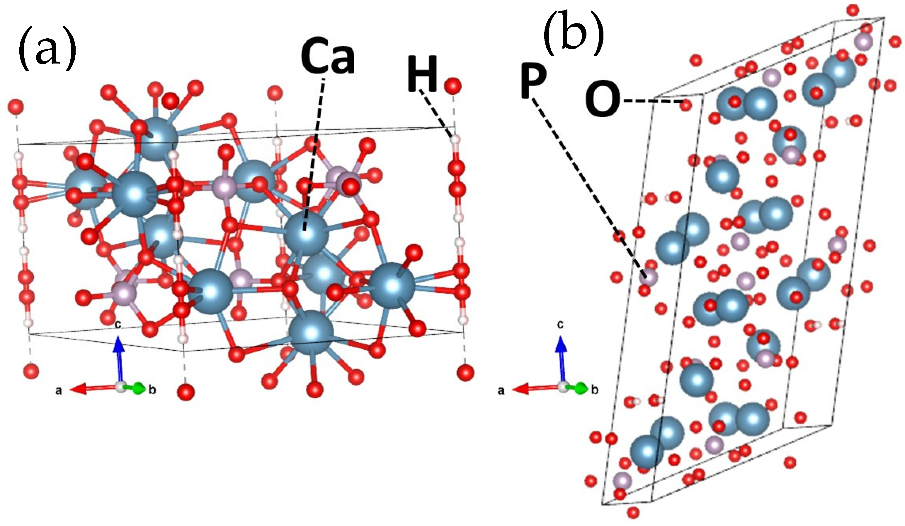
HAP exhibits two distinct crystal structures: Hexagonal and monoclinic, which are represented in Figure 2a,b. The hexagonal form has lattice parameters of a = b = 9.432 Å, c = 6.881 Å, and γ = 120°, while the monoclinic form has lattice parameters of a = 9.421 Å, b = 2a, c = 6.881 Å, and γ = 120° [48]. Therefore, it seems that one monoclinic unit cell contains two hexagonal cells. In other words, it is a pseudo-monoclinic structure. These crystal forms both adhere to a stoichiometric Ca/P ratio of 1.67 [49]. It is widely recognized that the lattice structure of HAP possesses a remarkable ability to accommodate nearly half of the elements found in the periodic system [50,51,52,53,54]. This unique characteristic allows for the substitution of various ions in its three main components: Ca2+, PO43−, and OH groups [55,56]. The high structural flexibility of hydroxyapatite enables the incorporation of both bivalent ions, such as Sr2+, Ba2+, Mg2+, and others, as well as monovalent cations, such as Na+ and K+, which typically replace Ca2+ ions [56,57]. In terms of anionic substitutions, they can occur either in the phosphate or hydroxyl positions. For example, F and Cl ions tend to occupy the OH sites, while CO32- can substitute for both PO43− and OH simultaneously or one by one [55]. Furthermore, SiO44− ions can replace PO43−, and there are instances where hydroxyl group sites may become unoccupied [58]. This inherent flexibility in the HAP lattice structure allows for the incorporation of a diverse range of ions, which provides significant opportunities for tailoring and changing its properties to meet specific application requirements. In addition, certain substitutional ions can distort the lattice by either expanding or contracting its size [59]. This depends on the difference in radius between the substituted ions, further influencing the overall properties and behavior of HAP.

2.2. Structure

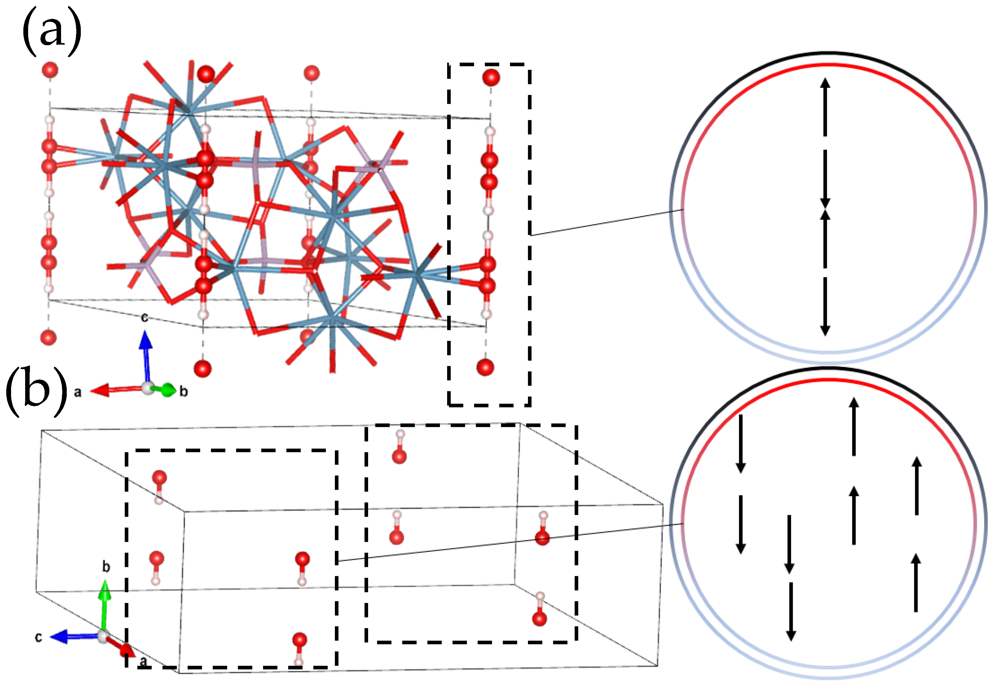
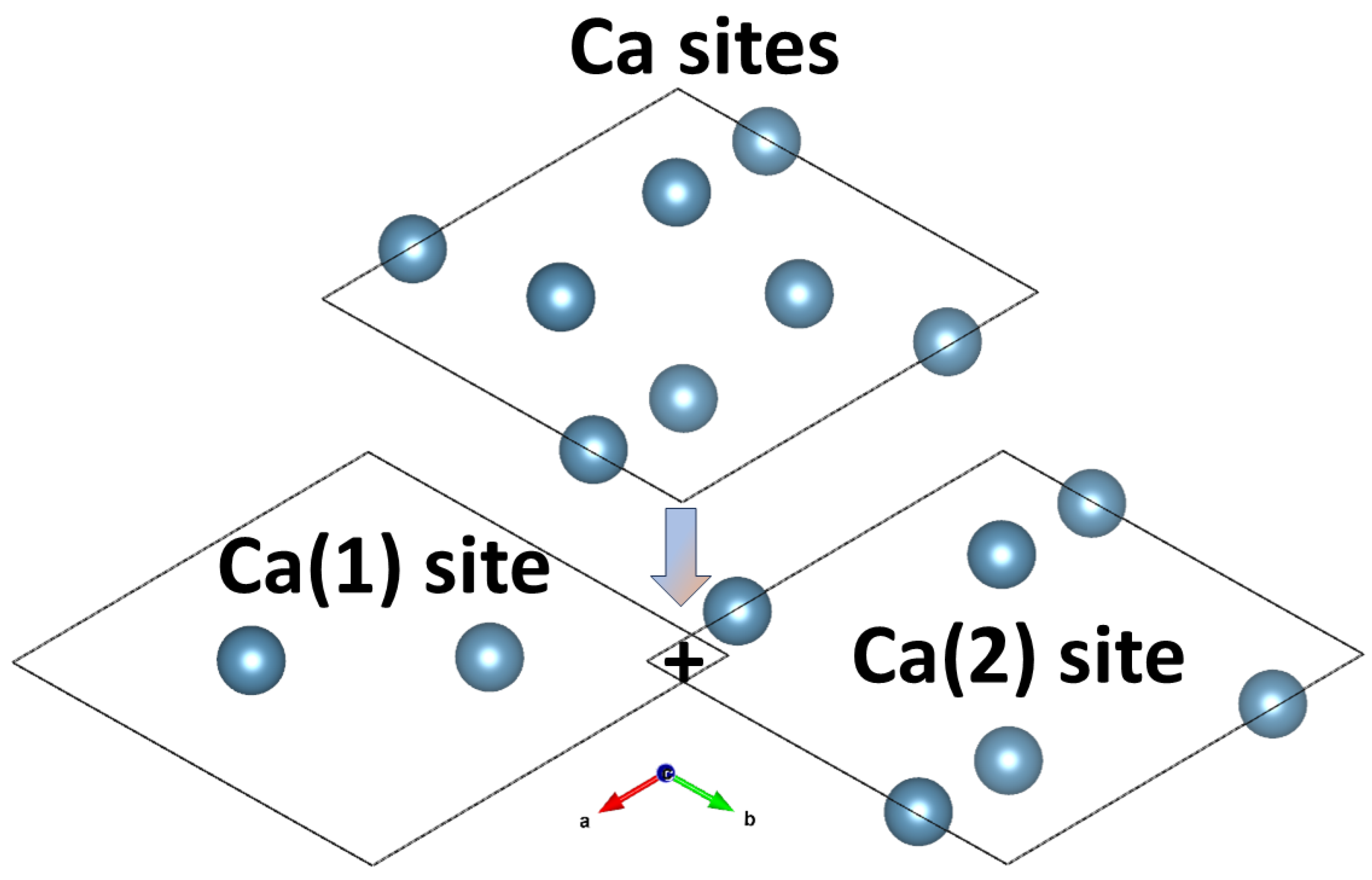
Naturally, HAP is one of the apatite family, which is calcium apatite represented by the chemical formula Ca5(PO4)3(OH) [60]. However, it is commonly denoted as Ca10(PO4)6(OH)2 to indicate that the crystal unit cell consists of two molecular units. One distinguishing characteristic of HAP is the OH ions existence that creates inner channels toward the c-axis [49,61]. These channels exhibit various random orientations of OH-dipoles under different external conditions. This unique structural arrangement allows for the transfer of protons along the c-axis under specific circumstances, such as elevated temperatures or the application of an electric field with critical values [62]. Further, HAP typically presents two primary crystalline structures, which are hexagonal, such as the P63/m space group, and monoclinic, such as the P21/b space group, as shown in Figure 3a,b [55]. There is a third structure which is an amorphous structure with no existing symmetry or long-range order [63]. However, the orientation of OH-dipoles in HAP leads to two distinct order variations. In the hexagonal disordered structure (P63/m), OH-dipoles are spatially randomly distributed throughout the crystal and encompass neighboring unit cells, as demonstrated in Figure 3. In contrast, the hexagonal ordered structure (P63) demonstrates a parallel orientation of OH-dipoles. The same concept applies to the monoclinic phase where the disordered structure (P21/b) features oppositely oriented OH-dipoles in adjacent OH-columns, while the ordered structure (P21) exhibits parallel OH-dipoles as shown in Figure 3b [62]. These various HAP structures have been experimentally determined using X-ray diffraction techniques [64,65]. Further, the hexagonal structure of HAP has 44 atoms per unit cell, while the monoclinic contains a double number of atoms, which is 88 atoms per unit cell [49]. Both the hexagonal and monoclinic phases of HAP exhibit a periodic repetition of their crystallographic structures throughout the entire crystal. These structures adhere to strict stoichiometry requirements. Their ideal stoichiometric ratio of Ca atoms to P atoms must be precisely 1.67 for the ideal chemical composition of the material. This strict adherence to stoichiometry is crucial for maintaining the desired properties and functionalities of HAP. It is common to observe deviations from the ideal crystallographic structures of HAP in actual mineral samples that existed in nature. These deviations mainly come from defects within the crystal lattice, such as vacancies, interstitials, and substitutions of multiple atoms. These defects can arise due to various factors, including impurities in the mineral composition, growth conditions, or external influences during crystal formation. These changes in the Ca/P ratio and the difference from the ideal structure can significantly impact the physical and chemical properties of HAP. They introduce structural irregularities and alter the arrangement of atoms within the crystal lattice. Therefore, studying and understanding these defects is crucial for exploiting the behavior and properties of HAP in practical applications. Moreover, there are two different positions for Ca2+ ions in the HAP structure [66]. (1) Ca(1) or Ca(I) is surrounded by six oxygen atoms from four orthophosphate groups, while (2) Ca(2) or Ca(II) is surrounded by seven oxygen atoms [67]. In Ca(2), six of the oxygen atoms come from four orthophosphate groups and one from hydroxyl ion, which is demonstrated in Figure 4. The crystallographic studies reported that Ca-O distance is longer in Ca(1) than Ca(2) [68].
HAP demonstrates limited ability to dissolve in water and remains insoluble in alkaline solutions. Nonetheless, in the presence of acids (low pH), the phosphate and hydroxide ions within the HAP structure can react with hydrogen ions, leading to the dissolution of HAP, according to (1) [69].
Ca 10 ( PO 4 ) 6 ( OH 2 ) + 14 H + + 10 Ca 2 + + 6 H 2 PO 4 + 2 H 2 O
Studies showed that doping HAP with dopants, such as manganese, magnesium, and carbonate, has the effect of increasing solubility and, therefore, decreasing chemical stability [69,70]. The preparation of nanosized HAP, however, might lead to higher reactivity and increased solubility in water. Moreover, previous studies revealed that HAP exhibits two distinct binding sites on its surface. (1) Ca-site and (2) P-site. The Ca-sites are composed of calcium ions and carry a positive charge, which leads to being more inclined to attract and adsorb negatively charged species, such as acidic molecules. On the other hand, the P-sites consist of phosphate groups that carry a negative charge and consequently interact preferentially and adsorb positive charges, such as basic molecules. This property of HAP allows for selective binding and interaction with molecules based on their acidic or basic nature. In addition, the selective binding phenomenon is a result of the distribution of charges and electrostatic forces in the HAP structure. Furthermore, HAP shows two different crystallographic planes with differences in their atomic arrangements. The a and b-planes predominantly expose positively charged calcium ions, while the c-planes expose negatively charged by the existence of phosphate and hydroxyl groups. This unique feature has prompted researchers to explore the potential for achieving selective adsorption behavior based on the orientation of the prepared HAP material. Therefore, the Ca/P ratio directly affects the adsorption behavior due to adjusting the ability to interact with negative charges to positive charges. The obtaining of this rare property of having both basic and acidic sites makes HAP a significantly important material that can be modified for the desired function [69].

3. Ionic Doping of HAP

Seventeen elements or nutrients are needed for plant growth and reproduction. While not all of the reviewed doped HAP materials were synthesized for nutrient transport and release purposes, the example shown in this section provides for the scope and potential of doped-HAP material synthesis for agricultural applications.

3.1. Carbonate Anion (CO32−)

Carbonated HAP (CHAP) is particularly similar to naturally formed HAP that is found in the human body [71]. CHAP exhibits more favorable formation during precipitation compared to the ideal stoichiometric HAP. The structure of CHAP is relatively complex due to its ability to be in various sites and consequently has three types: Type-A, where a CO32− ion replaces two OH ions, and Type-B, where a CO32− ion substitutes PO43− ion. In Type-AB CHAP, two CO32− ions simultaneously replace one PO43− and one OH. These types can be determined experimentally using Fourier Transform infrared spectroscopy (FTIR) [58,72,73,74].

3.2. Chlorine Anion (Cl)

Fahami et al. produced ClHAP and examined the crystal structure by XRD, and reported an increase in crystallite size upon substitution by Cl-. In addition, an increase in the lattice volume was noticed [75]. Naqshbandi et al. prepared ClHAP and studied the crystallinity behavior. They reported that the introduction of Cl ions into the crystal structure of HAP led to the substitution of OH ions. Therefore, a noticeable impact on the lattice parameters has been observed. The larger size of Cl ions relative to OH ions resulted in expanding the lattice parameters. Therefore, the incorporation of Cl ions results in a slight increase in both the crystallite size and the unit cell volume [76]. Moreover, Hsu et al. prepared ClHAP/polydopamine as a coating for Ti64 alloy. They reported that chloride release increases with time in SBF [77]. This indicates the ability to dope HAP with Cl and being used as a fertilizer for providing phosphorus, calcium, and chlorine.

3.3. Nitrate Anion (NO3)

Gafurov et al. mentioned that N can be substituted in HAP as NO3 and they can behave as CO32− because they are isoelectronic ions [78]. They reported that these impurities can be incorporated from the preparation by using calcium nitrate (Ca(NO3)2). Further, Biktagirove et al. reported that NO3 can locate in A-site and B-site instead of OH, and PO43− respectively [79]. These results open the ability to obtain HAP that might be used in N and P fertilizers at the same time without being in a composite. That means if urea is incorporated into the structure, the results might be effective in the agricultural field.

3.4. Sulfate Anion (SO42−)

Alshemary et al. reported a decrease in crystal volume at small amounts of substitution, then increased upon increasing the amount of SO42− due to its relatively large ionic radius. In addition, crystallite size decreased upon increasing the content of SO42− [80]. Another work by Alshemary et al. investigated sulfur-doped HAP (SHAP) in vitro towards osteoblast-like cells. They showed that ALP activity increased dramatically upon the existence of sulfur in the HAP structure. Furthermore, SHAP showed higher biocompatibility than unmodified HAP [81]. Furthermore, Radha G et al. examined the increment of S substitution and its biological effect on MC3T3-E1 cells. They demonstrated that cell proliferation enhanced until 10SHAP, while at 15SHAP, the proliferation decreased after 1, 3, and 7 days of culture [82].

3.5. Borate Anion (BO33−)

Boron is considered a micronutrient that is important for growth. In this regard, Atila et al. prepared nanocomposite of cellulose/B-HAP/gelatin, and they reported that the incorporation of B in HAP led to a decrease in the crystallinity of HAP. Further, mechanical properties were directly affected by this incorporation which led to an increase in Young’s modulus from 75.26 to 94.9 MPa. Moreover, the thermal stability in the existence of B in the HAP lattice was improved [83]. In addition, Tuncay et al. prepared B-HAP, and they mentioned the ability of B to enhance cell response and cell viability in vitro [84]. Furthermore, Arslan et al. reported that doping by B in HAP can directly affect biocompatibility, which improved as a result of this doping [85].

3.6. Iron (Fe2+ and Fe3+)

The inclusion of ferrous cation (Fe2+) and ferric cation (Fe3+) in hydroxyapatite affects the crystal parameters and local ion reorganization. The different electron configurations and ionic radii (70 pm for Fe2+ and 60 pm for Fe3+) contribute to their distinct interactions and positions within the crystal. A study was performed on an 88-atom monoclinic HAP supercell for incorporating ferrous and ferric cations in HAP. Makshakova et al. showed that cations can be introduced into HAP at both Ca(1) and Ca(2) positions. Further, Fe2+ can occupy either position without requiring additional charge compensation and resulting in the formula Ca9.5Fe0.52+ (PO4)6(OH)2. On the other hand, the substitution of Ca2+ by Fe3+ can lead to (Ca9.5Fe0.53+ (PO4)6O2H1.5), which is element perturbation. This possibly causes vacancy formation in the H+ or Ca2+ positions by releasing one Ca2+ for every two substituted atoms (for neutralizing the charge). Further, the release of H+ from OH was also considered to maintain a neutral overall charge and to minimize the cell contraction caused by charge compensation. The presence of OH- within the anion channel, surrounded by calcium ions, can show a minimal impact on the overall packing and density of the crystal [86]. In addition, they reported that Fe3+ substitution is more reactive with the surrounding.

3.7. Magnesium (Mg2+)

Karunakaran et al. showed that doping HAP with Mg2+ ions leads to less crystallinity and higher distortion in the lattice using XRD in addition to a growth in (a) crystal parameter [87]. Another study by Laurencin et al. reported the lattice distortion by Mg incorporation but mentioned falls in the lattice parameter (a) due to the substitution [88]. Further, Mg2+ can substitute Ca(1) or Ca(2) sites and lead to a shortening distance between Mg and O [89]. In addition, Goldberg et al. prepared Mg-HAP and reported that the substituting using Mg2+ ions led to an increase in BET-specific surface area from 23.35 to 86.91 m2/g from 0 to 1% substitution. They also reported that cell volume decreased upon increasing the amount of Mg2+ doping in addition to the adsorption capacity. Further, Mg-HAP led to an increase in the viable cells and proliferation of MG-63 cells after 24 h [68]. Predoi et al. reported the antibacterial properties of Mg-HAP in addition to its biocompatibility as a biofilm [90]. In another study by Predoi et al., they prepared a thin layer of Mg-HAP by dip coating procedure. It was reported that 25Mg-HAP has antibacterial activity against Staphylococcus aureus, Escherichia coli, and Candida albicans strains. In addition, the layer improved biocompatibility without affecting the cell behavior [91]. Further, Sharma et al. prepared Mg-HAP/urea, and they reported the improvement of nanohybrid compared to the traditional fertilizers [92].

3.8. Zinc (Zn2+)

Sharma et al. doped HAP with zinc and integrated ZnHAP with urea to be used as a nano-fertilizer. The incorporation of zinc can be applied to the growth of plants as an essential element of this process. Further, nitrogen can be taken up from urea, while phosphorus and calcium essentially come from HAP. They reported that growth is highly improved compared to the control sample, which confirms the ability of doped-HAP with the micronutrients to be significantly effective [92]. There are previous reports by Guerra-Lopez et al. that mentioned that Zn2+ prefers the Ca(2) site [93]. Furthermore, the same results are done by Chathurika et al. They reported that Zn-HAP/urea nano seed coating showed around 69% growth for Zea mays seeds [94]. Abeywardana et al. prepared Zn-HAP/urea nanohybrids to offer a sustainable solution for addressing urea and phosphorus deficiencies in plants while providing zinc supplementation. They reported that these biodegradable nanoparticles effectively deliver essential nutrients during early plant development and resulting in improved growth, development, and nutrient distribution in maize. The plant height reached around 140 cm for the nanocomposite and 80 cm for the control sample. In addition, the number of leaves and root lengths reached around 20 and 70 cm using Zn-HAP/urea, while 14 and 30 cm for the control sample [95].

3.9. Nickel (Ni2+)

Kurinjinathan et al. synthesized pure and Ni-doped hydroxyapatite (HAP) nanoparticles using ultrasonication-assisted wet-chemical techniques. The synthesized nanoparticles were systematically characterized using various techniques. X-ray diffraction (XRD) analysis revealed that the concentration of Ni2+ ions increased in the HAP structure while the crystallite sizes decreased. The average crystallite sizes for pure HAP and Ni-doped HAP with concentrations of 0.01 M, 0.05 M, and 0.1 M were found to be 29.09, 16.57, 14.33, and 13.66 nm, respectively [96]. Sebastimmal et al. employed the sol-gel method to synthesize Ni-doped HAP nanoparticles. SEM examination revealed that the particles exhibited a spherical or nearly spherical shape. Furthermore, Ni2+ doped HAP nanoparticles displayed remarkable antibacterial activity against a range of bacteria, including S. flexneri, E. coli, P. aeruginosa, K. pneumoniae, and S. aureus, as demonstrated by the disc diffusion technique. Thus, the importance of Ni dopants in HAP stricture is denoted by the applicability of these nanoparticles for medical applications as antibacterial agents. Moreover, these nanoparticles showed high levels of antifungal, antioxidant, and anticancer activities, making them highly promising for biomedical applications [97].

3.10. Copper (Cu2+)

Bazin et al. doped HAP with Cu, and they reported the ability of Cu dopant to increase lattice parameters and volume [98]. Unabia et al. prepared Cu-HAP coating, and they also mentioned the increase in lattice parameters and volume of HAP lattice upon increasing Cu content [99]. Imrie et al. prepared Cu-HAP, and they reported the expansion of the unit cell due to the doping, where volume and lattice parameters increased [100]. In addition, Stanic et al. synthesized Cu-HAP using the neutralization method, and they examined the released ions. They reported that HAP in phosphate buffer solution released extremely small amounts of Cu ions which are non-detectable [101]. Hidalgo-Robatto et al. studied the incorporation of Cu in HAP up to 3.31% and their effect on pre-osteoblast cells. They reported that cells proliferation and had healthy growth after 7 and 21 days in addition to their ability to prevent bacterial formation [102]. It should be mentioned that Cu is a plant micronutrient.

3.11. Potassium (K+)

Potassium is considered one of the essential macronutrients that are required for plant growth. It is a monovalent cation, however, it might replace Ca(II) ions partially. In this regard, to maintain neutrality, vacancies can be formed through the HAP structure, which might affect the chemical stability and the degradability of the formed crystals. Chahkandi et al. prepared K-HAP and revealed that particle size was, on average, 65–70 nm through XRD and TEM. Further, they reported the antibacterial activity of K-HAP in addition to the dye removal efficiency against Congo red (CR) [103].

3.12. Molybdenum (Mo2+)

The Molybdenum element is also considered one of the plant micronutrients. Abutalib et al. studied the incorporation of Mo in HAP lattice and reported that volume gradually decreased upon doping (from 0.05% to 5.00%) [104].

3.13. Co-Doping (Two or More Ions)

Makshakova et al. studied the doping of HAP by Mg and/or CO32− to obtain Mg-HAP, CHAP, and Mg-CHAP. They found that the incorporation of the lattice structure by both elements can be more efficient energetically [89]. On the other hand, Landi et al. synthesized SrMgHAP as a filler sample to be used in osteoporotic bone regeneration. They reported that the co-dopant substitution improved the cell response and bone metabolism, in particular at 10 mg/mL dose [105]. Sharma et al. prepared ZnMg-HAP/urea nanohybrids for improving wheat growth and uptake of nitrogen. They found that doping HAP nanoparticles enables the delivery of both macro and micronutrients to plants and makes them a biocompatible system suitable for agricultural use. Coating these nanoparticles with half and quarter doses of urea results in improved crop yield, nutrient levels, and leaf nitrogen content while simultaneously reducing nitrogen loss. The controlled and gradual release of nitrogen from the urea-coated ZnMg-HAP nanoparticles ensures an adequate and sustained supply of nitrogen during the crucial early growth stage of wheat plants. Furthermore, these nanohybrids effectively prevent the leaching or volatilization of urea from the soil, thereby ensuring a consistent and long-lasting nutrient delivery system [92]. However, the application of these nanohybrids showed no detrimental effects on wheat growth, which offers a promising alternative to conventional fertilizer methods in agriculture. Tang et al. prepared NiMo-HAP, and they reported that the incorporation of Ni and Mo showed a negative effect on the stability as a catalyst [106]. Moreover, Alshemary et al. demonstrated the incorporation of Sr2+ ions in SHAP, and they reported a high increase in biocompatibility and cytocompatibility of Sr-SHAP [81].

4. Synthesis of HAP

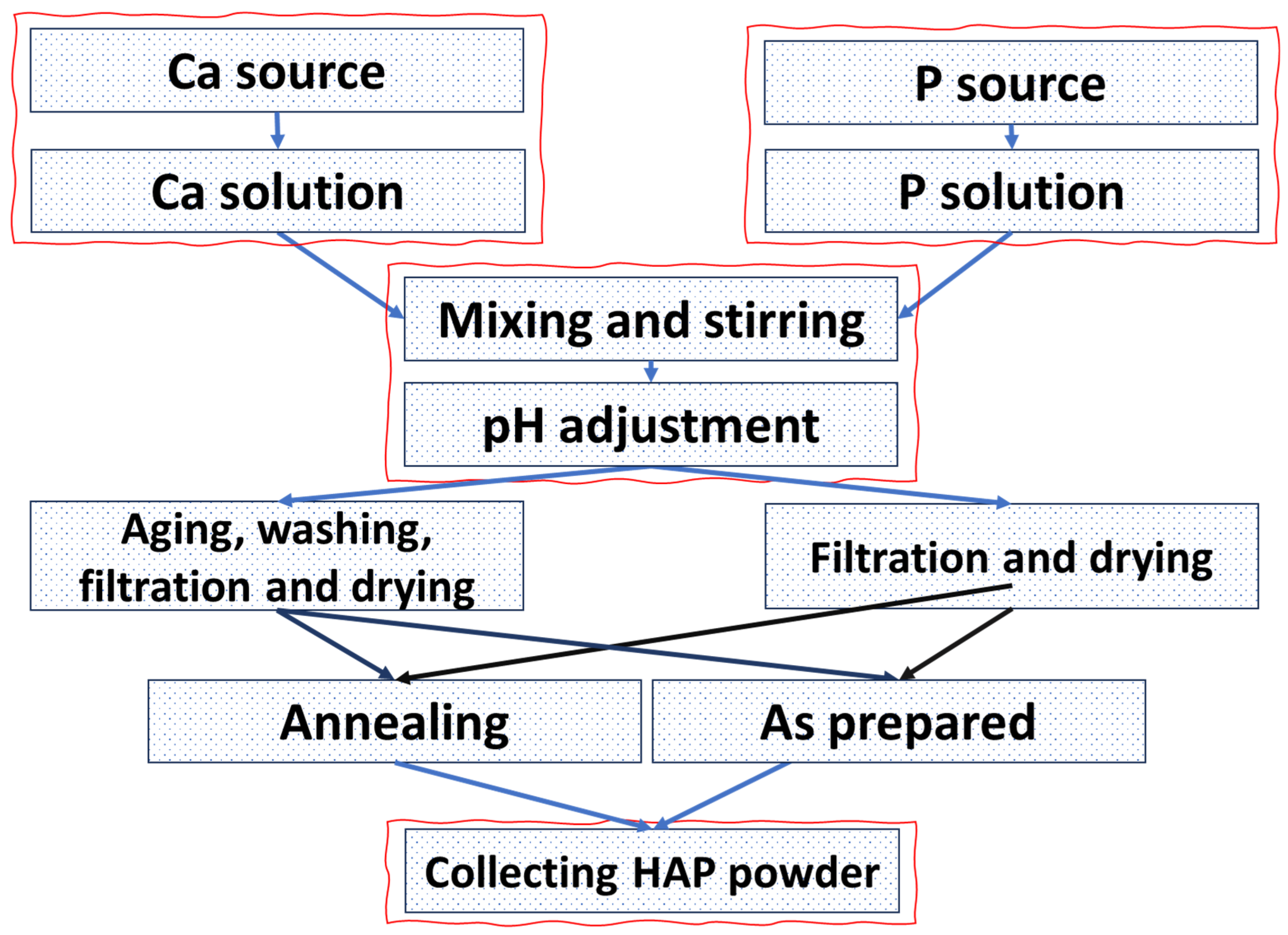
The synthesis of HAP involves several methods with distinct advantages and yielding HAP particles with specific properties. There are two main ways for obtaining HAP, which are (a) wet chemical methods, as described in Figure 5, and (b) solid-state reactions shown in Figure 6 and Figure 7.

4.1. Wet Chemical Synthesis

4.1.1. Coprecipitation

The wet chemical synthesis allows producing HAP nanoparticles with various ranges in scale, different morphologies, and crystallinity. One of the most simple and basic wet chemical synthesis techniques is the coprecipitation method which is frequently used [67,107]. The coprecipitation method involves the simultaneous precipitation of calcium and phosphate ions from a solution to form HAP nanoparticles. This is done by mixing calcium and phosphate precursors under controlled conditions, such as temperature, pH, and stirring rate [108]. This leads to the nucleation and growth of HAP particles. The coprecipitation process can be performed at ambient temperature or at elevated temperatures which depends on the desired properties of the nanoparticles. One of the key advantages of the coprecipitation method is its simplicity and cost-effectiveness, as presented in Table 1. [109]. This method has no complex requirements, such as complex equipment or specialized devices. Therefore, it is suitable for researchers to obtain samples and industries for large-scale production [110,111,112]. In addition, the materials needed for the synthesis are readily available and relatively inexpensive, which makes it more cost-effective. Another advantage of the coprecipitation method is its ability to provide high yields of HAP nanoparticles within a relatively short time. Furthermore, the reaction kinetics can be easily controlled by adjusting the temperature, pH, and concentrations in addition to the started precursors [113]. This availability of controlling various conditions can directly affect the properties of obtained HAP powder. By adjusting the concentrations of the calcium and phosphate precursors, the stoichiometry of the HAP can be finely tuned. Furthermore, controlling the reaction parameters, such as temperature (almost low temperature in this method) and pH, enables the modulation of particle size. This flexibility in composition and particle size control is crucial for tailoring the properties of HAP nanoparticles to suit specific applications. Another advantage from the point of view of ceramics production is the suboptimal crystallinity of the precipitated HAP nanoparticles in addition to the rapid precipitation process, often resulting in the formation of nanocrystalline and amorphous HAP. Therefore, lower crystallinity compared to other synthesis methods might affect the desired application [71].
Despite these advantages, the coprecipitation method has limitations and disadvantages. One of the main challenges is achieving a uniform particle size distribution and specific crystal morphology [117,118]. The coprecipitated nanoparticles tend to agglomerate, which leads to poor dispersion and affects their performance in certain applications. This issue can be addressed through additional processing steps, such as sonication or surface modification techniques [119]. These additional steps might be considered disadvantages. Moreover, the coprecipitation method can be sensitive to reaction conditions such as temperature, pH, and reaction time, which need to be carefully controlled to ensure the desired properties of the HAP nanoparticles. Any deviation in these parameters can lead to variations in particle size, composition, and crystallinity. Therefore, it affects the overall quality of the synthesized nanoparticles. Sciena et al. prepared HAP by the coprecipitation method and reported the improvement of P ion release [120].

4.1.2. Hydrothermal Method

The hydrothermal method is a widely used technique for the synthesis of HAP nanoparticles with controlled size, morphology, and crystallinity [121,122]. This method involves the reaction of calcium and phosphate sources in a closed system under high pressure and temperature [122]. The hydrothermal synthesis process promotes the transformation of amorphous calcium phosphate precursors into well-defined crystalline HAP nanoparticles [123]. One of the main advantages of the hydrothermal method is the ability to produce highly crystalline HAP nanoparticles with a narrow size distribution [123,124]. Well-defined crystal structures and high crystallinity can be obtained due to exposing a closed system to high-pressure and high-temperature conditions. In addition, controlling reaction time, temperature, and precursor concentrations can lead to tuning the particle size and crystallinity precisely. Further, the system under high temperature and pressure can directly affect the purity leaving the pure desired shape of HAP nanoparticles. The precise control over the crystalline nature of the nanoparticles contributes to their superior mechanical strength, stability, and biocompatibility, making them suitable for various applications. In addition, various morphologies can be obtained by adjusting the reaction conditions [125]. On the other hand, hydrothermal methods need specialized equipment to stand in these high conditions. The usage of high-pressure vessels and autoclaves increases the complexity and cost of the synthesis process, which directly limits its accessibility. Moreover, the reaction time, which ranges from several hours to several days, is another drawback of this method [126]. Moreover, the high-temperature and high-pressure conditions can promote particle agglomeration and lead to the formation of larger aggregates rather than individual nanoparticles. However, additional processing steps, such as sonication or surface modification techniques, may be employed to disperse and stabilize the HAP nanoparticles. In this regard, Tang et al. prepared HAP to be used in fertilizer application, and the preparation included a hydrothermal process. They obtained nanorod particles, which are efficient as P-delivery where phosphorus use efficiency reached around 47% compared to the chemical phosphorus fertilizer, which reached around 23% [7].

4.2. Solid-State Reactions

4.2.1. Solid-State Reaction and/or Thermal Decomposition

Solid-state reactions are employed for HAP synthesis in addition to the wet methods. These methods involve the direct heating or sintering of calcium and phosphate precursors at high temperatures. Usually, solid-state reactions involve high temperatures, which is a major requirement to activate the solid reactants for chemical interaction or crossing the activation barrier [127]. One widely used technique in nanoparticle preparation, in general, is the solid-state thermal decomposition method [128,129,130]. The precursors are heated to temperatures above 800 °C, and hence nucleation and growth occur to form crystalline HAP. The thermal decomposition method is relatively simple and cost-effective for small production but results in larger particle sizes and lower crystallinity compared to wet chemical synthesis methods.

4.2.2. Mechanochemical Synthesis

The mechanochemical method or mechanical activation is based on merging mechanical energy with solid-state reaction [27]. The conventional process of solid-state reactions consumes significant time and energy, but it can be replaced by incorporating mechanical energy [131]. Introducing mechanical energy into the process can achieve various benefits: (1) Increased reactivity, (2) overcoming the activation barrier, (3) improved diffusion, and (4) reduced reaction time. The technique can approach its purpose through grinding or milling by inducing mechanical stress to the solid reactants. This stress can disturb the surface-bound compounds leading to enhance the reaction. Further, grinding or milling leads directly to breaking down the particles into smaller ones. In more detail, when sample powders are crushed between colliding balls inside a ball mill pot, they undergo plastic deformation. That can lead to cold welding, fracturing, and rewelding. The impact force breaks crystallographic bonds and creates new surfaces on the particles. These new surfaces facilitate easy welding of particles together, resulting in an accelerated dissolution rate of the solid material [131]. Continued mechanical deformation further reduces particle size and increases surface energy, causing various changes in the properties such as surface-to-volume ratio and, consequently, reactivity [131]. In addition, chemical reactions typically require a certain amount of energy, which is commonly achieved by raising the temperature. However, the introduction of mechanical energy can contribute to the required energy and reduce the reliance on temperature and allow for lower temperatures to overcome the activation barrier. Furthermore, milling and grinding apply intense forces causing deformations, defects, and fractures. The decrease in particle size leads to more contact points between reactants which increases diffusivity and reactivity, then directly improving the reaction rate. The mechanochemical approach is a technically straightforward method used in the large-scale production of nanocrystalline HAP powder. When compared to the wet approach, it offers advantages in the aspect of simplicity and versatility [132]. The mechanochemical process does not require parameters, such as reactant concentrations, temperature, aqueous solution, or long processing times. That means the final morphology and size can be affected by other factors. The mechanochemical process requires different parameters that can be easily controlled such as speed or frequency, duration of milling, and milling media. To apply mechanical energy during the synthesis of HAP, commonly used equipment includes ball-milling or planetary mills. The device offers the option of adjusting the speed and duration of milling. Then, by utilizing high-energy milling, the interaction between the calcium source and the phosphorus source is stimulated. This leads to improved reaction rates. Further, this milling process enhances diffusion processes and allows for the synthesis of nanocrystalline HAP powder at lower temperatures and shorter reaction times. Therefore, the high-energy milling approach is highly advantageous for large-scale production of HAP powder with desirable properties. Moreover, there is no need for external temperature while the process is working. The collision and friction between the balls can generate heat (from kinetic energy), and this heat can facilitate the reaction. Various factors, including the reactant types, milling medium, milling ball type, and diameter, duration of milling steps and interval pauses, powder-to-ball mass ratio, and rotational speed, are crucial processing variables in the mechanochemical method [133]. In addition, it is reasonable to assume that the nanoscale characteristics of HAP obtained mechanochemically could influence the release of P ions in the soil. This is because nano scaling can increase the surface area and reactivity of HAP particles. That potentially affects the dissolution behavior and subsequent release of P ions. Studies have shown that nanosized materials can exhibit enhanced reactivity and solubility compared to their bulk counterparts, which may lead to altered release patterns of nutrient ions, such as P, in soil [134]. Moreover, a study conducted by Silva et al. prepared urea-loaded HAP using the mechanochemical method. They investigated the release of N from the urea-loaded HAP and found that the release kinetics followed the Higuchi model indicating a diffusion-controlled mechanism. However, it should be noted that in this study, they did not measure the release of P ions [135].

4.3. Economic Feasibility of the Mechanochemical HAP Synthesis Method

Han et al. highlighted the advantages of using mechanochemistry in sustainable large-scale processing as a green, solvent-free, scalable, and low-temperature process. In their study, they achieved significantly higher ammonia concentration compared to the traditional Haber–Bosch process. While the traditional method reached a concentration of only 25 vol%, the mechanochemical approach yielded an impressive 82.5 vol% ammonia concentration. The milling conditions for the mechanochemical method involved a temperature of around 45 °C and 1 bar pressure, whereas the Haber–Bosch process required much higher conditions with temperatures of around 450 °C and pressures of 200 bar. Despite the higher concentration achieved with mechanochemistry, the total energy consumption was remarkably lower, suggesting a potential for many practical applications. They evaluated the total energy consumption for the mechanochemical method at 4.5 × 10¹² J·m/t, while the thermal Haber–Bosch and electrochemical methods consumed 39 × 1012 J·m/t and 228 × 1012 J·m/t, respectively. It must be mentioned that these energy consumption values have relatively small yields compared to the mechanochemical method [136]. AlShamaileh et al. emphasized the need to reduce fertilizer consumption, particularly nitrogen (N) fertilizers, to mitigate environmental pollution. They pointed to the mechanochemical method as a potential solution for reducing consumption and its associated environmental impact [137]. Furthermore, Mohamed et al. discussed the benefits of the mechanochemical method for synthesizing cathode material containing lithium (Li), and iron (Fe), with a chemical formula of (Li2Fe)SO, in one step, leading to cost reduction [138]. In another study conducted by Palaz et al., they highlighted the advantages of milling as a continuous process, offering high productive capacity and low energy consumption. They compared this method to traditional approaches for producing phosphate fertilizer, which typically require 450 kWh/t in a periodic process lasting for 20 days and involve the use of reagents such as sulfuric acid (H2SO4). In contrast, the mechanochemical process was found to require 500 kWh/t, but only for a significantly shorter duration of 24 h, which aligns with the production process [139]. The remarkable difference lies in the elimination of the need for reagents in the mechanochemical approach, making it a more efficient and eco-friendly alternative. Moreover, its continuous nature ensures a more streamlined and continuous production, enhancing overall productivity [139]. Finally, these studies collectively demonstrate the potential of mechanochemistry as a sustainable and efficient approach for various applications, including fertilizer, such as HAP, production, and all contributing to reducing energy consumption and environmental impact.

4.4. Parameters Affecting Mechanochemical Synthesis Procedure

4.4.1. Time of Milling

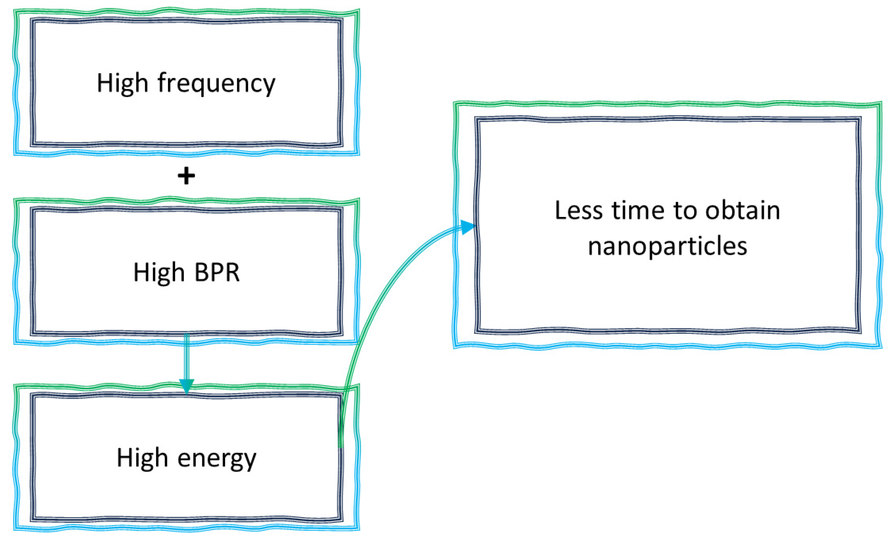
Silva et al. conducted a study focused on HAP synthesis using ball milling and investigated the effect of milling time. They found that crystallite size increased from 17.5 nm after 5 h of milling to 20.5 nm after 15 h [140]. This suggests that prolonged milling leads to a gradual increase in crystallite size. Nasiri-Tabrizi et al. focused on fluorapatite (FAP) (Ca10(PO4)6F2) and observed changes in crystal size with milling time. They found that the crystal size of FAP increased to approximately 90 nm after 50 min of milling, reached around 55 nm after 100 min, and eventually reduced to 35 nm after 300 min of milling [141]. The milling process also induced lattice strain, which increased from 0.2% to approximately 0.5% over 300 min. They noted that the milling process gradually eliminated the presence of CaO, with complete disappearance observed after 300 min of milling. Further, the morphology of the particles changed from flake-like shapes to more rounded particles as the milling time increased. These observations indicate that the milling duration and conditions can have a significant impact on the final crystal size of the synthesized HAP or its derivatives. Ebrahimi-Kahrizsangi et al. focused on the synthesis of spheroidal nanoparticles of FAP. They achieved an average crystalline size of 25 nm and an average lattice strain of 0.434%. The milling process included the use of zirconia balls with a diameter of 20 mm, a low rotational speed of 600 rpm, and a high ball-to-powder weight ratio (BPR) of 20:1. To prevent excessive temperature rise during milling, they conducted the process in steps, with 45 min of milling followed by a 15-min rest period for a total duration of 60 h. [142]. Fakharzadeh et al. reported the results of a study on HAP synthesis through ball milling and investigated the effects of milling time. They achieved a crystallite size of approximately 30 nm and a lattice strain of 0.5% after 3 h of milling. The milling conditions included a BPR of 15:1, a rotational speed of 600 rpm, and the use of steel balls with a diameter of 20 mm. The resulting HAP particles exhibited a spherical morphology and showed a tendency to agglomerate [143]. Further, it can be seen that at low rotational speed, more time is needed to obtain small crystallites. Bulina et al. investigated the synthesis of HAP particles through ball milling. They reported that after 30 min of milling at a high rotational speed of 1800 rpm, they obtained HAP particles with a non-uniform size distribution and a crystallite size of approximately 24.2 nm [35]. This study demonstrates the influence of milling parameters, such as rotational speed, on the particle size distribution and crystallite size of the synthesized HAP. It can be seen that 1800 rpm is relatively high, which indicates less time to obtain nanocrystals. Therefore, the use of high rotational speed allows for the efficient production of nanosized HAP particles within a short milling time. Chaikina et al. revealed a study focusing on the impact of milling time on the crystallite size of HAP. They demonstrated that longer milling times increased the crystallite size, ranging from approximately 20 to 32.7 nm. They conducted milling for durations of 3, 5, 10, 20, and 30 min, resulting in crystallite sizes of approximately 20, 20.6, 28.9, and 32.7 nm, respectively. The milling conditions included a high rotational speed of 1200 rpm and a high BPR of 20:1 [144]. These values shed light on the relationship between milling time, BPR, and rotational speed with crystallite size. Therefore, high speed and high BPR lead to reaching nanosized particles in a short time, and when milling time increases, the crystal size increases. In another study by Bulina et al., they observed spherical HAP particles with sizes less than 50 nm after 30 min of milling at 1200 rpm, as revealed by TEM [145]. The crystallite size determined by XRD was approximately 20 nm. The influence of milling parameters on particle morphology can be noticed. This study demonstrated the efficient synthesis of nanosized HAP particles within a short milling duration when the high rotational speed is incorporated.
Abdel-Aal et al. investigated the effect of milling time on the crystallite size of HAP. They found that the crystallite size decreased from around 13 to 6 nm after 3 and 5 h of milling, respectively. The experimental setup involved the use of a fine particle grinder instrument and a rotational speed of 2000 rpm [146]. This study demonstrated the impact of milling duration on the crystallite size of HAP, which showed that longer milling times resulted in a reduction in crystallite size. Moreover, it can be observed that the absence of grinding balls and being replaced with another instrument affect the ability to grind directly. Iwasaki et al. employed intermittent milling to avoid temperature rise during the process. The net wet milling time was 1 h, and they successfully synthesized HAP/La0.7Sr0.3MnO composite. The HAP particles showed sizes ranging from 50 to 100 nm. The milling conditions included a rotational speed of 600 rpm, zirconia balls with a diameter of 5 mm, and a BPR of 18:1 [147]. This study showcases the use of intermittent milling to control the synthesis of composite materials and achieve specific particle sizes. In addition, it can also be noticed that low rotational speed, low diameter, and high BPR increase the needed time to approach the nanoscale.
Galotta et al. investigated the synthesis of HAP particles through ball milling and showed that after 4 h of milling, then followed by drying at 150 °C, HAP particles had a crystallite size of approximately 21 nm. The particles exhibited a flaky morphology with nanoscale thickness. The experimental setup involved the use of zirconia balls with diameters of 6 mm, a rotational speed of 50 Hz (3000 rpm), and a BPR of 5:1 [37]. This reveals the influence of milling parameters on the morphology and size of the synthesized HAP particles. They used low BPR, low diameter, and high rotational speed. Fahami et al. focused on the synthesis of HAP/Ti nanocomposites through ball milling, and they observed a decrease in the crystallite size from 20 to 13 nm after 20 h of milling, which was accompanied by an increase in lattice strain. Morphologically flaky and ellipsoidal particles were observed, indicating a notable reduction in size after 10 h of milling. In addition, the experimental conditions included zirconia balls with a diameter of 20 mm, a rotational speed of 600 rpm, and a BPR of 20:1 [148]. It can be seen that the incorporation of TI led to an increase in the needed time for reducing the crystallite size, especially at a low rotational speed. Ferro et al. studied the synthesis of HAP from chicken eggshell and cuttlefish bone. The used balls are made of alumina with a diameter of 10 mm and BPR of 10:1 in addition to 400 and 600 rpm. They found that HAP structure was obtained after prolonged milling for approximately 10–12 h using high milling energy. XRD analysis revealed broad HAP peaks, indicating the nanosized of the HAP crystals [149]. That indicates the precursors used to synthesize HAP have a potential effect on the milling time, especially at the low rotational speed. Coreno A et al. conducted a study on HAP synthesis by milling the reactant mixture every 2 h. All reactants transformed into carbon-substituted HAP after 6 h of milling with particle sizes ranging from 10–20 nm. They used hardened steel balls with a BPR of 20:1 throughout the study [150]. Lala et al. investigated the doping effect of Zn, Mn, and Mg on HAP, which are prepared by ball milling. The preparation was done using a high planetary ball mill with a hardened chrome steel vial for 10 h in addition to pausing 10 min every 20 min. They reported that sintering the samples at 950 °C led to decomposing the undoped Mn, and Mg-HAP partially to β-TCP, while Zn-HAP decomposed partially to the tetracalcium phosphate phase [151].
Bulina et al. prepared magnesium-substituted HAP during a 20-min milling duration at a high rotational speed of 1200 rpm. They reported a crystallite size of approximately 18 nm, and the SEM images showed particles in spherical and rod shapes with sizes reaching 20 nm [152]. Youness et al. systematically investigated the effect of milling time on B-CHAP, varying the milling time from 1 to 10 h with 1-h increments and including a 30-min pause every 60 min. The milling process was at a high rotational speed of 1500 rpm using alumina balls with a diameter of 10 mm and a BPR of 10:1. They observed an increase in crystal size from 13 to 30 nm, while the lattice strain decreased from 0.9506% to 0.4420% [153]. The increase in the crystal size is due to using high BPR, high diameter, high speed, and long milling time. Hussain et al. studied preparing HAP from waste seashells by hydrothermal and ball milling under conditions of 40 min with a 10-min break every 15 min at 350 rpm and BPR 10:1. They showed that heat treatment at 900 °C led to the maximum crystallinity with near stoichiometric ratio with rod, spherical and needle shapes in addition to crystallite size around 36 nm [154].
Ravindranadh et al. synthesized calcium-lithium HAP (CLHAP) nanopowder using mechanochemical milling with a low rotational speed and 3 h of milling. They included pauses in the milling process, with a 15-min rest period every 60 min. The obtained CLHAP powder exhibited a crystallite size of 16 nm and a morphology resembling stone-like particles [155]. Furthermore, Nasiri-Tabrizi et al. prepared HAP under various mechanochemical conditions such as milling time, milling atmosphere, milling media, and precursor compositions. They reported that they fixed BPR 20:1 of zirconia balls of 20 mm diameter and rotational speed of 600 rpm. They studied the crystallite size concerning milling time and they found that crystallite size decreased from around 40 to 30 nm after 40 and 80 h, respectively. These results are estimated when they started the reaction using CaHPO4+Ca(OH)2 with air and argon atmosphere in addition to polyamide-6 (PA6) medium. There was no difference in the results estimated by changing the milling atmosphere. Moreover, they changed the precursor compounds to CaCO3+CaHPO4, and the results changed. The crystallite size of 40 h showed a smaller size in the air with a value of 32 nm. Furthermore, the argon medium showed 40 nm at the same duration of milling, and by increasing the milling time, the crystallite size decreased to 28 and 32 nm for the air and argon media, respectively. These results indicate the complex effect between precursors, milling media, and size. Furthermore, under the fixed previous conditions of speed, BPR, with 80 h of milling, changed atmosphere, precursors, and milling media. They found that in the polymeric media (PA6) starting with CaHPO4+Ca(OH)2 in air, the morphology of nanorods, cluster-like structures, and agglomerations. In the argon medium, a coalescence of nanorods is observed, which are chain-like agglomerations. In the metallic medium (only argon atmosphere), the only observed are coalescence agglomeration nanoparticles. By starting with CaCO3+CaHPO4, nanospheres are observed that make obvious chain-like clusters, especially in the argon atmosphere of polymeric medium [156]. This study showed that not only the milling factors are important to study, but also precursors, vial media, and even atmosphere are essentials.
A consistent trend can be observed in the previous studies investigating HAP synthesis using the mechanochemical method. It is evident that as the milling duration increases, there is a general decrease in the crystallite size of the material until a critical point is reached. Beyond this critical point, recrystallization occurs and results in an increase in the crystal size accompanied by a decrease in lattice strain. The duration of milling plays a significant role in this trend, but other factors related to the milling process also contribute. All the factors are translated into the material as energy which transfers into the sample.
Burgio et al. developed an equation to express the energy transferred into the sample from the planetary mechanical milling according to (2) [149,157]:
E i n p u t = ( 1 φ ) n b m b t ( w p w v ) ( w v 3 ( r v d b 2 ) w p + w v w p r p ) ( r v d b 2 )
The parameters involved in the (1) are as follows: φ is a numerical value dependent on vial filling, nb is the number of balls, mb refers to the mass of each ball, t is the milling time, wp refers to the angular rate of the mill, wv is the angular rate of the vial, rv indicates the vial radius, db is the ball diameter, and rp indicates the distance between the center of the plate and the center of the vial. This means all these parameters can affect the transferred energy into the sample. However, there are main parameters that can be mainly focused and controlled, such as rotational speed, time, and the ratio between the weight of balls and powder. For instance, Chaikina et al. reported an unexpected increase in crystallite size after only 3 min of milling. This can be attributed to the high rotational speed of 1200 rpm and a BPR value of 20:1 employed in their study. The combination of these factors led to the rapid and efficient breakage of grains, facilitating the cold welding and recrystallization process even within a short milling time [144]. Further, in the morphological aspect, the up-bottom technique of ball-milling breaks the grains, which tend to be in spherical and ellipsoidal shapes, unlike coprecipitation in which HAP particles tend to be in nanorod shape.

4.4.2. Effect of Ball Milling on Biological Properties

Angioni et al. investigated the biological response of HAP/bioglass named BGMS10 with 30 min of milling. They reported that this milling time was optimized with 800 °C of heat treatment at 70 MPa of pressure which led to covering SBF completely with HAP after 7 days of immersion. On the other hand, 120 min of milling led to lower bioactivity and lower mechanical properties [158]. Sharifianjazi et al. showed that PHAP showed around 60% cell viability in 7 days and around 100% ALP activities [159]. Lala et al. prepared CHAP by mechanochemical method at high energy for various milling times. They found that the best biocompatibility near the natural bone was in the sample after 2 h of milling. The high biocompatibility was estimated even at relatively high concentrations of 100 µg/mL, which was around 85% of cells were alive after 24 h of incubation. Furthermore, after 10 h of milling, the sample showed around 100% cell viability from the low concentrations to the relatively moderate of 60 µg/mL after 6, 12, and 24 h of incubation [160]. Fernandez et al. prepared HAP and β-TCP through chemical methods and combined them with bioactive glass containing 45SiO2-24.5CaO-24.5Na2O-6P2O5. Further, the biphasic bioceramic was treated mechanically, and they reported the development of biological response by decreasing the time of implantation into tibias with no inflammation for 30 days post-implantation [161]. Lala et al. prepared Mg-CHAP using a ball mill and showed that more than 95% of the cells were viable at low concentrations after 12, 24, and 48 h of incubation [162].

4.4.3. The Potential to Incorporate Dopants into HAP Structure Using Mechanochemical Synthesis

Lala et al. prepared Mg-doped-CHAP using a dry ball milling method in the existence of a hardened chrome steel vial for 10 h and 10 min pauses every 30 min with high energy planetary mill. They showed that crystallinity falls upon the increase of Mg ions. Furthermore, the lattice parameters were affected by Mg addition, and led to a decrease in the lattice parameters (a) and (c) [162]. Shu et al. prepared CHAP using zirconia balls with a maximum diameter of 15 mm in addition to a speed of 1000 rpm. Then they obtained a nanofibrous structure based on CHAP and reported that HAP was completely synthesized after 1 h of milling. The nanofiber-based bell-milled HAP exhibited a diameter of 8 nm and a length in the range of 50–150 nm [163]. Bulina et al. used water-cooled steel vials with a speed of 1200 rpm and BPR 1:20 for 30 min of milling. They estimated Si-HAP and revealed that substitution led to vacancy formation. In addition, the substitution increased the cell volume for the same milling conditions [164]. Coreno et al. reported that a completed reaction is achieved for the preparation of CHAP after 6 h of milling under conditions of 20:1 BPR with high energy mixing and hardened steel balls. Further, the particle size was in nanosize in the range of 10–50 nm [150]. In another work by Bulina et al., co-substituted HAP was prepared with the incorporation of Sr, and SiO4. They reported that SrSi-HAP prepared after 25 min of milling, which is estimated at 1300 rpm [165]. Furthermore, Chaikina et al. prepared Zn-HAP, and the structure was obtained after 30 min of milling with a high speed of 1800 rpm and 20:1 BPR of steel balls [34]. Moreover, Bulina et al. prepared Zn-HAP, and Cu-HAP at 1800 rpm after also 30 min in addition to Mg-HAP at 1200 rpm after 20 min [35,152]. The successful synthesis of Zn, Cu, Mg, and CHAP holds great significance in the field of fertilizer applications. These materials play a crucial role in enhancing plant growth and productivity. The process of milling has proven to be highly effective in achieving the desired outcomes within a short duration, which makes it suitable for industrial-scale production. By optimizing the milling parameters, such as milling time and energy input, as shown in Figure 8, high-quality doped-HAP nanoparticles with desirable properties for efficient fertilizer utilization can be obtained. The ability to produce these materials through efficient milling techniques paves the way for their widespread adoption in the agricultural industry, leading to improved crop yields and sustainable agricultural practices.

5. Summary and Future Perspectives

In this review, methods and examples of the doped-HAP synthesis and resulting properties are provided with an emphasis on the potential use of doped-HAP as a nutrient carrier. Several challenges have been identified in the preparation of HAP for nano fertilizers, especially using the mechanochemical method:
One of the challenges is the limited research on doping HAP with various elements for agricultural purposes specifically. While elements, such as Zn, have been widely studied for doping HAP, other elements, such as molybdenum (Mo), have not been extensively explored.
“Nitrogen-doped” HAP using urea are of particular interest and recently enjoyed lots of publicity. However, it is unclear from the crystallographic point of view what these entities entail as urea molecules cannot simply substitute ions in HAP, and the resulting composite materials are likely inclusion compounds at best. The resulting crystal structure of these HAP-molecular urea complexes needs to be scrutinized in light of the reported enhanced nitrogen release properties.
The number of studies that specifically discussed the combination of mechanochemical synthesis and doping of HAP is relatively low. Therefore, the combination between mechanochemical doped HAP and agricultural applications is still to be developed.
Practical scalability, energy input, and overall sustainability of mechanochemically synthesized nutrient-doped-HAP compared to the conventional large-scale methods need to be developed.
Overall, more research efforts are required to explore the potential of different elements for doping HAP as a nano fertilizer by investigating the ionic release rate and use efficiency of the plants. This essentially requires the elements which are important for plant growth, including the macronutrients, such as K, Mg, S, and N. In addition to the micronutrients involving Fe, Mn, Cu, Zn, B, Mo, Ni, and Cl. This will allow for a broader range of nutrients to be incorporated into the HAP structure, catering to specific agricultural needs. Moreover, further research and innovation in doped HAP based on mechanochemical synthesis can lead to an investigation of new nano fertilizer based on HAP for large-scale production. In addition, exploring modifications to the process parameters, such as milling duration and speed, can enhance the efficiency and scalability of the HAP synthesis process. This will enable the production of high-quality nano fertilizers with controlled properties.

Author Contributions

Conceptualization, M.A. and S.A.; writing—review and editing, M.A., S.A. and J.B.; supervision, J.B.; project administration, J.B.; funding acquisition, J.B. All authors have read and agreed to the published version of the manuscript.

Funding

This work is supported by the Engineering for Agricultural Production Systems program grant no. 2020-67022-31144 from the USDA National Institute of Food and Agriculture.

Institutional Review Board Statement

Not applicable.

Informed Consent Statement

Not applicable.

Data Availability Statement

Not applicable.

Conflicts of Interest

The authors declare no conflict of interest.

References

  1. Fellet, G.; Pilotto, L.; Marchiol, L.; Braidot, E. Tools for Nano-Enabled Agriculture: Fertilizers Based on Calcium Phosphate, Silicon, and Chitosan Nanostructures. Agronomy 2021, 11, 1239. [Google Scholar] [CrossRef]
  2. Alewell, C.; Ringeval, B.; Ballabio, C.; Robinson, D.A.; Panagos, P.; Borrelli, P. Global phosphorus shortage will be aggravated by soil erosion. Nat. Commun. 2020, 11, 4546. [Google Scholar] [CrossRef] [PubMed]
  3. Kirkby, E.A.; Johnston, A.E. Soil and fertilizer phosphorus in relation to crop nutrition. In The Ecophysiology of Plant-Phosphorus Interactions; Springer: Berlin/Heidelberg, Germany, 2008; pp. 177–223. [Google Scholar] [CrossRef]
  4. Montalvo, D.; McLaughlin, M.J.; Degryse, F. Efficacy of Hydroxyapatite Nanoparticles as Phosphorus Fertilizer in Andisols and Oxisols. Soil Sci. Soc. Am. J. 2015, 79, 551–558. [Google Scholar] [CrossRef]
  5. Elsayed, A.A.A.; El-Gohary, A.; Taha, Z.K.; Farag, H.M.; Hussein, M.S.; AbouAitah, K. Hydroxyapatite nanoparticles as novel nano-fertilizer for production of rosemary plants. Sci. Hortic. 2022, 295, 110851. [Google Scholar] [CrossRef]
  6. Abdelmigid, H.M.; Morsi, M.M.; Hussien, N.A.; Alyamani, A.A.; Alhuthal, N.A.; Albukhaty, S. Green synthesis of phosphorous-containing hydroxyapatite nanoparticles (nHAP) as a novel nano-fertilizer: Preliminary assessment on pomegranate (Punica granatum L.). Nanomaterials 2022, 12, 1527. [Google Scholar] [CrossRef]
  7. Tang, S.; Fei, X. Refractory Calcium Phosphate-Derived Phosphorus Fertilizer Based on Hydroxyapatite Nanoparticles for Nutrient Delivery. Sci. Hortic. 2021, 4, 1364–1376. [Google Scholar] [CrossRef]
  8. Huang, R.; Mao, P.; Xiong, L.; Qin, G.; Zhou, J.; Zhang, J.; Li, Z.; Wu, J. Negatively charged nano-hydroxyapatite can be used as a phosphorus fertilizer to increase the efficacy of wollastonite for soil cadmium immobilization. J. Hazard. Mater. 2023, 443, 130291. [Google Scholar] [CrossRef]
  9. Taskin, H.; Gunes, A. Synthetic nano-hydroxyapatite as an alternative phosphorus source for wheat grown under field conditions. J. Plant Nutr. 2023, 46, 3653–3666. [Google Scholar] [CrossRef]
  10. Sofyane, A.; Lahcini, M.; El Meziane, A.; Khouloud, M.; Dahchour, A.; Caillol, S.; Raihane, M. Properties of Coated Controlled Release Diammonium Phosphate Fertilizers Prepared with the Use of Bio-based Amino Oil. J. Am. Oil Chem. Soc. 2020, 97, 751–763. [Google Scholar] [CrossRef]
  11. Tiwari, K.; Kumar, Y.; Singh, T.; Nayak, R. Nano technology based P fertilizers for higher efficiency and agriculture sustainability. Ann. Plant Soil Res. 2022, 24, 198–207. [Google Scholar] [CrossRef]
  12. Fertahi, S.; Bertrand, I.; Ilsouk, M.; Oukarroum, A.; Amjoud, M.B.; Zeroual, Y.; Barakat, A. Impact of plasticizers on lignin–carrageenan formulation properties and on phosphorus release from a coated triple superphosphate fertilizer. Ind. Eng. Chem. Res. 2020, 59, 14172–14179. [Google Scholar] [CrossRef]
  13. Raniro, H.R.; Bettoni Teles, A.P.; Adam, C.; Pavinato, P.S. Phosphorus solubility and dynamics in a tropical soil under sources derived from wastewater and sewage sludge. J. Environ. Manag. 2022, 302, 113984. [Google Scholar] [CrossRef]
  14. Cowan, N.; Ferrier, L.; Spears, B.; Drewer, J.; Reay, D.; Skiba, U. CEA systems: The means to achieve future food security and environmental sustainability? Front. Sustain. Food Syst. 2022, 6, 891256. [Google Scholar] [CrossRef]
  15. Pradel, M.; Lippi, M.; Daumer, M.L.; Aissani, L. Environmental performances of production and land application of sludge-based phosphate fertilizers-a life cycle assessment case study. Environ. Sci. Pollut. Res. Int. 2020, 27, 2054–2070. [Google Scholar] [CrossRef] [PubMed]
  16. Sakhno, Y.; Ma, C.; Borgatta, J.; Jin, Y.; White, J.C.; Jaisi, D.P. Role of Cation Substitution and Synthesis Condition in a Calcium Phosphate-Based Novel Nanofertilizer on Lettuce (Lactuca sativa) Yield. ACS Sustain. Chem. Eng. 2022, 10, 15414–15422. [Google Scholar] [CrossRef]
  17. Tosun, U.G.; Sakhno, Y.; Jaisi, D.P. Synthesis of hydroxyapatite nanoparticles from phosphorus recovered from animal wastes. ACS Sustain. Chem. Eng. 2021, 9, 15117–15126. [Google Scholar] [CrossRef]
  18. Liu, R.; Lal, R. Synthetic apatite nanoparticles as a phosphorus fertilizer for soybean (Glycine max). Sci. Rep. 2014, 4, 5686. [Google Scholar] [CrossRef]
  19. Marchiol, L.; Filippi, A.; Adamiano, A.; Degli Esposti, L.; Iafisco, M.; Mattiello, A.; Petrussa, E.; Braidot, E. Influence of hydroxyapatite nanoparticles on germination and plant metabolism of tomato (Solanum lycopersicum L.): Preliminary evidence. Agronomy 2019, 9, 161. [Google Scholar] [CrossRef]
  20. Pohshna, C.; Mailapalli, D.R. Engineered urea-doped hydroxyapatite nanomaterials as nitrogen and phosphorus fertilizers for rice. ACS Agric. Sci. Technol. 2021, 2, 100–112. [Google Scholar] [CrossRef]
  21. Pradhan, S.; Durgam, M.; Mailapalli, D.R. Urea loaded hydroxyapatite nanocarrier for efficient delivery of plant nutrients in rice. Arch. Agron. Soil Sci. 2020, 67, 371–382. [Google Scholar] [CrossRef]
  22. Alrbaihat, M. Mechanochemical Synthesis of Dendrimers as Nanocarriers: A Review. Adv. Mater. Res. 2023, 1175, 37–46. [Google Scholar] [CrossRef]
  23. Garcia-Espejo, G.; Rodriguez-Padron, D.; Luque, R.; Camacho, L.; de Miguel, G. Mechanochemical synthesis of three double perovskites: Cs2AgBiBr6, (CH3NH3)2TlBiBr6 and Cs2AgSbBr6. Nanoscale 2019, 11, 16650–16657. [Google Scholar] [CrossRef]
  24. Tsuzuki, T. Mechanochemical synthesis of metal oxide nanoparticles. Commun. Chem. 2021, 4, 143. [Google Scholar] [CrossRef] [PubMed]
  25. de Oliveira, P.F.; Torresi, R.M.; Emmerling, F.; Camargo, P.H. Challenges and opportunities in the bottom-up mechanochemical synthesis of noble metal nanoparticles. J. Mat. Chem. A 2020, 8, 16114–16141. [Google Scholar] [CrossRef]
  26. Colacino, E.; Isoni, V.; Crawford, D.; García, F. Upscaling mechanochemistry: Challenges and opportunities for sustainable industry. Trends Chem. 2021, 3, 335–339. [Google Scholar] [CrossRef]
  27. Martinez, V.; Stolar, T.; Karadeniz, B.; Brekalo, I.; Uzarevic, K. Advancing mechanochemical synthesis by combining milling with different energy sources. Nat. Rev. Chem. 2023, 7, 51–65. [Google Scholar] [CrossRef] [PubMed]
  28. Dubadi, R.; Huang, S.D.; Jaroniec, M. Mechanochemical Synthesis of Nanoparticles for Potential Antimicrobial Applications. Materials 2023, 16, 1460. [Google Scholar] [CrossRef]
  29. Rehmanullah; Muhammad, Z.; Inayat, N.; Majeed, A. Application of nanoparticles in agriculture as fertilizers and pesticides: Challenges and opportunities. In New Frontiers in Stress Management for Durable Agriculture; Springer: Berlin/Heidelberg, Germany, 2020; pp. 281–293. [Google Scholar] [CrossRef]
  30. Naaz, H.; Rawat, K.; Saffeullah, P.; Umar, S. Silica nanoparticles synthesis and applications in agriculture for plant fertilization and protection: A review. Environ. Chem. Lett. 2023, 21, 539–559. [Google Scholar] [CrossRef]
  31. Pu’ad, N.M.; Haq, R.A.; Noh, H.M.; Abdullah, H.; Idris, M.; Lee, T. Synthesis method of hydroxyapatite: A review. Mater. Today Proc. 2020, 29, 233–239. [Google Scholar] [CrossRef]
  32. Baskaran, T.; Mohammad, N.F.; Saleh, S.S.M.; Nasir, N.F.M.; Daud, F.D.M. Synthesis Methods of Doped Hydroxyapatite: A Brief Review. J. Phys. Conf. Ser. 2021, 2071, 012008. [Google Scholar] [CrossRef]
  33. Xin, Y.; Shirai, T. Noble-metal-free hydroxyapatite activated by facile mechanochemical treatment towards highly-efficient catalytic oxidation of volatile organic compound. Sci. Rep. 2021, 11, 7512. [Google Scholar] [CrossRef] [PubMed]
  34. Chaikina, M.; Bulina, N.; Prosanov, I.Y.; Vinokurova, O.; Ishchenko, A. Structure formation of zinc-substituted hydroxyapatite during mechanochemical synthesis. Inorg. Mater. 2020, 56, 402–408. [Google Scholar] [CrossRef]
  35. Bulina, N.V.; Vinokurova, O.B.; Eremina, N.V.; Prosanov, I.Y.; Khusnutdinov, V.R.; Chaikina, M.V. Features of solid-phase mechanochemical synthesis of hydroxyapatite doped by copper and zinc ions. J. Solid State Chem. 2021, 296, 121973. [Google Scholar] [CrossRef]
  36. Dinda, S.; Bhagavatam, A.; Alrehaili, H.; Dinda, G.P. Mechanochemical synthesis of nanocrystalline hydroxyapatite from Ca (H2PO4)2. H2O, CaO, Ca(OH)2, and P2O5 mixtures. Nanomaterials 2020, 10, 2232. [Google Scholar] [CrossRef]
  37. Galotta, A.; Agostinacchio, F.; Motta, A.; Dirè, S.; Sglavo, V.M. Mechanochemical synthesis and cold sintering of mussel shell-derived hydroxyapatite nano-powders for bone tissue regeneration. J. Euro. Ceram. Soc. 2023, 43, 639–647. [Google Scholar] [CrossRef]
  38. Makarova, S.V.; Bulina, N.V.; Golubeva, Y.A.; Klyushova, L.S.; Dumchenko, N.B.; Shatskaya, S.S.; Ishchenko, A.V.; Khvostov, M.V.; Dudina, D.V. Hydroxyapatite Double Substituted with Zinc and Silicate Ions: Possibility of Mechanochemical Synthesis and In Vitro Properties. Materials 2023, 16, 1385. [Google Scholar] [CrossRef]
  39. Hendronursito, Y.; Sukmana, I.; Risano, A. Study of the potential utilization of local Lampung Province resources in development of dental implant bioceramics. IOP Conf. Ser. Mater. Sci. Eng. 2021, 1173, 012026. [Google Scholar] [CrossRef]
  40. Kumar, P.N.; Kannan, S.; Ferreira, J.M. Combined occupancy of gadolinium at the lattice sites of β-Ca3 (PO4) 2 and t-ZrO2 crystal structures. Eur. J. Inorg. Chem. 2020, 2020, 1163–1171. [Google Scholar] [CrossRef]
  41. Albulescu, R.; Popa, A.C.; Enciu, A.M.; Albulescu, L.; Dudau, M.; Popescu, I.D.; Mihai, S.; Codrici, E.; Pop, S.; Lupu, A.R.; et al. Comprehensive In Vitro Testing of Calcium Phosphate-Based Bioceramics with Orthopedic and Dentistry Applications. Materials 2019, 12, 3704. [Google Scholar] [CrossRef]
  42. Qi, C.; Musetti, S.; Fu, L.-H.; Zhu, Y.-J.; Huang, L. Biomolecule-assisted green synthesis of nanostructured calcium phosphates and their biomedical applications. Chem. Soc. Rev. 2019, 48, 2698–2737. [Google Scholar] [CrossRef]
  43. Braga, R.R. Calcium phosphates as ion-releasing fillers in restorative resin-based materials. Dent. Mater. 2019, 35, 3–14. [Google Scholar] [CrossRef] [PubMed]
  44. Pierre, C.; Bertrand, G.; Rey, C.; Benhamou, O.; Combes, C. Calcium phosphate coatings elaborated by the soaking process on titanium dental implants: Surface preparation, processing and physical-chemical characterization. Dent. Mater. 2019, 35, e25–e35. [Google Scholar] [CrossRef] [PubMed]
  45. Demina, V.A.; Krasheninnikov, S.V.; Buzin, A.I.; Kamyshinsky, R.A.; Sadovskaya, N.V.; Goncharov, E.N.; Zhukova, N.A.; Khvostov, M.V.; Pavlova, A.V.; Tolstikova, T.G.; et al. Biodegradable poly(l-lactide)/calcium phosphate composites with improved properties for orthopedics: Effect of filler and polymer crystallinity. Mater. Sci. Eng. C 2020, 112, 110813. [Google Scholar] [CrossRef]
  46. Fiume, E.; Magnaterra, G.; Rahdar, A.; Verné, E.; Baino, F. Hydroxyapatite for biomedical applications: A short overview. Ceramics 2021, 4, 542–563. [Google Scholar] [CrossRef]
  47. Costa-Pinto, A.R.; Lemos, A.L.; Tavaria, F.K.; Pintado, M. Chitosan and Hydroxyapatite Based Biomaterials to Circumvent Periprosthetic Joint Infections. Materials 2021, 14, 804. [Google Scholar] [CrossRef] [PubMed]
  48. Sans, J.; Arnau, M.; Turon, P.; Aleman, C. Permanently polarized hydroxyapatite, an outstanding catalytic material for carbon and nitrogen fixation. Mater. Horiz. 2022, 9, 1566–1576. [Google Scholar] [CrossRef]
  49. Bystrov, V.; Paramonova, E.; Avakyan, L.; Coutinho, J.; Bulina, N. Simulation and Computer Study of Structures and Physical Properties of Hydroxyapatite with Various Defects. Nanomaterials 2021, 11, 2752. [Google Scholar] [CrossRef]
  50. Neacsu, I.A.; Stoica, A.E.; Vasile, B.S.; Andronescu, E. Luminescent Hydroxyapatite Doped with Rare Earth Elements for Biomedical Applications. Nanomaterials 2019, 9, 239. [Google Scholar] [CrossRef]
  51. Zhang, C.; Bai, Y.; Li, D.; Wang, J.; Li, B.; Ma, W. Fabrication of Selenium-Doped Hydroxyapatite Coatings by Suspension Plasma Spraying: Characterization and Improvement of Coating Properties. J. Thermal Spray Technol. 2023, 32, 1893–1905. [Google Scholar] [CrossRef]
  52. Uskoković, V. Ion-doped hydroxyapatite: An impasse or the road to follow? Ceram. Int. 2020, 46, 11443–11465. [Google Scholar] [CrossRef]
  53. Hou, X.-R.; Wang, L.-L.; Zhou, Y.-M.; Liu, X.-X.; Tian, C.-Y. Effect of Different Element Doping on Drug Delivery Properties of Ordered Porous Hydroxyapatite. Russ. J. Phys. Chem. A 2022, 96, 2476–2481. [Google Scholar] [CrossRef]
  54. Jiang, Y.; Yuan, Z.; Huang, J. Substituted hydroxyapatite: A recent development. Mater. Technol. 2019, 35, 785–796. [Google Scholar] [CrossRef]
  55. Yook, H.; Hwang, J.; Yeo, W.; Bang, J.; Kim, J.; Kim, T.Y.; Choi, J.S.; Han, J.W. Design Strategies for Hydroxyapatite-Based Materials to Enhance Their Catalytic Performance and Applicability. Adv. Mater. 2022, e2204938. [Google Scholar] [CrossRef] [PubMed]
  56. Filip, D.G.; Surdu, V.-A.; Paduraru, A.V.; Andronescu, E. Current Development in Biomaterials—Hydroxyapatite and Bioglass for Applications in Biomedical Field: A Review. J. Func. Biomater. 2022, 13, 248. [Google Scholar] [CrossRef] [PubMed]
  57. Basu, S.; Basu, B. Doped biphasic calcium phosphate: Synthesis and structure. J. Asian Ceram. Soc. 2019, 7, 265–283. [Google Scholar] [CrossRef]
  58. Wang, M.; Wang, Q.; Lu, X.; Wang, K.; Ren, F. Computer simulation of ions doped hydroxyapatite: A brief review. J. Wuhan Univ. Technol. Mater. Sci. Ed. 2017, 32, 978–987. [Google Scholar] [CrossRef]
  59. Safavi, M.S.; Walsh, F.C.; Surmeneva, M.A.; Surmenev, R.A.; Khalil-Allafi, J. Electrodeposited Hydroxyapatite-Based Biocoatings: Recent Progress and Future Challenges. Coatings 2021, 11, 110. [Google Scholar] [CrossRef]
  60. Nayak, B.; Samant, A.; Misra, P.K.; Saxena, M. Nanocrystalline Hydroxyapatite: A Potent Material for Adsorption, Biological and Catalytic Studies. Mater. Today Proc. 2019, 9, 689–698. [Google Scholar] [CrossRef]
  61. Pandaa, R.N.; Hsieha, M.F.; Chunga, R.J.; Chin, T.S. FTIR, XRD, SEM and solid state NMR investigations of carbonate-containing hydroxyapatite nano-particles synthesized by hydroxide-gel technique. J. Phy. Chem. Solids 2003, 64, 193–199. [Google Scholar] [CrossRef]
  62. Bystrov, V.S. Computational studies of the hydroxyapatite nanostructures, peculiarities and properties. Math. Biol. Bioinform. 2017, 12, 14–54. [Google Scholar] [CrossRef]
  63. Bordea, I.R.; Candrea, S.; Alexescu, G.T.; Bran, S.; Baciut, M.; Baciut, G.; Lucaciu, O.; Dinu, C.M.; Todea, D.A. Nano-hydroxyapatite use in dentistry: A systematic review. Drug. Metab. Rev. 2020, 52, 319–332. [Google Scholar] [CrossRef] [PubMed]
  64. Broda, E.; Skwarek, E. Clay, Hydroxyapatite and Their Composites—Brief Review. In Nanooptics and Photonics, Nanochemistry and Nanobiotechnology, and Their Applications; Springer: Cham, Switzerland, 2020; pp. 255–272. [Google Scholar]
  65. Hartati, Y.W.; Irkham, I.; Zulqaidah, S.; Syafira, R.S.; Kurnia, I.; Noviyanti, A.R.; Topkaya, S.N. Recent advances in hydroxyapatite-based electrochemical biosensors: Applications and future perspectives. Sens. Bio-Sens. Res. 2022, 38, 100542. [Google Scholar] [CrossRef]
  66. Verma, R.; Mishra, S.R.; Gadore, V.; Ahmaruzzaman, M. Hydroxyapatite-based composites: Excellent materials for environmental remediation and biomedical applications. Adv. Colloid Interface Sci. 2023, 315, 102890. [Google Scholar] [CrossRef] [PubMed]
  67. Ressler, A.; Žužić, A.; Ivanišević, I.; Kamboj, N.; Ivanković, H. Ionic substituted hydroxyapatite for bone regeneration applications: A review. Open Ceram. 2021, 6, 100122. [Google Scholar] [CrossRef]
  68. Goldberg, M.A.; Fomin, A.S.; Murzakhanov, F.F.; Makshakova, O.N.; Donskaya, N.O.; Antonova, O.S.; Gnezdilov, O.I.; Mikheev, I.V.; Knotko, A.V.; Kudryavtsev, E.A.; et al. The improved textural properties, thermal stability, and cytocompatibility of mesoporous hydroxyapatite by Mg2+ doping. Mater. Chem. Phys. 2022, 289, 126461. [Google Scholar] [CrossRef]
  69. Ibrahim, M.; Labaki, M.; Giraudon, J.M.; Lamonier, J.F. Hydroxyapatite, a multifunctional material for air, water and soil pollution control: A review. J. Hazard. Mater. 2020, 383, 121139. [Google Scholar] [CrossRef]
  70. Zilm, M.; Thomson, S.D.; Wei, M. A Comparative Study of the Sintering Behavior of Pure and Manganese-Substituted Hydroxyapatite. Materials 2015, 8, 6419–6436. [Google Scholar] [CrossRef]
  71. Abdul Halim, N.A.; Hussein, M.Z.; Kandar, M.K. Nanomaterials-Upconverted Hydroxyapatite for Bone Tissue Engineering and a Platform for Drug Delivery. Int. J. Nanomed. 2021, 16, 6477–6496. [Google Scholar] [CrossRef]
  72. Ślósarczyk, A.; Paszkiewicz, Z.; Paluszkiewicz, C. FTIR and XRD evaluation of carbonated hydroxyapatite powders synthesized by wet methods. J. Mol. Struct. 2005, 744–747, 657–661. [Google Scholar] [CrossRef]
  73. Pebriani, C.; Sari, Y. Microwave-assisted precipitation of carbonated hydroxyapatite. J. Phys. Conf. Ser. 2019, 1248, 012078. [Google Scholar] [CrossRef]
  74. Sakhno, Y.; Jaisi, D.P. Novel Route to Enhance the Solubility of Apatite, a Potential Nanofertilizer, through Structural Incorporation of Sodium and Potassium Ions. ACS Agric. Sci. Technol. 2021, 1, 488–498. [Google Scholar] [CrossRef]
  75. Fahami, A.; Beall, G.W.; Betancourt, T. Synthesis, bioactivity and zeta potential investigations of chlorine and fluorine substituted hydroxyapatite. Mater. Sci. Eng. C 2016, 59, 78–85. [Google Scholar] [CrossRef] [PubMed]
  76. Naqshbandi, A.; Rahman, A. Synthesis and characterization of chlorinated hydroxyapatite as novel synthetic bone substitute with negative zeta potential. Ceram. Int. 2022, 48, 8112–8117. [Google Scholar] [CrossRef]
  77. Hsu, C.-S.; Haag, S.L.; Bernards, M.T.; Li, Q. Evaluation of chlorine substituted hydroxyapatite (ClHAP)/polydopamine composite coatings on Ti64. Colloid Surf. B Biointerfaces 2020, 189, 110799. [Google Scholar] [CrossRef]
  78. Gafurov, M.; Biktagirov, T.; Mamin, G.; Orlinskii, S. A DFT, X-and W-band EPR and ENDOR study of nitrogen-centered species in (nano) hydroxyapatite. Appl. Magn. Reson. 2014, 45, 1189–1203. [Google Scholar] [CrossRef]
  79. Biktagirov, T.; Gafurov, M.; Mamin, G.; Klimashina, E.; Putlayev, V.; Orlinskii, S. Combination of EPR measurements and DFT calculations to study nitrate impurities in the carbonated nanohydroxyapatite. J. Phys. Chem. A 2014, 118, 1519–1526. [Google Scholar] [CrossRef]
  80. Alshemary, A.Z.; Goh, Y.-F.; Akram, M.; Razali, I.R.; Abdul Kadir, M.R.; Hussain, R. Microwave assisted synthesis of nano sized sulphate doped hydroxyapatite. Mater. Res. Bull. 2013, 48, 2106–2110. [Google Scholar] [CrossRef]
  81. Alshemary, A.Z.; Pazarceviren, A.E.; Tezcaner, A.; Evis, Z. Mesoporous strontium doped nano sized sulphate hydroxyapatite as a novel biomaterial for bone tissue applications. RSC Adv. 2016, 6, 68058–68071. [Google Scholar] [CrossRef]
  82. Radha, G.; Venkatesan, B.; Jaisankar, S.N.; Rajashree, P.; Balakumar, S. Interplay between surface chemistry and osteogenic behaviour of sulphate substituted nano-hydroxyapatite. Mater. Sci. Eng. C 2021, 120, 111617. [Google Scholar] [CrossRef]
  83. Atila, D.; Karataş, A.; Evcin, A.; Keskin, D.; Tezcaner, A. Bacterial cellulose-reinforced boron-doped hydroxyapatite/gelatin scaffolds for bone tissue engineering. Cellulose 2019, 26, 9765–9785. [Google Scholar] [CrossRef]
  84. Tuncay, E.O.; Demirtas, T.T.; Gumusderelioglu, M. Microwave-induced production of boron-doped HAp (B-HAp) and B-HAp coated composite scaffolds. J. Trace Elem. Med. Biol. 2017, 40, 72–81. [Google Scholar] [CrossRef] [PubMed]
  85. Jodati, H.; Evis, Z.; Tezcaner, A.; Alshemary, A.Z.; Motameni, A. 3D porous bioceramic based boron-doped hydroxyapatite/baghdadite composite scaffolds for bone tissue engineering. J. Mech. Behav. Biomed. Mater. 2023, 140, 105722. [Google Scholar] [CrossRef] [PubMed]
  86. Makshakova, O.N.; Shurtakova, D.V.; Vakhin, A.V.; Grishin, P.O.; Gafurov, M.R. Incorporation of Iron(II) and (III) in Hydroxyapatite—A Theoretical Study. Crystals 2021, 11, 1219. [Google Scholar] [CrossRef]
  87. Karunakaran, G.; Cho, E.-B.; Kumar, G.S.; Kolesnikov, E.; Janarthanan, G.; Pillai, M.M.; Rajendran, S.; Boobalan, S.; Sudha, K.G.; Rajeshkumar, M.P. Mesoporous Mg-doped hydroxyapatite nanorods prepared from bio-waste blue mussel shells for implant applications. Ceram. Int. 2020, 46, 28514–28527. [Google Scholar] [CrossRef]
  88. Laurencin, D.; Almora-Barrios, N.; de Leeuw, N.H.; Gervais, C.; Bonhomme, C.; Mauri, F.; Chrzanowski, W.; Knowles, J.C.; Newport, R.J.; Wong, A.; et al. Magnesium incorporation into hydroxyapatite. Biomaterials 2011, 32, 1826–1837. [Google Scholar] [CrossRef] [PubMed]
  89. Makshakova, O.N.; Gafurov, M.R.; Goldberg, M.A. The Mutual Incorporation of Mg2+ and CO32−; into Hydroxyapatite: A DFT Study. Materials 2022, 15, 9046. [Google Scholar] [CrossRef] [PubMed]
  90. Predoi, D.; Iconaru, S.L.; Predoi, M.V.; Stan, G.E.; Buton, N. Synthesis, Characterization, and Antimicrobial Activity of Magnesium-Doped Hydroxyapatite Suspensions. Nanomaterials 2019, 9, 1295. [Google Scholar] [CrossRef]
  91. Predoi, D.; Iconaru, S.L.; Predoi, M.V.; Motelica-Heino, M.; Buton, N.; Megier, C. Obtaining and Characterizing Thin Layers of Magnesium Doped Hydroxyapatite by Dip Coating Procedure. Coatings 2020, 10, 510. [Google Scholar] [CrossRef]
  92. Sharma, B.; Afonso, L.O.B.; Singh, M.P.; Soni, U.; Cahill, D.M. Zinc- and magnesium-doped hydroxyapatite-urea nanohybrids enhance wheat growth and nitrogen uptake. Sci. Rep. 2022, 12, 19506. [Google Scholar] [CrossRef]
  93. Guerra-López, J.R.; Echeverría, G.A.; Güida, J.A.; Viña, R.; Punte, G. Synthetic hydroxyapatites doped with Zn (II) studied by X-ray diffraction, infrared, Raman and thermal analysis. J. Phys. Chem. Solids 2015, 81, 57–65. [Google Scholar] [CrossRef]
  94. Abeywardana, L.; de Silva, M.; Sandaruwan, C.; Dahanayake, D.; Priyadarshana, G.; Chathurika, S.; Karunaratne, V.; Kottegoda, N. Zinc-doped hydroxyapatite–urea nanoseed coating as an efficient macro–micro plant nutrient delivery agent. ACS Agric. Sci. Technol. 2021, 1, 230–239. [Google Scholar] [CrossRef]
  95. Abeywardana, L.; Sandaruwan, C.; Chathurika, S.; Karunaratne, V.; Kottegoda, N. Advanced coating on Zea mays seeds using modified hydroxyapatite nanoparticles as a plant nutrient delivery system for enhanced plant growth. CURR. SCI. 2023, 124, 599. [Google Scholar]
  96. Kurinjinathan, P.; Thanigai Arul, K.; Ramana Ramya, J.; Manikandan, E.; Hegazy, H.H.; Umar, A.; Algarni, H.; Ahmad, N. Effect of Nickel Doping on the Properties of Hydroxyapatite Nanoparticles. J. Nanosci. Nanotechnol. 2020, 20, 2482–2487. [Google Scholar] [CrossRef] [PubMed]
  97. Sebastiammal, S.; Fathima, A.S.L.; Henry, J.; Wadaan, M.A.; Mahboob, S.; Wadaan, A.M.; Manzoor, I.; Gopinath, K.; Rajeswary, M.; Govindarajan, M. Synthesis, Characterization, Antibacterial, Antifungal, Antioxidant, and Anticancer Activities of Nickel-Doped Hydroxyapatite Nanoparticles. Fermentation 2022, 8, 677. [Google Scholar] [CrossRef]
  98. Bazin, T.; Magnaudeix, A.; Mayet, R.; Carles, P.; Julien, I.; Demourgues, A.; Gaudon, M.; Champion, E. Sintering and biocompatibility of copper-doped hydroxyapatite bioceramics. Ceram. Int. 2021, 47, 13644–13654. [Google Scholar] [CrossRef]
  99. Unabia, R.B.; Bonebeau, S.; Candidato, R.T.; Jouin, J.; Noguera, O.; Pawłowski, L. Investigation on the structural and microstructural properties of copper-doped hydroxyapatite coatings deposited using solution precursor plasma spraying. J. Eur. Ceram. Soc. 2019, 39, 4255–4263. [Google Scholar] [CrossRef]
  100. Imrie, F.; Skakle, J.; Gibson, I. Preparation of copper-doped hydroxyapatite with varying x in the composition Ca10 (PO4) 6CuxOyHz. Bioceram. Dev. Appl. 2013, 1, 2013. [Google Scholar] [CrossRef]
  101. Stanić, V.; Dimitrijević, S.; Antić-Stanković, J.; Mitrić, M.; Jokić, B.; Plećaš, I.B.; Raičević, S. Synthesis, characterization and antimicrobial activity of copper and zinc-doped hydroxyapatite nanopowders. Appl. Surf. Sci. 2010, 256, 6083–6089. [Google Scholar] [CrossRef]
  102. Hidalgo-Robatto, B.M.; López-Álvarez, M.; Azevedo, A.S.; Dorado, J.; Serra, J.; Azevedo, N.F.; González, P. Pulsed laser deposition of copper and zinc doped hydroxyapatite coatings for biomedical applications. Surf. Coat. Technol. 2018, 333, 168–177. [Google Scholar] [CrossRef]
  103. Chahkandi, M.; Arami, S.R.S.; Mirzaei, M.; Mahdavi, B.; Hosseini-Tabar, S.M. A new effective nano-adsorbent and antibacterial material of hydroxyapatite. J. Iran. Chem. Soc. 2018, 16, 695–705. [Google Scholar] [CrossRef]
  104. Abutalib, M.M.; Yahia, I.S. Novel and facile microwave-assisted synthesis of Mo-doped hydroxyapatite nanorods: Characterization, gamma absorption coefficient, and bioactivity. Mater. Sci. Eng. C 2017, 78, 1093–1100. [Google Scholar] [CrossRef] [PubMed]
  105. Landi, E.; Uggeri, J.; Medri, V.; Guizzardi, S. Sr, Mg cosubstituted HA porous macro-granules: Potentialities as resorbable bone filler with antiosteoporotic functions. J. Biomed. Mater. Res. Part A 2013, 101, 2481–2490. [Google Scholar] [CrossRef] [PubMed]
  106. Tang, J.; Meng, J.; Pan, W.; Gu, T.; Zhang, Q.; Zhang, J.; Wang, X.; Bu, C.; Piao, G. Effect of hydroxyl and Mo doping on activity and carbon deposition resistance of hydroxyapatite supported NixMoy catalyst for syngas production via DRM reaction. Int. J. Hydrog. Energy 2023, 48, 19033–19045. [Google Scholar] [CrossRef]
  107. Gu, M.; Li, W.; Jiang, L.; Li, X. Recent progress of rare earth doped hydroxyapatite nanoparticles: Luminescence properties, synthesis and biomedical applications. Acta Biomater. 2022, 148, 22–43. [Google Scholar] [CrossRef]
  108. Du, M.; Chen, J.; Liu, K.; Xing, H.; Song, C. Recent advances in biomedical engineering of nano-hydroxyapatite including dentistry, cancer treatment and bone repair. Compos. Part B Eng. 2021, 215, 108790. [Google Scholar] [CrossRef]
  109. Pandya, H.M.; Anitha, P. Comprehensive Review of preparation methodologies of nano hydroxyapatite. J. Environ. Nanotechnol. 2014, 3, 101–121. [Google Scholar] [CrossRef]
  110. Arteaga-Díaz, S.J.; Meramo-Hurtado, S.I.; León-Pulido, J.; Zuorro, A.; González-Delgado, A.D. Environmental assessment of large scale production of magnetite (Fe3O4) nanoparticles via coprecipitation. Appl. Sci. 2019, 9, 1682. [Google Scholar] [CrossRef]
  111. Besenhard, M.O.; LaGrow, A.P.; Hodzic, A.; Kriechbaum, M.; Panariello, L.; Bais, G.; Loizou, K.; Damilos, S.; Cruz, M.M.; Thanh, N.T.K. Co-precipitation synthesis of stable iron oxide nanoparticles with NaOH: New insights and continuous production via flow chemistry. Chem. Eng. J. 2020, 399, 125740. [Google Scholar] [CrossRef]
  112. Zhang, J.; Fan, L.; Li, J.; Liu, X.; Wang, R.; Wang, L.; Tu, G. Growth mechanism of CsPbBr 3 perovskite nanocrystals by a co-precipitation method in a CSTR system. Nano Res. 2019, 12, 121–127. [Google Scholar] [CrossRef]
  113. Sadat-Shojai, M.; Khorasani, M.T.; Dinpanah-Khoshdargi, E.; Jamshidi, A. Synthesis methods for nanosized hydroxyapatite with diverse structures. Acta Biomater. 2013, 9, 7591–7621. [Google Scholar] [CrossRef]
  114. Abid, N.; Khan, A.M.; Shujait, S.; Chaudhary, K.; Ikram, M.; Imran, M.; Haider, J.; Khan, M.; Khan, Q.; Maqbool, M. Synthesis of nanomaterials using various top-down and bottom-up approaches, influencing factors, advantages, and disadvantages: A review. Adv. Colloid Interface Sci. 2022, 300, 102597. [Google Scholar] [CrossRef] [PubMed]
  115. Hong, Y.; Fan, H.; Li, B.; Guo, B.; Liu, M.; Zhang, X. Fabrication, biological effects, and medical applications of calcium phosphate nanoceramics. Mater. Sci. Eng. R Rep. 2010, 70, 225–242. [Google Scholar] [CrossRef]
  116. Jamkhande, P.G.; Ghule, N.W.; Bamer, A.H.; Kalaskar, M.G. Metal nanoparticles synthesis: An overview on methods of preparation, advantages and disadvantages, and applications. J. Drug Deliv. Sci. Technol. 2019, 53, 101174. [Google Scholar] [CrossRef]
  117. Farahmandjou, M.; Soflaee, F. Synthesis and characterization of α-Fe2O3 nanoparticles by simple co-precipitation method. Phys. Chem. Res. 2015, 3, 191–196. [Google Scholar] [CrossRef]
  118. Jarudilokkul, S.; Tanthapanichakoon, W.; Boonamnuayvittaya, V. Synthesis of hydroxyapatite nanoparticles using an emulsion liquid membrane system. Colloids Surf. A Physicochem. Eng. Asp. 2007, 296, 149–153. [Google Scholar] [CrossRef]
  119. Bader, N.; Benkhayal, A.A.; Zimmermann, B. Co-precipitation as a sample preparation technique for trace element analysis: An overview. Int. J. Chem. Sci. 2014, 12, 519–525. [Google Scholar]
  120. Sciena, C.R.; dos Santos, M.F.; Moreira, F.K.V.; Sena Neto, A.R.; Marconcini, J.M.; Correa, D.S.; Paris, E.C. Starch:Pectin Acidic Sachets Development for Hydroxyapatite Nanoparticles Storage to Improve Phosphorus Release. J. Poly. Environment 2019, 27, 794–802. [Google Scholar] [CrossRef]
  121. Nayak, A.K. Hydroxyapatite synthesis methodologies: An overview. Int. J. ChemTech Res. 2010, 2, 903–907. [Google Scholar]
  122. Kien, P.T.; Phu, H.D.; Linh, N.V.V.; Quyen, T.N.; Hoa, N.T. Recent Trends in Hydroxyapatite (HA) Synthesis and the Synthesis Report of Nanostructure HA by Hydrothermal Reaction. Adv. Exp. Med. Biol. 2018, 1077, 343–354. [Google Scholar] [CrossRef]
  123. Meng, L.-Y.; Wang, B.; Ma, M.-G.; Lin, K.-L. The progress of microwave-assisted hydrothermal method in the synthesis of functional nanomaterials. Mater. Today Chem. 2016, 1–2, 63–83. [Google Scholar] [CrossRef]
  124. Al-Khafaji, Z.; Adnan, M.; Radhi, N.S.; Musa, S.J.; Hadi, Z.M.; Radhi, S.S.; Mareai, B.M. The Growing Importance of Hydroxyapatite in Modern Biomedicine (HAP): A Review of Recent Advances and Challenges. Eng. Sci. 2023, 1. [Google Scholar] [CrossRef]
  125. Zastulka, A.; Clichici, S.; Tomoaia-Cotisel, M.; Mocanu, A.; Roman, C.; Olteanu, C.D.; Culic, B.; Mocan, T. Recent Trends in Hydroxyapatite Supplementation for Osteoregenerative Purposes. Materials 2023, 16, 1303. [Google Scholar] [CrossRef] [PubMed]
  126. Fatimah, M.; Shaaban, A.; Seliman, S. Overview: Process Parameters Forhydrothermal Synthesis OF Hydroxyapatite. J. Adv. Manuf. Technol. 2012, 2022, 3481677. [Google Scholar] [CrossRef]
  127. Cho, S.J.; Uddin, M.J.; Alaboina, P. Review of Nanotechnology for Cathode Materials in Batteries. In Emerging Nanotechnologies in Rechargeable Energy Storage Systems; Rodriguez-Martinez, L.M., Omar, N., Eds.; Elsevier: Boston, MA, USA, 2017; pp. 83–129. [Google Scholar]
  128. Fereshteh, Z.; Salavati-Niasari, M. Effect of ligand on particle size and morphology of nanostructures synthesized by thermal decomposition of coordination compounds. Adv. Colloid Interface Sci. 2017, 243, 86–104. [Google Scholar] [CrossRef]
  129. Khalaji, A.D.; Das, D. Synthesis and characterizations of NiO nanoparticles via solid-state thermal decomposition of nickel (II) Schiff base complexes. Int. Nano Lett. 2014, 4, 117. [Google Scholar] [CrossRef]
  130. Abdel-Monem, Y.K.; Emam, S.M.; Okda, H.M. Solid state thermal decomposition synthesis of CuO nanoparticles from coordinated pyrazolopyridine as novel precursors. J. Mater. Sci. Mater. Electron. 2017, 28, 2923–2934. [Google Scholar] [CrossRef]
  131. Zhang, Q.; Saito, F. A review on mechanochemical syntheses of functional materials. Adv. Powder Technol. 2012, 23, 523–531. [Google Scholar] [CrossRef]
  132. Sakamoto, H.; Matsuda, R.; Kitagawa, S. Systematic mechanochemical preparation of a series of coordination pillared layer frameworks. Dalton Trans. 2012, 41, 3956–3961. [Google Scholar] [CrossRef]
  133. Arokiasamy, P.; Abdullah, M.M.A.B.; Abd Rahim, S.Z.; Luhar, S.; Sandu, A.V.; Jamil, N.H.; Nabiałek, M. Synthesis methods of hydroxyapatite from natural sources: A review. Ceram. Int. 2022, 48, 14959–14979. [Google Scholar] [CrossRef]
  134. Carmona, F.J.; Guagliardi, A.; Masciocchi, N. Nanosized calcium phosphates as novel macronutrient nano-fertilizers. Nanomaterials 2022, 12, 2709. [Google Scholar] [CrossRef]
  135. de Silva, M.; Sandaruwan, C.; Hernandez, F.C.R.; Sahin, O.; Ashokkumar, M.; Ajayan, P.M.; Karunaratne, V.; Amaratunga, G.A.; Kottegoda, N. A Greener Mechanochemical Approach to the Synthesis of Urea-Hydroxyapatite Nanohybrids for Slow Release of Plant Nutrients. Res. Sq. 2022. [Google Scholar] [CrossRef]
  136. Han, G.F.; Li, F.; Chen, Z.W.; Coppex, C.; Kim, S.J.; Noh, H.J.; Fu, Z.; Lu, Y.; Singh, C.V.; Siahrostami, S.; et al. Mechanochemistry for ammonia synthesis under mild conditions. Nat. Nanotechnol. 2021, 16, 325–330. [Google Scholar] [CrossRef] [PubMed]
  137. AlShamaileh, E.; Al-Rawajfeh, A.E.; Alrbaihat, M. Mechanochemical synthesis of slow-release fertilizers: A review. Open Agric. J. 2018, 12, 11–19. [Google Scholar] [CrossRef]
  138. Mohamed, M.; Saadallah, H.; Gonzalez-Martinez, I.; Hantusch, M.; Valldor, M.; Büchner, B.; Hampel, S.; Gräßler, N. Mechanochemical synthesis of Li-rich (Li2Fe) SO cathode for Li-ion batteries. Green Chem. 2023, 25, 3878–3887. [Google Scholar] [CrossRef]
  139. Baláž, P.; Dutková, E. Fine milling in applied mechanochemistry. Miner. Eng. 2009, 22, 681–694. [Google Scholar] [CrossRef]
  140. Silva, C.C.; Graça, M.P.F.; Valente, M.A.; Sombra, A.S.B. Crystallite size study of nanocrystalline hydroxyapatite and ceramic system with titanium oxide obtained by dry ball milling. J. Mater. Sci. 2007, 42, 3851–3855. [Google Scholar] [CrossRef]
  141. Nasiri-Tabrizi, B.; Fahami, A.; Ebrahimi-Kahrizsangi, R.; Khazraei, A.; Yazdani, M.R.; Kajbafzadeh, M.J. A study on mechanochemical behavior of CaO–P2O5–CaF2–ZrO2 system to produce fluorapatite–zirconia composite nanopowders. Powder Technol. 2013, 243, 59–70. [Google Scholar] [CrossRef]
  142. Ebrahimi-Kahrizsangi, R.; Nasiri-Tabrizi, B.; Chami, A. Characterization of single-crystal fluorapatite nanoparticles synthesized via mechanochemical method. Particuology 2011, 9, 537–544. [Google Scholar] [CrossRef]
  143. Fakharzadeh, A.; Ebrahimi-Kahrizsangi, R.; Nasiri-Tabrizi, B.; Jefrey Basirun, W. Effect of dopant loading on the structural features of silver-doped hydroxyapatite obtained by mechanochemical method. Ceram. Int. 2017, 43, 12588–12598. [Google Scholar] [CrossRef]
  144. Chaikina, M.V.; Bulina, N.V.; Vinokurova, O.B.; Prosanov, I.Y.; Dudina, D.V. Interaction of calcium phosphates with calcium oxide or calcium hydroxide during the “soft” mechanochemical synthesis of hydroxyapatite. Ceram. Int. 2019, 45, 16927–16933. [Google Scholar] [CrossRef]
  145. Bulina, N.V.; Chaikina, M.V.; Prosanov, I.Y.; Komarova, E.G.; Sedelnikova, M.B.; Sharkeev, Y.P.; Sheikin, V.V. Lanthanum-silicate-substituted apatite synthesized by fast mechanochemical method: Characterization of powders and biocoatings produced by micro-arc oxidation. Mater. Sci. Eng. C 2018, 92, 435–446. [Google Scholar] [CrossRef] [PubMed]
  146. Abdel-Aal, E.A.; El-Midany, A.A.; El-Shall, H. Mechanochemical–hydrothermal preparation of nano-crystallite hydroxyapatite using statistical design. Mater. Chem. Phys. 2008, 112, 202–207. [Google Scholar] [CrossRef]
  147. Iwasaki, T.; Takeda, R. Mechanochemically assisted synthesis of La0.7Sr0.3MnO3 nanoparticles and induction heating properties of the composites with hydroxyapatite. Curr. Appl. Phys. 2021, 25, 12–17. [Google Scholar] [CrossRef]
  148. Fahami, A.; Ebrahimi-Kahrizsangi, R.; Nasiri-Tabrizi, B. Mechanochemical synthesis of hydroxyapatite/titanium nanocomposite. Solid State Sci. 2011, 13, 135–141. [Google Scholar] [CrossRef]
  149. Ferro, A.C.; Guedes, M. Mechanochemical synthesis of hydroxyapatite using cuttlefish bone and chicken eggshell as calcium precursors. Mater. Sci. Eng. C 2019, 97, 124–140. [Google Scholar] [CrossRef]
  150. Coreño, A.J.; Coreño, A.O.; Cruz, R.J.; Rodríguez, C.C. Mechanochemical synthesis of nanocrystalline carbonate-substituted hydroxyapatite. Opt. Mater. 2005, 27, 1281–1285. [Google Scholar] [CrossRef]
  151. Lala, S.; Maity, T.; Singha, M.; Biswas, K.; Pradhan, S. Effect of doping (Mg, Mn, Zn) on the microstructure and mechanical properties of spark plasma sintered hydroxyapatites synthesized by mechanical alloying. Ceram. Int. 2017, 43, 2389–2397. [Google Scholar] [CrossRef]
  152. Bulina, N.V.; Vinokurova, O.B.; Prosanov, I.Y.; Vorobyev, A.M.; Gerasimov, K.B.; Borodulina, I.A.; Pryadko, A.; Botvin, V.V.; Surmeneva, M.A.; Surmenev, R.A. Mechanochemical synthesis of strontium-and magnesium-substituted and cosubstituted hydroxyapatite powders for a variety of biomedical applications. Ceram. Int. 2022, 48, 35217–35226. [Google Scholar] [CrossRef]
  153. Youness, R.A.; Taha, M.A.; Elhaes, H.; Ibrahim, M. Molecular modeling, FTIR spectral characterization and mechanical properties of carbonated-hydroxyapatite prepared by mechanochemical synthesis. Mater. Chem. Phys. 2017, 190, 209–218. [Google Scholar] [CrossRef]
  154. Hussain, S.; Sabiruddin, K. Effect of heat treatment on the synthesis of hydroxyapatite from Indian clam seashell by hydrothermal method. Ceram. Int. 2021, 47, 29660–29669. [Google Scholar] [CrossRef]
  155. Ravindranadh, K.; Babu, B.; Manjari, V.P.; Rao, G.T.; Rao, M.; Ravikumar, R. Optical and structural properties of undoped and Mn2+ doped Ca–Li hydroxyapatite nanopowders using mechanochemical synthesis. J. Lumin. 2015, 159, 119–127. [Google Scholar] [CrossRef]
  156. Nasiri-Tabrizi, B.; Fahami, A.; Ebrahimi-Kahrizsangi, R. A comparative study of hydroxyapatite nanostructures produced under different milling conditions and thermal treatment of bovine bone. J. Ind. Eng. Chem. 2014, 20, 245–258. [Google Scholar] [CrossRef]
  157. Burgio, N.; Iasonna, A.; Magini, M.; Martelli, S.; Padella, F. Mechanical alloying of the Fe−Zr system. Correlation between input energy and end products. Il Nuovo Cim. D 1991, 13, 459–476. [Google Scholar] [CrossRef]
  158. Angioni, D.; Orrù, R.; Cao, G.; Garroni, S.; Bellucci, D.; Cannillo, V. Bioactivity enhancement by a ball milling treatment in novel bioactive glass-hydroxyapatite composites produced by spark plasma sintering. J. Eur. Ceram. Soc. 2023, 43, 1220–1229. [Google Scholar] [CrossRef]
  159. Sharifianjazi, F.; Esmaeilkhanian, A.; Moradi, M.; Pakseresht, A.; Asl, M.S.; Karimi-Maleh, H.; Jang, H.W.; Shokouhimehr, M.; Varma, R.S. Biocompatibility and mechanical properties of pigeon bone waste extracted natural nano-hydroxyapatite for bone tissue engineering. Mater. Sci. Eng. B 2021, 264, 114950. [Google Scholar] [CrossRef]
  160. Lala, S.; Brahmachari, S.; Das, P.K.; Das, D.; Kar, T.; Pradhan, S.K. Biocompatible nanocrystalline natural bonelike carbonated hydroxyapatite synthesized by mechanical alloying in a record minimum time. Mater. Sci. Eng. C 2014, 42, 647–656. [Google Scholar] [CrossRef]
  161. Fernández, C.; Martínez, C.; Prado, M.O.; Olmedo, D.; Ozols, A. Bone Regeneration with Wharton’s Jelly-Bioceramic-Bioglass Composite. Procedia Mater. Sci. 2015, 9, 205–212. [Google Scholar] [CrossRef]
  162. Lala, S.; Ghosh, M.; Das, P.K.; Das, D.; Kar, T.; Pradhan, S.K. Magnesium substitution in carbonated hydroxyapatite: Structural and microstructural characterization by Rietveld’s refinement. Mater. Chem. Phys. 2016, 170, 319–329. [Google Scholar] [CrossRef]
  163. Shu, C.; Yanwei, W.; Hong, L.; Zhengzheng, P.; Kangde, Y. Synthesis of carbonated hydroxyapatite nanofibers by mechanochemical methods. Ceram. Int. 2005, 31, 135–138. [Google Scholar] [CrossRef]
  164. Bulina, N.V.; Chaikina, M.V.; Andreev, A.S.; Lapina, O.B.; Ishchenko, A.V.; Prosanov, I.Y.; Gerasimov, K.B.; Solovyov, L.A. Mechanochemical Synthesis of SiO4–-Substituted Hydroxyapatite, Part II—Reaction Mechanism, Structure, and Substitution Limit. Eur. J. Inorg. Chem. 2014, 2014, 4810–4825. [Google Scholar] [CrossRef]
  165. Bulina, N.V.; Chaikina, M.V.; Prosanov, I.Y.; Dudina, D.V. Strontium and silicate co-substituted hydroxyapatite: Mechanochemical synthesis and structural characterization. Mater. Sci. Eng. B 2020, 262, 114719. [Google Scholar] [CrossRef]
Figure 1. The number of publications over the years including (hydroxyapatite, and fertilizer) and (hydroxyapatite, fertilizer, and nano) in the keywords, title, or abstract from Scopus.
Ceramics 06 00110 g001
Figure 2. The crystal structure of (a) Hexagonal HAP, and (b) monoclinic HAP, where monoclinic has a c-axis longer than a and b axes.
Ceramics 06 00110 g002
Figure 3. OH orientation in (a) hexagonal HAP with disordered OH dipoles, (b) in monoclinic HAP with ordered dipoles of OH; where a and b represent the HAP unit cell axis.
Ceramics 06 00110 g003
Figure 4. The various calcium sites in HAP hexagonal crystal structure which are mainly two sites Ca(1) and Ca(2); where a and b represent the HAP unit cell axis.
Ceramics 06 00110 g004
Figure 5. The schematic diagram for the general preparation pathway of the HAP coprecipitation method.
Ceramics 06 00110 g005
Figure 6. The general preparation route of HAP using the hydrothermal method.
Ceramics 06 00110 g006
Figure 7. The preparation steps of preparing HAP using the mechanochemical synthesis technique.
Ceramics 06 00110 g007
Figure 8. A schematic diagram for describing the formation of nanoparticles through short-time ball milling due to the existence of high frequency, high BPR, which leads to high input energy. These conditions should respect the lower and higher boundaries of the process. If the frequency or BPR is very high, the process stops, and no energy is transferred to the particles. Therefore, it can be done using high energy input, which is allowed to be transferred to the particles.
Ceramics 06 00110 g008
Table 1. The advantages and disadvantages of mentioned preparation methods.
Synthesis MethodAdvantagesDisadvantagesRefs
CoprecipitationNo need for special devices, simple, low-cost, low-crystalline particles, suitable for massive production.Particles are easily aggregated, and chemical residuals can affect plants, different steps, a wide range of sizes, and uncontrollable morphology.[114,115]
HydrothermalSize and morphology can be well controlled.Low yield and relatively long time, need a special device, high crystalline particles, unsuitable for large scale production.[116]
MechanochemicalSimple, low cost, no need for high temperature, appropriate for large production, reliability, safety, no chemical solutions, one-step synthesis.Needs special devices, nanoparticles tend to aggregate, contamination possibility from milling media, a wide range of particle sizes and shapes (inhomogeneous particulate)[114,116]
Disclaimer/Publisher’s Note: The statements, opinions and data contained in all publications are solely those of the individual author(s) and contributor(s) and not of MDPI and/or the editor(s). MDPI and/or the editor(s) disclaim responsibility for any injury to people or property resulting from any ideas, methods, instructions or products referred to in the content.