Article

Impact of Carrageenan-Based Encapsulation on the Physicochemical, Structural, and Antioxidant Properties of Freshwater Snail (Bellamya bengalensis) Protein Hydrolysates

by
Anand Vaishnav
1,
Naresh Kumar Mehta
1,*,
Mocherla Bhargavi Priyadarshini
1,
Soibam Khogen Singh
2,
Pratap Chandra Acharya
3,
Satyajeet Biswal
3,
Harjeet Nath
4,
Syed Arshad Hussain
5,
Prasenjit Pal
6,
Jham Lal
7,
Nongthongbam Sureshchandra Singh
1 and
Bikash Kumar Pati
1
1
Department of Fish Processing Technology and Engineering, College of Fisheries, Central Agricultural University (Imphal), Lembucherra 799210, Tripura, India
2
Krishi Vigyan Kendra, Indian Council of Agricultural Research, Regional Centre, NEH Region, Ukhrul 795142, Manipur, India
3
Drug Metabolomics Laboratory, Department of Pharmacy, Tripura University (A Central University), Suryamaninagar 799022, Tripura, India
4
Department of Chemical and Polymer Engineering, Tripura University (A Central University), Suryamaninagar 799022, Tripura, India
5
Department of Physics, Tripura University (A Central University), Suryamaninagar 799022, Tripura, India
6
Department of Fisheries Extension, Economics and Statistics, College of Fisheries, Central Agricultural University (Imphal), Lembucherra 799210, Tripura, India
7
Department of Aquaculture, College of Fisheries, Central Agricultural University (Imphal), Lembucherra 799210, Tripura, India
*
Author to whom correspondence should be addressed.
Colloids Interfaces 2025, 9(3), 29; https://doi.org/10.3390/colloids9030029
Submission received: 4 April 2025 / Revised: 1 May 2025 / Accepted: 11 May 2025 / Published: 13 May 2025
(This article belongs to the Special Issue Food Colloids: 3rd Edition)

Abstract

:
This study investigated the encapsulation of snail protein hydrolysates (SPHs) using carrageenan as a microencapsulating agent at concentrations of 1%, 2%, and 3%. SPHs were prepared from the soft tissue of freshwater snails (Bellamya bengalensis) through enzymatic hydrolysis using bromelain, resulting in a degree of hydrolysis of 48.05%. The encapsulation process was carried out using the spray-drying technique. Encapsulation with 3% carrageenan enhanced the yield, encapsulation efficiency (up to 84.96%), colloidal stability (up to −33.8 mV), and thermal stability (up to 75 °C). The particle size increased as the carrageenan concentration increased, reaching 206.9 nm at 3%, and the uniform polydispersity index (0.26) indicated stable encapsulation. While encapsulation reduces solubility and antioxidant activity (DPPH, FRAP, ABTS, and HRSA), it effectively protects SPH from environmental factors such as hygroscopicity and storage stability, thus maintaining high scavenging activity. Fourier transform infrared spectroscopy confirmed that carrageenan and SPH strongly interact. Scanning electron microscopy revealed that the particles had better shapes and smooth, cohesive surfaces. This study demonstrates the effectiveness of carrageenan as an encapsulating agent for SPH, enhancing its stability and bioactivity for potential applications in the food and nutraceutical industries as a bioactive additive and offering an alternative to conventional coating materials.

Graphical Abstract

1. Introduction

The global demand for functional food ingredients with bioactive properties has spurred interest in protein hydrolysates derived from diverse natural sources because of their multifaceted health benefits and functional properties [1,2]. This quest has led to the possibility of discovering diverse biological resources, including aquatic organisms. Researchers have targeted various aquatic sources, such as fresh and marine water fishes, shellfishes, seaweeds, and processing waste from aquatic foods, to produce bioactive peptides [3,4,5]. However, freshwater snails are often less explored despite their high protein content, essential amino acid content, and abundance in many regions of the world [6]. In addition to their potential nutritional and health benefits, snails are a cheap, sustainable, and rich source of bioactive compounds that could have positive effects on human health [7,8]. The integration of freshwater snails into food products may expand dietary choices and facilitate advancements in functional foods and nutraceuticals. They are excellent choices for making protein hydrolysates because of their high protein (13–17%) content [9,10] and essential amino acids such as lysine, leucine, histidine, and valine [11,12]. These snail-derived compounds potentially offer various health benefits, thus opening new possibilities for the development of health-promoting food products [13,14]. Protein hydrolysates derived via enzymatic hydrolysis are recognized for their bioactive peptides [15,16]. The facile digestion of peptides renders them optimal components for food items [17]. There is an increasing trend in the production of functional foods and nutraceuticals fortified with peptides.
Despite these advantages, their compatibility may be influenced by challenges such as susceptibility to degradation, oxidation, diminished bioactivity during storage and processing, undesirable bitter flavor, hygroscopicity, hydrophobicity, chemical instability, and decreased interactions with the food matrix [18,19]. Furthermore, obstacles such as restricted bioavailability and biostability hinder their successful integration into food. A viable solution to address these issues is the microencapsulation of protein hydrolysates via methods such as spray drying [20,21]. Microencapsulation is a method for enclosing bioactive substances in a protective matrix. Recently, many methods have been developed to improve the stability and functionality of sensitive compounds, making controlled release easier and making them more useful in a wider range of situations [22,23]. Spray drying is a prevalent method for microencapsulation in the food and nutraceutical sectors because of its efficiency, scalability, and cost-effectiveness [24]. This process involves atomizing a liquid mixture including the core material (e.g., bioactive compounds or protein hydrolysates) and the encapsulating material into fine droplets and subsequently subjecting it to fast drying with hot air to yield stable, dry particles [25]. Spray drying is beneficial for sensitive bioactive compounds since it reduces thermal degradation, enables precise control over particle size, extends shelf-life, and facilitates the incorporation of these powders into food products [26].
Among various encapsulation matrices, carrageenan, a natural, water-soluble, negatively charged galactose-based polysaccharide derived from red algae (Rhodophyceae), has shown excellent potential because its biocompatibility, gel-forming properties, and ability to form stable microcapsules through thermal or ion-induced gelation (hydrogel formation) make it suitable for this application [27]. In contrast to maltodextrin, gum arabic, or alginate, carrageenan provides a more robust gel network and enhanced moisture barrier characteristics, particularly beneficial for encapsulating animal-derived protein hydrolysates susceptible to oxidation and hygroscopicity. Its anionic characteristics facilitate electrostatic interactions with peptides, enhancing structural stability and prolonged release [27]. Furthermore, carrageenan conforms to the clean-label approach owing to its natural provenance and is more cost-effective and thermally stable than gum arabic, maltodextrin and alginate, rendering it a strategic option for food-grade encapsulation of bioactive peptides [28].
It has preventive benefits, enhances bioavailability, and corresponds with the increasing desire for clean-label products that are considered healthful and environmentally sustainable [28,29]. There are still relatively few investigations into carrageenan-based encapsulation techniques for animal-derived protein hydrolysates, as the majority of research has focused on plant-derived proteins. This gap underscores the need for a more comprehensive understanding of the effectiveness of carrageenan in encapsulating animal-derived bioactive peptides, especially from underused freshwater snail species such as Bellamya bengalensis. Carrageenan can be used for encapsulation through spray drying of numerous bioactive substances, such as antioxidants, vitamins, and essential oils, increasing their stability and function in food systems [30,31].
This process produces ultrafine particles that act as protective barriers, effectively protecting sensitive ingredients from exposure to environmental factors such as light, heat, and oxygen [32]. Using carrageenan as an encapsulating agent can also make some bioactive compounds more palatable in food products. This is because it masks the unpleasant taste or smell that some bioactive compounds have, making them more palatable [31]. Although encapsulated protein hydrolysates have potential for use in functional foods and nutraceuticals, studies on encapsulating protein hydrolysates from freshwater snails are scarce. Research has not yet thoroughly examined the effects of carrageenan encapsulation on the physicochemical and antioxidant characteristics of SPH, especially the improvement in shelf stability and the retention of bioactive properties post microencapsulation.
Therefore, the objective of this study is to evaluate the impact of carrageenan-based spray-drying encapsulation at varying concentrations (1%, 2%, and 3%) on the physicochemical, structural, and antioxidant properties of protein hydrolysates derived from freshwater snail (Bellamya bengalensis).

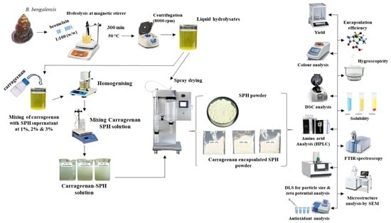
2. Materials and Methods

2.1. Chemicals

The B. bengalensis snails used in the experiment were purchased from a nearby market in Agartala, Tripura, India, and then immersed in filtered water for a period of 48 h to remove any contaminants. Before being frozen at −18 ± 2 °C for additional analysis, the meat was manually separated, rinsed, and analyzed. The microencapsulation agent’s carrageenan and pineapple stem tissue-derived bromelain (0.5 DMC U/mg) were purchased from HiMedia Ltd. (Thane (West), India) and stored in a refrigerator.

2.2. Preparation of Protein Hydrolysate

Snail protein hydrolysates (SPHs) were prepared according to Vaishnav et al. [33], with specific modifications. Figure 1 shows the detailed procedure of SPH preparation. Snail soft tissue was thawed at 27 ± 3 °C, sliced, and ground into a paste. Flash paste was mixed with double distilled water at a 1:1 ratio and homogenized at low speed for 2 min. The mixture was heated to 85 °C for 20 min to inactivate endogenous enzymes and then cooled, and the pH was adjusted to 7 with 0.1 N NaOH/HCl solutions. The material was subsequently agitated via a magnetic stirrer at 50 °C for hydrolysis. Bromelain was added at a 1:100 ratio (w/w) to initiate hydrolysis, and the pH was meticulously controlled. Following hydrolysis, the mixture was heated to 90 °C for 20 min to deactivate bromelain. After centrifugation at 8000 rpm for 10 min, the supernatant was collected and stored at −20 °C for future use.

2.3. Degree of Hydrolysis (DH)

The method outlined by Hoyle and Merritt [34] was employed to assess the degree of hydrolysis (DH). The DH was assessed at intervals of 60, 120, 180, 240, 300, and 360 min. At each time interval, a sample was collected and combined with 20% (1:1 ratio) trichloroacetic acid (TCA) to inactivate the enzymes. The mixture was then filtered, and the soluble protein content in the filtrate was analyzed. The DH was calculated as a ratio of the total protein content in the substrate, which was determined via the Kjeldahl method [35], to the amount of soluble proteins in the filtrate, which was measured via the Lowry method [36].

2.4. Encapsulation of Hydrolysates

Snail protein hydrolysates (SPH) were prepared through enzymatic hydrolysis (as described in the preceding Section 2.2) and subsequently subjected to microencapsulation using carrageenan as the wall material. Carrageenan was mixed with snail protein hydrolysate at concentrations of 1% (CN 1%), 2% (CN 2%), and 3% (CN 3%) (w/v) (as shown in Figure 2) and stirred on a magnetic stirrer (Neuation iStir HP 320, Gandhinagar, India) at 150 rpm for 10–15 min to ensure proper mixing. The mixtures were then spray-dried (SprayMate LSD-48, Jay Instruments & System Pvt. Ltd., Navi Mumbai, India) by pumping them into the spray dryer at a rate of 500 mL/h with a peristaltic pump. An atomizer operating at 1400 rpm was utilized for atomization in a downward flow arrangement, with an inlet temperature of 180 °C and an outflow temperature of 60 ± 3 °C. The resulting powder was separated via a cyclone separator of spray drier and collected in a container. The powdered samples were packed and stored in vacuum-sealed plastic bags composed of multilayer polyethylene (PE) with a total thickness of 100 µm. This type of packaging offers low oxygen and moisture permeability, making it suitable for long-term storage of sensitive bioactive compounds. The sealed samples were stored at −20 °C until further investigation.

2.5. Yield

The yield of the SPH and encapsulated SPH were determined by calculating the ratio of the mass of spray-dried hydrolysate powder to the initial mass of the raw material used for hydrolysis. The yield was expressed as a percentage, following the method described by Elavarasan et al. [37].

2.6. Encapsulation Efficiency (EE)

The encapsulation efficiency of the encapsulated protein hydrolysates was determined via the method described by Mendanha et al. [38]. The encapsulated hydrolysate powder was dissolved in water (5 mg/mL) and centrifuged to separate the nonencapsulated hydrolysates from the encapsulated particles. The amount of nonencapsulated hydrolysates in the supernatant was then quantified via the biuret method. The protein content of the encapsulated hydrolysates was also calculated. The encapsulation efficiency was calculated via the following formula:
EE   % = Total   protein   content Encapsulated   protein   content Total   protein   content × 100

2.7. Hygroscopicity

To determine moisture sorption, the method described by Unnikrishnan et al. [39] was used, and preweighed dry samples were placed in sealed desiccators containing a saturated sodium chloride solution, which maintained a relative humidity of 75.3% at 25 °C. After one week, the samples were removed and reweighed. The hygroscopicity was calculated as the percentage increase in weight due to moisture absorption relative to the initial dry weight of the samples.

2.8. Color Analysis

The color of the snail protein hydrolysates (SPHs) and encapsulated hydrolysates was measured via a colorimeter (ColorFlex, EZ, Hunter Associates Laboratory, Reston, VA, USA). The instrument measured standard color parameters, including lightness (L*), redness (a*), and yellowness (b*). The whiteness and chroma were calculated as described by Zaitounet al. [40].

2.9. Amino Acid Profiling

The amino acid profile of SPH was analyzed via high-resolution mass spectrometry (HRMS) with a Q-TOF mass spectrometer (6200 series TOF/6500 series Q-TOF, Agilent Technologies, Gurgaon, India) using the method described by Yin et al. [41]. A 2-µL aliquot of the sample was analyzed for amino acids, utilizing the amino acid MS method with MassHunter software (Version 12.1) for data acquisition and analysis. During profiling, individual amino acids were identified by their retention times, molecular formulas, and mass—charge ratios (m/z) against a reference database. Mass differences were also calculated to confirm accuracy, and compound identification was assigned a score on the basis of the reliability of the match.

2.10. Particle Size Distribution

The particle size distributions of the spray-dried SPH and encapsulated SPH samples were assessed via dynamic light scattering (Anton Paar, Graz, Austria). The samples were prepared under standardized conditions and evaluated to determine the size distribution and polydispersity index. The measurements were conducted at 25 °C with a scattering angle of 90°, and the particle sizes were measured after 120 s of autocorrelation.

2.11. Zeta Potential Measurements

Zeta potential measurements were conducted with Anton Paar (Austria) equipment to assess the surface charge of the hydrolysate particles. The electrophoretic mobility of the particles was assessed using laser doppler velocimetry technique. Each sample was measured thrice at 20 °C to ensure precision in the acquired zeta potential values.

2.12. Solubility

This method of Klompong et al. [42] was used to measure protein solubility. Two hundred milligrams of spray-dried SPH and encapsulated SPH were dissolved in 20 mL of deionized water. HCl (1 N) and NaOH (1 N) were used to adjust the solution pH to 2, 4, 6, 8, or 10. After 30 min of stirring at room temperature, the mixture was centrifuged at 7500× g for 15 min. The protein content of the supernatant was measured using the Biuret method [43]. The material was diluted in 0.5 N NaOH for protein analysis. Protein solubility was calculated via the following formula:
Solubility   % = Protein   content   in   supernatant Total   protein   content   in   sample   × 100

2.13. Differential Scanning Calorimetry (DSC)

The thermal characteristics of spray-dried SPH and encapsulated SPH were evaluated by differential scanning calorimetry (DSC 214, Polyma, NETZSCH, Selb, Bavaria, Germany). After equilibration at 25 °C in desiccators over saturated salt solutions, aluminum pans with 3 mg of the sample were hermetically sealed. DSC analysis was performed at 10 °C/min from 30 to 350 °C in a nitrogen atmosphere. Indium was used as a standard for instrument calibration.

2.14. Fourier Transform Infrared (FTIR) Spectroscopy

The spectral profiles of spray-dried SPH and encapsulated SPH were analyzed via Fourier transform infrared spectroscopy (Alpha FTIR spectrometer, Bruker, Bremen, Germany) within the range of 4500–400 cm−1. The reflectance spectra were converted to transmission spectra via the Kubelka–Munk algorithm for further interpretation.

2.15. Scanning Electron Microscopy (SEM)

The surface morphology of spray-dried SPH and encapsulated SPH was examined using a Sigma 300, Carl Zeiss scanning electron microscope (Oberkochen, Baden-Württemberg, Germany). A thin gold layer was applied to SEM stubs with carbon tabs to improve the conductivity. Accelerating voltages of 5 kV, 1.00K X and 3.00K X-ray micrographs were taken.

2.16. Bioactivity Property Analysis

2.16.1. DPPH (1,1-diphenyl-2-picrylhydrazyl) Radical-Scavenging Activity

The DPPH radical-scavenging activity of the protein hydrolysates was assessed by mixing the hydrolysate solutions (2, 4, 6, 8 and 10 mg/mL) with 0.1 mM DPPH. After a 30-min dark incubation, the absorbance was measured at 517 nm [44]. The DPPH scavenging activity (%) was calculated using the following formula:
D P P H   a c t i v i t y ( % ) = A c o n t r o l A s a m p l e A c o n t r o l × 100
where Acontrol is the absorbance of the DPPH solution without hydrolysate and Asample denotes the absorbance of the DPPH solution with hydrolysate.

2.16.2. Ferric-Reducing Antioxidant Power (FRAP) Assay

The ferric-reducing antioxidant power (FRAP) of the hydrolysates was assessed following the protocol of Pulido et al. [45] with some modifications. The FRAP reagent was prepared by mixing 25 mL of acetate buffer (pH 3.6), 2.5 mL of 10 mM TPTZ in 40 mM HCl, and 2.5 mL of 20 mM FeCl3·6H2O. A 30 µL aliquot of hydrolysate was combined with 900 µL of the FRAP reagent and incubated at 37 °C for 30 min. The absorbance was then measured at 595 nm. Antioxidant activity was measured via a standard curve generated from iron (II) sulfate solutions and expressed as millimoles of ferrous equivalents per gram of hydrolysate (mM Fe2⁺/g).

2.16.3. Hydroxyl Radical-Scavenging Activity (HRSA)

The hydroxyl radical-scavenging activity of the hydrolysates was evaluated using a modified method from You et al. [46]. The reaction mixture included 1.0 mL of phosphate-buffered saline (0.15 mol/L, pH 7.4), 1.0 mL of 1.0 mM safranin, 0.5 mL of 2.0 mmol/L EDTA-FeSO4, and 1.0 mL of hydrolysate supernatant. After mixing, 1.0 mL of 3% H2O2 was added, and the mixture was incubated at 37 °C for 30 min. The absorbance was measured at 520 nm, and the scavenging activity (%) was calculated via the following formula:
H R S A   a c t i v i t y   ( % ) = A 1 A 0 A 0 × 100
where A1, A2, and A0 represent the absorbances of the supernatant, the mixture without H2O2, and the control, respectively. All the experiments were conducted in triplicate.

2.16.4. ABTS (2,2-azinobis (3-ethylbenzothiazoline-6-sulfonic acid)) Radical-Scavenging Activity

To evaluate the ABTS radical-scavenging activity, an ABTS solution was prepared by combining 7 mM ABTS with 2.45 mM potassium persulfate in equal parts. The solution was then stored in the dark for 16 h. The solution was diluted with distilled water, resulting in an absorbance of 0.70 ± 0.02 at 734 nm. To assess the scavenging ability, 40 µL of hydrolysate was combined with 4 mL of diluted ABTS solution. The mixture was then vortexed for 30 s and allowed to settle for 6 min in the dark. The absorbance was measured at 734 nm [47]. The ABTS radical-scavenging activity (%) was estimated via the following formula:
A B T S   a c t i v i t y   ( % ) = A c o n t r o l A s a m p l e A c o n t r o l × 100
where Acontrol is the absorbance of the ABTS solution without the hydrolysate and Asample denotes the absorbance of the ABTS solution with the hydrolysate.

2.17. Statistical Analysis

The data from three replicates were statistically analyzed using analysis of variance (ANOVA). To identify significant differences among the means, Duncan’s multiple range test was employed, with a significance threshold of 5% (p < 0.05). The statistical analysis was performed using SPSS software (version 23.0 for Windows; SPSS Inc., Chicago, IL, USA).

3. Results and Discussion

3.1. Degree of Hydrolysis (DH)

The extent of protein degradation by bromelain was quantified by determining the DH. The DH values measured at different time intervals are presented in Figure 3. After 300 min, the DH plateaued, indicating no further increase in hydrolysis. After 300 min of enzyme reaction, hydrolysates were made, with a DH of 48.05% from the soft tissue proteins of the snail. This study’s DH value is slightly lower than the DH value reported by Auwal et al. [48] for stonefish protein hydrolysates made with bromelain after 360 min of hydrolysis. Different protein sources may have different amounts of DH because of changes in the structure and number of amino acids or because enzymes may not work properly [49,50]. According to Da Rosa Zavareze et al. [51], hydrolysates can be classified on the basis of DH into three categories, each with specific applications: low-DH hydrolysates improve protein functionality, medium-DH hydrolysates are generally used as flavor enhancers, and high-DH hydrolysates are suitable for nutritional supplements and specialized medical diets. Moreover, Vioque et al. [52] described hydrolyses with DH values above 10% as substantially hydrolyzed, whereas those with DH values less than 10% are regarded as somewhat hydrolyzed. With a DH of 48.05%, the hydrolysates developed in this work are thus highly hydrolyzed and can be used in food items to increase their bioactive qualities and increase their nutritional bioavailability.

3.2. Yield

The yields of SPH and carrageenan-encapsulated SPH were assessed at varying concentrations of wall material (1%, 2%, and 3%). The findings, illustrated in Table 1, demonstrate a significant increase in yield from 19.74% at 1% wall material to 33.46% at 2% wall material (p < 0.05). A minor increase to 35.28% was noted for the 3% wall combination. The yield of SPH without encapsulation was 8.92%, which was substantially lower (p < 0.05) than that of carrageenan-encapsulated SPH. The notable increase in yield (p < 0.05) from 1% to 2% wall material indicates that carrageenan efficiently encapsulates and forms a protective matrix around the protein hydrolysates [53]. The marginal increase in yield at 3% may be attributed to the increased viscosity of the solution, which may impede the encapsulation process [54]. The lower yield of SPH relative to carrageenan-encapsulated SPH may be attributed to differences in protein structure and interactions with the encapsulating agent. Carrageenan’s gelling properties may interact more effectively with the specific amino acid composition of SPH, enhancing encapsulation efficiency [55]. Additionally, SPH was more hygroscopic because of the higher content of charged N- or C-termini. During drying at high temperatures, greater evaporation of water occurred, reducing the moisture of the powders and increasing the capture of water molecules by the samples. When exposed to the environment, the dried powder rapidly assimilated moisture, causing the product to adhere inside the drying chamber [56]. In the presence of carrageenan, the particles were apparently larger than those constituting the soluble SPH.

3.3. Encapsulation Efficiency (EE)

Table 1 shows the encapsulation efficiency of carrageenan-encapsulated SPH at various wall material concentrations (1%, 2%, and 3%). The carrageenan concentration significantly improved the encapsulation efficiency (p < 0.05). The EE for the 1% wall material was 73.31%, which rose to 82.51% at 2% and 84.96% at 3%. These results indicate that increasing the proportion of carrageenan enhances the encapsulation efficiency, with the optimal efficiency achieved at 3%. Increased quantities of carrageenan enhance the encapsulation efficiency by forming a protective matrix surrounding protein hydrolysates. Carrageenan is recognized for its gelling and film-forming characteristics, which presumably enhance the stability and protection of SPH throughout the encapsulation process [57,58]. Recent studies on fish visceral protein hydrolysates indicate that increasing the concentration of wall materials improves encapsulation performance by forming a more cohesive barrier and minimizing the core material’s exposure to outside factors such as moisture and heat [59,60]. However, while the encapsulation efficiency increased with increasing carrageenan concentration, the difference between the 2% and 3% carrageenan concentrations was negligible. These data suggest that the encapsulation efficiency may stagnate over a specific concentration of wall material. Excess carrageenan may also increase the viscosity of the solution, preventing adequate encapsulation and distribution of the protein hydrolysates within the matrix. This behavior has been confirmed in other encapsulation investigations, where excessively viscous fluids might reduce the efficiency of the encapsulation process [61,62].

3.4. Hygroscopicity

Table 1 lists the hygroscopicity of SPH and carrageenan-encapsulated SPH at different concentrations of wall material. The results demonstrated that SPH without encapsulation resulted in a substantially greater hygroscopicity of 26.08%, whereas encapsulation with carrageenan dramatically reduced hygroscopicity across all concentrations of wall material. The hygroscopicity at 1% carrageenan was 16.88%, which decreased to 8.72% at 2% carrageenan and reached a minimum of 7.24% at 3% carrageenan. These findings indicate that carrageenan encapsulation markedly (p < 0.05) diminishes the hygroscopicity of SPH, with the most substantial reduction noted at the maximum wall material concentration of 3%. The encapsulating method formed a barrier around the SPH particles that prevented them from coming into contact with outside moisture. This made them less hygroscopic when the concentration of carrageenan was high. The presence of carrageenan, a hydrocolloid known for its remarkable capacity to retain water and gel, probably aids in the formation of this protective matrix by decreasing the number of exposed protein sites that could interact with water molecules [63]. Comparable findings have been reported in fish oil encapsulated with maltodextrin and gum arabic, which demonstrated reduced hygroscopicity due to the formation of a stable matrix that shields hydrophilic peptide chains [64]. Similarly, encapsulation of chicken meat protein hydrolysates with maltodextrin and gum arabic resulted in a significant decrease in moisture absorption, attributed to the hydrocolloid’s ability to form a dense, moisture-resistant coating [65]. The increased hygroscopicity observed in SPH without encapsulation can be attributed to the presence of charged amino acids and peptide chains that efficiently absorb moisture from the environment, resulting in increased water intake [66,67]. The significant reduction in hygroscopicity resulting from carrageenan encapsulation illustrates the effectiveness of this technique in improving the stability and shelf life of SPH, particularly in powdered formulations. An increase in wall material quantity (3%) may increase the viscosity of the encapsulation solution, perhaps leading to less uniform encapsulation. Nevertheless, the reduction in hygroscopicity at 3% indicates that carrageenan remains an effective barrier against moisture absorption. These findings correspond with prior studies that demonstrated the advantageous effects of carrageenan encapsulation in improving the functional properties of sensitive bioactive compounds [68].

3.5. Color Analysis

The color properties of the snail protein hydrolysate (SPH) and encapsulated SPH with varying concentrations of carrageenan (1%, 2%, and 3%) are presented in Table 1. The color properties of snail protein hydrolysate (SPH) and SPH encapsulated with carrageenan (at 1%, 2%, and 3% concentrations) showed notable variations that could improve its suitability for food applications. Encapsulation progressively increased the lightness (L*) from 89.33 in nonencapsulated SPH to 93.58 in samples with 3% carrageenan, suggesting a brightening effect due to the carrageenan matrix [69]. Both the redness (a*) and yellowness (b*) values decreased with increasing carrageenan concentration, indicating that encapsulation diminishes the red and yellow tones, likely due to the masking effect of carrageenan [70]. The color intensity, measured by chroma (C*), also decreased from 13.81 for the samples without encapsulation to 10.56 for the samples with 3% carrageenan encapsulation. This softening of color was accompanied by an increase in whiteness (W), which rose from 82.54 in nonencapsulated SPH to 87.64 with 3% carrageenan encapsulation, suggesting enhanced lightness and neutrality [71]. These findings show that encapsulated SPH may be advantageous in food products where a lighter and more neutral color is preferred, broadening its application potential.

3.6. Amino Acid Analysis

A total of 17 amino acids were identified in the SPH samples, as listed in Table 2, including seven essential amino acids (EAAs) (histidine, isoleucine, leucine, methionine, phenylalanine, tryptophan, and valine), two delicious amino acids (DAAs) (glutamine and proline), and four antioxidative amino acids (AAAs) (methionine, tyrosine, histidine and tryptophan). The findings revealed variable quantities of 17 amino acids, with histidine having the highest abundance (23.22%), followed by arginine (21.93%) and tyrosine (20.11%). These high quantities of basic and aromatic amino acids suggest a protein hydrolysate with potential nutritional value. Other important amino acids included proline (13.48%) and methionine (7.30%), whereas leucine (0.11%) and isoleucine (0.33%) were found at lower concentrations, indicating variation in essential amino acid distribution. The total percentage of hydrophobic amino acids was calculated to be approximately 24.31%, which is significant and suggests enhanced solubility and bioactivity, especially when these amino acids are included in antioxidant peptides or functional foods. Amino acids with antioxidant properties constitute approximately 52.54% of the total amino acids. This high antioxidative content reveals the resistance of the hydrolysate to oxidative stress, which is essential for cellular health and disease prevention [72]. The substantial presence of antioxidative amino acids, notably histidine (23.22%) and tyrosine (20.11%), suggests that Bellamya bengalensis protein hydrolysates might serve as valuable sources for the development of functional foods with antioxidant properties. Histidine is recognized for its capacity to buffer and chelate metal ions, potentially safeguarding cells from oxidative damage, which is vital in the prevention of aging and chronic diseases [72]. Tyrosine, another antioxidative amino acid, promotes neurotransmitter formation and may improve cognitive function, which contributes to the possible health advantages of this hydrolysate [73]. The presence of sulfur-containing amino acids, such as methionine (7.30%) and cysteine (1.91%), confirms their importance in protein synthesis and detoxification processes, both of which are critical for metabolic health [74]. The high abundance of hydrophobic amino acids (24.31%), particularly proline (13.48%) and methionine, was associated with increased solubility and bioactivity. Hydrophobic amino acids are frequently utilized in the synthesis of bioactive peptides, which can have antioxidant, antihypertensive, and antibacterial characteristics, making the hydrolysate appropriate for use in sports nutrition and medicinal supplements [75]. This is especially important considering the increasing global need for clean-label, bioavailable protein sources.
From a nutritional point of view, the essential amino acid content, which includes lysine, threonine, and methionine, suggests that it could be used as a supplementary protein source, especially in diets with low animal protein intake [76]. However, the low levels of essential amino acids, such as leucine and valine, in the hydrolysate may not be sufficient as a sole protein source. This limitation highlights the necessity of including other protein sources, such as plant-based proteins or dairy, to provide a balanced amino acid profile for human consumption that meets dietary requirements for essential amino acid intake [77]. The increased concentrations of tyrosine and histidine, together with the antioxidant properties of cysteine, increase the ability of hydrolysates to prevent lipid oxidation and microbial spoilage [78]. These results highlight the biofunctional properties of B. bengalensis protein hydrolysates. The presence of essential amino acids such as tyrosine, histidine, and cysteine make these hydrolysates appropriate for functional foods, nutraceuticals, and food preservation. Future studies should focus on isolating certain peptides and their use to optimize their potential.

3.7. Particle Size Distribution and Polydispersity Index (PDI)

The particle size of protein hydrolysates (SPHs) affects their functional properties considerably, including their solubility, dispersion, and bioavailability. In this study, the particle size of spray-dried SPH was analyzed with and without carrageenan as a wall material, and the results are presented in Figure 4. Without encapsulation, the average size of SPH particles was 81.74 nm, suggesting enhanced solubility and bioavailability due to their larger surface area, which supports better dispersion and potential for functional ingredient applications [79,80]. When carrageenan was incorporated as a wall material at concentrations of 1%, 2%, and 3%, the particle sizes of the hydrolysate increased by 55% (127.09 nm), 129% (187.35 nm), and 153% (206.9 nm), respectively. This increase in the particle size confirms the coating effect on the hydrolysate. Furthermore, the increase in particle size may be attributed to the thickening and gelling properties of carrageenan, which promote greater particle formation during spray drying [81]. The polysaccharide likely forms a protective layer around SPH, resulting in more stable particles [82]. However, a larger particle size, while enhancing stability, may reduce solubility and limit SPH functionality in specific applications [83].
Despite the significant differences in particle size, the zeta potential values showed negligible variation across carrageenan concentrations. This is because zeta potential measures surface charge, not physical size. The encapsulated particles retained similar surface electrostatic behavior, indicating that colloidal stability was maintained regardless of particle size differences. Thus, larger sizes are due to matrix thickness, while stable zeta potential reflects consistent charge repulsion [82,84].
The polydispersity index (PDI) values offer insight into particle uniformity, with lower PDI values (<0.4) indicating more homogeneous distributions [85,86]. The PDI of unencapsulated SPH was 0.41, indicating a relatively broad particle size distribution, which may be due to particle aggregation from hydrophobic interactions [87,88]. Encapsulation with 1%, 2% and 3% carrageenan reduced the PDI to 0.23, 0.24 and 0.26, respectively, reflecting more consistent particle sizes and improved stability across all concentrations [89]. The lowest PDI value at 1% carrageenan was attributed to optimal stability at this concentration. However, a slight increase in the PDI at higher concentrations may result from minor particle size variations due to a thicker encapsulating layer [90]. A lower PDI enhances the functional stability of encapsulated bioactive peptides by promoting uniform particle behavior, which is beneficial for applications requiring controlled release [90,91].

3.8. Zeta Potential Measurements

The surface charge of spray-dried SPH and its encapsulated forms with varying concentrations of carrageenan was assessed by measuring the zeta potential, as shown in Figure 5. These parameters provide insights into the stability, particle interactions, and electrokinetic properties, which are critical for applications in food preservation and bioactive delivery systems [92]. The zeta potential of unencapsulated SPH was recorded at −11.8 mV, indicating considerable electrostatic repulsion among the particles. Nonetheless, the use of carrageenan resulted in a remarkable increase in the negative charge. The zeta potential values for SPH containing 1%, 2%, and 3% carrageenan were −25.3, −31.9, and −33.8 mV, respectively. The increase in the negative charge is attributed to the sulfated groups in carrageenan, which provide supplementary negative charges to the system [93]. The elevated negative zeta potential values indicate the enhanced colloidal stability of the encapsulated SPH formulations, which is essential for sustaining dispersion and preventing aggregation in food systems [94]. These findings indicate that carrageenan serves as an excellent encapsulating agent for protein hydrolysates, enhancing their applicability in food preservation and controlled release applications. Subsequent research may investigate the bioactivity and shelf durability of these encapsulated systems in real food matrices.

3.9. Solubility

Table 3 presents the solubilities of spray-dried SPH and its encapsulated variants with carrageenan at various concentrations (1%, 2%, and 3%) throughout a range of pH values (2–10). Solubility is a major functional property of protein hydrolysates, considerably affecting their application in food and nutraceuticals [95]. The solubility of encapsulated hydrolysates is an essential indicator of their efficacy in delivering bioactive chemicals [96]. The solubility of nonencapsulated SPH was generally greater over the pH range, with minor fluctuations. At pH 2, the solubility was 97.32%, increasing to nearly complete solubility at pH 4 (99.97%) and sustaining a high level throughout the range, with a minor decrease to 94.61% at pH 10. These data demonstrate that SPH has exceptional solubility across both acidic and alkaline pH ranges. Encapsulation with carrageenan at different concentrations resulted in reduced solubility across all pH ranges. At 1% carrageenan, the solubility ranged from 72.13% at pH 2 to 70.87% at pH 10. With 2% carrageenan, the solubility ranged from 58.31% at pH 2 to 51.76% at pH 10. Finally, at 3% carrageenan, the solubility was the lowest, ranging from 43.28% at pH 2 to 41.10% at pH 10. The results indicate that the solubility of SPH is exceptionally high, particularly in acidic environments (pH 2–4), which is consistent with prior findings indicating that protein hydrolysates generally exhibit good solubility because of their relatively small peptide sizes and the presence of polar amino acid residues [19,97]. This high solubility across a broad pH range makes SPH a suitable ingredient for inclusion in various food matrices, where pH conditions may vary.
The encapsulation of SPH with carrageenan significantly reduced the solubility, with elevated concentrations of carrageenan (CN 3%) yielding the most substantial reduction. At pH 2, the solubility decreased from 97.32% for SPH to 43.28% for CN 3%. The reduction in solubility is probably attributable to the interaction between the carrageenan matrix and the protein hydrolysates, which may restrict the exposure of the peptides to the aqueous environment and decrease their solubility [98]. Despite this reduction, encapsulation could still be beneficial, mainly in applications where controlled release or protection of bioactive peptides is desired. A lower carrageenan concentration (1%) resulted in better solubility, suggesting that a balance between encapsulation and maintaining functional properties such as solubility can be achieved by optimizing the carrageenan content. However, the decreased solubility at higher carrageenan concentrations may limit the effectiveness of these encapsulated hydrolysates in certain food applications, especially in products requiring high solubility.

3.10. Differential Scanning Calorimetry (DSC)

The DSC thermograms for spray-dried SPH and its encapsulated forms with 1%, 2%, and 3% carrageenan are shown in Figure 6. DSC analysis provides insight into the thermal transitions and stability of samples, which is essential for understanding their encapsulation effects and potential applications in food preservation [99]. The DSC thermogram of SPH without encapsulation shows an endothermic peak near 60 °C, indicating the denaturation or thermal transition of protein components [100]. This is a common feature in protein hydrolysates due to the unfolding of peptide chains when subjected to heat. Beyond this temperature, the DSC curve gradually increases to approximately 325 °C, indicating the thermal degradation of the protein structure, which is characteristic of the breakdown of organic molecules into smaller volatile compounds [101].
The thermal profile with 1% carrageenan encapsulation showed a minor alteration in the initial endothermic transition at 65 °C. This transition indicates improved thermal stability attributable to the carrageenan matrix, which protects the protein hydrolysates. The widening of the transition indicates enhanced contact between the protein and the encapsulating agent, probably via hydrogen bonding or electrostatic interactions [102]. The delayed initiation of thermal degradation indicates enhanced resistance to heat-induced denaturation. The 2% carrageenan-encapsulated SPH sample exhibited a significant thermal transition at approximately 70 °C, indicating the stabilizing influence of the increased carrageenan concentration. An elevated concentration of carrageenan is expected to improve the encapsulation efficiency, offering enhanced protection against thermal denaturation [103]. A sharper peak indicates a more distinct thermal transition, perhaps resulting from the enhanced development of a protective matrix surrounding the protein hydrolysates. Above 250 °C, the degradation slope is less pronounced than that of the unencapsulated SPH, indicating enhanced thermal stability. The 3% carrageenan-encapsulated SPH mixture has a similar DSC curve with a stronger thermal transition peak at approximately 75 °C, indicating that it is the most thermally stable sample of the encapsulated formulations. The substantial increase in the endothermic peak signifies that the encapsulating matrix at this concentration is particularly efficient in stabilizing the protein hydrolysates against thermal degradation. Moreover, the initiation of thermal degradation was postponed relative to that of the lower-concentration samples, indicating that elevated carrageenan concentrations offer improved protection. Moreover, the extensive degradation profile above 300 °C indicates that carrageenan at elevated concentrations may undergo thermal degradation, thus affecting the overall thermal dynamics of the system [101].

3.11. Fourier Transform Infrared (FTIR) Spectroscopy

FTIR spectroscopy was used to evaluate the structural interactions and molecular characteristics of the encapsulated SPH at various concentrations (1%, 2%, and 3%) of carrageenan (CN). The spectra displayed in Figure 7 and Table 4 show significant peaks corresponding to various functional groups, which provide insights into the encapsulation process and the interaction between the protein hydrolysates and the encapsulating agent.
The FTIR spectra of SPH (black line) exhibited significant absorption bands at 3265.46 cm−1, 1644.50 cm−1, 1548.15 cm−1, 1407.77 cm−1, 1249.72 cm−1, 1031.29 cm−1, and 771.45 cm−1. The prominent peak at approximately 3265.46 cm⁻1 is attributed to the stretching vibrations of O–H and N–H bonds, indicating the presence of free hydroxyl and amine groups, which are characteristic of proteinaceous materials [104]. The amide I and amide II bands are detected at 1644.50 cm−1 and 1548.15 cm−1, respectively, corresponding to the C=O stretching and N-H bending vibrations of peptide bonds, confirming the presence of α-helices and β-sheets [105,106]. The peak at 1407.77 cm−1 is attributed to CH2 bending, whereas the peak at 1249.72 cm−1 is attributed to the amide III band, which is connected with N–H bending and C–N stretching. The peaks at 1031.29 cm−1 and 771.45 cm−1 signify C–O stretching and other secondary structures in the protein hydrolysates [107]. In the FTIR spectrum of samples encapsulated with 1% carrageenan (red line), the characteristic peaks of SPH are retained but show noticeable shifts, indicating interactions between SPH and carrageenan. The O–H and N–H stretching peaks slightly shifted to 3217.39 cm−1, indicating hydrogen bonding interactions between the protein hydrolysate and carrageenan. The amide I and amide II bands are observed at 1635.81 cm−1 and 1560.71 cm−1, showing a minor shift from the SPH spectrum, which suggests some encapsulation effects on the protein’s secondary structure [108]. Another peak at 702.24 cm−1 is attributed to the sulfate group (S=O stretching) of carrageenan, confirming its incorporation into the matrix [109]. For 2% CN (blue line), the FTIR spectrum shows further shifts in the key absorption bands. The O–H/N–H stretching band appears at 3285.15 cm−1, which is slightly similar to that of CN 1%, indicating continued hydrogen bonding between carrageenan and SPH. The amide I and amide II bands are observed at 1637.05 cm−1 and 1403.97 cm−1, suggesting that the secondary structure of SPH [108] is further affected by the increased carrageenan concentration. A notable peak at 620.64 cm−1 was observed, which may also be associated with the sulfate groups of carrageenan [109]. The additional sulfate peak and the shifting of the amide bands confirmed the increased interaction between carrageenan and the protein hydrolysates at this concentration. The FTIR spectrum of CN 3% (green line) shows more pronounced shifts in the O–H/N–H stretching region, with a peak at 3319.92 cm−1, which is a significant shift compared with both the SPH and the lower carrageenan concentrations. This suggests stronger hydrogen bonding and more extensive encapsulation interactions at the highest concentration of carrageenan. The amide I and II bands shifted to 1583.86 cm⁻1 and 1411.07 cm⁻1, respectively, indicating more significant conformational changes in the protein hydrolysate secondary structure due to encapsulation [110]. The peaks at 924.81 cm⁻1, 847.05 cm⁻1, and 702.42 cm⁻1 further confirmed the presence of carrageenan, with the sulfate group peaks becoming more pronounced as the concentration increased [109].
FTIR examination validated the effective encapsulation of SPH by carrageenan, as shown by shifts in the O–H/N–H and amide I/II bands, signifying hydrogen bonding and structural alterations. Elevated CN concentration exacerbated these changes and introduced sulphate group peaks, underscoring enhanced interactions. The 3% CN formulation exhibited the most significant spectrum alterations, indicating improved encapsulation efficiency. These findings endorse the efficacy of carrageenan as a viable encapsulating agent for protein hydrolysates in functional food applications.

3.12. Scanning Electron Microscopy (SEM) Analysis

SEM analysis of spray-dried SPH, both unencapsulated and encapsulated with 1%, 2%, and 3% carrageenan, as shown in Figure 8, revealed a significant variation in particle morphology, providing insights into the efficiency and characteristics of the encapsulation process. For the unencapsulated SPH particles, SEM images at 1.0 KX and 3.0 KX magnifications reveal a heterogeneous surface morphology, with a mix of spherical and irregularly shaped particles ranging from 2 to 10 microns. These spherical particles typically exhibit smooth surfaces, although some surface irregularities, cracks, and pores are apparent, likely due to the spray-drying process [111]. These morphological characteristics align with previous findings on spray drying, which reported atomization-driven formation of spherical particles with variable sizes [112]. Surface irregularities could indicate structural changes during drying, potentially impacting solubility and functional properties [113]. In contrast, encapsulated SPH with 1% carrageenan resulted in smoother and more uniform spherical particles, with fewer surface irregularities, suggesting successful encapsulation that formed a cohesive carrageenan matrix around the hydrolysates. Small pores visible at higher magnification may facilitate controlled release, supporting the effectiveness of carrageenan as an encapsulation material for enhancing stability and protecting bioactive compounds [114,115]. Increasing the carrageenan concentration to 2% and 3% further impacted the particle morphology, with SEM images at 1.0 KX and 3.0 KX showing denser, more compact particles, reduced porosity and smoother surfaces. This enhanced uniformity and stability likely contribute to prolonged release and increased encapsulation efficiency, which are crucial for applications in functional foods and nutraceuticals requiring sustained release [116,117].

3.13. Bioactivity Property Analysis

3.13.1. DPPH (1,1-diphenyl-2-picrylhydrazyl) Radical-Scavenging Activity

Figure 9 shows the DPPH radical-scavenging activity of SPH and encapsulated SPH using carrageenan at three different concentrations (1%, 2%, and 3%). The DPPH radical-scavenging activity of SPH and carrageenan-encapsulated SPH clearly increased in a dose-dependent manner across all the treatments, highlighting the strong antioxidant potential of SPH. For nonencapsulated SPH, the scavenging activity increased steadily from 54.64% at 2 mg/mL to 94.11% at 10 mg/mL, corroborating findings from previous studies on Skipjack tuna and Nile tilapia protein hydrolysates which exhibit increased radical scavenging capacity with increasing concentration [118,119]. The dose-dependent nature of antioxidant activity can be attributed to the increased availability of peptides that donate protons or electrons to neutralize free radicals as the concentration increases [120].
SPH encapsulated with carrageenan also exhibited significant radical-scavenging activity, although it was slightly lower than that of nonencapsulated SPH. At 10 mg/mL, the scavenging activity for 1% carrageenan (CN 1%) was 88.46%, that for 2% carrageenan (CN 2%) was 81.53%, and that for 3% carrageenan (CN 3%) was 80.03%, suggesting that encapsulation at higher carrageenan concentrations modestly reduces the antioxidant efficacy. This decrease is due to the interaction between carrageenan and the bioactive peptides in SPH, since polysaccharides such as carrageenan can partially encapsulate or protect the active regions of peptides, limiting their accessibility to DPPH radicals [121]. Furthermore, prior studies have demonstrated that encapsulation with a relatively high carrageenan concentration slightly slows the release of bioactive chemicals by forming a relatively complex, dense matrix [122]. However, the encapsulated SPH still exhibited significant scavenging activity, indicating that encapsulation does not significantly reduce bioactivity.
Despite the minor reduction in antioxidant efficiency, the encapsulation technique has significant advantages. Encapsulation with carrageenan may increase the stability of SPH by preserving the peptides from oxidation and degradation, making it more useful in applications requiring long-term antioxidant action [91]. Carrageenan, a natural polysaccharide, has also been found to improve the stability and bioavailability of active compounds, thereby supporting the functional use of encapsulated SPH in food preservation or nutraceutical formulations [69].

3.13.2. Ferric-Reducing Antioxidant Power (FRAP) Assay

Figure 10 depicts the ferric-reducing antioxidant power activity of SPH and its encapsulated forms at concentrations of 1%, 2%, and 3% carrageenan. The concentration of nonencapsulated SPH was substantially related to the FRAP activity, with values ranging from 57.08 mM Fe(II)/g at 2 mg/mL to 95.26 mM Fe(II)/g at 10 mg/mL. This pattern is consistent with prior studies, which determined that the antioxidant potential of protein hydrolysates from common carp and ray finned fish increases with increasing concentrations due to the presence of peptides that serve as electron donors, converting ferric ions to ferrous forms [123]. The linear increase in FRAP values suggests that SPH has strong electron-donating capabilities, most likely due to the bioactive peptides produced during hydrolysis, which have been demonstrated to significantly contribute to reducing power and consequently antioxidant capability [124,125]. High FRAP values are associated with the presence of particular amino acids, such as valine, leucine, proline, histidine, and tyrosine, which play important roles in increasing antioxidant activity when combined into peptide sequences. Among these, tyrosine is an excellent electron donor that efficiently captures electrons to prevent the oxidation of other molecules by reactive chemicals [126,127].
SPH encapsulated with carrageenan at 1%, 2%, and 3% concentrations showed a steady increase in FRAP activity as the SPH concentration increased, while the values were slightly lower than those of nonencapsulated SPH across all carrageenan concentrations. At 10 mg/mL SPH, the FRAP activity was 91.39 mM Fe(II)/g for 1% carrageenan, 83.84 mM Fe(II)/g for 2% carrageenan, and 83.47 mM Fe(II)/g for 3% carrageenan. This slight reduction in FRAP activity for the encapsulated samples could be attributed to the presence of carrageenan, which can partially inhibit the interaction between SPH peptides and ferric ions [69]. As the concentration of carrageenan increases, the encapsulation matrix may inhibit peptide diffusion, restricting the ability of the peptides to act as electron donors in the ferric ion reduction process [68]. Nasri et al. [1] reported that at higher biopolymer concentrations, the encapsulating matrix can become denser, limiting the diffusion of antioxidant molecules from within the encapsulation. This reduced release can lead to a decrease in the antioxidant’s immediate availability and overall activity, as the bioactive compounds are less accessible for interaction with target molecules.

3.13.3. Hydroxyl Radical-Scavenging Activity

Figure 11 shows the hydroxyl radical-scavenging activity of SPH and its encapsulated forms with carrageenan at different concentrations. The results of the hydroxyl radical-scavenging activity of SPH and its carrageenan-encapsulated derivatives indicated a positive relationship between SPH concentration and antioxidant potential. Hydroxyl radicals are the most reactive oxygen species, producing significant oxidative damage by damaging biomolecules such as lipids, proteins, and DNA [128,129]. The capacity of these protein hydrolysates to neutralize hydroxyl radicals suggests a potential role in reducing oxidative stress and its negative effects [130,131]. The nonencapsulated SPH exhibited a significant increase in scavenging activity with increasing SPH concentration, ranging from 28.37% at 2 mg/mL to 64.61% at 10 mg/mL. This trend is consistent with previous studies indicating that higher concentrations of protein hydrolysates improve antioxidant capacity, likely due to an increased number of bioactive peptides capable of interacting with and neutralizing reactive oxygen species (ROS) [11,132]. Bioactive peptides, particularly those containing amino acids such as tyrosine, histidine, and cysteine, have been shown to act as hydrogen donors, thereby stabilizing free radicals and reducing oxidative damage [133].
The encapsulated SPH also tended to increase in hydroxyl radical-scavenging activity as the SPH concentration increased across all carrageenan concentrations (1%, 2%, and 3%). However, the encapsulated forms generally exhibited lower activity than nonencapsulated SPH did, likely because the encapsulation matrix partially restricted the release and accessibility of the active peptides [98]. At 1% carrageenan, the scavenging activity increased from 23.16% at 2 mg/mL to 60.67% at 10 mg/mL, whereas at 2% and 3% carrageenan, the scavenging activity ranged from 18.79% to 48.7% and 17.62% to 46.83%, respectively. This reduction in scavenging efficiency at higher carrageenan concentrations is likely due to the denser biopolymer matrix, which can impede the release and diffusion of peptides, thereby limiting their interaction with hydroxyl radicals [1]. Higher carrageenan concentrations increase the matrix density, thereby reducing the bioavailability of peptides within the hydrolysates, as observed in other biopolymer encapsulation systems [134]. Polysaccharide-based encapsulation has been shown to protect peptides from degradation, thereby increasing their functional longevity. Thus, while encapsulation might reduce immediate antioxidant activity, it provides potential advantages for long-term applications in food preservation or nutraceutical formulations, where a controlled release of antioxidants is beneficial [135].

3.13.4. ABTS (2,2-azinobis (3-ethylbenzothiazoline-6-sulfonic acid)) Activity

Figure 12 shows the ABTS radical-scavenging activity of SPH and its encapsulated forms at various carrageenan concentrations (1%, 2%, and 3%). The ABTS radical-scavenging activity quantifies the antioxidant capability, with elevated percentages indicating enhanced scavenging of ABTS radicals [136]. The ABTS radical-scavenging activity of nonencapsulated and encapsulated SPH increased from 2 mg/mL to 10 mg/mL. At 2 mg/mL, the nonencapsulated SPH demonstrated 43.42% scavenging activity, which progressively increased to 93.92% at 10 mg/mL (Figure 1). This increase indicates that the antioxidant capacity of SPH is dependent on concentration, which is consistent with findings from similar studies where elevated concentrations of protein hydrolysates led to increased radical-scavenging activity [123]. The results demonstrate that SPH comprises peptides that may donate electrons, enabling them to interact with free radicals and transform them into more stable molecules, effectively ending the radical chain reaction [137]. Compared with the nonencapsulated SPH process, the encapsulation process led to slightly reduced activity. For samples encapsulated with 1% carrageenan, the ABTS scavenging activity ranged from 41.67% at 2 mg/mL to 90.09% at 10 mg/mL, demonstrating relatively less activity than SPH without encapsulation. Similarly, SPH encapsulated with 2% carrageenan revealed a decrease in scavenging activity from 34.52% at 2 mg/mL to 81.51% at 10 mg/mL, whereas samples encapsulated with 3% carrageenan demonstrated an activity range of 31.96% to 79.55%. These data indicate that while encapsulation did not fully suppress the antioxidant potential of SPH, it lowered the overall scavenging capacity, which may be attributed to the physical barriers established by the carrageenan matrix, which restricts the accessibility of SPH to ABTS radicals [1,69]. The encapsulation of bioactive compounds is frequently employed to protect sensitive compounds, regulate their release, and improve their stability. However, the results of the present study suggest that their functional properties, including their antioxidant activity, may be marginally limited. The decrease in activity could result from the reduced solubility or mobility of the encapsulated protein hydrolysates within the carrageenan matrix. The encapsulated SPH demonstrated considerable antioxidant ability, making it a potential candidate for use in controlled delivery systems for food preservation or therapeutic applications.
Although the in vitro antioxidant activity of encapsulated SPH is encouraging, its practical effectiveness in food systems or gastrointestinal environments requires more investigation. Food matrices may affect the release kinetics and interaction of SPH with reactive oxygen species, thereby altering bioactivity [138]. Furthermore, simulated digestion models indicate that encapsulated peptides can demonstrate delayed yet prolonged antioxidant action during gastrointestinal transit due to regulated release mechanisms [17,139]. Consequently, forthcoming in vivo or simulated gastrointestinal investigations are essential to confirm the functional applicability of encapsulated SPH in authentic dietary and physiological contexts.

4. Conclusions

The protein hydrolysates prepared from Bellamya bengalensis exhibited increased yield, stability, and antioxidant potential through microencapsulation assisted by carrageenan. Encapsulation enhances structural integrity and reduces hygroscopicity, a major factor for maintaining bioactivity and functionality in food applications. Increasing the carrageenan concentration to 3% substantially stabilized the encapsulated SPH, as evidenced by larger particle sizes, reduced polydispersity, and increased zeta potential, contributing to better shelf stability. Although the antioxidant activity slightly decreased with increasing concentrations of carrageenan, significant bioactivity retention by SPH encapsulated in this polysaccharide shows great promise for controlled-release applications in food preservation and nutraceuticals. Our findings indicate that carrageenan is an excellent natural microencapsulating agent that enhances the stability and functional profile of snail protein hydrolysates, thus enabling their wider application as a bioactive ingredient within different food technologies.
While carrageenan effectively enhanced the stability and functionality of SPH, its practical applicability may be restricted by a modest loss in antioxidant activity at higher concentration. However, the longer shelf life, structural protection, and ability for controlled release make carrageenan-encapsulated SPH a viable component for functional foods and nutraceuticals. Further studies optimizing encapsulation conditions could help maximize both stability and bioactivity.

Author Contributions

Writing—original draft preparation, investigation, and formal analysis, A.V.; conceptualization, manuscript editing, supervision, and visualization, N.K.M.; writing—review and editing and supervision, M.B.P.; writing—review and editing, S.K.S.; formal analysis, P.C.A., S.B., H.N. and S.A.H.; data curation, P.P.; investigation, J.L., N.S.S. and B.K.P. All authors have read and agreed to the published version of the manuscript.

Funding

This research received no external funding.

Institutional Review Board Statement

The research conducted in this study involved the use of freshwater snails, which are commonly consumed as food in the northeastern regions of India. As these animals are considered edible and are regularly harvested for human consumption, no specific ethical permission was required. The study adhered to all applicable local and institutional guidelines for the ethical use of biological materials.

Informed Consent Statement

Not applicable.

Data Availability Statement

The data that support the findings of this study are available from the corresponding author upon reasonable request.

Acknowledgments

The authors are thankful to the Dean of the College of Fisheries, Central Agricultural University (Imphal), Lembucherra, Tripura, India, for providing the facilities necessary to carry out this research. The authors are also greatly thankful for the chemical support received from the Department of Biotechnology, Government of India, under the Centre of Excellence project (DBT-NER/LIVS/05/2011 Phase II), Central Agricultural University, Imphal, Manipur, India. The authors also acknowledge the Drug Metabolomics Laboratory, Department of Pharmacy, Tripura University (A Central University), for providing the necessary facilities and support to carry out this study.

Conflicts of Interest

The authors declare no conflicts of interest.

References

  1. Nasri, R.; Hamdi, M.; Touir, S.; Li, S.; Karra-Chaâbouni, M.; Nasri, M. Development of delivery system based on marine chitosan: Encapsulation and release kinetic study of antioxidant peptides from chitosan microparticle. Int. J. Biol. Macromol. 2021, 167, 1445–1451. [Google Scholar] [CrossRef] [PubMed]
  2. Senadheera, T.R.; Hossain, A.; Shahidi, F. Marine bioactives and their application in the food industry: A review. Appl. Sci. 2023, 13, 12088. [Google Scholar] [CrossRef]
  3. Vásquez, P.; Cian, R.E.; Drago, S.R. Marine Bioactive Peptides (Fishes, Algae, Cephalopods, Molluscs, and Crustaceans). In Handbook of Food Bioactive Ingredients; Jafari, S.M., Rashidinejad, A., Simal-Gandara, J., Eds.; Springer: Cham, Switzerland, 2023; pp. 1–16. [Google Scholar] [CrossRef]
  4. Bar, A. Bellamya bengalensis: A review on its Ecological importance, Nutritional values & Ethnomedicinal importance. Eur. J. Pharm. Med. Res. 2020, 7, 315–319. [Google Scholar]
  5. Chakraborty, D.; Mukherjee, M.; Maity, J. Ethnomedicinal importance of viviparous gastropod, Bellamya bengalensis (Lamarck, 1822) in terms of estimation of its proximate protein and amino acid composition. Int. J. Sci. Res. 2015, 3, 245–248. [Google Scholar]
  6. Gupta, A.; Khanal, P. The potential of snails as a source of food and feed. J. Agric. Food Res. 2024, 18, 101330. [Google Scholar] [CrossRef]
  7. Pissia, M.A.; Matsakidou, A.; Kiosseoglou, V. Raw materials from snails for food preparation. Future Foods 2021, 3, 100034. [Google Scholar] [CrossRef]
  8. Tacias-Pascacio, V.G.; Castañeda-Valbuena, D.; Morellon-Sterling, R.; Tavano, O.; Berenguer-Murcia, Á.; Vela-Gutiérrez, G.; Fernandez-Lafuente, R. Bioactive peptides from fisheries residues: A review of use of papain in proteolysis reactions. Int. J. Biol. Macromol. 2021, 184, 415–428. [Google Scholar] [CrossRef]
  9. Khalua, R.K.; Tripathy, S.; Paul, B.; Bairy, D. Seasonal variation of carbohydrate, protein and lipid of common freshwater edible gastropod (Bellamya bengalensis) of Medinipur district, West Bengal. Res. J. Biol. 2014, 2, 49–52. [Google Scholar]
  10. Debnath, C.; Sahoo, L.; Haldar, A.; Datta, M.; Yadav, G.S.; Singha, A.; Bhattacharjee, J. Proximate and mineral composition of freshwater snails of Tripura, North–East India. Fish. Technol. 2016, 53, 307–312. [Google Scholar]
  11. Chatterjee, R.; Dey, T.K.; Roychoudhury, A.; Paul, D.; Dhar, P. Enzymatically excised oligopeptides from Bellamya bengalensis show potent antioxidative and anti-hypertensive activity. J. Food Sci. Technol. 2020, 57, 2586–2601. [Google Scholar] [CrossRef] [PubMed]
  12. Mondal, B.M. Bio-Chemistry and Nutritional health benefits of Bellamya bengalensis (Edible Gastropod): A Review. Afr. J. Bio. Sci. 2023, 5, 112–118. [Google Scholar]
  13. Baghele, M.; Mishra, S.; Meyer-Rochow, V.B.; Jung, C.; Ghosh, S. A review of the nutritional potential of edible snails: A sustainable underutilized food resource. Int. J. Nat. Prod. Res. 2022, 13, 47930. [Google Scholar] [CrossRef]
  14. Hayes, M.; Mora, L. Alternative Proteins as a Source of Bioactive Peptides: The Edible Snail and Generation of Hydrolysates Containing Peptides with Bioactive Potential for Use as Functional Foods. Foods 2021, 10, 276. [Google Scholar] [CrossRef]
  15. Ortizo, R.G.G.; Sharma, V.; Tsai, M.L.; Wang, J.X.; Sun, P.P.; Nargotra, P.; Dong, C.D. Extraction of novel bioactive peptides from fish protein hydrolysates by enzymatic reactions. Appl. Sci. 2023, 13, 5768. [Google Scholar] [CrossRef]
  16. Wu, Y.H.S.; Chen, Y.C. Trends and applications of food protein-origin hydrolysates and bioactive peptides. J. Food Drug Anal. 2022, 30, 172. [Google Scholar] [CrossRef]
  17. Amigo, L.; Hernández-Ledesma, B. Current evidence on the bioavailability of food bioactive peptides. Molecules 2020, 25, 4479. [Google Scholar] [CrossRef]
  18. Aguilar-Toalá, J.E.; Quintanar-Guerrero, D.; Liceaga, A.M.; Zambrano-Zaragoza, M.L. Encapsulation of bioactive peptides: A strategy to improve the stability, protect the nutraceutical bioactivity and support their food applications. RSC Adv. 2022, 12, 6449–6458. [Google Scholar] [CrossRef]
  19. Peighambardoust, S.H.; Karami, Z.; Pateiro, M.; Lorenzo, J.M. A review on health-promoting, biological, and functional aspects of bioactive peptides in food applications. Biomolecules 2021, 11, 631. [Google Scholar] [CrossRef]
  20. Mazloomi, N.; Safari, B.; Karaca, A.C.; Karimzadeh, L.; Moghadasi, S.; Ghanbari, M.; Assadpour, E.; Sarabandi, K.; Jafari, S.M. Loading bioactive peptides within different nanocarriers to enhance their functionality and bioavailability; in vitro and in vivo studies. Adv. Colloid Interface Sci. 2024, 334, 103318. [Google Scholar] [CrossRef]
  21. Akbarbaglu, Z.; Peighambardoust, S.H.; Sarabandi, K.; Jafari, S.M. Spray drying encapsulation of bioactive compounds within protein-based carriers; different options and applications. Food Chem. 2021, 359, 129965. [Google Scholar] [CrossRef] [PubMed]
  22. Culas, M.S.; Popovich, D.G.; Rashidinejad, A. Recent advances in encapsulation techniques for cinnamon bioactive compounds: A review on stability, effectiveness, and potential applications. Food Biosci. 2023, 57, 103470. [Google Scholar] [CrossRef]
  23. Vaishnav, A.; Lal, J.; Mehta, N.K.; Mohanty, S.; Yadav, K.K.; Priyadarshini, M.B.; Singh, S.K. Unlocking the Potential of Fishery Waste: Exploring Diverse Applications of Fish Protein Hydrolysates in Food and Nonfood Sectors. Environ. Sci. Pollut. Res. 2025, 22, 1–45. [Google Scholar] [CrossRef] [PubMed]
  24. Askari Vaselabadi, S.; Gharibzahedi, S.M.T.; Greiner, R.; Vale, J.M.; Ovenseri, A.C.; Rashidinejad, A.; Roohinejad, S. Advancements in Spray-Drying for the Microencapsulation of Fat-Soluble Vitamins: Stability, Bioavailability, and Applications. J. Food Biochem. 2025, 2025, 9974476. [Google Scholar] [CrossRef]
  25. Baltrusch, K.L.; Torres, M.D.; Domínguez, H.; Flórez-Fernández, N. Spray-drying microencapsulation of tea extracts using green starch, alginate or carrageenan as carrier materials. Int. J. Biol. Macromol. 2022, 203, 417–429. [Google Scholar] [CrossRef]
  26. Furuta, T.; Neoh, T.L. Microencapsulation of food bioactive components by spray drying: A review. Dry. Technol. 2021, 39, 1800–1831. [Google Scholar] [CrossRef]
  27. Usov, A.I. Polysaccharides of the red algae. In Advances in Carbohydrate Chemistry and Biochemistry; Academic Press: Cambridge, MA, USA, 2011; Volume 65, pp. 115–217. [Google Scholar] [CrossRef]
  28. Lomartire, S.; Gonçalves, A.M. Novel technologies for seaweed polysaccharides extraction and their use in food with therapeutically applications—A review. Foods 2022, 11, 2654. [Google Scholar] [CrossRef]
  29. Inguglia, E.S.; Song, Z.; Kerry, J.P.; O’Sullivan, M.G.; Hamill, R.M. Addressing clean label trends in commercial meat processing: Strategies, challenges and insights from consumer perspectives. Foods 2023, 12, 2062. [Google Scholar] [CrossRef]
  30. Jafari, S.; Jafari, S.M.; Ebrahimi, M.; Kijpatanasilp, I.; Assatarakul, K. A decade overview and prospect of spray drying encapsulation of bioactives from fruit products: Characterization, food application and in vitro gastrointestinal digestion. Food Hydrocoll. 2023, 134, 108068. [Google Scholar] [CrossRef]
  31. Chakraborty, S. Carrageenan for encapsulation and immobilization of flavour, fragrance, probiotics, and enzymes: A review. J. Carbohydr. Chem. 2017, 36, 1–19. [Google Scholar] [CrossRef]
  32. Mardani, M.; Siahtiri, S.; Besati, M.; Baghani, M.; Baniassadi, M.; Nejad, A.M. Microencapsulation of natural products using spray drying; an overview. J. Microencapsul. 2024, 41, 649–678. [Google Scholar] [CrossRef] [PubMed]
  33. Vaishnav, A.; Mehta, N.K.; Hussain, S.A.; Acharya, P.C.; Biswal, S.; Nath, H.; Priyadarshini, M.B.; Thangarani, A.J.; Pal, P.; Singh, S.K.; et al. Bromelain excised hydrolysates with potent bioactivity from Bellamya bengalensis soft tissues: Process optimization and characterization. J. Agric. Food Res. 2025, 17, 101595. [Google Scholar] [CrossRef]
  34. Hoyle, N.T.; Merritt, J.H. Quality of fish protein hydrolysates from herring (Clupea harengus). J. Food Sci. 1994, 59, 76–79. [Google Scholar] [CrossRef]
  35. AOAC. Official Method of Analysis of the AOAC International, 20th ed.; Association of Official and Analytical Chemists International: Washington, VA, USA, 2016. [Google Scholar]
  36. Lowry, O.H.; Rosebrough, N.J.; Farr, A.L.; Randall, R.J. Protein measurement with the Folin phenol reagent. J. Biol. Chem. 1951, 193, 265–275. [Google Scholar] [CrossRef]
  37. Elavarasan, K.; Naveen Kumar, V.; Shamasundar, B.A. Antioxidant and functional properties of fish protein hydrolysates from fresh water carp (Catla catla) as influenced by the Nature of Enzyme. J. Food Process. Preserv. 2014, 38, 1207–1214. [Google Scholar] [CrossRef]
  38. Mendanha, D.V.; Ortiz, S.E.M.; Favaro-Trindade, C.S.; Mauri, A.; Monterrey-Quintero, E.S.; Thomazini, M. Microencapsulation of casein hydrolysate by complex coacervation with SPI/pectin. Food Res. Int. 2009, 42, 1099–1104. [Google Scholar] [CrossRef]
  39. Unnikrishnan, P.; Kizhakkethil, B.P.; George, J.C.; Aliyamveetil Abubacker, Z.; Ninan, G.; Nagarajarao, R.C. Antioxidant peptides from dark meat of yellowfin tuna (Thunnus albacares): Process optimization and characterization. Waste Biomass Valorization 2021, 12, 1845–1860. [Google Scholar] [CrossRef]
  40. Zaitoun, B.J.; Palmer, N.; Amamcharla, J.K. Characterization of a Commercial Whey Protein Hydrolysate and Its Use as a Binding Agent in the Whey Protein Isolate Agglomeration Process. Foods 2022, 11, 1797. [Google Scholar] [CrossRef]
  41. Yin, B.; Li, T.; Zhang, S.; Li, Z.; He, P. Sensitive analysis of 33 free amino acids in serum, milk, and muscle by ultrahigh-performance liquid chromatography-quadrupole-orbitrap high resolution mass spectrometry. Food Anal. Methods 2016, 9, 2814–2823. [Google Scholar] [CrossRef]
  42. Klompong, V.; Benjakul, S.; Kantachote, D.; Shahidi, F. Antioxidative activity and functional properties of protein hydrolysate of yellow stripe trevally (Selaroides leptolepis) as influenced by the degree of hydrolysis and enzyme type. Food Chem. 2007, 102, 1317–1327. [Google Scholar] [CrossRef]
  43. Gaurav, K.; Mehta, N.K.; Priyadarshini, M.B.; Pal, P.; Nirmal, N.P.; Sharma, S.; Vaishnav, A. Fat Uptake Reduction During Deep-Fat Frying of Fish Fillets Coated with Soy Protein Isolate Edible Coating. J. Aquat. Food Prod. Technol. 2024, 33, 573–588. [Google Scholar] [CrossRef]
  44. Mohammadi, M.; Hamishehkar, H.; McClements, D.J.; Shahvalizadeh, R.; Barri, A. Encapsulation of Spirulina protein hydrolysates in liposomes: Impact on antioxidant activity and gastrointestinal behavior. Food Chem. 2023, 400, 133973. [Google Scholar] [CrossRef] [PubMed]
  45. Pulido, R.; Bravo, L.; Saura-Calixto, F. Antioxidant activity of dietary polyphenols as determined by a modified ferric reducing/antioxidant power assay. J. Agric. Food Chem. 2000, 48, 3396–3402. [Google Scholar] [CrossRef] [PubMed]
  46. You, L.; Zhao, M.; Liu, R.H.; Regenstein, J.M. Antioxidant and antiproliferative activities of loach (Misgurnus anguillicaudatus) peptides prepared by papain digestion. J. Agric. Food Chem. 2011, 59, 7948–7953. [Google Scholar] [CrossRef]
  47. Soltanzadeh, M.; Peighambardoust, S.H.; Ghanbarzadeh, B.; Mohammadi, M.; Lorenzo, J.M. Chitosan nanoparticles as a promising nanomaterial for encapsulation of pomegranate (Punica granatum L.) peel extract as a natural source of antioxidants. Nanomaterials 2021, 11, 61439. [Google Scholar] [CrossRef]
  48. Auwal, S.M.; Zarei, M.; Abdul-Hamid, A.; Saari, N. Optimization of bromelain-aided production of angiotensin I-converting enzyme inhibitory hydrolysates from stone fish using response surface methodology. Mar. Drugs 2017, 15, 104. [Google Scholar] [CrossRef]
  49. Yi, D.; Lin, Q.; Johns, P.W. Estimation of degree of hydrolysis of protein hydrolysates by size exclusion chromatography. Food Anal. Methods 2021, 14, 805–813. [Google Scholar] [CrossRef]
  50. Megrous, S.; Zhao, X.; Al-Dalali, S.; Yang, Z. Response surface methodology and optimization of hydrolysis conditions for the in vitro calcium-chelating and hypoglycemic activities of casein protein hydrolysates prepared using microbial proteases. J. Food Meas. Charact. 2024, 18, 3069–3084. [Google Scholar] [CrossRef]
  51. da Rosa Zavareze, E.; Telles, A.C.; El Halal, S.L.M.; da Rocha, M.; Colussi, R.; de Assis, L.M.; Prentice-Hernández, C. Production and characterization of encapsulated antioxidative protein hydrolysates from Whitemouth croaker (Micropogonias furnieri) muscle and byproduct. LWT-Food Sci. Technol. 2014, 59, 841–848. [Google Scholar] [CrossRef]
  52. Vioque, J.; Sánchez-Vioque, R.; Clemente, A.; Pedroche, J.; Millán, F. Partially hydrolysed rapeseed protein isolates with improved functional properties. J. Am. Oil Chem. Soc. 2000, 77, 447–450. [Google Scholar] [CrossRef]
  53. Fenton, T.; Kanyuck, K.; Mills, T.; Pelan, E. Formulation and characterization of kappa-carrageenan gels with nonionic surfactant for melting-triggered controlled release. Carbohydr. Polym. Technol. Appl. 2021, 2, 100060. [Google Scholar] [CrossRef]
  54. Udo, T.; Mummaleti, G.; Mohan, A.; Singh, R.K.; Kong, F. Current and emerging applications of carrageenan in the food industry. Food Res. Int. 2023, 173, 113369. [Google Scholar] [CrossRef]
  55. Gu, L.; McClements, D.J.; Li, J.; Su, Y.; Yang, Y.; Li, J. Formulation of alginate/carrageenan microgels to encapsulate, protect and release immunoglobulins: Egg Yolk IgY. Food Hydrocoll. 2021, 112, 106349. [Google Scholar] [CrossRef]
  56. Chuaychan, S.; Benjakul, S. Effect of maltodextrin on characteristics and antioxidative activity of spray-dried powder of gelatin and gelatin hydrolysate from scales of spotted golden goatfish. J. Food Sci. Technol. 2016, 53, 3583–3592. [Google Scholar] [CrossRef] [PubMed]
  57. Cheng, Z.; Zhang, B.; Qiao, D.; Yan, X.; Zhao, S.; Jia, C.; Xu, Y. Addition of κ-carrageenan increases the strength and chewiness of gelatin-based composite gel. Food Hydrocoll. 2022, 128, 107565. [Google Scholar] [CrossRef]
  58. Kokkuvayil Ramadas, B.; Rhim, J.W.; Roy, S. Recent progress of carrageenan-based composite films in active and intelligent food packaging applications. Polymers 2024, 16, 1001. [Google Scholar] [CrossRef]
  59. Fang, Z.; Bhandari, B. Encapsulation of polyphenols—A review. Trends Food Sci. Technol. 2010, 21, 510–523. [Google Scholar] [CrossRef]
  60. Kaveh, S.; Mahoonak, A.S.; Erfani-Moghadam, V.; Ghorbani, M.; Gholamhosseinpour, A.; Raeisi, M. Protein Hydrolysates of Niosome-Encapsulated Skipjack (Katsuwonus pelamis) Viscera: Antioxidant, Anticancer, and Release Properties. Appl. Food Res. 2025, 5, 100733. [Google Scholar] [CrossRef]
  61. Sablania, V.; Bosco, S.J.D.; Rohilla, S.; Shah, M.A. Microencapsulation of Murraya koenigii L. leaf extract using spray drying. J. Food Meas. Charact. 2018, 12, 892–901. [Google Scholar] [CrossRef]
  62. Permanadewi, I.; Kumoro, A.C.; Wardhani, D.H.; Aryanti, N. Effect of viscosity on iron encapsulation using alginate as a carrying agent in a controlled spray drying process. Food Res. 2022, 6, 56–67. [Google Scholar] [CrossRef]
  63. Ren, D.; Xue, G.; Zheng, H.; Yang, W.; Cao, W.; Lin, H.; Gao, J.; Qin, X.; Zhang, C. Thermal stabilization effects of κ-Carrageenan on water-soluble protein extracted from Pinctada martensii meat. J. Food Meas. Charact. 2022, 16, 4985–4995. [Google Scholar] [CrossRef]
  64. Annamalai, J.; Aliyamveetil Abubacker, Z.; Lakshmi, N.M.; Unnikrishnan, P. Microencapsulation of Fish Oil Using Fish Protein Hydrolysate, Maltodextrin, and Gum Arabic: Effect on Structural and Oxidative Stability. J. Aquat. Food Prod. Technol. 2020, 29, 293–306. [Google Scholar] [CrossRef]
  65. Kurozawa, L.E.; Park, K.J.; Hubinger, M.D. Effect of Carrier Agents on the Physicochemical Properties of a Spray Dried Chicken Meat Protein Hydrolysate. J. Food Eng. 2009, 94, 326–333. [Google Scholar] [CrossRef]
  66. Tonon, R.V.; Grosso, C.R.; Hubinger, M.D. Influence of emulsion composition and inlet air temperature on the microencapsulation of flaxseed oil by spray drying. Food Res. Int. 2011, 44, 282–289. [Google Scholar] [CrossRef]
  67. Mavelil-Sam, R.; Ouseph, E.M.; Morreale, M.; Scaffaro, R.; Thomas, S. Recent developments and formulations for hydrophobic modification of carrageenan bionanocomposites. Polymers 2023, 15, 1650. [Google Scholar] [CrossRef]
  68. Dong, Y.; Wei, Z.; Xue, C. Recent advances in carrageenan-based delivery systems for bioactive ingredients: A review. Trends Food Sci. Technol. 2021, 112, 348–361. [Google Scholar] [CrossRef]
  69. Marín-Peñalver, D.; Alemán, A.; Montero, M.P.; Gómez-Guillén, M.C. Entrapment of natural compounds in spray-dried and heat-dried iota-carrageenan matrices as functional ingredients in surimi gels. Food Funct. 2021, 12, 2137–2147. [Google Scholar] [CrossRef]
  70. Gómez-Mascaraque, L.G.; Llavata-Cabrero, B.; Martínez-Sanz, M.; Fabra, M.J.; López-Rubio, A. Self-assembled gelatin-ι-carrageenan encapsulation structures for intestinal-targeted release applications. J. Colloid Interface Sci. 2018, 517, 113–123. [Google Scholar] [CrossRef]
  71. Cheng, C.; Chen, S.; Su, J.; Zhu, M.; Zhou, M.; Chen, T.; Han, Y. Recent advances in carrageenan-based films for food packaging applications. Front. Nutr. 2022, 9, 1004588. [Google Scholar] [CrossRef]
  72. Sánchez, A.; Vázquez, A. Bioactive peptides: A review. Food Qual. Saf. 2017, 1, 29–46. [Google Scholar] [CrossRef]
  73. Guidea, A.; Zăgrean-Tuza, C.; Moț, A.C.; Sârbu, C. Comprehensive evaluation of radical scavenging, reducing power, and chelating capacity of free proteinogenic amino acids using spectroscopic assays and multivariate exploratory techniques. Spectrochim. Acta Part A Mol. Biomol. Spectrosc. 2020, 233, 118158. [Google Scholar] [CrossRef] [PubMed]
  74. Stipanuk, M.H. Metabolism of sulfur-containing amino acids: How the body copes with excess methionine, cysteine, and sulfide. J. Nutr. 2020, 150, 2494S–2505S. [Google Scholar] [CrossRef] [PubMed]
  75. Zaky, A.A.; Simal-Gandara, J.; Eun, J.B.; Shim, J.H.; Abd El-Aty, A.M. Bioactivities, applications, safety, and health benefits of bioactive peptides from food and byproducts: A review. Front. Nutr. 2022, 8, 815640. [Google Scholar] [CrossRef]
  76. Dimina, L.; Rémond, D.; Huneau, J.F.; Mariotti, F. Combining plant proteins to achieve amino acid profiles adapted to various nutritional objectives—An exploratory analysis using linear programming. Front. Nutr. 2022, 8, 809685. [Google Scholar] [CrossRef]
  77. Feng, L.; Wu, Y.; Han, Y.; Yao, X.; Li, Q.; Liu, M.; Cao, Y. Structural characteristics, functional properties and nutritional value of walnut protein by limited enzymatic hydrolysis. LWT 2024, 197, 115923. [Google Scholar] [CrossRef]
  78. Bielecka, M.; Cichosz, G.; Czeczot, H. Antioxidant, antimicrobial, and anticarcinogenic activities of bovine milk proteins and their hydrolysates—A review. Int. Dairy J. 2022, 127, 105208. [Google Scholar] [CrossRef]
  79. McClements, D.J. Nanoscale nutrient delivery systems for food applications: Improving bioactive dispersibility, stability, and bioavailability. J. Food Sci. 2015, 80, N1602–N1611. [Google Scholar] [CrossRef]
  80. Yeerong, K.; Chantawannakul, P.; Anuchapreeda, S.; Juntrapirom, S.; Kanjanakawinkul, W.; Müllertz, A.; Chaiyana, W. Chitosan Alginate Nanoparticles of Protein Hydrolysate from Acheta domesticus with Enhanced Stability for Skin Delivery. Pharmaceutics 2024, 16, 724. [Google Scholar] [CrossRef]
  81. Krempel, M.; Griffin, K.; Khouryieh, H. 13-Hydrocolloids as Emulsifiers and Stabilizers in Beverage Preservation. In Preservatives and Preservation Approaches in Beverages; Academic Press: Cambridge, MA, USA, 2019; pp. 427–465. [Google Scholar] [CrossRef]
  82. Song, X.; Chen, Y.; Sun, H.; Liu, X.; Leng, X. Physicochemical stability and functional properties of selenium nanoparticles stabilized by chitosan, carrageenan, and gum Arabic. Carbohydr. Polym. 2021, 255, 117379. [Google Scholar] [CrossRef]
  83. Li, X.; Chen, X.; Cheng, H. Impact of κ-Carrageenan on the Cold-Set Pea Protein Isolate Emulsion-Filled Gels: Mechanical Property, Microstructure, and In Vitro Digestive Behavior. Foods 2024, 13, 483. [Google Scholar] [CrossRef] [PubMed]
  84. Li, J.; Zhu, F. Whey protein hydrolysates and infant formulas: Effects on physicochemical and biological properties. Compr. Rev. Food Sci. Food Saf. 2024, 23, e13337. [Google Scholar] [CrossRef] [PubMed]
  85. Masarudin, M.J.; Cutts, S.M.; Evison, B.J.; Phillips, D.R.; Pigram, P.J. Factors determining the stability, size distribution, and cellular accumulation of small, monodisperse chitosan nanoparticles as candidate vectors for anticancer drug delivery: Application to the passive encapsulation of [14C]-doxorubicin. Nanotechnol. Sci. Appl. 2015, 67, 67–80. [Google Scholar] [CrossRef] [PubMed]
  86. Danaei, M.R.M.M.; Dehghankhold, M.; Ataei, S.; Hasanzadeh Davarani, F.; Javanmard, R.; Dokhani, A.; Khorasani, S.; Mozafari, M.R. Impact of particle size and polydispersity index on the clinical applications of lipidic nanocarrier systems. Pharmaceutics 2018, 10, 57. [Google Scholar] [CrossRef] [PubMed]
  87. Zabot, G.L.; Schaefer Rodrigues, F.; Polano Ody, L.; Vinícius Tres, M.; Herrera, E.; Palacin, H.; Córdova-Ramos, J.S.; Best, I.; Olivera-Montenegro, L. Encapsulation of bioactive compounds for food and agricultural applications. Polymers 2022, 14, 4194. [Google Scholar] [CrossRef]
  88. Wang, Y.H.; Wang, J.M.; Wan, Z.L.; Yang, X.Q.; Chen, X.W. Corn protein hydrolysate as a new structural modifier for soybean protein isolate based O/W emulsions. LWT 2020, 118, 108763. [Google Scholar] [CrossRef]
  89. Soukoulis, C.; Bohn, T. A comprehensive overview on the micro and nanotechnological encapsulation advances for enhancing the chemical stability and bioavailability of carotenoids. Crit. Rev. Food Sci. Nutr. 2018, 58, 1–36. [Google Scholar] [CrossRef]
  90. Đorđević, V.; Balanč, B.; Belščak-Cvitanović, A.; Lević, S.; Trifković, K.; Kalušević, A.; Kostić, I.; Komes, D.; Bugarski, B.; Nedović, V. Trends in encapsulation technologies for delivery of food bioactive compounds. Food Eng. Rev. 2015, 7, 452–490. [Google Scholar] [CrossRef]
  91. Hosseini, S.F.; Ramezanzade, L.; Nikkhah, M. Nano-Liposomal Entrapment of Bioactive Peptidic Fraction from Fish Gelatin Hydrolysate. Int. J. Biol. Macromol. 2017, 105, 1455–1463. [Google Scholar] [CrossRef] [PubMed]
  92. Midekessa, G.; Godakumara, K.; Ord, J.; Viil, J.; Lättekivi, F.; Dissanayake, K.; Kopanchuk, S.; Rinken, A.; Andronowska, A.; Bhattacharjee, S.; et al. Zeta potential of extracellular vesicles: Toward understanding the attributes that determine colloidal stability. ACS Omega 2020, 5, 16701–16710. [Google Scholar] [CrossRef]
  93. Liu, J.; Zhan, X.; Wan, J.; Wang, Y.; Wang, C. Review for carrageenan-based pharmaceutical biomaterials: Favourable physical features versus adverse biological effects. Carbohydr. Polym. 2015, 121, 27–36. [Google Scholar] [CrossRef] [PubMed]
  94. Feng, Y.; Kilker, S.R.; Lee, Y. Surface charge (zeta-potential) of nanoencapsulated food ingredients. In Characterization of Nanoencapsulated Food Ingredients; Academic Press: Cambridge, MA, USA, 2020; pp. 213–241. [Google Scholar] [CrossRef]
  95. Yousefi, N.; Abbasi, S. Food proteins: Solubility & thermal stability improvement techniques. Food Chem. Adv. 2022, 1, 100090. [Google Scholar] [CrossRef]
  96. Alu’datt, M.H.; Alrosan, M.; Gammoh, S.; Tranchant, C.C.; Alhamad, M.N.; Rababah, T.; Zghoul, R.; Alzoubi, H.; Ghatasheh, S.; Ghozlan, K.; et al. Encapsulation-based technologies for bioactive compounds and their application in the food industry: A roadmap for food-derived functional and health-promoting ingredients. Food Biosci. 2022, 50, 101971. [Google Scholar] [CrossRef]
  97. Rivera-Jiménez, J.; Berraquero-García, C.; Pérez-Gálvez, R.; García-Moreno, P.J.; Espejo-Carpio, F.J.; Guadix, A.; Guadix, E.M. Peptides and protein hydrolysates exhibiting anti-inflammatory activity: Sources, structural features, and modulation mechanisms. Food Funct. 2022, 13, 12510–12540. [Google Scholar] [CrossRef]
  98. Berraquero-García, C.; Pérez-Gálvez, R.; Espejo-Carpio, F.J.; Guadix, A.; Guadix, E.M.; García-Moreno, P.J. Encapsulation of bioactive peptides by spray-drying and electrospraying. Foods 2023, 12, 2005. [Google Scholar] [CrossRef]
  99. Gharanjig, H.; Gharanjig, K.; Hosseinnezhad, M.; Jafari, S.M. Differential scanning calorimetry (DSC) of nanoencapsulated food ingredients. In Characterization of Nanoencapsulated Food Ingredients; Academic Press: Cambridge, MA, USA, 2020; pp. 295–346. [Google Scholar] [CrossRef]
  100. Sooram, B.; Gupta, N.; Chethireddy, V.R.; Tripathi, T.; Saudagar, P. Applications of Differential Scanning Calorimetry in Studying Folding and Stability of Proteins. In Protein Folding Dynamics and Stability: Experimental and Computational Methods; Springer Nature: Singapore, 2023; pp. 37–60. [Google Scholar] [CrossRef]
  101. TIP, T. Interpreting DSC Curves Part 1: Dynamic Measurements. Available online: https://www.eng.uc.edu/~beaucag/Classes/Characterization/DSCParts/Artifacts%20in%20DSC%20Usercom_11.pdf (accessed on 20 February 2025).
  102. Zhou, H.X.; Pang, X. Electrostatic interactions in protein structure, folding, binding, and condensation. Chem. Rev. 2018, 118, 1691–1741. [Google Scholar] [CrossRef]
  103. Yang, X.; Lv, Z.; Han, C.; Zhang, J.; Duan, Y.; Guo, Q. Stability and encapsulation properties of daidzein in zein/carrageenan/sodium alginate nanoparticles with ultrasound treatment. Int. J. Biol. Macromol. 2024, 262, 130070. [Google Scholar] [CrossRef]
  104. Yathisha, U.G.; Vaidya, S.; Sheshappa, M.B. Functional properties of protein hydrolyzate from ribbon fish (Leptura canthus savala) as-prepared by enzymatic hydrolysis. Int. J. Food Prop. 2022, 25, 187–203. [Google Scholar] [CrossRef]
  105. Patil, U.; Gulzar, S.; Ma, L.; Zhang, B.; Benjakul, S. Pickering emulsion stabilized by fish myofibrillar proteins modified with tannic acid, as influenced by different drying methods. Foods 2023, 12, 1556. [Google Scholar] [CrossRef]
  106. Sun, W.; Zhou, F.; Zhao, M.; Yang, B.; Cui, C. Physicochemical changes of myofibrillar proteins during processing of Cantonese sausage in relation to their aggregation behavior and in vitro digestibility. Food Chem. 2011, 129, 472–478. [Google Scholar] [CrossRef]
  107. Wachirattanapongmetee, K.; Katekaew, S.; Weerapreeyakul, N.; Thawornchinsombut, S. Differentiation of protein types extracted from tilapia byproducts by FTIR spectroscopy combined with chemometric analysis and their antioxidant protein hydrolysates. Food Chem. 2024, 437, 137862. [Google Scholar] [CrossRef] [PubMed]
  108. Muyonga, J.H.; Cole, C.G.B.; Duodu, K.G. Fourier transform infrared (FTIR) spectroscopic study of acid-soluble collagen and gelatin from skins and bones of young and adult Nile perch (Lates niloticus). Food Chem. 2004, 86, 325–332. [Google Scholar] [CrossRef]
  109. Webber, V.; de Carvalho, S.M.; Ogliari, P.J.; Hayashi, L.; Barreto, P.L.M. Optimization of the extraction of carrageenan from Kappaphycus alvarezii using response surface methodology. Food Sci. Technol. 2012, 32, 812–818. [Google Scholar] [CrossRef]
  110. Sadat, A.; Joye, I.J. Peak fitting applied to Fourier transform infrared and Raman spectroscopic analysis of proteins. Appl. Sci. 2020, 10, 5918. [Google Scholar] [CrossRef]
  111. Sarabandi, K.; Gharehbeglou, P.; Jafari, S.M. Spray-drying encapsulation of protein hydrolysates and bioactive peptides: Opportunities and challenges. Dry. Technol. 2020, 38, 577–595. [Google Scholar] [CrossRef]
  112. Vicente, J.; Pinto, J.; Menezes, J.; Gaspar, F. Fundamental analysis of particle formation in spray drying. Powder Technol. 2013, 247, 1–7. [Google Scholar] [CrossRef]
  113. Gomes, M.H.G.; Kurozawa, L.E. Surface composition and microstructure of spray-dried microparticles: A review about their effect on functional properties. Dry. Technol. 2024, 42, 1665–1682. [Google Scholar] [CrossRef]
  114. Bakry, A.M.; Huang, J.; Zhai, Y.; Huang, Q. Myofibrillar protein with κ- or λ-carrageenans as novel shell materials for microencapsulation of tuna oil through complex coacervation. Food Hydrocoll. 2019, 96, 43–53. [Google Scholar] [CrossRef]
  115. Garzón, A.G.; Cian, R.E.; Drago, S.R. Effects of agar-carrageenan wall materials and core-to-wall material ratio on physicochemical properties and in vitro bioaccessibility of microencapsulated bioactive peptides. Food Hydrocoll. 2023, 139, 108570. [Google Scholar] [CrossRef]
  116. McClements, D.J. Recent developments in encapsulation and release of functional food ingredients: Delivery by design. Curr. Opin. Food Sci. 2018, 23, 80–84. [Google Scholar] [CrossRef]
  117. Klojdová, I.; Milota, T.; Smetanová, J.; Stathopoulos, C. Encapsulation: A strategy to deliver therapeutics and bioactive compounds? Pharmaceutics 2023, 16, 362. [Google Scholar] [CrossRef] [PubMed]
  118. Slizyte, R.; Rommi, K.; Mozuraityte, R.; Eck, P.; Five, K.; Rustad, T. Bioactivities of fish protein hydrolysates from defatted salmon backbones. Biotechnol. Rep. 2016, 11, 99–109. [Google Scholar] [CrossRef] [PubMed]
  119. Klomklao, S.; Benjakul, S. Protein hydrolysates prepared from the viscera of skipjack tuna (Katsuwonus pelamis): Antioxidative activity and functional properties. Turk. J. Fish. Aquat. Sci. 2018, 18, 69–79. [Google Scholar] [CrossRef] [PubMed]
  120. Yarnpakdee, S.; Benjakul, S.; Kristinsson, H.G.; Kishimura, H. Antioxidant and sensory properties of protein hydrolysate derived from Nile tilapia (Oreochromis niloticus) by one-and two-step hydrolysis. J. Food Sci. Technol. 2015, 52, 3336–3349. [Google Scholar] [CrossRef]
  121. Xue, J.; Luo, Y. Protein-polysaccharide nanocomplexes as nanocarriers for delivery of curcumin: A comprehensive review on preparation methods and encapsulation mechanisms. J. Future Foods 2023, 3, 99–114. [Google Scholar] [CrossRef]
  122. Pourashouri, P.; Shabanpour, B.; Heydari, S.; Raeisi, S. Encapsulation of fish oil by carrageenan and gum tragacanth as wall materials and its application to the enrichment of chicken nuggets. LWT 2021, 137, 110334. [Google Scholar] [CrossRef]
  123. Chalamaiah, M.; Jyothirmayi, T.; Diwan, P.V.; Kumar, B.D. Antioxidant activity and functional properties of enzymatic protein hydrolysates from common carp (Cyprinus carpio) roe (egg). J. Food Sci. Technol. 2015, 52, 5817–5825. [Google Scholar] [CrossRef]
  124. Tadesse, S.A.; Emire, S.A.; Barea, P.; Illera, A.E.; Melgosa, R.; Beltrán, S.; Sanz, M.T. Valorization of low-valued ray-finned fish (Labeo barbus nedgia) by enzymatic hydrolysis to obtain fish-discarded protein hydrolysates as functional foods. Food Bioprod. Process. 2023, 141, 167–184. [Google Scholar] [CrossRef]
  125. Pimentel, F.B.; Machado, M.; Cermeño, M.; Kleekayai, T.; Machado, S.; Rego, A.M.; FitzGerald, R.J. Enzyme-assisted release of antioxidant peptides from Porphyra dioica conchocelis. Antioxidants 2021, 10, 249. [Google Scholar] [CrossRef]
  126. Slizyte, R.; Mozuraityte, R.; Remman, T.; Rustad, T. Two-stage processing of salmon backbones to obtain high-quality oil and proteins. Int. J. Food Sci. Technol. 2018, 53, 2378–2385. [Google Scholar] [CrossRef]
  127. Nemati, M.; Shahosseini, S.R.; Ariaii, P. Review of fish protein hydrolysates: Production methods, antioxidant and antimicrobial activity and nanoencapsulation. Food Sci. Biotechnol. 2024, 33, 1789–1803. [Google Scholar] [CrossRef] [PubMed]
  128. Valko, M.; Leibfritz, D.; Moncol, J.; Cronin, M.T.; Mazur, M.; Telser, J. Free radicals and antioxidants in normal physiological functions and human disease. Int. J. Biochem. Cell Biol. 2007, 39, 44–84. [Google Scholar] [CrossRef] [PubMed]
  129. Afzal, S.; Abdul Manap, A.S.; Attiq, A.; Albokhadaim, I.; Kandeel, M.; Alhojaily, S.M. From imbalance to impairment: The central role of reactive oxygen species in oxidative stress-induced disorders and therapeutic exploration. Front. Pharmacol. 2023, 14, 1269581. [Google Scholar] [CrossRef] [PubMed]
  130. Shen, Q.; Ou, A.; Liu, S.; Elango, J.; Wang, S.; Henriques da Silva, T.; Bao, B. Effects of ion concentrations on the hydroxyl radical scavenging rate and reducing power of fish collagen peptides. J. Food Biochem. 2019, 43, e12789. [Google Scholar] [CrossRef]
  131. Xu, C.; Cai, C.; Liu, T.; Kang, J.; Li, S.; Zhang, J.; Wang, H. Enhancing the antioxidant activity of fish scale collagen hydrolysates through Plastein reaction. Food Bioprocess Technol. 2024, 17, 3693–3703. [Google Scholar] [CrossRef]
  132. Halliwell, B.; Gutteridge, J.M. Free Radicals in Biology and Medicine; Oxford University Press: New York, NY, USA, 2015. [Google Scholar]
  133. Sarmadi, B.H.; Ismail, A. Antioxidative peptides from food proteins: A review. Peptides 2010, 31, 1949–1956. [Google Scholar] [CrossRef]
  134. Cruz-Casas, D.E.; Ramos-González, R.; Prado-Barragán, L.A.; Aguilar, C.N.; Rodríguez-Herrera, R.; Iliná, A.; Esparza-González, S.C.; Flores-Gallegos, A.C. Antihypertensive amaranth protein hydrolysates encapsulation in alginate/pectin beads: Influence on bioactive properties upon in vitro digestion. Polysaccharides 2024, 5, 450–462. [Google Scholar] [CrossRef]
  135. Rezagholizade-Shirvan, A.; Soltani, M.; Shokri, S.; Radfar, R.; Arab, M.; Shamloo, E. Bioactive compound encapsulation: Characteristics, applications in food systems, and implications for human health. Food Chem. X 2024, 24, 101953. [Google Scholar] [CrossRef]
  136. Ilyasov, I.R.; Beloborodov, V.L.; Selivanova, I.A.; Terekhov, R.P. ABTS/PP decolorization assay of antioxidant capacity reaction pathways. Int. J. Mol. Sci. 2020, 21, 1131. [Google Scholar] [CrossRef]
  137. Jemil, I.; Jridi, M.; Nasri, R.; Ktari, N.; Salem, R.B.S.B.; Mehiri, M.; Hajji, M.; Nasri, M. Functional, antioxidant, and antibacterial properties of protein hydrolysates prepared from fish meat fermented by Bacillus subtilis A26. Process Biochem. 2014, 49, 963–972. [Google Scholar] [CrossRef]
  138. Rivero-Pino, F.; Espejo-Carpio, F.J.; Guadix, E.M. Evaluation of the Bioactive Potential of Foods Fortified with Fish Protein Hydrolysates. Food Res. Int. 2020, 137, 109572. [Google Scholar] [CrossRef]
  139. Xia, W.; Gao, Y.; Fang, X.; Jin, L.; Liu, R.; Wang, L.S.; Deng, Y.; Gao, J.; Yang, H.; Wu, W.; et al. Simulated Gastrointestinal Digestion of Walnut Protein Yields Anti-Inflammatory Peptides. Food Chem. 2024, 445, 138646. [Google Scholar] [CrossRef]
Figure 1. Preparation of protein hydrolysates from freshwater snails (B. bengalensis).
Colloids 09 00029 g001
Figure 2. Encapsulation of snail protein hydrolysates with 1%, 2%, and 3% carrageenan.
Colloids 09 00029 g002
Figure 3. Degree of hydrolysis of snail protein hydrolysates at different time intervals. The values are presented as the mean ± SD (n = 3).
Colloids 09 00029 g003
Figure 4. Particle size distributions of SPH and carrageenan-encapsulated SPH at different concentrations: (a) SPH without encapsulation, (b) 1% carrageenan-encapsulated SPH, (c) 2% carrageenan-encapsulated SPH, and (d) 3% carrageenan-encapsulated SPH.
Colloids 09 00029 g004
Figure 5. Zeta potential of SPH and carrageenan-encapsulated SPH: (a) SPH without encapsulation, (b) 1% carrageenan-encapsulated SPH, (c) 2% carrageenan-encapsulated SPH, (d) 3% carrageenan-encapsulated SPH.
Colloids 09 00029 g005
Figure 6. Differential scanning calorimetry curves of SPH and carrageenan-encapsulated SPH at different concentrations. The upward arrow indicating the direction of exothermic transitions.
Colloids 09 00029 g006
Figure 7. FTIR spectra of SPH and carrageenan-encapsulated SPH at different concentrations.
Colloids 09 00029 g007
Figure 8. Scanning electron microscopy (SEM) images of SPH and carrageenan-encapsulated SPH at different concentrations and magnifications: (a) SPH without encapsulation at 1 KX magnification; (b) SPH without encapsulation at 3 KX magnification; (c) 1% carrageenan-encapsulated SPH at 1 KX magnification; (d) 1% carrageenan-encapsulated SPH at 3 KX magnification; (e) 2% carrageenan-encapsulated SPH at 1 KX magnification; (f) 2% carrageenan-encapsulated SPH at 3KX magnification; (g) 3% carrageenan-encapsulated SPH at 1 KX magnification; (h) 3% carrageenan-encapsulated SPH at 3 KX magnification.
Colloids 09 00029 g008
Figure 9. DPPH activity of SPH and carrageenan-encapsulated SPH at different concentrations. The data are expressed as the mean ± SD (n = 3). Bars with different superscripts are significantly different (p < 0.05).
Colloids 09 00029 g009
Figure 10. FRAP activity of SPH and carrageenan-encapsulated SPH at different concentrations. The data are expressed as the mean ± SD (n = 3). Bars with different superscripts are significantly different (p < 0.05).
Colloids 09 00029 g010
Figure 11. Hydroxyl radical-scavenging activity of SPH and carrageenan-encapsulated SPH at different concentrations. The data are expressed as the mean ± SD (n = 3). Bars with different superscripts are significantly different (p < 0.05).
Colloids 09 00029 g011
Figure 12. ABTS activity of SPH and carrageenan-encapsulated SPH at different concentrations. The data are expressed as the mean ± SD (n = 3). Bars with different superscripts are significantly different (p < 0.05).
Colloids 09 00029 g012
Table 1. Physicochemical properties of snail protein hydrolysates (SPHs) and carrageenan-encapsulated SPH at different concentrations.
ParametersSPH Without Encapsulation1% Carrageenan Encapsulated SPH (CN 1%)2% Carrageenan Encapsulated SPH (CN 2%)3% Carrageenan
Encapsulated
SPH (CN 3%)
Yield (%)8.92 ± 0.45 a19.74 ± 0.65 b33.46 ± 0.17 c35.28 ± 0.61 d
EE (%)-73.31 ± 1.18 a82.51 ± 0.10 b84.96 ± 0.84 c
Hygroscopicity (%)26.08 ± 0.40 d16.88 ± 0.39 c8.72 ± 0.64 b7.24 ± 0.21 a
Lightness89.33 ± 0.54 a90.67 ± 0.81 b92.54 ± 0.21 c93.58 ± 0.30 d
Redness0.93 ± 0.005 d0.64 ± 0.015 c0.38 ± 0.010 b0.27 ± 0.005 a
Yellowness13.78 ± 0.56 d12.55 ± 0.38 c11.46 ± 0.28 b10.55 ± 0.30 a
Chroma13.81 ± 0.56 d12.57 ± 0.39 c11.47 ± 0.28 b10.56 ± 0.30 a
Whiteness82.54 ± 0.77 a84.33 ± 0.54 b86.31 ± 0.23 c87.64 ± 0.14 d
The values are presented as the mean ± SD (n = 3). Different superscripts indicate statistically significant differences between different groups.
Table 2. Amino acids identified in snail (Bellamya bengalensis) protein hydrolysates.
Amino AcidsPercentage (%)
Histidine **23.22
Serine1.07
Arginine21.93
Glycine1.11
Aspartic acid0.94
Glutamic acid0.35
Threonine3.07
Alanine *2.13
Proline *13.48
Cysteine **1.91
Lysine1.97
Tyrosine **20.11
Methionine *,**7.30
Valine *0.51
Isoleucine *0.33
Leucine *0.11
Phenylalanine *0.45
* Hydrophobic amino acids, ** Antioxidative amino acids.
Table 3. Solubility (%) of SPH and carrageenan-encapsulated SPH at different pH values.
Solubility (%)
pH246810
Snail protein hydrolysates97.31 ± 0.81 bD99.96 ± 0.020 dD99.26 ± 0.18 cdD98.35 ± 0.48 cD94.61 ± 0.65 aD
Carrageenan-encapsulated SPH (1%)72.13 ± 0.40 bC79.83 ± 0.58 eC77.84 ± 0.65 dC74.10 ± 0.98 cC70.87 ± 0.42 aC
Carrageenan-encapsulated SPH (2%)58.31 ± 0.90 bB63.32 ± 0.96 dB60.39 ± 0.55 cB57.06 ± 0.74 bB51.75 ± 0.84 aB
Carrageenan-encapsulated SPH (3%)43.28 ± 0.56 bA45.73 ± 0.63 cA46.89 ± 0.76 cA43.77 ± 0.62 bA41.10 ± 0.78 aA
The values are presented as the mean ± SD (n = 3). Different superscripts between pH values (lowercase letters) and groups (uppercase letters) indicate statistically significant differences.
Table 4. FTIR analysis of SPH and carrageenan-encapsulated SPH.
Band Position (cm−1)Functional Group/Vibration TypeSPH Without Encapsulation1% Carrageenan-Encapsulated SPH2% Carrageenan-Encapsulated SPH3% Carrageenan-Encapsulated SPH
3319.92–3265.46O–H and N–H stretching (hydrogen bonding)3265.463217.393285.153319.92
1583.86–1644.50Amide I band (C=O stretching of peptide bonds)1644.501635.811637.051583.86
1548.15–1411.07Amide II band (N–H bending, C–N stretching)1548.151560.711403.971411.07
1407.77–1403.97CH2 bending vibration1407.771506.571403.97Nil
1249.72–1223.24Amide III band (N–H bending, C–N stretching)1249.721246.601223.241234.60
1038.63–1031.29C–O stretching (carbohydrate/protein side chains)1031.291036.141038.631038.63
924.81–924.48C–O–S or C–O–C stretching (polysaccharide/carrageenan)Nil924.48924.45924.81
847.05Sulfate group (S=O symmetric stretching)NilNilNil847.05
702.42–702.24Sulfate group (S=O stretching in carrageenan)701.05702.24702.64702.42
771.54–771.45C–H out-of-plane bending or secondary protein structures771.45NilNilNil
620.64–619.88Sulfate group (possible S–O vibration)NilNil620.64619.88
Disclaimer/Publisher’s Note: The statements, opinions and data contained in all publications are solely those of the individual author(s) and contributor(s) and not of MDPI and/or the editor(s). MDPI and/or the editor(s) disclaim responsibility for any injury to people or property resulting from any ideas, methods, instructions or products referred to in the content.

