Article

Can Enzymatic Treatment of Sugar Beet Pectins Reduce Coalescence Effects in High-Pressure Processes?

by
Benjamin Bindereif
*,
Heike Petra Karbstein
and
Ulrike Sabine van der Schaaf
*
Karlsruhe Institute of Technology, Institute of Process Engineering in Life Sciences—Food Process Engineering, 76131 Karlsruhe, Germany
*
Authors to whom correspondence should be addressed.
Colloids Interfaces 2022, 6(4), 69; https://doi.org/10.3390/colloids6040069
Submission received: 28 October 2022 / Revised: 8 November 2022 / Accepted: 9 November 2022 / Published: 15 November 2022
(This article belongs to the Special Issue Food Colloids II)

Abstract

:
While sugar beet pectins (SBPs) are well known for effectively stabilizing fine oil droplets in low-fat food and beverages, e.g., low-fat dressings and soft drinks, it often fails in products of higher oil contents. The aim of this study was to improve the emulsifying properties of SBPs and, consequently, their ability to reduce coalescence during high pressure homogenization of products with increased oil content. Therefore, the molecular size of SBPs was reduced by partial cleavage of the homogalacturonan backbone using the enzymes exo- and endo-polygalacturonanase and varying incubation times. The sizes of SBPs were compared based on the molecular size distribution and hydrodynamic diameter. In addition, to obtain information on the interfacial activity and adsorption rate of SBPs, the dynamic interfacial tension was measured by drop profile analysis tensiometry. The (non)modified SBPs were used as emulsifying agents in 30 wt% mct oil–water emulsions stabilized with 0.5 wt% SBP at pH 3, prepared by high-pressure homogenization (400–1000 bar). By analyzing the droplet size distributions, conclusions could be drawn about the coalescence that occurred after droplet breakup. It could be shown that SBPs modified by exo-polygalacturonanase stabilized the oil–water interface more rapidly, resulting in less coalescence and the smallest oil droplets. In contrast, SBPs modified with endo-polygalacturonanase resulted in poorer emulsification properties, and thus larger oil droplets with increasing incubation time. The differences could be attributed to the different cleavage pattern of the enzymes used. The results suggest that a minimum molecular size is required for the stabilization of fine oil droplets with SBPs as emulsifiers.

Graphical Abstract

1. Introduction

Many food, pharmaceutical, and cosmetic products are emulsion-based, where fine oil droplets are dispersed in an aqueous phase. The droplet sizes and their distribution play an important role in stability, texture, color, and rheology. The production of emulsions with very fine droplets requires a high energy input, which is usually introduced by high-pressure systems [1]. To prevent immediate re-coalescence within seconds after the breakup of oil droplets, a high concentration of emulsifier molecules with fast adsorption kinetics and good emulsion-stabilizing properties are needed.
Natural polymers, such as hydrocolloids, are often used as emulsifiers in these products due to their emulsion-stabilizing functionality and high consumer acceptance. They adsorb onto the surface of the dispersed oil phase and form a thick protective layer that prevents oil droplets from coalescing [2,3]. However, there are some limitations and difficulties in using hydrocolloids due to their molecular structure. Most hydrocolloids possess high molecular weight, a broad molecular weight distribution, and a complex structure. Due to their size and complex structure, most hydrocolloids have a slow adsorption mechanism, leading to increased coalescence during high-pressure processing and large droplet sizes, especially in products of increased oil content. The viscosity-enhancing properties of hydrocolloids—helpful in preventing creaming—unfortunately often prevent the use of high-pressure systems above a certain hydrocolloid concentration. As a result, hydrocolloids have limited efficacy and usability in high-pressure homogenization processes. To overcome these difficulties, hydrocolloids can be modified by adjusting their molecular size and structure. Enzymatic degradation allows for a specific modification. Various enzymes from nature provide a toolbox that can be used for a targeted cleavage of linkages.
Sugar beet pectins (SBPs) can be used as a model hydrocolloid for investigations in this regard. SBPs are extracted from sugar beet pulp, a by-product of the sugar industry, and possess good emulsifying properties. Many authors have studied the relationship between particular structural features and their influence on the emulsifying properties of SBPs. It has often been described that the hydrophobic groups of the molecules, such as proteins and ferulic acids, serve as anchors to the oil phase and facilitate adsorption onto the interface [4,5,6,7,8]. In addition, it was shown that smaller droplets were obtained with SBPs that possessed a higher proportion of neutral sugar side chains [9]. In contrast to the side chains, the hydrophilic homogalacturonans (the backbone of (1,4)-linked α-D-galacturonic acid units) protrude into the aqueous phase and lead to steric and electrostatic stabilization [6]. However, the homogalacturonan seems to have less influence on the droplet size, compared to the neutral sugar side chains and hydrophobic groups [9].
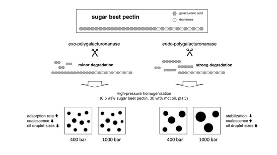
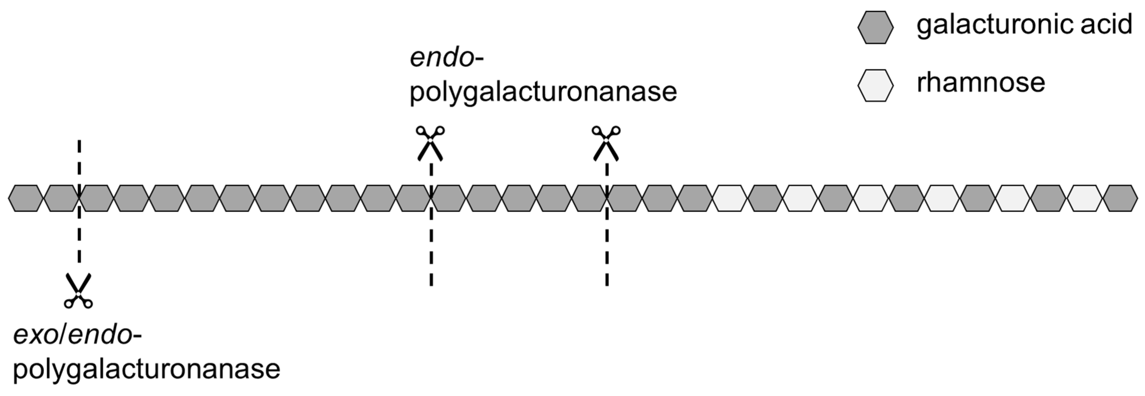
Therefore, modifying the homogalacturonan without degrading the side chains and functional groups might be useful to improve the adsorption kinetics of SBPs. A way to modify the homogalacturonan is the use of polygalacturonanases, such as exo- and endo-polygalacturonanase (exo-PG and endo-PG, respectively). Both are hydrolytic enzymes, which catalyze the hydrolysis of the O-glycosyl bond of α-D-(1→ 4)polygalacturonan but in a different pattern. Whereas endo-PG catalyzes the random cleavage of the O-glycosyl bond between unesterified α-1,4-D-linked galacturonic acid residues, exo-PG exclusively catalyzes the terminal cleavage of dimers from the non-reducing end [10,11]. As a result of the cleavage, the proportion of GalA in the molecule decreases and the proportion of neutral sugars increases [6,8,12]. Figure 1 shows schematically where the two enzymes operate on the pectic backbone.
The use of endo-PG leads to stronger fragmentation than the use of exo-PG, and thus to smaller SBPs with hydrophobic groups distributed over several fragments. On the one hand, a smaller size could have a positive effect on the adsorption rate, but, on the other hand, it could have a negative effect on the emulsion stabilizing properties.
Various enzymes to modify the structure of SBPs were used by Funami et al. [6]. They showed that an enzymatic treatment with 10 U/g SBP endo-PG led to larger emulsion droplets, compared to the unmodified control sample. Chen et al. [8] showed that the emulsifying properties of SBPs declined after enzymatic digestion with 10 U/g SBP endo-PG. This was determined by comparing droplet sizes of SBP-stabilized emulsions, creaming indices, and interfacial activities. SBPs with increased molecular weight due to laccase-mediated conjugation, on the other hand, showed better functionality and emulsion stabilization [13]. This was thought to be due to the formation of thick interfacial layers at the oil–water interface. Other authors demonstrated that pectin fractions of very low molecular weight resulted in lower interfacial activity and emulsions with larger droplet sizes due to insufficient steric stabilization [14,15,16].
Therefore, the enzymatic modification should not be excessive, as otherwise cleavage might lead to a loss of the emulsifying and emulsion-stabilizing properties.
The objective of this study was to determine if SBPs could be modified to improve their ability to reduce coalescence during high pressure homogenization. For this purpose, the size of SBPs was varied by the enzymatic cleavage of the homogalacturonan using exo- and endo-PG. The effect of the altered size on the adsorption of SBPs onto the oil–water interface was investigated using drop profile analysis tensiometry. Then, a high-pressure homogenizer was used to produce pectin-stabilized emulsions. The homogenization pressure was varied to achieve different degrees of droplet break-up and collision-induced coalescence rates, respectively. By analyzing the droplet size distributions, conclusions could be drawn about the coalescence that occurred immediately after droplet breakup.

2. Materials and Methods

2.1. Materials

Commercial sugar beet pectin was provided by Herbstreith & Fox KG (Neuenbürg, Germany). According to Bindereif et al. [9], pectins molecular characteristics were as follows: molecular weight, 104 ± 5 kDa; galacturonic acid, 66.1 ± 1.8%; protein content, 4.1 ± 0.2%; degree of methylation, 52.1 ± 1.7%; degree of acetylation, 23.1 ± 0.5%; trans-ferulic acid, 706 ± 21 mg/100 g.
The enzymes endo-polygalacturonanase (EC 3.2.1.15 from Aspergillus aculeatus) and exo-polygalacturonanase (EC 3.2.1.82 from Yersinia enterocolitica) were purchased from Megazyme (Bray, Ireland).
Medium chain triglyceride oil (mct) was purchased from Schumann & Sohn GmbH (Karlsruhe, Germany). According to supplier information, the mct oil was composed of 60% C8 and 40% C10 chains and had a density of 0.95 kg/L at room temperature. Unless stated otherwise, all chemicals used were of analytical grade and ultrapure water was used throughout the experiments.

2.2. Enzymatic Modification of Pectins

Pectins were enzymatically modified by endo-PG and exo-PG. As a first step of the treatment, 2.0 wt% SBP was dissolved in water at 60 °C using an Ultraturrax T-25 digital (IKA® Werke GmbH & Co. KG, Staufen, Germany) at a rotational speed of 10,000 rpm (tip speed 6.54 m/s) for 30 sec. After cooling to room temperature, the pH was adjusted to pH 5.5 for endo-PG experiments and pH 6.0 for exo-PG experiments using 0.5 M NaOH. Then, the solutions were left to equilibrate overnight for at least 15 h under gentle stirring. Then, the temperature was set to 50 °C (for endo-PG) or 60 °C (for exo-PG), respectively. To these solutions, 3 U/g pectin of endo-PG or exo-PG were added. After specific incubation times (10 min, 1 h, or 24 h, respectively), the enzymes were inactivated by heating the solution to 95 °C and holding the temperature for 5 min to stop the enzymatic reaction. The same procedure was performed for 24 h without the addition of enzymes in order to obtain reference samples. The solutions were then deep-frozen (−18 °C), freeze-dried, and ground to particles <0.5 mm. Pectins without enzymatic modification are referred to as exo-reference (reference pectin for treatments with exo-PG) and endo-reference (reference pectin for treatments with endo-PG). Furthermore, the modified pectins are referred to as exo-24h (treatment with exo-PG for 24 h), endo-10min, endo-1h, and endo-24h (treatment with endo-PG for 10 min, 1 h, and 24 h, respectively).

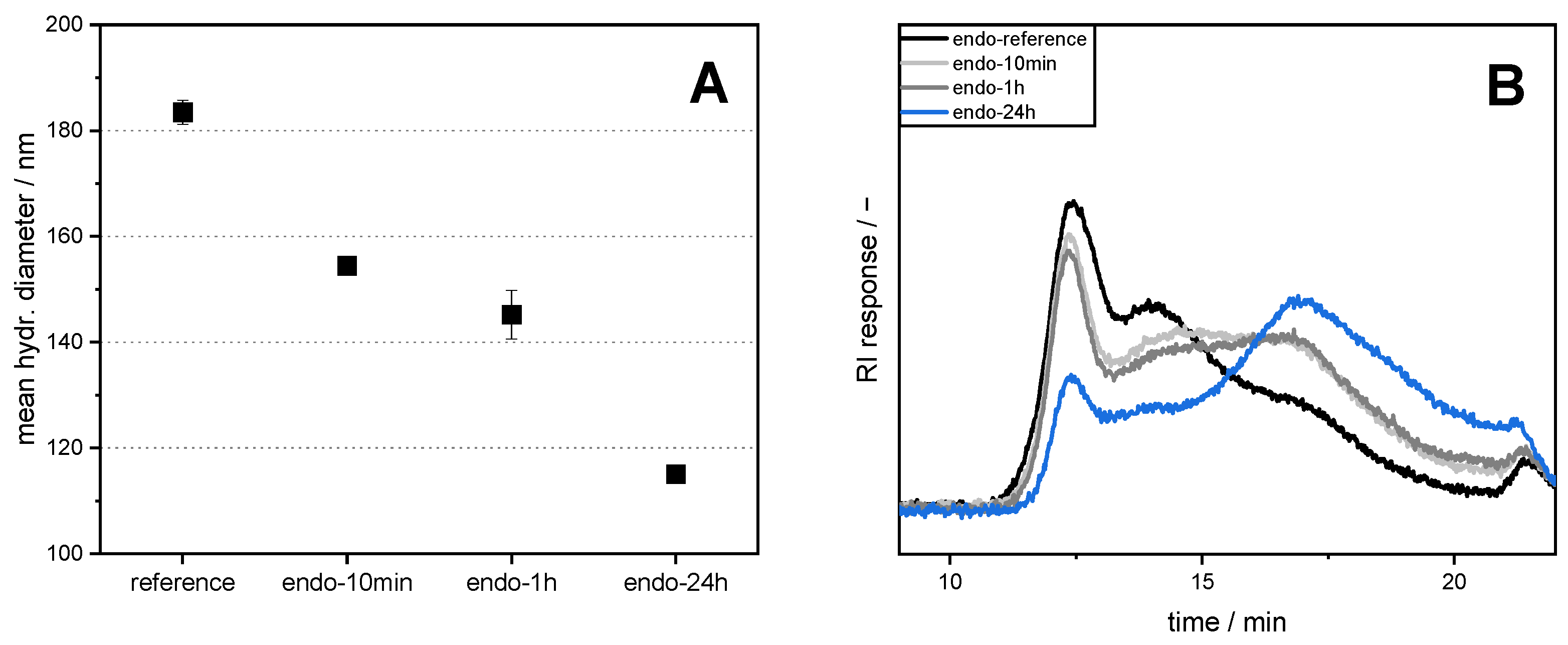
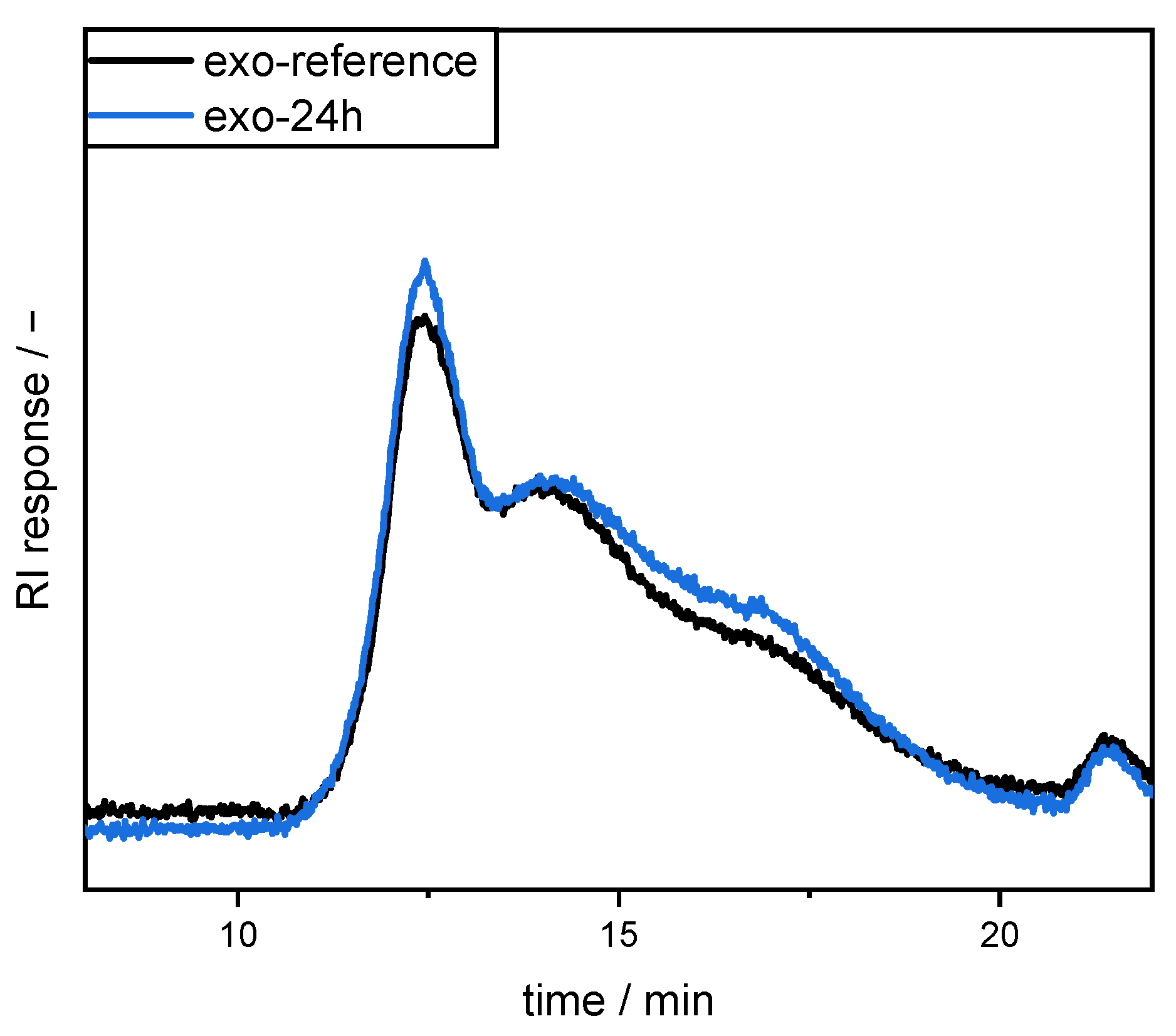
2.3. Molecular Size Distribution

High performance size exclusion chromatography (HPSEC) coupled with a refractive index (RI) detector (L-7490 LaChrom RI, Merck, Germany) was used to compare the molecular size distribution of the pectins. For this purpose, SBP samples were dissolved in 50 mM sodium nitrate (2 g/L). After centrifugation, the supernatant was used for SEC analysis on a Merck Hitachi L-7000 system equipped with a PWXL guard column (12 µm, 40 × 6 mm) connected in series with a TSKgel G6000PWXL (300 × 7.8 mm, 13 µm particle size) and a G4000PWXL (300 × 7.8 mm, 10 µm particle size, Tosoh Bioscience GmbH, Griesheim, Germany) column. Sodium nitrate (50 mM) was used as eluent at 40 °C (0.5 mL/min).

2.4. Preparation of Pectin Solutions

Sugar beet pectins (0.1 wt% or 0.5 wt%) were dissolved in water at 60 °C using an Ultraturrax T-25 digital (IKA® Werke GmbH & Co. KG, Staufen, Germany) at a rotational speed of 10,000 rpm for 30 s. The solution was cooled down, and the pH was adjusted to 3 using 1 M HCl at room temperature. Then, the pectin solutions were left to equilibrate for at least 15 h under constant magnetic stirring. The pH was checked again and readjusted to pH 3 if necessary.

2.5. Measurement of Hydrodynamic Diameter

Aqueous pectin solutions with a concentration of 0.1 wt% SBP were prepared, as described in Section 2.4. Then, the hydrodynamic diameters were measured, as described in Bindereif et al. [17]. In brief, SBP solutions at pH 3 were diluted further to yield 8 diluted samples (0.005–0.1 wt%). After an additional equilibration time of at least 15 h, three measurements of 10 runs each with 90 s run time per sample were performed. Refractive indices were set at n = 1.5470 for pectin and n = 1.333 for water. The hydrodynamic diameter was determined by plotting the measured z-average diameter versus SBP concentration and extrapolation to an SBP concentration of zero.

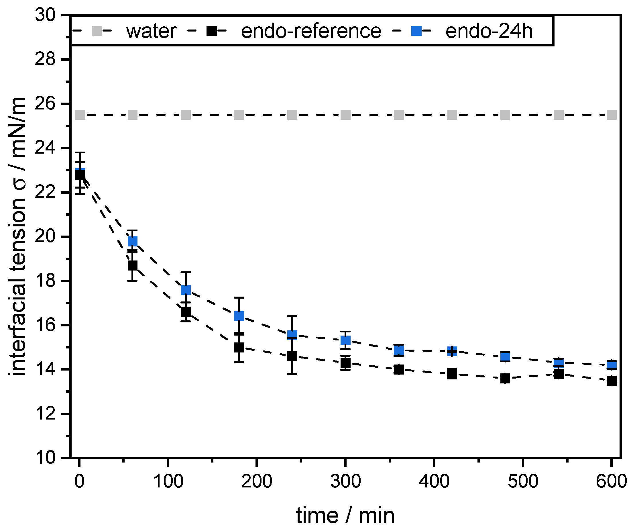
2.6. Measurement of Dynamic Interfacial Tension

Dynamic interfacial tension measurements at the mct oil–water interface were performed using the pendant drop technique (OCA 15 LJ, DataPhysics Instruments GmbH, Germany). Therefore, a droplet of mct oil with a volume of 12.5 µL was formed at the tip of the tensiometer capillary in a 0.1 wt% SBP solution (pH 3, see Section 2.4). All measurements were conducted in triplicate at 25 °C for 10 h. As a reference, the interfacial tension of the mct oil–water interface without the addition of pectins was also determined.

2.7. Viscosity Measurement

The viscosities of pectin solutions, mct oil, and emulsions were determined with a Physica MCR 301 rheometer equipped with a double gap geometry DG26.7 (Anton Paar, Graz, Austria). Rotational measurements were conducted at a temperature of 25 °C by applying a logarithmic shear rate profile recording 7 measurement points for each decade starting at 0.1 s−1 and rising up to 1000 s−1. Since SBP solutions and emulsions showed shear thinning behavior, the viscosity at 100 s−1 was used for comparison.

2.8. Preparation of Pectin-Stabilized Emulsions

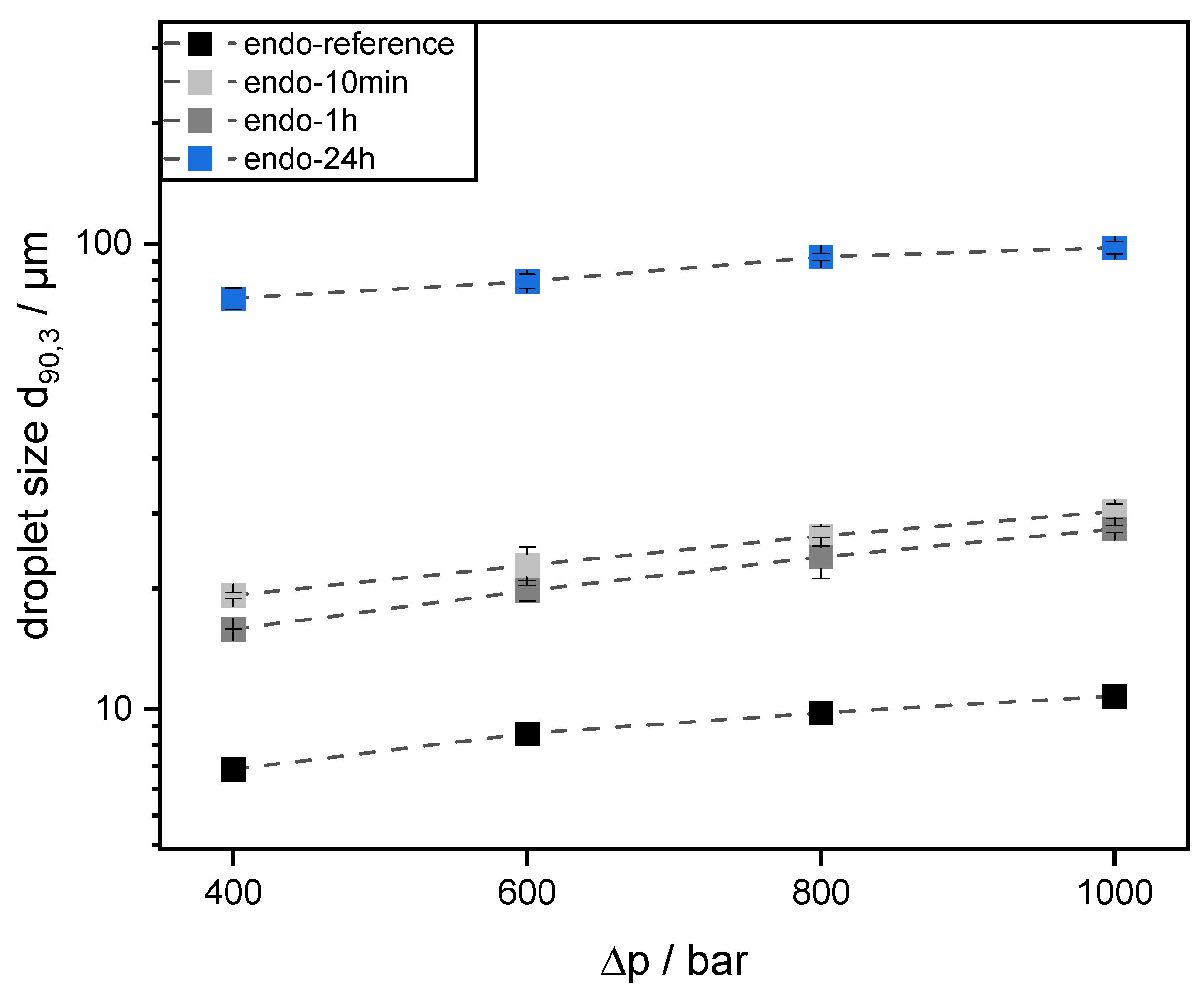
Emulsions were prepared using 30 wt% mct oil as dispersed phase and 70 wt% pectin solution (0.5 wt% SBP) as continuous aqueous phase. The pectin solutions were prepared, as described in 2.4. Then, mct oil was added to prepare an emulsion premix by using an Ultraturrax T-25 digital at 15,000 rpm (tip speed 9.81 m/s) for one minute. For further homogenization, the premix was transferred to a high-pressure homogenizer, Microfluidizer® MF 110 EH, equipped with a Y-type interaction chamber and with a microchannel diameter of 75 µm (Microfluidics Corporation, Newton, MA, USA). The interaction chamber is followed by an auxiliary processing module (APM) with a microchannel of 200 µm. The emulsions were homogenized at 400 bar, 600 bar, 800 bar, and 1000 bar. With increasing homogenizing pressure Δp, droplet break-up is increased. Droplet size decreases and the number of fine droplets increases. Since the number of droplets increases significantly more than their contact area, the collision rate increases strongly with increasing homogenization pressure.

2.9. Measurement of Droplet Size Distribution

Prepared emulsions were characterized by measuring their droplet size distributions (DSD) by static laser light scattering using a particle analyzer (HORIBA LA-950, Retsch Technology, Haan, Germany). The refractive indices were set at n = 1.333 for water and n = 1.4494 for mct oil for all measurements [18]. The measurements were conducted in triplicate at room temperature immediately after sample preparation. The results are depicted as the cumulative volume distribution Q3 and the 90th percentile of the volumetric cumulative size distribution (d90,3).

2.10. Statistical Analysis

All measurements and emulsions were prepared at least three times if not stated otherwise, per independent experiment. Statistical analysis, calculation of averages, and standard deviations were performed by using the software OriginPro 2020 (OriginLab Corp., Northampton, MA, USA).

3. Results and Discussion

3.1. Sugar Beet Pectins Modified by endo-Polygalacturonanase

3.1.1. Molecular Size Distribution and Hydrodynamic Diameter

The incubation with endo-PG resulted in significant changes in molecular size distribution and mean hydrodynamic diameter of the SBPs (see Figure 2). The hydrodynamic diameter of the reference sample (endo-reference) was 183 nm ± 2 nm. The longer the enzymatic treatment, the stronger the fragmentation of the pectins, and, accordingly, the smaller the hydrodynamic diameter. Although the differences between SBPs incubated for 10 min and 1 h were minor (hydrodynamic diameters of 154 nm ± 2 nm µm and 145 nm ± 5 nm, respectively), incubation for 24 h resulted in a significantly smaller hydrodynamic diameter (115 nm ± 1 nm).
Size exclusion chromatography revealed a broad size distribution and showed a shift towards smaller pectin molecules due to enzymatic modification. The longer the SBPs were enzymatically treated, the greater the proportion of the smaller molecules. Nevertheless, the molecular size distributions showed that fractions with large molecules and, consequently, without strong fragmentation were still present in all samples.

3.1.2. Emulsifying Properties

Surfactants of low molecular weight adsorb rapidly onto new interfaces and can, therefore, prevent re-coalescence immediately after droplet break-up. Slow adsorption, as found for typical polymeric emulsifiers, in contrast, leads to high re-coalescence rates [19,20,21,22]. High-pressure processes are often dominated by coalescence, leading to an increase in droplet size with increasing pressure difference Δp. This is pronounced in the products of higher oil content or for emulsifiers with reduced stabilization efficacy.
Figure 3 shows the volumetric oil droplet size distributions Q3 of the 30 wt% oil in water emulsions prepared at 400 bar. The use of SBPs with different sizes resulted in significant differences in the droplet size distribution of the prepared emulsions. An increasing shift to larger emulsion droplets was observed with increasing enzymatic degradation. Whereas emulsification with endo-reference showed a d90,3 of 7.4 µm ± 0.1 µm, incubation for 10 min and 1 h resulted in a d90,3 of 17.6 µm ± 0.3 µm and 14.8 µm ± 0.1 µm, respectively. The strong fragmentation that occurred in endo-24h resulted in further coalescence during emulsification and another significant increase in droplet size (d90,3 = 76.4 µm ± 4.2 µm). Moreover, the emulsion prepared with endo-24h showed a bimodal size distribution, which might be the consequence of a high coalescence rate.
The poorer performance of the enzymatically modified SBPs can be attributed to their severe fragmentation, resulting in insufficient emulsion-stabilizing properties. Nevertheless, the emulsions did not show strong changes in droplet size distribution over a storage period of 14 days. The d90,3 increased by only 5% for the endo-reference and 7% for the endo-24h samples, respectively (data not shown). This indicates that coalescence occurs immediately after droplet breakup until the interface is small enough to be sufficiently stabilized by the SBPs present. Thus, the differences in the emulsifying properties of SBPs due to the enzymatic modification resulted in short-term effects rather than long-term effects.
As the viscosities of the continuous phases were very low (<2.5 mPa·s) and were only slightly influenced by using 0.5 wt% SBP of different degrees of degradation, differences in droplet breakup between individual emulsion samples due to viscosity can be neglected. Instead, differences in droplet size distribution can be mainly attributed to the different emulsification properties of the SBP samples.
Figure 4 shows the droplets’ sizes of emulsions prepared at increasing pressure differences from 400 to 1000 bar. For the evaluation of the emulsifier efficacy, the characteristic droplet sizes d90,3 were used and plotted over Δp. At 600 bar, 800 bar, and 1000 bar, the same trends in droplet sizes can be seen as at 400 bar. With longer incubation time, and thus more fragmentation of SBPs, larger oil droplets were obtained during emulsification (d90,3: endo-reference < endo-1h ≈ endo-10min < endo-24h). With increasing Δp, droplet sizes d90,3 increased, indicating an increased rate of re-coalescence.
While the droplet size d90,3 stabilized with endo-reference was 7.4 µm ± 0.1 µm at 400 bar, it was 8.9 µm ± 0.5 µm at 600 bar, 9.8 µm ± 0.5 µm at 800 bar, and 10.7 µm ± 0.3 µm at 1000 bar, respectively. The increase in d90,3 using a Δp of 400 bar and 1000 bar corresponds to 44%. This trend could also be demonstrated for the modified SBP samples. Here, the increase corresponds to 52% for endo-10min, 64% for endo-1h, and 29% for endo-24h, respectively. Thus, with the chosen process conditions and formulation parameters, the use of both the endo-reference and all enzymatically treated samples resulted in coalescence-dominated emulsification processes. This can be attributed to too slow or insufficient stabilization of the oil droplets by the SBPs.
In previous studies, similar results were found for emulsions stabilized with pectins. Coalescence also dominated, for example, in the production of emulsions stabilized with pectinic acid with a molecular weight of 34 kDa [23]. Compared to pectinic acid, SBPs are more branched and compact but possess higher molecular weight (104 ± 5.0 kDa for the untreated SBPs) [9]. However, some of the obtained fragments after enzymatic treatment might be significantly smaller. Although these molecules could diffuse and adsorb faster, larger droplets were obtained than with the reference pectins. The small molecules, thus, appear to have reduced emulsion-stabilizing efficacy.

3.1.3. Interfacial Activity

To investigate whether the interfacial activity and adsorption rate of SBPs were greatly altered by the enzymatic treatment, the dynamic interfacial tension was measured by drop profile analysis tensiometry. Since SBPs with smaller hydrodynamic diameter diffuse more rapidly to the interface, the interfacial tension should be reduced faster than for large molecules [17]. Furthermore, it could be possible that the degradation of SBPs makes surface active groups more accessible so that the adsorption rate increases [16]. In addition, due to their smaller size, SBPs could adsorb in greater quantity onto the interface, which may lead to a stronger reduction in the interfacial tension.
Figure 5 compares the dynamic interfacial tension for a droplet of either 0.1 wt% endo-reference or 0.1 wt% endo-24h solution (see Section 2.4) in purified mct oil. Due to the smaller hydrodynamic diameter, and thus more rapid diffusion to the interface, a faster reduction in the interfacial tension was expected for endo-24h. However, enzymatically modified SBPs did not reduce the interfacial tension faster or more strongly than SBPs without enzymatic treatment. Using the endo-reference sample, the interfacial tension is reduced slightly more (13.5 mN/m ± 0.07 mN/m vs. 14.2 mN/m ± 0.17 mN/m for endo-24h after 10 h), but the differences appear minor. Since SBPs are available in excess in this analysis, reduction in interfacial tension could be due to the adsorption of large SBPs with low enzymatic degradation still present in the sample (see Figure 2). The differences could be due to competition at the interface between these SBPs with good interfacial activity and fragments of SBPs with reduced interfacial activity (e.g., parts of the homogalacturonan, with a low proportion of hydrophobic groups). This leads to a smaller reduction in the interfacial tension.
The assumption that mainly the large SBPs contribute to the stabilization of the interface could be supported by the preparation of emulsions with higher concentration of endo-24h. Increasing the concentration of endo-24h resulted in smaller droplets (see Figure 6). Nevertheless, approximately four times the amount of endo-24h (2.0 wt%) was required to obtain smaller droplet sizes than with endo-reference. Moreover, the emulsification processes were still dominated by coalescence even at higher concentrations of endo-24h (data not shown). Thus, the measured interfacial activity and droplet sizes strongly suggest that the large, barely modified SBPs are responsible for the emulsifying effect. However, this cannot be conclusively clarified without further purification or fractionation.
Overall, the enzymatic modification with endo-PG did not lead to the desired improvement in functionality, as coalescence could not be reduced.

3.2. Sugar Beet Pectins Modified by exo-Polygalacturonanase

As shown in Section 3.1, cleavage of SBPs with endo-PG resulted in SBP fragments with poorer emulsification properties, compared to the reference pectin. This could be due to the random cleavage of the backbone catalyzed by the enzymes [11]. To ensure that all important structures remained on a single SBP fragment and that the fragments did not become too small, more targeted cleavage was required. For this purpose, exo-PG was used for enzymatic modification.
The molecular weight distributions of exo-reference and exo-24h are shown in Figure 7. For both samples, the HPSEC-RI elution patterns showed broad molecular size distributions. Despite the enzymatic treatment, no significant shift towards smaller molecular sizes was observed. Other differences in the molecular size distributions were also not very pronounced. Because cleavage occurred exclusively at the non-reducing free ends of the homogalacturonan, the hydrodynamic diameter of SBPs changed only slightly (186 ± 4 nm for exo-reference vs. 175 ± 5 nm for exo-24h).

3.2.1. Emulsifying Properties

Although the differences in size between exo-reference and exo-24h were small, significant differences in emulsification properties were evident. Figure 8 shows the droplet size distribution of emulsions prepared with the respective SBPs as emulsifiers. When using exo-24h, smaller droplets (d90,3 = 5.9 ± 0.3 µm) were stabilized than when using exo-reference (d90,3 = 7.4 ± 0.1 µm). This could be due to the fact that modified SBPs adsorb faster and in greater numbers onto the interface due to lower steric hindrance. This resulted in the faster stabilization of the oil droplets after breakup and, consequently, smaller droplet sizes.
At higher Δp, increased coalescence outweighs the stronger droplet breakup when using 0.5 wt% exo-reference (see Figure 9). Despite stronger droplet breakup, an increase in homogenization pressure resulted in larger emulsion droplets. The characteristic droplet size d90,3 increased from 7.4 µm at 400 bar to 10.7 µm at 1000 bar, which corresponds to an increase of 45%.
In contrast to the use of exo-reference, no significant differences in droplet sizes were observed when exo-24h was used at various homogenization pressures. The droplet size d90,3 was 5.9 ± 0.3 µm at 400 bar, 6.3 ± 0.2 µm at 600 bar, 6.2 ± 0.2 µm at 800 bar, and 6.2 ± 0.2 µm at 1000 bar, respectively. Thus, with the chosen process conditions and formulation parameters, the use of exo-24h resulted in droplet breakup and coalescence balancing each other out.

3.2.2. Interfacial Activity

Figure 10 shows the dynamic interfacial tension, which initially decreased more rapidly when exo-24h was used, compared to exo-reference. This supports the statement of the faster adsorption of SBPs after modification with exo-PG. As a result, when using exo-24h, the coalescence during emulsification is significantly reduced and smaller droplets are obtained than when using exo-reference.

4. Conclusions

In the present study, we attempted to improve the emulsifying properties of SBPs in order to reduce coalescence during high-pressure homogenization. For this purpose, SBPs were subjected to enzymatic treatment to increase their adsorption rate due to smaller hydrodynamic sizes. As shown in the literature, neutral sugar side chains and functional groups play an important role for the emulsion-stabilizing properties. Therefore, two enzymes (endo-PG and exo-PG) were used to cleave glycosidic linkages in the homogalacturonan without degrading other structural features. HPSEC and hydrodynamic diameter measurements showed that fragmentation increased with increasing incubation time. Furthermore, the use of endo-PG resulted in significantly smaller SBPs than the use of exo-PG. This could be attributed to the different nature of the enzymatic cleavage of endo-PG, compared to exo-PG.
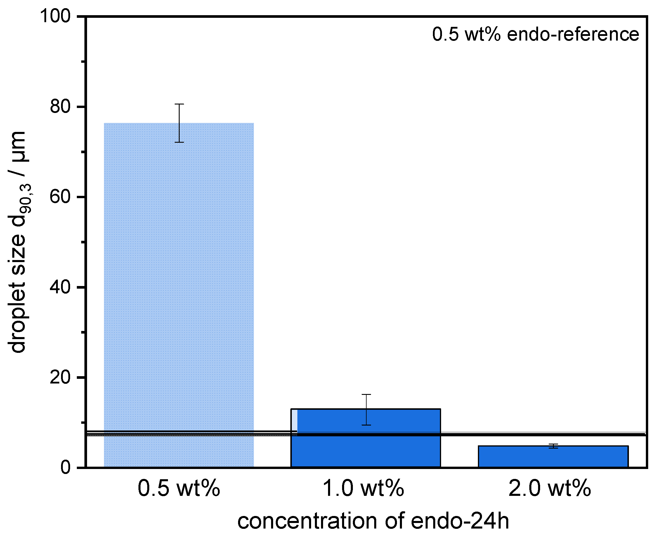
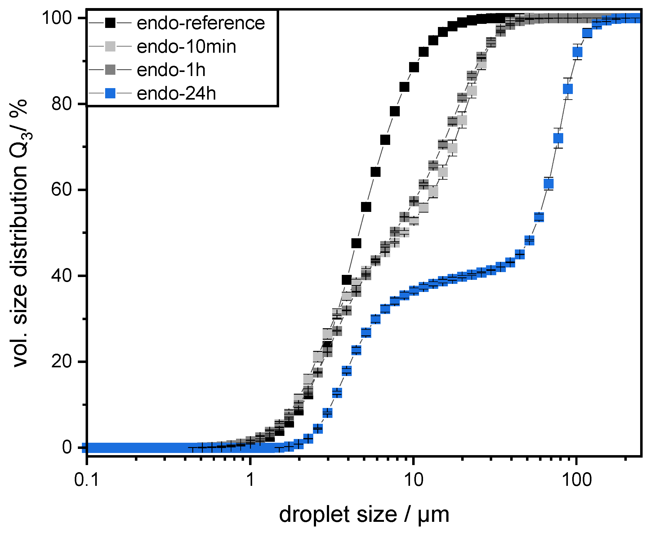
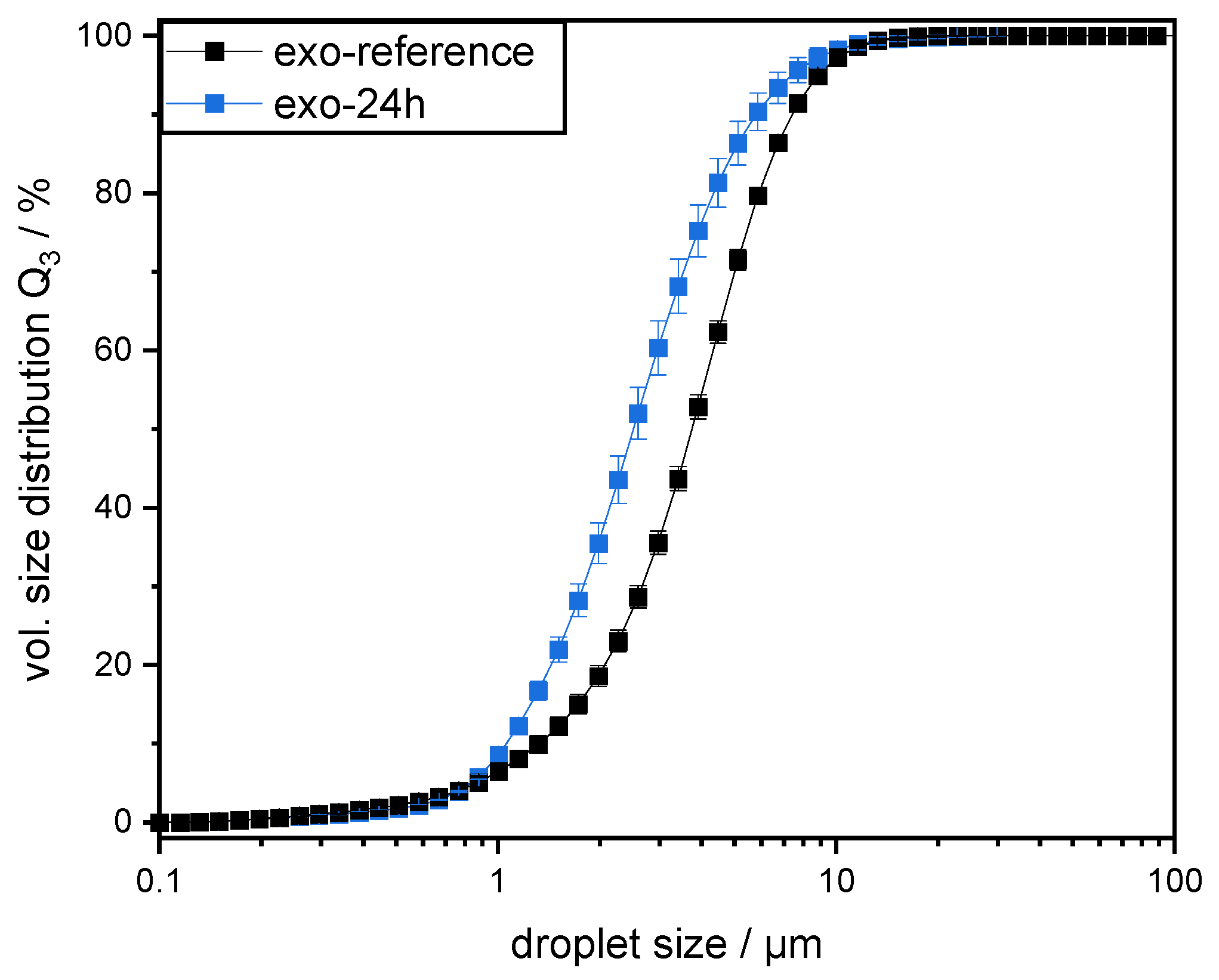
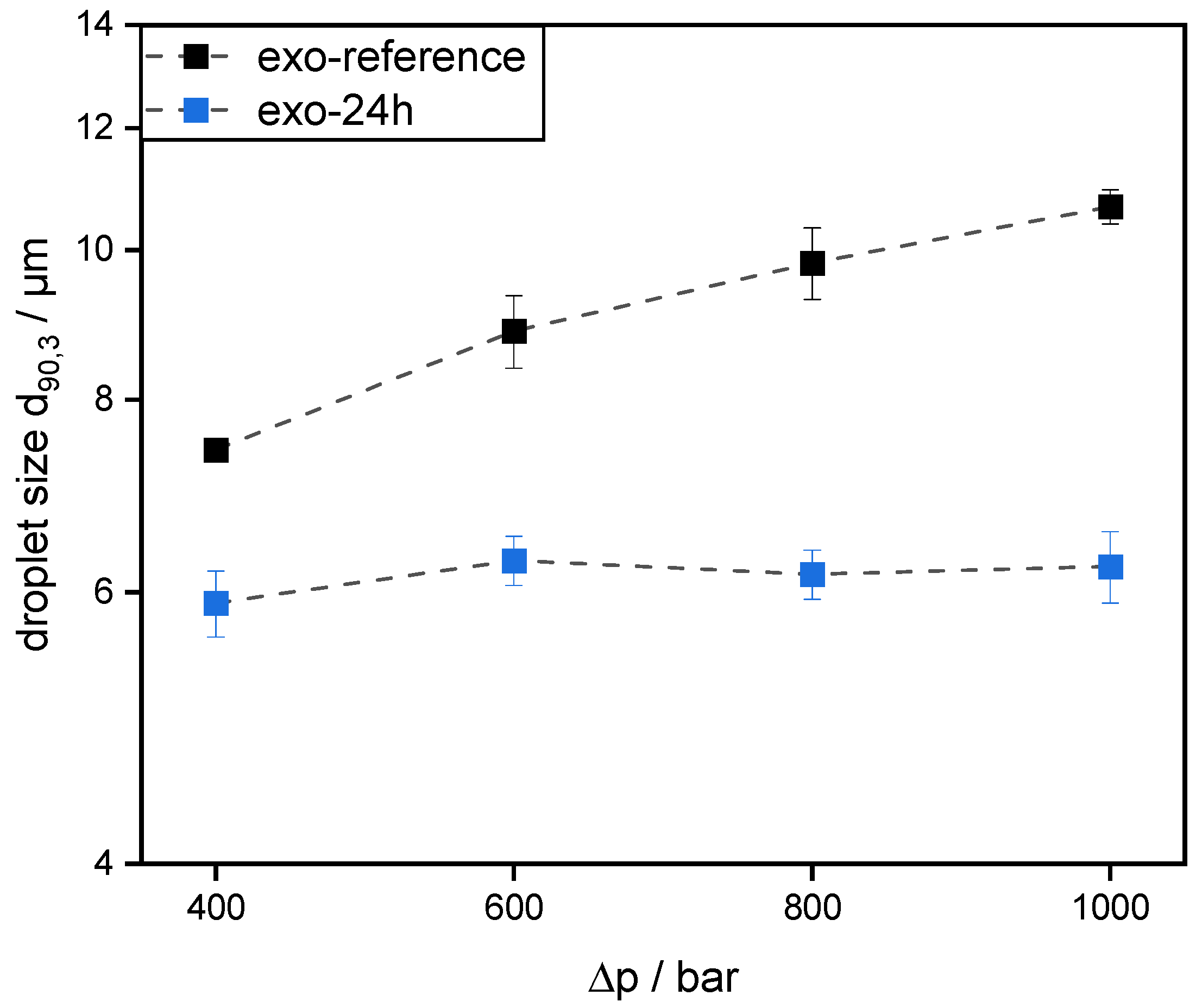
The preparation of emulsions (0.5 wt% SBP, 30 wt% mct oil, pH 3) and measurement of droplet size distributions provided information on the coalescence that occurred during emulsification. It was observed that emulsions prepared with SBPs modified with endo-PG showed larger droplet sizes than nonmodified SBPs due to enhanced coalescence. The coalescence-dominated emulsification process was also evident when the homogenization pressure was increased. With increasing Δp, larger droplets were obtained. This could be attributed to the reduced emulsion stabilizing properties of SBPs due to their strong fragmentation after enzymatic modification.
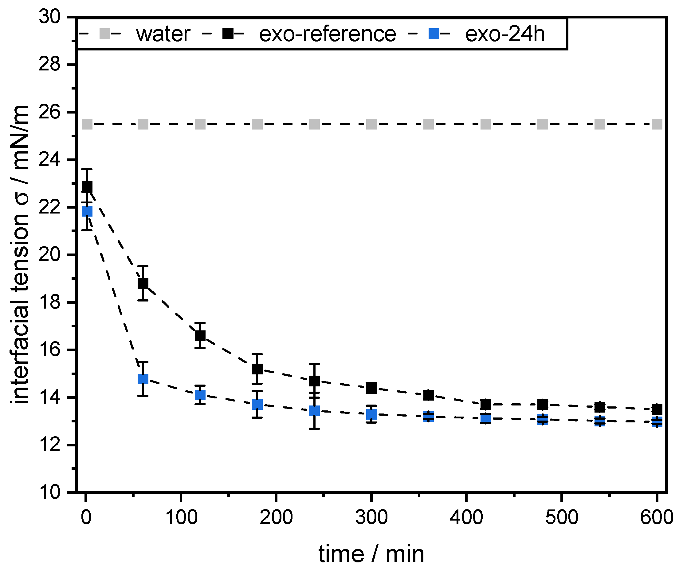
However, this was not the case for SBPs incubated with exo-PG. An enzymatic treatment of 24 h significantly improved their emulsifying properties. Faster adsorption, confirmed by droplet profile analysis tensiometer, resulted in less coalescence during emulsification. As a result, smaller droplets were obtained than with the reference pectins. In addition, increasing the homogenization pressure did not result in larger droplets, despite higher collision rates.
Overall, the present study showed that SBPs can be modified to alter their emulsifying properties. This allows coalescence to be reduced or increased during high-pressure homogenization, depending on modification. Based on this knowledge, other pectins and hydrocolloids could also be enzymatically modified to modify their emulsifying properties. However, this is only possible in a targeted manner if the structures that are decisive for the emulsifying properties are known. Structures that are insignificant for emulsion stabilization can be cleaved off to decrease the viscosity, increase the adsorption rate, and optimize the conformation at the interface. For this purpose, however, it is important to first establish the structure–property functions of the respective hydrocolloids in order to specifically modify the emulsifying properties.

Author Contributions

Conceptualization, B.B. and U.S.v.d.S.; methodology, B.B.; validation, B.B.; investigation, B.B.; resources, H.P.K. and U.S.v.d.S.; data curation, B.B.; writing—original draft preparation, B.B.; writing—review and editing, U.S.v.d.S. and H.P.K.; visualization, B.B.; supervision, H.P.K. and U.S.v.d.S.; project administration, B.B., H.P.K. and U.S.v.d.S.; funding acquisition, H.P.K. and U.S.v.d.S. All authors have read and agreed to the published version of the manuscript.

Funding

This research was conducted in the frame of IGF Project no. 20218 of the FEI and was supported via AiF within the programme for promoting the Industrial Collective Research (IGF) of the German Ministry of Economic Affairs and Energy (BMWi), based on a resolution of the German Parliament.

Institutional Review Board Statement

Not applicable.

Informed Consent Statement

Not applicable.

Data Availability Statement

The datasets generated for this study are available on request to the corresponding author.

Acknowledgments

The authors thank Herbstreith & Fox KG (Neuenbürg, Germany) for providing sugar beet pectins; Lydia Schütz, Yujing Wang, and Jessica Wezstein for conducting experiments; Hendrik Eichhöfer for his help with HPSEC measurements; and Gabriela Itziar Saavedra Isusi for proofreading. We acknowledge support by the KIT-Publication Fund of the Karlsruhe Institute of Technology.

Conflicts of Interest

The authors declare no conflict of interest.

References

  1. Floury, J.; Desrumaux, A.; Lardières, J. Effect of high-pressure homogenization on droplet size distributions and rheological properties of model oil-in-water emulsions. Innov. Food Sci. Emerg. Technol. 2000, 1, 127–134. [Google Scholar] [CrossRef]
  2. Dickinson, E. Hydrocolloids acting as emulsifying agents—How do they do it? Food Hydrocoll. 2018, 78, 2–14. [Google Scholar] [CrossRef]
  3. Dickinson, E. Hydrocolloids as emulsifiers and emulsion stabilizers. Food Hydrocoll. 2009, 23, 1473–1482. [Google Scholar] [CrossRef]
  4. Siew, C.K.; Williams, P.A. Role of protein and ferulic acid in the emulsification properties of sugar beet pectin. J. Agric. Food Chem. 2008, 56, 4164–4171. [Google Scholar] [CrossRef] [PubMed]
  5. Schmidt, U.S.; Schmidt, K.; Kurz, T.; Endreß, H.-U.; Schuchmann, H.P. Pectins of different origin and their performance in forming and stabilizing oil-in-water-emulsions. Food Hydrocoll. 2015, 46, 59–66. [Google Scholar] [CrossRef]
  6. Funami, T.; Nakauma, M.; Ishihara, S.; Tanaka, R.; Inoue, T.; Phillips, G.O. Structural modifications of sugar beet pectin and the relationship of structure to functionality. Food Hydrocoll. 2011, 25, 221–229. [Google Scholar] [CrossRef]
  7. Funami, T.; Zhang, G.; Hiroe, M.; Noda, S.; Nakauma, M.; Asai, I.; Cowman, M.K.; Al-Assaf, S.; Phillips, G.O. Effects of the proteinaceous moiety on the emulsifying properties of sugar beet pectin. Food Hydrocoll. 2007, 21, 1319–1329. [Google Scholar] [CrossRef]
  8. Chen, H.M.; Fu, X.; Luo, Z.G. Effect of molecular structure on emulsifying properties of sugar beet pulp pectin. Food Hydrocoll. 2016, 54, 99–106. [Google Scholar] [CrossRef]
  9. Bindereif, B.; Eichhöfer, H.; Bunzel, M.; Karbstein, H.P.; Wefers, D.; van der Schaaf, U.S. Arabinan side-chains strongly affect the emulsifying properties of acid-extracted sugar beet pectins. Food Hydrocoll. 2021, 7, 106968. [Google Scholar] [CrossRef]
  10. Yahia, E.M.; Carrillo-López, A.; Bello-Perez, L.A. Carbohydrates. Postharvest Physiology and Biochemistry of Fruits and Vegetables; Elsevier: Amsterdam, The Netherlands, 2019; pp. 175–205. ISBN 9780128132784. [Google Scholar]
  11. Burns, J.K. The Polygalacturonases and Lyases. In The Chemistry and Technology of Pectin; Elsevier: Amsterdam, The Netherlands, 1991; pp. 165–188. ISBN 9780080926445. [Google Scholar]
  12. Guo, X.; Guo, X.; Yu, S.; Kong, F. Influences of the different chemical components of sugar beet pectin on the emulsifying performance of conjugates formed between sugar beet pectin and whey protein isolate. Food Hydrocoll. 2018, 82, 1–10. [Google Scholar] [CrossRef]
  13. Jung, J.; Wicker, L. Laccase mediated conjugation of sugar beet pectin and the effect on emulsion stability. Food Hydrocoll. 2012, 28, 168–173. [Google Scholar] [CrossRef]
  14. Leroux, J.; Langendorff, V.; Schick, G.; Vaishnav, V.; Mazoyer, J. Emulsion stabilizing properties of pectin. Food Hydrocoll. 2003, 17, 455–462. [Google Scholar] [CrossRef]
  15. Yapo, B.M.; Robert, C.; Etienne, I.; Wathelet, B.; Paquot, M. Effect of extraction conditions on the yield, purity and surface properties of sugar beet pulp pectin extracts. Food Chem. 2007, 100, 1356–1364. [Google Scholar] [CrossRef]
  16. Akhtar, M.; Dickinson, E.; Mazoyer, J.; Langendorff, V. Emulsion stabilizing properties of depolymerized pectin. Food Hydrocoll. 2002, 16, 249–256. [Google Scholar] [CrossRef]
  17. Bindereif, B.; Karbstein, H.; Zahn, K.; van der Schaaf, U. Effect of conformation of sugar beet pectin on the interfacial and emulsifying properties. Foods 2022, 11, 214. [Google Scholar] [CrossRef] [PubMed]
  18. Saavedra Isusi, G.I.; Madlindl, L.B.; Karbstein, H.P.; van der Schaaf, U.S. Microstructures and conformational arrangement in emulsions caused by concentration ratios of pectin-based microgels and oil. Colloids Surf. A Physicochem. Eng. Asp. 2020, 602, 125166. [Google Scholar] [CrossRef]
  19. Stang, M.; Karbstein, H.; Schubert, H. Adsorption kinetics of emulsifiers at oil—Water interfaces and their effect on mechanical emulsification. Chem. Eng. Process. Process Intensif. 1994, 33, 307–311. [Google Scholar] [CrossRef]
  20. Karbstein, H.; Schubert, H. Developments in the continuous mechanical production of oil-in-water macro-emulsions. Chem. Eng. Process. Process Intensif. 1995, 34, 205–211. [Google Scholar] [CrossRef]
  21. Jafari, S.M.; Assadpoor, E.; He, Y.; Bhandari, B. Re-coalescence of emulsion droplets during high-energy emulsification. Food Hydrocoll. 2008, 22, 1191–1202. [Google Scholar] [CrossRef]
  22. Karbstein, H.P. Untersuchungen zum Herstellen und Stabilisieren von Öl-in-Wasser-Emulsionen. Ph.D. Thesis, University of Karlsruhe, Karlsruhe, Germany, 1994. [Google Scholar]
  23. Saavedra Isusi, G.I.; Bindereif, B.; Karbstein, H.P.; van der Schaaf, U.S. Polymer or microgel particle: Differences in emulsifying properties of pectin as microgel or as individual polymer chains. Colloids Surf. A Physicochem. Eng. Asp. 2020, 598, 124793. [Google Scholar] [CrossRef]
Figure 1. Schematic illustration of the enzymatic modification with the enzymes exo- and endo-polygalacturonanase used in this study.
Colloids 06 00069 g001
Figure 2. (A) Hydrodynamic diameter of the endo-reference sugar beet pectin and the enzymatically modified sugar beet pectins (endo-10min, endo-1h, and endo-24h, respectively) in solution at pH 3. (B) Elution patterns of the high performance size exclusion chromatography coupled with refractive index detection (HPSEC-RI) of the respective sugar beet pectins.
Colloids 06 00069 g002
Figure 3. Volumetric oil droplet size distributions Q3 of oil–water emulsions prepared with 0.5 wt% of the respective pectin and 30 wt% mct oil. The emulsions were prepared at pH 3 using a high-pressure homogenizer at 400 bar.
Colloids 06 00069 g003
Figure 4. Droplet sizes d90,3 of emulsions prepared with 0.5 wt% sugar beet pectin with or without (endo-reference) enzymatic modification using endo-polygalacturonanase. The pectins were enzymatically treated for 10 min (endo-10min), 1 h (endo-1h), and 24 h (endo-24h), respectively. The emulsions were prepared at pH 3 with 30 wt% mct oil at varying energy densities.
Colloids 06 00069 g004
Figure 5. The mct oil–water interfacial tension, using 0.1 wt% pectin solutions with and without enzymatic modification (24 h enzymatic treatment using endo-polygalacturonanase), measured over time at 25 °C. As a reference, the interfacial tension was measured without the addition of pectin.
Colloids 06 00069 g005
Figure 6. Droplet sizes d90,3 of emulsions prepared with 0.5 wt%, 1.0 wt%, and 2.0 wt% endo-24h (24 h enzymatic treatment using endo-polygalacturonanase), respectively. A reference line was drawn for 0.5 wt% endo-reference. The emulsions were prepared at pH 3 with 30 wt% mct oil at 400 and 1000 bar.
Colloids 06 00069 g006
Figure 7. Elution patterns of the high performance size exclusion chromatography coupled with refractive index detection (HPSEC-RI) of the reference pectin (exo-reference) and the enzymatically modified pectin (exo-24h).
Colloids 06 00069 g007
Figure 8. Volumetric oil droplet size distributions Q3 of emulsions (30 wt% mct oil in water) prepared with 0.5 wt% exo-reference and 0.5 wt% exo-24h, respectively. The emulsions were prepared at pH 3 using a high-pressure homogenizer at 400 bar.
Colloids 06 00069 g008
Figure 9. Droplet sizes d90,3 of emulsions prepared with 0.5 wt% pectin with (exo-24h) or without (exo-reference) enzymatic modification using exo-polygalacturonanase. The emulsions were prepared at pH 3 with 30 wt% mct oil at varying energy densities.
Colloids 06 00069 g009
Figure 10. The mct oil–water interfacial tension, using 0.1 wt% pectin solutions with and without enzymatic modification (24 h enzymatic treatment using exo-polygalacturonanase), measured over the time at 25 °C. As a reference, the interfacial tension was measured without the addition of pectin.
Colloids 06 00069 g010
Publisher’s Note: MDPI stays neutral with regard to jurisdictional claims in published maps and institutional affiliations.

