Article

Feasibility of In Vivo Semen Collection and Description of the Morphology and Ultrastructure of the Spermatozoa of Arapaima gigas (Schinz, 1822)

by
Lucas S. Torati
1,
Júlia T. Lopes
2,
Adriana F. Lima
1,
Velmurugu Puvanendran
3,*,
Sergio R. Batlouni
4 and
Luciana N. Ganeco-Kirschnik
1,*
1
EMBRAPA Fisheries and Aquaculture, Palmas 77008-900, Brazil
2
Instituto Federal do Tocantins—IFTO, Campus Pedro Afonso, Pedro Afonso 77710-000, Brazil
3
Center for Marine Aquaculture & Production Biology, Nofima AS, Muninbakken 9, 9019 Tromsø, Norway
4
Centro de Aquicultura da UNESP—CAUNESP, Universidade Estadual Paulista—UNESP, Jaboticabal 14884-900, Brazil
*
Authors to whom correspondence should be addressed.
Fishes 2024, 9(1), 24; https://doi.org/10.3390/fishes9010024
Submission received: 1 December 2023 / Revised: 2 January 2024 / Accepted: 3 January 2024 / Published: 4 January 2024
(This article belongs to the Special Issue Reproductive Biology and Breeding of Fish)

Abstract

:
The pirarucu, Arapaima gigas, is an emblematic endangered Amazon osteoglossid with high potential for aquaculture development. In this study, we report the in vivo collection of semen from pirarucu for the first-time and a complete description of the spermatozoa ultrastructure using scanning and transmission electron microscopy (SEM and TEM). The spermatozoon of pirarucu is biflagellate with no acrosomal cell, comprising a spherical head (8.26 ± 2.19 µm2), a short intermediate piece region and two long flagella (68.34 ± 5.69 µm). The nucleus is spherical and electron-dense, and at its base, there are two nuclear fossae where the centrioles are found. The anterior part of the flagellum is positioned at the lateral base of the nucleus, forming an intermediate piece. Lateral to the intermediate piece, an abundant number of mitochondria are present and occupy most of the cytoplasmic volume. The two flagella were found with a 9 + 2 axonemal structure (nine peripheral doublets and two central microtubules). The central microtubules are aligned with two side fins formed laterally by the axoneme plasma membrane. Side fins were observed from the anterior region to the posterior end. The results show that the spermatozoa of A. gigas are biflagellate, mitochondria-rich and robust, corroborating the external fertilization characteristic of the species. The novel description of sperm morphology presented herein will contribute to advancing the knowledge of and aiding future research on the reproductive biology of this species.
Key Contribution: Our work, for the first time, reports the in vivo collection of sperm from A. gigas male broodstock and describes the spermatozoon ultrastructure. A. gigas has a rare biflagellate, mitochondria-rich and robust spermatozoon which corroborates the external fertilization characteristic of this species. This study provides novel insights into the reproductive biology of the male of this species and creates opportunities for future studies in this aspect.

1. Introduction

As the most diversified vertebrate group, teleosts display great variety in modes of reproduction, from internal fertilization as in viviparous species to the external fertilization of gametes as in oviparous species in fresh, salt and brackish waters [1]. Some fish species are group spawners, while others mate as couples. Spawning and fertilization can be pelagic, but egg laying and fertilization can also occur in nests or on stones, grass, and many other substrates [2]. Some species will reproduce only once during their lifetime (semelparous), while others will produce offspring multiple times during adulthood (iteroparous) [3]. Not surprisingly, these widespread reproductive methods lead to a great source of variability in the ultrastructure of fish oocytes and spermatozoa, and these are often associated with fertilization particularities that also provide key information about fertilization modes and the natural histories of the different groups [4].
Fish spermatozoa are widely divergent in gross morphology, and they display aflagellate to biflagellate conditions with wide range of shapes, size, and structures. The variation exists in head size and shape, midpiece size, and the number and length of flagella [5]. So far, the morphological diversity of fish spermatozoa has been described for over 280 species and 100 teleost families, with diverse character states described from a common ancestor and used for phylogenetic reconstructions [4]. With the vast, divergent gross morphology of sperm structure, Mattei [4] found it difficult to correlate sperm morphology with taxonomic models in Teleostei. Within Osteoglossiformes, tremendous variability has been reported with families having either monoflagellate (Pantodontidae and Notopteridae), biflagellate (Osteoglossidae) or even aflagellate (Mormyridae and Gymnarchidae) spermatozoon types [6,7]. Although bearing aflagellate spermatozoa suggests internal fertilization, species such as Mormyridae and Gymnarchidae have external fertilization with low sperm competition [8,9]. Monoflagellate spermatozoa are the most frequently found type among teleosts, described for species with internal fertilization and external fertilization [6,10]. The presence of biflagellate spermatozoa is widespread in the animal kingdom from flatworms to mammals [5,11]; however, in fishes, the biflagellate condition seems to be less frequent and has only been described for over 16 families from seven orders [10].
Arapaima gigas (Schinz, 1822) is an Amazon osteoglossid considered the largest scaled freshwater fish in the world [12]. The species is dioecious and iteroparous, with couples spawning in nests built in shallow areas of flooded forest during the rainy period in the Amazon [13,14]. After spawning, males provide intense parental care by guarding the nest for up to three months post spawning, and parental care ceases at the end of the rainy season [15]. Like other osteoglossids, in A. gigas, only the left gonad is functional [16]. The testis of the pirarucu begins its differentiation 43 days after hatching [17], when individuals still measure around 75 cm in total length [18]. The testis has been described as a “cord-like” structure, with a diameter ranging from 1–1.5 cm, and is connected to the genital papilla through a spermatic duct [19,20]. Spermatogenesis in A. gigas has been divided into four developmental stages (immature, maturing, ripe and spent), and for being a multiple-spawner species, its testis can become ripe again weeks after the first spermiation, allowing for multiple breeding events [20,21]. So far, there is lack of information about the spermatozoa ultrastructure of A. gigas, a key species of Amazonian fauna with enormous potential for aquaculture development in the tropics. This lack of information includes the overall aspects of semen characteristics in the species, which hampers the development of biotechnologies, such as semen cryopreservation, which are necessary to ensure continuous seed production and thus control its reproduction in captivity. The feasibility of semen collection has never been reported for A. gigas, possibly due to an overall lack of knowledge about its reproductive biology. In this study, we report the in vivo collection of semen from male broodstock for the first time and describe the spermatozoa ultrastructure.

2. Materials and Methods

2.1. Semen Sampling Procedure

Before attempting semen collection in a live broodstock, a pilot study was conducted aiming to evaluate the possibility of semen collection via stripping the dissected testis of a hormonally stimulated Arapaima male. To do so, a mature, four-year-old male (18.9 kg bodyweight, 1.31 cm total length) was selected and provided two 250 µg sGnRHa implants (Ovaplant®, Syndel Laboratories, Canada) in the dorsal musculature (26.4 µg.kg−1). Twenty-four hours after implantation, the male was sacrificed after a brain concussion, and its cord-like testis was carefully dissected to preserve the most of its caudal region. Then, the testis was gently stripped, and a translucent and viscous semen sample was collected (~0.5 mL). The presence of spermatozoa was confirmed by examining the sample under a light microscope. The following insights came after this pilot test: (1) semen in A. gigas is likely to be translucent; (2) semen volume in A. gigas is likely to be very low (~1 mL) in males of the size we analysed; (3) male body anatomy, with the testis lying tight between the body walls, could allow for a successful stripping of live broodstock; and (4) even after dissecting the fish, the position of the spermatic duct opening (at the genital papilla or urinary channel) could not be easily identified, and this still requires further investigation.
After this pilot study, a total of eight PIT-tagged adult male A. gigas broodstock individuals (>7 years of age) held at the facilities of Embrapa Fisheries and Aquaculture (Palmas, TO, Brazil), measuring 152.56 ± 7.93 cm in total length and weighing 32.96 ± 5.11 kg, were used for sperm collection. These males had been paired with single females in eight different 300 m2 earthen ponds since 2019. Semen collections were carried out by 4 May 2021 (time A) in all males (baseline). At this time, five males were implanted intramuscularly with mGnRHa Evac implants (Center of Marine Biotechnology, Baltimore, MD, USA; 43.60 ± 2.04 µg.kg−1). After 21 days, on 25 May 2021 (time B), sperm were collected from all eight fish. After the collection at Time B, the same five males were implanted with the hormone implants again (42.24 ± 1.28 µg.kg−1). Then, sperm from all fish were collected for the third time on 15 June 2021 (Time C). At all three time points evaluated, sperm from three males not receiving any hormonal therapy were collected. Prior to each sperm collection, the fish were fasted for 24 h, netted from the earthen ponds, and kept contained outside water on a soft, wet mattress for approximately 10 min. Anaesthetics were not used during sperm collection because anaesthetics could potentially compromise welfare and can result in mortality because A. gigas is an air-breather [22]. Fish breathing behaviour was closely monitored (engulfing air at regular intervals of four to six minutes). After each sperm collection, the fish were monitored until they resumed a normal breathing behaviour and were returned to their respective ponds. Prior to sperm collection, the urinary bladder was emptied by applying gentle suction with the aid of a soft silicon cannula (3 mm internal diameter) connected to a 20 mL syringe. Then, we inserted a perforated silicon canula filled with cotton into the urinary canal, aiming to prevent urine contamination. Then, the fish was stripped by applying a cephalocaudal pressure on its abdominal left side, starting 25 cm from the urogenital papilla (Figure 1). The released semen was a translucent and viscous liquid, as observed in the pilot study, and it was suctioned using a 1.0 mL sterile syringe and then stored in 1.5 mL cryovials at room temperature. Although we emptied each fish’s urinary bladder, the release of semen was accompanied by the release of a transparent liquid (possibly urine or coelomic fluid). The spermatozoa were observed in the initial viscous fraction of the seminal fluid, and volumes varied from 0.5 to 1.5 mL. In the more aqueous fractions collected after the initial viscous fraction (>5 mL), no spermatozoa were observed. There were no apparent differences in terms of semen volume, colour, or density between hormone-implanted and not-implanted fish.

2.2. Spermatozoon Ultrastructure

For ultrastructure analyses, semen samples from GnRHa-implanted and un-implanted males at all three time points (A, B and C) were separately centrifugated at 13,000 rpm for 10 min. Then, the supernatant was pipetted out, and 1 mL of 3% glutaraldehyde solution (diluted in 0.1 M phosphate buffer (pH 7.4)) was added over the remaining pellet, aiming to achieve cell fixation. The fixed material was then refrigerated at 4.0 °C for 4 h. After fixing, the glutaraldehyde was washed out and replaced with 1 mL of 0.1 M phosphate buffer (pH 7.4). Samples were then kept at 4.0 °C until microscopic analyses.

2.2.1. Scanning Electron Microscopy (SEM) of Sperm

For the SEM analysis, 200 µL of buffered sample solution was pipetted onto a sterile glass coverslip previously treated with Biobond® (Electron Microscopy Sciences, Hatfield, PA, USA). The coverslip was kept at room temperature for 20 min to allow for cell adhesion. We then followed the protocol described in Faustino et al. [23], with post-fixation carried out in 1% osmium tetroxide at 4.0 °C for 2 h and washed with phosphate buffer. Dehydration was then carried out using progressive ethanol concentrations (30%, 50%, 70%, 80%, 90% and 95% for 10 min each) and three washes in 100% ethanol for 20 min. Then, the samples were dried in a Critical Point with liquid CO2 (BAL-TEC model CPD 030, Oberland, Liechtenstein). The samples were then mounted over a cooper support, metallised in gold (BAL-TEC model SCD 050, Oberland, Principality of Liechtenstein) and finally analysed for the identification of spermatic cells and photographically documented using a scanning electron microscope (JEOL model JSM-6610LV, Tokyo, Japan).

2.2.2. Transmission Electron Microscopy (TEM) of Sperm

For the TEM analysis, we followed the protocol described in Faustino et al. [23], in which a buffered sample solution was initially embedded in 2% agar, and the block was fixed in 1% osmium tetroxide and then dehydrated in progressive acetone concentrations (30%, 50%, 70%, 80%, 90% and 95% for 10 min each), followed by three washes in 100% acetone for 20 min. A pre-infiltration step was carried out overnight using 1:1 100% acetone/araldite. Infiltration in araldite was conducted at 40 °C for 2 h, followed by inclusion and polymerization at 60 °C for 72 h. The block was then trimmed, and selected sections were cut into ultra-thin 70 nm sections with a diamond blade. The samples were then stained with uranyl acetate and lead citrate and observed and analysed in a transmission electron microscope for cell structure identification (JEOL model JEM-100 CX II, Tokyo, Japan).

2.3. Spermatozoon Morphometrics

To analyse sperm morphology, 10 µL of milt sample was fixed in 90 µL of 4% formaldehyde–citrate (1:9 v:v milt/fixative) and kept in 2 mL microtubes (Eppendorf, Hamburg, Germany) until being analysed. For a morphological analysis, 5 µL of this fixed milt sample was stained with 100 µL of 3% Rose Bengal dye. Then, 5 µL of stained milt was smeared on a glass slide and analysed under a light microscope (1000× magnification; Leica DM2500, Heerbrugg, Switzerland) equipped with a Leica DFC 500 camera system. Based on the captured images, spermatozoon parameters (head length and width, head area and flagellum length) were measured using ImageJ v.1.49 software [24]. In total, we collected semen samples from five males (n = 5), considering different sampling dates (A—baseline; B—21 days post first GnRHa implantation; C—21 days post second GnRHa implantation) (Table 1), and 25 spermatozoa were measured from each sample (Table 1).

2.4. Cell Membrane Integrity

The cell membrane integrity of all sperm samples was evaluated following a methodology adapted from Blom [25] for bull sperm, and a similar method was also used in fish [26]. Briefly, a 10 µL semen aliquot was stained with eosin–nigrosine (5% eosin and 10% nigrosine, Sigma-Aldrich, Steinheim, Germany; pH = 6.9) at a 1:4 v/v milt/dye ratio. Then, 5 µL of the stained milt was smeared on a glass slide and exposed to ambient air until completely dry. The dried slide was then observed under a light microscope (400× magnification; Leica DM500, Heerbrugg, Switzerland), and cells were classified as either as intact (colourless cytoplasm) or ruptured (stained cell cytoplasm) (Supplementary Figure S1). In total, 100 cells were counted, and percentages of intact and raptured cells were calculated.

3. Results

3.1. Spermatozoon Ultrastructure

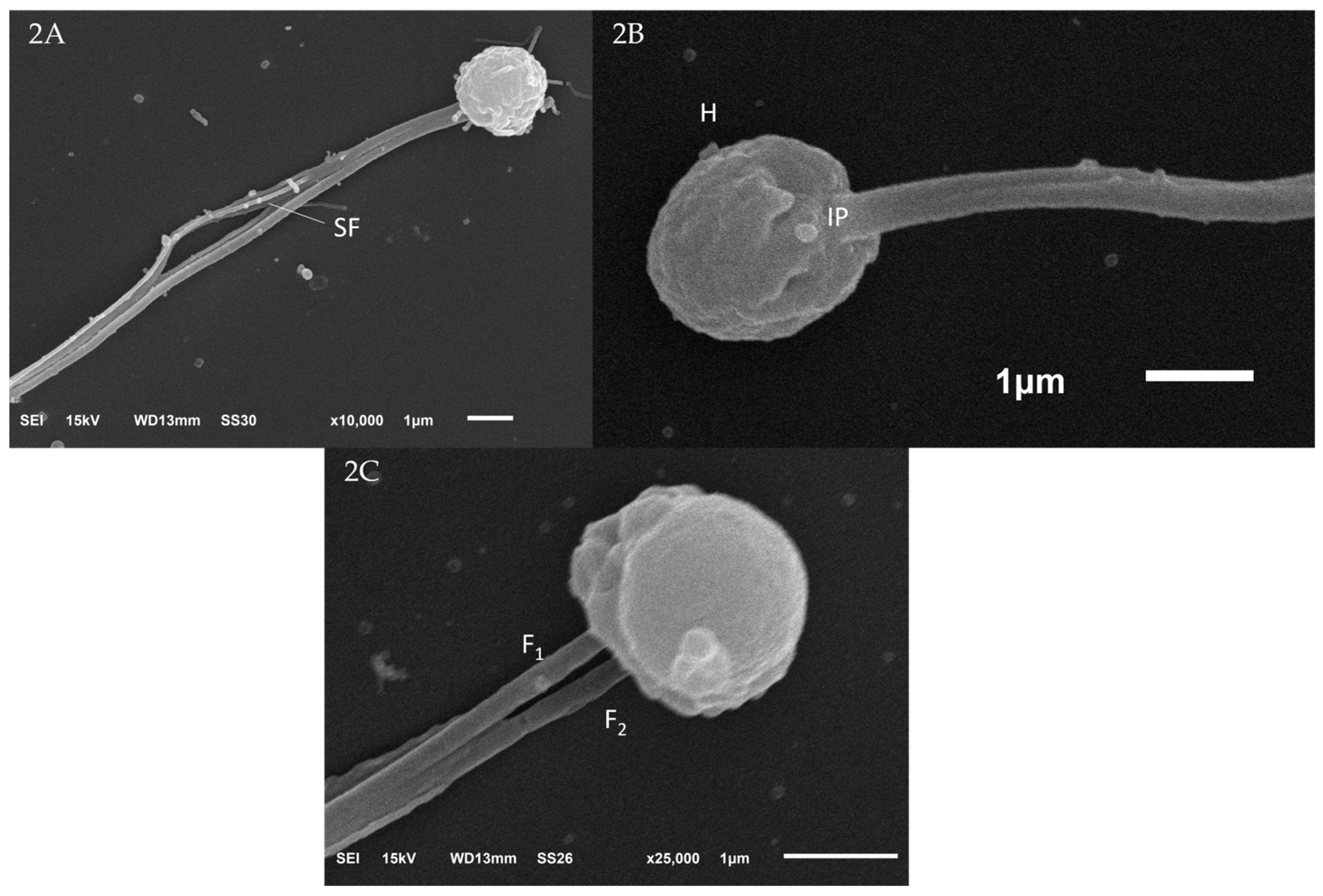
3.1.1. Scanning Electron Microscopy (SEM) of Sperm

The spermatozoon of A. gigas is a biflagellate cell comprising a spherical head, a short intermediate piece region and two flagella (Figure 2). The mean head length and width were 3.32 ± 0.49 µm and 2.87 ± 0.39 µm, respectively (Table 1). The flagellum length was 68.34 ± 5.69 µm (Table 1).

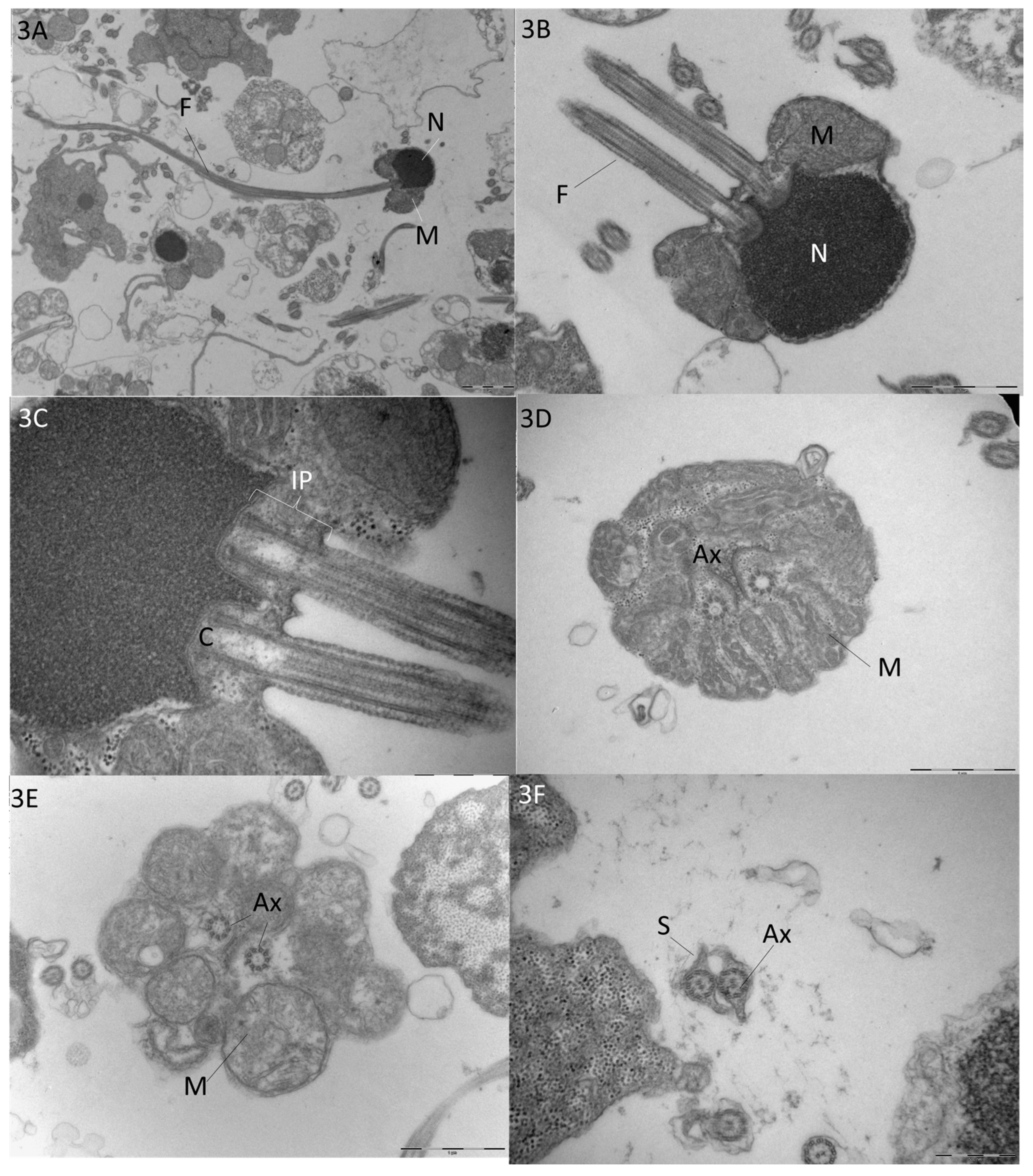
3.1.2. Transmission Electron Microscopy (TEM) of Sperm

The nucleus is spherical and electron-dense, and its envelope is found adjacent to the plasma membrane (Figure 2B and Figure 3A). Membrane integrity was 72.1% (Table 1). No acrosome vesicle was found. At the base of the nucleus, there are two nuclear fossae where the centrioles are positioned (Figure 3C). The anterior part of the flagella is positioned at the lateral base of the nucleus, forming an intermediate piece. Lateral to the intermediate piece, forming two sets, tubular cristae mitochondria are abundant and occupy most of the cytoplasmic volume (Figure 3B,C). The two flagella were found with 9 + 2 axonemal structures, with an axoneme formed by nine peripheral doublets with two central microtubules (Figure 3D,E). The central microtubules are aligned with side fins formed laterally by the axoneme plasma membrane (Figure 3F). Side fins were observed from the anterior region to the posterior end.

3.1.3. Spermatozoon Morphometrics and Cell Membrane Integrity

Sperm samples were collected from five males (out of eight) at different sampling times (A, B and C); these males were implanted or not implanted with GnRHa (Table 1). The mean spermatozoan head length was 3.32 ± 0.49 µm, the head width was 2.87 ± 0.39, the head area was 8.26 ± 2.19 µm2, and the flagellum length 68.34 ± 5.69 µm (Table 1). The mean membrane integrity was 72.1%, ranging from 66.5 to 78.8% in all analysed samples (Table 1).

4. Discussion

In this study, for the first time, we report the feasibility of the in vivo collection of semen in the Amazon osteoglossid A. gigas. The collection was carried out with and without hormonal stimulation using GnRHa implants, also in the same fish at different time points. The semen of the pirarucu was found to be translucent and viscous, different from the whitish semen produced by most freshwater species [27,28]. The collected samples obtained were likely mixed with urine since they were activated when observed under a light microscope soon after collection [29]. Membrane integrity is a key index of cell viability, and any cell (here, sperm) with a damaged membrane cannot carry out its functions, i.e., it cannot fertilise eggs [30]. A membrane integrity analysis showed that more than 70% of the spermatozoa sampled were alive and thus had fertilization capability. Cell membrane integrity is dependent on several factors, including fish nutrition and other physical/environmental factors, especially the temperature upon gametogenesis [31,32], and any improvement in membrane integrity needs further investigation.
Interestingly, we found the spermatozoa of A. gigas have two flagella. The presence of biflagellate spermatozoa has been reported in 31 fish species from 16 families [10], but the information available so far indicates that there is no obvious phylogenetic relationship in the evolution of biflagellate spermatozoa [10]. However, future studies on sperm ultrastructure using electron microscopy could reveal more species with biflagellate sperm, as was the case with the Channel catfish, which was once considered a monoflagellate [33,34]. So far, the only osteoglossid found to bear two flagella is Heterotis niloticus [7], considered to be an Arapaima sister group. Both share similar sperm structures with a spherical, electron-dense nucleus about 2–3 µm in diameter, two nuclear fossae connected to two centrioles and a short intermediate piece at the anterior part of the flagellum [7]. Both species are phylogenetically closely related [35], sharing many biological similarities such as a low gonadosomatic index [19,36], with only the left gonad functional, and possess similar reproductive behaviours [19,37]. It is thus possible that semen collection in H. niloticus could be similar to Arapaima, and its collection would then likewise be doable, although the semen characteristics of H. niloticus have not been reported [7]. In general, fish species with biflagellate spermatozoa are external fertilisers; however, an internally fertilising ocean pout, Macrozoarces americanus from the family Zoarcidae, has biflagellate spermatozoa [38]. Among catfish, two species, Ictalurus punctatus and I. nebulosus, have been reported to have biflagellate sperm [34,39], and both have species have spherical nuclei with condensed chromatin, a fossa at the posterior end, and a midpiece that extends anteriorly around the posterior portion of the nucleus. Similarly, A. gigas has spherical nuclei and a short intermediate piece but two nuclear fossae. The significance of these differences in biflagellate sperm ultrastructure is not known because no information is available on sperm behaviour, physiology or fertilisation dynamics. Nevertheless, the findings presented in this study are valuable on biological and biotechnological grounds for A. gigas, as we will discuss further.
So far, a great variety of sperm types have been described within Osteoglossiformes, such as monoflagellated sperm in Pantodontidae and Notopteridae, biflagellate in Osteoglossidae and aflagellate in Mormyrydae and Gymnarchidae [6]. Our findings for A. gigas support the biflagellate character previously assigned to Osteoglossidae based on H. niloticus [7]. It has been previously postulated that biflagellate sperm evolved independently in several groups and has been reported previously for over 32 fish species from seven different orders [6,10]. In addition to the biflagellate condition, the ultrastructure of A. gigas spermatozoon is similar to that reported for H. niloticus [7]. In both species, the spermatozoon cytoplasm is embodied by mitochondria at the nuclear base, the flagella insertion forms a very short intermediate piece, axonemes have a 9 + 2 pattern and side fins are found along the flagella. Differently, the nucleus of the A. gigas spermatozoon was found to be larger (~2.9 µm) in comparison to H. niloticus (2 µm). A previous study reported an 11 µm diameter aperture for the outer micropylar canal in the eggs of A. gigas [40]; thus, 3 µm should be precisely the diameter of the micropylar inner aperture.
For fish species studied so far, it is still not clear whether a biflagellate condition would increase spermatozoa motility and fertility capabilities or improve manoeuvrability, allowing for the blocking of the micropylar canal and thus impeding the entrance of other spermatozoa [6,10]. For A. gigas, it some degree of competition between spermatozoa is expected since the genetic contribution of more than one male to offspring has been reported [41]. Females of A. gigas lay eggs on nests built on a sandy bottom in shallow (c. 1 m) areas [13], and external fertilization should occur just after the eggs are laid. The rounded head of the pirarucu spermatozoon is characteristic of a species with external fertilization, which, together with its biflagellate condition and large number of mitochondria, suggests a long motility period is necessary for egg fertilization after eggs are deposited in the turbid waters in which the nests are built. The relatively larger length of A. gigas sperm (72 µm, including the head and flagella) is also a characteristic of external fertilization because the length of sperm is likely to correlated to their swimming speed and will aid in finding a suitable egg before they become inactive [42]. However, courtship and fertilization events in A. gigas have not been properly described or captured using imaging devices in natural or captive environments, which would enlighten the understanding of reproductive behaviour and the fertilization process.
The size of the intermediate piece of the spermatozoon appeared to be positively correlated to the long lifespan of the sperm and sperm dispersal [43]. In general, most teleost species displaying internal fertilization have a larger intermediate piece and contain a greater number of mitochondria that provide energy via oxidative phosphorylation for sperm motility [5]. In our study, A. gigas had a shorter intermediate piece, which is a common characteristic of many externally fertilizing fishes [44]. Mitochondria provide the required energy for fish spermatozoa motility and integrity, hence determining fertilizing potential [45]. Although it was reported that the mitochondrial richness of the intermediate piece in fish species with external fertilization is low, A. gigas sperm had an abundant number of mitochondria. Nonetheless, the possibility of semen collection in A. gigas opens future investigation possibilities, such as to understanding motility patterns using computer-aided sperm analysis (CASA), longevity and sperm metabolism (respiration activity, ATP content and enzyme activity), among others. Such information is lacking for teleosts with biflagellate sperm [6].
The possibility of semen collection is also relevant to the control of A. gigas reproduction in captivity, one of the current bottlenecks for its aquaculture development [21,46]. The verification of semen after fish stripping could be used for male identification, complementing the cannulation technique currently applied for female identification [40]. Sex identification in the species is a key step since farmers today isolate couples in earth ponds, attempting to stimulate natural reproduction [46,47,48]. The possibility of semen collection could also be applied for artificial fertilization in A. gigas. Although egg collection is still not feasible for A. gigas after hormonal therapy, ovulating females are often found during broodstock handling [40,49]. In this context, future research should be carried out to better understand the opening position of the spermatic duct in the species, thus allowing for the collection of inactivated semen suitable for the development of cryopreservation protocols.

5. Conclusions

In conclusion, this study showed for the first-time feasibility of the in vivo collection of semen from A. gigas independent of hormonal stimulation. We provided a complete description of the spermatozoa ultrastructure after SEM and TEM analyses, showing a biflagellate spermatozoon in the species. The results show that the spermatozoa of A. gigas are biflagellate, mitochondria-rich and robust, corroborating the external fertilization characteristic of the species. This study provides novel insights into the reproductive biology of the species and creates opportunities for future studies of the reproductive biology of this fish.

Supplementary Materials

The following supporting information can be downloaded at https://www.mdpi.com/article/10.3390/fishes9010024/s1, Figure S1: Sample micrographs from used for membrane integrity analysis of Arapaima gigas sperm. Sperm smear slides stained with 5% eosin, 10% nigrosine and observed under a light microscope (400×). Spermatozoa with a colourless cytoplasm were classified as intact, while spermatozoa with a stained cytoplasm were considered ruptured.

Author Contributions

Conceptualization, L.S.T. and L.N.G.-K.; methodology, J.T.L., L.S.T. and L.N.G.-K.; software, L.S.T. and L.N.G.-K.; validation, J.T.L., A.F.L., V.P., S.R.B. and L.N.G.-K.; formal analysis, L.S.T. and L.N.G.-K.; investigation, L.S.T., J.T.L., A.F.L., V.P., S.R.B. and L.N.G.-K.; resources, L.S.T.; data curation, L.S.T., J.T.L., A.F.L., V.P., S.R.B. and L.N.G.-K.; writing—original draft preparation, L.S.T.; writing—review and editing, L.S.T., J.T.L., A.F.L., V.P., S.R.B. and L.N.G.-K.; visualization, L.S.T. and L.N.G.-K.; supervision, L.S.T. and L.N.G.-K.; project administration, L.S.T.; funding acquisition, L.S.T. All authors have read and agreed to the published version of the manuscript.

Funding

This research was funded by the European Union’s Horizon 2020 Research and Innovation Programme under grant agreement N° 818173.

Institutional Review Board Statement

This study was conducted in accordance with the Brazilian guidelines for the care and use of animals for scientific and educational purposes(DBCA) and was approved by the National System for the Management of Genetic Heritage and Associated Traditional Knowledge—SISGEN (ADBE614) and by the Ethics Committee for the Use of Animals(CEUA) of the National Research Centre on Fisheries, Aquaculture and Agricultural Systems (CNPASA) (specific certificate n˚69/2021).

Data Availability Statement

Data supporting reported results can be found at https://zenodo.org/records/8359643 (accessed on 2 January 2024).

Acknowledgments

The authors thank Maria Dolores Ferreira and José Augusto Maulin for scanning electron microscopy and transmission electron microscopy analyses performed at the Laboratory of Electron Microscopy, Faculty of Medicine, São Paulo University (FMRP/USP), Ribeirão Preto-SP.

Conflicts of Interest

The authors declare no conflicts of interest. The funders had no role in the design of the study; in the collection, analyses, or interpretation of data; in the writing of the manuscript; or in the decision to publish the results.

References

  1. Breder, C.M.; Rosen, D.E. Modes of Reproduction in Fishes; The Natural History Press: Garden City, NY, USA, 1966. [Google Scholar]
  2. Rocha, M.J.; Arukwe, A.; Kapoor, B.G. Fish Reproduction; Science Publishers: Enfield, CT, USA, 2008. [Google Scholar]
  3. Sloman, K.A. The Diversity of Fish Reproduction: An Introduction. In Encyclopedia of Fish Physiology—From Genome to Environment; Farrell, A., Ed.; Academic Press: Cambridge, MA, USA, 2011; Volume I, pp. 613–615. [Google Scholar]
  4. Mattei, X. Spermatozoon ultrastructure and its systematic implications in fishes. Can. J. Zool. 1991, 69, 3038–3055. [Google Scholar] [CrossRef]
  5. Pitnick, S.; Hosken, D.J.; Birkhead, T.R. Sperm morphological diversity. In Sperm Biology: An Evolutionary Perspective; Pitnick, S., Hosken, D.J., Birkhead, T.R., Eds.; Elsevier: Amsterdam, The Netherlands, 2009; pp. 69–149. [Google Scholar]
  6. Koenig, L.A.; Gallant, J.R. Sperm competition, sexual selection and the diverse reproductive biology of Osteoglossiformes. J. Fish Biol. 2021, 99, 740–754. [Google Scholar] [CrossRef]
  7. Mattei, X.; Marchand, B.; Quilichini, Y. A biflagellate spermatozoon in the African bonytongue Heterotis niloticus (Teleostei, Osteoglossidae). J. Fish Biol. 2019, 94, 335–338. [Google Scholar] [CrossRef]
  8. Morrow, E.H. How the sperm lost its tail: The evolution of aflagellate sperm. Biol. Rev. 2004, 79, 795–814. [Google Scholar] [CrossRef]
  9. Bratton, B.O.; Kramer, B. Patterns of the electric organ discharge during courtship and spawning in the mormyrid fish, Pollimyrus isidori. Behav. Ecol. Sociobiol. 1989, 24, 349–368. [Google Scholar] [CrossRef]
  10. Montgomerie, R.; Fitzpatrick, J.L. Testes, sperm, and sperm competition. In Reproductive Biology and Phylogeny of Fishes (Agnathans and Bony Fishes): Phylogeny, Reproductive System, Viviparity, Spermatozoa; Jamieson, B.G., Ed.; CRC Press: Boca Raton, FL, USA, 2009; pp. 1–53. [Google Scholar]
  11. Du Plessis, L.; Soley, J.T. Structural peculiarities associated with multiflagellate sperm in the emu, Dromaius novaehollandiae. Theriogenology 2012, 78, 1094–1101. [Google Scholar] [CrossRef]
  12. Nelson, J.S.; Grande, T.C.; Wilson, M.V.H. Fishes of the World, 5th ed.; Wiley: Hoboken, NJ, USA, 2016. [Google Scholar]
  13. Castello, L. Nesting habitat of Arapaima gigas (Schinz) in Amazonian floodplains. J. Fish Biol. 2008, 72, 1520–1528. [Google Scholar] [CrossRef]
  14. Gurdak, D.J.; Stewart, D.J.; Castello, L.; Arantes, C.C. Diversity in reproductive traits of arapaima (Arapaima spp., Müller, 1843) in Amazonian várzea floodplains: Conservation implications. Aquat. Conserv. 2019, 29, 245–257. [Google Scholar] [CrossRef]
  15. Castello, L. Lateral migration of Arapaima gigas in floodplains of the Amazon. Ecol. Freshw. Fish 2008, 17, 38–46. [Google Scholar] [CrossRef]
  16. Fontenele, O. Contribuição para o conhecimento da biologia do Pirarucú, “Arapaima gigas” (Cuvier), em cativeiro (Actinopterygii, Osteoglossidae). Rev. Bras. Biol. 1948, 8, 445–459. [Google Scholar]
  17. Zelada-Mázmela, R.F.; Gutiérrez, G.A.; Zelada-Mázmela, E. Histological description of the early gonadal development of Arapaima gigas, paiche. J. World. Aquac. Soc. 2022, 53, 754–764. [Google Scholar] [CrossRef]
  18. Amaral, A.C.; Lima, A.F.; Ganeco-Kirschnik, L.N.; Almeida, F.L. Morphological characterization of pirarucu Arapaima gigas (Schinz, 1822) gonadal differentiation. J. Morphol. 2020, 281, 491–499. [Google Scholar] [CrossRef] [PubMed]
  19. Godinho, H.P.; Santos, J.E.; Formagio, P.S.; Guimarães-Cruz, R.J. Gonadal morphology and reproductive traits of the Amazonian fish Arapaima gigas (Schinz, 1822). Acta Zool. Stock. 2005, 86, 289–294. [Google Scholar] [CrossRef]
  20. Núñez, J.; Duponchelle, F. Towards a universal scale to assess sexual maturation and related life history traits in oviparous teleost fishes. Fish Physiol. Biochem. 2009, 35, 167–180. [Google Scholar] [CrossRef]
  21. Núñez, J.; Chu-Koo, F.; Berland, M.; Arévalo, L.; Ribeyro, O.; Duponchelle, F.; Renno, J. Reproductive success and fry production of the paiche or pirarucu, Arapaima gigas (Schinz), in the region of Iquitos, Perú. Aquac. Res. 2011, 42, 815–822. [Google Scholar] [CrossRef]
  22. Farrel, A.P.; Randall, D.J. Air-breathing mechanics in two Amazonian teleosts, Arapaima gigas and Hoplerythrinus unitaeniatus. Can. J. Zool. 1978, 56, 939–945. [Google Scholar] [CrossRef]
  23. Faustino, F.; Silva, R.C.; Hilbig, C.C.; Makino, L.C.; Senhorini, J.A.; Ninhaus-Silveira, A.; Nakaghi, L.S.O. Spermatozoon ultrastructure and semen parameters of Brycon vermelha (Characiformes, Characidae). Anim. Reprod. Sci. 2015, 157, 17–23. [Google Scholar] [CrossRef]
  24. Schneider, C.A.; Rasband, W.S.; Eliceiri, K.W. NIH Image to ImageJ: 25 years of image analysis. Nat. Methods 2012, 9, 671–675. [Google Scholar] [CrossRef]
  25. Blom, E. A one-minute live-dead sperm stain by means of eosin-nigrosin. Fertil. Steril. 1950, 1, 176–177. [Google Scholar] [CrossRef]
  26. Lubzens, E.; Daube, N.; Pekarsky, I.; Magnus, Y.; Cohen, A.; Yusefovich, F.; Feigin, P. Carp (Cyprinus carpio L.) spermatozoa cryobanks—Strategies in research and application. Aquaculture 1997, 155, 13–30. [Google Scholar] [CrossRef]
  27. Woynárovich, A.; Anrooy, R.V. Field Guide to the Culture of Tambaqui (Colossoma macropomum, Cuvier, 1816); Food and Agriculture Organization of the United Nations: Rome, Italy, 2019. [Google Scholar]
  28. Mataveli, M.; Moraes, G.V.; Vargas, L.D.; Toninato, M.J.C.; Sakaguti, E.S.; Barbosa, R.C.; Streit, D.P., Jr.; Merlini, L. Avaliação da qualidade do sêmen de tilápia-do-nilo (Oreochromis niloticus), linhagem chitralada, suplementada com diferentes concentrações de vitamina c. Bol. Inst. Pesca 2007, 33, 1–7. [Google Scholar]
  29. Król, J.; Żarski, D.; Bernáth, G.; Palińska-Żarska, K.; Krejszeff, S.; Długoński, A.; Horváth, Á. Effect of urine contamination on semen quality variables in Eurasian perch Perca fluviatilis L. Anim. Reprod. Sci. 2018, 197, 240–246. [Google Scholar] [CrossRef] [PubMed]
  30. Lahnsteiner, F. Characterization of seminal plasma proteins stabilizing the sperm viability in rainbow trout (Oncorhynchus mykiss). Anim. Reprod. Sci. 2007, 97, 151–164. [Google Scholar] [CrossRef]
  31. Drokin, S.I. Phospholipids and fatty acids of phospholipids of sperm from several freshwater and marine species of fish. Comp. Biochem. Physiol. Part B 1993, 104, 423–428. [Google Scholar] [CrossRef]
  32. Labbé, C.; Maisse, G. Influence of rainbow trout thermal acclimation on sperm cryopreservation: Relation to change in the lipid composition of the plasma membrane. Aquaculture 1996, 145, 281–294. [Google Scholar] [CrossRef]
  33. Jaspers, E.J.; Avault, J.W.; Russell, J.D. Spermatozoal morphology and ultrastructure of channel catfish, Ictalurus punctatus. Trans. Am. Fish. Soc. 1976, 105, 475–480. [Google Scholar] [CrossRef]
  34. Poirier, G.R.; Nicholson, N. Fine-structure of the testicular spermatozoa from the channel catfish, Ictalurus punctatus. J. Ultrastruct. Res. 1982, 80, 104–110. [Google Scholar] [CrossRef]
  35. Lavoue, S. Was Gondwanan breakup the cause of the intercontinental distribution of Osteoglossiformes? A time-calibrated phylogenetic test combining molecular, morphological, and paleontological evidence. Mol. Phylogenet. Evol. 2016, 99, 34–43. [Google Scholar] [CrossRef]
  36. Irene, K.F.K.; Tiéhoua, K.; Yaya, S.; Konan, N. Reproduction De Heterotis Niloticus (Cuvier, 1829) De La Riviere Agneby (Cote d’Ivoire). Eur. J. Sci. Res. 2016, 12, 1857–7881. [Google Scholar]
  37. Monentcham, S.E.; Kouam, J.; Pouomogne, V.; Kestemont, P. Biology and prospect for aquaculture of African bonytongue, Heterotis niloticus (Cuvier, 1829): A review. Aquaculture 2009, 289, 191–198. [Google Scholar] [CrossRef]
  38. Yao, Z.; Crim, L.W. Spawning of ocean pout (Macrozoarces americanus L.): Evidence in favour of internal fertilization of eggs. Aquaculture 1995, 130, 361–372. [Google Scholar] [CrossRef]
  39. Burke, M.G.; Leatherland, J.F. Seasonal changes in testicular histology of brown bullheads, Ictalurus nebulosus Lesueur. Can. J. Zool. 1984, 62, 1185–1194. [Google Scholar] [CrossRef]
  40. Torati, L.S.; Lima, A.F.; Ganeco-Kirschnik, L.N.; Migaud, H. Endoscopy and Cannulation as Non-Invasive Tools to Identify Sex and Monitor Reproductive Development in Arapaima gigas. Copeia 2019, 107, 287–296. [Google Scholar] [CrossRef]
  41. Farias, I.P.; Leão, A.; Crossa, M.; Almeida, Y.S.; Honczaryk, A.; Verba, J.T.; Hrbek, T. Evidence of polygamy in the socially monogamous Amazonian fish Arapaima gigas (Schinz, 1822) (Osteoglossiformes, Arapaimidae). Neotrop. Ichthyol. 2015, 13, 195–204. [Google Scholar] [CrossRef]
  42. Fitzpatrick, J.L.; Desjardins, J.K.; Milligan, N.; Montgomerie, R.; Balshine, S. Reproductive-tactic-specific variation in sperm swimming speeds in a shell-brooding cichlid. Biol. Reprod. 2007, 77, 280–284. [Google Scholar] [CrossRef]
  43. Smith, C.C.; Ryan, M.J. Evolution of sperm quality but not quantity in the internally fertilized fish Xiphophorus nigrensis. J. Evol. Biol. 2010, 23, 1759–1771. [Google Scholar] [CrossRef] [PubMed]
  44. Lahnsteiner, F.; Berger, B.; Weismann, T.; Patzner, R.A. Motility of spermatozoa of Alburnus alburnus (Cyprinidae) and its relationship to seminal plasma composition and sperm metabolism. Fish Physiol. Biochem. 1996, 15, 167–179. [Google Scholar] [CrossRef]
  45. Coward, K.; Bromage, N.R.; Hibbitt, O.; Parrington, J. Gamete physiology, fertilization and egg activation in teleost fish. Rev. Fish Biol. Fish. 2002, 12, 33–58. [Google Scholar] [CrossRef]
  46. Torati, L.S.; Taylor, J.; Mesquita, P.E.C.; Migaud, H. GnRHa implants and size pairing effects on plasma and cephalic secretion sex steroids in Arapaima gigas. Gen. Comp. Endocrinol. 2020, 299, 113614. [Google Scholar] [CrossRef]
  47. Chu-Koo, F.; Dugue, R.; Alvan Aguilar, M.; Casanova Daza, A.; Alcantara Bocanegra, F.; Chavez Veintemilla, C.; Duponchelle, F.; Renno, J.F.; Tello, S.; Nunez, J. Gender determination in the Paiche or Pirarucu (Arapaima gigas) using plasma vitellogenin, 17β-estradiol, and 11-ketotestosterone levels. Fish Physiol. Biochem. 2009, 35, 125–136. [Google Scholar] [CrossRef]
  48. Lima, A.F. The influence of sex ratio on the reproduction of pirarucu, Arapaima gigas, in captivity. Acta Amaz. 2018, 48, 38–41. [Google Scholar] [CrossRef]
  49. Torati, L.S.; Varges, A.P.S.; Galvão, J.A.S.; Mesquita, P.E.C.; Migaud, H. Endoscopy application in broodstock management of Arapaima gigas (Schinz, 1822). J. Appl. Ichthyol. 2016, 32, 353–355. [Google Scholar] [CrossRef]
Figure 1. Collection of semen from live broodstock of Arapaima gigas, applying a cephalocaudal pressure on its abdominal left side and suctioning samples using a sterile 1 mL syringe.
Fishes 09 00024 g001
Figure 2. Spermatozoa structure of Arapaima gigas observed through scanning electron microscopy (SEM). (A) General view of an entire spermatozoon depicting the head, the two flagella and side fins (SF). (B) Magnification of the head (H) region depicting the intermediate piece (IP) area. (C) Magnified view of the head with a clearer view of both F1 and F2 flagella.
Fishes 09 00024 g002
Figure 3. Ultrastructure of Arapaima gigas spermatozoa, obtained via transmission electron microscopy (TEM). (A,B) Longitudinal sections of striped spermatozoa depicting two flagella (F) and the head with the nucleus (N) and tubular cristae mitochondria (M). (C) Head detail showing the centrioles (C) and intermediate piece (IP) region. (D,E) Intermediate piece cross-section depicting both axonemes (Ax) arranged in a 9 + 2 pattern and surrounded by mitochondria (M). (F) Flagella cross-section depicting axonemes (Ax) and side fins (S).
Fishes 09 00024 g003
Table 1. Spermatozoa of Arapaima gigas. Mean (±SD) values obtained for the head length (µm), head width (µm), head area (µm2), flagellum length (µm), and mean membrane integrity (%). The time column corresponds to dates A—4 May 2021 (baseline); B—25 May 2021 (21 days post first GnRHa implantation) and C—15 June 2021 (21 days post second GnRHa implantation) for males which were (or not) GnRHa-implanted. For each sample, 25 spermatozoa were measured. For membrane integrity, the number of observed spermatozoa is indicated in the table.
Male IDGnRHa
Implanted
TimeHead Length (µm)Head Width (µm)Head Area (µm2)Flagellum Length (µm)Membrane Integrity (%)
M1YesB3.84 ± 0.683.22 ± 0.4410.60 ± 3.1567.01 ± 5.9466.8 (n = 542)
M1YesC2.73 ± 0.262.38 ± 0.345.66 ± 1.2272.15 ± 3.9278.8 (n = 330)
M3NoA3.18 ± 0.322.87 ± 0.248.30 ± 1.2467.69 ± 5.3078.4 (n = 385)
M3NoB3.58 ± 0.303.08 ± 0.259.32 ± 0.9872.44 ± 2.24n.a.
M4YesA2.33 ± 0.302.89 ± 0.198.41 ± 1.2768.81 ± 1.8462.5 (n = 387)
M7YesA3.18 ± 0.312.69 ± 0.267.42 ± 1.2668.79 ± 4.2870.4 (n = 81)
M7YesC3.39 ± 0.542.74 ± 0.447.53 ± 2.2563.13 ± 7.9478.8 (n = 142)
M8YesB3.36 ± 0.253.05 ± 0.258.71 ± 1.6365.62 ± 6.4969.0 (n = 113)
Overall mean3.32 ± 0.492.87 ± 0.398.26 ± 2.1968.34 ± 5.6972.1 (n = 1980)
Disclaimer/Publisher’s Note: The statements, opinions and data contained in all publications are solely those of the individual author(s) and contributor(s) and not of MDPI and/or the editor(s). MDPI and/or the editor(s) disclaim responsibility for any injury to people or property resulting from any ideas, methods, instructions or products referred to in the content.

