Article

Antioxidant Defense of Mytilus galloprovincialis Mussels Induced by Marine Heatwaves in Correlation with Marteilia Pathogen Presence

by
Athanasios Lattos
1,
Dimitrios K. Papadopoulos
1,
Konstantinos Feidantsis
2,*,
Ioannis A. Giantsis
3,
Ioannis Georgoulis
1,
Dimitrios Karagiannis
4 and
Basile Michaelidis
1,*
1
Laboratory of Animal Physiology, Department of Zoology, School of Biology, Faculty of Science, Aristotle University of Thessaloniki, GR-54124 Thessaloniki, Greece
2
Department of Fisheries & Aquaculture, University of Patras, GR-26504 Mesolonghi, Greece
3
Department of Animal Science, Faculty of Agricultural Sciences, University of Western Macedonia, GR-531 00 Florina, Greece
4
School of Veterinary Medicine, Aristotle University of Thessaloniki, GR-54124 Thessaloniki, Greece
*
Authors to whom correspondence should be addressed.
Fishes 2023, 8(8), 408; https://doi.org/10.3390/fishes8080408
Submission received: 23 July 2023 / Revised: 29 July 2023 / Accepted: 1 August 2023 / Published: 8 August 2023

Abstract

:
Background: The effects of climate change negatively affect marine bivalves’ health. Lately, the intensity of marine heatwaves poses serious threats to the aquatic environment setting of high-risk bivalve farming. Since temperature increases can jeopardize bivalves’ immunity response, pathogen infection becomes more evident. Reactive oxygen species (ROS) production, increased during the process of phagocytosis, is confronted by the animals’ antioxidant defense system. However, apart from pathogenic infections, antioxidant defense responses are also induced by seawater temperature increases; Methods: To enlighten the antioxidant status of Mytilus galloprovincialis originating from mortality events enhanced by intense heatwaves in Thermaikos Gulf, northern Greece, along with Marteilia refringens infection, we examined the expression of genes related to antioxidant defense (catalase, CuSOD and mt10) along with the lipid peroxidation levels and activity levels of antioxidant enzymes (catalase, SOD and glutathione reductase); Results: Our results exhibited increased levels of all these biomarkers. This increase was intensified in the Marteilia infected individuals; Conclusions: Our findings shed light on the oxidative and antioxidant status of farmed mussels led to mortality in the context of Marteilia infection. The latter is augmented by the synergistic effect of heatwaves causing a significant increase in oxidative damage and subsequent antioxidant defense.
Key Contribution: Heatwaves increased all examined oxidative and antioxidant-related bio-indicators in M. galloprovincialis mussels, especially those infected with Marteilia, the major pathogen of farmed mussels. These results enlighten the existing biological impact of heatwaves, providing insights for future management of the marine aquatic sector.

Graphical Abstract

1. Introduction

The effects of climate change constitute a major inhibition factor in global marine aquaculture production [1,2,3]. Specifically, ocean acidification and acute temperature precipitation pattern changes pose a negative impact on the sector [4,5]. Furthermore, the consequences of the aforementioned factors directly affect farmed species, rendering them more vulnerable to opportunistic microorganisms [3]. Hence, microorganisms belonging to many taxonomic clades, such as bacteria, viruses and protozoan and metazoan parasites, take advantage of the physiological stress and the subsequent immunological depression of aquatic farmed species, resulting in infections that lead to severe population reductions or even mass mortalities [6,7,8,9,10].
The effects of climate change have been proven to greatly downregulate the physiological performance of aquatic species. Regarding marine invertebrates, which constitute a valuable group of animals in terms of food production, the effects of climate change affect their productivity in many ways [3]. Acute modification of the values of several abiotic factors affects both the growth performance and welfare of cultured species [3,6,11]. Additionally, it has been demonstrated that changes in salinity, pH and temperature affect immunity in marine bivalves [12]. Since marine invertebrates rely mainly on their innate immune system to confront microorganism infection, phagocytosis is the basal immune mechanism that copes with pathogens [13,14]. During the process of phagocytosis, reactive oxygen species (ROS) production, a process known as respiratory burst (resulting from the interaction of several substrates with the NADPH—oxidase multiprotein complex) is taking place in tissues [7,15,16,17]. Due to the fact that this process results in pathogen-derived reactive oxygen intermediates (ROIs) and ROS production, marine bivalves possess several antioxidant mechanisms [18] in order to prevent severe cellular damage from excessive oxidative stress and finally maintain an equilibrium between antioxidants and pro-oxidants [19]. Thus, invertebrates such as marine bivalves possess enzymes, such as superoxide dismutase (SOD), catalase (CAT) and glutathione reductase, as a part of their antioxidant defense system [19]. These enzymatic processes can catalyze the conversion of hydrogen peroxide into less-reactive oxygen molecules and water [20,21]. Nevertheless, apart from pathogenic infections, antioxidant defense responses are also induced by rapid changes in abiotic factors such as ambient seawater temperature [22,23].
Due to the ongoing climate change, the strength, frequency and duration of marine heatwaves are expected to increase in the future [24], which are periods during which the water temperature is abnormally warm for the time of the year relative to historical temperatures (with temperatures warmer than the 90th percentile based on a 30-year historical baseline period) with that extreme warmth persisting for days (five or more) to months [24,25]. In recent years, climate change has become a serious inhibitory factor in mussel aquaculture, and it has become clear that marine heatwaves tend to worsen the aforementioned factor. Specifically, as described in Lattos et al. [6], marine heatwaves with the synergistic effects of Marteilia refringens limited the production in North Greece in 2022 and even annihilated mussels resulting in the complete loss of production in 2021 [6]. This phenomenon was initiated after strong precipitation events; these resulted in a temporal temperature decrease during mid-summer (July) and continued in the next days with an acute temperature increase. At the same time, protozoan parasites such as M. refringens were favored from the temperature increase and the heat stress of the hosts and multiplied uncontrollably, creating infections in the cultured species [6]. As a result, farmers applying the long-line system of mussel culture are trying to submerge their equipment in deeper spots. On the other hand, farmers using the traditional type of farming (bouchot) as described in Lattos et al. [6] face the phenomenon occasionally without being able to save their production unless they have the ability to transfer the cultured species to greater depths.
Aquatic organisms’ biochemical and physiological responses during acclimation to increasing temperature have been thoroughly studied under laboratory conditions. These studies support the establishment of the physiological mechanisms that are involved during both various exposures and acclimation to mimicked field conditions [26,27,28,29]. However, so far, only a few studies examining the organisms’ physiological responses to seasonality have been employed in the field. These studies eventually demonstrate the clear links between the results obtained under laboratory and field conditions. Under this prism, field studies on farmed aquatic organisms would be a useful tool to reveal climate-induced effects. Thus, the main objective of the present study was to investigate the antioxidant responses of the Mediterranean mussel Mytilus galloprovincialis (Lamarck, 1819) in both culture types (traditional bouchot culture (TC) and long-line system (LC)) under the presence or absence of the cercozoan parasite M. refringens. Further, the investigation was carried out during the increase in the ambient sea water temperature during a heatwave event in order to enlighten the underlying mechanisms of antioxidant defense and complement our previous results regarding mussels’ pathophysiological responses to marine heatwaves and, in addition, to the synergistic effect of Marteilia infection.

2. Materials and Methods

2.1. Sampling Procedure

The sampling procedure is described in detail in Lattos et al. [6]. Specifically, adult mussels (M. galloprovincialis) with a weight of 22.43 ± 3.21 g (mean ± SD), a shell length of 5.62 ± 0.35 cm and a shell width of 2.89 ± 0.11 cm were sampled. The sampling was conducted from both the traditional bouchot culture (TC) and long-line system (LC). Variations in sea water physicochemical parameters were measured by the employment of a Multiparameter Water Quality Meter (Model WQC-24, DKK-TOA Company, Bad Nauheim, Germany). Thus, apart from the sea water temperature, the pH, the concentration of oxygen and the salinity were also recorded (at both 12 a.m. and 12 p.m. daily on a monthly basis). The samplings were conducted in the marine area of Thermaikos Gulf at the end of May when the surface sea water temperature was 18 °C, at the beginning of July when the surface sea water temperature reached 28 °C, on 25 July 2022 when the surface sea water temperature increased to 28.2 °C and finally on 1 August 2022 when the surface sea water temperature reached 29.5 °C (Figure 1). The selection of 26 May as the initial sampling point was due to the fact that mussels exhibit their optimal physiological performance at approximately 18 °C [30,31]. It should be mentioned that the highest surface sea water temperature value in the period from 2009 to 2022 was 27.2 °C, thus underlying the fact that the present study’s samplings were conducted during a marine heatwave (Figure 1). Other sea water physicochemical parameters remained unchanged and thus are not presented (Salinity (g/L): 26 May = 34.9, 13 July = 34.9, 25 July = 35.2, 1 August = 35.1; oxygen (mg/L): 26 May = 8.1, 13 July = 8.2, 25 July = 8.3, 1 August = 8.1; and pH: 26 May = 8.1, 13 July = 8.2, 25 July = 8.3, 1 August = 8.1). Mortalities increased parallel to the increasing seawater temperature and reached 100% in the bouchot culture. From each culture process, ten individuals were sampled. From each sample animal, half of the digestive gland and half of the mantle were extracted, stored in 1.5 mL eppendorf tubes and immediately frozen in liquid nitrogen. Then, all samples were transferred to the laboratory, where they were stored at −80 °C.

2.2. Histopathogical Detection of M. refringens

The remaining part of the digestive gland after dissection was stored in Davidson fixative agent as described in Lattos et al. [6] according to Shaw and Battle [32]. Parasitic infections were evaluated via histological sections stained with hematoxylin and eosin according to Howard et al. [33].

2.3. RNA Extraction and cDNA Synthesis

Total RNA extraction was conducted from the mantle of the mussels using the NucleoZOL reagent (Macherey-Nagel, Düren, Germany). This process was carried out according to the manufacturer’s instructions. However, the optional separation step was not performed. Briefly, approximately 50 mg of the mantle was homogenized with piston pestling in 500 μL NucleoZOL. Then, into the lysate, RNAase-free water was added. Subsequently, for RNA precipitation, centrifugation and then the addition of isopropanol followed. The samples were again centrifuged, and the RNA pellet was subjected to two ethanol washes. Finally, dilution in 60 μL nuclease-free water of the extracted RNA was performed. A Quawell UV–Vis 5000 spectrophotometer (Quawell Technology, San Jose, CA, USA) was used for the estimation of RNA purity and concentration. The extracted RNA was stored at −80 °C until reverse transcription. For the reverse transcription process, approximately 500 ng of total RNA from each sample was subjected to cDNA synthesis using the PrimeScript kit (Takara, Japan) and the oligodT primers according to the manufacturer’s protocol. Similarly to RNA, the evaluation of cDNA concentration and purity was performed. Thereafter, the samples were stored at −20 °C until qPCR application.

2.4. Gene Expression Analysis

The gene expression of three antioxidant genes (Catalase, Cu/Zn superoxide dismutase and Metallothionein-10) was calculated through quantitative real-time PCR (qPCR). The comparative CT method (2−ΔΔCT method) [34] was employed for the quantification of the relative expression of the above-mentioned genes. The cDNA originating from the samples collected on the 26th of May was designated as the control samples. The Ct values of all studied genes were normalized to the Ct values of the reference gene EF1-a. For each gene, three different cDNA from each group of mussels were subjected to real-time PCR. The PCR reactions were performed by the employment of the KAPA SYBR® FAST qPCR Master Mix (2X) kit in a 10 μL volume. The reaction wells contained 2 μΜ of each primer, 5 μL of KAPA SYBR® FAST qPCR Master Mix (2X), 10 ng of template cDNA and PCR-grade water up to 10 μL. The qPCR Thermocycler Eco 48 Real-time PCR (PCRmax, San Diego, CA, USA) was used in order to perform the runs for 40 cycles. The gene-specific primers utilized for the qPCR reactions are shown in Table 1.

2.5. Assays of Antioxidant Enzymes and Lipid Peroxidation (TBARS)

The activities of the antioxidant enzymes catalase (CAT), superoxide dismutase (SOD) and glutathione reductase (GR) in the mantle of the sampled mussels were measured in the homogenized tissue samples’ supernatants (2000× g, 4 °C, 15 min) [39]. Specifically, catalase (EC 1.11.1.6) enzymatic activity resulted from the changes in absorbance of hydrogen peroxide (H2O2) (240 nm) [40]. SOD (EC 1.15.1.1) total enzymatic activity was the result of the inhibition of NADH by β-mercaptoethanol [41], while GR (EC 1.8.1.7) enzymatic activity levels resulted from the absorbance changes in NADPH (240 nm) [42]. Protein concentrations were determined using the BioRad protein assay. All enzymatic activities are expressed as U per mg protein.
Homogenization of the mantle samples for the measurement of TBARS and the activities of the cytosolic and mitochondrial enzymes was performed according to Buege and Aust [43]. The terminal product, malondialdehyde (MDA), formed by the decomposition of polyunsaturated fatty acids mediated by free radicals was quantified as thiobarbituric acid reactive substances.

2.6. Statistical Analysis

To test for significance at p < 0.05 (5%) between all experimental groups, one-way analysis of variance (ANOVA) (GraphPad Instat 3.0) was employed. This was followed by Bonferroni post hoc analysis. Friedman’s nonparametric test and Dunn’s post hoc test were applied, since normality tests exhibit little power to test the homogeneity of small sample size data. For the assessment of the significance of factor interactions, two-way ANOVA (GraphPad Prism 5.0) was also performed with the following as fixed factors: different aquaculture techniques and Marteilia spp. presence. Moreover, for the determination of the correlation between the examined variables, principal components analysis (PCA) was applied using the FactoMineR package in R [44].

3. Results

3.1. Marteilia Detection

As already mentioned in Lattos et al. [6], no infected individuals were detected in the LC in the sampling from the 13 July 2022, thus indicating a 0% prevalence rate. On the other hand, in the TC from the same date, one sample was heavily infected with marteiliosis. In the sampling from the 25 July 2022, the prevalence rate of marteiliosis was 20% in both the LC and TC, while in both culture types, individuals were divided into one lightly and one heavily infected. In the sampling from the 1 August 2022, M. refringens was detected only in the LC (Figure 1 and Figure 2).

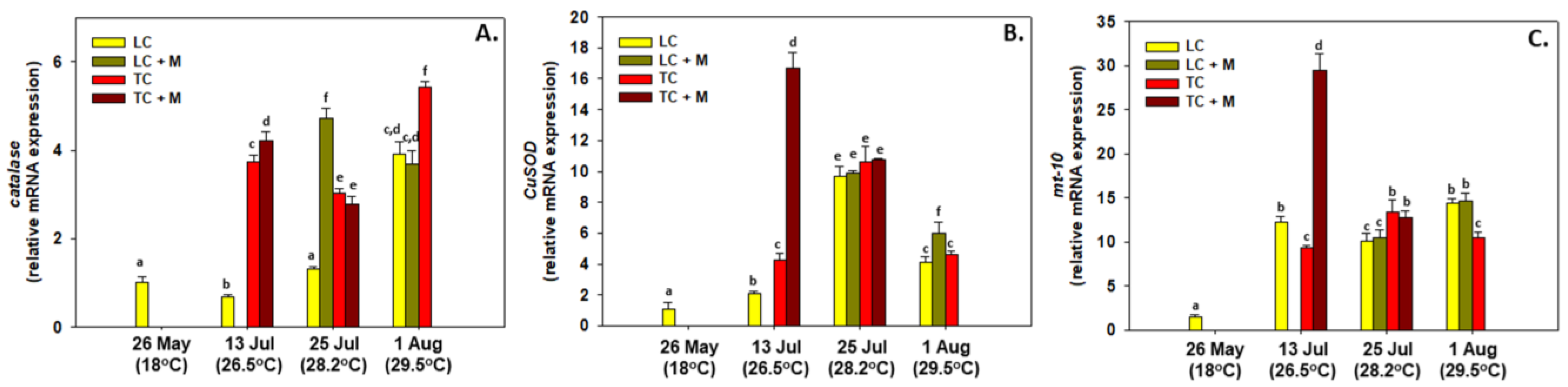
3.2. Gene Expression

A generally increasing pattern of expression was observed in all three genes examined from the samples infected with Marteilia spp., exhibiting higher levels compared to the non-infected samples (Figure 3). Specifically, catalase gene expression levels increased in all samplings compared to the one from May with the sampling on the 1 August of the TC treatment exhibiting the highest values compared to all other samplings. Although the LC samples infected with Marteilia spp. in this sampling exhibited no differences compared to the LC samples in the sampling from the 13 July, the TC samples infected with Marteilia spp. exhibited higher levels compared to the TC samples. This was also the case for the LC samples and the LC samples infected with Marteilia spp. (Figure 3A). Contrary to catalase, CuSOD and mt10 gene expression levels exhibited their highest levels in the TC samples infected with Marteilia spp. in the sampling from the 13 July compared to the non-infected TC samples of the same sampling. However, in the next sampling, no differences were observed between Marteilia spp. infected and non-infected individuals (Figure 3B,C).

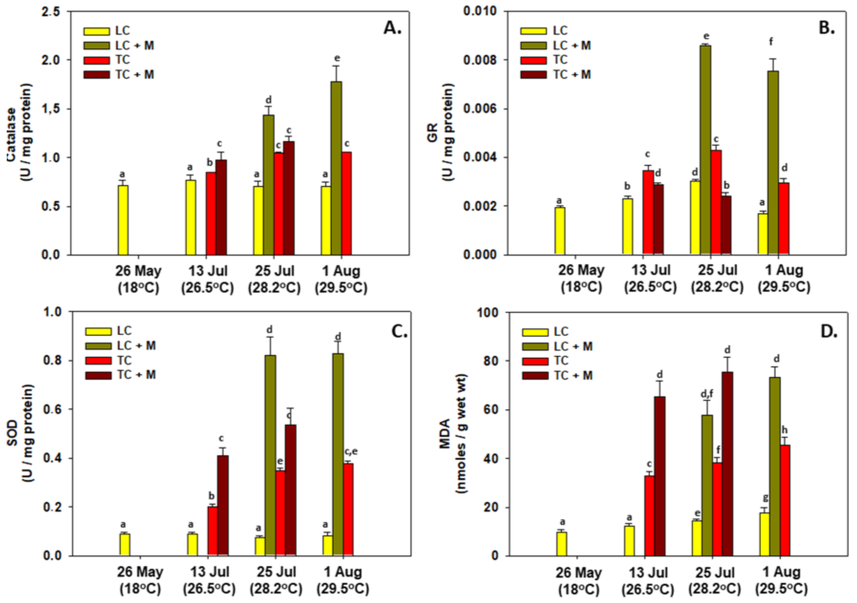
3.3. Enzymatic Activity

The activity levels of antioxidant enzymes followed a pattern similar to that of the expression of genes. Specifically, the samples infected with Marteilia spp. exhibited higher activity levels compared to the non-infected samples. The latter was also exhibited in the MDA levels, depicting higher lipid peroxidation levels in the Marteilia spp. infected samples (Figure 4). While catalase, GR and SOD exhibited their highest activity levels in the LC samples infected with Marteilia spp. in the samplings from the 25 July and the 1 August compared to the other samplings, the lipid peroxidation levels depicted their highest levels in the TC samples infected with Marteilia spp. in the samplings from the 13 and 25 July compared to the other samplings (Figure 4).

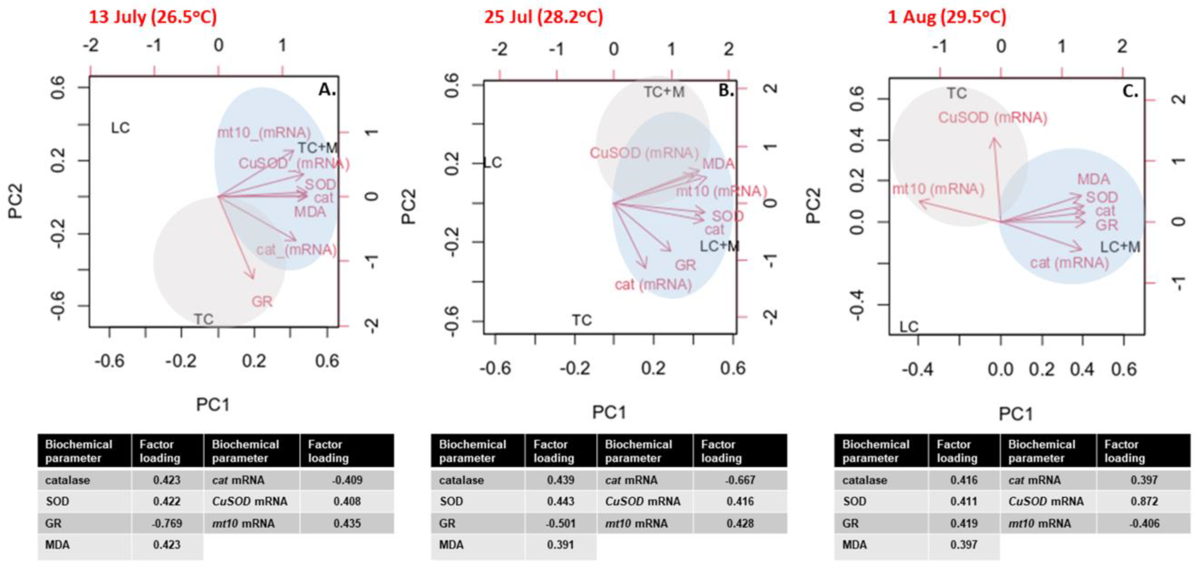
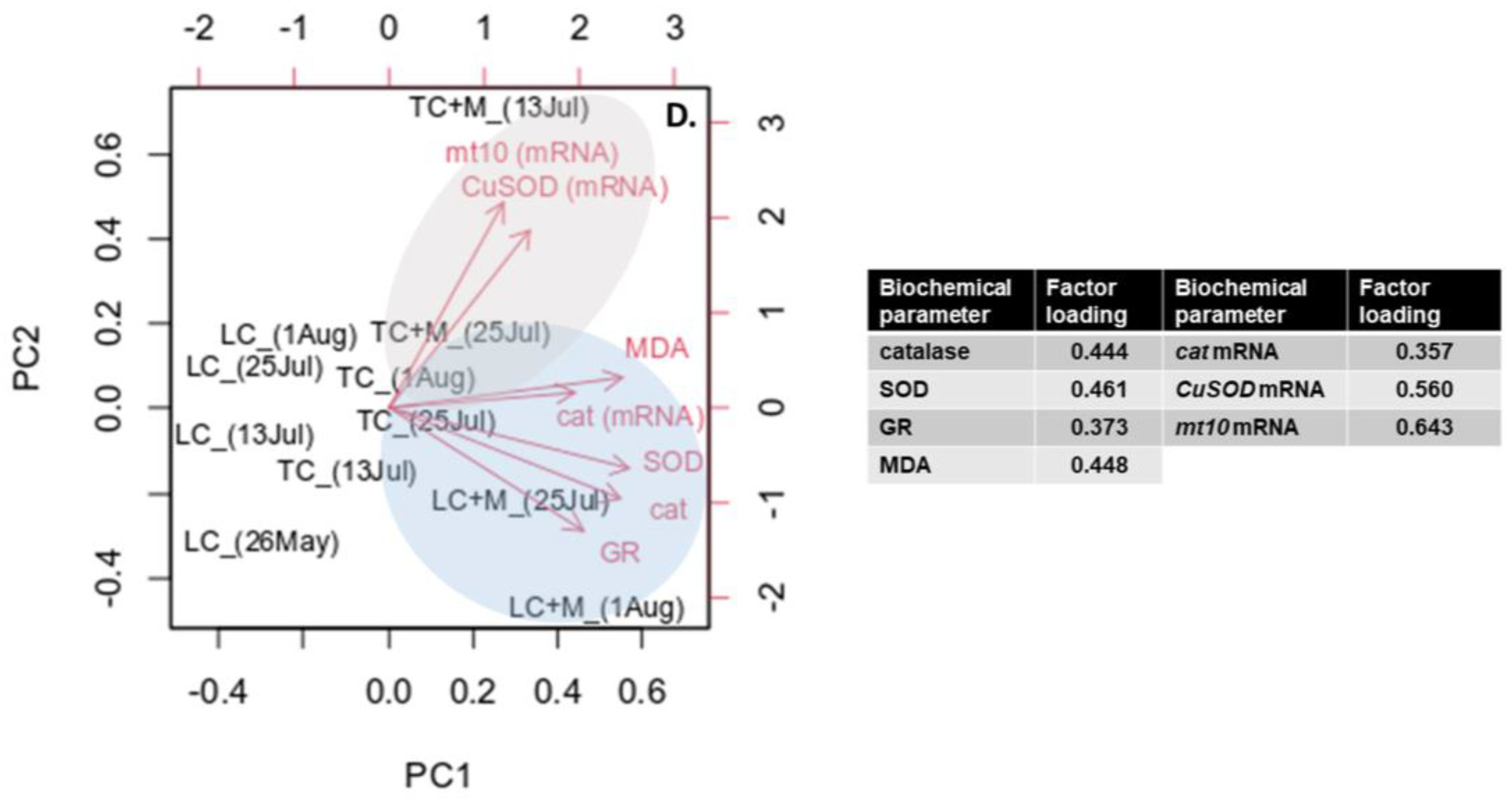
3.4. Multivariate Analysis

PCA analysis was performed for each sampling in order to examine each sea water temperature effect and for all samplings together in order to provide the whole effect of seasonality.
Specifically, in the sampling from the 13 July, clear clusters were formed according to the culture technique and the infection. Specifically, a cluster displaying higher levels of all examined biochemical parameters except that of GR formed in the TC samples infected with Marteilia spp., while the latter parameter formed a cluster with the TC samples. Overall, 79.65% of the variance was attributed to PC1, while 20.35% was attributed to PC2. Cumulatively, PC1 and PC2 explained 100% of the total variance in the dataset (Figure 5A).
In the sampling from the 25 July, two clear clusters were formed: LC samples infected with Marteilia spp. and TC samples infected with Marteilia spp. The LC samples infected with Marteilia spp. formed a cluster with all examined biochemical indicators and shared MDA, CuSOD and mt10 gene expression levels with the cluster formed with the TC samples infected with Marteilia spp. A total of 71.19% of the variance was attributed to PC1, and 16.09% was attributed to PC2. Cumulatively, PC1 and PC2 explained 87.28% of the total variance in the dataset (Figure 5B).
Likewise, in the sampling from the 1 August, two clear clusters were formed: TC samples and LC samples infected with Marteilia spp. Specifically, the TC samples formed a cluster with CuSOD and mt10 gene expression levels, while the LC samples infected with Marteilia spp. formed a cluster with catalase gene expression; catalase, SOD and GR activity levels; and MDA levels. A total of 81.3% of the variance was attributed to PC1, while 18.7% was attributed to PC2. Cumulatively, PC1 and PC2 explained 100% of the total variance in the dataset (Figure 5C).
Regarding the overall effect of seasonality, two clear clusters were formed: TC samples and LC samples, both infected with Marteilia spp. Specifically, the TC samples infected with Marteilia spp. formed a cluster with CuSOD and mt10 gene expression levels, while the LC samples infected with Marteilia spp. formed a cluster with all the remaining biochemical indicators. A total of 61.01% of the variance was attributed to PC1, while 22.97% was attributed to PC2. Cumulatively, PC1 and PC2 explained 83.97% of the total variance in the dataset (Figure 5D).

4. Discussion

The current field study demonstrates the effect of intense phenomena, in particular marine heatwaves favored by climate change, on the oxidative status and the antioxidant defense of the Mediterranean mussel M. galloprovincialis, occasionally infected with M. refringens. The results demonstrate the synergistic effects of a temperature rise and parasitic infection in the antioxidant responses of M. galloprovincialis. Temperature alone is considered to be an important abiotic factor, seriously affecting the physiology of marine bivalves [6,7,45], since a temperature rise can lead to higher energy costs due to an increase in oxygen consumption, which subsequently leads to an increase in ROS production [46,47]. Enhanced and prolonged ROS production can result in oxidative damage that, in turn, may lead to cellular damage and eventually mortalities in marine invertebrates [48,49]. However, the recruitment of antioxidant defense mechanisms may ameliorate these oxidative stress effects [6,7,50].
Due to the stress provoked by climate change impacts, disease etiology tends to become more complex, especially when phenomena such as marine heatwaves weaken the immune responses of the aquatic species [8,51]. M. refringens, the causative agent of ‘’Aber disease’’, has been implicated several times in mortalities of wild and cultured populations of marine bivalves along Greek coastlines [1,52,53]. It is widely known that the dynamics of M. refringens in aquatic environments are highly correlated to temperature rises, although there is evidence of overwintering in hosts [1]. Aber disease has been correlated with physiological and immunological depression in marine bivalves [54,55]. Specifically, infections with M. refringens in marine bivalves may result in the downregulation of metabolism and physiological processes and, finally, energy allocation disruptions, leading to mortalities [6,56,57].
Oxidative stress damage by means of lipid peroxidation levels was increased in the mussels exposed to increased sea water temperature. Generally, increased temperature elevates the lipid peroxidation levels in various bivalve species [58,59,60,61]. However, studies specifically on M. galloprovincialis, Crassostrea virginica (Gmelin, 1791) and Scrobicularia plana (da Costa, 1778) have shown that heat stress may not lead to increased levels of lipid peroxidation [62,63,64]. Therefore, it seems that the intensity of the oxidative stress and its subsequent effects probably rely on the differences of species, temperature, intensity and form of temperature-induced stress. Lipid peroxidation levels in the present study were additionally increased in Marteilia infected individuals. It has been previously documented that the implication of pathogenic agents induces oxidative stress, which leads to an alarm of pathogenic invasion to the host species [65,66]. Hemocytes of bivalves seem to trigger a burst of respiratory activity under pathogen stimulation, which involves the production of ROS that eliminates the phagocytized material [17,67]. ROS overproduction is restrained by antioxidant defense systems to eliminate tissue peroxidation and oxidative damage. The latter has been demonstrated in Ruditapes decussatus (Linnaeus, 1758) clams infected with Perkinsus olseni [68]. Comparing the two types of culture, uninfected mussels exhibited higher levels of lipid peroxidation with the traditional culture (TC). This observation can be attributed to the small depth of the culture site and the possible intense and rapid temperature shifts during the day. On the other hand, Marteilia infection elevated lipid peroxidation levels regardless of the culture type or the sampling date. Similar to the above, lipid peroxidation levels were found to be increased in Pinna nobilis (Linnaeus, 1758) specimens infected with the parasite Haplosporidium pinnae [18].
Similar to the levels of lipid peroxidation, the activity levels of antioxidant enzymes as well as gene expression levels regarding antioxidant responses are also in line with temperature rises. The latter indicates the fact that a temperature rise induces the antioxidant defense mechanism in mussels [58,59]. Specifically, CuSOD gene expression demonstrated higher levels in high ambient sea water temperature (26.5 °C). It must be highlighted that these levels were additionally increased in infected individuals. Concerning the antioxidant defense mechanisms, it has been shown that infections with protistan parasites can result in variations in the activity levels of antioxidant enzymes during infections from the effort of the host to confront the invasive parasitic pathogen [7]. However, it should be noted that, although at this temperature increase mortality cases have been documented to be higher compared to lower temperatures, the infection rate is lower compared to lower temperatures [69,70]. In the same way, SOD activity levels exhibited higher values in infected individuals of both culture types exposed to higher ambient sea water temperature. This observation is in line with other studies documenting higher values in SOD during an encounter of a marine invertebrate with a protozoan parasite such as Bonamia ostreae [71]. Regarding catalase activity, the results presented an upregulation in infected individuals, while uninfected individuals followed different patterns. More specifically, our results regarding catalase expression (mRNA and enzymatic activity) are in line with the ones presented by Lattos et al. [7], demonstrating an upregulation of catalase activity in Pinna nobilis individuals infected with Haplosporidium pinnae. These levels were even more amplified under the prism of seasonal temperature rises due to the presence of marine heatwaves. Concerning mt10 gene expression, which acts as a cytoprotective agent in oxidative stress, gene expression showed an initial upregulation in temperatures until 28.2 °C and then a downregulation at the peak temperature (29.5 °C) [35]. The aforementioned results are in line with those demonstrated by Feidantsis et al. [49], which also indicated a downregulation during a temperature increase from 26 °C to 28 °C. The downregulation of mt10 expression is correlated with higher oxidative stress under this temperature increase, which probably surpasses the ability of the organism to induce some antioxidant defense mechanisms. However, this seems to be extremely versatile regarding which of these mechanisms will be down- or up-regulated. On the contrary to the above-mentioned results, GR activity levels do not exhibit any correlation with the parasitic infection in mussels. Specifically, GR exhibited higher values in the presence of M. refringens in the LC culture, while in the TC type of culture, GR exhibited lower values in the rise of the temperature.
It should be underlined that greater activities of antioxidant enzymes during pathogenic conditions in bivalves may also not be accompanied with increased lipid peroxidation levels, thus indicating the presence of sophisticated antioxidant defense mechanisms to avoid tissue peroxidation [72]. The latter strategy protects the organism from cellular damage and suggests great capacities of bivalve species to adapt to infections and changing temperatures. However, the incidents of most mass mortalities are attributed to the synergistic effects of multiple factors [73]. The increased frequency and/ or duration of extreme climatic events could destroy the adaptive mechanisms and could decrease the ability of bivalve Mollusks to counteract multiple stressors, such as increased temperature, diseases and harmful algal blooms, leading to repeated mortality outbreaks.

5. Conclusions

Due to the ongoing extreme phenomena of climate change, such as the intensity of marine heatwaves, drastic management measures should be considered for the amelioration of this phenomena in order for the restoration of marine production to be accomplished. The latter is of great necessity since the synergy of M. refringens infection and heatwaves have resulted in devastating mass mortalities of M. galloprovincialis in northern Greece’s farms in 2020 and 2021. Since long-term exposure to such conditions can lead to uncontrollable effects in the future, investigation in this field of research should be encouraged. To this end, the present study aimed to shed light on the underlying molecular mechanisms of mussels exposed to both M. refringens infection as well as to the synergy of marine heatwaves and M. refringens infection. As it has been shown by the present results and confirmed by the employed PCA analysis, M. refringens infection intensifies the responses of this bivalve to the marine heatwaves. Although bivalves seem to recruit sophisticated mechanisms for the maintenance of their physiological fitness and homeostasis, environmental stress seems to exceed these defenses, subsequently leading to extensive mortalities.

Author Contributions

Conceptualization, A.L., D.K.P. and K.F.; methodology, A.L., D.K.P. and I.G.; software, K.F.; validation, K.F., I.A.G. and B.M.; formal analysis, K.F.; investigation, A.L.; resources, A.L.; data curation, K.F.; writing—original draft preparation, A.L. and K.F.; writing—review and editing, A.L., D.K.P., K.F., I.A.G., I.G., D.K. and I.A.G.; visualization, D.K.; supervision, B.M.; project administration, B.M.; funding acquisition, D.K. and B.M. All authors have read and agreed to the published version of the manuscript.

Funding

The research work was supported by the Hellenic Foundation for Research and Innovation (HFRI) under the HFRI PhD Fellowship grant (Fellowship Number: 1654).

Institutional Review Board Statement

Animals received proper care in compliance with the “Guidelines for the Care and Use of Laboratory Animals” published by US National Institutes of Health (NIH publication No 85-23, revised 1996) and the “Principles of laboratory animal care” published by the Greek Government (160/1991) based on EU regulations (86/609). The protocol as well as surgery and sacrifice were approved by the Committee on the Ethics of Animal Experiments of the Directorate of Veterinary Services of Prefecture of Thessaloniki under the license number EL 54 BIO 05.

Informed Consent Statement

Not applicable.

Data Availability Statement

Available after a reasonable request from the corresponding authors.

Conflicts of Interest

The authors declare no conflict of interest.

References

  1. Lattos, A.; Chaligiannis, I.; Papadopoulos, D.; Giantsis, I.A.; Petridou, E.I.; Vafeas, G.; Staikou, A.; Michaelidis, B. How Safe to Eat Are Raw Bivalves? Host Pathogenic and Public Health Concern Microbes within Mussels, Oysters, and Clams in Greek Markets. Foods 2021, 10, 2793. [Google Scholar] [CrossRef]
  2. Froehlich, H.E.; Gentry, R.R.; Halpern, B.S. Global change in marine aquaculture production potential under climate change. Nat. Ecol. Evol. 2018, 2, 1745–1750. [Google Scholar] [CrossRef] [PubMed]
  3. Zgouridou, A.; Tripidaki, E.; Giantsis, I.A.; Theodorou, J.A.; Kalaitzidou, M.; Raitsos, D.E.; Lattos, A.; Mavropoulou, A.M.; Sofianos, S.; Karagiannis, D.; et al. The current situation and potential effects of climate change on the microbial load of marine bivalves of the Greek coastlines: An integrative review. Environ. Microbiol. 2022, 24, 1012–1034. [Google Scholar] [CrossRef] [PubMed]
  4. Maulu, S.; Hasimuna, O.J.; Haambiya, L.H.; Monde, C.; Musuka, C.G.; Makorwa, T.H.; Munganga, B.P.; Phiri, K.J.; Nsekanabo, J.D.M. Climate Change Effects on Aquaculture Production: Sustainability Implications, Mitigation, and Adaptations. Front. Sustain. Food Syst. 2021, 5, 609097. [Google Scholar] [CrossRef]
  5. Chapman, E.J.; Byron, C.J.; Lasley-Rasher, R.; Lipsky, C.; Stevens, J.R.; Peters, R. Effects of climate change on coastal ecosystem food webs: Implications for aquaculture. Mar. Environ. Res. 2020, 162, 105103. [Google Scholar] [CrossRef]
  6. Lattos, A.; Papadopoulos, D.K.; Feidantsis, K.; Karagiannis, D. Are Marine Heatwaves Responsible for Mortalities of Farmed Mytilus galloprovincialis? A Pathophysiological Analysis of Marteilia infected Mussels from Thermaikos gulf, Greece. Animals 2022, 12, 2805. [Google Scholar] [CrossRef] [PubMed]
  7. Lattos, A.; Papadopoulos, D.K.; Giantsis, I.A.; Feidantsis, K.; Georgoulis, I.; Karagiannis, D.; Carella, F.; Michaelidis, B. Investigation of the highly endangered Pinna nobilis’ mass mortalities: Seasonal and temperature patterns of health status, antioxidant and heat stress responses. Mar. Environ. Res. 2023, 188, 105977. [Google Scholar] [CrossRef] [PubMed]
  8. Cascarano, M.C.; Stavrakidis-Zachou, O.; Mladineo, I.; Thompson, K.D.; Papandroulakis, N.; Katharios, P. Mediterranean aquaculture in a changing climate: Temperature effects on pathogens and diseases of three farmed fish species. Pathogens 2021, 10, 1205. [Google Scholar] [CrossRef] [PubMed]
  9. Callaway, R.; Shinn, A.P.; Grenfell, S.E.; Bron, J.E.; Burnell, G.; Cook, E.J.; Crumlish, M.; Culloty, S.; Davidson, K.; Ellis, R.P.; et al. Review of climate change impacts on marine aquaculture in the UK and Ireland. Aquat. Conserv. Mar. Freshw. Ecosyst. 2012, 22, 389–421. [Google Scholar] [CrossRef]
  10. Nnadi, N.E.; Carter, D.A. Climate change and the emergence of fungal pathogens. PLoS Pathog. 2021, 17, e1009503. [Google Scholar] [CrossRef]
  11. Pourmozaffar, S.; Tamadoni Jahromi, S.; Rameshi, H.; Sadeghi, A.; Bagheri, T.; Behzadi, S.; Gozari, M.; Zahedi, M.R.; Abrari Lazarjani, S. The role of salinity in physiological responses of bivalves. Rev. Aquac. 2020, 12, 1548–1566. [Google Scholar] [CrossRef]
  12. Matozzo, V.; Marin, M.G. Bivalve immune responses and climate changes: Is there a relationship? Invertebr. Surviv. J. 2011, 8, 70–77. [Google Scholar]
  13. Canesi, L.; Gallo, G.; Gavioli, M.; Pruzzo, C. Bacteria-hemocyte interactions and phagocytosis in marine bivalves. Microsc. Res. Tech. 2002, 57, 469–476. [Google Scholar] [CrossRef] [PubMed]
  14. Allam, B.; Raftos, D. Immune responses to infectious diseases in bivalves. J. Invertebr. Pathol. 2015, 131, 121–136. [Google Scholar] [CrossRef] [PubMed] [Green Version]
  15. Dailianis, S. Production of superoxides and nitric oxide generation in haemocytes of mussel Mytilus galloprovincialis (Lmk.) after exposure to cadmium: A possible involvement of Na þ/H þ exchanger in the induction of cadmium toxic effects. Fish Shellfish Immunol. 2009, 27, 446–453. [Google Scholar] [CrossRef] [PubMed]
  16. Choi, K. The known and unknown sources of reactive oxygen and nitrogen species in haemocytes of marine bivalve molluscs. Fish Shellfish Immunol. 2015, 42, 91–97. [Google Scholar] [CrossRef]
  17. Lambert, C.; Soudant, P.; Jegaden, M.; Delaporte, M.; Labreuche, Y.; Moal, J.; Samain, J. In vitro modulation of reactive oxygen and nitrogen intermediate (ROI/RNI) production in Crassostrea gigas hemocytes. Aquaculture 2007, 270, 413–421. [Google Scholar] [CrossRef] [Green Version]
  18. Box, A.; Capó, X.; Tejada, S.; Catanese, G.; Grau, A.; Deudero, S.; Sureda, A.; Valencia, J.M. Reduced antioxidant response of the fan mussel Pinna nobilis related to the presence of Haplosporidium pinnae. Pathogens 2020, 9, 932. [Google Scholar] [CrossRef]
  19. Burgos-Aceves, M.A.; Cohen, A.; Smith, Y.; Faggio, C. MicroRNAs and their role on fish oxidative stress during xenobiotic environmental exposures. Ecotoxicol. Environ. Saf. 2018, 148, 995–1000. [Google Scholar] [CrossRef]
  20. Barber, S.C.; Mead, R.J.; Shaw, P.J. Oxidative stress in ALS: A mechanism of neurodegeneration and a therapeutic target. Biochim. Biophys. Acta Mol. Basis Dis. 2006, 1762, 1051–1067. [Google Scholar] [CrossRef] [Green Version]
  21. Monari, M.; Matozzo, V.; Foschi, J.; Cattani, O.; Serrazanetti, G.P.; Marin, M.G. Effects of high temperatures on functional responses of haemocytes in the clam Chamelea gallina. Fish Shellfish Immunol. 2007, 22, 98–114. [Google Scholar] [CrossRef]
  22. Abele, D.; Burlando, B.; Viarengo, A.; Pörtner, H.O. Exposure to elevated temperatures and hydrogen peroxide elicits oxidative stress and antioxidant response in the Antarctic intertidal limpet Nacella concinna. Comp. Biochem. Physiol. B Biochem. Mol. Biol. 1998, 120, 425–435. [Google Scholar] [CrossRef] [Green Version]
  23. Rahman, M.A.; Henderson, S.; Miller-Ezzy, P.; Li, X.X.; Qin, J.G. Immune response to temperature stress in three bivalve species: Pacific oyster Crassostrea gigas, Mediterranean mussel Mytilus galloprovincialis and mud cockle Katelysia rhytiphora. Fish Shellfish Immunol. 2019, 86, 868–874. [Google Scholar] [CrossRef] [PubMed]
  24. Masson-Delmotte, V.; Zhai, P.; Pirani, A.; Connors, S.L.; Péan, C.; Berger, S.; Caud, N.; Chen, Y.; Goldfarb, L.; Gomis, M.I.; et al. (Eds.) IPCC, 2021: Annex VII: Glossary. [Matthews, J.B.R.; Möller, V.; van Diemen, R.; Fuglestvedt, J.S.; Masson-Delmotte, V.; Méndez, C.; Semenov, S.; Reisinger, A. (Eds.)]; In Climate Change 2021: The Physical Science Basis. Contribution of Working Group I to the Sixth Assessment Report of the Intergovernmental Panel on Climate Change; Cambridge University Press: Cambridge, UK; New York, NY, USA, 2023; pp. 2215–2256. [Google Scholar] [CrossRef]
  25. Hobday, A.J.; Alexander, L.V.; Perkins, S.E.; Smale, D.A.; Straub, S.C.; Oliver, E.C.; Benthuysen, J.A.; Burrows, M.T.; Donat, M.G.; Feng, M.; et al. A hierarchical approach to defining marine heatwaves. Prog. Oceanogr. 2016, 141, 227–238. [Google Scholar] [CrossRef] [Green Version]
  26. Pörtner, H.O.; Knust, R. Climate change affects marine fishes through the Oxygen Limitation of Thermal Tolerance. Science 2007, 315, 95–97. [Google Scholar] [CrossRef] [PubMed] [Green Version]
  27. Anestis, A.; Lazou, A.; Pörtner, H.O.; Michaelidis, B. Behavioural, metabolic and molecular stress indicators in the marine bivalve Mytilus galloprovincialis during long-term acclimation at increasing ambient temperature. Am. J. Physiol. 2007, 293, R911–R921. [Google Scholar] [CrossRef] [Green Version]
  28. Anestis, A.; Pörtner, H.O.; Lazou, A.; Michaelidis, B. Metabolic and molecular stress responses of sublittoral bearded horse mussel Modiolus barbatus to warming sea water: Implications for vertical zonation. J. Exp. Biol. 2008, 211, 2889–2898. [Google Scholar] [CrossRef] [Green Version]
  29. Katsikatsou, M.; Anestis, A.; Pörtner, H.O.; Vratsistas, A.; Aligizaki, K.; Michaelidis, B. Field studies and projections of climate change effects on the bearded horse mussel Modiolus barbatus in the Gulf of Thermaikos, Greece. Mar. Ecol. Prog. Ser. 2012, 449, 183–196. [Google Scholar] [CrossRef] [Green Version]
  30. Kroeker, K.J.; Gaylord, B.; Hill, T.M.; Hosfelt, J.D.; Miller, S.H.; Sanford, E. The Role of Temperature in Determining Species’ Vulnerability to Ocean Acidification: A Case Study Using Mytilus galloprovincialis. PLoS ONE 2014, 9, e100353. [Google Scholar] [CrossRef] [Green Version]
  31. Galimany, E.; Ramón, M.; Ibarrola, I. Feeding behavior of the mussel Mytilus galloprovincialis (L.) in a Mediterranean estuary: A field study. Aquaculture 2011, 314, 236–243. [Google Scholar] [CrossRef]
  32. Shaw, B.L.; Battle, H.I. The Gross and Microscopic Anatomy of the Digestive Tract of the Oyster Crassostrea Virginica (Gmelin). Can. J. Zool. 1957, 35, 325–347. [Google Scholar] [CrossRef]
  33. Howard, D.W.; Lewis, E.J.; Keller, B.J.; Smith, C.S. Histological Techniques for Marine Bivalve Mollusks and Crustaceans; NOAA Technical Memorandum NOS NCCOS 5; NOAA: Silver Spring, MD, USA, 2004; 218p.
  34. Livak, K.J.; Schmittgen, T.D. Analysis of relative gene expression data using real-time quantitative PCR and the 2-ΔΔCT method. Methods 2001, 25, 402–408. [Google Scholar] [CrossRef]
  35. Giannetto, A.; Maisano, M.; Cappello, T.; Oliva, S.; Parrino, V.; Natalotto, A.; De Marco, G.; Fasulo, S. Effects of Oxygen Availability on Oxidative Stress Biomarkers in the Mediterranean Mussel Mytilus galloprovincialis. Mar. Biotechnol. 2017, 19, 614–626. [Google Scholar] [CrossRef]
  36. Wang, Q.; Yuan, Z.; Wu, H.; Liu, F.; Zhao, J. Molecular characterization of a manganese superoxide dismutase and copper/zinc superoxide dismutase from the mussel Mytilus galloprovincialis. Fish Shellfish Immunol. 2013, 34, 1345–1351. [Google Scholar] [CrossRef] [PubMed]
  37. Dondero, F.; Piacentini, L.; Marsano, F.; Rebelo, M.; Vergani, L.; Venier, P.; Viarengo, A. Gene transcription profiling in pollutant exposed mussels (Mytilus spp.) using a new low-density oligonucleotide microarray. Gene 2006, 376, 24–36. [Google Scholar] [CrossRef] [PubMed]
  38. Moreira, R.; Pereiro, P.; Costa, M.M.; Figueras, A.; Novoa, B. Evaluation of reference genes of Mytilus galloprovincialis and Ruditapes philippinarum infected with three bacteria strains for gene expression analysis. Aquat. Living Resour. 2015, 27, 147–152. [Google Scholar] [CrossRef] [Green Version]
  39. Salach, J.I. Preparation of Monoamine Oxidase from Beef Liver Mitochondria. Methods Enzymol. 1978, 53, 495–501. [Google Scholar] [CrossRef] [PubMed]
  40. Cohen, G.; Dembiec, D.; Marcus, J. Measurement of catalase activity in tissue extracts. Anal. Biochem. 1970, 34, 30–38. [Google Scholar] [CrossRef]
  41. Paoletti, F.; Mocali, A. Determination of superoxide dismutase activity by purely chemical system based on NAD(P)H oOxidation. Methods Enzymol. 1990, 186, 209–220. [Google Scholar] [CrossRef]
  42. Carlberg, I.; Mannervik, B. Glutathione reductase. Methods Enzymol. 1985, 113, 484–490. [Google Scholar] [CrossRef]
  43. Buege, J.A.; Aust, S.D. Biomembranes—Part C: Biological Oxidations. Methods Enzymol. 1978, 52, 302–310. [Google Scholar] [PubMed]
  44. Lê, S.; Josse, J.; Husson, F. FactoMineR: A Package for Multivariate Analysis. J. Stat. Softw. 2008, 25, 1–18. [Google Scholar] [CrossRef] [Green Version]
  45. Gagnaire, B.; Frouin, H.; Moreau, K.; Thomas-Guyon, H.; Renault, T. Effects of temperature and salinity on haemocyte activities of the Pacific oyster, Crassostrea gigas (Thunberg). Fish Shellfish Immunol. 2006, 20, 536–547. [Google Scholar] [CrossRef] [PubMed] [Green Version]
  46. Vasquez, M.C.; Houston, C.T.; Alcantar, C.Y.; Milshteyn, L.; Brazil, C.A.; Zepeda, O.G. Interactive effects of multiple stressors on the physiological performance of the invasive mussel Mytilus galloprovincialis. Mar. Environ. Res. 2022, 178, 105665. [Google Scholar] [CrossRef] [PubMed]
  47. Banni, M.; Hajer, A.; Sforzini, S.; Oliveri, C.; Boussetta, H.; Viarengo, A. Transcriptional expression levels and biochemical markers of oxidative stress in Mytilus galloprovincialis exposed to nickel and heat stress. Comp. Biochem. Physiol. C Toxicol. Pharmacol. 2014, 160, 23–29. [Google Scholar] [CrossRef]
  48. Regoli, F.; Winston, G.W. Applications of a new method for measuring the total oxyradical scavenging capacity in marine invertebrates. Mar. Environ. Res. 1998, 46, 439–442. [Google Scholar] [CrossRef]
  49. Feidantsis, K.; Giantsis, I.A.; Vratsistas, A.; Makri, S.; Pappa, A.Z.; Drosopoulou, E.; Anestis, A.; Mavridou, E.; Exadactylos, A.; Vafidis, D.; et al. Correlation between intermediary metabolism, Hsp gene expression, and oxidative stress-related proteins in long-term thermal-stressed Mytilus galloprovincialis. Am. J. Physiol. Regul. Integr. Comp. Physiol. 2020, 319, R264–R281. [Google Scholar] [CrossRef]
  50. Carella, F.; Aceto, S.; Mangoni, O.; Mollica, M.P.; Cavaliere, G.; Trinchese, G.; Aniello, F.; De Vico, G. Assessment of the health status of mussels Mytilus galloprovincialis along the campania coastal areas: A multidisciplinary approach. Front. Physiol. 2018, 9, 683. [Google Scholar] [CrossRef] [Green Version]
  51. Dang, V.T.; Speck, P.; Benkendorff, K. Influence of elevated temperatures on the immune response of abalone, Haliotis rubra. Fish Shellfish Immunol. 2012, 32, 732–740. [Google Scholar] [CrossRef]
  52. Virvilis, C.; Angelidis, P. Presence of the parasite Marteilia sp. in the flat oyster (Ostrea edulis L.) in Greece. Aquaculture 2006, 259, 1–5. [Google Scholar] [CrossRef]
  53. Karagiannis, D.; Angelidis, P. Infection of cultured mussels Mytilus galloprovincialis by the protozoan Marteilia sp. in the Thermaikos Gulf (N Greece). Bull. Eur. Assoc. Fish Pathol. 2007, 27, 131–141. [Google Scholar]
  54. Karagiannis, D.; Michaelidis, B.; Theodoridis, A.; Angelidis, P.; Feidantsis, K.; Staikou, A. Field studies on the effects of Marteilia sp. on growth of mussel Mytilus galloprovincialis in Thermaikos Gulf. Mar. Environ. Res. 2018, 142, 116–123. [Google Scholar] [CrossRef]
  55. Anestis, A.; Pörtner, H.O.; Karagiannis, D.; Angelidis, P.; Staikou, A.; Michaelidis, B. Response of Mytilus galloprovincialis (L.) to increasing seawater temperature and to marteliosis: Metabolic and physiological parameters. Comp. Biochem. Physiol. A Mol. Integr. Physiol. 2010, 156, 57–66. [Google Scholar] [CrossRef]
  56. Fuentes, J.; López, J.; Mosquera, E.; Vázquez, J.; Villalba, A.; Álvarez, G. Growth, mortality, pathological conditions and protein expression of Mytilus edulis and M. galloprovincialis crosses cultured in the Ría de Arousa (NW of Spain). Aquaculture 2002, 213, 233–251. [Google Scholar] [CrossRef]
  57. Elgharsalli, R.; Seguineau, C.; Arzul, I.; Aloui-Bejaoui, N.; Quere, C.; Moal, J. Effect of infection by the protistan parasite Marteilia refringens on the enzyme activity and energy reserves of oyster Ostrea stentina (Payraudeau, 1826) in Tunisia. J. Mar. Biol. Assoc. UK 2018, 98, 161–170. [Google Scholar] [CrossRef]
  58. Abele, D.; Puntarulo, S. Formation of reactive species and induction of antioxidant defence systems in polar and temperate marine invertebrates and fish. Comp. Biochem. Physiol. A Mol. Integr. Physiol. 2004, 138, 405–415. [Google Scholar] [CrossRef] [PubMed]
  59. Matoo, O.B.; Ivanina, A.V.; Ullstad, C.; Beniash, E.; Sokolova, I.I. Interactive effects of elevated temperature and CO2 levels on metabolism and oxidative stress in two common marine bivalves (Crassostrea virginica and Mercenaria mercenaria). Comp. Biochem. Physiol. A Mol. Integr. Physiol. 2013, 164, 545–553. [Google Scholar] [CrossRef]
  60. Verlecar, X.N.; Jena, K.B.; Chainy, G.B.N. Biochemical markers of oxidative stress in Perna viridis exposed to mercury and temperature. Chem.-Biol. Interact. 2007, 167, 219–226. [Google Scholar] [CrossRef]
  61. Taylor, A.M.; Maher, W.A.; Ubrihien, R.P. Mortality, condition index and cellular responses of Anadara trapezia to combined salinity and temperature stress. J. Exp. Mar. Biol. Ecol. 2017, 497, 172–179. [Google Scholar] [CrossRef]
  62. Matozzo, V.; Chinellato, A.; Munari, M.; Bressan, M.; Marin, M.G. Can the combination of decreased pH and increased temperature values induce oxidative stress in the clam Chamelea gallina and the mussel Mytilus galloprovincialis? Mar. Pollut. Bull. 2013, 72, 34–40. [Google Scholar] [CrossRef]
  63. Lannig, G.; Flores, J.F.; Sokolova, I.M. Temperature-dependent stress response in oysters, Crassostrea virginica: Pollution reduces temperature tolerance in oysters. Aquat. Toxicol. 2006, 79, 278–287. [Google Scholar] [CrossRef] [Green Version]
  64. Amorim, V.E.; Gonçalves, O.; Capela, R.; Fernández-Boo, S.; Oliveira, M.; Dolbeth, M.; Arenas, F.; Cardoso, P.G. Immunological and oxidative stress responses of the bivalve Scrobicularia plana to distinct patterns of heatwaves. Fish Shellfish Immunol. 2020, 106, 1067–1077. [Google Scholar] [CrossRef] [PubMed]
  65. Soudant, P.; Chu, F.L.E.; Volety, A. Host-parasite interactions: Marine bivalve molluscs and protozoan parasites, Perkinsus species. J. Invertebr. Pathol. 2013, 114, 196–216. [Google Scholar] [CrossRef] [PubMed]
  66. De Zoysa, M.; Whang, I.; Nikapitiya, C.; Oh, C.; Choi, C.Y.; Lee, J. Transcriptional analysis of disk abalone (Haliotis discus discus) antioxidant enzymes against marine bacteria and virus challenge. Fish Shellfish Immunol. 2011, 31, 155–160. [Google Scholar] [CrossRef]
  67. Labreuche, Y.; Lambert, C.; Soudant, P.; Boulo, V.; Huvet, A.; Nicolas, J. Cellular and molecular hemocyte responses of the Pacific oyster, Crassostrea gigas, following bacterial infection with Vibrio aestuarianus strain 01/32. Microbes Infect. 2006, 8, 2715–2724. [Google Scholar] [CrossRef] [PubMed] [Green Version]
  68. Fernández-Boo, S.; Duarte, C.; Guévélou, E.; Sousa, J.; Freitas, R.; Joaquim, S.; Costas, B.; Magalhães, L.; Matias, D.; Cruz, A. Effect of the alveolate parasite Perkinsus olseni infection on sexual maturation and spawning efficiency of the clam Ruditapes decussatus. Parasitology 2023, 150, 321–328. [Google Scholar] [CrossRef]
  69. Haffnerl, P. Experimental transmission of Marteilia refringens with special consideration of its life cycle. Dis. Aquat. Org. 1998, 34, 135–144. [Google Scholar]
  70. Audemard, C.; Barnaud, A.; Collins, C.M.; Le Roux, F.; Sauriau, P.G.; Coustau, C.; Blachier, P.; Berthe, F.C.J. Claire ponds as an experimental model for Marteilia refringens life-cycle studies: New perspectives. J. Exp. Mar. Bio. Ecol. 2001, 257, 87–108. [Google Scholar] [CrossRef]
  71. Morga, B.; Renault, T.; Faury, N.; Chollet, B.; Arzul, I. Cellular and molecular responses of haemocytes from Ostrea edulis during in vitro infection by the parasite Bonamia ostreae. Int. J. Parasitol. 2011, 41, 755–764. [Google Scholar] [CrossRef] [Green Version]
  72. Genard, B.; Miner, P.; Nicolas, J.L.; Moraga, D.; Boudry, P.; Pernet, F.; Tremblay, R. Integrative Study of Physiological Changes Associated with Bacterial Infection in Pacific Oyster Larvae. PLoS ONE 2013, 8, e64534. [Google Scholar] [CrossRef] [Green Version]
  73. Callaway, R.; Burdon, D.; Deasey, A.; Mazik, K.; Elliott, M. The riddle of the sands: How population dynamics explains causes of high bivalve mortality. J. Appl. Ecol. 2013, 50, 1050–1059. [Google Scholar] [CrossRef]
Figure 1. Kimina site (red dot) (40.474678, 22.723543) in Thermaikos Gulf (northern Greece) where seasonal sampling took place in both culture techniques. The incorporated table depicts the sample prevalence rate of seasonal infection with M. refringens, while the incorporated figure depicts climatological (long-term mean) seasonal cycles of satellite-derived sea surface temperature (SST).
Fishes 08 00408 g001
Figure 2. Histological display of Marteilia refringens (Arrow) developmental stages in the digestive glands of M. galloprovincialis. Hematoxylin and eosin staining X10.
Fishes 08 00408 g002
Figure 3. Relative mRNA expression levels of catalase (A), CuSOD (B) and mt10 (C) in the mantle of infected and non-infected by Marteilia spp. (M) M. galloprovincialis specimens. The collection of the samples was performed in Thermaikos Gulf from both the traditional bouchot culture (TC) and long-line system (LC) during a summer heatwave. Values constitute means ± SD of n = 5 (preparations from different animals). Statistically significant differences (p < 0.05) between samples are indicated by lower case letters.
Fishes 08 00408 g003
Figure 4. Enzymatic activity levels of catalase (A), GR (B), SOD (C) and levels of MDA (D) in the mantle of infected or non-infected by Marteilia spp. (M) M. galloprovincialis specimens. The collection of the samples was performed in Thermaikos Gulf from both the traditional bouchot culture (TC) and long-line system (LC) during a summer heatwave. Values constitute means ± SD of n = 5 (preparations from different animals). Statistically significant differences (p < 0.05) between samples are indicated by lower case letters.
Fishes 08 00408 g004
Figure 5. Multivariate component analysis depicting biochemical parameters’ correlations with the PC1 and PC2 (principal components) in the mantle of M. galloprovincialis in the samplings from 13 July (A), 25 July (B), 1 August (C) and the overall samplings (D). The PCA was generated from the complete biochemical and physiological dataset (parameters with red vector arrows were included as predictors in constructing the PCA). Analytical table of the contribution of biochemical parameters studied according to factor loadings and variable correlations with each of the first two principal components (PCs) in the multivariate analysis.
Fishes 08 00408 g005aFishes 08 00408 g005b
Table 1. Primer pair sequences, amplicon sizes and GenBank accession numbers for the analyzed genes through real-time PCR.
Target GeneForward Primer (5′→3′)Amplicon Size (bp)GenBank Accession No.Reference
Reverse Primer (5′→3′)
Catalase (cat)CTCTGACCGTGGAACCCCTGA193AY743716.2[35]
ATCACGGATGGCATAATCTGGA
Cu/Zn superoxide dismutase (Cu/Zn sod)AGGCGCAATCCATTTGTTAC212JN863296.1[36]
CATGCCTTGTGTGAGCATCT
Metallothionein-10 (mt-10)GGGCGCCGACTGTAAATGTTC93AY566248.1[37]
CACGTTGAAGGCCCTGTACACC
Elongation factor (EF1-α)GATATGCGCCAGTCTTGGAT223AB162021[38]
CTCATGTCTCGGACAGCAAA
Disclaimer/Publisher’s Note: The statements, opinions and data contained in all publications are solely those of the individual author(s) and contributor(s) and not of MDPI and/or the editor(s). MDPI and/or the editor(s) disclaim responsibility for any injury to people or property resulting from any ideas, methods, instructions or products referred to in the content.

