Article

Molecular Characterization and Expression Analysis of Four Janus Kinases (JAK1, JAK2a, JAK3 and TYK2) from Golden Pompano (Trachinotus ovatus)

1
College of Animal Science and Technology, Guangxi University, Nanning 530004, China
2
Development Research Institute of Agro-Animal Husbandry Industry, Guangxi University, Nanning 530004, China
3
Guangxi Academy of Fishery Sciences, Guangxi Key Laboratory of Aquatic Genetic Breeding and Healthy Aquaculture, Nanning 530021, China
*
Authors to whom correspondence should be addressed.
Fishes 2023, 8(5), 245; https://doi.org/10.3390/fishes8050245
Submission received: 11 April 2023 / Revised: 1 May 2023 / Accepted: 5 May 2023 / Published: 8 May 2023
(This article belongs to the Special Issue Interactions between Fish and Pathogens in Aquaculture)

Abstract

:
Golden pompano, Trachinotus ovatus, is a perciform fish with great economic value and is widely cultured in the coastal areas of China. The wide occurrence of bacterial, parasitic and viral diseases has seriously threatened the sustainable development of the golden pompano culture industry. Janus kinases (JAKs) play important roles in most cytokine-mediated inflammatory responses, antiviral immune responses, autoimmune responses and disease pathologies. The present study aimed to obtain the full-length cDNA sequences of JAKs (JAK1, JAK2a, JAK3 and TYK2) from golden pompano and investigate their roles following stimulation with lipopolysaccharide (LPS), polyriboinosinic-polyribocytidylic acid (poly I:C) and Vibrio alginolyticus using RT-PCR, RACE-PCR and real-time qPCR methods. All four JAK proteins of golden pompano shared similar conserved domains, had high identities and clustered well with their teleost counterparts in phylogenetic analysis. Furthermore, these four genes were expressed in all examined tissues from healthy fish and induced in head kidney (HK), spleen, liver and gill post LPS, poly I:C and V. alginolyticus stimulation. Knowledge of the roles of JAKs in the immune response to different microbial pathogens provides a basis for further understanding of these functions.
Key Contribution: The JAK1, JAK2a, JAK3 and TYK2 genes were cloned from golden pompano. The sequence features of JAK1, JAK2a, JAK3 and TYK2 were analyzed. The expressional patterns of JAK1, JAK2a, JAK3 and TYK2 in normal tissues and following LPS, poly I:C and Vibrio alginolyticus stimulation were analyzed.

1. Introduction

JAK kinases (JAKs) are crucial components of the JAK/STAT pathway, which is involved in inflammation and antiviral response [1,2,3,4]. The mammalian JAK family consists of four members, JAK1, JAK2, JAK3 and tyrosine kinase (TYK2) [5]. All four JAKs contain a N-terminal 4.1, Ezrin, Radixin, Moesin (FERM) domain, an Src homology 2 (SH2) domain, a serine/threonine/tyrosine protein kinase (STYKc) domain and a tyrosine kinase (Tyrkc) domain [3,6]. At present, JAKs are confirmed to play a critical part in the immune system [7,8,9,10,11] and are considered as important drug targets in mammals [12,13]. To date, JAKs have been characterized in some fish species [14,15,16,17,18], with the surprising finding that bony fishes have five JAK members, JAK1, JAK2a, JAK2b, JAK3 and TYK2 [19]. Fish JAKs share similar structures and functions as mammalian JAKs [15,17], which can be induced by bacteria and viruses. For example, JAK1 can be induced and phosphorylated in the mid-kidney and spleen of blunt-snout bream (Megalobrama amblycephala) post Aeromonas hydrophila infection [20]. Furthermore, the expression of JAK1 was significantly up-regulated in skin cells from Mandarin fish (Siniperca chuatsi) post rhabdovirus infection [21]. JAKs were involved in the regulation of antiviral response, B-cell proliferation and specific antibody production in large yellow croaker (Larimichthys crocea) [22,23]. In recent years, extensive studies based on RNA-seq technology have revealed that JAKs are activated in response to different microbial pathogens in fishes, such as largemouth bass (Micropterus salmoides) [24], marbled rockfish (Sebastiscus marmoratus) [25], Chinese tongue sole (Cynoglossus semilaevis) [26], turbot (Scophthalmus maximus) [27,28], black rockfish (Sebastes schlegelii) [29], rainbow trout (Oncorhynchus mykiss) [30] and crucian carp (Carassius auratus) [31]. These results indicate that fish JAKs play important roles in immune response, providing a theoretical basis for understanding the biological functions of JAKs in fishes.
Golden pompano (Trachinotus ovatus) is one of the most economically important fish species and is widely cultured in the southeastern part of China [32,33]. In 2021, the production of golden pompano reached 243,908 tons in China [34]. However, the annual increase of pathogenic diseases caused huge economic losses to the golden pompano culture industry [35]. Until now, although one study based on transcriptome analysis has revealed that the JAK-STAT pathway of golden pompano was activated in response to Streptococcus agalactiae infection [32], the sequence characteristics and specific expression levels of JAKs in golden pompano challenged with different pathogens have not been determined. More information about JAKs in golden pompano is not yet available, and studies regarding JAKs in golden pompano remain largely limited.
In the present study, four JAK homologues of golden pompano (trJAK1, trJAK2a, trJAK3 and trTYK2) were isolated and characterized. The basal level of expression of these four JAKs in various tissues, and following LPS, poly I:C and Vibrio alginolyticus stimulation, were determined. These results provide a solid basis for further research on the mechanism of JAKs regulating immune responses in golden pompano.

2. Materials and Methods

2.1. Fish

Golden pompanos (weight 207.4 ± 30.6 g) were acquired from an aquaculture farm in Nanning (Guangxi, China) and kept in recirculating seawater at 26 ± 2 °C for 3 weeks before the experiment.

2.2. Total RNA Extraction and cDNA Synthesis

Total RNA extraction, total RNA quality assessment and cDNA synthesis were conducted in line with the methods described in our previous studies [35]. First-strand cDNA was synthesized from 2 μL of total RNA (1.19 μg/μL), and the cDNA was diluted 50-fold for gene cloning.

2.3. Molecular Cloning of JAKs cDNA

Degenerate primers (Table 1) were designed to obtain the partial sequences of the four JAKs genes according to the conserved sequences of JAKs in greater amberjack (Seriola dumerili) and large yellow croaker (GenBank accession No.: XM_022743111.1 and XM_019265251.2 for trJAK1, XM_022762102.1 and XM_010736794.3 for trJAK2a, XM_022740405.1 and XM_010742102.3 for trJAK3, XM_022739387.1 and XM_019275336.2 for trTYK2). The methods for amplifying the four JAK genes followed those of our previous study with some modifications [36]. The annealing temperature was 68 °C and the extension time was 210 s. The 5′- and 3′-ends of trJAK genes were amplified with universal (UPM/NUP) and specific primers (Table 1). The full-length cDNA sequences of trJAK1, trJAK2a, trJAK3 and trTYK2 were assembled by DNAstar software 7.0 (http://www.dnastar.com/, accessed on 25 February 2020) separately.

2.4. DNA Sequence Analysis

The complete cDNA sequences of trJAK1, trJAK2a, trJAK3 and trTYK2 were annotated on the BLASTX online website (http://blast.ncbi.nlm.nih.gov/Blast.cgi, accessed on 29 February 2020) and the corresponding amino acids were deduced using the translate tool on the ExPASy website (http://web.expasy.org/translate/, accessed on 11 April 2023). The homology of the four JAK proteins with those of other vertebrate was calculated using MegAlign software in the DNA star 7 software package. The protein motifs of these four JAKs were predicted using the simple modular architecture research tool (SMART) (http://smart.emblheidelberg.de/, accessed on 12 November 2022). The SignalP 4.1 software (http://www.cbs.dtu.dk/services/SignalP/, accessed on 12 November 2022) was used to determine the signal peptide. The phylogenetic analysis was carried out by the MEGA 7.0 program using the Neighbor-Joining method [37] with the bootstrap setting of 10,000.

2.5. Basal Tissue Expression of trJAK1, trJAK2a, trJAK3 and trTYK2

Nine tissues, including spleen, head kidney (HK), skin, muscle, gill, heart, liver, brain and intestine, were separately sampled from five healthy golden pompanos for basal expression analysis. The methods for total RNA extraction and cDNA synthesis followed those of a previous study [35]. The expression levels of trJAK1, trJAK2a, trJAK3 and trTYK2 in each tissue were detected using the qPCR method.

2.6. Expression of trJAK1, trJAK2a, trJAK3 and trTYK2 post LPS, Poly I:C and V. alginolyticus Stimulation

The method for determining the transcription changes for trJAK1, trJAK2a, trJAK3 and trTYK2 followed those of our previous study [35]. Briefly, the fish in four groups were intraperitoneally (i.p.) stimulated with LPS, poly I:C, live V. alginolyticus and PBS, separately. At 0, 6, 12, 24 and 48 h post-injection (hpi), the important immune-related organs, i.e., head kidney (HK) [38], spleen [39], gill [40] and liver [41], of five fish in each group were sampled. The qPCR method was used to detect the relative expression changes of the four JAKs in golden pompano.

2.7. qPCR

The specific primers (Table 2) used in qPCR were designed according to the full-length cDNA of trJAKs, and the amplification efficiency exceeded 90%. The qPCR was performed using a LightCycler480 (Roche, Germany). The qPCR reaction volume and qPCR program were consistent with those of a previous study [35] except for the primers. All samples were analyzed in triplicate. The β-actin gene was chosen as the internal normalized control because it is stable across selected tissues and is not affected by LPS, poly I:C, or V. alginolyticus stimulation [35]. The relative expressions of the four trJAKs in normal tissues and following V. alginolyticus, LPS and poly I:C stimulation were determined using the 2−ΔΔCT method [42]. The qPCR data were met prior to data analysis and are presented as mean ± standard error (SE), and were analyzed using the SPSS statistics package 24 (SPSS, USA) as described in previous studies [35,36].

3. Results

3.1. Sequence Characteristics of trJAK1, trJAK2a, trJAK3 and trTYK2

In the present study, JAK1, JAK2a, JAK3 and TYK2 from golden pompano were isolated and identified by RT-PCR and RACE-PCR. The full cDNA sequence of trJAK1 (GenBank accession number: MT240840.1) was 4974bp in length, including a 99bp 5′-untranslated region (UTR), a 3531bp open reading frame (ORF) encoding 1176 amino acids (aa), and a 1344bp 3′-UTR (Figure 1).
The complete cDNA sequence of trJAK2a (GenBank accession number: MN820448.1) was 4301bp, containing a 371bp 5′-UTR, a 3402bp ORF encoding 1133 aa, and a 528bp 3′-UTR (Figure 2).
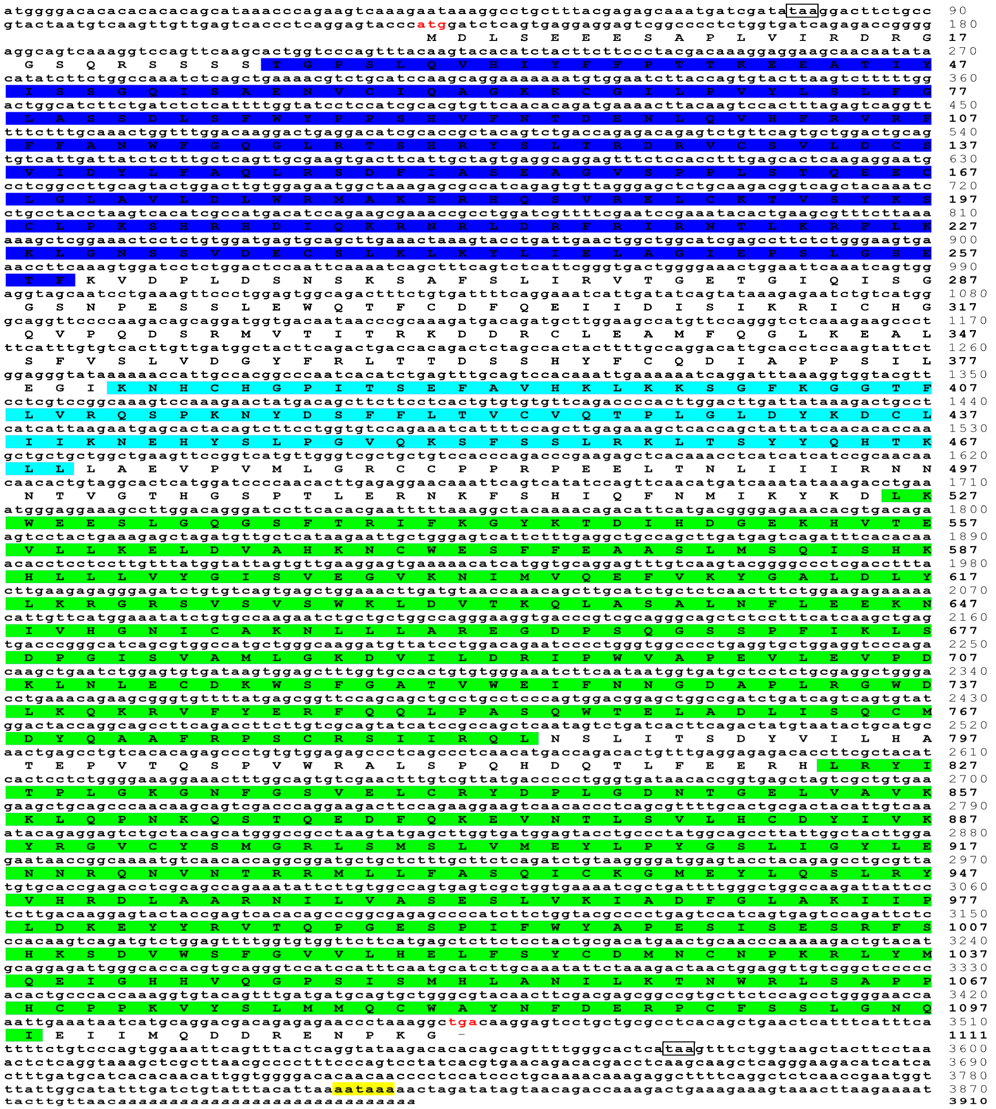
The full-length cDNA sequence of trJAK3 (GenBank accession number: MN782002.1) was 3910bp, containing a 130bp 5′-UTR, a 3336bp ORF encoding 1111 aa and a 444bp 3′-UTR (Figure 3).
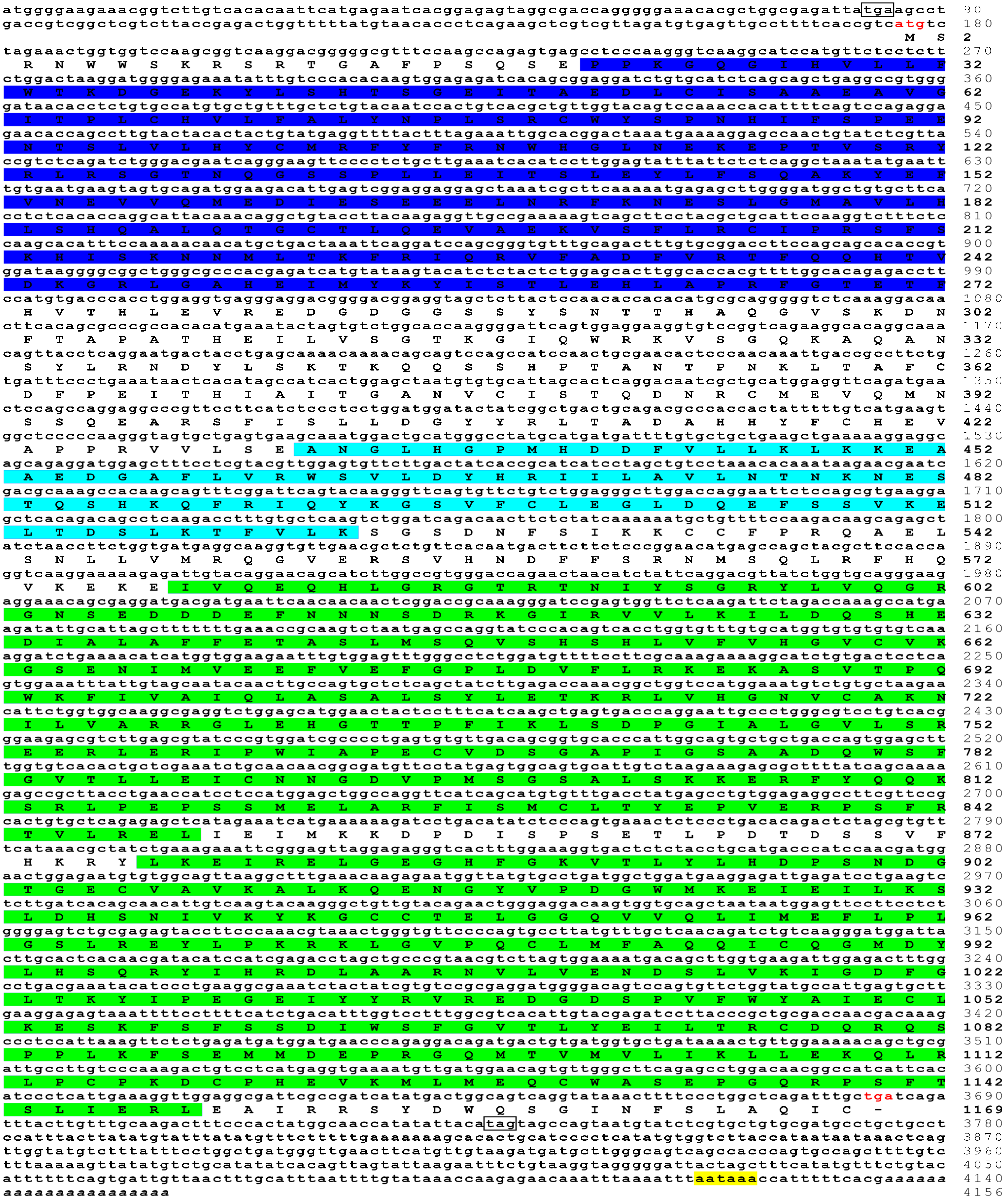
The complete cDNA sequence of trTYK2 (GenBank accession number: MT240841.1) was 4156bp long, including a 175bp 5′-UTR, a 471bp 3′-UTR and a 3510bp ORF encoding 1169 aa (Figure 4).
The polyadenylation signal (aataaa) upstream of the poly (A) tail and the poly (A) tail were found in the 3′-UTR of the four JAK genes. Further, two stop codons were found in the 5′-UTR and 3′-UTR of the four JAK genes. These findings indicate that the full-length cDNA sequences of the four JAK genes from golden pompano had been obtained. The predicted molecular weights of the four trJAK proteins were 134.71 kDa, 129.99 kDa, 126.48 kDa, 133.12 kDa, respectively, and the theoretical pIs were 6.77, 6.55, 6.55 and 6.62, respectively. No signal peptides were found in the four JAK proteins. Protein domain and sequence alignment analyses showed that all four trJAK proteins shared the same protein structures with JAKs of large yellow croaker and zebrafish (Danio rerio), including a FERM domain, an SH2 domain and two Tyrkc domains (Supplementary Figure S1).

3.2. Homology and Phylogenetic Analysis

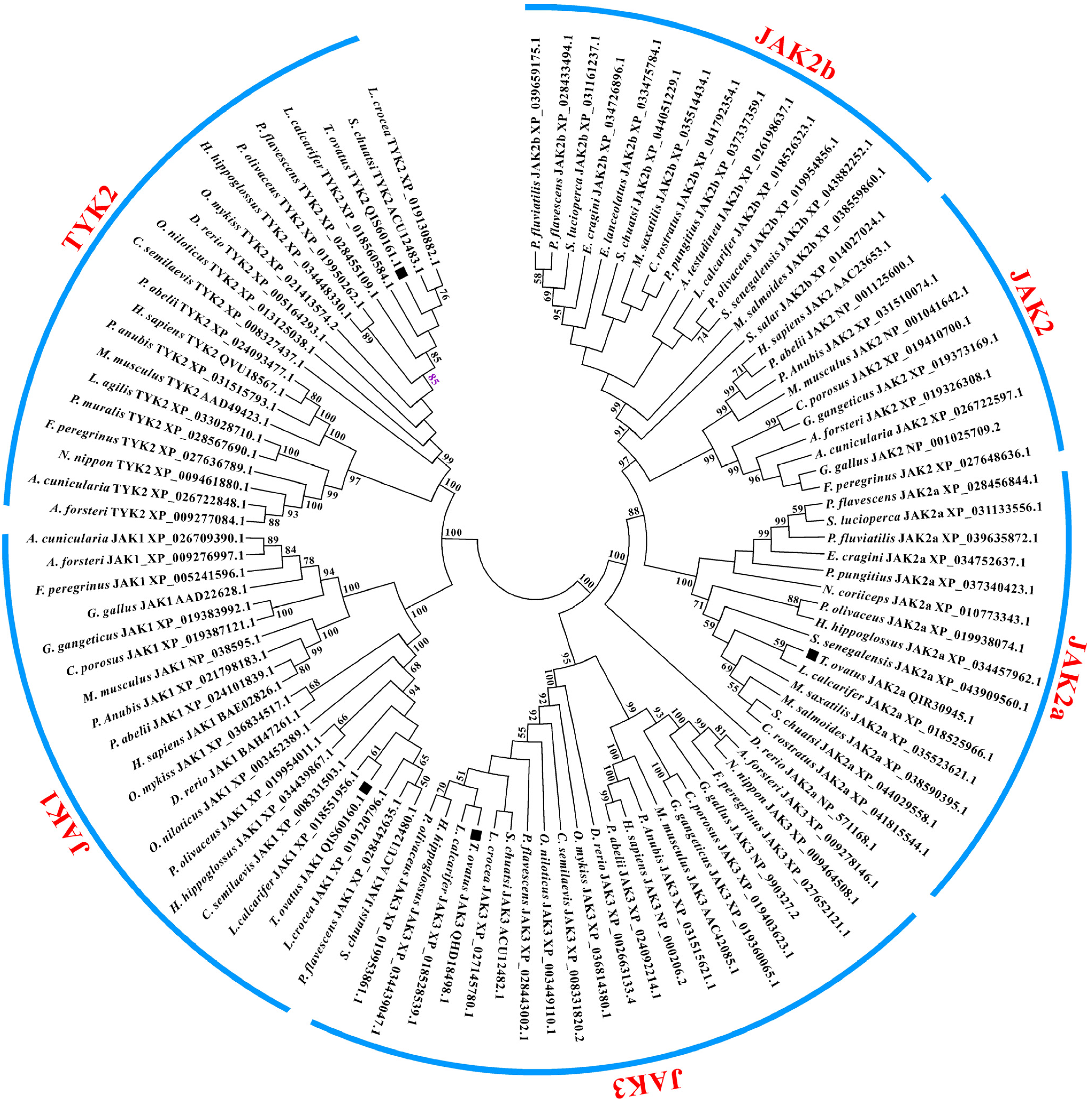
The four trJAK proteins shared higher sequence identities with their counterparts of other fish species than with mammalian homologs (Table 3). trJAK1 had 73.8–92.9% sequence identities with fish JAK1. trJAK2a shared 69.1–92.3% sequence identities with JAK2a of other fish species. trJAK3 had 67.0–88.5% sequence identities with their teleost counterparts, whist TYK2 had 66.3–86.3% sequence identities with fish JAK3. A phylogenetic tree was constructed to understand the evolutionary relationships of JAKs with their counterparts from other species using the neighbor-joining method according to the putative protein sequences. The results showed that all four JAK proteins of golden pompano were clustered well with their fish counterparts (Figure 5). Further, the four proteins were closely clustered with the JAKs of Asian seabass (Lates calcarifer) (Figure 5).

3.3. Expression Profiles of trJAK1, trJAK2a, trJAK3 and trTYK2

qPCR was performed to investigate mRNA transcription of the four JAKs in gill, skin, head kidney (HK), heart, brain, liver, muscle, spleen and intestine. All four trJAK genes were ubiquitously expressed in all examined tissues (Figure 6). The levels of expression of trJAK1 were relatively high in the liver, spleen, intestine and gill, but relatively low in the heart, brain and skin. trJAK2a was highly expressed in the liver, followed by the spleen, brain, intestine, gill, HK, skin, heart and muscle. High expression levels of trJAK3 were observed in the spleen, gill, HK, liver and intestine. trTYK2 was highly expressed in the liver, brain, gill and intestine, but weakly expressed in the skin, heart and muscle.

3.4. Expression of trJAK1, trJAK2a, trJAK3 and trTYK2 after LPS, Poly I:C and V. alginolyticus Stimulation

To investigate the roles of JAKs from golden pompano during pathogen invasion, transcription level changes in the HK, spleen, liver and gill were detected using the qPCR method (Figure 7). Following LPS stimulation, the expression of trJAK1 was strongly increased in the HK, spleen, liver and gill at 6 h, and showed no significant changes at 24 h and 48 h. The expression levels of trJAK2a, trJAK3 and trTYK2 were significantly up-regulated in the HK, spleen and liver at 6 h and 12 h. Following poly I:C stimulation, the expression levels of trJAK1, trJAK3 and trTYK2 were significantly increased in the HK, spleen and liver at 6 h, but no significant changes were observed in HK at 12 h. The expression of trJAK2a was dramatically increased in the four tissues at 6 h and 48 h. Following V. alginolyticus challenge, the expression levels of trJAK1, trJAK2a and trJAK3 in the HK and spleen were significantly up-regulated at 12 h, and then returned to normal levels at 48 h. The expression levels of trTYK2 in the HK, liver and gill were increased at 6 h and 48 h, but no significant changes were found at 12 h and 24 h.

4. Discussion

The JAK/STAT signaling pathway is critical to various physiological processes, including cell growth, differentiation, apoptosis and resistance to pathogens [43,44]. In arthropods and insects, the JAK/STAT signaling pathway has been proven to play important roles in antiviral and antibacterial responses [45,46,47,48]. JAKs are important components of the JAK/STAT pathway, and the activation of JAKs is essential in the majority of cytokine signaling. However, the JAKs are not well studied in teleost fishes. In the current study, JAK1, JAK2a, JAK3 and TYK2 from golden pompano were cloned, and their mRNA expression in healthy tissues and following stimulation with LPS, poly I:C, and V. alginolyticus were investigated. All four JAKs contained a FERM domain, an SH2 domain and two Tyrkc domains. Conversely, a STYKc domain was found in JAKs of mandarin fish, channel catfish, javeline goby, yellow catfish and mammals [15,16,17,18,49,50]. The STYKc domain is involved in modulating the Tyrkc domain activity of JAKs [51]. Meanwhile, the Tyrkc domain plays crucial roles in the phosphorylation of receptors and STAT transcription factors [6]. The absence of the STYKc domain in the four JAKs of golden pompano indicates that pompano JAKs have been mutated during the evolution of fishes [52]. Phylogenetic analysis showed that all four JAKs were closely clustered with their counterparts of other fish species, indicating that fish JAKs are evolutionarily homologous [53]. In addition, JAK2a and JAK2b were found only in fishes, suggesting that the mechanism of the JAK/STAT signal pathway in fish is relatively complex compared to that in mammals.
qRT-PCR results showed that trJAK1, trJAK2a, trJAK3 and trTYK2 were constitutively expressed across all the examined tissues. Similar expression profiles were observed in JAKs of javeline goby, channel catfish and yellow catfish [16,17,18]. The distinct expression of the four JAKs in different tissues revealed that pompano JAKs may perform diverse biological processes in different tissues [18,54]. In addition, the expression of trJAK1 and trJAK2a were highest in the liver. Similar results were observed in channel catfish JAK1 and JAK2a [18], yellow catfish JAK1 and JAK2a [16], common carp (cyprinus carpio) JAK1 [55], mandarin fish JAK1 and JAK2 [15], and grass carp JAK2 [56], implying that trJAK1 and trJAK2a play vital roles in the liver. The spleen, an important immune organ in fishes, contains many immune cells, such as lymphocytes, macrophages and granulocytes [57]. The high expressions of trJAK3 were observed in the spleen, in agreement with that of JAK3 in javeline goby [17], indicating that trJAK3 is actively involved in the immune response of fish. Meanwhile, trTYK2 was highly expressed in the liver, followed by the brain, gill, intestine, spleen, heart, skin, HK and muscle. However, different tissue-expression profiles of TYK2s were observed in Atlantic salmon (Salmo salar) [54], common carp [55], and blunt-snout bream [58]. These results imply that trTYK2 expression might be tissue-specific and species-specific.
Following LPS stimulation, the expression of trJAK1, trJAK2a, trJAK3 and trTYK2 was up-regulated in the HK, spleen and liver at 6 h, and the expression of trJAK2a, trJAK3 and trTYK2 was significantly increased in the gill at 12 h, indicating that the four pompano JAKs are involved in LPS-induced immune responses. It has been found that the JAK/STAT signaling pathway is activated in Prenant’s schizothoracin (Schizothorax prenanti) stimulated with LPS [59]. Snakehead (Channa argus) JAK1 and JAK3 were activated in HK leucocytes following stimulation with LPS [60]. Furthermore, the JAK2 of mouse was activated in macrophage cells induced by LPS [61], and the TYK2-null mouse was resistant to LPS-induced endotoxin shock [62]. These results demonstrate that JAKs play crucial roles in the LPS-induced immune response. After poly I:C stimulation, the expressions of trJAK1, trJAK2a, trJAK3 and trTYK2 were significantly up-regulated in the HK, spleen, liver and gill at 48 h. Similar results were observed in other fish species. For example, JAKs of mandarin fish were significantly induced in M-IFF cells post poly I:C stimulation [15], and mandarin fish JAK1 was also induced in the liver, spleen and HK after red sea bream iridovirus infection [63]. The expression of JAK1, JAK2a and JAK3 strongly increased in the spleen of large yellow croaker following stimulation with poly I:C [64]. Grass carp JAK1 and TYK2 were induced in kidney cells post poly I:C stimulation [53]. JAK1 was significantly induced in spleen of yellowhead catfish (Tachysurus fulvidraco) post poly I:C stimulation [65]. The expression of JAK1 was significantly up-regulated in the HK of silver crucian carp (Carassius auratus gibeli) post CyHV-2 infection [66,67]. These results imply that fish JAKs perform a similar role during viral infection. After V. alginolyticus stimulation, the expressions of trJAK1, trJAK2a, trJAK3 and trTYK2 were significantly increased in the HK and spleen at 6 h and 12 h. The expressions of trJAK2a and trTYK2 were up-regulated in liver and gill at 6 h, and the expressions of trJAK1 and trJAK3 were significantly up-regulated in liver and gill at 24 h or 48 h. The changes in the expression patterns of the four pompano JAKs were consistent with those observed in previous studies of other fishes. For example, JAKs were induced in the gill and liver of channel catfish after E. ictaluri infection [18]. The expression of JAK1 was significantly up-regulated in the HK of rainbow trout post Tetracapsuloides bryosalmonae and Myxobolus cerebralis co-infection [68]. Up-regulated expression was also observed for JAK3 of sea bass (Dicentrarchus labrax) after Photobacterium damsela stimulation [69], Reeves shade (Tenualosa reevesii) post A. hydrophila infection and catfish (Silurus asotus) challenge with E. ictaluri [70,71]. The expression of JAK3 and TYK2 was also significantly increased in the liver and middle kidney of blunt-snout bream after A. hydiophila challenge [58,72]. In addition, transcriptome analysis revealed that the expression of JAKs was up-regulated in the HK, spleen and liver from golden pompano after S. agalactiae infection [32]. These results collectively indicate that trJAK1, trJAK2a, trJAK3 and trTYK2 play important roles in the immune defense of fishes against bacterial infection.
It has been found that mammalian cytokine responses are mediated by the JAK/STAT signaling pathway [73]. Fish JAKs share similar functions with mammalian JAKs. For example, type I IFN of large yellow croaker triggered an antiviral response via the JAK/STAT pathway [22]. The JAK/STAT pathway of Atlantic salmon was involved in regulation of type I and II IFN signaling [74]. The JAK/STAT pathway of Grass carp could modulate the expression of IFN I [75]. The JAK/STAT signaling pathway of mandarin fish was activated to regulate the expression of IRF-1 and Mx following poly I:C stimulation. In the current study, JAKs of golden pompano were activated in the spleen, HK, liver and gill response to LPS, poly I:C and V. alginolyticus stimulation. In addition, golden pompano IFN gamma was dramatically induced in the liver and kidney post LPS, poly I:C and S. agalactiae stimulation [32,76]. The expressions of IL-1 beta, TNF alpha and JAKs were significantly up-regulated in the liver, spleen, and head kidney of golden pompano after S. agalactiae infection [32], whilst the expressions of IL-1 beta, IL-11 and IL-34 were significantly increased in the liver, spleen, kidney, gill, and skin of golden pompano following V. harveyi, S. agalactiae, and VNNV infection [77]. These results suggest that the functions of JAKs may be conserved from teleost to mammals, and JAKs paly essential roles in immune response. How the JAKs in golden pompano are involved in modulation of inflammatory cytokines (IL beta, TNF alpha and IFN gamma, etc.) requires further study.

5. Conclusions

In conclusion, JAK1, JAK2a, JAK3 and TYK2 were isolated and characterized from golden pompano. These four pompano JAKs shared high sequence identities with JAKs of other fish species. Further, inducible expression of the four JAK genes was observed in the HK, spleen, liver and gill post LPS, poly I:C and V. alginolyticus stimulation, indicating the roles of JAKs in response to pathogen invasion. Nonetheless, similar results according to immune stimulation indicate that the JAK/STAT pathway of golden pompano may share conserved functions with their counterparts of other vertebrates. Further in vitro studies are required to demonstrate this. Co-expressing all JAK proteins in cultured cells from golden pompano and protein–protein interactions analysis will directly reveal the regulatory roles of JAKs on cytokines. Biochemical and functional studies must be carried out to elucidate how pompano JAK/STAT is assembled, how it can be activated and, finally, how it is involved in regulation of the expression of cytokines. X-ray crystallographic analysis of JAKs will reveal disparities and parities between mammals and fishes. Currently, we have isolated the macrophages from golden pompano, and are using them to study the regulatory effects of JAKs on inflammatory cytokines.

Supplementary Materials

The following supporting information can be downloaded at: https://www.mdpi.com/article/10.3390/fishes8050245/s1, Figure S1: Comparison of protein sequences of JAKs between golden pompano, large yellow croaker, zebrafish and human, and their GeneBank accession numbers are listed in Figure 5. The protein domains were predicted by Simple Modular Architecture Research Tool (SMART) (http://smart.emble-heidelberg.de, accessed on 12 November 2022). The FERM domain is marked in green. The STYKc domain of human is marked in red and the Tyrkc domain is marked in bule.

Author Contributions

Conceptualization, Y.W.; methodology, M.Y. and M.C.; software, Y.X. and P.H.; validation, Y.W.; formal analysis, M.C. and P.H.; investigation, M.Y.; resources, Y.W.; data curation, Y.X.; writing—original draft preparation, Y.X. and M.C.; writing—review and editing, Y.X. and Y.W.; visualization, Y.X. and X.L.; supervision, X.L.; project administration, Y.W. and Z.L.; funding acquisition, Z.L. All authors have read and agreed to the published version of the manuscript.

Funding

This research was funded by the National Natural Science Foundation of China, grant number 31860736.

Institutional Review Board Statement

This study was performed in accordance with the recommendation from the Management and Use of Laboratory Animals of Guangxi Autonomous Region, and the study was conducted with the approval of the Animal Care and Welfare Committee of Guangxi University (GXU-2023-0005).

Informed Consent Statement

Not applicable.

Data Availability Statement

Data supporting the results of this study can be found in the Supplementary Materials of this paper.

Conflicts of Interest

The authors declare no conflict of interest.

References

  1. Zhang, X.; Wu, J.; Ye, B.; Wang, Q.; Xie, X.; Shen, H. Protective effect of curcumin on TNBS-induced intestinal inflammation is mediated through the JAK/STAT pathway. BMC Complement. Altern. Med. 2016, 16, 299. [Google Scholar] [CrossRef] [PubMed]
  2. Sabaawy, H.E.; Ryan, B.M.; Khiabanian, H.; Pine, S.R. JAK/STAT of all trades: Linking inflammation with cancer development, tumor progression and therapy resistance. Carcinogenesis 2021, 42, 1411–1419. [Google Scholar] [CrossRef]
  3. Villarino, A.V.; Kanno, Y.; Ferdinand, J.R.; O’Shea, J.J. Mechanisms of Jak/STAT Signaling in Immunity and Disease. J. Immunol. 2015, 194, 21–27. [Google Scholar] [CrossRef] [PubMed]
  4. Ezeonwumelu, I.J.; Garcia-Vidal, E.; Ballana, E. JAK-STAT Pathway: A Novel Target to Tackle Viral Infections. Viruses 2021, 13, 2379. [Google Scholar] [CrossRef]
  5. Raftery, N.; Stevenson, N.J. Advances in anti-viral immune defence: Revealing the importance of the IFN JAK/STAT pathway. Cell. Mol. Life Sci. 2017, 74, 2525–2535. [Google Scholar] [CrossRef] [PubMed]
  6. Morris, R.; Kershaw, N.J.; Babon, J.J. The molecular details of cytokine signaling via the JAK/STAT pathway. Protein Sci. 2018, 27, 1984–2009. [Google Scholar] [CrossRef] [PubMed]
  7. Xu, F.; Wang, S.; Wang, Y.; Hu, L.; Zhu, L. Inhibition of gp130 alleviates LPS-induced lung injury by attenuating apoptosis and inflammation through JAK1/STAT3 signaling pathway. Inflamm. Res. 2023, 72, 493–507. [Google Scholar] [CrossRef]
  8. Li, X.; Liu, J. FANCD2 inhibits ferroptosis by regulating the JAK2/STAT3 pathway in osteosarcoma. BMC Cancer 2023, 23, 179. [Google Scholar] [CrossRef]
  9. Ociepa, K.; Danilewicz, M.; Wągrowska-Danilewicz, M.; Peterson-Jęckowska, R.; Wójcicka-Rubin, A.; Lewkowicz, N.; Zajdel, R.; Żebrowska, A. Expression of the Selected Proteins of JAK/STAT Signaling Pathway in Diseases with Oral Mucosa Involvement. Int. J. Mol. Sci. 2023, 24, 323. [Google Scholar] [CrossRef]
  10. Wu, J.; Del Duca, E.; Espino, M.; Gontzes, A.; Cueto, I.; Zhang, N.; Estrada, Y.D.; Pavel, A.B.; Krueger, J.G.; Guttman-Yassky, E. RNA Sequencing Keloid Transcriptome Associates Keloids with Th2, Th1, Th17/Th22, and JAK3-Skewing. Front. Immunol. 2020, 11. [Google Scholar] [CrossRef]
  11. Rizi, F.Z.; Ghorbani, A.; Zahtab, P.; Darbaghshahi, N.N.; Ataee, N.; Pourhamzeh, P.; Hamzei, B.; Dolatabadi, N.F.; Zamani, A.; Hooshmand, M. TYK2 single-nucleotide variants associated with the severity of COVID-19 disease. Arch. Virol. 2023, 168, 1–8. [Google Scholar] [CrossRef]
  12. Villarino, A.V.; Gadina, M.; O’Shea, J.J.; Kanno, Y. Snapshot: JAK-STAT signaling II. Cell 2020, 181, 1696.e1. [Google Scholar] [CrossRef]
  13. Gadina, M.; Chisolm, D.A.; Philips, R.L.; McInness, I.B.; Changelian, P.S.; O’Shea, J.J. Translating JAKs to Jakinibs. J. Immunol. 2020, 204, 2011–2020. [Google Scholar] [CrossRef] [PubMed]
  14. Leu, J.H.; Yan, S.J.; Lee, T.F.; Chou, C.M.; Chen, S.T.; Hwang, P.P.; Chou, C.K.; Huang, C.J. Complete genomic organization and promoter analysis of the round-spotted pufferfish JAK1, JAK2, JAK3, and TYK2 genes. DNA Cell Biol. 2000, 19, 431–446. [Google Scholar] [CrossRef] [PubMed]
  15. Guo, C.-J.; Zhang, Y.-F.; Yang, L.-S.; Yang, X.-B.; Wu, Y.-Y.; Liu, D.; Chen, W.-J.; Weng, S.-P.; Yu, X.-Q.; He, J.-G. The JAK and STAT family members of the mandarin fish Siniperca chuatsi: Molecular cloning, tissues distribution and immunobiological activity. Fish Shellfish Immunol. 2009, 27, 349–359. [Google Scholar] [CrossRef] [PubMed]
  16. Wu, K.; Tan, X.-Y.; Xu, Y.-H.; Chen, Q.-L.; Pan, Y.-X. JAK and STAT members of yellow catfish Pelteobagrus fulvidraco and their roles in leptin affecting lipid metabolism. Gen. Comp. Endocrinol. 2016, 226, 14–26. [Google Scholar] [CrossRef]
  17. Wu, K.; Tan, X.-Y.; Xu, Y.-H.; Shi, X.; Fan, Y.-F.; Li, D.-D.; Liu, X. JAK family members: Molecular cloning, expression profiles and their roles in leptin influencing lipid metabolism in Synechogobius hasta. Comp. Biochem. Physiol. Part B Biochem. Mol. Biol. 2017, 203, 122–131. [Google Scholar] [CrossRef]
  18. Jin, Y.; Zhou, T.; Li, N.; Liu, S.; Xu, X.; Pan, Y.; Tan, S.; Shi, H.; Yang, Y.; Yuan, Z.; et al. JAK and STAT members in channel catfish: Identification, phylogenetic analysis and expression profiling after Edwardsiella ictaluri infection. Dev. Comp. Immunol. 2018, 81, 334–341. [Google Scholar] [CrossRef]
  19. Ward, A.E.; Rosenthal, B.M. Evolutionary responses of innate immunity to adaptive immunity. Infect. Genet. Evol. 2014, 21, 492–496. [Google Scholar] [CrossRef]
  20. Sun, Q.; Zhang, J.; Wang, J.; Wang, H.; Gao, Z.; Liu, H. Janus kinase 1 in Megalobrama amblycephala: Identification, phylogenetic analysis and expression profiling after Aeromonas hydrophila infection. Fish Shellfish Immunol. 2023, 135. [Google Scholar] [CrossRef]
  21. Meng, X.Y.; Wang, Z.H.; Yu, X.D.; Zhang, Q.Y.; Ke, F. Development and characterization of a skin cell line from Chinese perch (Siniperca chuatsi ) and its application in aquatic animal viruses. J. Fish Dis. 2022, 45, 1439–1449. [Google Scholar] [CrossRef] [PubMed]
  22. Chen, J.; Guan, Y.; Guan, H.; Mu, Y.; Ding, Y.; Zou, J.; Ouyang, S.; Chen, X. Molecular and Structural Basis of Receptor Binding and Signaling of a Fish Type I IFN with Three Disulfide Bonds. J. Immunol. 2022, 209, 806–819. [Google Scholar] [CrossRef] [PubMed]
  23. Mu, P.; Teng, Y.; Wu, H.; Li, X.; Huo, J.; Ao, J.; Chen, X. Large yellow croaker (Lrimichthys crocea) IL-2 modulates humoral immunity via the conserved JAK-STAT5 signal pathway. Fish Shellfish Immunol. 2023, 133. [Google Scholar] [CrossRef] [PubMed]
  24. Xu, Z.; Liao, J.; Zhang, D.; Liu, S.; Zhang, L.; Kang, S.; Xu, L.; Chen, H.; Peng, W.; Zhou, S.; et al. Isolation, Characterization, and Transcriptome Analysis of an ISKNV-Like Virus from Largemouth Bass. Viruses 2023, 15, 398. [Google Scholar] [CrossRef] [PubMed]
  25. Zhang, Y.; Wang, X.; Shi, H.; Song, N.; Han, F.; Chai, X.; Liu, Q.; Wang, Y.; Gao, T. Comparative transcriptomic analysis of the liver and spleen in marbled rockfish (Sebastiscus marmoratus) challenged with polyriboinosinic polyribocytidylic acid (poly(I:C)). Aquaculture 2022, 554, 738144. [Google Scholar] [CrossRef]
  26. Zhang, X.; Hao, X.; Ma, W.; Zhu, T.; Zhang, Z.; Wang, Q.; Liu, K.; Shao, C.; Wang, H.-Y. Transcriptome Analysis Indicates Immune Responses against Vibrio harveyi in Chinese Tongue Sole (Cynoglossus semilaevis). Animals 2022, 12, 1144. [Google Scholar] [CrossRef]
  27. Song, Y.; Dong, X.; Hu, G. Transcriptome analysis of turbot (Scophthalmus maximus) head kidney and liver reveals immune mechanism in response to Vibrio anguillarum infection. J. Fish Dis. 2022, 45, 1045–1057. [Google Scholar] [CrossRef]
  28. Fu, Q.; Li, Y.; Zhao, S.; Wang, H.; Zhao, C.; Zhang, P.; Cao, M.; Yang, N.; Li, C. Comprehensive identification and expression profiling of immune-related lncRNAs and their target genes in the intestine of turbot (Scophthalmus maximus L.) in response to Vibrio anguillarum infection. Fish Shellfish Immunol. 2022, 130, 233–243. [Google Scholar] [CrossRef]
  29. Gao, C.; Cai, X.; Ma, L.; Sun, P.; Li, C. Systematic analysis of circRNA-related ceRNA networks of black rockfish (Sebastes schlegelii) in response to Aeromonas salmonicides infection. Fish Shellfish Immunol. 2023, 135, 108648. [Google Scholar] [CrossRef]
  30. Zhao, L.; Huang, J.; Wu, S.; Li, Y.; Pan, Y. Integrative analysis of miRNA and mRNA expression associated with the immune response in the intestine of rainbow trout (Oncorhynchus mykiss) infected with infectious hematopoietic necrosis virus. Fish Shellfish Immunol. 2022, 131, 54–66. [Google Scholar] [CrossRef]
  31. Di, G.; Li, H.; Zhao, Y.; Lin, Y.; Lan, D.; Kong, X.; Chen, X. Comprehensive transcriptomic analysis reveals insights into the gill response to hypoxia and Poly I:C in Qihe crucian carp Carassius auratus. Aquac. Rep. 2022, 24, 101154. [Google Scholar] [CrossRef]
  32. Gao, J.; Guo, H.-Y.; Liu, M.-J.; Zhu, K.-C.; Liu, B.; Liu, B.-S.; Zhang, N.; Jiang, S.-G.; Zhang, D.-C. Transcriptome Analysis of the Immune Process of Golden Pompano (Trachinotus ovatus) Infected with Streptococcus agalactiae. Fishes 2023, 8, 52. [Google Scholar] [CrossRef]
  33. Guo, H.-Y.; Li, W.-F.; Zhu, K.-C.; Liu, B.-S.; Zhang, N.; Liu, B.; Yang, J.-W.; Zhang, D.-C. Pathology, Enzyme Activity and Immune Responses after Cryptocaryon irritans Infection of Golden Pompano Trachinotus ovatus (Linnaeus 1758). J. Mar. Sci. Eng. 2023, 11, 262. [Google Scholar] [CrossRef]
  34. Zhou, J.; Dong, X.; Kong, B.; Sun, Q.; Ji, H.; Liu, S. Effects of magnetic field-assisted immersion freezing at different magnetic field intensities on the muscle quality of golden pompano (Trachinotus ovatus). Food Chem. 2023, 407, 135092. [Google Scholar] [CrossRef]
  35. Xie, Y.; Gao, S.; Cao, Y.; Ji, Y.; Zhang, Q.; Wei, Y.; Qi, Z. Molecular characterization and functional analysis of DIGIRR from golden pompano (Trachinotus ovatus). Front. Immunol. 2022, 13, 974310. [Google Scholar] [CrossRef]
  36. Xie, Y.; Lei, K.; He, J.; Wei, Y. Molecular Characterization and Expression Analysis of TAK1, TAB1 and TAB2 of Golden Pompano (Trachinotus ovatus). Fishes 2022, 7, 173. [Google Scholar] [CrossRef]
  37. Kumar, S.; Stecher, G.; Tamura, K. MEGA7: Molecular evolutionary genetics analysis version 7.0 for bigger datasets. Mol. Biol. Evol. 2016, 33, 1870–1874. [Google Scholar] [CrossRef]
  38. Geven, E.J.; Klaren, P.H. The teleost head kidney: Integrating thyroid and immune signalling. Dev. Comp. Immunol. 2017, 66, 73–83. [Google Scholar] [CrossRef]
  39. He, Y.; Wang, E.; Wang, K.; Wang, J.; Fan, W.; Chen, D.; Yang, Q. Morphology of the Spleen in Oreochromis niloticus: Splenic Subregions and the Blood-Spleen Barrier. Animals 2021, 11, 2934. [Google Scholar] [CrossRef]
  40. Huo, J.; Hu, X.; Bai, J.; Lv, A. Multiomics analysis revealed miRNAs as potential regulators of the immune response in Carassius auratus gills to Aeromonas hydrophila infection. Front. Immunol. 2023, 14, 1098455. [Google Scholar] [CrossRef]
  41. Akiyoshi, H.; Inoue, A. Comparative Histological Study of Teleost Livers in Relation to Phylogeny. Zoöl. Sci. 2004, 21, 841–850. [Google Scholar] [CrossRef] [PubMed]
  42. Livak, K.J.; Schmittgen, T.D. Analysis of relative gene expression data using real-time quantitative PCR and the 2(T)(-Delta Delta C) method. Methods 2001, 25, 402–408. [Google Scholar] [CrossRef] [PubMed]
  43. Hu, X.; Li, J.; Fu, M.; Zhao, X.; Wang, W. The JAK/STAT signaling pathway: From bench to clinic. Signal Transduct. Target. Ther. 2021, 6, 1–33. [Google Scholar] [CrossRef] [PubMed]
  44. Villarino, A.V.; Kanno, Y.; O’Shea, J.J. Mechanisms and consequences of Jak–STAT signaling in the immune system. Nat. Immunol. 2017, 18, 374–384. [Google Scholar] [CrossRef] [PubMed]
  45. Ruan, Z.; Wan, Z.; Yang, L.; Li, W.; Wang, Q. JAK/STAT signalling regulates antimicrobial activities in Eriocheir sinensis. Fish Shellfish Immunol. 2019, 84, 491–501. [Google Scholar] [CrossRef] [PubMed]
  46. Ran, X.-Q.; Gao, L.; Yan, M.; Kang, C.-J. Peroxiredoxin 4 Interacts with Domeless and Participates in Antibacterial Immune Response Through the JAK/STAT Pathway. Front. Immunol. 2022, 13, 907183. [Google Scholar] [CrossRef]
  47. Gao, J.; Zhao, B.-R.; Zhang, H.; You, Y.-L.; Li, F.; Wang, X.-W. Interferon functional analog activates antiviral Jak/Stat signaling through integrin in an arthropod. Cell Rep. 2021, 36, 109761. [Google Scholar] [CrossRef]
  48. Bang, I.S. JAK/STAT signaling in insect innate immunity. Èntomol. Res. 2019, 49, 339–353. [Google Scholar] [CrossRef]
  49. Takahashi, T.; Shirasawa, T. Molecular cloning of rat JAK3, a novel member of the JAK family of protein tyrosine kinases. FEBS Lett. 1994, 342, 124–128. [Google Scholar] [CrossRef]
  50. Haan, C.; Kreis, S.; Margue, C.; Behrmann, I. Jaks and cytokine receptors—An intimate relationship. Biochem. Pharmacol. 2006, 72, 1538–1546. [Google Scholar] [CrossRef]
  51. Shuai, K.; Liu, B. Regulation of JAK–STAT signalling in the immune system. Nat. Rev. Immunol. 2003, 3, 900–911. [Google Scholar] [CrossRef] [PubMed]
  52. Haan, S.; Margue, C.; Engrand, A.; Rolvering, C.; de Leur, H.S.-V.; Heinrich, P.C.; Behrmann, I.; Haan, C. Dual Role of the Jak1 FERM and Kinase Domains in Cytokine Receptor Binding and in Stimulation-Dependent Jak Activation. J. Immunol. 2008, 180, 998–1007. [Google Scholar] [CrossRef]
  53. Hou, Q.; Gong, R.; Liu, X.; Mao, H.; Xu, X.; Liu, D.; Dai, Z.; Wang, H.; Wang, B.; Hu, C. Poly I:C facilitates the phosphorylation of Ctenopharyngodon idellus type I IFN receptor subunits and JAK kinase. Fish Shellfish Immunol. 2017, 60, 13–20. [Google Scholar] [CrossRef] [PubMed]
  54. Sobhkhez, M.; Hansen, T.; Iliev, D.B.; Skjesol, A.; Jørgensen, J.B. The Atlantic salmon protein tyrosine kinase Tyk2: Molecular cloning, modulation of expression and function. Dev. Comp. Immunol. 2013, 41, 553–563. [Google Scholar] [CrossRef] [PubMed]
  55. Wu, P.; Gao, Q.; Liu, D.; Fu, Y.; Yuan, Y.; Wen, C. Molecular identification of tyrosine kinase 2 (TYK2) gene from common carp Cyprinus carpio and its expression pattern. Acta Hydrobiol. Sin. 2015, 39, 229–233. [Google Scholar]
  56. Wu, X.; Li, X.; Leng, X.; Guan, L.; Guo, T. Cloning and tissue expression of JAK2 gene in grass carp (Ctenopharyngodon idella). J. Shanghai Ocean Univ. 2012, 21, 21–27. [Google Scholar]
  57. Bjørgen, H.; Koppang, E.O. Anatomy of teleost fish immune structures and organs. Immunogenetics 2021, 73, 53–63. [Google Scholar] [CrossRef]
  58. Zhang, J.; Wang, J.; Wu, X.; Liu, H. Molecular cloning and expression of TYK2 in Megalobrama amblycephala. J. Huazhong Agric. Univ. 2021, 40, 197–205. [Google Scholar]
  59. Li, Y.; Xia, P.; Wu, J.; Huang, A.; Bu, G.; Meng, F.; Kong, F.; Cao, X.; Han, X.; Yu, G.; et al. The potential sensing molecules and signal cascades for protecting teleost fishes against lipopolysaccharide. Fish Shellfish Immunol. 2020, 97, 235–247. [Google Scholar] [CrossRef]
  60. Li, D.; Cui, Z.; Zhao, F.; Zhu, X.; Tan, A.; Deng, Y.; Lai, Y.; Huang, Z. Characterization of snakehead (Channa argus) interleukin-21: Involvement in immune defense against two pathogenic bacteria, in leukocyte proliferation, and in activation of JAK–STAT signaling pathway. Fish Shellfish Immunol. 2022, 123, 207–217. [Google Scholar] [CrossRef]
  61. Kimura, A.; Naka, T.; Muta, T.; Takeuchi, O.; Akira, S.; Kawase, I.; Kishimoto, T. Suppressor of cytokine signaling-1 selectively inhibits LPS-induced IL-6 production by regulating JAK–STAT. Proc. Natl. Acad. Sci. USA 2005, 102, 17089–17094. [Google Scholar] [CrossRef] [PubMed]
  62. Kamezaki, K.; Shimoda, K.; Numata, A.; Matsuda, T.; Nakayama, K.-I.; Harada, M. The role of Tyk2, Stat1 and Stat4 in LPS-induced endotoxin signals. Int. Immunol. 2004, 16, 1173–1179. [Google Scholar] [CrossRef] [PubMed]
  63. Liu, X.; Chen, N.; Gao, X.; Zhang, Y.; Li, X.; Zhang, Y.; Bing, X.; Huang, H.; Zhang, X. The infection of red seabream iridovirus in mandarin fish (Siniperca chuatsi) and the host immune related gene expression profiles. Fish Shellfish Immunol. 2018, 74, 474–484. [Google Scholar] [CrossRef] [PubMed]
  64. Mu, Y.; Li, M.; Ding, F.; Ding, Y.; Ao, J.; Hu, S.; Chen, X. De Novo Characterization of the Spleen Transcriptome of the Large Yellow Croaker (Pseudosciaena crocea) and Analysis of the Immune Relevant Genes and Pathways Involved in the Antiviral Response. PLoS ONE 2014, 9, e97471. [Google Scholar] [CrossRef]
  65. Liu, Q.-N.; Tang, Y.-Y.; Zhou, M.-J.; Luo, S.; Li, Y.-T.; Wang, G.; Zhang, D.-Z.; Yang, H.; Tang, B.-P.; He, W.-F. Differentially expressed genes involved in immune pathways from yellowhead catfish (Tachysurus fulvidraco) after poly (I:C) challenge. Int. J. Biol. Macromol. 2021, 183, 340–345. [Google Scholar] [CrossRef]
  66. Lu, J.; Xu, D.; Shen, Z.; Lu, L. Differential expression of miRNA in Carassius auratus gibelio in response to cyprinid herpesvirus 2 infection. Dev. Comp. Immunol. 2018, 82, 1–6. [Google Scholar] [CrossRef]
  67. Lu, J.; Xu, D.; Jiang, Y.; Kong, S.; Shen, Z.; Xia, S.; Lu, L. Integrated analysis of mRNA and viral miRNAs in the kidney of Carassius auratus gibelio response to cyprinid herpesvirus 2. Sci. Rep. 2017, 7, 13787. [Google Scholar] [CrossRef]
  68. Kotob, M.H.; Kumar, G.; Saleh, M.; Gorgoglione, B.; Abdelzaher, M.; El-Matbouli, M. Differential modulation of host immune genes in the kidney and cranium of the rainbow trout (Oncorhynchus mykiss) in response to Tetracapsuloides bryosalmonae and Myxobolus cerebralis co-infections. Parasites Vectors 2018, 11, 326. [Google Scholar] [CrossRef]
  69. Neves, J.V.; Caldas, C.; Wilson, J.M.; Rodrigues, P.N.S. Molecular mechanisms of hepcidin regulation in sea bass (Dicentrarchus labrax). Fish Shellfish Immunol. 2011, 31, 1154–1161. [Google Scholar] [CrossRef]
  70. Yan, H.; Wang, H.; Yu, B.; Huang, S.; Li, Y.; Wu, J. Study on separation of hemolytic Aeromonas hydrophila strain XF-6 from Tenualosa reevesii and its effect on iron metabolism. Southwest China. J. Agric. Sci. 2018, 31, 862–868. [Google Scholar]
  71. Yan, H.; Wang, H.; Yu, B.; Huang, S.; Li, Y.; Wu, J. Isolation of pathogenic Edwardsiella tarda strain CA26 from Silurus asotus and its effect on immune factors. J. Henan Agric. Sci. 2018, 47, 144–149, 160. [Google Scholar]
  72. Yang, Y.; Fu, X.; Chen, B.; Wang, W.; Liu, H. Effect of Aeromonas hydiophila infection on iron metabolism in Megalobrama amblycephala liver. J. Huazhong Agric. Univ. 2015, 34, 97–101. [Google Scholar]
  73. Majoros, A.; Platanitis, E.; Kernbauer-Hölzl, E.; Rosebrock, F.; Müller, M.; Decker, T. Canonical and Non-Canonical Aspects of JAK–STAT Signaling: Lessons from Interferons for Cytokine Responses. Front. Immunol. 2017, 8, 29. [Google Scholar] [CrossRef]
  74. Skjesol, A.; Liebe, T.; Iliev, D.B.; Thomassen, E.I.S.; Tollersrud, L.G.; Sobhkhez, M.; Joensen, L.L.; Secombes, C.J.; Jørgensen, J.B. Functional conservation of suppressors of cytokine signaling proteins between teleosts and mammals: Atlantic salmon SOCS1 binds to JAK/STAT family members and suppresses type I and II IFN signaling. Dev. Comp. Immunol. 2014, 45, 177–189. [Google Scholar] [CrossRef]
  75. Liu, Y.; Du, H.; Wang, S.; Lv, Y.; Deng, H.; Chang, K.; Zhou, P.; Hu, C. Grass carp (Ctenopharyngodon idella) TNK1 modulates JAK-STAT signaling through phosphorylating STAT1. Dev. Comp. Immunol. 2021, 116, 103951. [Google Scholar] [CrossRef] [PubMed]
  76. Zhu, K.-C.; Guo, H.-Y.; Zhang, N.; Liu, B.-S.; Guo, L.; Jiang, S.-G.; Zhang, D.-C. Functional characterization of IRF8 regulation of type II IFN in golden pompano (Trachinotus ovatus). Fish Shellfish Immunol. 2019, 94, 1–9. [Google Scholar] [CrossRef] [PubMed]
  77. Wu, Y.; Zhou, Y.; Cao, Z.; Sun, Y.; Chen, Y.; Xiang, Y.; Wang, L.; Zhang, S.; Guo, W. Comparative analysis of the expression patterns of IL-1β, IL-11, and IL-34 in golden pompano (Trachinotus ovatus) following different pathogens challenge. Fish Shellfish Immunol. 2019, 93, 863–870. [Google Scholar] [CrossRef]
Figure 1. The nucleotide sequence, deduced amino acid sequence and predicted domains of trJAK1. The start codon and the stop codon are marked in red, the FERM domain (from 30 aa to 284 aa) is marked with a blue background, the SH2 domain (from 448 aa to 543 aa) is marked with a cyan background, the TyrKc domain (from 595 aa to 861 aa, from 889 aa to 1163 aa) is marked with a green background, and the poly-A tail is italicized. Two stop codons are in-frame and the polyadenylation signal (aataaa) upstream of the poly (A) tail is marked in yellow.
Fishes 08 00245 g001
Figure 2. The nucleotide sequence, deduced amino acid sequence and predicted domains of trJAK2a. The start codon and the stop codon are marked in red, the FERM domain (from 38 aa to 283 aa) is marked with a blue background, the SH2 domain (from 401 aa to 491 aa) is marked with a cyan background, the TyrKc domain (from 547 aa to 807 aa, from 850 aa to 1124 aa) is marked with a green background, and the poly-A tail is italicized. Two stop codons are in-frame and the polyadenylation signal (aataaa) upstream of the poly (A) tail is marked in yellow.
Fishes 08 00245 g002
Figure 3. The nucleotide sequence, deduced amino acid sequence and predicted domains of trJAK3. The start codon and the stop codon are marked in red, the FERM domain (from 26 aa to 259 aa) is marked with a blue background, the SH2 domain (from 381 aa to 469 aa) is marked with a cyan background, the TyrKc domain (from 526 aa to 784 aa, from 824 aa to 1098 aa) is marked with a green background, and the poly-A tail is italicized. Two stop codons are in-frame and the polyadenylation signal (aataaa) upstream of the poly (A) tail is marked in yellow.
Fishes 08 00245 g003
Figure 4. The nucleotide sequence, deduced amino acid sequence and predicted domains of trTYK2. The start codon and the stop codon are marked in red, the FERM domain (from 21 aa to 272 aa) is marked with a blue background, the SH2 domain (from 432 aa to 523 aa) is marked with a cyan background, the TyrKc domain (from 578 aa to 848 aa, from 877 aa to 1148 aa) is marked with a green background, and the poly-A tail is italicized. Two stop codons are in-frame and the polyadenylation signal (aataaa) upstream of the poly (A) tail is marked in yellow.
Fishes 08 00245 g004
Figure 5. Phylogenetic analysis of the JAKs in vertebrates. The tree was conducted using MEGA 7.0 with the N-J method and the bootstrap value is set as 10,000. Pompano JAK1, JAK2a, JAK3 and TYK2 are marked by black boxes. The tree was constructed from 104 protein sequences and the Genbank number for each sequence is listed after the gene name.
Fishes 08 00245 g005
Figure 6. Expression profiles of trJAK1 (A), trJAK2a (B), trJAK3 (C) and trTYK2 (D) in the skin, gill, head kidney (HK), heart, brain, liver, muscle, spleen and intestine tissues from healthy golden pompano. The levels of expression of these four genes in the muscle were set to 1, and the relative expression levels of target genes were normalized to that of β-actin. The results are presented with mean ± SE (N = 5). Bars that do not share a letter exhibit a significant difference (p < 0.05).
Fishes 08 00245 g006
Figure 7. Expression patterns of trJAK1, trJAK2a, trJAK3 and trTYK2 in the spleen, head kidney (HK), liver and gill post LPS, poly I:C and Vibrio alginolyticus stimulation (AP). The relative expression of target genes was normalized to that of β-actin. The expressional changes of these four genes were analyzed using the 2−ΔΔCT method and expressed as fold-change. Results shown as means ± SE (N = 5). * p < 0.05, ** p < 0.01.
Fishes 08 00245 g007
Table 1. Primers used for cloning of trJAK genes.
Primer NameSequence (5′ to 3′)Application
Nested-PCR
JAK1-F1AACGAAACGGAAGTAACTTGCCAtrJAK1 partial sequence
JAK1-F2GGCTCAACTGTCCTCTTCTCCTACT
JAK1-R1AAATGAAGCCGCACAACAGATG
JAK1-R2CCCAGAAAGTCAAACAGTCGGA
JAK2a-F1GACGAGGCAGCTACTTTTATTGGCAtrJAK2a partial sequence
JAK2a-F2CAGCAGAGGACAACAGGCAGAGTG
JAK2a-R1TGCCGTCACTCCAACATCTTCATT
JAK2a-R2TGCTGCCATAAACCCAGACATCAC
JAK3-F1CACAGCATAAACCCAGAAGTCAAAGAAtrJAK3 partial sequence
JAK3-F2GGTCCCAGTTTACAAGTACACATCT
JAK3-R1GCTTGCTTGAGGTCGTGTCTGTTC
JAK3-R2GTGCCCAAAACTGCTGTGTGTCTTA
TYK2-F1GAGAATCACGGAGAGTAGGCGACCtrTYK2 partial sequence
TYK2-F2TGTAACACCCTCAGAAGCTCGTCG
TYK2-R1ACCCATCAGCCAGGAAATAAAGACA
TYK2-R2GCATCGCACAGCACGAGATACATT
RACE-PCR
JAK1-5′R1TCCTCCTCTGAGTTTGCTGATGCAtrJAK1 5′-UTR
JAK1-5′R2TTCAGTGGTGCCATGCCAATTTC
JAK1-3′F1AGAGCATCAAGGACAACGAGGGATAtrJAK1 3′-UTR
JAK1-3′F2GACTCCTCCAAGAGCCCGATGA
JAK2a-5′R1GGGCATCAGCGACCAGTCTGTAtrJAK2a 5′-UTR
JAK2a-5′R2TATTGGCCTGCTTGATGCTGATG
JAK2a-3′F1AGATCCTCAAGTCCCTCCACCATGtrJAK2a 3′-UTR
JAK2a-3′F2AAGACGGAACCTGCGGCTGAT
JAK3-5′R1CATCCACAGAGGAGTTTCCGAGCtrJAK3 5′-UTR
JAK3-5′R2CGGTTTCGCTTCTGGATGTCAT
JAK3-3′F1TTTGGCAGTGTCGAACTTTGTCGtrJAK3 3′-UTR
JAK3-3′F2CCGGTGAGCTAGTCGCTGTGAA
TYK2-5′R1AAGGCGGTCAATTTGTTGGGAGTtrTYK2 5′-UTR
TYK2-5′R2GAACCTCCATGCAGCGATTGT
TYK2-3′F1GGAGTCTGCGAGAGTACCTTCCCAtrTYK2 3′-UTR
TYK2-3′F2CATCGAGACCTAGCTGCCCGTAA
UPM-LongCTAATACGACTCACTATAGGGCAAGCAGTGGTATCAACGCAGAGTRACE universal primers
UPM-ShortCTAATACGACTCACTATAGGGC
NUPAAGCAGTGGTATCAACGCAGAGT
The UPM primers used in RACE-PCR were mixed with UPM-Long primers and UPM-Short primers according to the mole ratio of 1:5.
Table 2. Primers used in qPCR.
Primer NameSequence (5′ to 3′)Application
JAK1-qFACGACCCCAAGAAGAGACCTtrJAK1 expression
JAK1-qRCCCGAGGATCATAGCGACAC
JAK2a-qFGTTTCCACCTGGGGAGTACGtrJAK2a expression
JAK2a-qRCTGGCTGACTGGTCGAGTTT
JAK3-qFCTGCGACATGAACTGCAACCtrJAK3 expression
JAK3-qRACTGTACACCTTTGGTGGGC
TYK2-qFGGAAGAGCGTCTTGAGCGTAtrTYK2 expression
TYK2-qRAGGTAAGCGGCTCTTTTGCT
β-actin-FGCTACGTCGCCCTGGACTTCGene expression
β-actin-RCTCATGGATTCCGCAGGACTC
Table 3. Identity (%) of JAK proteins between golden pompano and other vertebrates.
T. ovatusL. calcariferS. chuatsiP. flavescensH. hippoglossusP. olivaceusD. rerioH. sapiensM. musculus
JAK192.990.089.486.986.873.860.961.3
JAK2a95.392.391.489.988.969.169.569.1
JAK390.587.687.185.388.567.050.650.2
TYK285.586.384.382.183.266.351.951.7
Disclaimer/Publisher’s Note: The statements, opinions and data contained in all publications are solely those of the individual author(s) and contributor(s) and not of MDPI and/or the editor(s). MDPI and/or the editor(s) disclaim responsibility for any injury to people or property resulting from any ideas, methods, instructions or products referred to in the content.