Article

Characterization of SNX5 in Orange-Spotted Grouper (Epinephelus coioides) during In Vitro Viral Infection

1
College of Fishery, Guangdong Ocean University, Zhanjiang 524088, China
2
Provincial Key Laboratory of Aquatic Animal Disease Control and Healthy Culture, Zhanjiang 524088, China
3
Guangdong Key Laboratory of Control for Diseases of Aquatic Economic Animals, Zhanjiang 524088, China
4
Southern Marine Science and Engineering Guangdong Laboratory, Zhanjiang 524002, China
5
College of Marine Sciences, South China Agricultural University, Guangzhou 510642, China
6
Guangxi Key Lab for Marine Natural Products and Combinational Biosynthesis Chemistry, Guangxi Beibu Gulf Marine Research Centre, Guangxi Academy of Sciences, Nanning 530007, China
*
Author to whom correspondence should be addressed.
Fishes 2023, 8(5), 231; https://doi.org/10.3390/fishes8050231
Submission received: 11 March 2023 / Revised: 19 April 2023 / Accepted: 26 April 2023 / Published: 28 April 2023
(This article belongs to the Special Issue Interactions between Fish and Pathogens in Aquaculture)

Abstract

:
SNX5 is a protein that is involved in endosomal sorting, signal transduction and endocytosis pathways. However, the roles of fish SNX5 were largely unknown. In this study, we identified an SNX5 homolog (EcSNX5) from an orange-spotted grouper (E. coioides) and investigated its role during viral infection. EcSNX5 encoded 412 amino acids with a PX domain and a BAR domain. In addition, it shared high identities with other known fish SNX5. Through quantitative real-time polymerase chain reaction (qRT-PCR), the high expression of EcSNX5 was observed in the head, kidney and heart. After stimulation with the red-spotted grouper nervous necrosis virus (RGNNV) in vitro, EcSNX5 expression was significantly induced. After RGNNV infection in vitro, EcSNX5 overexpression enhanced the expression of RGNNV genes, including coat protein (CP) and RNA-dependent RNA polymerase (RdRp). EcSNX5 knockdown downregulates expression of CP and RdRp. The TCID50 assay showed a higher viral titer when EcSNX5 is over expressed. Moreover, EcSNX5 overexpression could reduce the expression of interferon genes (IRF1, IRF3, IRF7, MX1, ISG15, ISG56, MDA5 and TRIF) and inflammatory genes (IL6, IL8, IL-1β and TNF-α). EcSNX5 knockdown could promote the expression of interferon factors and inflammatory factors. Moreover, EcSNX5 overexpression suppresses the expression of autophagy genes (LC3-II, BECN1, ATG5 and ATG16L1) and upregulates the expression of apoptosis genes (Bax, BNIP3), but EcSNX5 knockdown had the opposite effect. According to the subcellular localization, EcSNX5 is localized in the cytoplasm and co-localizaed with RGNNV CP protein. The results showed EcSNX5 can influence viral infections by regulating the expression of interferon factors and inflammatory factors as well as adjusting virus-induced autophagy. These data will contribute to a better understanding of the immune response of fish during virus infection.
Key Contribution: To date, the roles of fish SNX5 during viral infection remain unclear. This study investigates how Ec-SNX5 impacts RGNNV infections, and the present data will expand our knowledge on fish SNX.

1. Introduction

Sorting nexins (SNXs) is the family of phosphoinositide-binding proteins which mediate proteins’ retrograde transport from endosomes to Golgi bodies or plasma membranes [1]. The SNX family has thirty-three members (SNX1-SNX33) with conserving domains, including the PX domain, BAR domain and SH3 domain [2]. SNX can maintain the stability of various substances in cells by sorting and transporting cellular cargo and participate in intracellular signaling [2]. Among them, SNX5 and SNX27 are involved in the virus life cycle, such as virus entry into the cells, virus replication and the assembly of virions [3].
Sorting nexin 5 (SNX5) is a member of the SNX family that has been widely studied in mammals [4]. SNX5 possesses two important domains: a PX domain that binds to phosphoinositol (e.g., PtdIns(3)P and PtdIns(3, 4)P2); a BAR domain that senses and drives the bending of the membrane structure [5]. SNX5 not only participates in endosomal sorting but also phospholipinositol signaling pathways [6]. SNX5 can control membrane transport and protein sorting [7], such as forming the heterodimer to recognize autophagosome membrane proteins and generating membrane curvature on autolysosome through their BAR domains to mediate the cargo sorting process [8]. Furthermore, the BAR domain of SNX5 serves as a direct cargo-selecting module for a large number of transmembrane proteins passing through endosomes [9,10]. A recent study indicates that SNX5 mediates viral-induced autophagy to protect mice from multiple human virus infections. Mechanistically, SNX5 improves the curvature of the membrane via BAR domain and facilitates autophagy-associated PI3KC3-C1 kinase complex formation, generating an autophagy initiation signal PtdIns(3)P on the endosomal membrane to activate autophagy [5]. SNX5 also can influence viral invasion via modulating virus entry, virus replication and virions assembly [3,11]. However, the roles of fish SNX5 during viral infection are rarely studied.
Grouper is distributed in tropical or subtropical areas, such as the Pacific Ocean and the Indian Ocean, which are rare, farmed fish in the coastal areas of Asian countries [12]. Viral nervous necrosis (VNN) is one of the serious diseases of grouper caused by the nervous necrosis virus (NNV) [13] which is a major viral pathogen in marine farming [14,15]. NNV infection induces autophagy that is characterized by lysosomal vacuolation [16,17]. Cell death induced by RGNNV infection also requires new protein synthesis and the release of cytochrome c [18]. To date, the mechanism of RGNNV replication and transport in the cell is not fully clear yet. Recent studies have shown that MmHSP90ab1 is one of the specific receptors for RGNNV and mediates the internalization of RGNNV through clathrin-mediated endocytosis [19]. RGNNV also can exploit the p53-dependent pathway to influence cell cycle stagnation in the G1 phase of the host cell and facilitate viral replication [20]. Cellular fatty acid synthesis and mitochondrial β-oxidation are critical to the completion of the viral life cycle of RGNNV which are important components for RGNNV replication [14]. Furthermore, Glutamine is essential for RGNNV replication and, thus, can be regulated by the tricarboxylic acid cycle [21]. In this study, we identified the SNX5 gene (EcSNX5) from E. coioides and investigated the function of SNX5 and regulatory effects during RGNNV infection. This data will provide new insight to further investigate the immune defense of fish against viral infection.

2. Materials and Methods

2.1. Fish, Cell Line and Virus

Orange-spotted grouper, E. coioides (70–80 g) were purchased and raised in a lab fish farming area. Grouper spleen cells (GS) were kindly supplied by Prof. Qiwei Qin. The GS cells were sub-cultured in cell culture fluid which contained Leibovitz’s L15 medium and 15% fetal bovine serum (FBS, Gibco, AUS) at 26 °C in a cell chamber [22]. Red-spotted grouper nervous necrosis virus (RGNNV) was thawed from the refrigerator to liquid, and then centrifuged at low speed before use [23]. The newly prepared RGNNV had to be repeatedly freeze–thawed three times before it could be used.

2.2. Cloning of EcSNX5 and Bioinformatics Analysis

Based on the EST sequences of EcSNX5 from the grouper spleen transcriptome library, the open reading frames of EcSNX5 were amplified using the primers (EcSNX5-ORF-F and EcSNX5-ORF-R, Table 1) [24]. PCR program was set as follows: pre-degeneration at 94 °C for 3 min, 94 °C for 30 s, 57 °C for 30 s, 72 °C for 1 min and 72 °C for 5 min. The PCR amplicons were electrophoresed in 1.0% agarose gels. The resulting products were purified using specialized kits (TaKaRa). DNAMAN9.0 software was applied to study the nucleotide sequences of EcSNX5 and predicted amino acid sequences of EcSNX5. Logging on the corresponding website (https://www.ncbi.nlm.nih.gov/Structure/cdd/wrpsb.cgi (accessed on 4 July 2021)) to indicate the conservative functional domain. MUSCLE software (Multiple Sequence Alignment, MSA) was used to align the required amino acid sequences, and then the results were further sorted out. UPGMA method was applied to structure the phylogenetic tree on MEGA version 6.0, and 1000 bootstrapping tests were performed.

2.3. Plasmids Construction

For elucidating the function of EcSNX5 after RGNNV infection, EcSNX5 was inserted into the empty eukaryotic vector pcDNA3.1(+). The ligation product was used to transform competent E. coli cells and the positive clones were selected for sequencing. The recombined plasmid (assigned pcDNA-EcSNX5) and empty vector pcDNA3.1(+) were extracted using the Free Endotoxin Plasmid Extraction Maxi Kit according to the instructions and then used for further analysis.

2.4. Tissue Distribution of EcSNX5

Orange-spotted grouper tissues, including liver, brain, muscle, skin, intestines, gill, spleen, heart, stomach and head kidney were sampled from healthy grouper. The total RNA was abstracted from ten tissues of healthy E. coioides using TRIzol reagent (Invitrogen, Waltham, MA, USA) and examined through 1% agarose gel electrophoresis. The cDNA was synthesised by the extracted RNA through the reverse transcription kit (Biomarker Script II 1st Strand cDNA Synthesis Kit). The qRT–PCR was employed to detect the expression quantity of EcSNX5.
RT primers of Table 1 were used for quantitative real-time PCR analysis (qRT–PCR). The qRT–PCR was performed in 10 μL reaction system. A total of 0.5 μL of each primer and cDNA, 3.5 μL of ultra-pure grade water and 5 μL of SYBR® Select Master Mix (Applied Biosystems, Foster City, CA, USA) were added sequentially. QRT–PCR was carried out using a LightCycler 480 Real-Time PCR system from Roche, Germany. Every test condition was implemented in triplicate employing the conditioning cycles (94 °C for 5 min, 44 cycles at 94 °C for 5 s, 60 °C for 10 s and 71 °C for 15 s). The 18S rRNA was applied as a control. Through 2−∆∆Ct method, the relative expression level of all genes was calculated [25].

2.5. Temporal Expression of EcSNX5 after RGNNV Infection

RGNNV was employed to inoculate the GS cell at a Multiplicity of Infection (MOI) of 0.1. RNA extraction method was gathered at six time periods (0, 4, 8, 12, 24 and 48 h). RNA from GS cells was converted to cDNA synthesis for qRT–PCR. Three replicates were set up at each time point.

2.6. Effects of EcSNX5 on RGNNV Infection

GS cells were transfected with pcDNA3.1(+) empty vector, pcDNA-EcSNX5 by Lipofectamine3000 (Invitrogen). At 24 h after transfection, the transfected cells were incubated with RGNNV at an MOI of 0.1. The cells of the control group and the experimental group were collected at 48 h after RGNNV infection for analysis.
For virus titer, pcDNA-EcSNX5 and pcDNA3.1(+) empty carriers were transfected into GS cells that were trained in 24-well plates and labeled. The transfected cells accepted the incubation of RGNNV at an MOI of 0.1 for 48 h and repeatedly freeze–thawed three times to obtain two samples. The virus titer was confirmed according to the median tissue culture infectious dose (TCID50). The virus samples were diluted with a cell culture medium at a 10-fold ratio. A total of 100 μL diluted samples per well were used to replace the medium in the cell culture plate, and each sample had 8 Wells. After 7 days of incubation, cell death was observed and recorded. The viral titer was calculated with the Karber method.
For EcSNX5 Knockdown, the EcSNX5 siRNA (siEcSNX5: 5′GGAGAAGACAUUUCUGCUUTT-3′, 50 μM) or negative control with the same bulk were transfected into GS cells using Lipofectamine3000. At 24 h after transfection, the cells were incubated with RGNNV at an MOI of 0.1 for 48 h. GS cells were collected after the incubation period for total RNA extraction.

2.7. Subcellular Localization of EcSNX5

PEGFP-N1 or pEGFP-EcSNX5 were prepared as described in Section 2.3 and transfected into GS cells. The transfected cells were incubated with RGNNV at an MOI of 0.1. In order to do immunofluorescence staining, 4% paraformaldehyde in PBS was used to fix GS cells for 24 h in an environment of 4 °C. Then, the GS cells proceeded permeabilized treatment for 0.2% Triton X-100 in PBS for 16 min at normal temperature. GS cells imposed restrictions on 5% bovine serum albumin (Sigma, St. Louis, MO, USA) for half an hour and hatched with two hundredfold dilutions of the diluted anti-C antibody in PBS that contained 0.2% bovine serum albumin for about two hours at normal temperature. After using PBS to wash repeatedly, GS cells were hatched with two hundredfold dilutions of fluorescent isothiocyanate-conjugated goat anti-rabbit IgG for one hour at normal temperature. Subsequently, 4′,6-diamidino-2-phenylindole (1 mg/mL) was used to stain GS cells for 15 min. The results were exposed under the inverted fluorescence microscope (Zeiss, Oberkochen, Germany) [26].

2.8. Statistical Analysis

All statistics were represented as average values and standard deviations (SD). The quantitative analysis of the data was conducted for SPSS 22.0 software. The figures were created with GraphPad Prism 8.0 (Table 2). The asterisks denoted statistically remarkable differences (* p < 0.05; ** p < 0.01).

3. Results

3.1. Sequence Analysis of EcSNX5

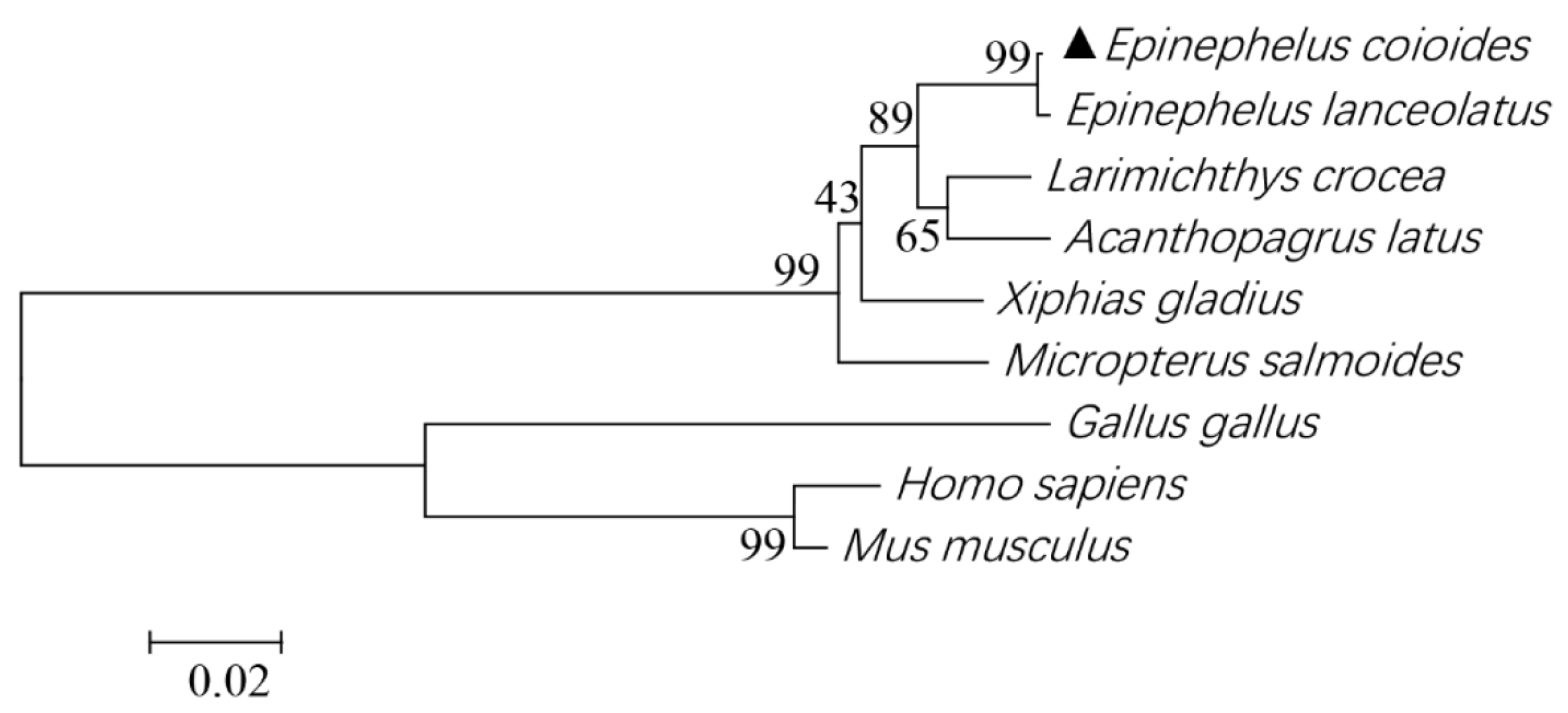
The ORF of EcSNX5 (Accession No. op810504) encoded a polypeptide of 412 amino acids with typical Phox Homology (PX) (28–164aa) and BAR (177–398aa) domains. The multisequence alignment of amino acids found that E. coioides (Accession No: op810504) had the highest identities (98.56%) with E. lanceolatus and the lowest identities (71.94%) with Homo sapiens (Figure 1). Based on the amino acid sequence of EcSNX, E. coioides and E. lanceolatus SNX5 proteins were clustered into one group, indicating the closest evolutionary of them (Figure 2).

3.2. Tissue Distribution of EcSNX5 Gene

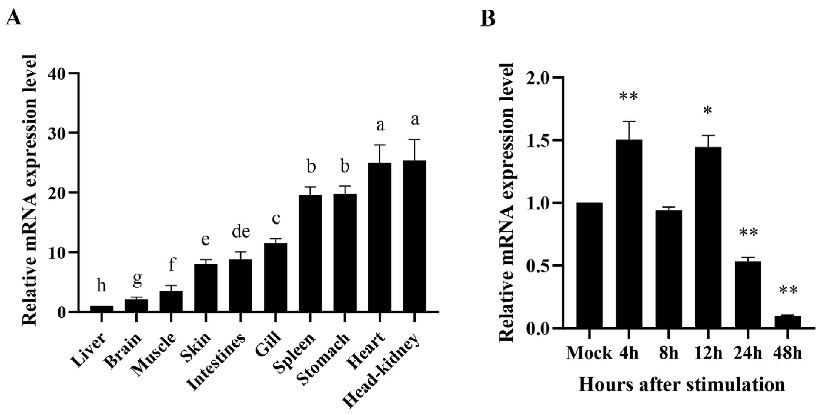
The expression of the EcSNX5 gene in different tissues were shown in Figure 3A. Additionally, the highest expression level was seen in head kidney, followed by heart, stomach, spleen, gills, intestine, skin, muscle, brain and liver.

3.3. Expression Pattern of EcSNX5 after RGNNV Stimulation In Vitro

For the sake of analyzing the influence of RGNNV infection on EcSNX5 expression, the expression quantity of EcSNX5 was tested by qRT–PCR after stimulation of GS cells with RGNNV. The results are shown in (Figure 3B), the expression trend was up-down-up-down, and the expression peak is 4 h after infection.

3.4. EcSNX5 Promoted RGNNV Replication

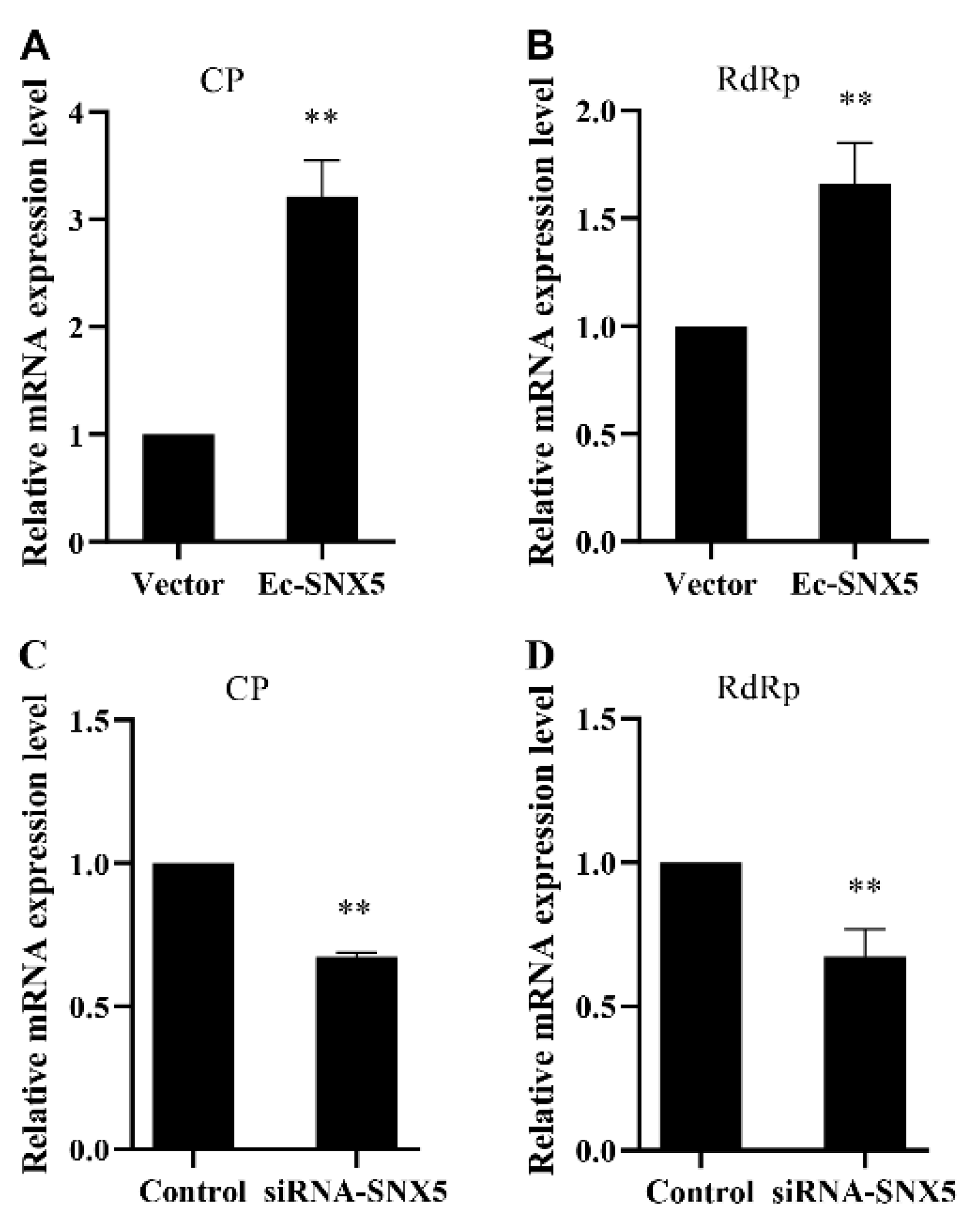
To clarify the impacts of over-expressed EcSNX5 and EcSNX5 knockdown on RGNNV infection, EcSNX5 or EcSNX5 siRNA were transfected into GS cells. The transfected cells were infected with RGNNV. The results of qRT–PCR showed that the expression of CP and RdRp was remarkably improved in EcSNX5-transfected cells compared with vector-transfected cells (Figure 4A,B). EcSNX5 knockdown decreased the expression of CP and RdRp (Figure 4C,D). Additionally, the virus titers were increased significantly in EcSNX5 transfected cells (Figure 5). All of the data demonstrate that EcSNX5 promoted RGNNV replication.

3.5. Co-Localization of EcSNX5 and RGNNV CP

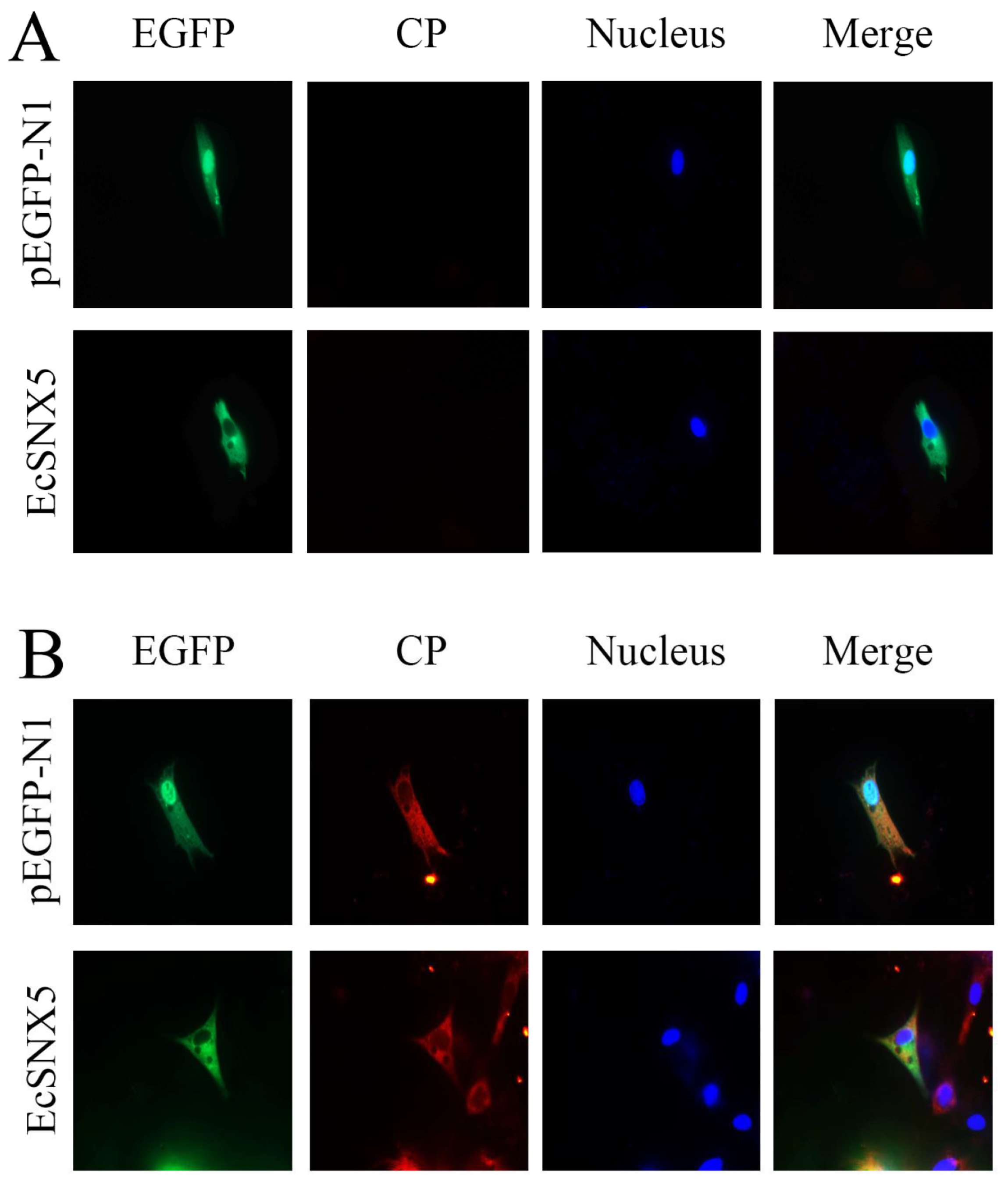
As shown in Figure 6A, EcSNX5 was distributed in the cytoplasm of GS cells. Further immunofluorescence staining experiments were performed to investigate the co-localization of EcSNX5 and RGNNV CP. The fluorescence signals of EcSNX5 and RGNNV CP overlapped, suggesting that EcSNX5 was co-localized with RGNNV CP.

3.6. EcSNX5 Affects Interferon Pathway and Inflammatory Pathway

For clarifying the impacts of EcSNX5 on the expressions of interferon or inflammatory pathway, the expression of interferon- and inflammatory-related factors were examined. Compared with the negative control, EcSNX5 overexpression could reduce the expression of interferon-relative genes (IRF1, IRF3, IRF7, MX1, ISG15, ISG56, MDA5 and TRIF) and inflammation-relative genes (IL6, IL8, IL-1β and TNF-α) (Figure 7A). EcSNX5 knockdown showed the opposite effects on the expressions of genes described above (Figure 7B).

3.7. EcSNX5 Affects the Expression of Autophagy-Related Factors and Apoptosis Factor

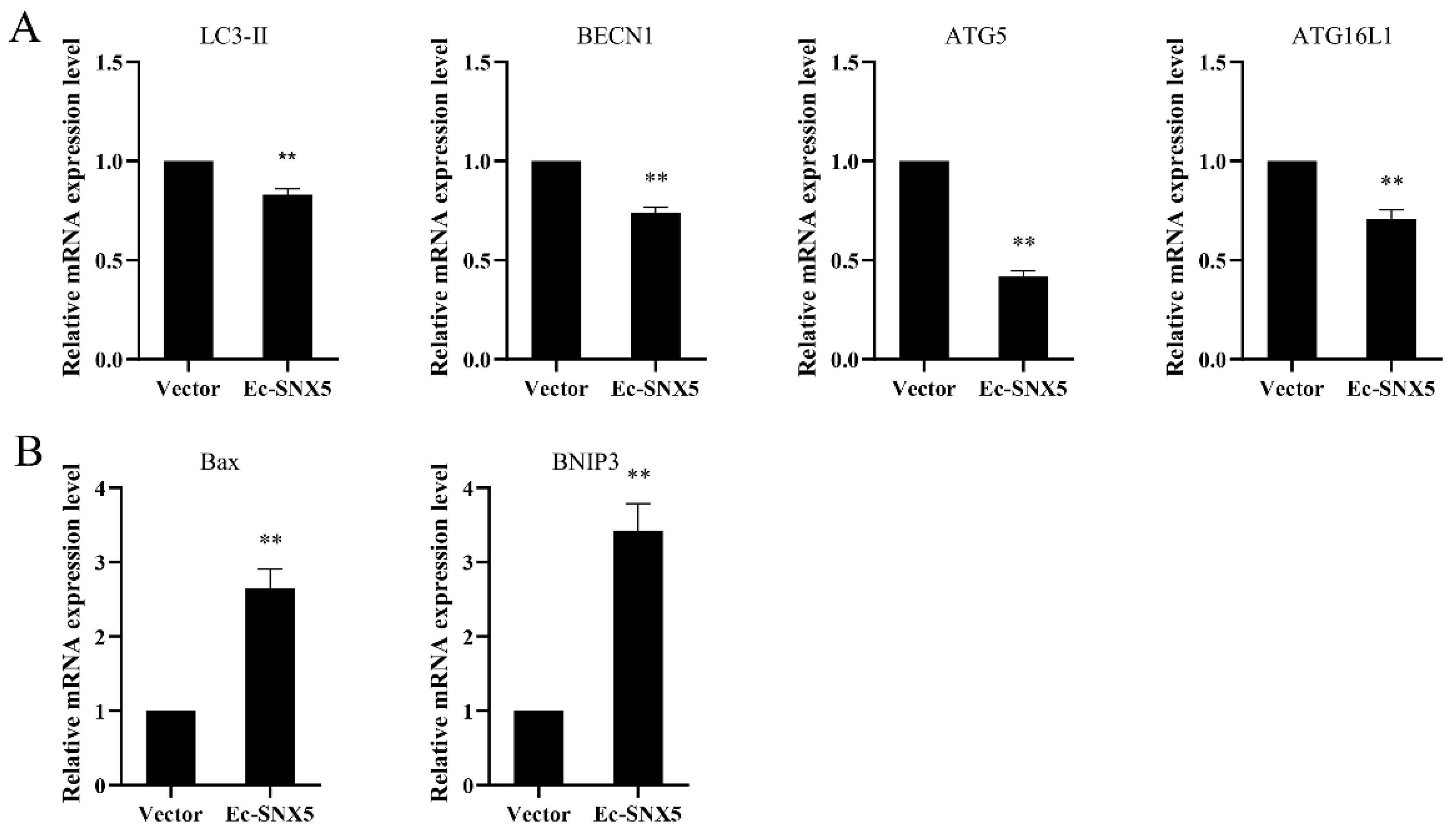
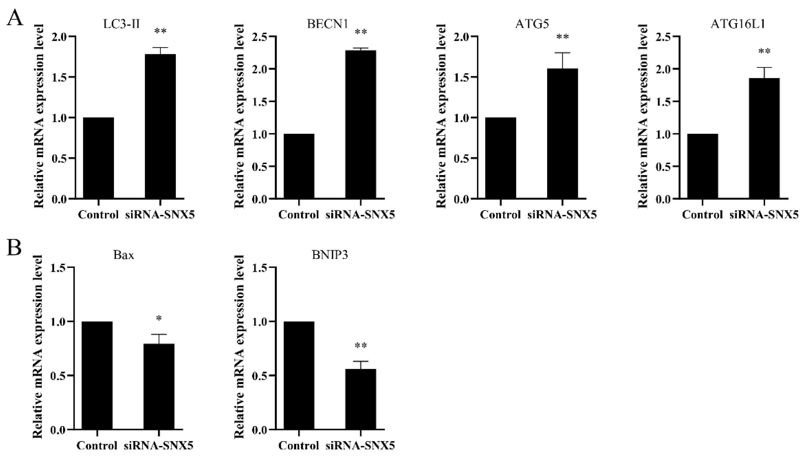
For illustrating the impact of EcSNX5 on cell death caused by viruses, RGNNV was applied to inoculate EcSNX5-transfected cells. Compared with the negative control, the expressions of autophagy-related factors (LC3-II, BECN1, ATG5 and ATG16L1) were decreased and the expression quantity of apoptosis factor (Bax, BNIP3) were enhanced in the EcSNX5-transfected cells. (Figure 8). After the EcSNX5 knockdown, the expressions of autophagy-related factors were enhanced, and the expressions of apoptosis factor were impaired (Figure 9). These data indicated that EcSNX5 inhibited autophagy and promoted apoptosis.

4. Discussion

SNX5 has a key role in membrane transport, controlling membrane transport, protein sorting and participating in intracellular signaling [3]. So far, SNX5 has been extensively studied in mammals, but there are few studies on SNX5 in fish. From our article, we identified and characterized an SNX5 homolog (EcSNX5) from an orange-spotted grouper, E. coioides. EcSNX5 encodes 412 amino acids. It was predicted to have PX and BAR domains. EcSNX5 shared the highest identities (98.56%) and were most closely related with E. lanceolatus SNX5 protein. The deduced EcSNX5 protein contains conserved PX and BAR domains, indicating that EcSNX5 might have semblable functions to extra reported SNX5 proteins. All examined tissues of the grouper could test EcSNX5. In the head kidney, the level of expression was the highest, followed by the heart, similar to those reported in zebrafish [27]. SNX5 has high expression quantity in both hematopoietic progenitor cells and endothelial progenitor cells in zebrafish [27], suggesting that EcSNX5 may also play a key role in the cell-fate decision during early growth. The tissue distribution of EcSNX5 implied that it may have a similar function, such as zebrafish SNX5. After viral infection in vitro, the expression of EcSNX5 was induced and reached its peak at 4 h after infection, then began to decrease, and increased significantly at 12 h and decrease again. Previous studies have shown that SNX5 could induce autophagy to resist virus invasion [5]. SNX5 can also be exploited by viruses to negatively regulate immune response [28]. The reason for the downregulation of the EcSNX5 gene after 12 h may be the massive cell death after virus infection.
Previous studies have suggested that SNX5 is essential for virus replication [29]. The present data showed that EcSNX5 was significantly induced after stimulation with RGNNV in vitro, showing the potential involvement of EcSNX5 in the immune response of RGNNV. To further understand the role of EcSNX5 after NNV infection, the expression levels of viral genes were explored. The results indicated that overexpression of EcSNX5 enhanced the expression of viral genes CP and RdRp, while the knockdown of EcSNX5 significantly downregulated CP and RdRp. Accordingly, the viral titer was also significantly enhanced after EcSNX5 transfection. RdRp is the key viral protein during NNV infection which can induce an endoplasmic reticulum stress response, regulate viral replication and host cell mitochondria-mediated cell death [30]. CP also can induce cell apoptosis, followed by secondary necrosis and cell death via the activation of caspase-3 or caspase-8 [31]. Furthermore, CP inhibits type I IFN response by enhancing the ubiquitination degradation of TRAF3 and IRF3 and promotes NNV replication [32]. It has been recorded that human SNX5 can interact with the human cytomegalovirus UL35 protein for transporting HCMV glycoprotein B to assemble virion particle which promoted the replication of the virus [33]. In order to clarify whether there are similar effects of EcSNX5 during viral infection, a co-localization analysis was performed. The subcellular localization analysis showed that EcSNX5 was co-localized with viral CP protein during viral infection, suggesting that EcSNX5 might promote viral replication by facilitating the viral CP protein transporting and virion assembly.
For investigating the role of EcSNX5 on the immune response to viral infection, the transcripts of several interferon factors and inflammatory factors were examined using qRT–PCR method. The overexpression of ECSNX5 remarkably decreased the expression quantity of IRF1, IRF3, IRF7, MX1, ISG15, ISG56, MDA5, TRIF, IL6, IL8, IL-1β and TNF-α. After the EcSNX5 gene knockdown, the transcriptional levels of these genes were increased. This data demonstrated that EcSNX5 promoted viral replication of NNV by inhibiting immune-factors expression. It has been reported that overexpression of SNX5 inhibited virus-induced IFN-β promoter ISRE, NF-kappa B and IRF3 activation, enhanced K48 ubiquitination of RIG-I and attenuated its K63 ubiquitination, thereby inhibiting the production of type I interferon and pro-inflammatory factors [28]. Whether EcSNX5 has a similar mechanism is needed to clarify in future research. Except for immunity, SNX5 also can impact virus infection through modulating autophagy [5]. For understanding the impact of EcSNX5 on cell death after viral infection, autophagy- and apoptosis-related genes were detected. The results exhibited that EcSNX5 overexpression downregulated the expressions of LC3-II, BECN1, ATG5 and ATG16L1 and upregulated the expressions of Bax and BNIP3 after NNV infection, indicating that the overexpression of EcSNX5 inhibited autophagy and promoted apoptosis. Accordingly, the knockdown of EcSNX5 upregulated the expressions of autophagy genes and downregulated the expressions of apoptosis genes. Given that inhibition of autophagy and promotion of apoptosis were conducive to the persistent infection of virus [34], the present data suggested that EcSNX5 had positive regulatory roles on viral infection. In mammals, SNX5 can increase the expression of ERK 1/2 that inhibits autophagy by activating the mTOR signaling pathway [35], suggesting that EcSNX5 may also have unique way of inhibiting autophagy. Additionally, given that autophagy contributes to RGNNV replication [36], these data suggested that EcSNX5 promote virus proliferation by inhibiting autophagy.

5. Conclusions

In this study, EcSNX5 was characterized by an orange-spotted grouper (E. coioides). The highest expression level of EcSNX5 was observed in the head kidney and responded to RGNNV stimulation in vitro. EcSNX5 may interact with viral CP protein for promoting viral replication and impacts viral infection by regulating virus-induced cell death, interferon pathways and inflammatory responses. The current data will expand the understanding for the role of fish SNX protein during immune response against viral infection and provide useful information for the control strategy development of viral diseases in fish farming.

Supplementary Materials

The following supporting information can be downloaded at: https://www.mdpi.com/article/10.3390/fishes8050231/s1, Figure S1: The nucleotide and derived amino acid sequences of EcSNX5; Table S1: GenBank accession numbers of SNX5 used in the article; Table S2: Full title of the genes used in the article.

Author Contributions

Investigation and Original draft preparation, R.W.; Formal analysis and Visualization, J.L., Z.L. and H.H.; Methodology, Y.H. and B.W.; Resource, J.T. and J.J.; Conceptualization, Writing—Reviewing and Editing, J.C. All authors have read and agreed to the published version of the manuscript.

Funding

This research was funded by National Natural Science Foundation of China (U20A2065, 32073006), Natural Science Foundation of Guangxi (2020GXNSFAA297243), National Key R&D Program of China (2018YFD0900501), Natural Science Foundation of Guangdong Province (No. 2021A1515010532), the Fund of Southern Marine Science and Engineering Guangdong Laboratory (Zhanjiang) (No. ZJW-2019-06), the project of Guangxi Mangrove coastal wetland ecological protection and sustainable utilization Talent Highland (BGMRC202101), Guangdong Provincial Key Laboratory of Pathogenic Biology and Epidemiology for Aquatic Economic Animals (PBEA2020YB01).

Institutional Review Board Statement

All animal experiments were conducted strictly based on the recommendations in the ‘Guide for the Care and Use of Laboratory Animals’ set by the National Institutes of Health. The animal experiments were approved by the Animal Ethics Committee of Guangdong Ocean University (Zhanjiang, China), approval code: (2022)2, approval date: 5 May 2022.

Informed Consent Statement

Not applicable.

Data Availability Statement

The ORF of EcSNX5 has been deposited in NCBI, and the accession number is Accession No. op810504. (https://www.ncbi.nlm.nih.gov/ (accessed on 12 November 2022))

Acknowledgments

The authors thank Qiwei Qin (College of Marine Sciences, South China Agricultural University, Guangdong, China) for kindly providing the Red-spotted grouper nervous necrosis virus and the GS cells. The authors would also like to thank all the laboratory members for their suggestions on this manuscript.

Conflicts of Interest

The authors declare no conflict of interest.

References

  1. Cullen, P.J.; Korswagen, H.C. Sorting nexins provide diversity for retromer-dependent trafficking events. Nat. Cell Biol. 2012, 14, 29–37. [Google Scholar] [CrossRef]
  2. Teasdale, R.D.; Collins, B.M. Insights into the PX (phox-homology) domain and SNX (sorting nexin) protein families: Structures, functions and roles in disease. Biochem. J. 2012, 441, 39–59. [Google Scholar] [CrossRef]
  3. Lu, Y.; He, P.; Zhang, Y.; Ren, Y.; Zhang, L. The emerging roles of retromer and sorting nexins in the life cycle of viruses. Virol. Sin. 2022, 37, 321–330. [Google Scholar] [CrossRef]
  4. Villar, V.A.M.; Armando, I.; Sanada, H.; Frazer, L.C.; Russo, C.M.; Notario, P.M.; Lee, H.; Comisky, L.; Russell, H.A.; Yang, Y.; et al. Novel role of sorting nexin 5 in renal D-1 dopamine receptor trafficking and function: Implications for hypertension. FASEB J. 2013, 27, 1808–1819. [Google Scholar] [CrossRef]
  5. Dong, X.; Yang, Y.; Zou, Z.; Zhao, Y.; Ci, B.; Zhong, L.; Bhave, M.; Wang, L.; Kuo, Y.C.; Zang, X.; et al. Sorting nexin 5 mediates virus-induced autophagy and immunity. Nature 2021, 589, 456–461. [Google Scholar] [CrossRef]
  6. Merino-Trigo, A.; Kerr, M.C.; Houghton, F.; Lindberg, A.; Mitchell, C.; Teasdale, R.D.; Gleeson, P.A. Sorting nexin 5 is localized to a subdomain of the early endosomes and is recruited to the plasma imembrane following EGF stimulation. J. Cell Sci. 2004, 117, 6413–6424. [Google Scholar] [CrossRef]
  7. Yong, X.; Hu, W.; Zhou, X.; Wang, J.; Burstein, E.; Jia, D. Expression and purification of the SNX1/SNX6 complex. Protein Expr. Purif. 2018, 151, 93–98. [Google Scholar] [CrossRef]
  8. Zhou, C.; Wu, Z.; Du, W.; Que, H.; Wang, Y.; Ouyang, Q.; Jian, F.; Yuan, W.; Zhao, Y.; Tian, R.; et al. Recycling of autophagosomal components from autolysosomes by the recycler complex. Nat. Cell Biol. 2022, 24, 497–512. [Google Scholar] [CrossRef]
  9. Sun, Q.; Yong, X.; Sun, X.; Yang, F.; Dai, Z.; Gong, Y.; Zhou, L.; Zhang, X.; Niu, D.; Dai, L.; et al. Structural and functional insights into sorting nexin 5/6 interaction with bacterial effector IncE. Signal Transduct. Target. Ther. 2017, 2, 17030. [Google Scholar] [CrossRef]
  10. Yong, X.; Zhao, L.; Deng, W.; Sun, H.; Zhou, X.; Mao, L.; Hu, W.; Shen, X.; Sun, Q.; Billadeau, D.D.; et al. Mechanism of cargo recognition by retromer-linked SNX-BAR proteins. PLoS Biol. 2020, 18, e3000631. [Google Scholar] [CrossRef]
  11. Cai, J.; Wei, S.; Yu, D.; Song, R.; Lu, Y.; Wu, Z.; Qin, Q.; Jian, J. BNIP3, a cell pro-apoptotic protein, involved in response to viral infection in orange spotted grouper, Epinephelus coioides. Fish Shellfish Immunol. 2017, 64, 407–413. [Google Scholar] [CrossRef]
  12. Soyano, K.; Amagai, T.; Yamaguchi, T.; Mushirobira, Y.; Xu, W.-G.; Pham, N.T.; Murata, R. Endocrine Regulation of Maturation and Sex Change in Groupers. Cells 2022, 11, 825. [Google Scholar] [CrossRef]
  13. Ariff, N.; Abdullah, A.; Azmai, M.N.A.; Musa, N.; Zainathan, S.C. Risk factors associated with viral nervous necrosis in hybrid groupers in Malaysia and the high similarity of its causative agent nervous necrosis virus to reassortant red-spotted grouper nervous necrosis virus/striped jack nervous necrosis virus strains. Vet. World 2019, 12, 1273–1284. [Google Scholar] [CrossRef]
  14. Huang, Y.; Zhang, Y.; Zheng, J.; Wang, L.; Qin, Q.; Huang, X. Metabolic profiles of fish nodavirus infection in vitro: RGNNV induced and exploited cellular fatty acid synthesis for virus infection. Cell. Microbiol. 2020, 22, e13216. [Google Scholar] [CrossRef]
  15. Su, Z.D.; Shi, C.Y.; Huang, J.; Shen, G.M.; Li, J.; Wang, S.Q.; Fan, C. Establishment and application of cross-priming isothermal amplification coupled with lateral flow dipstick (CPA-LFD) for rapid and specific detection of red-spotted grouper nervous necrosis virus. Virol. J. 2015, 12, 149. [Google Scholar] [CrossRef]
  16. Guo, X.; Wang, W.; Zheng, Q.; Qin, Q.; Huang, Y.; Huang, X. Comparative transcriptomic analysis reveals different host cell responses to Singapore grouper iridovirus and red-spotted grouper nervous necrosis virus. Fish Shellfish Immunol. 2022, 128, 136–147. [Google Scholar] [CrossRef]
  17. Tu, J.; Chen, W.; Fu, X.; Lin, Q.; Chang, O.; Zhao, L.; Lan, J.; Li, N.; Lin, L. Susceptibility of Chinese Perch Brain (CPB) Cell and Mandarin Fish to Red-Spotted Grouper Nervous Necrosis Virus (RGNNV) Infection. Int. J. Mol. Sci. 2016, 17, 740. [Google Scholar] [CrossRef]
  18. Chen, S.-P.; Wu, J.-L.; Su, Y.-C.; Hong, J.-R. Anti-Bcl-2 family members, zfBcl-x(L) and zfMcl-1a, prevent cytochrome c release from cells undergoing betanodavirus-induced secondary necrotic cell death. Apoptosis 2007, 12, 1043–1060. [Google Scholar] [CrossRef]
  19. Zhang, W.; Jia, K.; Jia, P.; Xiang, Y.; Lu, X.; Liu, W.; Yi, M. Marine medaka heat shock protein 90ab1 is a receptor for red-spotted grouper nervous necrosis virus and promotes virus internalization through clathrin-mediated endocytosis. PLoS Pathog. 2020, 16, e1008668. [Google Scholar] [CrossRef]
  20. Mai, W.; Liu, H.; Chen, H.; Zhou, Y.; Chen, Y. RGNNV-induced cell cycle arrest at G1/S phase enhanced viral replication via p53-dependent pathway in GS cells. Virus Res. 2018, 256, 142–152. [Google Scholar] [CrossRef]
  21. Asim, M.; Jiang, S.; Yi, L.; Chen, W.; Sun, L.; Zhao, L.; Khattak, M.N.K.; Tu, J.; Lin, L. Glutamine is required for red-spotted grouper nervous necrosis virus replication via replenishing the tricarboxylic acid cycle. Virus Res. 2017, 227, 245–248. [Google Scholar] [CrossRef] [PubMed]
  22. Huang, X.; Huang, Y.; Sun, J.; Han, X.; Qin, Q. Characterization of two grouper Epinephelus akaara cell lines: Application to studies of Singapore grouper iridovirus (SGIV) propagation and virus-host interaction. Aquaculture 2009, 292, 172–179. [Google Scholar] [CrossRef]
  23. Ni, S.; Yu, Y.; Wei, J.; Zhou, L.; Wei, S.; Yan, Y.; Huang, X.; Huang, Y.; Qin, Q. MicroRNA-146a promotes red spotted grouper nervous necrosis virus (RGNNV) replication by targeting TRAF6 in orange spotted grouper, Epinephelus coioides. Fish Shellfish Immunol. 2018, 72, 9–13. [Google Scholar] [CrossRef]
  24. Zou, Z.; Zheng, Q.; Cai, J.; Tang, J.; Xia, L.; Li, P.; Jian, J. Identification of BAG5 from orange-spotted grouper (Epinephelus coioides) involved in viral infection. Dev. Comp. Immunol. 2021, 116, 103916. [Google Scholar] [CrossRef]
  25. Livak, K.J.; Schmittgen, T.D. Analysis of relative gene expression data using real-time quantitative PCR and the 2(-Delta Delta C(T)) Method. Methods 2001, 25, 402–408. [Google Scholar] [CrossRef] [PubMed]
  26. Han, H.L.; Wang, Y.X.; Xu, S.F.; Han, C.Z.; Qin, Q.W.; Wei, S.N. High-density lipoproteins negatively regulate innate immunity and facilitate red-spotted grouper nervous necrosis virus entry via scavenger receptor B type 1. Int. J. Biol. Macromol. 2022, 215, 424–433. [Google Scholar] [CrossRef] [PubMed]
  27. Yoo, K.-W.; Kim, E.-H.; Jung, S.-H.; Rhee, M.; Koo, B.-K.; Yoon, K.-J.; Kong, Y.-Y.; Kim, C.-H. Snx5, as a Mind bomb-binding protein, is expressed in hematopoletic and endothelial precursor cells in zebrafish. FEBS Lett. 2006, 580, 4409–4416. [Google Scholar] [CrossRef] [PubMed]
  28. Li, J.; Chen, T.; Xie, T.; Yang, Y.-X.; He, T.-S.; Xu, L.-G. SNX5 inhibits RLR-mediated antiviral signaling by targeting RIG-I-VISA signalosome. Biochem. Biophys. Res. Commun. 2020, 522, 889–896. [Google Scholar] [CrossRef]
  29. Schuchman, R.; Kilianski, A.; Piper, A.; Vancini, R.; Ribeiro, J.M.C.; Sprague, T.R.; Nasar, F.; Boyd, G.; Hernandez, R.; Glaros, T. Comparative Characterization of the Sindbis Virus Proteome from Mammalian and Invertebrate Hosts Identifies nsP2 as a Component of the Virion and Sorting Nexin 5 as a Significant Host Factor for Alphavirus Replication. J. Virol. 2018, 92, e00694-18. [Google Scholar] [CrossRef]
  30. Su, Y.-C.; Wu, J.-L.; Hong, J.-R. Betanodavirus up-regulates chaperone GRP78 via ER stress: Roles of GRP78 in viral replication and host mitochondria-mediated cell death. Apoptosis 2010, 16, 272–287. [Google Scholar] [CrossRef]
  31. Huang, H.-L.; Liu, Y.-T.; Chen, M.-C.; Wu, J.-L.; Hong, J.-R. Zebrafish anti-apoptotic gene Bcl-xL can prevent aquatic birnavirus-induced cell death in fish cells without affecting expression of viral proteins. Fish Shellfish. Immunol. 2011, 31, 970–977. [Google Scholar] [CrossRef] [PubMed]
  32. Jia, P.; Zhang, W.; Xiang, Y.; Lu, X.; Chen, X.; Pan, H.; Yi, M.; Jia, K. The Capsid Protein of Nervous Necrosis Virus Antagonizes Host Type I IFN Production by a Dual Strategy to Negatively Regulate Retinoic Acid–Inducible Gene-I–like Receptor Pathways. J. Immunol. 2022, 209, 326–336. [Google Scholar] [CrossRef] [PubMed]
  33. Maschkowitz, G.; Gaertner, S.; Hofmann-Winkler, H.; Fickenscher, H.; Winkler, M. Interaction of Human Cytomegalovirus Tegument Proteins ppUL35 and ppUL35A with Sorting Nexin 5 Regulates Glycoprotein B (gpUL55) Localization. J. Virol. 2018, 92, e00013-18. [Google Scholar] [CrossRef]
  34. Chen, Q.; Jia, D.; Ren, J.; Cheng, Y.; Wu, H.; Guo, S.; Wei, T. VDAC1 balances mitophagy and apoptosis in leafhopper upon arbovirus infection. Autophagy 2022, 1–15. [Google Scholar] [CrossRef] [PubMed]
  35. Li, Z. Overexpression of lncRNA HOXA-AS2 promotes the progression of oral squamous cell carcinoma by mediating SNX5 expression. BMC Cell Biol. 2022, 23, 59. [Google Scholar] [CrossRef] [PubMed]
  36. Li, C.; Liu, J.; Zhang, X.; Yu, Y.; Huang, X.; Wei, J.; Qin, Q. Red grouper nervous necrosis virus (RGNNV) induces autophagy to promote viral replication. Fish Shellfish Immunol. 2020, 98, 908–916. [Google Scholar] [CrossRef] [PubMed]
Figure 1. Multiple sequence alignment of EcSNX5 proteins. The conserved amino acids (AA) were marked with dark blue (all the same), pink (more than six sequences), and light blue (six or less than sequences). Table 2 lists an accession number of diverse proteins employed in the article.
Fishes 08 00231 g001
Figure 2. The phylogenetic analysis between EcSNX5 protein and other SNX5 proteins. The triangle symbol indicated the species studied in this paper. GenBank accession numbers for the SNX5 sequence are shown in Table 2.
Fishes 08 00231 g002
Figure 3. (A) Tissue distribution of EcSNX5. Significant differences were indicated by different letters. The datum was expressed as the rate of EcSNX5 mRNA expression quantity in liver. (B) The expression quantity of EcSNX5 in GS cells after infection with RGNNV. EcSNX5 expression was standardized for mock treatment in GS cells. The vertical bar represents the mean value ± SD (n = 3), and remarkable difference of EcSNX5 expression quantity between the infected and mock specimens was bespoken with two asterisks (**) at p < 0.01 and one asterisk (*) at p < 0.05.
Fishes 08 00231 g003
Figure 4. (A,B) Over-expressed EcSNX5 improved the expression quantity of CP and RdRp. (C,D) Knockdown of the EcSNX5 downregulated the expression levels of CP and RdRp. Vector: Gene expression in GS cells transfected with pcDNA-3.1. Control: Gene expression in GS cells transfected with negative control siRNA. Datum on behalf of the mean value for three independent experiments, the error bar represents the SD. (** p < 0.01).
Fishes 08 00231 g004
Figure 5. Viral titer was counted as TCID50 and counted employing Karber method. Datum on behalf of the mean value for three independent experiments, the error bar represents SD. (** p < 0.01).
Fishes 08 00231 g005
Figure 6. EcSNX5 co-localized with CP of RGNNV. (A) Non-infected GS Cells. (B) GS Cells infected with RGNNV. Transfected with pEGFP-EcSNX5 or pEGFP-N1 for 24 h and started to fix it after RGNNV inoculation for 24 h or without RGNNV inoculation. A total of 24 h for RGNNV CP dyeing by the anti-RGNNV-CP antibody and observed employing inverted fluorescence Microscope. The green fluorescence exhibits the location of GFP-tagged protein, the blue fluorescence exhibits the location of nucleus and the red fluorescence exhibits the location of RGNNV coat protein.
Fishes 08 00231 g006
Figure 7. (A) Expression of interferon pathway-related gene and inflammatory factor. Over-expressed EcSNX5 downregulated the expression quantity of interferon pathway-related gene and inflammatory factor. (B) Expression of interferon pathway-related gene and inflammatory factor. Knockdown of the EcSNX5 enhanced the expression quantity of interferon pathway-related gene and inflammatory factor. Vector: Gene expression in GS cells transfected with pcDNA-3.1. Control: Gene expression in GS cells transfected with negative control siRNA. The expression levels of these genes were detected by means of qRT–PCR method. Datum on behalf of the mean value for three independent experiments, the error bar represents the SD. (** p < 0.01, * p < 0.05).
Fishes 08 00231 g007
Figure 8. (A) Expression of autophagy genes. (B) Expression of apoptotic genes. Overexpressed EcSNX5 lowered the expression quantity of autophagy genes and improved the expression quantity of apoptotic genes. The full names of the genes employed in the article are displayed in supplementary materials. Datum on behalf of the mean value for three independent experiments, the error bar represents the SD. (** p < 0.01).
Fishes 08 00231 g008
Figure 9. (A) Expression of autophagy genes. (B) Expression of apoptotic genes. Knockdown of the EcSNX5 improved the expression quantity of autophagy genes and reduced the expression quantity of apoptotic genes. The expression quantity of these genes was detected by means of qRT–PCR method. Datum on behalf of the mean value for three independent experiments, the error bar represents the SD. (** p < 0.01, * p < 0.05).
Fishes 08 00231 g009
Table 1. Primers used in the article. EcSNX5-ORF-F/R is the primer for amplification of EcSNX5 ORF. The pcDNA-EcSNX5-F/R is the primer for construction of eukaryotic expression vector. In addition, the rest of the primers are used for QRT–PCR.
NameSequence (5′–3′)Size
EcSNX5-ORF-FATGACATCCACAATAGACGAAAG1236 bp
EcSNX5-ORF-RCTTGGTAATGGCAGGACATGGAT
pcDNA-EcSNX5-FCCGCTCGAGATGACATCCACAATAGACGAAAG1255 bp
pcDNA-EcSNX5-RCCGGAATTCCCTTGGTAATGGCAGGACATGGAT
EcSNX5-RT-FTCCACAATAGACGAAAGCAAT136 bp
EcSNX5-RT-RGGACAGTAAACTTGACTTTATCCC
18S–RT-FATTGACGGAAGGGCACCACCAG158 bp
18S-RT-RTCGCTCCACCAACTAAGAACGG
RGNNV-CP-RT-FCAACTGACAACGATCACACCTTC162 bp
RGNNV-CP-RT-RCAATCGAACACTCCAGCGACA
RGNNV-RdRp-RT-FGTGTCCGGAGAGGTTAAGGATG192 bp
RGNNV-RdRp-RT-RCTTGAATTGATCAACGGTGAACA
EcIRF1-RT-FAGGGAGCCAGTGGAGTGAATC457 bp
EcIRF1-RT-RGATGCCTGTGCCCAAAGTTAT
EcIRF3-RT-FATGGTTTAGATGTGGGGGTGTCGGG148 bp
EcIRF3-RT-RGAGGCAGAAGAACAGGGAGCACGGA
EcIRF7-RT-FCAACACCGGATACAACCAAG153 bp
EcIRF7-RT-RGTTCTCAACTGCTACATAGGGC
EcMxI-RT-FCGAAAGTACCGTGGACGAGAA117 bp
EcMxI-RT-RTGTTTGATCTGCTCCTTGACCAT
EcISG15-RT-FCCTATGACATCAAAGCTGACGAGAC179 bp
EcISG15-RT-RGTGCTGTTGGCAGTGACGTTGTAGT
EcISG56-RT-FCAGGCATGGTGGAGTGGAAC126 bp
EcISG56-RT-RCTCAAGGTAGTGAACAGCGAGGTA
EcMDA5-RT-FACCTGGCTCTCAGAATTACGAACA142 bp
EcMDA5-RT-RTCTGCTCCTGGTGGTATTCGTTC
EcTRIF-RT-FAAACCAACCACTGGACCAAACTT189 bp
EcTRIF-RT-RGATGGCATCCTCGACACACCTCA
EcIL6-RT-FACCTGGCTCTCAGAATTACGAACA169 bp
EcIL6-RT-RTCATCTTCAAACTGCTTTTCGTG
EcIL8-RT-FGCCGTCAGTGAAGGGAGTCTAG131 bp
EcIL8-RT-RATCGCAGTGGGAGTTTGCA
EcIL-1β-RT-FCTCCACCGACTGATGAGGATATG128 bp
EcIL-1β-RT-RGGCTGTTATTGACCCGAACTAAG
EcTNFa-RT-FATGAACAAAGAAGTAGATTGGGTCG162 bp
EcTNFa-RT-RGTCCGACTTGATTAGTGCTT
EcLC3-II-RT-FGCACCCCAACAAGATACCAGT145 bp
EcLC3-II-RT-RCGTAGACCTCGGAGATGGCA
EcBecline1-RT-FGAGATACCGTCTGGTCCCGTAT265 bp
EcBecline1-RT-RCCTTTTTCCACCTCCTCTTTGA
EcATG5-RT-FCCACTGAGGAGGGAGGCTT88 bp
EcATG5-RT-RCAGATGAAACAGGGCGAAA
EcATG16L1-RT-FGAACAGCCAACTCCTTCAGCACG154 bp
EcATG16L1-RT-RTCCTTTAGGCTCACAGCGACCAG
EcBax-RT-FGACCCAAATACCAAGAGG47 bp
EcBax-RT-RTGTGGGACTGAGTGAAGAG
EcBNIP3-RT-FATGAACAAAGAAGTAGATTGGGTCG232 bp
EcBNIP3-RT-RGTGAGATGAGTAAGGAAGGGATGA
Table 2. The software and websites for analysis in this study.
Analysis ContentWebsite/Software
Primer designPrimer 5.0
Nucleic acid and protein sequence analysisDNAMAN 9.0
Conservative functional domain predictionhttps://www.ncbi.nlm.nih.gov/Structure/cdd/wrpsb.cg (accessed on 4 July 2021)
Amino acid sequence alignment analysisMUSCLE
Evolutionary tree constructionMEGA version 6.0
Quantitative analysis of dataSPSS 22.0
Graphic creationGraphPad Prism 8.0
Disclaimer/Publisher’s Note: The statements, opinions and data contained in all publications are solely those of the individual author(s) and contributor(s) and not of MDPI and/or the editor(s). MDPI and/or the editor(s) disclaim responsibility for any injury to people or property resulting from any ideas, methods, instructions or products referred to in the content.