Article

Successful Cryopreservation of Spermatogonia Stem Cells of Neotropical Catfish (Rhamdia quelen) and Enriched Germ Cell Transplantation into Common Carp (Cyprinus carpio) Testes

by
Ivana F. Rosa
1,†,
Emanuel R. M. Martinez
1,†,
Melanie Digmayer
1,
Lucas B. Doretto
1,2,3,* and
Rafael H. Nóbrega
1,*
1
Department of Structural and Functional Biology, Institute of Biosciences, São Paulo State University (UNESP), Botucatu 01049-010, Brazil
2
Yellow Sea Fisheries Research Institute, Chinese Academy of Fishery Sciences, Qingdao 266071, China
3
National Key Laboratory of Mariculture Biobreeding and Sustainable Goods, Qingdao 266071, China
*
Authors to whom correspondence should be addressed.
These authors contributed equally to this work.
Fishes 2023, 8(10), 478; https://doi.org/10.3390/fishes8100478
Submission received: 14 August 2023 / Revised: 14 September 2023 / Accepted: 21 September 2023 / Published: 25 September 2023
(This article belongs to the Special Issue Reproductive Biology and Breeding of Fish)

Abstract

:
Cryopreservation and transplantation of spermatogonial stem cells (SSCs) offer new possibilities in the conservation of valuable genetic resources. Therefore, the present study developed a cryopreservation method for whole testicular tissue and for spermatogonial stem cells of jundia catfish (Rhamdia quelen) and developed an enriched germ cell transplantation of jundia catfish into depleted common carp (Cyprinus carpio) testes. Our findings from whole testes indicate that the cryoprotectants MeOH (1.3 M), DMSO (1.4 M), and EG (1.4 M) resulted in high cell viability rates of 67%, 62%, and 51.5%, respectively. Notably, in the case of enriched post-thaw SSCs, DMSO exhibited the highest cell viability at 27%, followed by EG at 16% and MeOH at 7%. Additionally, we observed the successful colonization and proliferation of jundia germ cells within the recipient gonads of common carp following transplantation. Notably, Sertoli cells were identified in the recipient gonads, providing support to the stained donor germ cells and indicated the formation of cysts. Our data suggest that cryopreserving entire testicular tissue presents a viable alternative to cryopreserving isolated testicular cells, and the spermatogonial cells isolated from testes of jundia retained transplantability characteristics. Nonetheless, more investigations are required to reach the goal of functional gamete and to assess the effectiveness of transplantation using these cryopreserved tissues. Taken together, proper cryopreservation methodology and transplantation technology could aid the preservation practice of fish genetic resources.
Key Contribution: This study suggests that cryopreserving entire testicular tissue is a practical alternative to cryopreserving isolated testicular cells of jundia. Furthermore, enriched spermatogonial testicular cells isolated from jundia demonstrated successful colonization and proliferation within the depleted common carp testes. These findings have implications for the conservation of fish genetic resources and offer potential advancements in preservation practices.

1. Introduction

Spermatogonial cryopreservation has emerged as an attractive tool to preserve fish genetic resources and facilitates artificial reproduction [1,2,3,4,5]. In the recent years, the cryopreservation of oogonia, primordial germ cells (PGCs), and spermatogonial stem cells (SSC) has attained a notable degree of advancement, and these cell types have been successfully cryopreserved across numerous species [5,6,7,8,9,10,11,12,13,14,15,16,17]. In this sense, SSCs can be cryopreserved to establish a valuable resource in the form of a spermatogonial stem cell bank for threatened or economically significant species [1,3,5,6].
In addition to spermatogonial cryopreservation technology, the SSC transplantation has introduced novel avenues for safeguarding valuable genetic resources [6,15,18,19]. The growing interest in the biology of SSCs in teleost fish has been primarily influenced by the experiments by Brinster and Avarbock [20], who developed an SSC transplant technique in mammalian species. In this study, mouse spermatogonial stem cells (SSCs) were successfully transplanted into depleted mouse recipient testes, resulting in production of functional donor-derived spermatozoa [20].
In fish species, recent advances in spermatogonia transplantation and the subsequent production of donor-derived sperm have been achieved in masu salmon (Oncorhynchus masou) [21], rainbow trout (Oncorhynchus mykiss) [5,6,7], tilapia (Oreochromis niloticus) [22,23], medaka (Oryzias latipes) [11], pufferfish (Carinotetraodon travancoricus) [12], and zebrafish (Danio rerio) [10,24]. For instance, Hettiarachchi et al. (2022) [25] demonstrated the capability of spermatogonial stem cells from blue catfish (Ictalurus furcatus) to colonize and proliferate in triploid channel catfish recipients. Similarly, testicular cells from yellowtail (Seriola quinqueradiata) were successfully transplanted into allogeneic recipients, with type A spermatogonia from the thawed cells colonizing recipient gonads and differentiating into functional sperm [26]. In this sense, germ cell transplantation techniques offer an efficient alternative for conservation of genotypes of commercially important fish species [4,16,21,22,26].
Rhamdia quelen, commonly known as the silver catfish or jundia, is a neotropical catfish highly valued as a protein source for human consumption and aquaculture in southern Brazil, Argentina, and Uruguay [27,28,29,30]. With its increasing significance, extensive research has focused on feeding management for growth [31,32], breeder nutrition [33,34,35], and the reproductive processes, encompassing sperm quality and gamete conservation [19,36,37,38,39,40,41]. However, the literature lacks comprehensive coverage of several reproductive parameters, especially those related to testis function, including cryopreservation, transplantation, and spermatogenesis [18,19,23,39]. Therefore, it is imperative to conduct studies on the reproductive biology of R. quelen to enhance its utilization in genetic improvement programs and for conservation efforts.
In this study, we established a cryopreservation technique for both whole testicular tissue and enriched spermatogonial stem cells of jundia catfish (R. quelen). Additionally, we conducted a cross-species transplantation using enriched SSCs of jundia catfish, belonging to the Siluriformes order [42], into a depleted adult male common carp (Cyprinus carpio) from the Cypriniformes order [43].

2. Materials and Methods

2.1. Animals Maintenance

Nine sexually mature male jundia (R. quelen) and eighteen depleted adult male common carp (C. carpio) were used in this study. Both species were obtained from commercial farmers and raised in the aquarium facility at the Department of Structural and Functional Biology, Institute of Biosciences, São Paulo State University (Botucatu, Brazil). Fish used for the experiments were kept in 500 L tanks in a recirculation system with controlled photothermal conditions (27 °C, pH 7.6; conductivity of 750 μS) under 14 h:10 h (light, dark) photoperiod. Parameters such as salinity, pH, dissolved oxygen, and ammonia were monitored daily. All animal handling and experimental procedures were performed following the Ethical Principles of Animal Experimentation adopted by the National Council for the Control of Animal Experimentation (CONCEA/Brazil). All procedures conducted in this study were approved by the CEUA (Committee on Ethics in the Use of Animals) of São Paulo State University, protocol number: 666-CEUA for the jundia and 672-CEUA for the common carp.

2.2. Enzymatic Dissociation of Testes

Before germ cell cryopreservation of jundia, enzymatic dissociation of testes was performed. Briefly, testes from three adult jundia were collected and washed in Hank’s Balanced Salt Solution (HBSS, Merck KGaA, Darmstadt, Germany) containing 0.1% penicillin–streptomycin (10,000 IU/10 mg/mL) [6,7,20,24].
Further, enzymatic dissociation of testicular tissue was performed using 0.2% collagenase in Dulbecco’s Modified Eagle’s Medium/Nutrient Mixture F-12 Ham (DMEM/F-12 Ham) (Gibco, Life Technologies, Andover, MA, USA) followed by incubation for 3 h at 28 °C. Subsequently, the tissue was incubated with a mixture of 0.25% trypsin/1 mM EDTA (Life Technologies, Andover, MA, USA) and 0.03% DNase I (Invitrogen™, Life Technologies, Andover, MA, USA) under similar conditions for 30 min. To inhibit trypsin activity, an equal volume of FBS (Gibco, Life Technologies, Andover, MA, USA) was added.
The resultant cell suspension was filtered through a 60 µm Corning cell strainer (Sigma Aldrich, San Luis, MI, USA) to remove any undissociated cell clumps, followed by centrifugation at 200× g for 10 min. Subsequently, the pellet was resuspended in DMEM/F-12 Ham (Gibco, Life Technologies, Andover, MA, USA) and collected for germ cells cryopreservation protocol. Moreover, an enriched type A spermatogonia cell suspension was obtained with Percoll gradient centrifugation according to methods previously described [23,44,45].

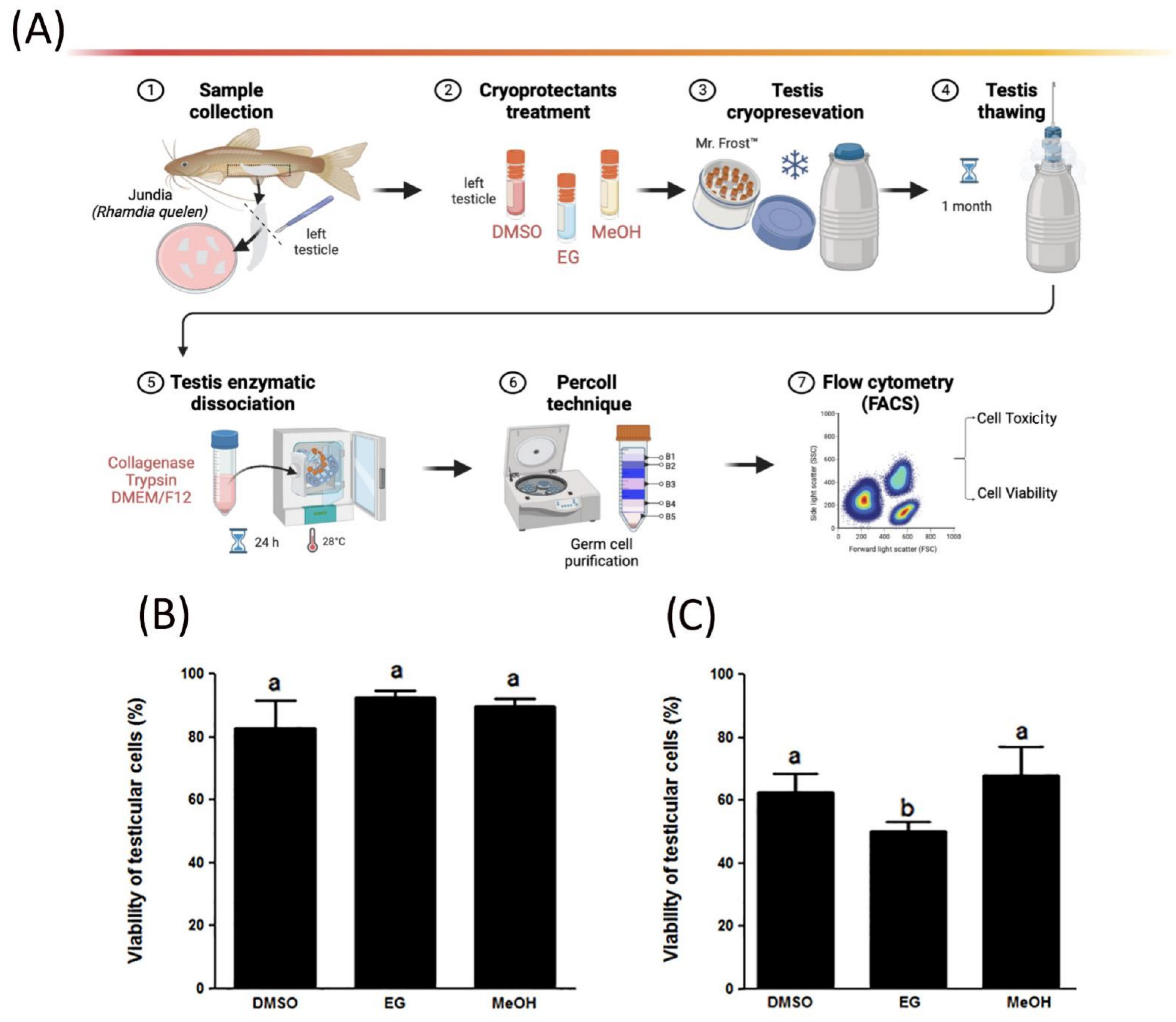
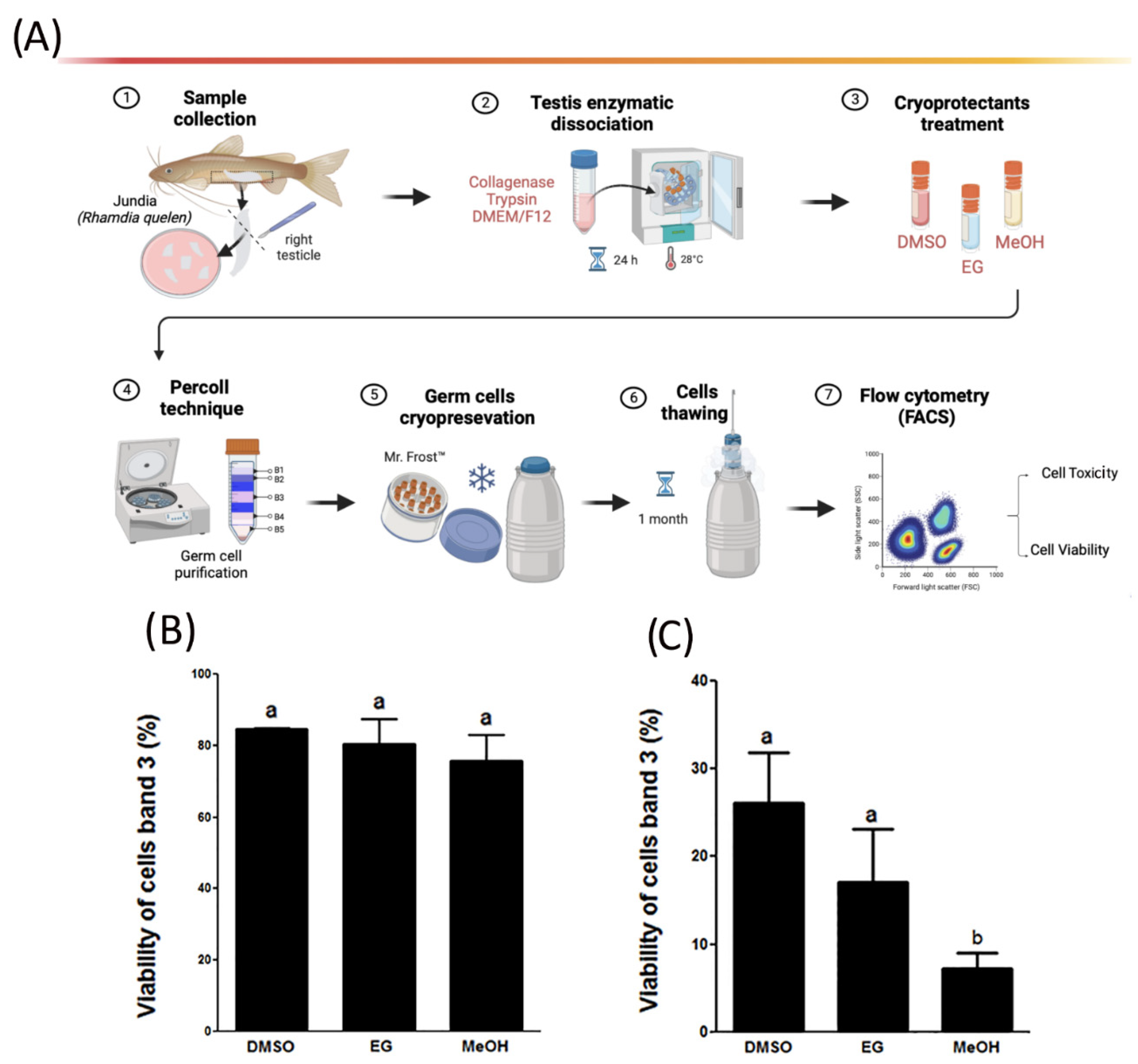
2.3. Testes and Germ Cells’ Cryopreservation Protocol

To establish an effective germ cells’ cryopreservation protocol, both intact testes and dissociated testicular cells from jundia were subjected to cryopreservation for one month, followed by a subsequent assessment of cell viability. Moreover, enriched germ cells with Percoll technique were also cryopreserved and evaluated for cell viability.
Briefly, intact testes from adult jundia (n = 5) were collected and washed in DMEM solution, cut into fragments of 5 × 5 × 5 mm, and transferred into 1.5 mL cryogenic vials with 500 uL of cryomedia containing a permeating cryoprotectant solution. The vials remained submerged in ice for 60 min to reduce the toxicity of the cryoprotectants and stabilize the solution in the tissue. After this period, the vials were transferred to a Mr. Frosty™ freezing container (Thermo Scientific, Waltham, MA, USA) in a −80 °C freezer for at least 90 min before being plunged into liquid nitrogen for 1 month.
The cryoprotectants used were 1.3 M methanol (MeOH), 1.4 M dimethyl sulfoxide (DMSO), and 1.8 M ethylene glycol (EG) in Dulbecco’s Modified Eagle’s Medium (DMEM) (Gibco, Life Technologies, Andover, MA, USA) combined with 0.75% bovine serum albumin (BSA Merck KGaA, Darmstadt, Germany) and 10% fetal bovine serum (FBS). The concentrations of DMSO and EG were selected according to Lacerda et al. [44] and Yoshizaki et al. [46], respectively. Moreover, MeOH concentration was selected according to Lee and Yoshizaki et al. [6].
For germ cell cryopreservation, cells obtained previously from testes dissociation was first quantified using a Neubauer chamber, and aliquots of 107 cells were added to 1.2 mL cryogenic tubes containing the cooled cryoprotectant solution described above. Subsequently, the tubes were placed on ice for 30 min to stabilize the cell solution, followed by immediate transfer to the Mr. Frosty™ freezing container and storage in an ultra-freezer at −80 °C for at least 90 min before being plunged into liquid nitrogen for 1 month. Before this period, samples were held in liquid nitrogen for at least 24 h before thawing. For the thawed process, cryogenic vials containing germ cells and testes were placed in a water bath at 30 °C for 2 min followed by washing in DMEM/0.75% BSA to remove the cryoprotectants. Subsequently, testes were subjected to the dissociation process followed by germ cell purification using the Percoll technique. An enriched type A spermatogonia cell suspension was obtained with Percoll gradient centrifugation according to methods previously described [23,44,45,47]. Moreover, the cryopreserved germ cells were collected for toxicity assay and cell viability analysis.

2.4. Toxicity, Viability, and Cell DNA Assessment Using Flow Cytometry

To assess the toxicity of the cryoprotectant solutions, we evaluated both cell viability and cell cycle dynamics, leading to the establishment of four distinct experimental groups (Table S1). The determination of cell viability was executed utilizing the Fluorescence-Activated Cell Sorting (FACS) technique. In this approach, cell suspensions were stained with annexin V conjugated to fluorescein isothiocyanate (FITC), serving to detect live cells, while the employment of the membrane-impermeable propidium iodide (PI) nucleic acid dye (Thermo Fisher Scientific, Gibco®, Mississauga, ON, Canada) enabled the discernment of deceased cells. For this purpose, germ cells obtained from band 3, as performed by Lacerda et al. [47] of Percoll were first washed with DMEM/binding buffer, incubated for 5 min in the dark, and followed by PI for a further 5 min incubation. After incubation, cells were analyzed with the FACSCanto™ flow cytometry system (BD Biosciences, Piscataway, NJ, USA). Nonetheless, all dissociated cells from the four groups (Table S1) were submitted to viability and toxicity analyses. The percentage of viable cells in each group was estimated using the FlowJo program, and 20,000 events were recorded in each analysis.
Additionally, the cell DNA content from the dissociation of three testes was also determined by staining the cells with propidium iodide and measuring fluorescence using FACSCanto™ II. The dissociated cells from cell suspension and the 5 bands obtained with Percoll gradients were resuspended and fixed at −20 °C with 70% ethanol and Milli-Q water. The fixed cells were incubated in a solution containing 100 µg/mL of RNase and 50 µg/mL of propidium iodide for 30 min at 37 °C. FACS was used to analyze 10,000 cells from each population, and the percentage of cells in each cell cycle phase was calculated. The cells were classified as being in interphase stages G0/G1, S, or G2/M based on the intensity of the fluorescence peaks [48].

2.5. Light Microscopy for Percoll Bands

For light microscopy, cells from Percoll bands were fixed in 4% paraformaldehyde in Sorensen buffer (0.1 M, pH 7.2) for at least 24 h. Posteriorly, cells were dehydrated in a graded ethylic series and embedded in Historesin (Leica HistoResin). Historesin blocks were sectioned (3 µm thickness), and histological sections were stained with toluidine blue. Germ cells at different stages of development were counted based on the morphological criteria from Lacerda et al. [47]. The histological sections were examined and documented using Leica DMI6000 microscope (Leica, Hessen, Germany).

2.6. Spermatogenesis Depletion

In the present study, endogenous spermatogenesis was depleted in eighteen adult male carp specimens (Table S2) according to the procedures already described by Lacerda et al. [45]. The average weight of the carp specimens was 20.2 ± 4.3 g, and the average length was 13 cm. The specimens were kept in 250 L tanks with constant aeration and weekly water changes. Water temperature was adjusted daily and maintained at 35 °C for at least 2 weeks before receiving intraperitoneal injections of busulfan (Sigma, St. Louis, MO, USA). After 7 days at this temperature, the first dose of busulfan was injected (18 mg/kg of live weight), and the second dose (in the same concentration) was injected after a two-week interval from the first injection. In order to validate spermatogenesis depletion, depleted testes from adult male carp specimens were collected for histological analysis as described below.

2.7. Donor Germ Cell Labeling with PKH26 and Transplantation

Dissociated cells (n = 3 testes) (band 3), previously purified with Percoll, from jundia were used for intraperitoneal transplantation into depleted male common carp (n = 18) (Tables S2 and S3). To accurately identify the donors’ germ cells in the recipients’ testes, germ cells were labeled with PKH26 (Sigma Aldrich, St. Louis, MO, USA) for 5 min following the manufacturer’s protocol. After staining, the cells were then suspended in DMEM/F12 at a concentration of 107 cells/mL. For transplantation, recipient common carp (n = 18) were anesthetized with Quinaldin solution (Sigma Aldrich, San Luis, MI, USA), and the donor germ cells were transplanted using a glass micropipette (outside diameter of 70 lm) under a Zeiss stereomicroscope. Approximately 1 mL of cell suspension was injected into each recipient. Following transplantation, recipient fish were maintained in a 500 L tank, and the water temperature was gradually decreased (1–2 °C) per day until 26 °C was reached.

2.8. Microscopic Observation of Donor-Derived Germ Cells in Recipient Common Carp

In order to evaluate the germ cell transplantation, recipient fish testes were collected at 24 h and 1, 2, 3, 4, 5, 6, and 12 weeks post-spermatogonial transplantation. Therefore, collected testis were first fixed with 4% paraformaldehyde in PBS overnight. The material was then dehydrated in an increasing series of concentrations of alcohol (70% for 4 h; 95% for 4 h), infiltrated, and embedded in paraffin (Leica Wetzlar, Hessen, Germany). Finally, the sections were stained with DAPI (targeting DNA in the cell nucleus) and then analyzed under a confocal microscope (Leica DMI 4000B).

2.9. Statistical Analyses

Data were initially checked for deviations from variance normality and homogeneity through the Shapiro–Wilk and Bartlett’s tests, respectively. Significant differences among groups were identified using a paired Student’s t-test, at 5% probability. GraphPad Prism 8.01 (Graphpad Software, Inc., San Diego, CA, USA) was used for the statistical analysis.

3. Results

3.1. Cell Viability of Cryopreserved Testicular Germ Cells and Toxicity of Cryoprotectants

In order to evaluate the cryoprotectant viability and toxicity, intact testes and dissociated testicular cells were subjected to cryopreservation for one month, followed by a subsequent assessment of cell viability through the FACS technique (Figure 1A, Figure 2A, Figures S1 and S2). Our results showed that none of the used cryoprotectants were found to be toxic in non-cryopreserved cells (Figure 1B and Figure 2B). Among the cryoprotectants, EG showed 92% of cell viability when used on whole testes (Figure 1B), followed by MeOH (89%) and DMSO (82%). No significant differences between the groups were identified.
Furthermore, following a one-month cryopreservation period, the thawing and dissociation of testis samples were undertaken to evaluate cell viability (Figure 1B,C). Our findings indicated a cell viability of 67% and 62% for cryomedium containing MeOH and DMSO, respectively, with EG demonstrating a viability of 51.5% (Figure 1C). Notably, statistical analysis revealed a significant difference between the EG solution and both DMSO and MeOH solutions (Figure 1C).
Additionally, we also evaluated the viability rates of non-cryopreserved and cryopreserved testicular cells enriched with the Percoll technique (Figure 2A–C). Our results showed that non-cryopreserved testicular cells (band 3) have significantly higher viabilities in cryomedium containing DMSO (84%) and EG (81%) than those cryopreserved in MeOH (76%) (Figure 2B). However, the viability rates of testicular cells recovered after cryopreservation (band 3) were lower than non-cryopreserved dissociated cells (Figure 2C). The DMSO exhibits the highest cell viability (27%) compared to cryoprotectants EG (16%) and MeOH (7%) (Figure 2C), although no significant differences were identified between cells cryopreserved in EG and MeOH.

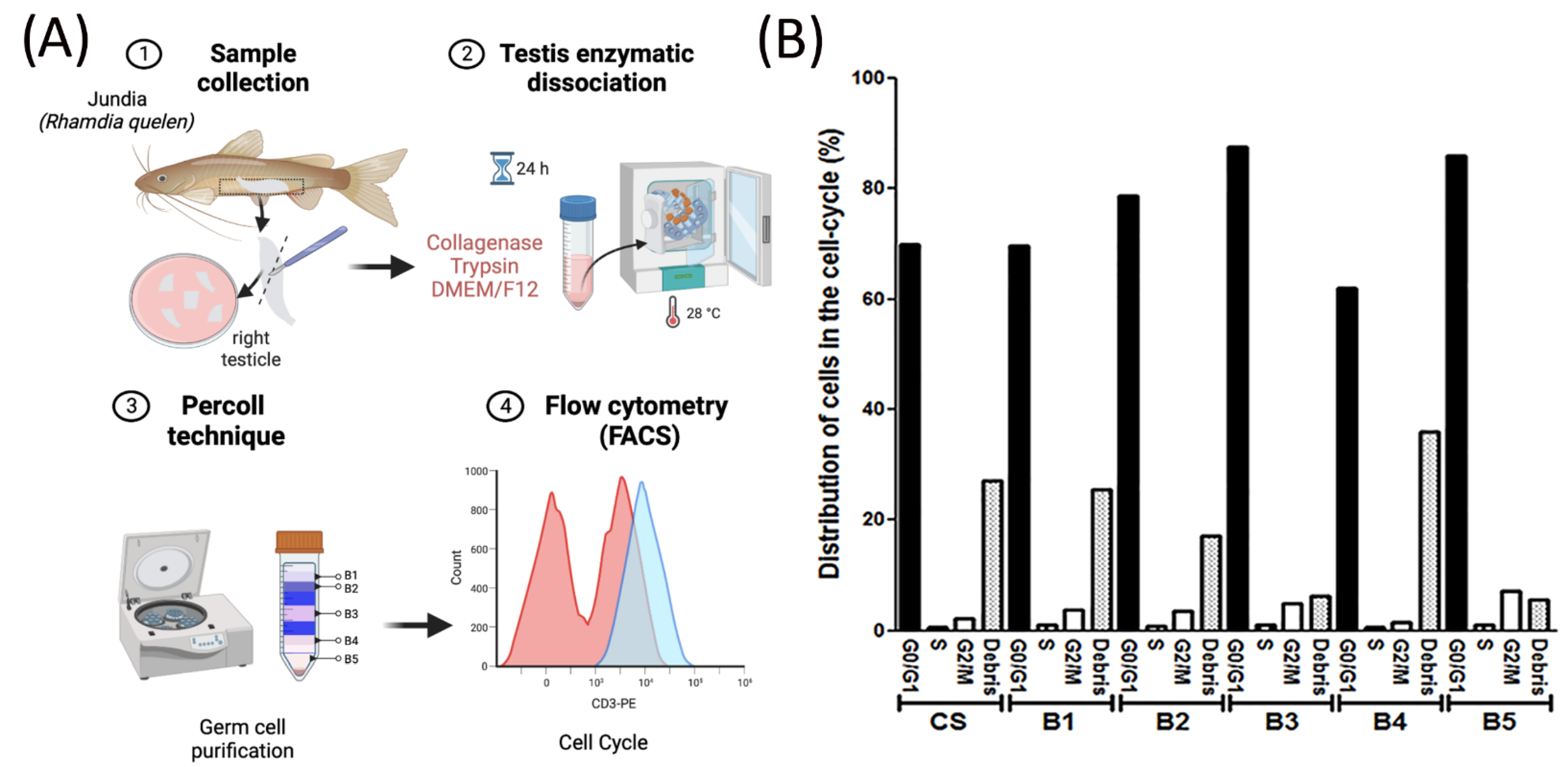
3.2. Cell Cycle Analysis

Flow cytometry with propidium iodide was used in order to evaluate the cell cycle of suspensions obtained with Percoll technique (Figure 3A). The relative percentage of the proportions of cells in the G0/G1, S, and G2/M stages was calculated (Figure 3B, Figures S3 and S4), and representative histograms for the cell cycle stage distributions of all Percoll bands are shown in Figure S3.
In our study, distinct stages of interphase within the cell cycle were observed for both whole suspensions and separate bands, determined through analysis of their fluorescent DNA content (Figure 3B and Figure S3). Upon aggregation of the bands and suspensions from all three fish (Figure 3B), G0/G1 emerged as the predominant phase of the cell cycle in band 3, constituting 87%. Moreover, a minority of cells were identified within the G2/M stage, and around 2% of cells were observed to be in a quiescent state within the S stage for all bands and cells suspension (Figure 3B). As expected, a notable prevalence of type Aund was found in band 3 as evidenced by the histological analysis (Figure S4).

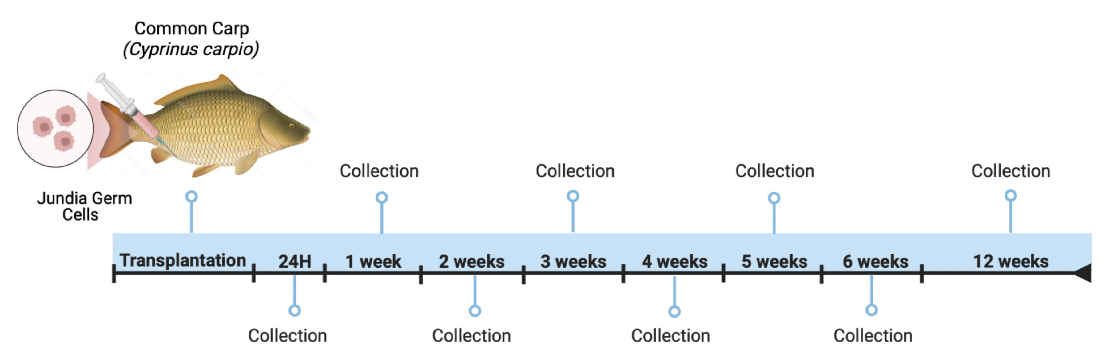
3.3. Jundia PKH26-Labeled Germ Cells in the Common Carp Testes

Jundia germ cells marked with PKH26 were transplanted and then tracked for 12 weeks in recipients (common carp) (Figure 4 and Figure 5A–O). PKH26-labeled germ cells were observed in the recipient seminiferous tubules in the first week (7 days) of transplantation (Figure 5A–C), while the control group showed no fluorescence (Figure 5A). More specifically, the incorporated PKH26-positive cells at 1 week (Figure 5B,C) and 3 weeks (Figure 5E,F) had an approximate diameter of 10 μm, which is a typical morphological characteristic of spermatogonia [19] with the presence of Sertoli cells surrounding them.
At 28, 35, and 84 days post-transplantation (4, 5, and 12 weeks) (Figure 5G–O), the cells were arranged in spermatogenic cystic structures inside the seminiferous tubules, while the control had no fluorescence distribution (Figure 5G,J,M). Additionally, some recipient Sertoli cells were also found to be surrounding the cells labeled with PKH26, and the fluorescence was gradually weakened. Furthermore, all recipient common carp testes presented PKH26-positive donor germ cells.

4. Discussion

Spermatogonial cryopreservation and SSC transplantation technology have emerged as attractive tools to preserve fish genetic resources due to SSC characteristics such as self-renewal and capability of sperm production [5,6,10,15,45,49]. In this regard, this research enabled the creation of a cryopreservation protocol for whole testes and enriched testicular cells from jundia (R. quelen), and performed an enriched germ cell transplantation into a depleted testis of common carp.
For this purpose, our investigation first assessed the toxicity and cryopreservation viability of whole testes and enriched spermatogonial cells from jundia. In the current study, our results showed that 1.4 M of MeOH (67%), 1.3 M DMSO (62%), and 1.3 M of EG (51, 5%) were suitable cryoprotectants for whole testis cryopreservation (Figure 1). In the meantime, the enriched testicular cells from band 3 exhibited 27% cell viability with DMSO, followed by 16% with EG, and 7% with MeOH (Figure 2).
Several studies have shown the efficacy of MeOH, EG, and DMSO at varying concentrations in other fish species [5,13,17,44,50,51]. More accurately, studies conducted by Lee et al. [5] and Lacerda et al. [44] also indicated that 1.4 M DMSO serves as a suitable cryoprotectant for preserving testicular germ cells in Brachymystax lenok and Oreochromis niloticus, respectively. Meanwhile, 1.3 DMSO is the best cryopreservation outcome for other few fish species, including Acipenser baerii [8], Asterropteryx semipuncatata [13], Melanotaenia fluviatilis [17], and Pangasianodon hypophthalmus [52].
Concerning MeOH protectant, our data showed that MeOH (1.4 M) was a high-performing cryoprotectant for whole testis cryopreservation, meanwhile, it was consistently the worst-performing cryoprotectant for preserving enriched post-thaw SSCs. In this regard, we propose that the influence of each cryoprotectant is subject to variation based on the type of cryopreserved cells and is also species-specific. Furthermore, our collective findings strongly suggest that cryopreserving entire testes represents an alternative for preserving R. quelen spermatogonial cells compared to enriched SSCs, and could be used for further transplantation studies. This approach aligns with previous studies suggesting that cryopreserving whole testes is not only practical in terms of sample collection but also presents a great potential use in spermatogonial cell transplantation [5,6,8,10,13,14,51].
To evaluate the effectiveness of enriching type A spermatogonia for subsequent transplantation, we conducted cell cycle and histological analysis. Our findings revealed that band 3, isolated using the Percoll gradient (Figure 3A), exhibited the highest proportion of type Aund, as indicated by the percentage of cells residing in the G0/G1 phase and according to the histological analysis (Figure 3B, Figures S3 and S4). Previous research [23] suggests a quiescent state of undifferentiated cells (Aund) possess developmental plasticity and sexual bipotency, enabling them to successfully incorporate, proliferate, and generate functional gametes after transplantation [22].
Given the plasticity of undifferentiated cells [22], we transplanted enriched germ cells from jundia into depleted common carp testes to assess the viability of such cross-species transplantation (Figure 4). Our results revealed the successful colonization and proliferation of jundia PKH26-labeled germ cells within the recipient gonads of common carp following transplantation (Figure 5). Interestingly, we observed Sertoli cells in the recipient gonad supporting stained donor germ cells, indicating the formation of new cysts. Hence, our results corroborate that the spermatogonial cells isolated from testes of jundia retained their transplantability characteristics as homing capacity, and the testicular microenvironment of common carp supports the colonization of germ cells from other fish species.
It is known that spermatogenesis lasts approximately one week in jundia [19], and according to the literature, a delay in the development of donor-derived spermatogenesis after transplantation had been reported previously in different fish species [4,19,44]. More precisely, Silva et al. [19] transplanted jundia germ cells into depleted tilapia and observed signs of semen 17 weeks after the transplantation, meanwhile, Majhi et al. [4] demonstrated that O. hatcheri testes needed a duration of 24 weeks to produce O. bonariensis sperm. Our findings in common carp are consistent with these studies, as sperm production here was not observed in the recipients after 12 weeks (84 days) post-transplant. These results suggests that the time required for spermatogenesis is considered species-specific, and re-establishment of exogenous spermatogenesis in recipient testes was not immediate [4,19,44]. In this sense, these results could be potentially linked to the duration required for cell colonization, the development of spermatogenic cysts, and the maturation of sperm cells [4,19,44]. Furthermore, the relationship between the genetic distance of the donor-recipient and the frequency of donor-derived offspring needs to be further elucidated and more investigations are required to reach the goal of functional gamete production.

5. Conclusions

In conclusion, we have demonstrated for the first time that cryopreservation of whole testicular tissue yielded a high percentage of cell cryosurvival in R. quelen. These results indicate that cryopreservation of whole testicular tissue is indeed a valid alternative method to cryopreservation of testicular cells and could be used for further transplantation studies. Moreover, we also demonstrate successful colonization and proliferation of jundia germ cells within the recipient gonads of common carp after transplantation. Nonetheless, more investigations are required to achieve functional gamete production and assess the effectiveness of transplantation using these cryopreserved tissues. Therefore, the cryopreservation protocol developed in the present study could be led to the development and implementation of new alternative conservation strategies for R. quellen germ cells.

Supplementary Materials

The following supporting information can be downloaded at: https://www.mdpi.com/article/10.3390/fishes8100478/s1. Figure S1: Histograms of the cell viability analysis used to measure the toxicity of the cryoprotectants before freezing; fish number 1 is used as an example of the analyses; Figure S2: Histograms of cell viability after thawing; fish number 1 is used as an example of the analyses; Figure S3: Flow cytometry analysis with propidium iodide to determine cells’ phases in the cell cycle and showing the distribution of the cells; Figure S4: Germ cell count and histological analysis from dissociated and Percoll bands cells from Rhandia quelen testes; Table S1: Spermatogenesis depletion process performed on recipient carp specimens; Table S2: Collection period and number of carp specimens for histological analysis after transplantation of jundia spermatogonial cells.

Author Contributions

Conceptualization, R.H.N., E.R.M.M. and M.D.; methodology, E.R.M.M. and M.D.; formal analysis, E.R.M.M. and M.D.; investigation, I.F.R. and L.B.D.; data curation, I.F.R. and L.B.D.; writing—original draft preparation, I.F.R. and L.B.D.; writing—review and editing, I.F.R., L.B.D. and R.H.N.; visualization, I.F.R., L.B.D. and R.H.N.; supervision, R.H.N. and L.B.D. All authors have read and agreed to the published version of the manuscript.

Funding

This research was funded by the Fundação de Amparo à Pesquisa do Estado de São Paulo (FAPESP) (13/22778-3 granted to E.R.M.M; 2020/15237-0 granted to I.F.R.; 21/06742-5 granted to R.H.N.) and the Conselho Nacional de Desenvolvimento Científico e Tecnológico (CNPq, 449289/2014-1).

Institutional Review Board Statement

The current research was conducted following the Ethical Principles on Animal Experimentation adopted by the National Council for the Control of Animal Experimentation (CONCEA/Brazil). All procedures used in this study were approved by the CEUA (Committee on Ethics in the Use of Animals) of São Paulo State University (UNESP), protocol number 666-CEUA for jundia and 672-CEUA for common carp, and reporting followed the recommendations in the arrive guidelines.

Data Availability Statement

The raw data supporting the conclusions of this article will be made available by the authors, without undue reservation, to any qualified researcher. The data are available from the author, I.F.R., upon request.

Acknowledgments

The authors gratefully acknowledge the Reproductive and Molecular Biology Research Group for their support in the development of this study. We would also like to thank Ramon Kaneno and his research group at the Laboratory of Flow Cytometry (Department of Microbiology and Immunology, UNESP-Botucatu, SP) for their assistance in the FACS technique.

Conflicts of Interest

The authors declare no conflict of interest.

References

  1. Majhi, S.K.; Hattori, R.S.; Yokota, M.; Watanabe, S.; Strüssmann, C.A. Germ cell transplantation using sexually competent fish: An approach for rapid propagation of endangered and valuable germlines. PLoS ONE 2009, 4, e6132. [Google Scholar] [CrossRef] [PubMed]
  2. Zhang, T.; Rawson, D.M.; Pekarsky, I.; Blais, I.; Lubzens, E. Low-temperature preservation of fish gonad cells and oocytes. In The Fish Oocyte: From Basic Studies to Biotechnological Applications; Springer: Berlin/Heidelberg, Germany, 2007; pp. 411–436. [Google Scholar]
  3. Pukazhenthi, B.; Comizzoli, P.; Travis, A.J.; Wildt, D.E. Applications of emerging technologies to the study and conservation of threatened and endangered species. Reprod. Fertil. Dev. 2005, 18, 77–90. [Google Scholar] [CrossRef] [PubMed]
  4. Majhi, S.K.; Hattori, R.S.; Rahman, S.M.; Strüssmann, C.A. Surrogate production of eggs and sperm by intrapapillary transplantation of germ cells in cytoablated adult fish. PLoS ONE 2014, 9, e95294. [Google Scholar] [CrossRef]
  5. Lee, S.; Iwasaki, Y.; Shikina, S.; Yoshizaki, G. Generation of functional eggs and sperm from cryopreserved whole testes. Proc. Natl. Acad. Sci. USA 2013, 110, 1640–1645. [Google Scholar] [CrossRef] [PubMed]
  6. Lee, S.; Yoshizaki, G. Successful cryopreservation of spermatogonia in critically endangered Manchurian trout (Brachymystax lenok). Cryobiology 2016, 72, 165–168. [Google Scholar] [CrossRef]
  7. Lee, S.; Iwasaki, Y.; Yoshizaki, G. Long-term (5 years) cryopreserved spermatogonia have high capacity to generate functional gametes via interspecies transplantation in salmonids. Cryobiology 2016, 73, 286–290. [Google Scholar] [CrossRef]
  8. Pšenička, M.; Saito, T.; Rodina, M.; Dzyuba, B. Cryopreservation of early stage Siberian sturgeon Acipenser baerii germ cells, comparison of whole tissue and dissociated cells. Cryobiology 2016, 72, 119–122. [Google Scholar] [CrossRef]
  9. Marinović, Z.; Lujić, J.; Kása, E.; Bernáth, G.; Urbányi, B.; Horváth, Á. Cryosurvival of isolated testicular cells and testicular tissue of tench Tinca tinca and goldfish Carassius auratus following slow-rate freezing. Gen. Comp. Endocrinol. 2017, 245, 77–83. [Google Scholar] [CrossRef]
  10. Marinović, Z.; Li, Q.; Lujić, J.; Iwasaki, Y.; Csenki, Z.; Urbányi, B.; Yoshizaki, G.; Horváth, Á. Preservation of zebrafish genetic resources through testis cryopreservation and spermatogonia transplantation. Sci. Rep. 2019, 9, 13861. [Google Scholar] [CrossRef]
  11. Seki, S.; Kusano, K.; Lee, S.; Iwasaki, Y.; Yagisawa, M.; Ishida, M.; Hiratsuka, T.; Sasado, T.; Naruse, K.; Yoshizaki, G. Production of the medaka derived from vitrified whole testes by germ cell transplantation. Sci. Rep. 2017, 7, 43185. [Google Scholar] [CrossRef]
  12. Yoshikawa, H.; Ino, Y.; Shigenaga, K.; Katayama, T.; Kuroyanagi, M.; Yoshiura, Y. Production of tiger puffer Takifugu rubripes from cryopreserved testicular germ cells using surrogate broodstock technology. Aquaculture 2018, 493, 302–313. [Google Scholar] [CrossRef]
  13. Hagedorn, M.M.; Daly, J.P.; Carter, V.L.; Cole, K.S.; Jaafar, Z.; Lager, C.V.; Parenti, L.R. Cryopreservation of fish spermatogonial cells: The future of natural history collections. Sci. Rep. 2018, 8, 6149. [Google Scholar] [CrossRef] [PubMed]
  14. RFraněk, R.; Marinović, Z.; Lujić, J.; Urbányi, B.; Fučíková, M.; Kašpar, V.; Pšenička, M.; Horváth, Á. Cryopreservation and transplantation of common carp spermatogonia. PLoS ONE 2019, 14, e0205481. [Google Scholar]
  15. Abualreesh, M.; Myers, J.N.; Gurbatow, J.; Johnson, A.; Xing, D.; Wang, J.; Li, S.; Coogan, M.; Vo, K.; El Husseini, N.; et al. Development of a spermatogonia cryopreservation protocol for blue catfish, Ictalurus furcatus. Cryobiology 2020, 97, 46–52. [Google Scholar] [CrossRef] [PubMed]
  16. Octavera, A.; Yoshizaki, G. Production of Chinese rosy bitterling offspring derived from frozen and vitrified whole testis by spermatogonial transplantation. Fish Physiol. Biochem. 2020, 46, 1431–1442. [Google Scholar] [CrossRef] [PubMed]
  17. Rivers, N.; Daly, J.; Jones, R.; Temple-Smith, P. Cryopreservation of testicular tissue from murray river rainbowfish, Melanotaenia fluviatilis. Sci. Rep. 2020, 10, 19355. [Google Scholar] [CrossRef]
  18. Mylonas, C.C.; Fostier, A.; Zanuy, S. Broodstock management and hormonal manipulations of fish reproduction. Gen. Comp. Endocrinol. 2010, 165, 516–534. [Google Scholar] [CrossRef]
  19. Silva, M.A.; Costa, G.M.; Lacerda, S.M.S.N.; Brandão-Dias, P.F.P.; Kalapothakis, E.; Júnior, A.S.; Alvarenga, E.R.; França, L.R.D. Successful xenogeneic germ cell transplantation from Jundia catfish (Rhamdia quelen) into adult Nile tilapia (Oreochromis niloticus) testes. Gen. Comp. Endocrinol. 2016, 230, 48–56. [Google Scholar] [CrossRef]
  20. Brinster, R.L.; Avarbock, M.R. Germline transmission of donor haplotype following spermatogonial transplantation. Proc. Natl. Acad. Sci. USA 1994, 91, 11303–11307. [Google Scholar] [CrossRef]
  21. Takeuchi, Y.; Yoshizaki, G.; Takeuchi, T. Surrogate broodstock produces salmonids. Nature 2004, 430, 629–630. [Google Scholar] [CrossRef]
  22. Okutsu, T.; Shikina, S.; Kanno, M.; Takeuchi, Y.; Yoshizaki, G. Production of trout offspring from triploid salmon parents. Science 2007, 317, 1517. [Google Scholar] [CrossRef] [PubMed]
  23. Lacerda, S.M.S.N.; Batlouni, S.R.; Silva, S.B.G.; Homem, C.S.P.; França, L.R. Germ cells transplantation in fish: The Nile-tilapia model. Anim. Reprod. (AR) 2018, 3, 146–159. [Google Scholar]
  24. Nobrega, R.H.; Greebe, C.D.; van de Kant, H.; Bogerd, J.; de Franca, L.R.; Schulz, R.W. Spermatogonial stem cell niche and spermatogonial stem cell transplantation in zebrafish. PLoS ONE 2010, 5, e12808. [Google Scholar] [CrossRef]
  25. Hettiarachchi, D.U.; Alston, V.N.; Bern, L.; Shang, M.; Wang, J.; Xing, D.; Li, S.; Su, B.; Coogan, M.P.; Johnson, A.; et al. Producing xenogenic channel catfish, Ictalurus punctatus with cryopreserved testes and ovarian tissues of blue catfish, I. furcatus. Aquaculture 2022, 561, 738691. [Google Scholar] [CrossRef]
  26. Morita, T.; Miwa, M.; Kumakura, N.; Morishima, K.; Miki, T.; Takeuchi, Y.; Yoshizaki, G. Production of functional sperm from cryopreserved testicular germ cells following intraperitoneal transplantation into allogeneic surrogate in yellowtail (Seriola quinqueradiata). Cryobiology 2021, 100, 32–39. [Google Scholar] [CrossRef]
  27. Carneiro, P.C.F.; Bendhack, F.; Mikos, J.D.; Schorer, M.; Oliveira Filho, P.R.C.; Baldisserotto, B.; Golombieski, J.I. Jundiá: Um grande peixe para a região sul. Panor. Aqüicultura 2002, 12, 41–46. [Google Scholar]
  28. Fracalossi, D.M.; Meyer, G.; Santamaria, F.M.; Weingartner, M.; Zaniboni Filho, E. Performance of Jundiá, Rhamdia quelen, and Dourado, Salminus brasiliensis, in earth ponds of southern Brazil. Acta Sci.-Anim. Sci. 2004, 26, 345–352. [Google Scholar]
  29. Sampaio, E.V.; Sato, Y. Biologia reprodutiva e desova induzida de duas espécies de bagres (Osteichthyes: Siluriformes) da bacia rio São Francisco. Acta Sci. Biol. Sci. 2006, 28, 263–268. [Google Scholar] [CrossRef]
  30. Signor, A.; Feiden, A.; Boscolo, W.R.; Signor, A.A.; Gonçalves, G.S.; Sary, C.; Klein, S. Eventos reprodutivos do jundiá Rhamdia voulezi cultivado em tanques-rede. Rev. Bras. Reprodução Anim. 2013, 37, 272–277. [Google Scholar]
  31. Montes-Girao, P.J.; Fracalossi, D.M. Dietary lysine requirement as basis to estimate the essential dietary amino acid profile for jundiá, Rhamdia quelen. J. World Aquac. Soc. 2006, 37, 388–396. [Google Scholar] [CrossRef]
  32. Barcellos, L.J.G.; Marqueze, A.; Trapp, M.; Quevedo, R.M.; Ferreira, D. The effects of fasting on cortisol, blood glucose and liver and muscle glycogen in adult jundiá Rhamdia quelen. Aquaculture 2010, 300, 231–236. [Google Scholar] [CrossRef]
  33. Bombardelli, R.A.; de Oliveira, E.J.; Syperreck, M.A.; de Oliveira Pedreira, A.C.; de Freitas, J.M.A.; Marques, A.E.M.L.; Meurer, F. Silver catfish (Rhamdia quelen) breeders fed on crude glycerin-containing diets exhibited metabolic alterations and increased sperm concentration. Aquaculture 2021, 530, 735724. [Google Scholar] [CrossRef]
  34. Gomes, R.L.M.; Moro, E.B.; Bruno dos Santos, S.O.S.A.; Damasceno, D.Z.; Rodrigues, M.L.; Bittencourt, F. Sources of lipids in diets for silver catfish (Rhamdia quelen) juveniles. Bol. Inst. Pesca 2019, 45, e465. [Google Scholar] [CrossRef]
  35. Prois Flores, D.; Pianesso, D.; Kelm Battisti, E.; Ghedini Martinelli, S.; de Freitas, I.L.; Radünz Neto, J.; Picolli da Silva, L.; Aldrighi Tavares, R.; Lazzari, R. L-threonine requirement of silver catfish (Rhamdia quelen): Growth performance, body composition, plasma and liver metabolites. J. Appl. Anim. Res. 2023, 51, 358–365. [Google Scholar] [CrossRef]
  36. Barcellos, L.J.; Wassermann, G.F.; Scott, A.P.; Woehl, V.M.; Quevedo, R.M.; Ittzés, I.; Krieger, M.H.; Lulhier, F. Steroid profiles in cultured female jundia, the siluridae Rhamdia quelen (Quoy and Gaimard, Pisces Teleostei), during the first reproductive cycle. Gen. Comp. Endocrinol. 2001, 121, 325–332. [Google Scholar] [CrossRef] [PubMed]
  37. de Souza França, T.; Gomes, I.C.; Sanches, E.A.; Atehortúa, M.P.; Teixeira, N.S.; Rodrigues, R.B.; de Freitas, T.R.; Galuppo, A.G.; Quirino, M.; Benato, J.L.; et al. Post-thaw dilution of Rhamdia quelen sperm improves the reproductive success. Anim. Reprod. Sci. 2022, 243, 107018. [Google Scholar] [CrossRef]
  38. Morón-Alcain, E.; Mendia, A.C.; Muñoz, L.H.; Boaglio, A.C.; Cerutti, P.A.; Hernández, D.R.; López, P.A.; Vigliano, F.A. Effects of heat and cold shock-induced triploidy on productive parameters of silver catfish (Rhamdia quelen) late-hatched in the reproductive season. Aquaculture 2017, 473, 303–309. [Google Scholar] [CrossRef]
  39. da Costa, B.B.; Marques, L.S.; Lassen, P.G.; Rodrigues, R.B.; Streit Jr, D.P. Cryopreservation-induced morphological changes in the sperm of South American silver catfish (Rhamdia quelen). J. Appl. Ichthyol. 2019, 35, 987–993. [Google Scholar] [CrossRef]
  40. Pérez-Atehortúa, M.; Galuppo, A.G.; Rodrigues, R.B.; de Souza França, T.; dos Santos Teixeira, N.; de Freitas, T.R.; Marques, L.S.; Gomes, I.C.; Benato, J.L.; Flores, T.; et al. The use of differential separation and density gradient with AllGrad 90% after thawing improves the sperm quality of South American catfish (Rhamdia quelen). Aquaculture 2022, 553, 738072. [Google Scholar] [CrossRef]
  41. de Oliveira Pedreira, A.C.; Malacarne, A.M.; Dalmaso, A.C.S.; Carvalho, K.I.F.S.; Chagas, T.V.; da Silva Gambetta, M.I.R.; Chiella, R.J.; Bombardelli, R.A. L-carnitine solution used on Rhamdia quelen thawed sperm activation boosts sperm movement, maintains larval quality, and permits to optimize the sperm use. Anim. Reprod. Sci. 2022, 245, 107054. [Google Scholar] [CrossRef]
  42. Malabarba, L.R.; Malabarba, M.C. Phylogeny and classification of Neotropical fish. In Biology and Physiology of Freshwater Neotropical Fish; Academic Press: Cambridge, MA, USA, 2020; pp. 1–19. [Google Scholar]
  43. Ye, X.; Lv, Y.; Wei, L.; Huang, J.; Wen, Y.; Zhang, G.; Zhang, S.; Yang, Z.; Liu, K. The complete mitochondrial genome of Jinbian carp Cyprinus carpio (Cypriniformes: Cyprinidae). Mitochondrial DNA Part B 2018, 3, 1096–1097. [Google Scholar] [CrossRef] [PubMed]
  44. Lacerda, S.M.; Batlouni, S.R.; Costa, G.M.; Segatelli, T.M.; Quirino, B.R.; Queiroz, B.M.; Kalapothakis, E.; França, L.R. A new and fast technique to generate offspring after germ cells transplantation in adult fish: The nile tilapia (Oreochromis niloticus) model. PLoS ONE 2010, 5, e10740. [Google Scholar] [CrossRef] [PubMed]
  45. Lacerda, S.M.S.N.; Batlouni, S.R.; Assis, L.H.C.; Resende, S.M.; Campos-Silva, S.M.; França, L.R. Spermatogonial transplantation in the Nile-tilapia. In Proceedings of the International Symposium on Animal Biology of Reproduction, Belo Horizonte, Brazil, 15–18 November 2006; Volume 3, p. 183. [Google Scholar]
  46. Yoshizaki, G.; Fujinuma, K.; Iwasaki, Y.; Okutsu, T.; Shikina, S.; Yazawa, R.; Takeuchi, Y. Spermatogonial transplantation in fish: A novel method for the preservation of genetic resources. Comp. Biochem. Physiol. Part D Genom. Proteom. 2011, 6, 55–61. [Google Scholar] [CrossRef]
  47. Lacerda, S.M.S.N.; Martinez, E.R.M.; Mura, I.L.D.D.; Doretto, L.B.; Costa, G.M.; Silva, M.A.; Digmayer, M.; Nóbrega, R.H.; França, L.R. Duration of spermatogenesis and identification of spermatogonial stem cell markers in a Neotropical catfish, Jundiá (Rhamdia quelen). Gen. Comp. Endocrinol. 2019, 273, 249–259. [Google Scholar] [CrossRef] [PubMed]
  48. Crissman, H.A.; Mullaney, P.F.; Steinkamp, J.A. Chapter 12 methods and applications of flow systems for analysis and sorting of mammalian cells. Meth. Cell Biol. 1975, 9, 179–246. [Google Scholar]
  49. Caires, K.; Broady, J.; McLean, D. Maintaining the male germline: Regulation of spermatogonial stem cells. J. Endocrinol. 2010, 205, 133. [Google Scholar] [CrossRef]
  50. Corrales, C.; Leliaert, F.; Forrest, L.; Martín, M.P.; Vandelook, F.; Thines, M.; Poczai, P.; Kahila, G.; Mulcahy, D.; Haring, E.; et al. Cryopreservation. In Biodiversity Biobanking—A Handbook on Protocols and Practices; Pensoft Publishers, University of Helsinki: Helsinki, Finland, 2023; Available online: https://helda.helsinki.fi/server/api/core/bitstreams/fe66dc36-885f-4f7a-b3b4-4b98e57ac763/content (accessed on 12 September 2023).
  51. Saleedang, A.; Chotigeat, W.; Musikarun, P.; Sakunrang, C.; Wonglapsuwan, M. Production of semah mahseer (Tor douronensis) donor-derived offspring through xenogeneic germ cell transplantation. Aquaculture 2022, 560, 738528. [Google Scholar] [CrossRef]
  52. Boonanuntanasarn, S.; Sreebun, S.; Booncherd, K.; Khaosa-art, P.; Sooksawat, T.; Ichida, K.; Pirarat, N.; Yazawa, R. Cryopreservation of testicular cell in striped catfish (Pangasianodon hypophthalmus) and its effects on apoptosis, germ-cell specific gene expression and germ cell transplantability. Aquaculture 2023, 570, 739370. [Google Scholar] [CrossRef]
Figure 1. Cell viability analysis of non-cryopreserved and cryopreserved testicular cells from whole testes based on FACS. (A) Experimental protocol. (B) Viability of cryoprotectants in the testes before cryopreservation. (C) Viability of the dissociated testicular cells after thawing. Dimethyl sulfoxide (DMSO), methanol (MeOH), and ethylene glycol (EG). Different letters indicate significant differences among groups, and bars represent the mean ± SEM (Student’s t-test p < 0.05).
Fishes 08 00478 g001
Figure 2. Cell viability analysis of non-cryopreserved enriched cells (band 3) and cryopreserved enriched cells (band 3) based on FACS. (A) Experimental protocol. (B) Viability of the band 3 cells before thawing. (C) Viability of the band 3 cells after thawing. Dimethyl sulfoxide (DMSO), methanol (MeOH), and ethylene glycol (EG). Different letters indicate significant differences among groups, and bars represent the mean ± SEM (Student’s t-test p < 0.05).
Fishes 08 00478 g002
Figure 3. Graph showing the distribution of cells’ phases in the cell cycle in three fish (SubG0 is included in the analysis with G0/G1). (A) Experimental protocol. (B) Phases of the cell cycle: Gap 0/Gap 1 (G0/G1); Synthesis (S); and Mitosis or Meiosis (G2/M). Cell debris: CS; B1: band 1 of Percoll; B2: band 2 of Percoll; B3: band 3 of Percoll; B4: band 4 of Percoll; and B5: band 5 of Percoll.
Fishes 08 00478 g003
Figure 4. PKH26-labeled germ cells derived from jundia were transplanted via the urogenital papilla into recipient-depleted testes of common carp, and recipient testes were collected at different time points for subsequent evaluation.
Fishes 08 00478 g004
Figure 5. Histological evaluation of cross-species transplantation of germ cells from jundia into depleted male carp based on fluorescence microscopy. The photographs show the histological preparations of the testes with germ cells labeled with PKH26 (B,E,H,K,N) and stained with DAPI (A,D,G,J,M), as well as photos in which both stains are present (merged) (C,F,I,L,O). The spermatogenic cysts are highlighted (dotted line). White arrows indicate spermatogonia (Sg) and Sertoli cells (Se). Scale bar: 10 µm.
Fishes 08 00478 g005
Disclaimer/Publisher’s Note: The statements, opinions and data contained in all publications are solely those of the individual author(s) and contributor(s) and not of MDPI and/or the editor(s). MDPI and/or the editor(s) disclaim responsibility for any injury to people or property resulting from any ideas, methods, instructions or products referred to in the content.

