Article

Isolation, Identification and Characteristics of Aeromonas caviae from Diseased Largemouth Bass (Micropterus salmoides)

1
Yangtze River Fisheries Research Institute, Chinese Academy of Fishery Sciences, Wuhan 430223, China
2
Department of Aquatic Animal Medicine, College of Fisheries, Huazhong Agricultural University, Wuhan 430070, China
*
Author to whom correspondence should be addressed.
Fishes 2022, 7(3), 119; https://doi.org/10.3390/fishes7030119
Submission received: 22 April 2022 / Revised: 24 May 2022 / Accepted: 26 May 2022 / Published: 28 May 2022
(This article belongs to the Special Issue Diseases in Fish and Shellfish)

Abstract

:
The largemouth bass (Micropterus salmoides) is one of the most economically valuable fish species in China. In this study, a bacterial pathogen was isolated from the internal organs of diseased M. salmoides, and the strain was named WH21406. This isolate was identified as Aeromonas caviae on the basis of its morphology, biochemical features and 16S rDNA phylogenetic analysis. Four virulence genes related to pathogenicity, namely, flagella (fla), elastase (ela), haemolysin (hly) and aerolysin (aer), were detected in this isolate. The median lethal dosage (LD50) of A. caviae WH21406 for M. salmoides was calculated to be 3.46 × 10⁵ CFU mL−1. The histopathological analysis showed obvious tissue damage in the gill, liver, kidney, spleen and gut of the diseased fish. The antibiotic susceptibility test demonstrated that strain WH21406 was highly sensitive to enrofloxacin, norfloxacin, streptomycin and amikacin. The results of this study provide a foundation for the diagnosis, prevention and treatment of A. caviae infection in M. salmoides.

1. Introduction

The largemouth bass (Micropterus salmoides) is indigenous to the Mississippi River Valley in North America, and it was introduced to various countries, including China [1,2]. Owing to its rapid growth and delicious flesh, M. salmoides has become one of the most economically valuable fish species in China [3,4,5]. According to the China Fishery Statistical Yearbook, the annual output of M. salmoides already reached 619,519 tons in 2020 (Fisheries and Fisheries Administration Bureau of Ministry of Agriculture and Rural Affairs, 2021). However, the aquatic environment is deteriorating as a result of the rapid expansion of largemouth bass aquaculture, resulting in increasing incidences of serious pathogen outbreaks that threaten the sustainable development of largemouth bass aquaculture [3,6,7]. Outbreaks caused by bacterial pathogens such as Nocardia seriolae, Aeromonas hydrophila, Aeromonas veronii, Aeromonas sobria, Edwardsiella piscicida, Edwarsiella tarda and Flavobacterium columnare are increasing in frequency and causing huge economic losses [8,9,10,11,12,13,14].
Aeromonas spp. are widely found in aquatic environments; they are the causative agents of major diseases in fish, leading to high mortality and deterioration of product quality [15,16,17]. Aeromonas caviae is a gram-negative bacterium that belongs to the family Aeromonadaceae [18]. A. caviae can infect many types of aquatic animals, such as Indian catfish (Clarias batrachus), rainbow trout (Oncorhynchus mykiss), white shrimp (Penaeus vannamei), crayfish (Procambarus clarkia), common carp (Cyprinus carpio) and grass carp (Ctenopharyngodon idellus) [19,20,21,22]. The typical clinical symptoms of A. caviae infection include surface swimming, listlessness, inappetence, haemorrhagic septicaemia, ulceration and abdominal distention [19,23]. The widespread infections of A. caviae in fish species seriously threaten the healthy development of aquaculture [24].
The pathogenicity of Aeromonas species is complex and confounding. Aeromonas produces various toxins that can harm its hosts such as hemolysin, aerolysin and cytotonic enterotoxins [25]. Dallal et al. found the presence of 5 virulence genes such as alt, ast, act, aer and hly among 12 strains of Aeromonas isolates, including A. veronii, A. hydrophila, and A. caviae [26]. Hence, it is imperative to detect the virulence genes in Aeromonas in order to assess its potential pathogenicity.
To the best of our knowledge, this is the first report of A. caviae as a causative agent in the largemouth bass (M. salmoides). The diseased M. salmoides typically showed anabrosis and redness of the body surface, and a large number of deaths were detected. The present study aimed to investigate the etiology of this disease and provide scientific reference for its diagnosis and treatment. An A. caviae strain was isolated from the diseased fish and identified through morphological observation, bacterial biochemical identification and 16S rDNA sequence analysis. The pathogenicity of this A. caviae strain was confirmed. In addition, histopathological observation of the diseased fish, virulent gene analysis and antibiotic susceptibility screening were performed to obtain information for the healthy breeding of M. salmoides.

2. Materials and Methods

2.1. Fish Specimens

Diseased M. salmoides specimens (15 ± 2 cm in length) were collected from the M. salmoides breeding base in Wuhan, Hubei Province, China. The diseased M. salmoides showed reduced feeding and slow swimming. The clinical symptoms of diseased M. salmoides included lethargy, surface swimming, redness of the skin and mouth, ulcers and turgidity. The illness of M. salmoides was lasted for 10 days, and the condition was controlled after drug treatment. The moribund fish were maintained in oxygenated bags and immediately transported to the laboratory for diagnosis and pathogen isolation. Healthy M. salmoides specimens (15 ± 2 cm in length) with no history of disease were obtained from the Yangtze River Fisheries Research Institute, Chinese Academy of Fishery Sciences. The fish were maintained in recirculating aquaria (300 L) for 14 days to allow them to acclimatize to the environment. During the acclimatization period, the water temperature was 28 °C ± 1 °C, the fish were fed with commercial feed twice a day and the water was renewed daily at a rate of 30%. All experimental procedures were conducted according to the guidelines of the Animal Experimental Ethical Inspection of Laboratory Animal Centre, Yangtze River Fisheries Research Institute, Chinese Academy of Fishery Sciences (ID Number: YFI2021-zhouyong-05).

2.2. Pathogen Detection and Identification

Samples of the gills, skin mucus, liver, kidney and spleen were subjected to standardized virological and parasitological analyses for diagnostic purposes. The fish liver, spleen and kidney were homogenized and filtered with 0.22 μm micron membrane for virus examination. The liver cell line of M. salmoides was used for virus isolation. The parasites were detected in diseased fish using an inverted microscope. Bacterial isolation was performed in a class II biosafety cabinet (ESCO, Singapore). The moribund fish were anesthetized in 40 mg L−1 tricaine methane sulfonate (MS-222; Sigma, Saint Louis, MO, USA), and the body surface was disinfected before dissection. The liver and kidney of each moribund fish were removed with an inoculation loop, inoculated onto brain heart infusion (BHI) agar plates (Difco, Detroit, MI, USA) and incubated at 28 °C for 24 h. The colonies were subcultured twice, inoculated into BHI liquid medium and cultured at 28 °C with shaking at 200 rpm. Purified bacterial cultures were used for Gram staining (Jiancheng, Nanjing, China) and observed with a scanning electron microscope (Hitachi, Tokyo, Japan). The obtained strain was designated WH21406.

2.3. 16S Ribosomal DNA Sequencing Analysis

The genomic DNA of WH21406 was extracted using a bacterial genomic DNA kit (Tiangen, Beijing, China). Universal primers 27F and 1492R (Table 1) were used to amplify the 16S rDNA. The amplification program was as follows: 95 °C for 5 min, followed by 35 cycles of 94 °C for 1 min, 55 °C for 1 min and 72 °C for 1 min and 72 °C for 10 min. The amplified products were analysed using 1% agarose gel electrophoresis and visualized with an ultraviolet light transilluminator (Bio-Rad, Hercules, CA, USA). The amplified products were collected and sent to Huayu Gene Technology Co., Ltd., Wuhan, China, for sequencing; the results were compared against the NCBI database (http://blast.ncbi.nlm.nih.gov, accessed on 19 May 2022).

2.4. Bacterial Biochemical Identification

The bacterial biochemical characteristics were examined using a Biolog automatic microbial identification system (Biolog, Hayward, CA, USA). The bacterial strains were inoculated onto BUG solid medium at 30 °C for 24 h. A single colony was selected and evenly distributed in IF-A inoculation solution, which was then inoculated into a GEN III identification plate at a dose of 100 μL per well. The GEN III identification plate was placed in the Biolog automatic microbial identification system (Biolog, Hayward, CA, USA) for cultivation and automatic identification [33].

2.5. Screening of Virulence Genes

Eight virulence genes, namely, serine protease (ahp), flagella (fla), cytotoxic enterotoxin (act), elastase (ela), haemolysin (hly), heat-labile cytotonic enterotoxin (alt), lipase (lip) and aerolysin (aer), were screened using PCR. The primers used for amplification of the virulence genes are listed in Table 1. The amplification programs were similar to those used for 16S rRNA gene amplification reported previously, except that different annealing temperatures were used. The amplified products were analysed with 1% agarose gel electrophoresis and visualized using the ultraviolet light transilluminator (Bio-Rad).

2.6. Histopathological Analysis

Tissues of the liver, spleen, kidney and intestine were collected from diseased and healthy M. salmoides specimens. The tissues were fixed in 4% paraformaldehyde, dehydrated with an ethanol gradient series, embedded in paraffin and sectioned using a microtome (Thermo, Waltham, MA, USA), stained with haematoxylin-eosin (Solarbio, Beijing, China) and observed under an optical microscope (Olympus, Tokyo, Japan) [34].

2.7. Antibiotic Susceptibility Test

The antibiotic susceptibility test for strain WH21406 was performed using the Kirby– Bauer disk diffusion method [35]. WH21406 was incubated in BHI at 28 °C for 24 h. Then, the concentration of bacterial cells was adjusted to 1 × 10⁸ CFU mL−1 with sterile phosphate-buffered saline (PBS; Cytiva, Marlborough, CT, USA). The bacterial solution was pipetted onto Muller Hilton agar plates (Difco, Detroit, MI, USA) and spread evenly. The plates were left for 5 min, and then the drug-sensitive test papers were added. Ten antibacterial drugs, namely, florfenicol (30 μg), enrofloxacin (10 μg), neomycin sulphate (30 μg), doxycycline (30 μg), norfloxacin (10 μg), gentamicin (10 μg), streptomycin (10 μg), trimethoprim-sulfamethoxazole (25:75; 25 μg), tetracyclines (30 μg) and amikacin (30 μg), were selected (Hangwei, Hangzhou, China). The diameter of the inhibition zone was measured after incubation at 28 °C for 24 h [36]. The results were classified as sensitive (S), moderately sensitive (M) and resistant (R), according to the instructions for drug-sensitive test papers (Hangwei, Hangzhou, China).

2.8. Pathogenicity Assays

The pathogenicity assay was performed using healthy M. salmoides specimens, according to the methodology described in a previous study [37]. The median lethal dose (LD50) was calculated using the improved Kohl’s method [38]. The bacterial strain WH21406 was cultured in BHI at 28 °C for 20 h, harvested via centrifugation at 4 °C and 6000 rpm for 5 min and washed three times with sterile PBS. The bacteria were re-suspended in sterile PBS at a 10-fold ratio to 1.0 × 10⁴, 1.0 × 10⁵, 1.0 × 10⁶, 1.0 × 10⁷ and 1.0 × 10⁸ CFU mL−1. The concentration of the bacterial suspension was determined using the plate colony counting method [38]. One hundred and eighty M. salmoides specimens were randomly divided into six groups (30 fish per group). The infected groups were intraperitoneally injected with 0.2 mL of each bacterial suspension (1.0 × 10⁴, 1.0 × 10⁵, 1.0 × 10⁶, 1.0 × 10⁷ and 1.0 × 10⁸ CFU mL−1). The control group was injected with 0.2 mL of sterile PBS. The experiment was repeated twice. The mortality of all groups was recorded daily for 14 days, and the dead fish were subjected to bacterial isolation and identification.

3. Results

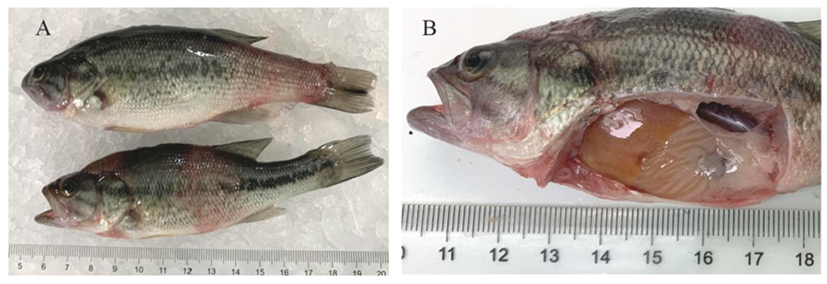
3.1. Clinical Symptoms

Diseased M. salmoides swam on the water surface and swam alone by the pond. Redness of the skin and mouth, ulcers and turgidity were observed (Figure 1A). After dissection, liver yellowing and blood loss were observed. No ascites was found in the abdominal cavity, the kidney was swollen and the muscle was red and bleeding (Figure 1B).

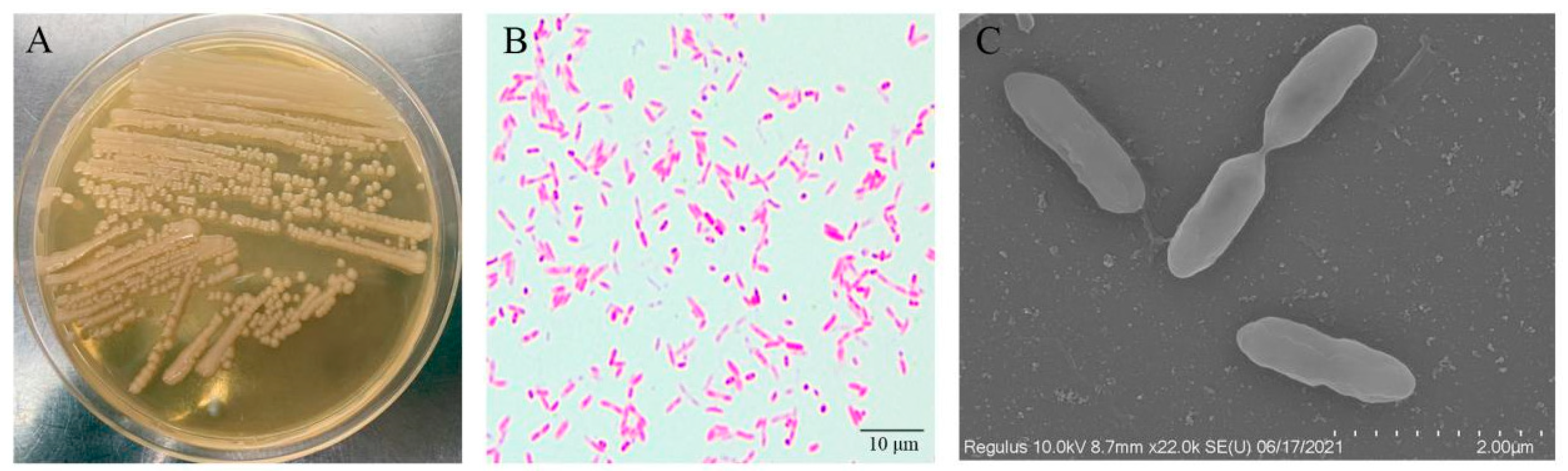
3.2. Pathogen Isolation and Characterization

Buff, translucent, circular and convex bacterial colonies were observed on the BHI agar plates after incubation for 24 h (Figure 2A). No discernible differences in color or form were detected among all colonies on the plates. Additionally, no viruses or parasites were detected in any of the sampled fish. The bacteria from the diseased fish were gram-negative and rod-shaped (Figure 2B). The scanning electron microscope (Hitachi) also demonstrated that the bacteria were nearly rod-shaped (approximately 2.4 × 0.8 μm; Figure 2C).
The bacterial strain WH21406 was identified as Aeromonas caviae with the Biolog Automatic Microbial Identification System. According to the test report, the WH21406 matched the A. caviae reference strain, and comparison with the reference strain in the database yielded a satisfactory score (Table 2).

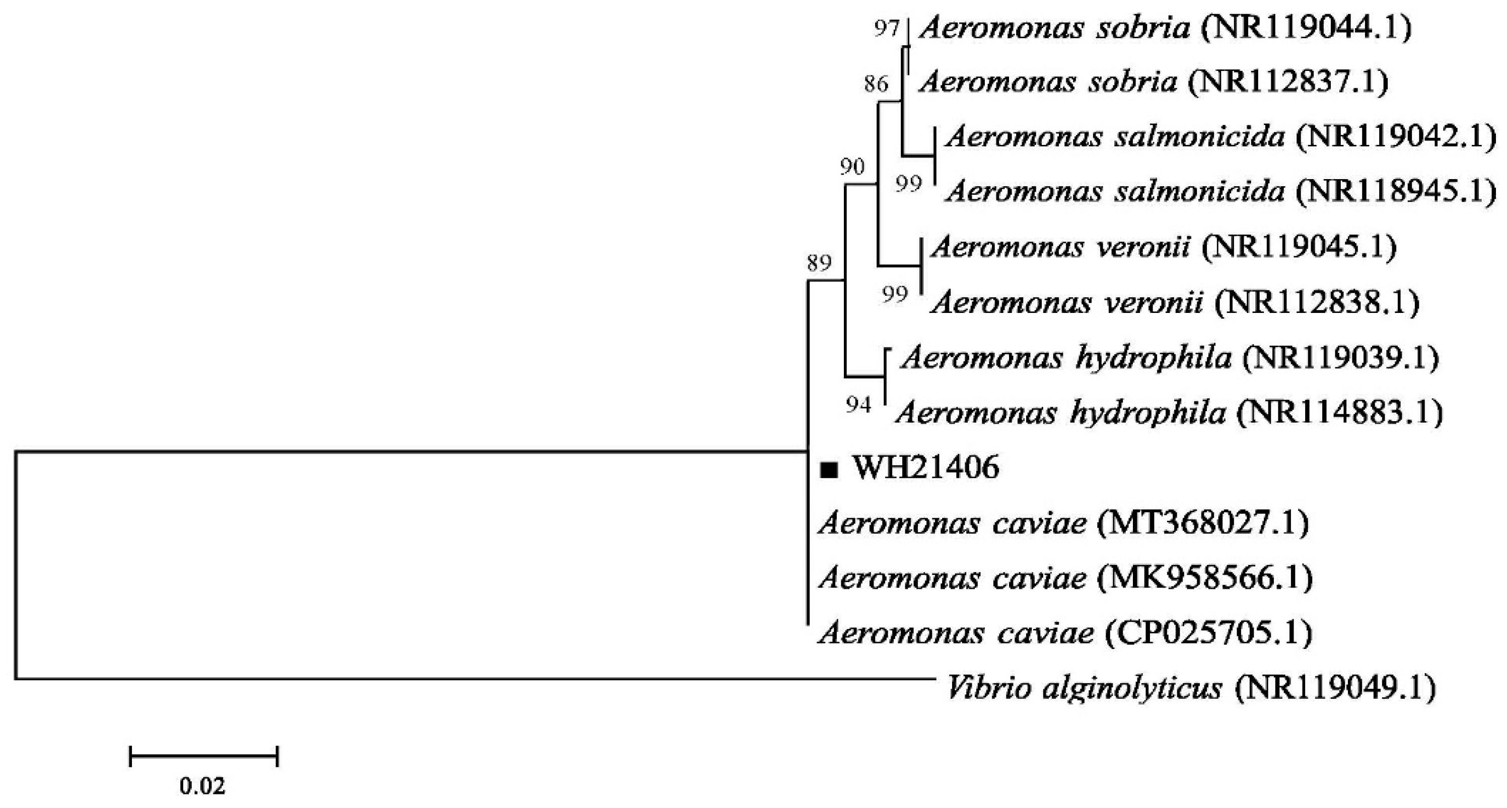
3.3. Bacterial 16S rDNA Sequence Analysis

The 16S rDNA of strain WH21406 is 1407 bp in length. BLAST results of the 16S rDNA sequences showed that WH21406 was 100% similar to A. caviae (MT368027.1). The phylogenetic was constructed using the neighbor-joining method [39] using MEGA 6.0 software. The phylogenetic analysis results indicated that WH21406 and A. caviae (MT368027.1, MK958566.1, CP025705.1) aggregated into a branch (Figure 3).

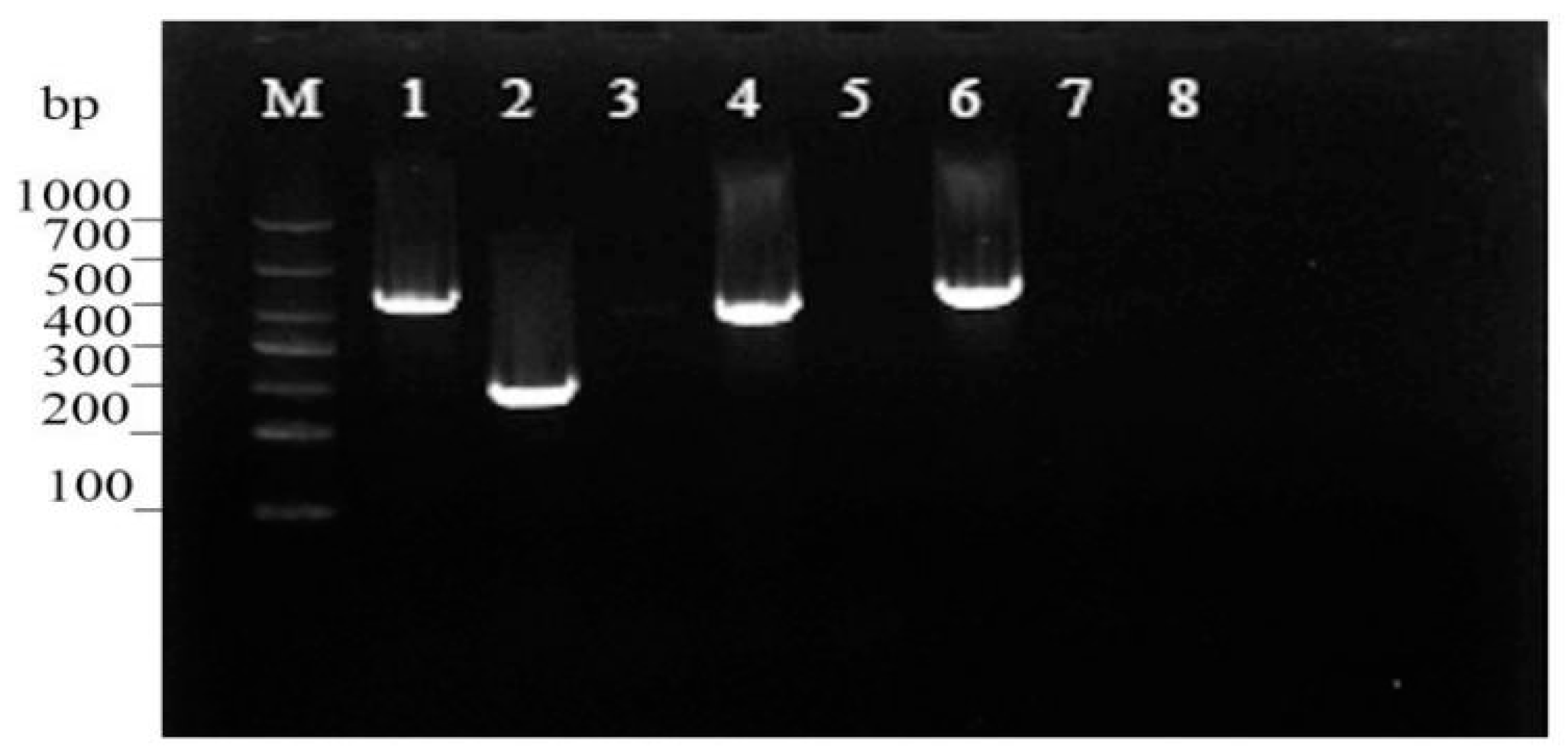
3.4. Virulence Gene Assessment

According to the PCR profiles of the eight virulence genes, fla, aer, ela and hly were found in WH21406, and act, ahp, alt and lip genes were not detected (Figure 4).

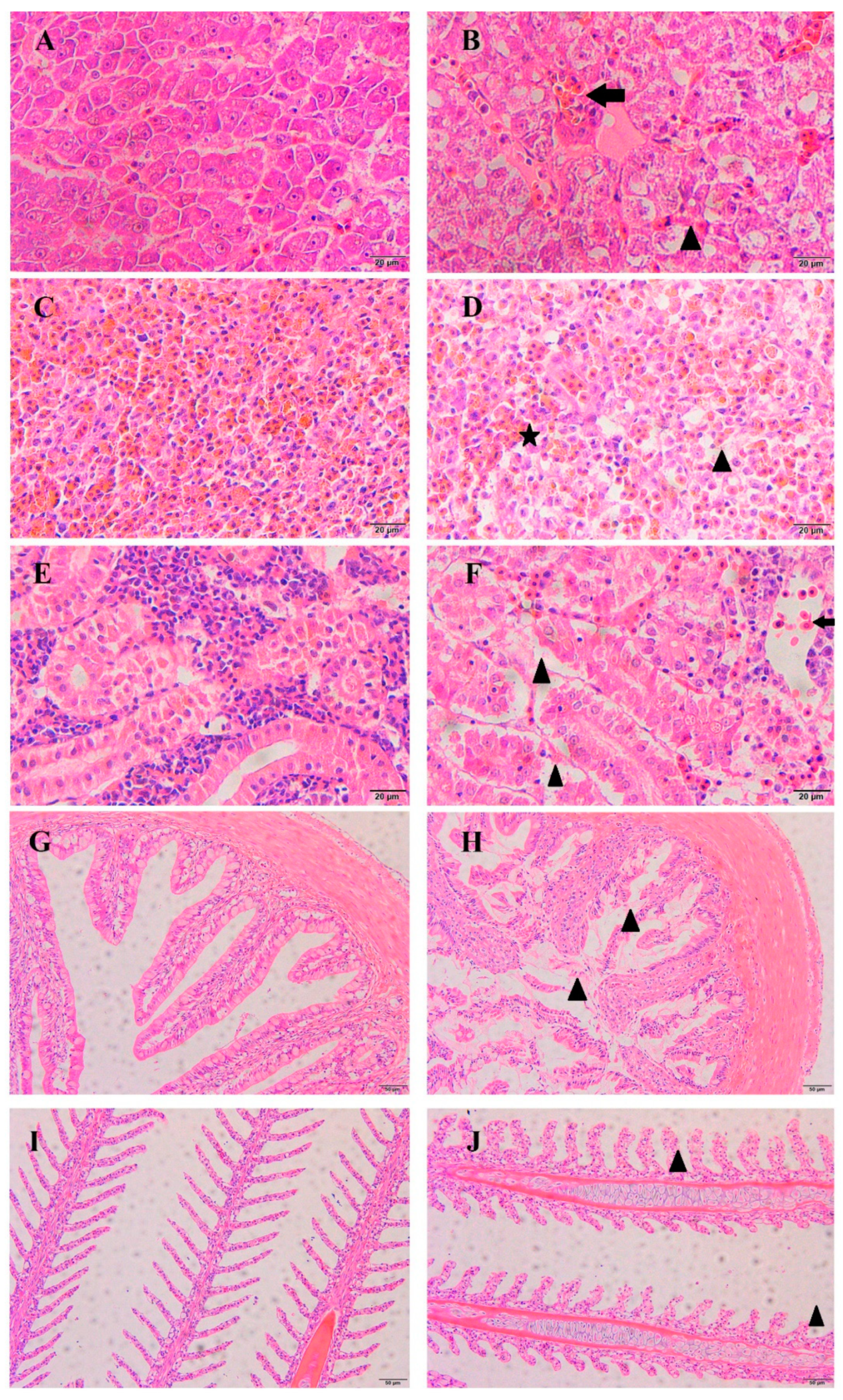
3.5. Histopathological Observations

The tissues of the diseased fish showed obvious haemorrhaging and necrosis. In the diseased fish, a large number of inflammatory cells had infiltrated the liver (Figure 5B, arrow), and the hepatocyte showed vacuolation (Figure 5B, triangle). The splenic cells of the healthy fish were tightly arranged, but the splenic tissue of the diseased fish showed extensive necrocytosis (Figure 5D, triangle) and hemosiderin deposits (Figure 5D, asterisk). In the diseased fish, a large number of inflammatory cells had infiltrated the kidney (Figure 5F, arrow), and necrosis was observed in the glomerulus (Figure 5F, triangle). In the intestine of the diseased fish, considerable damage and abnormal changes in the villi structure were detected (Figure 5H, triangle). In the gills of the diseased fish, necrosis of the epithelia and swelling were observed (Figure 5J, triangle).

3.6. Antibiotic Susceptibility Test

The antibiotic susceptibility test demonstrated that strain WH21406 was highly sensitive to enrofloxacin, norfloxacin, streptomycin and amikacin and moderately sensitive to gentamicin (Table 3). Additionally, WH21406 was resistant to florfenicol, neomycin sulphate, compound sulfamethoxazole, doxycycline and tetracyclines.

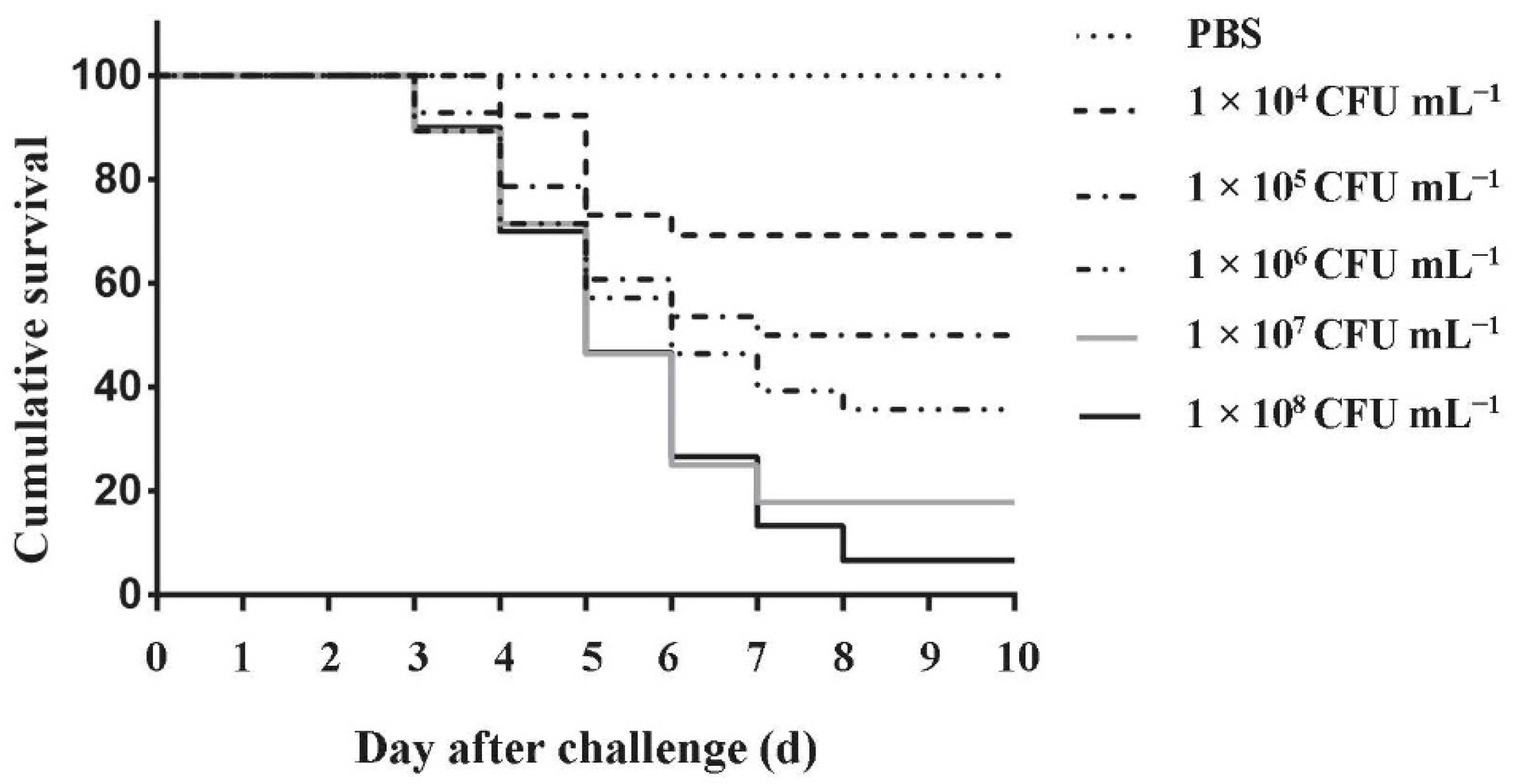
3.7. Pathogenicity Assays

The cumulative survival rate of M. salmoides challenged with the isolate A. caviae WH21406 for 10 days is shown in Figure 6. The fish injected with A. caviae WH21406 died between the fourth and seventh day post-injection. The LD50 of A. caviae WH21406 injected intraperitoneally was calculated to be 3.46 × 10⁵ CFU mL−1 using the improved Kou’s method. The symptoms of the artificially infected fish were consistent with those of the naturally infected fish, which showed ulcers and turgidity on the body surface and fin rot. No death or clinical symptoms were observed in the control group. Furthermore, A. caviae was re-isolated from the artificially infected fish. Mortality and symptoms were consistent in the repeated trials. These results indicated that the isolate A. caviae WH21406 was the pathogen.

4. Discussion

The genus Aeromonas contains many opportunistic pathogens that cause infections in many aquatic and terrestrial animals, including human beings [40,41,42]. A. caviae is a mesophilic species, and it is widely distributed in various aquatic ecosystems such as wastewater, aquaculture water and urban drinking water [18,43,44]. Recently, many studies revealed that the incidence of A. caviae infection in fish has increased, and the clinical symptoms are in accordance with the typical signs of Aeromonas spp. infection [23,44]. In this study, A. caviae WH21406 was isolated from diseased M. salmoides and identified using morphological characterization, biochemical identification and 16S rDNA sequence analysis. Additionally, the clinical signs of the artificially infected fish were similar to those of the naturally infected fish, and A. caviae was re-isolated from the artificially infected fish. These results indicated that A. caviae was the pathogen in the diseased fish. To the best of our knowledge, this is the first report of A. caviae infection in M. salmoides, and this strain caused a large number of deaths.
In this study, the LD50 of A. caviae strain WH21406 was calculated to be 3.46 × 10⁵ CFU mL−1. According to previous studies on M. salmoides, the LD50 of A. veronii HN1903 strain was 3.72 × 10⁴ CFU (g fish)−1 [10], and the LD50 of A. hydrophila LY4 strain was 5. 7 × 10⁵ CFU mL−1 [45]. These results demonstrate that A. caviae strain WH21406 is highly virulent to M. salmoides.
Virulence genes are good indicators of the pathogenicity of a microorganism [46]. We detected virulence genes fla, aer, ela and hly in A. caviae strain WH21406 (Figure 4). These virulence genes were extensively utilized to investigate the potential pathogenicity of Aeromonas spp. [47]. Flagella coded by fla are required for cellular propulsion. Motility is key for pathogenic bacteria that adhere to host cells and cause disease [25]. Aerolysin coded by aer is a pore-forming toxin that can damage epithelial cells in the intestine [48]. Elastase (ela), a secreted protein, can induce epithelial cell apoptosis. Haemolysins (hly) produce cytotoxic effects and lysis of erythrocytes, and they contribute to bacterial evasion from host inflammatory responses [49]. The virulence genes present in A. caviae WH21406 indicate that the synergistic effects conferred by combinations of these genes are key contributors to its high pathogenicity. And act, ahp, alt and lip genes were not detected in A. caviae WH21406. Mohamad [50] found that approximately 30 percent of A. caviae harbour ahp and lip genes. Some Aeromonas species establish in the gastrointestinal tract and can produce enteritis via elaboration of enterotoxigenic molecules such as cytotoxic enterotoxin (act) and cytotonic heat-labile enterotoxin (alt) [40]. No obvious enteritis symptoms occurred in the M. salmoides, which may be related to the absence of act and alt genes in A. caviae WH21406.
The histopathological analysis showed that the A. caviae isolate can cause obvious tissue damage and hemosiderin accumulation in the gill, liver, kidney, spleen and gut, which was similar to the damage found in Silurus meridionalis and Rhamdia quelen infected by A. caviae [23,51]. Cytopathic changes caused by several bacterial toxins may be an important cause of tissue damage. Previous studies indicated that haemolysin produced by A. caviae has a strong virulence and causes rupture and dissolution of red blood cells [23]. This may be the main reason for hemosiderin accumulation in the spleen and kidney.
Few studies were conducted regarding the antibiotic sensitivity of A. caviae. Antibiotic susceptibility testing for A. caviae WH21406 provided a reference for the treatment of A. caviae infection. The isolate WH21406 was highly sensitive to enrofloxacin, norfloxacin, streptomycin and amikacin and resistant to florfenicol, neomycin sulphate, compound sulfamethoxazole, doxycycline and tetracyclines (Table 3). The A. caviae strain from Macrobrachium rosenbergii was highly sensitive to chloramphenicol, florfenicol, tetracycline and doxycycline and resistant to norfloxacin, erythromycin, streptomycin, compound sulfamethoxazole and rifampicin [52]. A. caviae strains from different fish exhibit different antibiotic sensitivity characterizations. Therefore, drug sensitivity testing is particularly important for the treatment of diseases in the production process.

5. Conclusions

In this study, pathogenic A. caviae WH21406 was isolated from diseased M. salmoides. The artificial infection test showed that A. caviae WH21406 is highly pathogenic to M. salmoides, and this isolate is highly sensitive to enrofloxacin, norfloxacin, streptomycin and amikacin. The results of this study provide a reference for the diagnosis and treatment of fish infection with A. caviae.

Author Contributions

Conceptualization, M.X. and Y.Z.; methodology, M.X.; software, Z.X.; validation, Y.L., N.J. and M.X.; formal analysis, W.L.; investigation, Y.M.; resources, Z.X.; data curation, M.X.; writing—original draft preparation, M.X.; writing—review and editing, Y.Z.; visualization, N.J.; supervision, L.Z.; project administration, Y.F.; funding acquisition, Y.Z. All authors have read and agreed to the published version of the manuscript.

Funding

This research was funded by the National Key Research Development Program of China, Grant/Award number: 2019YFD0900105; the National Natural Science Foundation of Hubei, Grant/Award number: 2021CFB486; the Central Public-interest Scientific Institution Basal Research Fund, Grant/Award number: 2020TD44; the open project of Agriculture Ministry Key Laboratory of Healthy Freshwater Aquaculture, Grant/Award number:ZJK202210.

Institutional Review Board Statement

The study was conducted in accordance with the guidelines of the Animal Experimental Ethical Inspection of Laboratory Animal Centre, and approved by the Institutional Review Board of the Yangtze River Fisheries Research Institute, Chinese Academy of Fishery Sciences (ID Number: YFI2021-zhouyong-05).

Data Availability Statement

The data presented in this study are available on request from the corresponding author.

Acknowledgments

We thank the anonymous reviewers for their insightful comments and suggestions, and we also thank the National Key Research Development Program of China (2019YFD0900105), the National Natural Science Foundation of Hubei(2021CFB486), the Central Public-interest Scientific Institution Basal Research Fund (2020TD44), and the open project of Agriculture Ministry Key Laboratory of Healthy Freshwater Aquaculture (ZJK202210).

Conflicts of Interest

The authors declare that they have no known competing financial interests or personal relationships that may have appeared to influence the work reported in this paper.

References

  1. Coyle, S.D.; Tidwell, J.H.; Webster, C.D. Response of Largemouth Bass Micropterus salmoides to Dietary Supplementation of Lysine, Methionine, and Highly Unsaturated Fatty Acids. J. World Aquac. Soc. 2010, 31, 89–95. [Google Scholar] [CrossRef]
  2. Mizanur, R.M.; Li, X.; Sharifuzzaman, S.; He, M.; Leng, X. Dietary threonine requirement of juvenile largemouth bass, Micropterus salmoides. Aquaculture 2021, 543, 736884. [Google Scholar]
  3. Gong, Y.; Yang, F.; Hu, J.; Liu, C.; Xie, S. Effects of dietary yeast hydrolysate on the growth, antioxidant response, immune response and disease resistance of largemouth bass (Micropterus salmoides). Fish Shellfish. Immunol. 2019, 94, 548–557. [Google Scholar] [CrossRef] [PubMed]
  4. He, M.; Li, X.; Poolsawat, L.; Guo, Z.; Yao, W.; Zhang, C.; Leng, X. Effects of fish meal replaced by fermented soybean meal on growth performance, intestinal histology and microbiota of largemouth bass (Micropterus salmoides). Aquac. Nutr. 2020, 26, 1058–1071. [Google Scholar] [CrossRef]
  5. Zhao, Y.; Yang, C.; Zhua, X.; Feng, L.; Liu, Y.; Jiang, W.-D.; Wu, P.; Huang, X.; Chen, D.; Yang, S.-Y.; et al. Dietary methionine hydroxy analogue supplementation benefits on growth, intestinal antioxidant status and microbiota in juvenile largemouth bass Micropterus salmoides. Aquaculture 2022, 556, 738279. [Google Scholar] [CrossRef]
  6. Guo, Z.R.; Zhao, Z.; Zhang, C.; Jia, Y.J.; Wang, G.X. Carbon nanotubes-loaded subunit vaccine can increase protective immunity against rhabdovirus infections of largemouth bass (Micropterus Salmoides). Fish Shellfish. Immunol. 2020, 99, 548–554. [Google Scholar] [CrossRef] [PubMed]
  7. Ma, D.; Deng, G.; Bai, J.; Li, S.; Yu, L.; Quan, Y.; Yang, X.; Jiang, X.; Zhu, Z.; Ye, X. A Strain of Siniperca chuatsi Rhabdovirus Causes High Mortality among Cultured Largemouth Bass in South China. J. Aquat. Anim. Health 2013, 25, 197–204. [Google Scholar] [CrossRef]
  8. Shimahara, Y.; Huang, Y.F.; Tsai, M.A.; Wang, P.C.; Chen, S.C. Immune response of largemouth bass, Micropterus salmoides, to whole cells of different Nocardia seriolae strains. Fish. Sci. 2010, 76, 489–494. [Google Scholar] [CrossRef]
  9. Huizinga, H.W.; Esch, G.W.; Hazen, T.C. Histopathology of red-sore disease (Aeromonas hydrophila) in naturally and experimentally infected largemouth bass Micropterus salmoides (Lacepede). J. Fish Dis. 2010, 2, 263–277. [Google Scholar] [CrossRef]
  10. Pei, C.; Song, H.; Zhu, L.; Qiao, D.; Yan, Y.; Li, L.; Zhao, X.; Zhang, J.; Jiang, X.; Kong, X. Identification of Aeromonas veronii isolated from largemouth bass Micropterus salmoides and histopathological analysis. Aquaculture 2021, 540, 736707. [Google Scholar] [CrossRef]
  11. Camus, A.; Griffin, M.; Armwood, A.; Soto, E. A Spontaneous Outbreak of Systemic Edwardsiella piscicida Infection in Largemouth Bass Micropterus salmoides (Lacépède, 1802) in California, USA. J. Fish Dis. 2019, 42, 759–763. [Google Scholar] [CrossRef] [PubMed]
  12. Jianchou, L.W.W.A.C. Study on Antioxidation Function of Micropterus salmoides Tissues by Aeromonas Sobria Artificial Infection. Hubei Agric. Sci. 2009, 48, 1719–1721. [Google Scholar] [CrossRef]
  13. Francis-Floyd, R.; Reed, P.; Bolon, B.; Estes, J.; Mckinney, S. An epizootic of Edwardsiella tarda in largemouth bass (Micropterus salmoides). J. Wildl. Dis. 1993, 29, 334. [Google Scholar] [CrossRef] [Green Version]
  14. Bowker, J.D.; Carty, D.; Trushenski, J.T.; Bowman, M.P.; Wandelear, N.; Matthews, M. Controlling Mortality Caused by External Columnaris in Largemouth Bass and Bluegill with Chloramine-T or Hydrogen Peroxide. N. Am. J. Aquac. 2013, 75, 342–351. [Google Scholar] [CrossRef]
  15. Martinez-Murcia, A.J.; Saavedra, M.J.; Mota, V.R.; Maier, T.; Stackebrandt, E.; Cousin, S. Aeromonas aquariorum sp. nov., isolated from aquaria of ornamental fish. Int. J. Syst. Evol. Microbiol. 2008, 58, 1169–1175. [Google Scholar] [CrossRef] [PubMed] [Green Version]
  16. De Silva, B.C.J.; Hossain, S.; Dahanayake, P.S.; Heo, G.J. Aeromonas spp. from marketed Yesso scallop (Patinopecten yessoensis): Molecular characterization, phylogenetic analysis, virulence properties and antimicrobial susceptibility. J. Appl. Microbiol. 2019, 126, 288–299. [Google Scholar] [CrossRef]
  17. Williams, M.A.; Addo, S.; Terhune, J.S.; Davis, D.A.; Carrias, A.A.; Liles, M.R. Effects of Bacillus subtilis strains and the prebiotic Previda((R)) on growth, immune parameters and susceptibility to Aeromonas hydrophila infection in Nile tilapia, Oreochromis niloticus. Aquac. Res. 2017, 48, 4798–4810. [Google Scholar]
  18. Lim, Y.L.; Ee, R.; Yin, W.F.; Chan, K.G. Quorum Sensing Activity of Aeromonas Caviae Strain YL12, A Bacterium Isolated from Compost. Sensors 2014, 14, 7026–7040. [Google Scholar] [CrossRef] [Green Version]
  19. Thomas, J.; Madan, N.; Nambi, K.; Majeed, S.A.; Basha, A.N.; Hameed, A.S. Studies on ulcerative disease caused by Aeromonas caviae-like bacterium in Indian catfish, Clarias batrachus (Linn). Aquaculture 2013, 376, 146–150. [Google Scholar] [CrossRef]
  20. Zepeda-Velázquez, A.P.; Vega-Sánchez, V.; Ortega-Santana, C.; Rubio-Godoy, M.; de Oca-Mira, D.M.; Soriano-Vargas, E. Pathogenicity of Mexican isolates of Aeromonas sp. in immersion experimentally-infected rainbow trout (Oncorhynchus mykiss, Walbaum 1792). Acta Trop. J. Biomed. Sci. 2017, 169, 122–124. [Google Scholar] [CrossRef]
  21. Xiankai, Z.; Haipeng, C.; Chunhua, M. Solation and antibiotic susceptibility of an Aeromonas caviae pathogen from yellow leg disease-infected Penaeus vannamei. J. Aquac. 2020, 41, 6. [Google Scholar]
  22. Haipeng, C.; Lefu, W.; Yibin, Y.; Tingting, S.; Shan, H. Isolation and biological characterisitics of an Aeromonas caviae pathogen from Procambarus clarkii. Acta Hydrobiol. Sin. 2014, 38, 7. [Google Scholar]
  23. Baldissera, M.; Souza, C.F.; Júnior, G.; Verdi, C.M.; Moreira, K.; Rocha, M.; Veiga, M.; Santos, R.; Vizzotto, B.S.; Baldisserotto, B. Aeromonas caviae alters the cytosolic and mitochondrial creatine kinase activities in experimentally infected silver catfish: Impairment on renal bioenergetics. Microb. Pathog. 2017, 110, 439–443. [Google Scholar] [CrossRef] [PubMed]
  24. Sunniva, H.; Olav, V.; Jakobsen, A.N. Species Distribution and Prevalence of Putative Virulence Factors in Mesophilic Aeromonas spp. Isolated from Fresh Retail Sushi. Front. Microbiol. 2017, 8, 931. [Google Scholar]
  25. Sen, K.; Rodgers, M. Distribution of six virulence factors in Aeromonas species isolated from US drinking water utilities: A PCR identification. J. Appl. Microbiol. 2004, 97, 1077–1086. [Google Scholar] [CrossRef] [Green Version]
  26. Dallal, M.; Fard, R.; Talkhabi, M.K.; Aghaiyan, L.; Salehipour, Z. Prevalence, virulence and antimicrobial resistance patterns of Aeromonas spp. isolated from children with diarrhea. Germs 2016, 6, 91. [Google Scholar] [CrossRef] [Green Version]
  27. Jensen, S.; Bergh, Ø.; Enger, Ø.; Hjeltnes, B. Use of PCR-RFLP for genotyping 16S rRNA and characterizing bacteria cultured from halibut fry. Can. J. Microbiol. 2002, 48, 379–386. [Google Scholar] [CrossRef]
  28. Hu, M.; Wang, N.; Pan, Z.H.; Lu, C.P.; Liu, Y.J. Identity and virulence properties of Aeromonas isolates from diseased fish, healthy controls and water environment in China. Lett. Appl. Microbiol. 2012, 55, 224–233. [Google Scholar] [CrossRef]
  29. Gui-Hong, F.U.; Xiao, D.; Kun, H.U.; Yang, X.L. Multiplex PCR and ERIC-PCR genotype in virulence genes of Aeromonas hydrophila in Crucian carp. Mar. Fish. 2014, 36, 549. [Google Scholar]
  30. de Pádua, S.B.; Jerônimo, G.T.; de Menezes-Filho, R.N.; Taboga, S.R.; Martins, M.L.; Belo, M.A.A. Pathological assessment of farmed yellowtail tetra Astyanax altiparanae infested by Acusicola sp. (Ergasilidae). Aquac. Rep. 2015, 2, 63–66. [Google Scholar] [CrossRef] [Green Version]
  31. Nawaz, M.; Khan, S.A.; Khan, A.A.; Sung, K.; Tran, Q.; Kerdahi, K.; Steele, R. Detection and characterization of virulence genes and integrons in Aeromonas veronii isolated from catfish. Food Microbiol. 2010, 27, 327–331. [Google Scholar] [CrossRef] [PubMed]
  32. Wang, G.; Clark, C.G.; Liu, C.; Pucknell, C.; Munro, C.K.; Kruk, T.; Caldeira, R.; Woodward, D.L.; Rodgers, F.G. Detection and Characterization of the Hemolysin Genes in Aeromonas hydrophila and Aeromonas sobria by Multiplex PCR. J. Clin. Microbiol. 2003, 41, 1048–1054. [Google Scholar] [CrossRef] [PubMed] [Green Version]
  33. Xu, J.; Zeng, X.; Jiang, N.; Zhou, Y.; Zeng, L. Pseudomonas alcaligenes infection and mortality in cultured Chinese sturgeon, Acipenser sinensis. Aquaculture 2015, 446, 37–41. [Google Scholar] [CrossRef]
  34. Jiang, N.; Jin, X.U.; Jie, M.A.; Fan, Y.; Zhou, Y.; Liu, W.; Zeng, L.; Disease, D.; Institute, Y.; Sciences, C. Histopathology and Ultrastructural Pathology of Cyprinid Herpesvirus Ⅱ(CyHV-2) Infection in Gibel Carp, Carassius auratus gibelio. Wuhan Univ. J. Nat. Sci. 2015, 20, 413–420. [Google Scholar] [CrossRef]
  35. Barry, A.L.; Coyle, M.B.; Thornsberry, C.; Gerlach, E.H.; Hawkinson, R.W. Methods of measuring zones of inhibition with the Bauer-Kirby disk susceptibility test. J. Clin. Microbiol. 1979, 10, 885. [Google Scholar] [CrossRef] [Green Version]
  36. Huang, X.; Gu, Y.; Zhou, H.; Xu, L.; Gai, C. Acinetobacter venetianus, a potential pathogen of red leg disease in freshwater-cultured whiteleg shrimp Penaeus vannamei. Aquac. Rep. 2020, 18, 100543. [Google Scholar] [CrossRef]
  37. Santos, Y.; Laixier, R.; Bandin, I.; Lamas, J.; Toranzo, A. Susceptibility of turbot (Scophthalmus maximus), coho salmon (Oncorhynchus kisutch, and rainbow trout of. my kiss) to strains of Vibrio anguillarum and their exotoxins. J. Appl. Ichthyol. 2010, 7, 160–167. [Google Scholar]
  38. Zou, Y.; Zhang, P.; Mo, Z.; Liu, T.; Xu, Y. Isolation and identification of the pathogenic bacteria from Scophthamus maximus. Gaojishu Tongxun 2004, 14, 89–93. [Google Scholar]
  39. Saitou, N. The neighbor-joining method: A new method for reconstructing phylogenetic trees. Mol. Biol. Evol. 1987, 4, 406–425. [Google Scholar]
  40. Janda, J.M.; Abbott, S.L. The genus Aeromonas: Taxonomy, pathogenicity, and infection. Clin. Microbiol. Rev. 2010, 23, 35–73. [Google Scholar] [CrossRef] [Green Version]
  41. Mary, L.R.; Kurcheti, P.P.; Babu, G.; Purushothaman, C.S. Effect of Aeromonas hydrophila Infection on Caspase-3 Expression and Activity in Rohu, Labeo rohita. J. Aquac. Res. Dev. 2013, 4, 370–386. [Google Scholar]
  42. Woo, S.J.; Kim, M.S.; Jeong, M.G.; Do, M.Y.; Hwang, S.D.; Kim, W.J. Establishment of Epidemiological Cut-Off Values and the Distribution of Resistance Genes in Aeromonas hydrophila and Aeromonas veronii Isolated from Aquatic Animals. Antibiotics 2022, 11, 343. [Google Scholar] [CrossRef] [PubMed]
  43. Carvalho, M.J.; Martínez-Murcia, A.; Esteves, A.C.; Correia, A.; Saavedra, M.J. Phylogenetic diversity, antibiotic resistance and virulence traits of Aeromonas spp. from untreated waters for human consumption. Int. J. Food Microbiol. 2012, 159, 230–239. [Google Scholar] [CrossRef]
  44. Baldissera, M.D.; Souza, C.F.; Bottari, N.B.; Verdi, C.M.; Santos, R.C.V.; Vizzotto, B.S.; Baldisserotto, B. Purinergic signalling displays an anti-inflammatory profile in the spleen of fish experimentally infected with Aeromonas caviae: Modulation of the immune response. J. Fish Dis. 2018, 41, 683–687. [Google Scholar] [CrossRef] [PubMed]
  45. Ye, W.; Guo, C.; Cao, H.; Yang, X. Isolation, identification, pathogenicity and in vitro antibacterial drugs of pathogenic Aeromonas hydrophila from Micropterus salmoides with hemorrhagic disease. Freshw. Fish. 2018, 48, 54–60. [Google Scholar]
  46. Senderovich, Y.; Ken-Dror, S.; Vainblat, I.; Blau, D.; Izhaki, I.; Halpern, M. A Molecular Study on the Prevalence and Virulence Potential of Aeromonas spp. Recovered from Patients Suffering from Diarrhea in Israel. PLoS ONE 2012, 7, e30070. [Google Scholar] [CrossRef]
  47. Sun, J.; Zhang, X.; Gao, X.; Jiang, Q.; Wen, Y.; Lin, L. Characterization of Virulence Properties of Aeromonas veronii Isolated from Diseased Gibel Carp (Carassius gibelio). Int. J. Mol. Sci. 2016, 17, 496. [Google Scholar] [CrossRef]
  48. Abrami, L.; Fivaz, M.; Glauser, P.E.; Sugimoto, N.; Zurzolo, C.; van der Goot, F.G. Sensitivity of Polarized Epithelial Cells to the Pore-Forming Toxin Aerolysin. Infect. Immun. 2003, 71, 739. [Google Scholar] [CrossRef] [Green Version]
  49. Gao, S.; Zhao, N.; Amer, S.; Qian, M.; Lv, M.; Zhao, Y.; Su, X.; Cao, J.; He, H.; Zhao, B. Protective efficacy of PLGA microspheres loaded with divalent DNA vaccine encoding the ompA gene of Aeromonas veronii and the hly gene of Aeromonas hydrophila in mice. Vaccine 2013, 31, 5754–5759. [Google Scholar] [CrossRef]
  50. Azzam-Sayuti, M.; Ina-Salwany, M.Y.; Zamri-Saad, M.; Yusof, M.T.; Amal, M. The prevalence, putative virulence genes and antibiotic resistance profiles of Aeromonas spp. isolated from cultured freshwater fishes in peninsular Malaysia. Aquaculture 2021, 540, 736719. [Google Scholar] [CrossRef]
  51. Wang, K.; Xiao, D.; He, Y.; Chen, D.; Geng, Y.; Liu, T.; Huang, G. Isolation, Purification and pathogenicity of the haemolysin produced by Aeromonas caviae from Silurus meridionalis Chen. Oceanol. Et Limnol. Sin. 2012, 43, 1122–1127. [Google Scholar]
  52. Guo, Y.; Zhou, M.; Li, Y.; Wang, W. Aeromonas caviae from Macrobrachium rosenbergii: Isolation and identification. Chin. Agric. Sci. Bull. 2020, 36, 147–153. [Google Scholar]
Figure 1. Clinical symptoms of diseased largemouth bass. (A) Clinical symptoms of body surface, (B) Clinical symptoms of internal organs.
Fishes 07 00119 g001
Figure 2. Colonial morphology, Gram staining and scanning electron microscopy image of the isolate WH21406. (A) Colonial morphology of WH21406, (B) Gram staining image of WH21406, (C) Scanning electron microscopy image of WH21406.
Fishes 07 00119 g002
Figure 3. Phylogenetic tree for strain WH21406 on the basis of 16S rDNA sequences (the numbers represent bootstrap values).
Fishes 07 00119 g003
Figure 4. Agarose gel electrophoresis of PCR products of the virulence genes. M: DL1000 marker, 1: fla, 2: aer, 3: act, 4: ela, 5: ahp, 6: hly, 7: alt, 8: lip.
Fishes 07 00119 g004
Figure 5. Histological observation of the tissues of healthy and diseased largemouth bass specimens. (A) Normal liver tissue; (B) Liver tissue from diseased largemouth bass, inflammatory cell infiltration (arrow), hepatocyte vacuolation (triangle); (C) Normal spleen tissue; (D) Spleen tissue from diseased largemouth bass, necrocytosis (triangle), hemosiderin deposits (asterisk); (E) Normal kidney tissue; (F) Kidney tissue from diseased largemouth bass, inflammatory cell infiltration (arrow), glomerulus necrosis (triangle); (G) Normal gut tissue; (H) Gut tissue from diseased largemouth bass, intestinal villi damage (triangle); (I) Normal gill filaments; (J) Gill tissue from diseased largemouth bass, epithelia necrosis and swelling (triangle).
Fishes 07 00119 g005
Figure 6. Survival rates of largemouth bass specimens challenged with different doses of A. caviae WH21406 for 10 days post-infection.
Fishes 07 00119 g006
Table 1. Primers for 16S rRNA and virulence gene testing.
GenePrimer Sequence (5′−3′)Product Size
(bp)
Optimal Annealing
Temperature (°C)
References
16SrRNA-FAGAGTTTGATCATGGCTCAG150055Jensen et al. [27]
16SrRNA-RTACGGTTACCTTGTTACGACTT
ahp-FATTGGATCCCTGCCTATCGCTTCAGTTCA91155Hu et al. [28]
ahp-RGCTAAGCTTGCATCCGTGCCGTATTCC
ela-FACACGGTCAAGGAGATCAAC51355Sen and Rodgers [25]
ela-RCGCTGGTGTTGGCCAGCAGG
act-FATCGTCAGCGACAGCTTCTT50055Fu et al. [29]
act-RCTCATCCCTTGGCTTGTTGT
fla-FTCCAACCGTYTGACCTC60855Hu et al. [28]
fla-RGMYTGGTTGCGRATGGT
hly-FGGCCGGTGGCCCGAAGATACGGG59765Heuzenroeder et al. [30]
hly-RGGCGGCGCCGGACGAGACGGG
alt-FTGACCCAGTCCTGGCACGGC44264Nawaz et al. [31]
alt-RGGTGATCGATCACCACCAGC
aer-FCAAGAACAAGTTCAAGTGGCCA30957Wang et al. [32]
aer-RACGAAGGTGTGGTTCCAGT
lip-FATCTTCTCCGACTGGTTCGG38255Sen and Rodgers [25]
lip-RCCGTGCCAGGACTGGGTCTT
Table 2. Results of Biolog identification of strain WH21406.
ReagentResult *ReagentResult *
A1 Negative ControlNE1 GelatinP
A2 DextrinPE2 Glycyl-l-ProlineP
A3 d-MaltosePE3 l-AlanineP
A4 d-TrehalosePE4 l-ArginineP
A5 d-CellobiosePE5 l-Aspartic AcidP
A6 GentiobioseBE6 l-Glutamic AcidP
A7 SucrosePE7 l-HistidineP
A8 d-TuranoseBE8 l-Pyroglutamic AcidP
A9 StachyoseBE9 l-SerineP
A10 Positive ControlPE10 LincomycinB
A11 PH6PE11 Guanidine HClB
A12 PH5NE12 Niaproof 4N
B1 d-RaffinosePF1 PectinP
B2 α-d-LactosePF2 d-Galacturonic AcidP
B3 d-MelibiosePF3 l-Galactonic Acid LactoneP
B4 β-Methyl-d-GlucosidePF4 d-Gluconic AcidP
B5 d-SalicinPF5 d-Glucuronic AcidP
B6 N-Acetyl-d-GlucosaminePF6 GlucuronamideN
B7 N-Acetyl-β-d-MannosaminePF7 Mucic AcidP
B8 N-Acetyl-d-GalactosaminePF8 Quinic AcidP
B9 N-Acetyl Neuraminic AcidBF9 d-Saccharic AcidP
B10 1% NaClPF10 VancomycinP
B11 4% NaClBF11 Tetrazolium VioletP
B12 8% NaClNF12 Tetrazolium BlueP
C1 α-d-GlucosePG1 P-Hydroxy-Phenylacetic AcidN
C2 d-MannosePG2 Methyl PyruvateP
C3 d-FructosePG3 d-Lactic Acid Methyl EsterP
C4 d-GalactoseBG4 l-Lactic AcidP
C5 3-Methyl GlucosePG5 Citric AcidP
C6 d-FucosePG6 6α-Keto-Glutaric AcidP
C7 l-FucosePG7 d-Malic AcidL
C8 l-RhamnosePG8 l-Malic AcidP
C9 InosinePG9 Bromo-Succinic AcidP
C10 1%Sodium LactatePG10 Nalidixic AcidP
C11 Fusidic AcidNG11 Lithium ChlorideP
C12 d-SerinePG12 Potassium TelluriteN
D1 d-SorbitolPH1 Tween 40P
D2 d-MannitolPH2 γ-Amino-Butyric AcidP
D3 d-ArabitolBH3 α-Hydroxy-Butyric AcidP
D4 Myo-inositolBH4 β-Hydroxy-d, l-Butyric AcidP
D5 GlycerolPH5 α-Keto-Butyric AcidP
D6 d-Glucose-6-PO4PH6 Acetoacetic AcidP
D7 d-Fructose-6-PO4PH7 Propionic AcidP
D8 d-Aspartic AcidPH8 Acetic AcidP
D9 d-SerinePH9 Formic AcidB
D10 TroleandomycinPH10 AztreonamB
D11 Rifamycin SVPH11 Sodium ButyrateB
D12 MinocyclineNH12 Sodium BromateN
* Notes: P = Positive, N = Negative, B = Borderline, L = Less than A1 well.
Table 3. Antibiotic susceptibility test results.
Drug NameInhibition Zone (mm)Sensitivity *
Florfenicol6R
Enrofloxacin22S
Neomycin sulfate6R
Doxycycline6R
Norfloxacin18S
Gentamicin12I
Streptomycin22S
Compound Sulfamethoxazole6R
Tetracyclines6R
Amikacin17S
* Notes: S, susceptible; I, intermediate; R, resistant.
Publisher’s Note: MDPI stays neutral with regard to jurisdictional claims in published maps and institutional affiliations.

