Article

Identification and Characterization of Immunoglobulin T Heavy Chain in Large Yellow Croaker (Larimichthys crocea)

1
Key Laboratory of Marine Biotechnology of Fujian Province, Institute of Oceanology, Fujian Agriculture and Forestry University, 15 Shangxiadian Road, Fuzhou 350002, China
2
Institute of Animal Husbandry and Veterinary Medicine of Fujian Academy of Agricultural Sciences, Fuzhou 350013, China
3
Southern Marine Science and Engineering Guangdong Laboratory (Zhuhai), Zhuhai 519000, China
*
Author to whom correspondence should be addressed.
These authors contributed equally to this work.
Fishes 2022, 7(1), 29; https://doi.org/10.3390/fishes7010029
Submission received: 6 December 2021 / Revised: 21 January 2022 / Accepted: 24 January 2022 / Published: 27 January 2022
(This article belongs to the Special Issue Discovery and Utilization of Fish Resistance Genes)

Abstract

:
Three immunoglobulin (Ig) isotypes have been identified in teleosts, IgM, IgD, and IgT or IgZ. IgT, a new teleost Ig isotype, plays a vital role in mucosal immunity. However, information on molecular and functional characteristics of fish IgT is still limited. In this study, an IgT heavy chain (LcIgT) gene was cloned and characterized in large yellow croaker (Larimichthys crocea). Complete cDNA of LcIgT was 1930 bp in length, encoding a protein of 554 amino acids. The deduced LcIgT contains a VH region and only three CH regions (CH1, CH2, CH4), but no transmembrane region was predicted. Phylogenetic analysis showed that IgT heavy chain sequences from all fish species are grouped together. Homology comparison showed that LcIgT shares the highest amino acid identity of 58.73% with IgT heavy chain in Scophthalmus maximus. The VH domain of LcIgT has the highest identity of 72.50% with that of Scophthalmus maximus IgT. Relatively, each constant domain of LcIgT exhibits the highest amino acid identity with that of IgT in Oreochromis niloticus (67.61% identity for CH1, 61.11% identity for CH2, and 63.74% identity for CH4). LcIgT was constitutively expressed in various tissues tested, with the highest levels in mucosa-associated tissues such as gills and skin. After Cryptocaryon irritans infection, the mRNA levels of LcIgT were significantly up-regulated in the spleen (3.27-fold) at 4 d, in the head kidney (3.98-fold) and skin (2.11-fold) at 7 d, and in gills (4.45-fold) at 14 d. The protein levels in these detected tissues were all significantly up-regulated; the peak of its up-regulation was 6.33-fold at 28d in gills, 3.44-fold at 7d in skin, and 3.72-fold at 14d in spleen. These results showed that IgT response could be simultaneously induced in both systemic and mucosal tissues after parasitic infection and that IgT may be involved in systemic immunity and mucosal immunity against parasitic infection.

1. Introduction

Immunoglobulins (Igs) consist of two light (L) chains and two heavy (H) chains [1], which have a critical role in the vertebrate adaptive immune system as an important class of immune effector molecules. Five Igs isotypes have been identified in mammals: IgM, IgG, IgA, IgD, and IgE, based on the difference of heavy chain [2]. In teleosts, only three Igs isotypes, IgM, IgD, and IgT or IgZ are discovered as yet [3,4,5,6,7]. IgM is the most predominant and stable Ig in systemic immunity, while IgT or IgZ is considered an Ig specialized in mucosal immunity in teleosts [8]. IgD has an unknown function in teleosts, but studies have suggested that IgD might be involved in surveillance and immune regulation as an antigen-binding receptor [9].
IgT or IgZ has been identified in several teleost fish, including rainbow trout (Oncorhynchus mykiss) [6], zebrafish (Danio rerio) [10], fugu (Takifugu rubripes) [11], common carp (Cyprinus carpio) [12], Japanese flounder (Paralichthys olivaceus) [13], turbot (Scophthalmus maximus) [14], European seabass (Dicentrarchus labrax L.) [15], three-spined stickleback (Gasterosteus aculeatus) [15], Pacific bluefin tuna (Thunnus orientalis) [16], and Atlantic salmon (Salmo salar) [17]. Interestingly, some teleost species possess multiple IgT subclasses, such as rainbow trout with three subclasses, termed IgT1, IgT2, and IgT3 [18], zebrafish with two functional molecules, IgZ and IgZ-2 [7], common carp with IgZ1 and IgZ2 [12], and Atlantic salmon with IGH-A and IGH-B [17]. Nevertheless, no IgT ortholog has been found in channel catfish (Ictalurus punctatus) [19] and medaka (Oryzias latipes) [20] until now.
Two forms of IgT, membrane-bound IgT (mIgT) and secreted IgT (sIgT), exist in teleosts. The heavy chain of both IgT forms contains a variable region (VH) and 2–4 constant regions (CH), while the mIgT possesses additional single- or double-spinning transmembrane regions (TMs). The CH number of IgT varies among different species; for example, four CH domains are found in most teleosts, whereas only two exist in fugu [11] and three in stickleback [21] and emerald rock cod [22]. IgH gene locus in teleosts has multiple variables (V), diversity (D), joining (J), and constant (C) segments in the following order: (Vn-Dn-Jn-Cn) [10,16]. RAG-1 and RAG-2 assemble the V, D, and J segments by recognizing the recombination signal sequences at the borders of the V, D, and J segments [23]. IgT heavy chain (τ) exons are located either in the 5’ regions of the D and J gene segments of IgM and IgD heavy chains in zebrafish [10] and stickleback [24] or inserted within the V gene segments of IgM and IgD heavy chains in rainbow trout [6] and Atlantic salmon [25], which has resulted in the evolution of a distinct B cell lineage. The structure and feature of the IgT heavy chain genes in teleosts are diversified with the evolution of species, which might cause differential functional characteristics of IgT. Thus, the cloning of IgT genes from more fish species could increase the understanding of the molecular structure of fish IgT.
Tissue expression of fish IgT genes has been widely studied, and patterns were variable among different species. The mucosa-associated tissues such as gills and gut were found to be the primary tissues that IgT was expressed in several species [6,17,21,22]. The zebrafish IgZ1 was detected only in the kidney, whereas IgZ-2 was in the kidney and skin [7]. The high IgT expression levels in common carp were found in gills, head kidney, muscle, and brain [26]. The flounder IgT was highly expressed in the spleen, liver, and gills [13]. After Edwardsiella tarda infection, the flounder IgT expression was strongly induced in gills, skin, spleen, and head kidney [13]. The turbot IgT was up-regulated after infection with Vibrio anguillarum in gills, skin, kidney, spleen, liver, and gut [14]. The trout IgT expression was significantly increased when infected with parasites or bacteria, mainly in mucosal tissues including gut [27], skin [28], gill [29], and nasal [30]. These data suggested that teleost IgT may participate in both systemic and mucosal immunity against pathogens, especially in mucosal immunity. Therefore, expression analyses of IgT molecules based on various pathogen infections will provide valuable clues to fish IgT function in-depth knowledge.
Large yellow croaker, belonging to the Perciformes, is an important mariculture fish in China, with the highest annual production among marine species [31]. In recent years, the outbreak of infectious diseases caused by various pathogens, including bacteria, viruses, and parasites, has severely affected the development of large yellow croaker farming [32,33,34]. Cryptocaryon irritans, a ciliate ectoparasite, can infect large yellow croakers, leading to high mortality and huge economic losses [35]. Understanding the function of the immune system of L. crocea will contribute to the prevention and control of the infectious diseases that occur in this species. Igs are an important class of immune effector molecules in the vertebrate adaptive immune system. A previous study elucidated the molecular characteristics of IgM heavy and light chain genes in large yellow croakers, and their expression patterns upon immune stimuli were also analyzed [36,37]. Here, we describe the cloning and molecular characterization of a secretory IgT heavy chain (LcIgT) in large yellow croaker. The expression pattern of LcIgT in several tissues was analyzed. After a challenge with C. irritans, the expression changes of LcIgT mRNA and protein were determined by real-time PCR (RT-PCR) and western blotting. These results will contribute to the understanding of the molecular and functional characteristics of fish IgT.

2. Materials and Methods

2.1. Experimental Infection and Tissue Preparation

Healthy large yellow croakers (body weight: 20 ± 3.6 g; length: 9 ± 1.5 cm) were obtained from a mariculture farm at Fujian, China. The fish were maintained in a 3000-L aerated water tank with a flow-through seawater supply at 25 °C. The individuals were acclimatized for 7 days prior to being used for the challenge experiments. Then, fish were divided into two groups and cultured in separate 3000-L closed tanks. Thirty fish in the experimental group were infected with 3000 theronts of C. irritans per fish by bath for 4 h. The infection was performed in a 100-L fiberglass aquarium with about 60-L water containing a proper number of theronts of C. irritans and then transferred into 3000-L aerated water tanks with fresh seawater. Another thirty fish were treated in the same way but without C. irritans, and designated as the control group. The fish were euthanized with eugenol, and various tissues including spleen, head kidney, skin, and gills were sampled from five fish of each group at different time points (4 d, 7 d, 14 d, 21 d, 28 d, 35 d) after infection. These isolated tissues were frozen immediately in liquid nitrogen and stored at −80 °C before RNA extraction. The Committee on the Ethics of Animal Experiments of the Chinese Academy of Sciences approved all experiments on animals.

2.2. Complete cDNA Cloning and Sequence Analysis of LcIgT

To obtain the full-length cDNA of large yellow croaker IgT heavy chain (LcIgT) gene, 5′ and 3′ RACE-PCR were performed using the 5′ and 3′-Full RACE Kit (TaKaRa, China). Primers (Table S1) for the 5′ and 3′ RACE were designed based on IgT heavy chain gene sequences obtained from the genome database of large yellow croaker (JRPU00000000) [38]. Both 5′ and 3′ RACE-PCR were performed according to the manufacturer′s instructions. The resulting PCR product was cloned into the pMD19-T simple vector (TaKaRa, Beijing, China) and sequenced. All sequences were assembled to obtain the full-length cDNA of the IgT heavy chain. The deduced amino acid sequence of IgT heavy chain was analyzed for a signal peptide using SignalP software and N-glycosylation sites with the NetNGlyc 1.0 Server. Sequence similarity analysis was performed using the BLAST program (http://blast.ncbi.nlm.nih.gov/Blast.cgi, accessed on 12 May 2021). Protein domains were predicted by Simple Modular Architecture Research Tool (SMART) (http://smart.embl-heidelberg.de/, accessed on 17 May 2021). The isoelectric point (pI) and the molecular weight of the deduced IgT protein were calculated by the ExPASy-Compute pI/Mw tool (https://web.expasy.org/compute_pi/, accessed on 23 June 2021). Multiple sequence alignment was performed using the ClustalW 2.0. A phylogenetic tree was conducted by the neighbor-joining (NJ) method using the MEGA 6 program [39]. The bootstrap method with 1000 replicates was used to examine the veracity of these trees and the statistical significance of each branch. The amino acid similarity among each CH region of IgT was also analyzed using protein Blastp at the NCBI (https://blast.ncbi.nlm.nih.gov/Blast.cgi, accessed on 6 July 2021), and the corresponding phylogenetic trees were constructed as performed above.

2.3. Tissue Distribution of LcIgT by RT-PCR

To determine the tissue distribution of LcIgT, eleven tissues, including liver, heart, head kidney, gills, brain, spleen, stomach, intestine, skin, muscle, and blood, were collected from at least five healthy juveniles. Total RNA was extracted from these tissues using Eastep® Super Total RNA Extraction Kit (Promega, Shanghai, China), referring to the manufacturer’s protocol. Subsequently, Oligo dT-Adaptor primer (Promega, Shanghai, China) was used to reverse-transcribe 1 μg total RNA into the first-strand cDNA. The cDNA was diluted with RNase-free water and stored at −20 °C before RT-PCR. The special primers (Supplementary Table S1) were used to determine the transcriptional levels of LcIgT in all tissues with a QuantStudioTM 1 (Applied biosystems, Thermo, Waltham, MA, USA) equipped with Design & Analysis Software v1.5.1. PCR was performed in a reaction volume of 20 μL. PCR amplification was performed under the following conditions: one cycle of 95 °C for 5 min, 40 cycles of 95 °C for 30 s, 57 °C for 30 s, and 72 °C for 1 min, followed by a final extension of 72 °C for 10 min. In addition, β-actin was amplified as the internal control for RT-PCR. The expression levels of LcIgT were normalized by β-actin using the 2−ΔΔCT method and expressed as the ratio of LcIgT expression levels in the blood [40]. The collected tissues were examined in triplicate.

2.4. LcIgT Expression Analysis after Infection with C. irritans

To further understand the modulation of LcIgT expression upon C. irritans infection, four tissues (spleen, head kidney, gills, and skin) were isolated at 4 d, 7 d, 14 d, 21 d, 28 d, and 35 d post-infection as above. Total RNA extraction and first-strand cDNA synthesis were performed as the previous protocol. Then these templates were used for RT-PCR with respective primer sets of LcIgT and β-actin genes. RT-PCR conditions were the same as described above. The relative expression levels of LcIgT were normalized by β-actin and expressed as fold changes by comparing the normalized gene expression levels of C. irritans-infected fish with those of the non-infected fish (defined as 1) at the same time point. The samples were detected in triplicate.

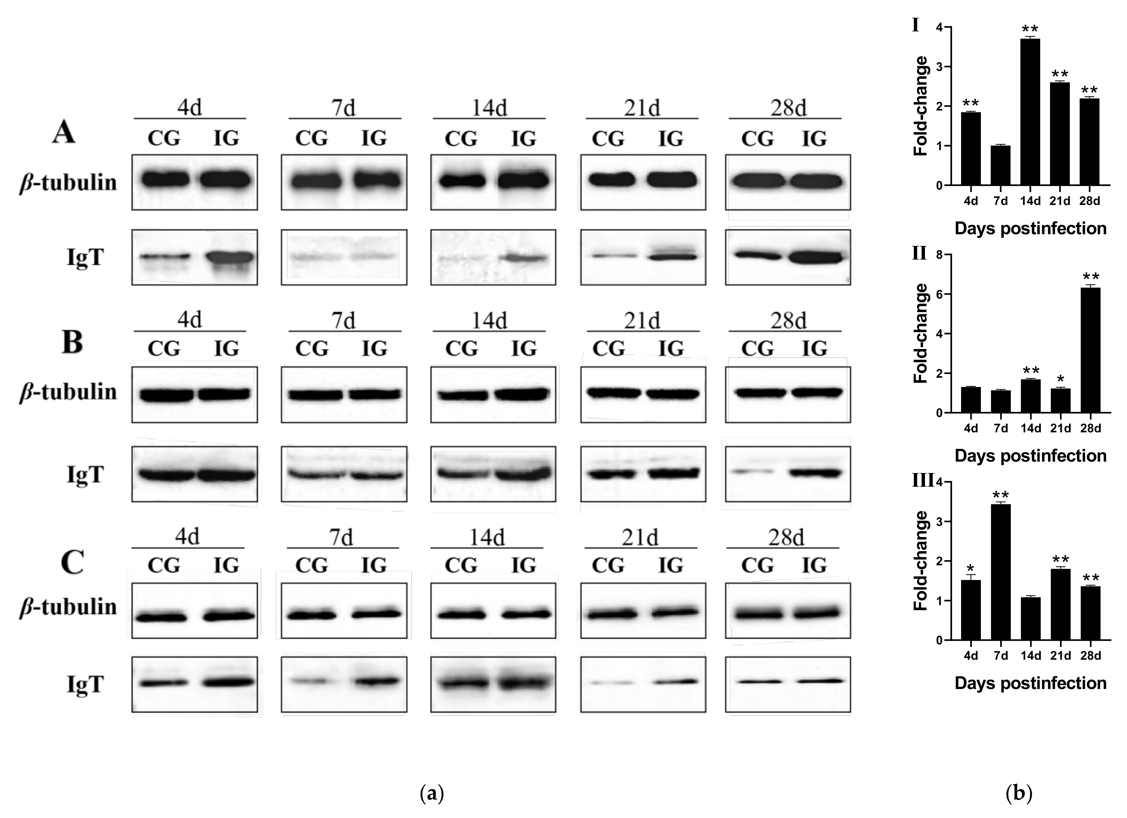
2.5. Western Blotting Analysis of LcIgT Expression

To detect the modulation of LcIgT protein levels, each tissue was homogenized in a homogenizer IKA T10 (Truelab, Shanghai, China) with RIPA lysis buffer (Beyotime, Shanghai, China) containing 2% protease inhibitor, and instantly centrifuged at 4 °C (13,000× g for 5 min). Under the reducing conditions, the supernatants of lysate were separated by 12% SDS-PAGE, and then the proteins were transferred to polyvinylidene difluoride (PVDF) membranes (GE, Boston, MA, USA). The membranes were immersed in blocking buffer, consisting of TBST buffer containing 0.2% Tween-20 (Sangon Biotech, Shanghai, China) and 5% nonfat dried milk for 1 h at room temperature with shaking to block completely. Then the membranes were incubated in blocking buffer with primary antibody (1.2 μg/mL mouse anti-IgT mAb, 1:1000; prepared in our laboratory [41]) overnight at 4 ℃. The membranes were rinsed twice with TBST for 10 min each time, following incubation with diluted secondary antibody, HRP-labelled Goat Anti-Mouse IgM (1:5000, Thermo Fisher Scientific, Waltham, MA, USA), shaken for 1 h at ambient temperature. After three extra washes with TBST as described above, the membranes were covered up with detection solution, a mixture of an equal amount of detection solution A and B, and placed in a gel imaging system (Azure Biosystems, Dublin, CA, USA) to detect. Alternatively, β-tubulin was selected as the internal control to achieve protein quantification. The samples were detected in triplicate. Image J v1.8.0 software was used to analyze the protein gray values of IgT.

2.6. Statistical Analysis

All data of each repeated experiment were analyzed by GraphPad Prism 8 software and denoted as the mean ± standard error of the mean. Using IBM SPSS Statistics 19.0, the analysis of independent samples t-test and one-way ANOVA was applied to the data of each group, and the p-value < 0.05 represents statistical difference.

3. Results

3.1. Identification and Characterization of LcIgT cDNA

The cloned LcIgT cDNA sequence (GenBank accession number: MW450786) was 1930 bp long, containing an open reading frame (ORF) of 1665 bp, a 5′-untranslated region (5′-UTR) of 31 bp, and a 3′-UTR of 234 bp with a putative atypical polyadenylation signal sequence AATAAA (Supplement Figure S1). The ORF of LcIgT encodes a protein of 554-amino acids (aa), containing a putative 29-aa signal peptide, a 117-aa variable region (VH, H47-E163), and three constant domains (CH1, D164-G236; CH2, T259-T331; CH4, M453-N542), but no transmembrane region was predicted, indicating that LcIgT cDNA sequence cloned here encodes a secreted form of IgT heavy chain. The LcIgT sequence contains twelve conserved cysteine residues (Cys51, Cys123, Cys158, Cys170, Cys227, Cys264, Cys321, Cys361, Cys422, Cys478, Cys528, and Cys553) and two potential N-glycosylation sites (N398 and N499). Additionally, LcIgT has a predicted molecular weight of 58.69 kDa and an isoelectric point of 7.18.

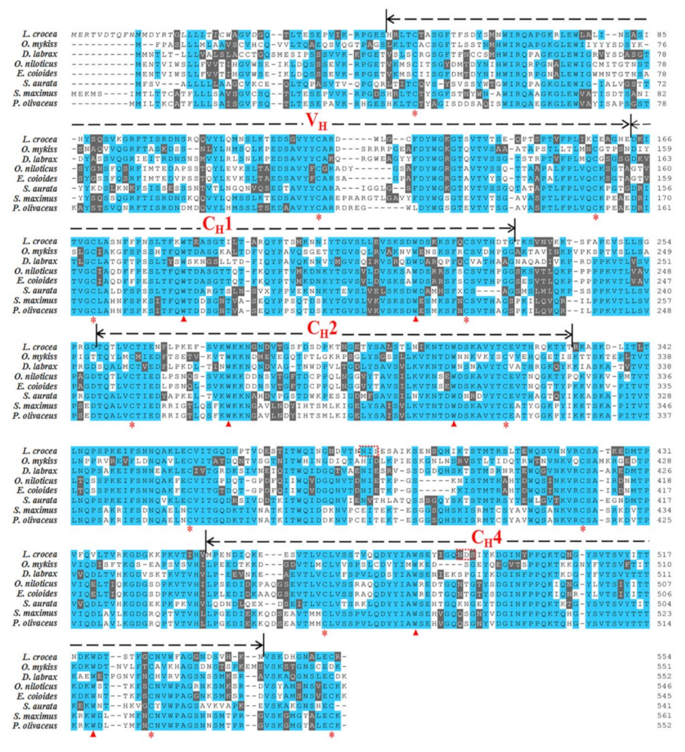
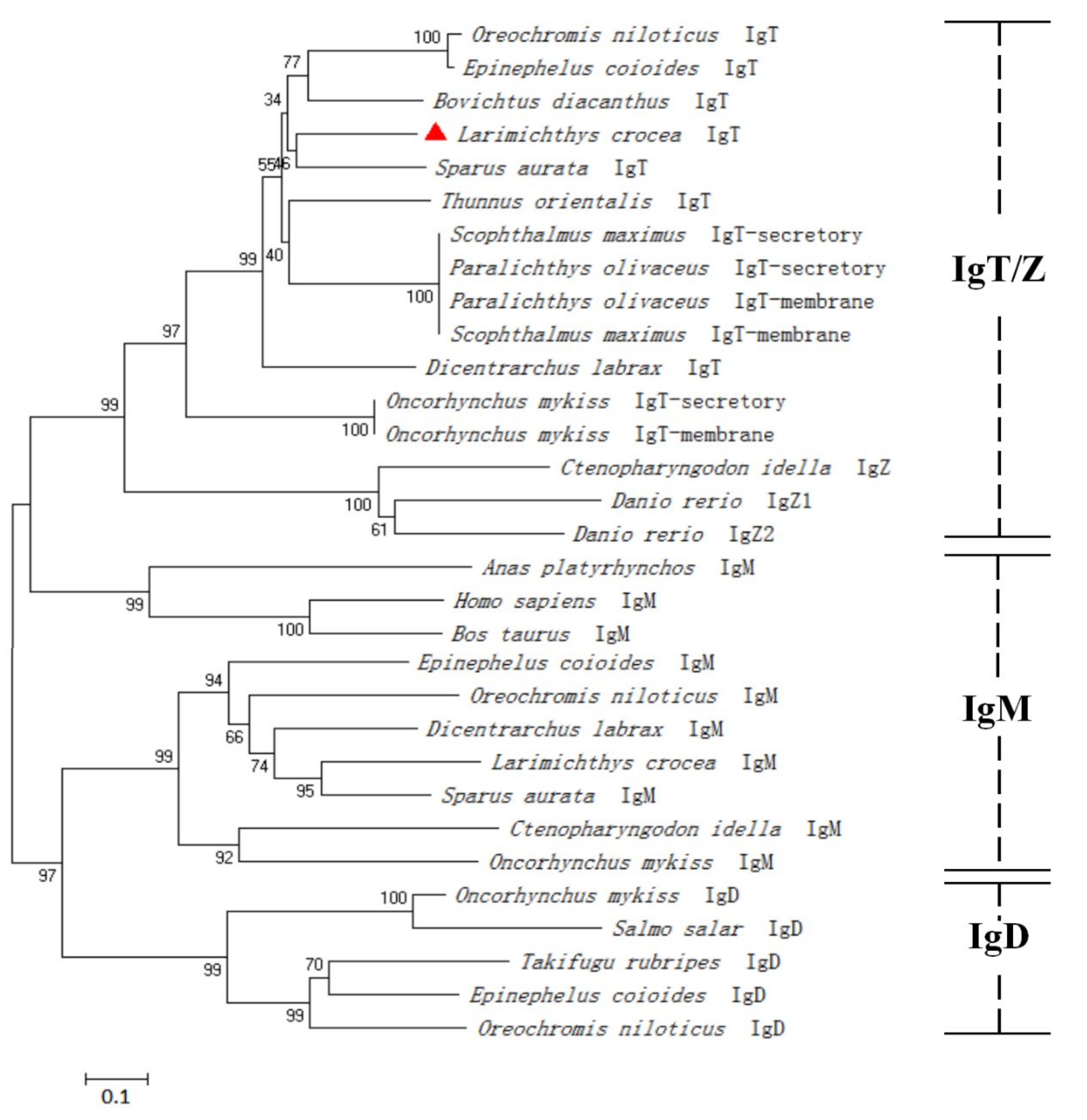
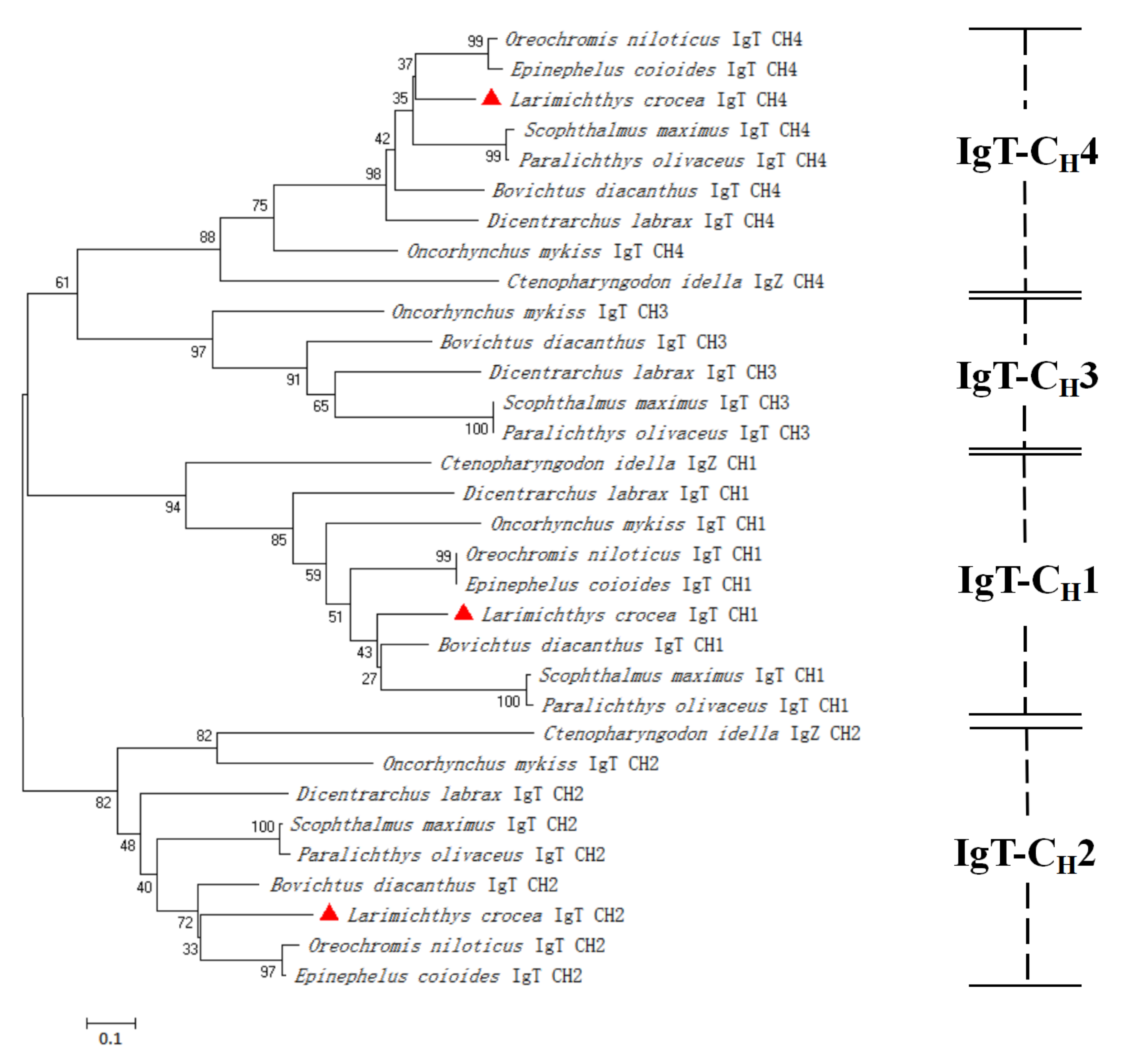
3.2. Multiple Sequence Alignment and Phylogenetic Analysis of LcIgT

The multiple alignments of amino acid sequences of LcIgT and other known teleost IgT heavy chains showed the conservation of typical Ig structural domains (Figure 1), with a VH and two-four CH domains. LcIgT contains three CH domains, CH1, CH2, and CH4, which were different from other fish IgT heavy chains at CH number and arrangement. Homology comparison showed that LcIgT shares the highest amino acid identity of 58.73% to Scophthalmus maximus IgT heavy chain (Table 1). Then, the VH and CH domains of LcIgT were compared with the corresponding VH and CH domains in other teleosts IgT. As shown in Table 2, the VH domain of LcIgT has the highest identity of 72.50% to that of Scophthalmus maximus IgT. Nevertheless, the CH1, CH2, and CH4 domains of LcIgT all share the highest identity to those of Oreochromis niloticus IgT with 67.61%, 61.11%, and 63.74% identities, respectively.
To understand the relationship between the IgT heavy chain of large yellow croaker and other teleosts, a phylogenetic tree was constructed with the protein sequences from various species. As shown in Figure 2, the IgT, IgM, and IgD of all species were clustered into three respective groups. Additionally, the constructed phylogenetic tree also showed that the corresponding CH1, CH2, CH3, and CH4 domains of the IgT heavy chain of the teleosts were clustered together, although LcIgT lacks CH3 domain (Figure 3).

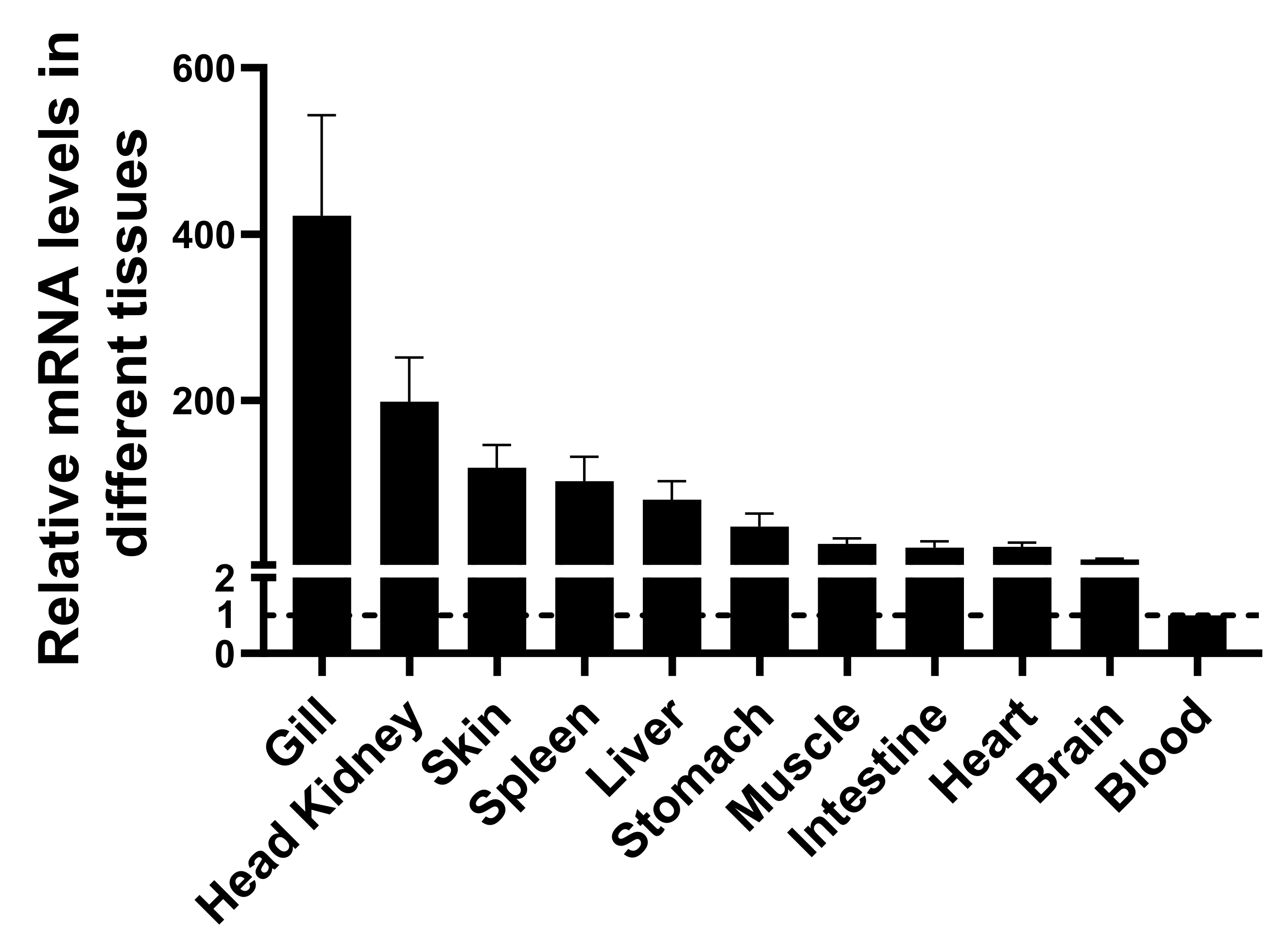
3.3. Tissue Distribution of LcIgT by RT-PCR

Tissue expression analysis showed that LcIgT mRNA was expressed in all tested tissues of large yellow croaker, including liver, heart, head kidney, gills, brain, spleen, stomach, intestine, skin, muscle, and blood (Figure 4). The highest expression levels of LcIgT were detected in gills, while the lowest levels were in blood. These results indicated that LcIgT transcripts were constitutively expressed in all tissues tested, with the highest expression levels in gills.

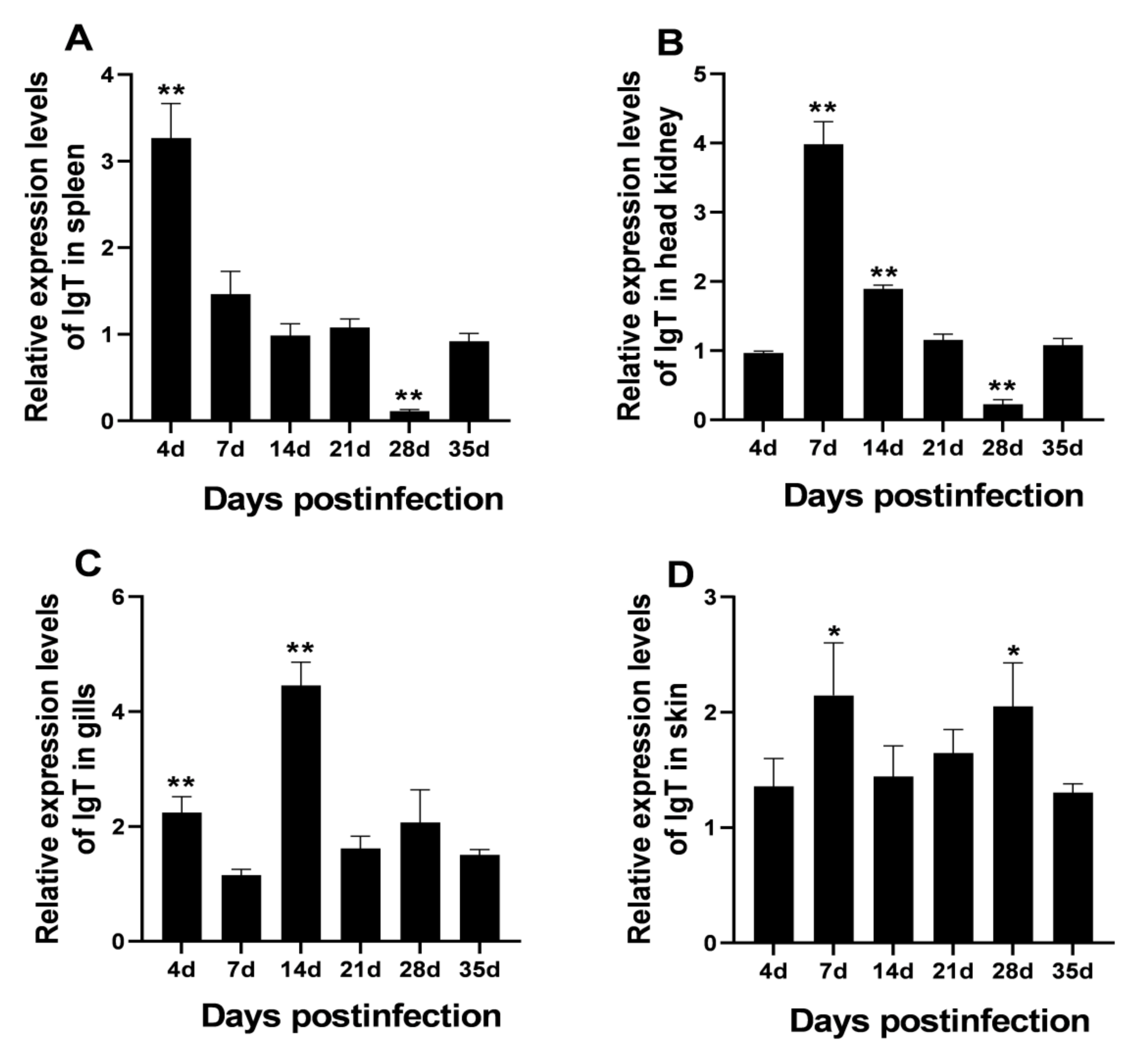
3.4. Modulation of LcIgT Gene Expression after C. irritans Infection

We further analyzed the modulation of LcIgT gene expression after C. irritans infection. As shown in Figure 5, expression levels of LcIgT in gills, skin, spleen, and head kidney were significantly up-regulated after C. irritans infection at several time points. LcIgT transcripts were increased in the spleen at 4 d (3.27-fold increase) and in head kidney and skin at 7 d (3.98- and 2.11-fold increases, respectively). In comparison, its expression levels in the gills were substantially up-regulated at 14 d post-infection (4.45-fold increase). Thus, LcIgT expression was differentially modulated in different tissues after C. irritans infection.

3.5. Expression Change at Protein Levels of LcIgT after C. irritans Infection

Finally, we investigated the expression change at the protein levels of LcIgT after C. irritans infection. The protein expression of LcIgT was significantly increased in gills, skin, and spleen upon C. irritans infection. The results of gray values analysis showed that the protein levels of LcIgT in the C. irritans-infected group were commonly higher than those in the control group, with a 3.43-fold increase in the skin at 7 d, 3.72-fold increase in the spleen at 14 d, and 6.33-fold in gills at 28 d (Figure 6).

4. Discussion

The present study characterized a secretory IgT heavy chain (LcIgT) in large yellow croaker. The deduced LcIgT has the conserved Ig structural characteristics, including a VH and three CH domains (CH1, CH2, and CH4), lacking the CH3 domain (Figure 2). IgT heavy chain in most known teleosts possesses four CH domains, but stickleback and emerald rock cod IgT heavy chains contain CH1, CH3, and CH4 domains (with a lack of CH2 domain) [21,22], and fugu IgT heavy chain contain only two CH domains, CH1 and CH4 [11]. These data indicated that the CH domains of IgT heavy chain might vary in CH number and arrangement among different teleosts. The cysteine and tryptophan residues, likely to contribute to the formation of disulfide bonds and the maintenance of the spatial structure of immunoglobulin [13,14], are well conserved in LcIgT (Figure 2; Cys170, Trp184, Trp219, Cys227, Cys264, Trp241, Trp313, Cys321, Cys478, Trp492, Trp521, and Cys528). The third conserved cysteine residue, involved in forming an interchain disulfide bond, and the two cysteine residues in each CH domain, indispensable for intrachain disulfide bridge in the tertiary structure of IgT [14], are also conserved in large yellow croaker. The neighbor-joining phylogeny revealed that LcIgT formed a well-supported clade with IgT molecules from other fish species. Single domain comparison showed that each CH domain of LcIgT shared a high amino acid identity with the corresponding domain of IgT in other species, with the highest identity with those of Oreochromis niloticus IgT. This further supported that the equivalent evolution extent may occur in each CH domain in teleosts.
Expression analyses showed that LcIgT was constitutively expressed in all tissues tested, with the highest levels in gills (Figure 5), which was consistent with the results observed in trout [30], flounder [13], European seabass [15], blunt snout bream [42], and emerald rock cod [43]. In addition, IgT was also highly expressed in the main mucosal tissues such as skin and intestine in large yellow croaker and other teleosts [13,14]. Thus, IgT plays a role in the mucosal immunity of fish. After bacterial infection, the flounder and turbot IgT expressions were up-regulated in several tissues, especially in gills and skin [13,14]. In trout, the IgT mRNA was significantly increased in gills, skin, nasopharynx, and gut after infection with Ichthyophthirius multifiliis or Ceratomyxa shasta [28,30,44]. Besides, significant increases of IgT-specific titers and IgT+ B-cells were detected in the gill, skin, and nasal mucus of I. multifiliis-infected trout [45]. In our study, the expression levels of both LcIgT mRNA and protein were found to be up-regulated in gills, skin, and spleen after C. irritans infection (Figure 6), suggesting that LcIgT may be involved in the systemic immunity and mucosal immunity against parasitic infection. In the previous study, LcIgT protein was expressed at the highest levels in the skin [41]. This may be the reason that the fold change of LcIgT protein in the skin (3.43-fold increase) was not very remarkable after C. irritans infection. All these data indicated that IgT plays a vital role in the immune defense against pathogenic infections in fish.

5. Conclusions

In conclusion, we cloned and characterized a secretory IgT heavy chain (LcIgT) from large yellow croaker. LcIgT was constitutively expressed in all tissues tested, with the highest levels in mucosa-associated tissues such as gills and skin. After C. irritans infection, both mRNA and protein levels of LcIgT were significantly up-regulated in systemic and mucosal immune tissues, suggesting that LcIgT may play a role in the systemic immunity and mucosal immunity against parasitic infection. However, further investigations are required to clarify the exact roles in immunity against parasitic infections of IgT in large yellow croakers.

Supplementary Materials

The following supporting information can be downloaded at: https://www.mdpi.com/article/10.3390/fishes7010029/s1, Figure S1: Nucleotide and deduced amino acid sequences of LcIgT gene (GenBank accession number: MW450786.1), Table S1: Primers and their sequences used in this study.

Author Contributions

Conceptualization, Y.T. (Yan Teng) and Q.F.; methodology, Y.T. (Yan Teng), Q.F., Y.T. (Yuanzhen Tan), Y.D. (Yangyang Ding) and Y.D. (Yang Ding); software, Y.T. (Yan Teng) and Q.F.; validation, Y.T. (Yan Teng), Q.F. and Y.T. (Yuanzhen Tan); formal analysis, Y.T. (Yan Teng) Q.F. and Y.T. (Yuanzhen Tan); investigation, Y.T. (Yan Teng), Q.F., Y.T. (Yuanzhen Tan) and Y.D. (Yang Ding); resources, Y.T. (Yan Teng), Q.F., Y.T. (Yuanzhen Tan), Y.D. (Yangyang Ding), Y.D. (Yang Ding) and X.C.; data curation, Q.F. and X.C.; writing—original draft preparation, Y.T. (Yan Teng) and Q.F.; writing—review and editing, Y.T. (Yan Teng), Q.F. and X.C.; visualization, X.C.; supervision, X.C.; project administration, X.C.; funding acquisition, X.C. All authors have read and agreed to the published version of the manuscript.

Funding

The work was supported by grants from the National Natural Science Foundation of China (U1905204 and 32073007), China Agriculture Research System of MOF and MARA (CARS-47), Fujian Science and Technology Department (2021N5008), and Institute of Oceanology of Fuzhou (2021F02).

Institutional Review Board Statement

The studies were carried out in strict accordance with the Regulations of the Administration of Affairs Concerning Experimental Animals, under protocol license number: SYXK(MIN)2007-0004, approved by the Institutional Animal Care and Use Committee of Fujian Province. All of the surgery was performed under Tricaine-S anesthesia, and all efforts were made to minimize suffering.

Acknowledgments

The work was supported by grants from the National Natural Science Foundation of China (U1905204 and 32073007), China Agriculture Research System of MOF, and MARA (CARS-47), Fujian Science and Technology Department (2021N5008), and Institute of Oceanology of Fuzhou (2021F02). ORCID: 0000-0003-2379-7596 (X.H.C.). Finally, we thank the referees for the thoughtful comments and recommendations.

Conflicts of Interest

The authors declare no conflict of interest. The funders had no role in the design of the study; in the collection, analyses, or interpretation of data; in the writing of the manuscript, or in the decision to publish the results.

References

  1. Schroeder, H.J.; Cavacini, L. Structure and function of immunoglobulins. J. Allergy Clin. Immunol. 2010, 125 (Suppl. 2), S41–S52. [Google Scholar] [CrossRef] [PubMed] [Green Version]
  2. Sunyer, J.O. Fishing for mammalian paradigms in the teleost immune system. Nat. Immunol. 2013, 14, 320–326. [Google Scholar] [CrossRef] [PubMed]
  3. Bradshaw, C.M.; Richard, A.S.; Sigel, M.M. IgM antibodies in fish mucus. Proc. Soc. Exp. Biol. Med. 1971, 136, 1122–1124. [Google Scholar] [CrossRef] [PubMed]
  4. Sunyer, J.O. Evolutionary and functional relationships of B cells from fish and mammals: Insights into their novel roles in phagocytosis and presentation of particulate antigen. Infect. Disord. Drug Targets 2012, 12, 200–212. [Google Scholar] [CrossRef]
  5. Edholm, E.S.; Bengten, E.; Wilson, M. Insights into the function of IgD. Dev. Comp. Immunol. 2011, 35, 1309–1316. [Google Scholar] [CrossRef]
  6. Hansen, J.D.; Landis, E.D.; Phillips, R.B. Discovery of a unique Ig heavy-chain isotype (IgT) in rainbow trout: Implications for a distinctive B cell developmental pathway in teleost fish. Proc. Natl. Acad. Sci. USA 2005, 102, 6919–6924. [Google Scholar] [CrossRef] [Green Version]
  7. Hu, Y.L.; Xiang, L.X.; Shao, J.Z. Identification and characterization of a novel immunoglobulin Z isotype in zebrafish: Implications for a distinct B cell receptor in lower vertebrates. Mol. Immunol. 2010, 47, 738–746. [Google Scholar] [CrossRef]
  8. Fillatreau, S.; Six, A.; Magadan, S.; Castro, R.; Sunyer, J.O.; Boudinot, P. The astonishing diversity of Ig classes and B cell repertoires in teleost fish. Front. Immunol. 2013, 4, 28. [Google Scholar] [CrossRef] [Green Version]
  9. Edholm, E.S.; Bengten, E.; Stafford, J.L.; Sahoo, M.; Taylor, E.B.; Miller, N.W.; Wilson, M. Identification of two IgD+ B cell populations in channel catfish, Ictalurus punctatus. J. Immunol. 2010, 185, 4082–4094. [Google Scholar] [CrossRef] [Green Version]
  10. Danilova, N.; Bussmann, J.; Jekosch, K.; Steiner, L.A. The immunoglobulin heavy-chain locus in zebrafish: Identification and expression of a previously unknown isotype, immunoglobulin Z. Nat. Immunol. 2005, 6, 295–302. [Google Scholar] [CrossRef]
  11. Savan, R.; Aman, A.; Sato, K.; Yamaguchi, R.; Sakai, M. Discovery of a new class of immunoglobulin heavy chain from fugu. Eur. J. Immunol. 2005, 35, 3320–3331. [Google Scholar] [CrossRef] [PubMed]
  12. Ryo, S.; Wijdeven, R.H.; Tyagi, A.; Hermsen, T.; Kono, T.; Karunasagar, I.; Rombout, J.H.; Sakai, M.; Verburg-van Kemenade, B.M.; Savan, R. Common carp have two subclasses of bonyfish specific antibody IgZ showing differential expression in response to infection. Dev. Comp. Immunol. 2010, 34, 1183–1190. [Google Scholar] [CrossRef] [PubMed]
  13. Du, Y.; Tang, X.; Zhan, W.; Xing, J.; Sheng, X. Immunoglobulin Tau Heavy Chain (IgT) in Flounder, Paralichthys olivaceus: Molecular Cloning, Characterization, and Expression Analyses. Int. J. Mol. Sci. 2016, 17, 1571. [Google Scholar] [CrossRef] [PubMed] [Green Version]
  14. Tang, X.; Du, Y.; Sheng, X.; Xing, J.; Zhan, W. Molecular cloning and expression analyses of immunoglobulin tau heavy chain (IgT) in turbot, Scophthalmus maximus. Vet. Immunol. Immunopathol. 2018, 203, 1–12. [Google Scholar] [CrossRef] [PubMed]
  15. Buonocore, F.; Stocchi, V.; Nunez-Ortiz, N.; Randelli, E.; Gerdol, M.; Pallavicini, A.; Facchiano, A.; Bernini, C.; Guerra, L.; Scapigliati, G.; et al. Immunoglobulin T from sea bass (Dicentrarchus labrax L.): Molecular characterization, tissue localization and expression after nodavirus infection. BMC Mol. Biol. 2017, 18, 8. [Google Scholar] [CrossRef] [Green Version]
  16. Mashoof, S.; Pohlenz, C.; Chen, P.L.; Gatlin, D., 3rd; Buentello, A.; Criscitiello, M.F. Expressed IgH mu and tau transcripts share diversity segment in ranched Thunnus orientalis. Dev. Comp. Immunol. 2014, 43, 76–86. [Google Scholar] [CrossRef]
  17. Tadiso, T.M.; Lie, K.K.; Hordvik, I. Molecular cloning of IgT from Atlantic salmon, and analysis of the relative expression of tau, mu, and delta in different tissues. Vet. Immunol. Immunopathol. 2011, 139, 17–26. [Google Scholar] [CrossRef] [Green Version]
  18. Zhang, N.; Zhang, X.J.; Chen, D.D.; Sunyer, J.O.; Zhang, Y.A. Molecular characterization and expression analysis of three subclasses of IgT in rainbow trout (Oncorhynchus mykiss). Dev. Comp. Immunol. 2017, 70, 94–105. [Google Scholar] [CrossRef] [Green Version]
  19. Bengten, E.; Clem, L.W.; Miller, N.W.; Warr, G.W.; Wilson, M. Channel catfish immunoglobulins: Repertoire and expression. Dev. Comp. Immunol. 2006, 30, 77–92. [Google Scholar] [CrossRef]
  20. Magadan-Mompo, S.; Sanchez-Espinel, C.; Gambon-Deza, F. Immunoglobulin heavy chains in medaka (Oryzias latipes). BMC Evol. Biol. 2011, 11, 165. [Google Scholar] [CrossRef] [Green Version]
  21. Bao, Y.; Wang, T.; Guo, Y.; Zhao, Z.; Li, N.; Zhao, Y. The immunoglobulin gene loci in the teleost Gasterosteus aculeatus. Fish Shellfish Immunol. 2010, 28, 40–48. [Google Scholar] [CrossRef] [PubMed]
  22. Giacomelli, S.; Buonocore, F.; Albanese, F.; Scapigliati, G.; Gerdol, M.; Oreste, U.; Coscia, M.R. New insights into evolution of IgT genes coming from Antarctic teleosts. Mar. Genom. 2015, 24 Pt 1, 55–68. [Google Scholar] [CrossRef]
  23. Schatz, D.G.; Swanson, P.C. V(D)J recombination: Mechanisms of initiation. Annu. Rev. Genet. 2011, 45, 167–202. [Google Scholar] [CrossRef] [PubMed] [Green Version]
  24. Gambon-Deza, F.; Sanchez-Espinel, C.; Magadan-Mompo, S. Presence of an unique IgT on the IGH locus in three-spined stickleback fish (Gasterosteus aculeatus) and the very recent generation of a repertoire of VH genes. Dev. Comp. Immunol. 2010, 34, 114–122. [Google Scholar] [CrossRef] [PubMed]
  25. Yasuike, M.; de Boer, J.; von Schalburg, K.R.; Cooper, G.A.; McKinnel, L.; Messmer, A.; So, S.; Davidson, W.S.; Koop, B.F. Evolution of duplicated IgH loci in Atlantic salmon, Salmo salar. BMC Genom. 2010, 11, 486. [Google Scholar] [CrossRef] [PubMed] [Green Version]
  26. Savan, R.; Aman, A.; Nakao, M.; Watanuki, H.; Sakai, M. Discovery of a novel immunoglobulin heavy chain gene chimera from common carp (Cyprinus carpio L.). Immunogenetics 2005, 57, 458–463. [Google Scholar] [CrossRef] [PubMed]
  27. Zhang, Y.A.; Salinas, I.; Li, J.; Parra, D.; Bjork, S.; Xu, Z.; LaPatra, S.E.; Bartholomew, J.; Sunyer, J.O. IgT, a primitive immunoglobulin class specialized in mucosal immunity. Nat. Immunol. 2010, 11, 827–835. [Google Scholar] [CrossRef]
  28. Xu, Z.; Parra, D.; Gomez, D.; Salinas, I.; Zhang, Y.A.; von Gersdorff Jørgensen, L.; Heinecke, R.D.; Buchmann, K.; LaPatra, S.; Sunyer, J.O. Teleost skin, an ancient mucosal surface that elicits gut-like immune responses. Proc. Natl. Acad. Sci. USA 2013, 110, 13097–13102. [Google Scholar] [CrossRef] [Green Version]
  29. Xu, Z.; Takizawa, F.; Parra, D.; Gómez, D.; von Gersdorff Jørgensen, L.; LaPatra, S.E.; Sunyer, J.O. Mucosal immunoglobulins at respiratory surfaces mark an ancient association that predates the emergence of tetrapods. Nat. Commun. 2016, 7, 10728. [Google Scholar] [CrossRef] [Green Version]
  30. Dong, F.; Yin, G.M.; Meng, K.F.; Xu, H.Y.; Liu, X.; Wang, Q.C.; Xu, Z. IgT Plays a Predominant Role in the Antibacterial Immunity of Rainbow Trout Olfactory Organs. Front. Immunol. 2020, 11, 583740. [Google Scholar] [CrossRef]
  31. Mu, Y.; Li, M.; Ding, F.; Ding, Y.; Ao, J.; Hu, S.; Chen, X. De novo characterization of the spleen transcriptome of the large yellow croaker (Pseudosciaena crocea) and analysis of the immune relevant genes and pathways involved in the antiviral response. PLoS ONE 2014, 9, e97471. [Google Scholar] [CrossRef] [PubMed] [Green Version]
  32. Chen, X.H.; Lin, K.B.; Wang, X.W. Outbreaks of an iridovirus disease in maricultured large yellow croaker, Larimichthys crocea (Richardson), in China. J. Fish Dis. 2003, 26, 615–619. [Google Scholar] [CrossRef] [PubMed]
  33. Yi, T.; Yujia, S.; Lingmin, Z.; Xu, X.; Huang, L.; Qin, Y.; Su, Y.; Yi, G.; Yan, Q. Mechanistic insight into the roles of Pseudomonas plecoglossicida clpV gene in host-pathogen interactions with Larimichthys crocea by dual RNA-seq. Fish Shellfish Immunol. 2019, 93, 344–353. [Google Scholar]
  34. Li, C.; Wang, S.; Ren, Q.; He, T.; Chen, X. An outbreak of visceral white nodules disease caused by Pseudomonas plecoglossicida at a water temperature of 12 degrees C in cultured large yellow croaker (Larimichthys crocea) in China. J. Fish Dis. 2020, 43, 1353–1361. [Google Scholar] [CrossRef] [PubMed]
  35. Kong, S.; Ke, Q.; Chen, L.; Zhou, Z.; Pu, F.; Zhao, J.; Bai, H.; Peng, W.; Xu, P. Constructing a High-Density Genetic Linkage Map for Large Yellow Croaker (Larimichthys crocea) and Mapping Resistance Trait Against Ciliate Parasite Cryptocaryon irritans. Mar. Biotechnol. 2019, 21, 262–275. [Google Scholar] [CrossRef] [PubMed]
  36. Tian, C.; Ding, Y.; Ao, J.; Chen, X. Three isotypes of immunoglobulin light chains in large yellow croaker, Pseudosciaena crocea: Molecular cloning, characterization, and expression analysis. Fish Shellfish Immunol. 2011, 30, 1249–1256. [Google Scholar] [CrossRef] [PubMed]
  37. Tian, C.; Chen, X.; Ao, J. The up-regulation of large yellow croaker secretory IgM heavy chain at early phase of immune response. Fish Physiol. Biochem. 2010, 36, 483–490. [Google Scholar] [CrossRef]
  38. Ao, J.; Mu, Y.; Xiang, L.X.; Fan, D.; Feng, M.; Zhang, S.; Shi, Q.; Zhu, L.Y.; Li, T.; Ding, Y.; et al. Genome sequencing of the perciform fish Larimichthys crocea provides insights into molecular and genetic mechanisms of stress adaptation. PLoS Genet. 2015, 11, e1005118. [Google Scholar] [CrossRef] [Green Version]
  39. Huang, M.; Mu, P.; Li, X.; Ren, Q.; Zhang, X.Y.; Mu, Y.; Chen, X. Functions of TNF-alpha1 and TNF-alpha2 in large yellow croaker (Larimichthys crocea) in monocyte/macrophage activation. Dev. Comp. Immunol. 2020, 105, 103576. [Google Scholar] [CrossRef]
  40. Livak, K.J.; Schmittgen, T.D. Analysis of relative gene expression data using real-time quantitative PCR and the 2(-Delta Delta C(T)) Method. Methods 2001, 25, 402–408. [Google Scholar] [CrossRef]
  41. Fu, Q.; Wei, Z.; Chen, Y.; Xie, J.; Zhang, X.; He, T.; Chen, X. Development of monoclonal antibody against IgT of a perciform fish, large yellow croaker (Larimichthys crocea) and characterization of IgT(+) B cells. Dev. Comp. Immunol. 2021, 119, 104027. [Google Scholar] [CrossRef] [PubMed]
  42. Xia, H.; Liu, W.; Wu, K.; Wang, W.; Zhang, X. sIgZ exhibited maternal transmission in embryonic development and played a prominent role in mucosal immune response of Megalabrama amblycephala. Fish Shellfish Immunol. 2016, 54, 107–117. [Google Scholar] [CrossRef] [PubMed]
  43. Coscia, M.R.; Simoniello, P.; Giacomelli, S.; Oreste, U.; Motta, C.M. Investigation of immunoglobulins in skin of the Antarctic teleost Trematomus bernacchii. Fish Shellfish Immunol. 2014, 39, 206–214. [Google Scholar] [CrossRef] [PubMed]
  44. Yu, Y.Y.; Kong, W.; Yin, Y.X.; Dong, F.; Huang, Z.Y.; Yin, G.M.; Dong, S.; Salinas, I.; Zhang, Y.A.; Xu, Z. Mucosal immunoglobulins protect the olfactory organ of teleost fish against parasitic infection. PLoS Pathog. 2018, 14, e1007251. [Google Scholar] [CrossRef] [PubMed] [Green Version]
  45. Wang, Q.; Yu, Y.; Zhang, X.; Xu, Z. Immune responses of fish to Ichthyophthirius multifiliis (Ich): A model for understanding immunity against protozoan parasites. Dev. Comp. Immunol. 2019, 93, 93–102. [Google Scholar] [CrossRef] [PubMed]
Figure 1. Alignment of the predicted large yellow croaker IgT heavy chain amino acid sequence with other known IgT heavy chain molecules. The conserved amino acids are indicated in blue fill. The predicted signal peptide sequence of large yellow croaker IgT heavy chain is indicated in italics. The CH domains are divided into three domains (CH1, CH2, and CH4). The conserved cysteines and tryptophan are marked with red asterisk and triangle. The first cysteine corresponds to the cysteine required for binding to the light chain. Potential glycosylation sites (NIS and NDS) are shown with a red dotted box.
Fishes 07 00029 g001
Figure 2. Phylogenetic tree of IgT heavy chain molecules from large yellow croaker and other fish species. A polygenetic tree was constructed to show the genetic relationship between large yellow croaker IgT and other fish species IgT. IgM, IgD heavy chain molecules from fish and vertebrates, were used as an outgroup. The tree was constructed by MEGA software using the Neighbor Joining (NJ) method, and the numbers on nodes represent the frequency with which this node is recovered per 100 bootstrap replications in a total of 1000. Large yellow croaker IgT was marked with a solid red triangle. GenBank accession numbers for IgT heavy chain sequences are as follows: MW450786 Larimichthys crocea (large yellow croaker) IgT heavy chain, AKA09828.1 Bovichtus diacanthus (Bovichtus) IgT heavy chain, AUV64181.1 Oreochromis niloticus (Nile tilapia) IgT, ACZ54909.1 Epinephelus coioides (juvenile grouper) IgZ heavy chain, ASK39430.1 Sparus aurata (juvenile seabream) IgT heavy chain secretory form, ANS12794.1 Paralichthys olivaceus (Japanese flounder) IgT membrane form, ANS12795.1 Japanese flounder IgT secretory form, AMQ49170.1 Scophthalmus maximus (turbot) IgT secretory form, AHC31432.1 Thunnus orientalis (Pacific bluefin tuna), AKK32388.1 Dicentrarchus labrax (European seabass), AAW66980.1 Oncorhynchus mykiss (rainbow trout) IgT membrane form, AAW66978.1 rainbow trout IgT secretory form, ANS12795.1 Paralichthys olivaceus (Japanese flounder), ABF19723.1 Ctenopharyngodon idella (grass carp), AAT67444.1 Danio rerio (zebrafish) IgZ1, ACH92959.1 zebrafish IgZ2. GenBank accession numbers for IgM heavy chain sequences are as follows: AAK69167.1 zebrafish IgM secretory form, ABD76396.1 grass carp IgM heavy chain, AAB27359.2 rainbow trout IgM heavy chain, ACM24795.1 large yellow croaker IgM heavy chain, ACH87158.1 bovichtus IgM secretory form, AAX78211.1 juvenile grouper IgM heavy chain, AAA49774.1 Xenopus laevis (African clawed frog) IgM heavy chain, CAC43280.1 Anas platyrhyncho (duck) IgM heavy chain, NP_001192115.1 Bos taurus (cattle) IgM heavy chain, and AAS01770.1 Homo sapiens (human) IgM heavy chain. GenBank accession numbers for IgD heavy chain sequences are: AAW66977.1 rainbow trout IgD membrane form, AHY86392.1 Nile tilapia IgD membrane form, BAD34541.1 Takifugu rubripes (fugu) IgD, AFI33218.1 juvenile grouper IgD heavy chain, ADD59896.1 IgD heavy chain constant region.
Fishes 07 00029 g002
Figure 3. Phylogenetic tree of each constant domain of fish IgT heavy chains. A polygenetic tree was constructed to show the genetic relationship of each constant domain of IgT heavy chains from large yellow croakers and other fish species. The tree was constructed by MEGA software using the Neighbor Joining (NJ) method, and the numbers on nodes represent the frequency with which this node is recovered per 100 bootstrap replications in a total of 1000. The single constant domain of large yellow croaker IgT was marked with a solid red triangle.
Fishes 07 00029 g003
Figure 4. Tissue expression profile of LcIgT. The expression levels of the LcIgT gene in eleven tissues of healthy large yellow croaker (liver, heart, head kidney, gills, brain, spleen, stomach, intestine, skin, muscle, and blood) were detected by real time-PCR. Total RNA from various tissues of five fish was isolated and then transcribed into the first cDNA. Error bars represent the mean ± standard error of the mean (mean ± SEM).
Fishes 07 00029 g004
Figure 5. Expression modulation of LcIgT in large yellow croakers infected by C. irritans. Expression change of LcIgT mRNA upon C. irritans infection was analyzed by real-time PCR. Four tissues, spleen (A), head kidney (B), gills (C), and skin (D), were collected from five C. irritans-infected fish or non-infected fish and used for total RNA extraction. The relative expression levels of LcIgT expressed as fold changes by comparing the normalized gene expression levels of C. irritans-infected fish with those of non-infected fish at the same time point. Error bars represent the mean ±SEM. Statistically significant differences are indicated with asterisks where * p < 0.05, ** p < 0.01.
Fishes 07 00029 g005
Figure 6. Western blotting analysis of LcIgT expression in large yellow croaker infected by C. irritans. Western blotting analyzed the expression of LcIgT protein upon C. irritans infection. (a) Three tissues, spleen (A), gills (B), and skin (C), were collected from five C. irritans-infected fish or non-infected fish at 4 d, 7 d, 14 d, 21 d, and 28 d post-infection. CG: control group; IG: infection group. (b) The fold change values of spleen, gills, and skin, marked with (I), (II), and (III), respectively, were calculated by normalizing to the control group. Statistically significant differences are indicated with asterisks where * p < 0.05 and ** p < 0.01.
Fishes 07 00029 g006
Table 1. The IgT heavy chain protein sequences used for phylogenetic analysis.
SpeciesIgT Heavy Chain (Identity, %)NCBI Accession Number
Entire Sequence
Larimichthys crocea100MW450786
Scophthalmus maximus58.73AMQ49170.1
Sparus aurata56.10ASK39430.1
Paralichthys olivaceus55.88ANS12795.1
Dicentrarchus labrax55.04AKK32388.1
Epinephelus coioides53.96ACZ54909.1
Oncorhynchus mykiss44.14AAW66978.1
Oreochromis niloticus36.84AUV64181.1
Danio rerio32.14ABF19723.1
Ctenopharyngodon idella28.24AAT67444.1
Table 2. The amino acid identity analysis of the VH and each CH domain of IgT heavy chain between L. crocea and other fish species.
SpeciesIgT Heavy Chain (Identity, %)
VHCH1CH2CH4
Larimichthys crocea100100100100
Scophthalmus maximus72.5061.2952.0563.33
Oreochromis niloticus41.0567.6161.1163.74
Paralichthys olivaceus56.1061.2952.0563.33
Epinephelus coioides43.9766.2056.9463.74
Sparus aurata47.4762.9061.1160.24
Dicentrarchus labrax51.9047.8956.9458.43
Oncorhynchus mykiss44.2150.0040.8547.73
Ctenopharyngodon idella45.3035.1435.1425.97
Publisher’s Note: MDPI stays neutral with regard to jurisdictional claims in published maps and institutional affiliations.

