Article

Effects of Continuous Light (LD24:0) Modulate the Expression of Lysozyme, Mucin and Peripheral Blood Cells in Rainbow Trout

1
Laboratorio de Piscicultura y Patología Acuática, Departamento de Oceanografía, Facultad de Ciencias Naturales y Oceanográficas, Universidad de Concepción, Concepción 4030000, Chile
2
Laboratorio de Toxicología, Departamento de Farmacia, Facultad de Farmacia, Universidad de Concepción, Concepción 4030000, Chile
3
Laboratorio Aquagestión, Universidad de Concepción, Concepción 4030000, Chile
4
Laboratorio de Biotecnología y Biofármacos, Departamento Fisiopatología, Facultad de Ciencias Biológicas, Universidad de Concepción, Concepción 4030000, Chile
5
Departamento de Microbiología, Facultad de Ciencias Biológicas, Universidad de Concepción, Concepción 4030000, Chile
6
Facultad de Agronomía e Ingeniería Forestal, Pontificia Universidad Católica de Chile, Santiago 8940833, Chile
*
Author to whom correspondence should be addressed.
Fishes 2022, 7(1), 28; https://doi.org/10.3390/fishes7010028
Submission received: 9 December 2021 / Revised: 14 January 2022 / Accepted: 20 January 2022 / Published: 25 January 2022
(This article belongs to the Topic Animal Diseases in Agricultural Production Systems)

Abstract

:
Continuous photoperiod is extensively used in fish farming, to regulate the reproductive cycle, despite evidence suggesting that artificial photoperiods can act as a stressor and impair the immune system. We evaluated the potential effects of an artificial photoperiod on mucus components: lysozyme and mucin, in juvenile rainbow trout (Oncorhynchus mykiss) after exposure for one month to natural photoperiod (LD12:12) or constant light (LD24:0) artificial photoperiod. For each treatment, we assessed changes in peripheral blood cells (erythrocytes and leukocytes) and skin mucus component concentrations. Our results show a decrease in lysozyme concentration, while mucin levels are increased. Similarly, we find elevated monocytes and polymorphonuclears under constant light photoperiod. These findings suggest that LD24:0 regulates lysozyme, mucin, and leukocytes, implying that artificial photoperiods could be a stressful.

1. Introduction

The light-dark cycle is one of the most important inputs that control many biological activities in fish [1,2]. Hence, the use of an extended artificial photoperiod in aquaculture is widely recognized as a biocompatible and environmentally friendly tool to control sexual maturation and fish development [2,3,4]. Artificial photoperiods are extensively used across fish farms to accelerate or delay spawning time, to synchronize smoltification and/or for growth enhancement [5,6,7,8]. For instance, salmonids are normally raised under constant light (light, L; dark, D = LD24:0) since the first feeding to accelerate growth during the freshwater phase. As standard protocol, the onset of smoltification is triggered using constant light (LD24:0), followed by a period of artificial winter (LD12:12) during six to eight weeks, and followed by additional six weeks in LD24:0 [7].
However, studies show that changes in photoperiod and light intensity may act as a chronic stressor in fish [8,9]. Although the application of artificial photoperiod is considered a clean technique, a recent study in trout estimated mortality rates over 25% in fish subjected to artificial photoperiods compared to mortality rates of 7% in fish maintained in natural light cycles [9,10]. In addition, application of artificial light indues stress that leads to immunosuppression and leaves salmonids and Nile tilapia prone to diseases [8,10,11,12].
While some studies in rainbow trout Oncorhynchus mykiss under a constant light regime show an increased in plasma cortisol concentration—a readout for physiological stress response [11], other studies in trout and red sea bream (Pagrus major) show unchanged cortisol levels and no evidence of a stress response [12,13,14].
Fish skin is the first line of defence against microorganisms, acting as a physicochemical barrier against infectious agents with mucin as an integral part of the mucosal barrier [10]. Histological analysis of the skin show a chronic response to artificial photoperiods, possibly related to alterations of non-specific skin defense mechanisms (mucus enzymes such as lysozyme or mucin) [10].
Moreover, recent studies in tilapia demonstrated that the mucosal immunity follow a circadian rhythm that is affected by comparing equal length of day and night (LD12:12, LD) and total darkness [15].
On the other hand, among leukocytes, neutrophils, monocytes and macrophages are particularly important in the defense system in fish and function in conjunction with lysozymes. This suggests that artificial photoperiods could increase susceptibility to diseases by inducing alterations in the concentration or activity of skin mucus enzymes that represent the principal barrier for pathogens [9,10,11,12,13,14,15,16,17].
Given the economic and cultural importance, and the scale of the aquaculture industry, particularly salmonids, it is necessary to properly evaluate the effect of rhythmicity and immune response of extended artificial photoperiod regimes on fish health and welfare. The aim of this study was to evaluate the effect of applying an extended artificial photoperiod (practical use for aquaculture) on the lysozyme and mucin levels of the skin mucus, and its effect on the levels of peripheral blood leukocytes in rainbow trout.

2. Materials and Methods

2.1. Experimental Design

One thousand and two hundred juvenile rainbow trouts (O. mykiss, mean weight 317 ± 12 g and length 17.7 ± 3.2 cm) were purchased from the “Salmones Pangue” fish farm (Bio-Bio Region, Chile, 36°41′11″ S; 72°52′04″ W) and kept in four circular tanks of 30,000 L each, with flow-through freshwater, gentle aeration, and fed on a ration of 4% body weight per day. Fish were randomly allocated to each experimental tank (300 fish each), achieving an initial density below 3.2 kg m−3. Fish were acclimated to these conditions for two weeks before photoperiod exposures started.
After the initial two-week acclimation period, tanks were randomly allocated to one of two treatments, natural photoperiod (LD: 11:1,-12:2,12:9,11:8) or artificial extended photoperiod (LD24:0). Therefore, each treatment was exposed to its light regime for one month with two replicate tanks per treatment (due to space constrictions on the fish farm). The artificial photoperiod protocol was applied using a 3600 lux cold halogen lamp suspended approximately 60 cm above the water surface. To avoid the influence of daylight, tanks were isolated using black polyethylene sheets and covered with black “Raschel” mesh. The average water temperature was maintained at 11 °C ± 2, water pH between 6 and 6.8, dissolved oxygen at 7–8 mg/L, and ammonium levels were always lower than 0.1 mg/L. From each tank, 10 fish were randomly selected and sampled (without replacement) at days 0, 3, 7, 14 and 30 of photoperiod exposure period. Fish were anaesthetized for 3 min with ethyl p-amino benzoate (BZ-20 Veterquímica, Santiago, Chile) using 1 mL of BZ-20 in 5 L of water from the aquarium. Whilst under anesthesia, weight and length measurements were taken from which Fulton’s condition factor (K) was calculated in both groups using the following formula: K = 100 × (W/L3). Where, W indicates final weight (g), and L is the length (cubic length), during 0, 3, 7, 14 and 30 days.

2.2. Preparation of Reduced Mucins

Mucus samples were collected before size measurement and blood sampling to reduce handling stress. Approximately 500 µL of skin mucus was collected from each fish (within 5 to 10 s), by dragging a sterilized spatula from posterior to anterior direction along the dorsal side. The mucus sample was then deposited into a sterile tube and snap frozen (and held) in liquid nitrogen until transported to the laboratory at the Universidad de Concepción. Once in the laboratory, samples were thawed on ice and centrifuged at 10,000 rpm at 4 °C for 10 min. Briefly, whole mucins were treated with GuHCl and DTT. Iodoacetamide was added and incubated in the dark overnight at room temperature.

2.3. Lysozyme Concentration

Lysozyme activity in the mucus was measured using the turbidometric assay based on Ellis [18]. Briefly, mucus samples (100 µL) were added to a 1 mL Micrococcus lysodeikticus suspension (0.1 µg in 500 mL of 0.1 M sodium phosphate buffer, pH 6.2). After 3 min of mixing, the absorbance of the resultant mixture was measured at 623 nm using a spectrophotometer (PHOTOMETER 5010 V5+5010, Riele, Berlin, Germany). Commercial hen egg white lysozyme (Merck, Darmstadt, Germany) was used to generate a standard curve and calculate the samples concentration. The lysozyme units are expressed as µg/mL.

2.4. Mucin Concentration

The concentration of mucin in the mucus was determined following the method described in Datta and Datta [19], with modifications. Briefly, 50 µL of mucus was mixed with 25 µL of phenol (80% w/v) and 1 mL of concentrated H2SO4. The mixture was allowed to react for 10 min and the absorbance of the sample was then read at 490 nm in a spectrophotometer as previously described. Mucin concentration was then calculated based on a glucose calibration curve.

2.5. Blood Samples

Blood samples (0.5 mL) were withdrawn from the caudal vein/artery using a 3-mL disposable syringe. A portion of that blood was immediately transferred to capillary tubes for hematocrit determination after centrifugation at 12,000 rpm for 5 min according to [20]. The remaining blood sample was then stored in an Eppendorf tube containing 10 µL EDTA 10% and kept on ice until analysis which was performed within 2 h of their arrival at the laboratory [21,22]. Total erythrocytes and leukocyte were counted in Neubauer chambers from a solution of blood and Rees Ecker reagent (1:100 v/v) [22].

2.6. Statistical Analysis

To evaluate statistical significance in the observed differences in Fulton’s condition factor, hematological parameters, lysozyme and mucin concentrations during the treatments, a two-way analysis of variance (ANOVA) was performed with treatment and time as fixed factors, followed by a Tukey HSD test. Data normality and homogeneity of variances were first assessed by a Shapiro Wilk and a Levene test, respectively. Results were considered significant at a p ˂ 0.05.

3. Results

In groups of trout (O. mykiss) exposed to natural or artifical photoperiods regimes, we observe statistically significant differences in the Fulton’s condition factor (Figure 1) on days 3 (p = 0.0002, n = 10) and 30 (p < 0.0001, n = 10), unlike days 7 and 14, where the differences did not reach statistical significance (p = 0.8, n = 10; p = 0.2, n = 10, respectively). Hematocrit and erythrocytes show a trending increase from day 0 to 14 in both treatment groups, however, no statistically significant differences were observed at each time point between treatment groups (Figure 2A,B). Haematocrit levels fluctuated between values of 29 and 38% in both groups during the experiment (p > 0.05), while erythrocytes fluctuated between 1.53 to 2.75 × 10¹² cell/L (p > 0.05).
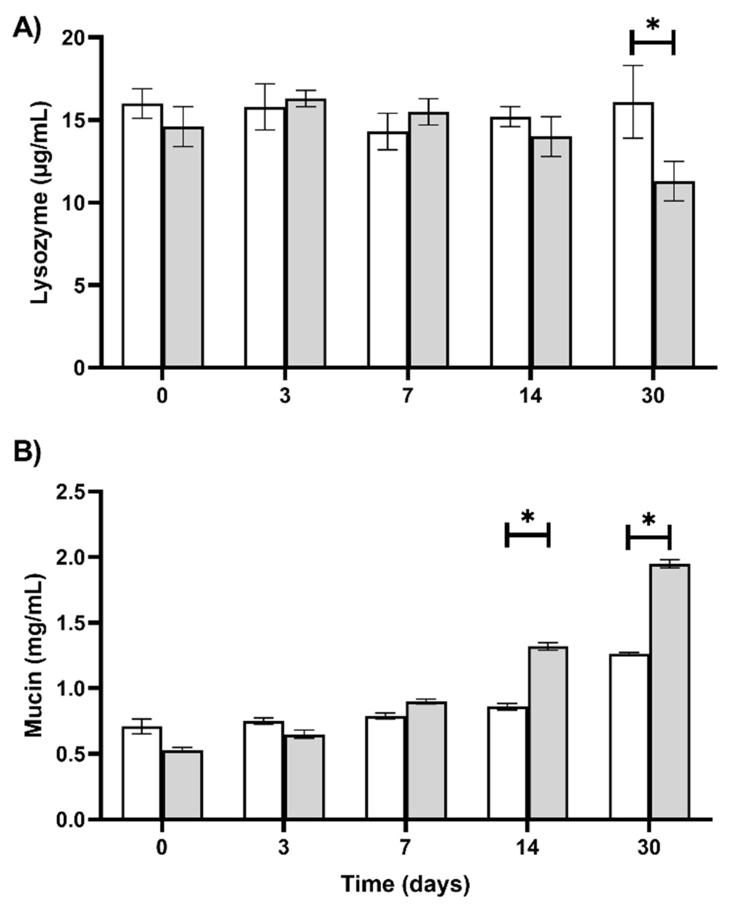
Regarding the white blood cell counts (WBC), total leukocyte numbers decrease in fish maintained under artificial photoperiod within 3 days of exposure and until day 7, compared to the natural photoperiod group. At days 14 and 30, the leukocyte numbers decrease to similar levels between treatment groups. (Figure 2C). Similarly, lymphocytes between days 3 and 7 in fish exposed to artificial photoperiod (p < 0.05 Figure 2D), and polymorphonuclears (Figure 2F) decreased between 3 and 7 days (0.6–0.7 × 10⁹ cell/L LD24:0 vs. 1.4–1.2 × 10⁹ cells/L, respectively). Monocyte counts (Figure 2E) increased after 3 days in LD24:0 fish (p < 0.05) (1.1 × 10⁹ cells/L) but at day 7 showed a significant decrease compared to the control group (p < 0.05) (0.2 × 10⁹ cell/L). Lysozyme and mucin concentrations ranged between 13.9 to 16.3 µg/mL and 0.53 to 1.95 µg/mL, respectively, during the duration of the experiment (Figure 3). Lysozyme concentration showed no changes between treatments during the first 14 days of exposure (p < 0.05). However, trout exposed to LD24:0 artificial photoperiod show a significant decrease (p < 0.05) (11.3 µg/mL), compared to the control group at day 30 (16.6 µg/mL) (Figure 3A). Mucin concentration showed no significant changes until day 7 of exposure, ranging from 0.71 µg/mL to 0.86 µg/mL. However, the LD24:0 artificial photoperiod group exhibit elevated concentrations of mucin at day 14 (p < 0.05) and day 30 (16.1 µg/mL) compared to the group under natural photoperiod that show mucin values of 11.3 µg/mL at day 30 (Figure 3B).

4. Discussion

Fulton’s condition factor (K) is used to compare fish of similar lengths and to evaluate the volumetric ratio as a function of weight, and as an indicator of animal welfare in fish populations that undergo changes in environmental factors, nutritional status, and reproduction. In fish farms, K changes as a function of disease or other environmental stressors [23]. Our results show that fish under continuous artificial photoperiod have a lower K. This suggests that continuous light may acts as a stressor in fish by demanding higher energy expenditure.
In concordance to previous findings in rainbow trout [11,12,13,15,16], continuous light for 30 days has not effect on haematocrit and erythrocytes in fish, haematology remains as a valuable diagnostic tool, despite the lack of reference values, as exist in mammals.
The decrease in leukocyte counts within 3 days of the continuous artificial light might represent aninitial signaling of acute stress. The leukopenia and lymphopenia detected after 3 days of exposure to artificial photoperiod—that is reversed after 7 days- are similar to those observed in Atlantic salmon, rainbow, brown and brook trout [24,25]. Acute stress under artificial light treatment could also explain the changes in neutrophils counts on day 3. Under acute stress conditions neutrophil counts increase due to a shift into circulating blood in response to glucocorticoids [26,27]. Additionally, neutrophils and the monocyte/macrophage system are an important part of the non-specific defense system, characterized by an elevated phagocytic ability, and mobility to migrate into specific tissues and consequently to elevate lysozyme content (stored in lysosomal granules). Thus, the reduction in white blood leukocytes could be explained by a migration into lymphoid tissues [28,29] during acute stress as a result of the photoperiod treatment [8,9].
Neutrophils and monocytes-macrophages are not only located in the peripheral blood, but they are also present in the skin and mucous membranes [30,31,32]. The skin’s mucus acts as a semi-permeable barrier allowing the gas, water, and nutrient exchange [33,34,35]. Mucus is also involved in respiration, osmoregulation, swimming, excretion, and defense against pathogens in fish [36,37,38]. Also, mucus has been described as an important part of innate humoral and cellular responses [31,39,40]. The presence of mucins and lysozyme, among other enzymes described in the fish skin mucus, highlights the role that the mucus plays in the response to disease resistance [41]. Lysozyme is known primarily as a non-specific defense enzyme in fish. It has been described in neutrophils, associated to the monocyte-macrophage lysosome system, and found extracellularly in leukocyte-rich tissues such as blood and mucus and its activity is an important index of innate immunity of fish [42,43,44,45]. Lysozyme concentrations with bacteriolytic properties have been shown to vary in plasma under both acute and chronic stress conditions [46,47,48,49]. It has been described that both serum and tissue lysozyme levels decrease, indicating that its concentration in the liver (main lysozyme producing organ along with the skin) is directly related to lysozyme concentrations found in the blood [42,43,44,45].
In our study, lysozyme concentrations decreased after 30 days of artificial light treatment, in agreement to previous reports [45], showing that highly stressed fish had elevated cortisol levels and lower lysozyme concentrations than moderately stressed fish. Similar results were described by Burgos et al. [50], showing that lysozyme concentration decreases in fish mucus using a continuous light photoperiod. However, the opposite trend has also been described [46], which might be explained by factors like handling and water ammonia levels that have been described to affect lysozyme concentration.
We suggest that fish under artificial photoperiod may be more prone to infectious diseases after 30 days of exposure to LD24:0 considering the importance of the first defense barrier [41]. Nevertheless, more studies are needed to confirm this hypothesis.
Whilst most work on fish lysozyme has been performed using serum measurements, Guardiola et al. [32] showed in sea bream (Sparus aurata) that some immune parameters in skin mucus were always higher than in the serum. This finding does not only show the importance of mucus as a first line of defense, but also shows the relationship between mucus and serum lysozyme. In this sense, the results obtained in the present study suggest that artificial photoperiod can modulate lysozyme production and that should receive more attention to prevent diseases in production systems.
Mucins are the predominant molecules present in mucus and can be divided into secreted gel-forming (main component of mucus) or associated with the cell-surface [31]. This carbohydrate (highly O-glycosylated proteins) has been described as an agglutinating receptor molecule for microorganisms [51,52], playing an important role in eliminating pathogens and parasitic infestation [53].
Recent research shows interaction with pathogens relevant to salmon farming in fresh and seawater phase, with different affinities related with the mucin’s origin (skin vs. gastrointestinal tract) and complexity of the glycans [54]. Since it has been described those infections only occur when pathogens are able to colonize mucosal integuments [55], it is this agglutinating-adhesive property of mucin that confers a defensive function by trapping and immobilizing pathogens [56]. However, it must also be considered that an increase in viscosity of mucus may support or allow colonization by pathogens, particularly if bacteriostatic or bacteriolytic activity is compromised, as noted in our study (increased mucin and diminished lysozyme).
Taken together, in this study the increased in mucin concentration in fish after exposure to the continuous light photoperiod, could explain the previously reported elevated rates of infections observed in fish exposed to artificial photoperiod [10]. Our results suggest that mucin and lysozyme concentrations change under a prolonged light exposure, implying that a LD24:0 artificial photoperiod could act as a stimulus increasing their secretion in mucus. This could alter the viscous-elastic properties, due to the elevated content of the high molecular weight glycoprotein mucin, which is produced and secreted by skin epithelial cells and glands [57]. This mucus production is a specific stress response in fish and tremendous energy expenditure in the production of mucins [58]. Along with differences in Fulton’s condition factor, we speculate that the difference in mucin concentration is not due to differences in size.
Consequently, the application of an artificial photoperiod (LD24:0) in trout may induce defense suppression and could cause increased susceptibility to diseases [41]. The findings presented here will contribute to better understand the side effects in the use of artificial photoperiod in fish farms, and develop preventive measures, especially during the first 30 days of application of the photoperiod.

5. Conclusions

We highlight the finding that the decrease in lysozyme is inversely correlated to the significant increase in mucin in trout subjected to continues light photoperiod. Our study contributes to a better comprehension of the effects of photoperiods as a productive strategy in fish farming and its potential effects on fish welfare.

Author Contributions

Conceptualization, A.V., J.A. Methodology, I.R., B.S., V.C. Formal analysis, A.V., R.C., B.S., S.E.-A., I.R. Investigation, A.V., I.R., B.S., V.C., J.A. resources, A.V., V.C. data curation, S.E.-A. writing—original draft preparation, A.V., R.C., S.E.-A. writing—review and editing, S.E.-A., R.C. visualization, R.C. supervision, S.E.-A., A.V. funding acquisition, A.V. All authors have read and agreed to the published version of the manuscript.

Funding

This research was funded by FONDECYT 11090246.

Institutional Review Board Statement

The study was conducted according to the guidelines of the Declaration of Helsinki, and approved by the Ethics Committee of Universidad de Concepción, Chile (protocol code CBB 1084-2021).

Informed Consent Statement

Not applicable.

Data Availability Statement

The study did not report any data.

Acknowledgments

We thank Matias Escobar for revising and editing the manuscript.

Conflicts of Interest

The authors declare no conflict of interest. The funders had no role in the design of the study; in the collection, analyses, or interpretation of data; in the writing of the manuscript, or in the decision to publish the results.

References

  1. Esteban, M.A.; Cuesta, A.; Rodríguez, A.; Meseguer, J. Effect of photoperiod on the fish innate immune system: A link between fish pineal gland and the immune system. J. Pineal Res. 2006, 41, 261–266. [Google Scholar] [CrossRef] [PubMed]
  2. Morro, B.; Balseiro, P.; Albalat, A.; Pedrosa, C.; Mackenzie, S.; Nakamura, S.; Shimizu, M.; Nilsen, T.O.; Sveier, H.; Ebbesson, L.O.; et al. Effects of different photoperiod regimes on the smoltification and seawater adaptation of seawater-farmed rainbow trout (Oncorhynchus mykiss): Insights from Na+, K+–ATPase activity and transcription of osmoregulation and growth regulation genes. Aquaculture 2019, 30, 282–292. [Google Scholar] [CrossRef]
  3. Karlsen, Ø.; Norberg, B.; Kjesbu, O.S.; Taranger, G.L. Effects of photoperiod and exercise on growth, liver size, and age at puberty in farmed Atlantic cod (Gadus morhua L.). ICES Mar. Sci. Symp. 2006, 63, 355–364. [Google Scholar] [CrossRef] [Green Version]
  4. Iversen, M.; Mulugeta, T.; West, A.C.; Jørgensen, E.H.; Samuel, A.M.; Simen Rød Sandve, M.; Hazlerigg, D. Photoperiod-dependent developmental reprogramming of the transcriptional response to seawater entry in Atlantic salmon (Salmo salar). G3 2021, 11, 1–9. [Google Scholar] [CrossRef] [PubMed]
  5. Wilkinson, R.J.; Longland, R.; Woolcott, H.; Porter, J.R. Effect of elevated winter–spring water temperature on sexual maturation in photoperiod manipulated stocks of rainbow trout (Oncorhynchus mykiss). Aquaculture 2010, 22, 236–244. [Google Scholar] [CrossRef]
  6. Taranger, G.L.; Carrillo, M.; Schulz, R.; Fontaine, P.; Zanuy, S.; Felip, A.; Weltzien, F.A.; Dufourf, S.; Karlseng, Ø.; Norbergg, B.; et al. Control of puberty in farmed fish. Gen. Comp. Endocrinol. 2010, 3, 483–515. [Google Scholar] [CrossRef] [Green Version]
  7. Pino Martinez, E.; Balseiro, P.; Pedrosa, C.; Haugen, T.S.; Mitchell, F.S.; Handeland, S.O. The effect of photoperiod manipulation on Atlantic salmon growth, smoltification and sexual maturation: A case study of a commercial RAS. Aquac. Res. 2020, 52, 2593–2608. [Google Scholar] [CrossRef]
  8. Wang, K.; Li, K.; Liu, L.; Tanase, C.; Mols, R.; van der Meer, M. Effects of light intensity and photoperiod on the growth and stress response of juvenile Nile tilapia (Oreochromis niloticus) in a recirculating aquaculture system. Aquac. Fish. 2020. [Google Scholar] [CrossRef]
  9. Cabillon, N.A.R.; Lazado, C.C. Mucosal barrier functions of fish under changing environmental conditions. Fishes 2019, 4, 2. [Google Scholar] [CrossRef] [Green Version]
  10. Valenzuela, A.; Campos, V.; Yañez, F.; Alveal, K.; Gutiérrez, P.; Rivas, M.; Contreras, N.; Klempau, A.; Fernandez, I.; Oyarzún, C. Application of artificial photoperiod in fish: A factor that increases susceptibility to infectious diseases? Fish. Physiol. 2012, 38, 943–950. [Google Scholar] [CrossRef]
  11. Leonardi, M.; Klempau, A. Artificial photoperiod influence on the immune system of juvenile rainbow trout Oncorhynchus mykiss. Aquaculture 2003, 221, 581–591. [Google Scholar] [CrossRef]
  12. Valenzuela, A.E.; Silva, V.M.; Klempau, A.E. Effects of constant light on haematological parameters of cultured rainbow trout (Oncorhynchus mykiss) in the Southern Hemisphere. Fish. Physiol. Biochem. 2006, 32, 113–120. [Google Scholar] [CrossRef]
  13. Biswas, A.K.; Seoka, M.; Tanaka, Y.; Takii, K.; Kumai, H. Effect of photoperiod manipulation on the growth performance and stress response of juvenile red sea bream (Pagrus major). Aquaculture 2006, 258, 350–356. [Google Scholar] [CrossRef]
  14. Lazado, C.C.; Vilhelm Skov, P. Secretory proteins in the skin mucus of Nile Tilapia (Oreochromis niloticus) are modulated temporally by photoperiod and bacterial endotoxin cues. Fishes 2019, 4, 57. [Google Scholar] [CrossRef] [Green Version]
  15. Valenzuela, A.E.; Silva, V.M.; Klempau, A.E. Some changes in the haematological parameters of rainbow trout (Oncorhynchus mykiss) exposed to three artificial photoperiod regimes. Fish. Physiol. Biochem. 2008, 33, 45–48. [Google Scholar] [CrossRef]
  16. Valenzuela, A.E.; Silva, V.M.; Klempau, A.E. Effects of different artificial photoperiods and temperatures on haematological parameters of rainbow trout (Oncorhynchus mykiss). Fish. Physiol. Biochem. 2008, 34, 159–167. [Google Scholar] [CrossRef] [PubMed]
  17. Xua, Z.; Parra, D.; Gómez, D.; Salinas, I.; Zhang, Y.A.; Jørgensend, L.V.G.; Heinecked, R.D.; Buchmannd, K.; LaPatrae, S.; Sunyera, J.O. Teleost skin, an ancient mucosal surface that elicits gut-like immune responses. Proc. Natl. Acad. Sci. USA 2013, 110, 13097–13102. [Google Scholar] [CrossRef] [Green Version]
  18. Ellis, A. E Immunity to bacteria in fish. Fish. Shell Fish Immun. 1999, 9, 291–308. [Google Scholar] [CrossRef]
  19. Datta, S.; Datta, S.C. Purification and characterization of fish surface mucin. Ital. J. Biochem. 1987, 36, 143–152. [Google Scholar]
  20. Urbina, M.A.; Glover, C.N. Should I stay or should I go?: Physiological, metabolic and biochemical consequences of voluntary emersion upon aquatic hypoxia in the scaleless fish Galaxias maculatus. J. Comp. Physiol. B Biochem. Syst. Environ. Physiol. 2012, 182, 1057–1067. [Google Scholar] [CrossRef]
  21. Campbell, T.W.; Murru, F. An introduction to fish hematology. Comp. Cont. Ed. Vet. Sci 1990, 12, 525–533. [Google Scholar]
  22. Klontz, G.W. Fish hematology. In Techniques in Fish Immunology, 3rd ed.; Stolen, J.D., Rowley, A.F., Zelikoff, J.T., Kaatari, S.L., Smith, S.A., Eds.; SOS Publications: Fair Haven, NJ, USA, 1994; pp. 121–131. [Google Scholar]
  23. Jones, R.; Petrell, R.; Pauly, D. Using modified length-weight relationships to assess the condition of fish. Aquac. Eng. 1999, 20, 261–276. [Google Scholar] [CrossRef]
  24. Angelidis, P.; Baudin-Laurencin, F.; Youinou, P. Stress in rainbow trout, Salmo gairdneri: Effects upon phagocyte chemiluminescence, circulating leucocytes and susceptibility to Aeromonas salmonicida. J. Fish. Biol. 1987, 31, 113–122. [Google Scholar] [CrossRef]
  25. Pickering, A.D.; Griffiths, R.; Pottinger, T.G. A comparison of the effects of overhead cover on the growth, survival and haematology of juvenile Atlantic salmon, Salmo salar L.; brown trout, Salmo trutta L.; and rainbow trout, Salmo gairdneri Richardson. Aquaculture 1987, 66, 109–124. [Google Scholar] [CrossRef]
  26. Melingen, G.O.; Pettersen, E.F.; Wergeland, H.I. Leucocyte populations and response to immunization and photoperiod manipulation in Atlantic salmon (Salmo salar L.) 0 + smolt. Aquaculture 2002, 214, 381–396. [Google Scholar] [CrossRef]
  27. Grzelak, A.K.; Davis, D.J.; Caraker, S.M.; Crim, M.J.; Spitsbergen, J.M.; Wiedmeyer, C.E. Stress Leukogram Induced by Acute and Chronic Stress in Zebrafish (Danio rerio). Comp. Med. 2017, 67, 263–269. [Google Scholar]
  28. Maule, A.G.; Schreck, C.B. Changes in numbers of leukocytes in immune organs of juvenile coho salmon after acute stress or cortisol treatment. J. Aquat. Animal Health 1990, 2, 298–304. [Google Scholar] [CrossRef] [Green Version]
  29. Heinzel, K.; Benz, C.; Bleu, C.C. A silent chemokine recptor regulates steady-state leukocyte homing in vivo. Proc. Natl. Acad. Sci. USA 2007, 104, 8421–8426. [Google Scholar] [CrossRef] [Green Version]
  30. Lie, O.; Evensen, O.; Sorensen, A.; Froysadal, E. Study of lysozyme activity in some fish species. Dis. Aquat. Org. 1989, 6, 1–5. [Google Scholar] [CrossRef]
  31. Gomez, D.; Sunyer, J.O.; Salinas, I. The mucosal immune system of fish: The evolution of tolerating commensals while fighting pathogens. Fish. Shellfish Immunol. 2013, 35, 1729–1739. [Google Scholar] [CrossRef] [Green Version]
  32. Guardiola, F.A.; Cuesta, A.; Abellan, E.; Meseguer, J.; Esteban, M.A. Comparative analysis of the humoral immunity of skin mucus from several marine teleost fish. Fish. Shellfish Immunol. 2014, 40, 24–31. [Google Scholar] [CrossRef] [PubMed]
  33. Ingram, G.A. Substances involved in the natural resistance of fish to infection a review. J. Fish. Biol. 1980, 16, 23–60. [Google Scholar] [CrossRef]
  34. Fletcher, T. Non-antibody molecules and the defense mechanisms of fish. In Stress and Fish; Pickering, A.D., Ed.; Academy Press: Washington, DC, USA, 1981; pp. 171–183. [Google Scholar]
  35. Esteban, M.A. An overview of the immunological defences in fish skin. Int. Sch. Res. Not. 2012, 2012, 853470. [Google Scholar]
  36. Shepard, K.L. Functions for fish mucus. Rev. Fish Biol. Fish. 1994, 4, 401–429. [Google Scholar] [CrossRef]
  37. Urbina, M.A.; Meredith, A.S.; Forster, M.E.; Glover, C.N. The importance of cutaneous gas exchange in aquatic and aerial mediums in galaxiid fishes. J. Fish. Biol. 2014, 84, 759–773. [Google Scholar] [CrossRef]
  38. Urbina, M.A.; Glover, C.N. Effect of salinity on osmoregulation, metabolism and nitrogen excretion in the amphidromous fish, inanga (Galaxias maculatus). J. Exp. Mar. Bio. Ecol. 2015, 473, 7–15. [Google Scholar] [CrossRef]
  39. Tiralongo, F.; Messina, G.; Lombardo, B.M.; Longhitano, L.; Li Volti, G.; Tibullo, D. Skin mucus of marine fish as a source for the development of antimicrobial agents. Front. Mar. Sci. 2020, 7, 760. [Google Scholar] [CrossRef]
  40. Dash, S.; Das, S.K.; Samal, J.; Thatoi, H.N. Epidermal mucus, a major determinant in fish health: A review. Iran. J. Vet. Res. 2018, 19, 72–81. [Google Scholar]
  41. Minniti, G.; Rød Sandve, S.; Padra, J.T.; Heldal Hagen, L.; Lindén, S.; Pope, P.B.; Arntzen, M.Ø.; Vaaje-Kolstad, G. The farmed Atlantic Salmon (Salmo salar) skin-mucus proteome and its nutrient potential for the resident bacterial community. Genes 2019, 10, 515. [Google Scholar] [CrossRef] [Green Version]
  42. Holloway, J.; Shoemaker, C.A.; Ottinger, C.A. Serum lysozyme levels in paddle Fish walleye. J. Aquat Anim Health 1993, 5, 324–326. [Google Scholar] [CrossRef]
  43. Shailesh, S.; Sahoo, P.K. Lysozyme: An important defence molecule of fish innate immune system. Aquac. Res. 2008, 39, 223–239. [Google Scholar]
  44. Moeck, A.; Peters, G. Lysozyme activity in rainbow trout, Oncorhynchus mykiss (Walbaum), stressed by handling, transport and water pollution. J. Fish. Biol. 1990, 37, 873–885. [Google Scholar] [CrossRef]
  45. Røed, K.; Larsen, H.; Linder, R.; Refstie, T. Genetic variation in lysozyme activity in rainbow trout (Oncorhynchus mykiss). Aquaculture 1993, 109, 237–244. [Google Scholar] [CrossRef]
  46. Demers, N.E.; Bayne, C.J. The immediate effects of stress on hormones and plasma lysozyme in rainbow trout. Dev. Comp. Immunol. 1997, 21, 363–373. [Google Scholar] [CrossRef]
  47. Cortés, R.; Teles, M.; Trídico, R.; Acerete, L.; Tort, L. Effects of cortisol administered through slow-release implants on innate immune responses in rainbow trout (Oncorhynchus mykiss). Int. J. Genom. 2013, 2013, 619714. [Google Scholar] [CrossRef] [Green Version]
  48. Sunyer, J.O.; Gomez, E.; Navarro, V.; Quesada, J.; Tort, L. Depression of humoral components of the immune system and physiological responses in gilthead sea bream Sparus aurata after daily acute stress. Can. J. Fish. Aquat. 1995, 52, 2339–2346. [Google Scholar] [CrossRef]
  49. Ortuño, J.; Esteban, M.A.; Meseguer, J. Effects of short-term crowding stress on the gilthead seabream (Sparus aurata L.) innate immune response. Fish. Shellfish Immunol. 2001, 11, 187–197. [Google Scholar] [CrossRef] [PubMed]
  50. Burgos, A.; Valenzuela, A.; Gonzalez, M.; Klempau, A. Non-specific defence mechanisms of rainbow trout (Oncorhynchus mykiss) during artificial photoperiod. Bull. Eur. Ass. Fish. Phatol. 2004, 24, 240–245. [Google Scholar]
  51. Redondo, M.J.; Alvarez-Pellitero, P. Carbohydrate patterns in the digestive tract of Sparus aurata (L.) and Psetta maxima (L.) (Teleostei) parasitized by Enteromyxum leei and E. scophthalmi (Myxozoa). Parasitol. Int. 2010, 59, 445–453. [Google Scholar] [CrossRef] [Green Version]
  52. Estensoro, I.; Jung-Schroers, V.; Alvarez-Pellitero, P.; Steinhagen, D.; Sitja-Bobadilla, A. Effects of Enteromyxum leei (Myxozoa) infection on gilthead sea bream (Sparus aurata) (Teleostei) intestinal mucus: Glycoprotein profile and bacterial adhesion. Parasitol. Res. 2013, 112, 567e76. [Google Scholar] [CrossRef]
  53. Hasnain, S.Z.; Wang, H.; Ghia, J.E.; Haq, N.; Deng, Y.; Velcich, A.; Grencis, R.K.; Thornton, D.J.; Khan, W.I. Mucin gene deficiency in mice impairs host resistance to an enteric parasitic infection. Gastroenterology 2010, 138, 1763–1771. [Google Scholar] [CrossRef] [PubMed]
  54. Padra, J.T.; Murugan, A.V.M.; Sundell, K.; Sundh, H.; Benktander, J. Fish pathogen binding to mucins from Atlantic salmon and Arctic char differs in avidity and specificity and is modulated by fluid velocity. PLoS ONE 2019, 24, e0215583. [Google Scholar] [CrossRef] [PubMed]
  55. Mayer, L. Mucosal immunity. Pediatrics 2003, 111, 1595–1600. [Google Scholar] [CrossRef] [PubMed]
  56. Hoste, H. Adaptive physiological processes in the host during gastrointestinal parasitism. Int. J. Parasitol. 2001, 31, 231–244. [Google Scholar] [CrossRef]
  57. Busch, L.; Borda, E. Mucinas salivales: Estructura química, mecanismos de liberación y participación en la defensa no inmunológica de la cavidad bucal. Revista de la Facultad de Odontología 2009, 24, 171–176. [Google Scholar]
  58. Zuchelkowski, E.; Pinkstaff, C.; Hinton, D. Mucosubstance histochemistry in control and acid-stressed epidermis of brown bullhead catfish Ictalurus nebulosus (LeSueur). Anat. Rec. 1985, 212, 327–335. [Google Scholar] [CrossRef]
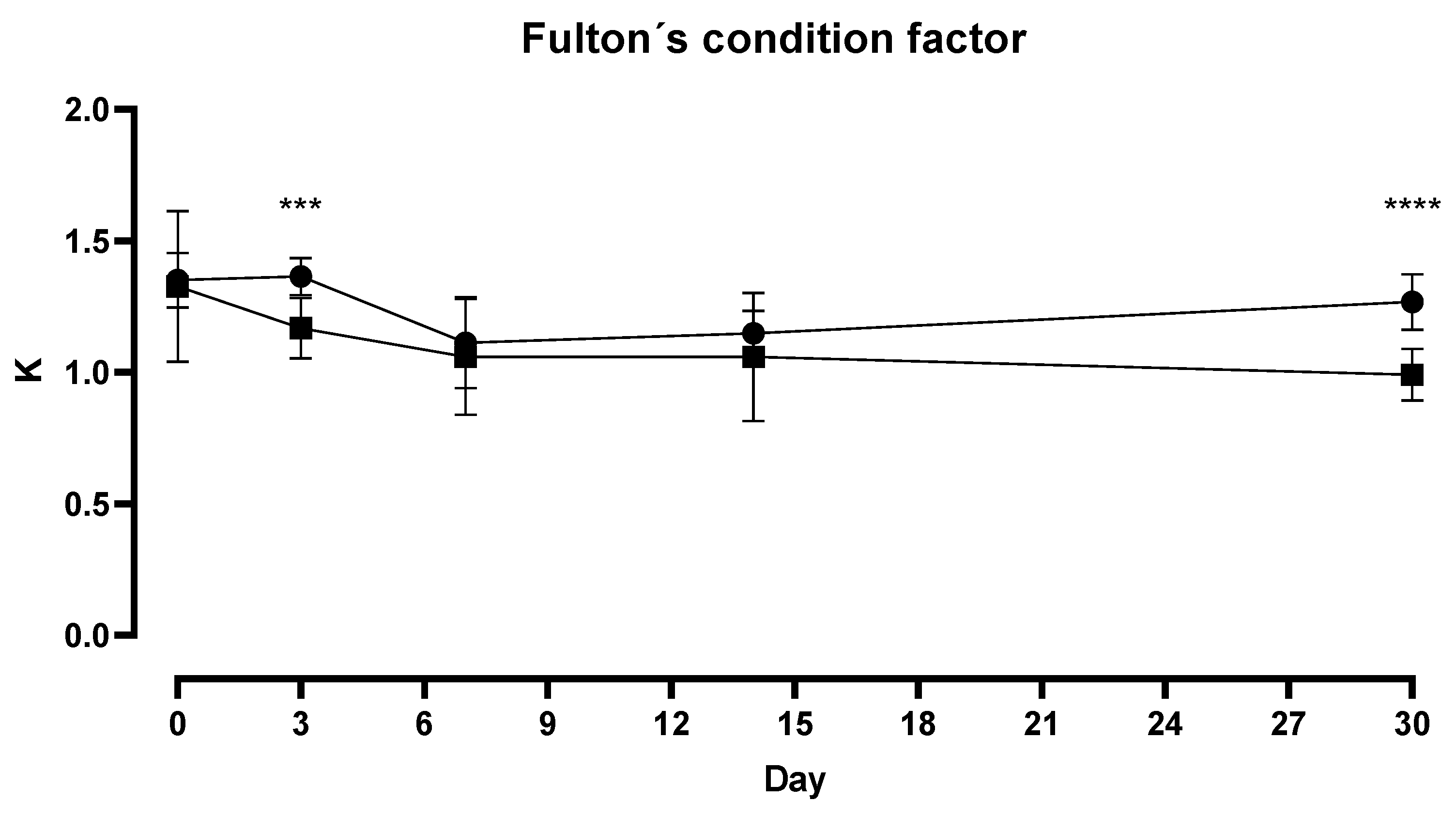
Figure 1. Fulton’s condition factor of trout (O. mykiss) exposed to control (natural photoperiod; solid circle) and artificial (LD24:0; solid square) photoperiods. Asterisks (*** and ****) indicate significant differences between treatments (day 3; p = 0.0002 and day 30; p < 0.0001, respectively). Spots represent mean and ± Standard deviation.
Fishes 07 00028 g001
Figure 2. Changes in Haematocrit (A), Erythrocytes (B) and Leucocytes (C), Lymphocytes (D), Monocytes (E) and Polymorphonuclear (F) in trout (O. mykiss) exposed to control (natural photoperiod; empty bars) and artificial (LD24:0; grey bars) photoperiods. Asterisks indicate significant differences between treatments. Bars represent mean and ± Standard Error.
Fishes 07 00028 g002
Figure 3. Lysozyme (A) and mucin (B) concentrations quantified in mucus of O. mykiss under LD24:0 photoperiod (grey bars) and controls (empty bars) during a period of 30 days. Asterisks indicate significant differences between treatments. Bars represent mean and ± Standard Error.
Fishes 07 00028 g003
Publisher’s Note: MDPI stays neutral with regard to jurisdictional claims in published maps and institutional affiliations.