1. Introduction
With the rapid growth of the global human population, demand for seafood has surged, leading to the continued expansion of aquaculture in both scale and industry [
1,
2,
3]. However, advancement of high-density, intensive aquaculture has led to the deterioration of water quality and the aquatic environment [
4,
5]. This deterioration directly compromises the immune function and disease resistance of aquatic animals, reduces survival rates and growth performance, and ultimately increases susceptibility to disease outbreaks [
6,
7]. To enhance the resilience of aquatic animals under suboptimal water conditions, farmers frequently apply substantial quantities of chemical agents and antibiotics [
8,
9,
10]. However, the excessive use of drugs has multiple adverse effects, including drug residues, immune imbalances in aquatic animals, microbial resistance, immunosuppression, and environmental contamination [
11,
12,
13]. Consequently, there is an increasing demand for safer antibiotic and immune-boosting alternatives in aquaculture.
Plant extracts are rich in bioactive compounds, including alkaloids, terpenes, tannins, saponins, glycosides, steroids, and essential oils, and can enhance stress resistance in aquaculture animals [
14]. Various active ingredients in plant extracts have shown excellent application results in aquatic animals, and developing these extracts as alternatives to antibiotics is a priority. For example, dietary supplementation with artichoke (
Cynara scolymus) leaf extract can enhance the tolerance of Nile Tilapia (
Oreochromis niloticus) to fluoride stress [
15]. Similarly, dietary supplementation with oak (
Quercus castaneifolia) leaf extract improved the tolerance of common carp (
Cyprinus carpio) to overcrowding [
16]. Plant extracts have also accelerated the growth of cultured organisms, improving feed intake, enhancing vitality and immune response, promoting gonadal maturation, with added aphrodisiac and antipathogenic effects [
17]. Supplementation with compound plant extracts of
Solanum ferox combined with either
Boesenbergia pandurata or
Zingiber zerumbet markedly improved the growth performance, molting rate, and survival of pond-reared
Scylla serrata [
18].
Pandanus leaf extract could also upregulate the expression of immune-related genes (Hsp70, proPO, and crustin) in shrimp (
Litopenaeus vannamei), improving survival rates after infection with
Vibrio parahaemolyticus [
19]. In addition, dietary supplementation with
Annona squamosa leaf extract enhanced the antioxidant capacity of Nile tilapia (
Oreochromis niloticus) [
20].
Macleaya cordata is a medicinal plant rich in isoquinoline alkaloids, with sanguinarine and chelerythrine as its major bioactive constituents, along with protopine and allocryptopine [
21,
22,
23,
24].
Macleaya cordata extract (MCE) is anti-inflammatory, antibacterial, insecticidal, and has antitumor effects [
25,
26,
27], and the application of MCE as a feed additive in livestock and poultry production has been extensively investigated [
28,
29]. The dietary inclusion of MCE improved egg quality, enhanced antioxidant capacity and immune activity, and regulated reproductive hormone secretion in Xuefeng black-bone chickens [
30]. In addition, dietary supplementation with MCE reduced diarrhea incidence and enhanced intestinal barrier function in growing piglets [
31]. Recent studies have also demonstrated the efficacy of MCE in aquatic animals, where dietary supplementation with 50–100 mg/kg MCE could improve growth performance, serum biochemical parameters, intestinal morphology, and antioxidant capacity in juvenile American eels (
Anguilla rostrata) [
32]. Dietary MCE supplementation also improved haemocyte-related immune responses and the activities of superoxide dismutase (SOD) and phenoloxidase (PO), enhancing resistance against
Vibrio parahaemolyticus and alleviating visceral pathological damage in
Litopenaeus vannamei [
33]. Therefore, MCE holds great potential for boosting the immune and antioxidant capacities of aquatic animals.
The red claw crayfish (
Cherax quadricarinatus), native to Australia and New Guinea, is one of the largest freshwater decapod crustaceans [
34,
35]. As a food source, it exhibits several advantages, including tender and palatable meat, high edible yield, tolerance to long-distance transportation, a short farming cycle, and the ability to be marketed live [
36,
37,
38]. Despite the profitability of crayfish farming, the industry still faces challenges associated with water quality stress under intensive farming systems [
39,
40,
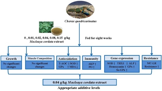
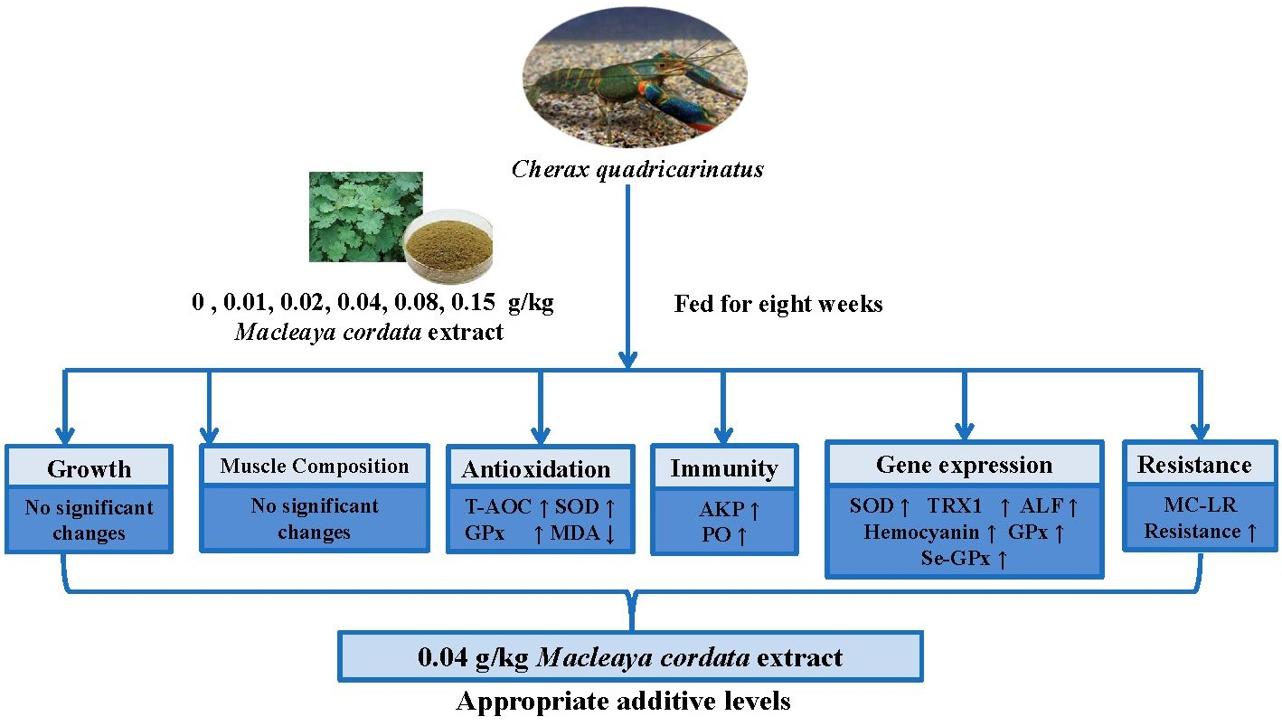
41], while research on feed additives that mitigate stress in crayfish remains limited. Therefore, this study aimed to evaluate the effects of MCE on the growth performance, muscle composition, antioxidant capacity, immunity, and stress resistance of juvenile red claw crayfish. The findings provide a theoretical basis for the development of plant-derived immunostimulants and the implementation of environmentally friendly strategies to mitigate environmental stress in intensive crayfish aquaculture.
2. Materials and Methods
2.1. Experimental Diets
The formulation and composition of the experimental diets are presented in
Table 1. The experiment consisted of six groups: a control group and five experimental groups supplemented with 0.01 (Diet 2), 0.02 (Diet 3), 0.04 (Diet 4), 0.08 (Diet 5), and 0.15 (Diet 6) g/kg MCE (Sanguinarine ≥ 1.50%, Chelerythrine ≥ 0.75%; Hunan Micolta Bioresource Inc., Changsha, China), respectively. All ingredients were finely ground and passed through an 80-mesh sieve, then mixed with oil until homogeneous. Distilled water was then added to each diet mixture, followed by thorough mixing and extrusion into pellets with a diameter of 1.5 mm using a pelletizer. The prepared diets were finally air-dried and stored at −20 °C until use.
2.2. Animal and Experimental Design
Juvenile crayfish were obtained from a commercial aquaculture farm in Wenchang, Hainan, China, and transported to the Wenchang Experimental Base of the Chinese Academy of Tropical Agricultural Sciences for the feeding trial. Before the experiment, crayfish were acclimated in a recirculating aquaculture system for two weeks and fed a basal diet. After 24 h of starvation, crayfish (initial body weight: 0.22 ± 0.02 g) were randomly assigned to rearing tanks (500 L) with three replicates per group, and 40 crayfish in each replicate. To reduce cannibalism, 40 PVC pipes were placed in each tank to serve as shelter. During the eight-week experimental period, feeding was conducted twice daily (08:00 and 18:00) to apparent satiation, and mortality was recorded. Throughout the experiment, the water temperature was maintained at 23–26 °C, the pH at 7.2–8.0, and the ammonia nitrogen below 0.05 mg/L.
2.3. Sample Collection
At the end of the feeding trial, following a 24 h fasting period, all the crayfish were collected from each tank, their numbers recorded, and their body weight and body length were measured for growth performance analysis. Hemolymph samples were collected from the base of the third pereiopod using a 2.5 mL syringe preloaded with an equal volume of anticoagulant solution (26.3 g/L NaCl, 18.0 g/L glucose, 6.3 g/L citric acid, 6.7 g/L sodium citrate, and 2.9 g/L EDTA). The hemolymph samples were centrifuged at 3000 rpm for 10 min at 4 °C, after which the supernatant was separated and stored at −80 °C for subsequent analyses. Hepatopancreas samples were rapidly dissected, immediately frozen in liquid nitrogen, and stored at −80 °C for further analysis. Dorsal muscle samples were also collected and stored at −20 °C for muscle nutritional composition determination.
2.4. Growth Performance
Growth performance indices were calculated using the following equations [
42]:
- (1)
Weight gain rate (WGR, %) = 100 × (Final body weight − Initial body weight)/Initial body weight;
- (2)
Specific growth rate (SGR, %·day−1) = 100 × (ln Final body weight − ln Initial body weight)/Number of rearing days;
- (3)
Survival rate (SR, %) = 100 × (Final number/Initial number).
2.5. Composition Analysis
The crude protein, crude lipid, moisture, and ash contents of the diets and muscle samples were determined according to standard methods of the AOAC (1995) [
43]. Moisture content was determined by drying samples in an oven at 105 °C until a constant weight was achieved. A semi-automatic Kjeldahl apparatus (Kjeltec™ 8400, Foss, Hillerød, Denmark) determined the crude protein content following acid digestion, while the crude lipid content was measured using a Soxhlet extraction system (SZF-06A, Hongji, Shanghai, China). Ash content was determined by incineration in a muffle furnace (SX2-2.5-10, Yiheng, Shanghai, China) at 550 °C for 12 h.
2.6. Antioxidant and Immune Index Assays
The total antioxidant capacity (T-AOC), superoxide dismutase (SOD), catalase (CAT), glutathione peroxidase (GPx), glutathione S-transferase (GST), acid phosphatase (ACP), and alkaline phosphatase (AKP), glutathione (GSH) and malondialdehyde (MDA) were determined using assay kits (Nanjing Jiancheng Bioengineering Institute, Nanjing, China) following to the manufacturer’s instructions. Finally, PO was measured using a double-antibody sandwich enzyme-linked immunosorbent assay (ELISA) kit (Dongguan Enzyme-linked Biotechnology Co., Ltd., Dongguan, China).
2.7. RNA Extraction and cDNA Synthesis
Hepatopancreas samples from three crayfish per tank were pooled for total RNA extraction using TRIzol reagent (Thermo Fisher Scientific, Waltham, MA, USA). The RNA concentration was measured with a NanoDrop One spectrophotometer (NanoDrop Technologies, Wilmington, DE, USA), and RNA integrity was evaluated by 1% agarose gel electrophoresis. Subsequently, the total RNA was reverse transcribed into complementary DNA (cDNA) using the PrimeScript RT reagent kit with gDNA Eraser (Takara, Kusatsu, Japan) according to the manufacturer’s instructions. The resulting cDNA was stored at −20 °C for quantitative real-time PCR (qRT-PCR) analysis.
2.8. Quantitative Real-Time PCR (qRT-PCR)
Based on the transcriptome data generated previously [
44,
45], specific primers (
Table 2) were designed using Primer Premier V5 and synthesized by BGI Biotech Co., Ltd. (Shenzhen, China). The qRT-PCR was performed using 2 × SYBR
® Green qPCR Mix (Takara, Japan) on a Stratagene Mx3005P real-time PCR system (Agilent Technologies, Santa Clara, CA, USA). The thermal cycling conditions were as follows: initial denaturation at 94 °C for 3 min, followed by 40 cycles of 94 °C for 15 s, 58 °C for 15 s, and 72 °C for 20 s. Relative gene expression levels were calculated using the 2
−ΔΔCt method [
46].
2.9. Microcystin-LR (MC-LR) Challenge
Following sample collection, 10 crayfish were randomly selected from each culture tank for the MC-LR stress challenge. The challenge dose (108 μg/kg BW) was selected based on previous data [
47]. All selected crayfish were injected intramuscularly with MC-LR and returned to their original tanks. The cumulative mortality was monitored and recorded over 48 h, and the survival rate of each dietary group was calculated.
2.10. Data Analysis
All data are expressed as mean ± standard deviation (SD). Statistical analyses were performed in SPSS 18.0 (SPSS Inc., Chicago, IL, USA). Significant differences among groups were determined by one-way analysis of variance (ANOVA) followed by Tukey’s test, and differences were considered statistically significant at p < 0.05.
4. Discussion
Numerous plant-derived extracts have been reported to enhance the growth performance of crustaceans, including
Forsythia suspensa extract in
Penaeus monodon [
48], banana peel extract in
Macrobrachium rosenbergii [
49], and
Psidium guajava leaf powder in
Penaeus monodon [
50]. Here, results demonstrated that dietary MCE supplementation did not significantly affect the growth performance of red claw crayfish, which contrasts with the findings of a previous study on fish [
51], but aligns with the results of Zhang et al., who showed that dietary supplementation with MCE did not significantly improve WGR and SGR in
Marsupenaeus japonicus [
52]. This may be due to the biological properties of the major active constituents of MCE, the isoquinoline alkaloids sanguinarine and chelerythrine. These possess antibacterial, detoxifying, and anti-inflammatory activities [
53,
54,
55], suggesting that the primary mechanism of MCE may be physiological regulation and health promotion, rather than growth stimulation.
In the current study, dietary MCE supplementation did not significantly affect muscle composition in crayfish. Haitham et al. also showed that dietary supplementation with Ashwagandha (
Withania somnifera) extracts did not significantly alter the moisture, crude protein, or lipid contents of
Litopenaeus vannamei [
56]. This suggests that dietary supplementation with appropriate levels of plant-derived bioactive compounds has limited effects on proximate body composition, while reported benefits are more likely associated with physiological regulation, antioxidant defense, and health maintenance rather than nutrient deposition. The application of MCE in animal production has primarily focused on its antibacterial and anti-inflammatory properties, and information on its effects on growth performance and body composition in aquatic animals remains limited. Therefore, additional research is needed to clarify the mechanisms by which MCE influences nutrient utilization, growth regulation, and body composition in aquatic species.
Antioxidant enzymes are important indicators of health and oxidative stress responses in aquatic animals [
57,
58]. Both SOD and CAT are key enzymes in the antioxidant defense system, while T-AOC reflects the overall antioxidant capacity, and MDA content serves as an indicator of lipid peroxidation and cellular damage [
59,
60]. Glutathione peroxidase (GPx) scavenges reactive oxygen species using glutathione (GSH) as a substrate, whereas glutathione S-transferase (GST) participates in the detoxification of harmful compounds [
61,
62]. Previous studies have shown that MCE can enhance the activities of antioxidant enzymes and reduce lipid peroxidation in farmed animals [
63,
64]. In the present study, dietary MCE supplementation increased SOD and GPx activities and T-AOC levels, while decreasing MDA content in both the hepatopancreas and plasma of
C. quadricarinatus. Similarly, dietary paprika extract significantly increased CAT and SOD activities in
Litopenaeus vannamei [
65], while dietary supplementation with
Scutellaria baicalensis polysaccharides enhanced the antioxidant capacity of
Macrobrachium rosenbergii, thereby improving the prawn’s resistance to oxidative stress [
66]. Also, Pavarist et al. (2019) reported that dietary MCE increased the SOD activities in
Litopenaeus vannamei [
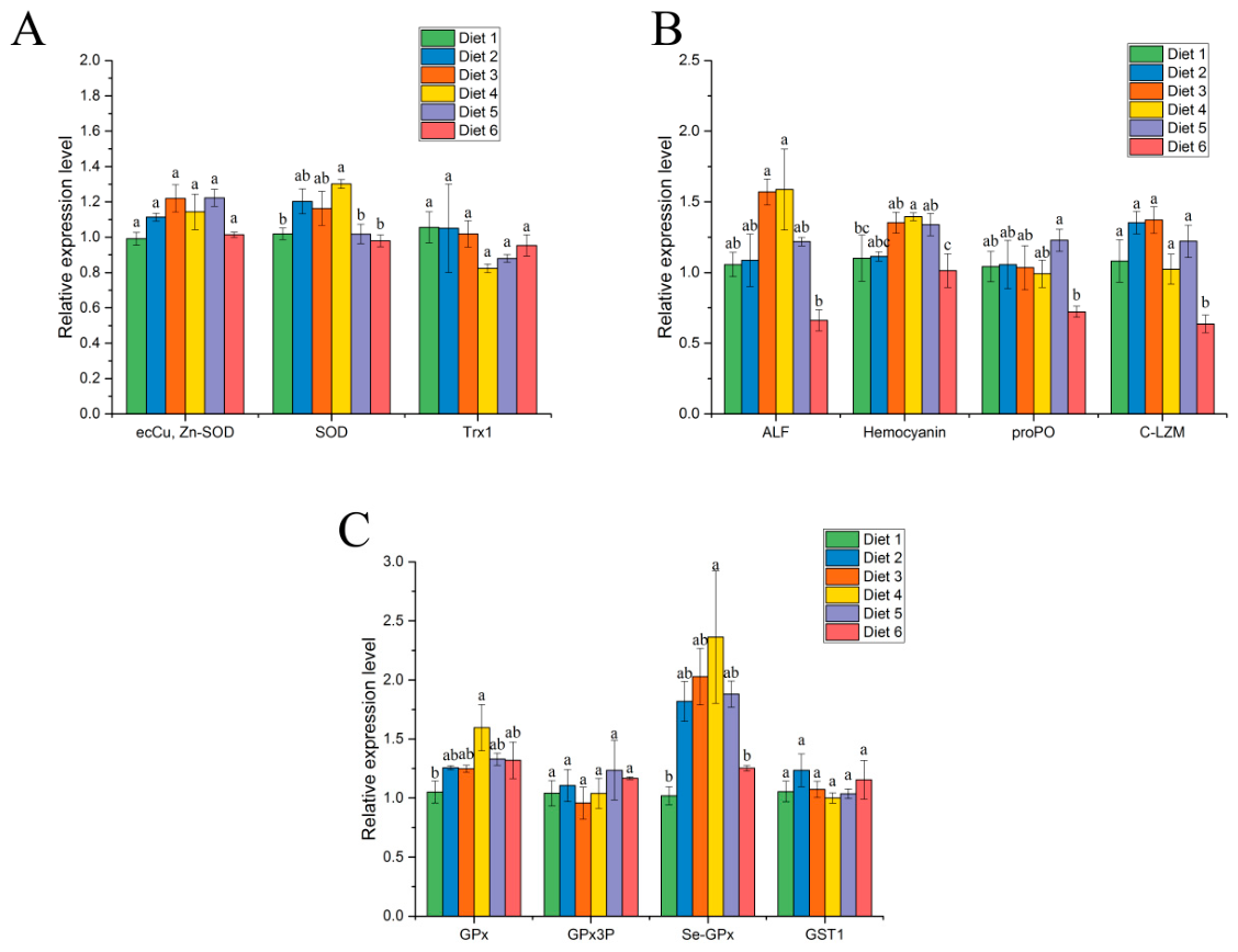
33]. In this study, dietary MCE significantly up-regulated SOD, GPx, and Se-GPx expression levels in the hepatopancreas, which was consistent with the enzyme activity results. Overall, these findings indicate that dietary MCE enhances antioxidant capacity and mitigates oxidative stress in
C. quadricarinatus.
In crustaceans, acid phosphatase (ACP), alkaline phosphatase (AKP), and phenoloxidase (PO) are widely recognized as important indicators of innate immune function [
67]. Specifically, AKP is involved in nutrient metabolism and disease resistance, whereas ACP functions as a lysosomal marker enzyme that participates in immune responses [
68,
69]. Phenoloxidase is a key component of the prophenoloxidase activation system and plays a central role in pathogen recognition and defense reactions in crustaceans [
70]. Previous studies have demonstrated that plant-derived bioactive compounds, including polysaccharides, flavonoids, and alkaloids, can enhance the immune response of aquatic animals by modulating immune-related enzyme activities. Dietary supplementation with
Mallotus japonicus bark extract enhanced the innate immune response of
Heterotilapia buttikoferi, as evidenced by increased lysozyme activity, serum bactericidal activity, phagocytic activity, respiratory burst activity, and serum protein levels [
71]. Puerarin supplementation also significantly increased ACP and AKP activities in
Litopenaeus vannamei under ammonia stress [
72]. In the present study, dietary MCE supplementation increased AKP activity and PO levels in both the hepatopancreas and plasma of
C. quadricarinatus, suggesting an enhanced innate immune response. This is supported by a previous study, which showed that MCE can enhance immune responses in
Litopenaeus vannamei [
33].
Immune factors play an important role in the non-specific immune response of crustaceans [
73,
74]. Antimicrobial peptides, including ALF, LZM, and hemocyanin, serve as key immune effectors in crustaceans, while the proPO-activating system is critical for immune recognition in invertebrates. Non-self molecules can activate the proPO cascade, releasing proPO, which is subsequently converted into active PO by serine proteases [
75,
76,
77]. The hepatopancreas is a multifunctional organ that plays a central role in both metabolism and immunity, and its antioxidant status and stress response are closely linked to overall host immune function [
78]. In the present study, MCE significantly induced the expression of ALF and hemocyanin in the hepatopancreas of crayfish. It is known that MCE can influence immune function by regulating immune factors such as TNF, NF-κB, and MyD88 [
79], where MCE reduced the expression of cryptdin-4 and cryptdin-5 in mice [
64]. Similar plant-derived immunoregulatory effects have been reported in crustaceans; for example, dietary supplementation with
Echinacea purpurea enhanced antioxidant capacity, lysozyme activity, hemocyte parameters, and immune-related gene expression in
Litopenaeus vannamei [
80]. Likewise, mint (
Mentha piperita L.) leaf extract increased lysozyme and superoxide dismutase activities in
Penaeus semisulcatus, suggesting modulation of innate immune responses [
81]. These findings suggest that plant-derived bioactive compounds can broadly influence crustacean immune function, although the regulatory pathways and response patterns may vary specifically.
Cyanobacterial blooms have become a major environmental concern in aquaculture, as the MC-LR they produce can induce oxidative stress, immunotoxicity, tissue damage, and mortality in aquatic organisms [
82,
83,
84]. In the present study, the effect of dietary supplementation with MCE on the survival of crayfish under MC-LR challenge was evaluated. Previous studies have confirmed that plant-derived extracts can enhance immune defenses and improve the survival of aquatic animals under pathogenic or environmental stress [
85,
86]. For instance, dietary supplementation with 1 g/kg dandelion extract increased the survival of golden pompano (
Trachinotus ovatus) following a
Vibrio harveyi challenge [
87], whereas
Forsythia suspensa extract (0.01–0.02%) enhanced the resistance to and improved survival of black tiger shrimp (
Penaeus monodon) following
Vibrio parahaemolyticus infection [
48]. Similarly, Wongsasak et al. reported that probiotics improved the survival of Pacific white shrimp (
Litopenaeus vannamei) under ammonia-nitrogen stress [
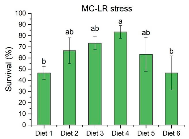
88]. Here, dietary supplementation with 0.04 g/kg MCE significantly increased the survival rate of crayfish exposed to MC-LR stress, suggesting that MCE can enhance crayfish tolerance to MC-LR by boosting their antioxidant capacity and immune function. Although the precise mechanisms underlying the protective effects of MCE in crayfish remain to be elucidated, its antioxidant and immunostimulatory properties likely contribute to the improved resilience under environmental stress. Further studies are warranted to clarify the molecular and physiological pathways through which MCE mitigates MC-LR toxicity in crustaceans.