Article

Feeding Morphology Supports Carnivorous Habits in Algansea lacustris: A Multitrait Approach

by
Citlali Wendolin Rodriguez-Paramo
1,
María Cristina Chávez-Sánchez
2,
Pamela Navarrete-Ramírez
3,*,
Carlos Antonio Martínez-Palacios
1,
Andrea Gutiérrez-Contreras
1 and
Carlos Cristian Martínez-Chávez
1,*
1
Instituto de Investigaciones Agropecuarias y Forestales, Universidad Michoacana de San Nicolás de Hidalgo, San Juanito Itzícuaro, Morelia C.P. 58337, Michoacán, Mexico
2
Centro de Investigación en Alimentación y Desarrollo, A.C. Subsede Mazatlán, Mazatlán C.P. 82112, Sinaloa, Mexico
3
SECIHTI-Instituto de Investigaciones Agropecuarias y Forestales, Universidad Michoacana de San Nicolás de Hidalgo, San Juanito Itzícuaro, Morelia C.P. 58337, Michoacán, Mexico
*
Authors to whom correspondence should be addressed.
Fishes 2026, 11(3), 167; https://doi.org/10.3390/fishes11030167
Submission received: 16 February 2026 / Revised: 11 March 2026 / Accepted: 12 March 2026 / Published: 14 March 2026
(This article belongs to the Special Issue Trophic Ecology of Freshwater and Marine Fish Species)

Abstract

Accurate classification of fish trophic strategies based solely on gut contents can be misleading, especially when plant material is ingested incidentally during predatory benthic foraging. The Pátzcuaro chub (Algansea lacustris) is a critically endangered cyprinid endemic to Central Mexico. It has historically been described as omnivorous with a tendency toward algivory, despite limited anatomical evidence. In this study, integrated anatomical, morphometric, and functional approaches were used to reassess the feeding strategy of A. lacustris and inform conservation-oriented aquaculture. Double-staining techniques revealed a specialised filtering and crushing branchial–pharyngeal system adapted to capture and process animal prey. Relative intestinal length (RIL) was measured from freshly dissected intestines. Intestinal transit time was experimentally evaluated using a formulated diet and live Artemia. Algansea lacustris exhibited a short intestine (RIL = 0.86 ± 0.10) and rapid intestinal transit (<30 min), both of which are characteristics of carnivorous teleosts. These results provide consistent anatomical and physiological evidence that A. lacustris is primarily adapted to a low-trophic carnivorous or insectivorous feeding strategy, with important implications for its ecological characterisation. Moreover, intestinal transit was faster after ingestion of live Artemia than after the formulated diet, likely due to differences in moisture content. The observed short transit times indicate the need for more frequent feeding and support the refinement of diet formulation and feeding strategies in conservation aquaculture programmes.
Key Contribution: By applying a multitrait anatomical and physiological framework rather than relying solely on gut content analysis, this study demonstrates that Algansea lacustris is functionally adapted to a low-trophic, carnivorous or insectivorous feeding strategy, reshaping its trophic classification and guiding conservation-oriented aquaculture practices.

1. Introduction

Classifying fish as herbivorous, carnivorous, or omnivorous solely based on stomach or intestinal contents can be misleading. Gut content analyses are often biased toward less digestible components or materials incidentally ingested with primary prey, obscuring actual diet selection [1,2]. This limitation complicates the accurate identification of natural feeding habits. In teleost fishes, agastria is not a reliable proxy for defining herbivory or carnivory, as stomachless lineages occur across multiple trophic strategies [3,4]. Dietary specialisation is better inferred from the combined evaluation of feeding structures and intestinal traits, because herbivorous fish generally have longer intestines than omnivorous and carnivorous taxa, likely reflecting longer retention times and a greater dependence on microbial processing of plant-rich diets [5]. This interpretation is consistent with evidence that endogenous cellulase activity in teleosts remains uncertain, whereas cellulolytic intestinal bacteria and hindgut fermentation with short-chain fatty acid production have been documented in herbivorous and omnivorous fish [6,7,8]. These issues highlight the need for more robust indicators of trophic strategy, such as anatomical traits. Relative intestinal length (RIL), mouth morphology, mandibular dentition, and the structure of the pharyngeal teeth, in particular, provide consistent evidence of species-specific feeding adaptations [2,9,10,11]. Integrative studies combining dietary and morphological approaches remain scarce, despite their relevance to aquaculture management. The Pátzcuaro chub (Algansea lacustris) was originally classified as a benthic omnivore based solely on gut content observations, namely the presence of filamentous algae and similar proportions of different benthic microcrustaceans [12]. However, this classification contrasts with the species’ relatively simple digestive configuration, particularly its short intestine. In cyprinids, relative gut length is more consistently associated with trophic tendency than agastria itself, with shorter guts generally occurring in species with more carnivorous diets and longer, more complex intestines in species with herbivorous tendencies [9,13]. A comparable digestive configuration is observed in the Mexican pike silverside (Chirostoma estor), which cohabits Lake Pátzcuaro. Despite sharing similar anatomical traits, C. estor has been characterised as a low-trophic-level carnivore due to its preference for zooplankton and its high protein requirements [2,14].
Algansea lacustris is currently classified as critically endangered due to habitat degradation, invasive species, and overfishing [15], and practical information regarding its culture remains scarce. In light of these threats, current aquaculture conservation efforts emphasise the study of its basic biology and nutritional requirements as a foundation for conservation aquaculture programmes. Accurately identifying the dietary habits of A. lacustris is therefore essential to understand its biology and to define appropriate feed formulations and feeding strategies for this species aimed at sustaining viable wild and captive populations.
The objective of this study was to characterise key anatomical and functional feeding traits in A. lacustris, including relative intestinal length and pharyngeal teeth morphology, to clarify its trophic strategy. Additionally, intestinal transit time was evaluated using both live and formulated feeds to assess practical differences in digestive passage associated with varying moisture contents and establish a baseline for evidence-based feeding protocols for this species in captivity.

2. Materials and Methods

2.1. Biological Material and Ethical Procedures

This study used Algansea lacustris F2 specimens reared in the Laboratorio de Biotecnología Acuícola at the Universidad Michoacana de San Nicolás de Hidalgo (IIAF-UMSNH). In all cases, fish were euthanised by an overdose exposure to a clove oil–water solution (doTERRA, Pleasant Grove, UT, USA), in accordance with the ethical standards established for the handling of aquatic organisms [16,17,18].

2.2. Double-Staining Procedure

To visualise the arrangement of cartilage and mineralised structures (bone and teeth), including pharyngeal teeth (specialised teeth located in the throat region) and the masticatory plate (the part used for grinding food), juvenile specimens (n = 8; 1.01 ± 0.20 g; 3.9 ± 0.26 cm total length) were processed using a double-staining protocol adapted from Darias et al. (2010) [19], which stains cartilage blue and mineralised tissues red. The total length of each specimen was recorded to set immersion times for each step (Table 1).
Juvenile specimens were rinsed in distilled water for 15 min. They were then fixed for 24 h in 4% buffered formalin (JT Baker, Phillipsburg, NJ, USA; 0.1 M phosphate buffer, pH 7) at 4 °C. After fixation, the specimens were washed twice in distilled water (5 min each). Next, specimens were placed individually in perforated 8 cm plastic containers for simultaneous processing in a single staining chamber.
Cartilage was stained in an alcian blue solution (100 mg/L alcian blue [Meyer, Edo. Mex., Mexico], 800 mL/L 95% ethanol, and 200 mL/L acetic acid), with incubation times based on fish size (Table 1).
Residual acid from cartilage staining was neutralised by immersion for 3 min in 100% ethanol with 1% KOH (JT Baker, Phillipsburg, NJ, USA). Specimens were then rehydrated through a descending ethanol series (95%, 70%, 40%, 15%). Each step used two 15 min immersions, followed by three 5 min rinses in distilled water.
Bleaching was carried out at 30 °C in a solution of 1 volume 3% H2O2 (Fermont, Monterrey, NL, Mexico) and 9 volumes 1% KOH. Exposure times were adjusted by fish length (Table 1). Specimens were then rinsed in running water. Next, they were transferred to a clearing solution (7 parts distilled water, 3 parts sodium borate, JT Baker, Phillipsburg, N.J, USA, and 2 g trypsin, JT Baker, Phillipsburg, NJ, USA) for 20 h at 30 °C.
Bone was stained in an alizarin red solution (a dye that stains bone red; 5 g/L alizarin red [MEYER, Mexico City, Edo. Mex., Mexico] in 1% KOH), again with an incubation time determined by length (Table 1). After staining, specimens were rinsed in distilled water, excess dye was removed in 1% KOH (a solution commonly used to clear tissue), and samples were transferred through glycerol–KOH solutions (mixtures of glycerol and potassium hydroxide used to clear and preserve tissues: 40% glycerol/60% 1% KOH for 2 h; then, 70% glycerol/30% 1% KOH for 6 h). Cleared and stained specimens were stored in 100% glycerol for long-term preservation.

2.3. Gill Raker and Pharyngeal Teeth Analysis

Gill arches and pharyngeal teeth were dissected from each specimen (n = 5; 37.9 ± 5.4 g; 13.19 ± 7.2 cm) and processed using a two-stage cleaning protocol. First, the structures were submerged in Tween 80® (Sigma-Aldrich) to soften muscle tissue, facilitating removal with fine forceps. They were then transferred to a sodium hydroxide solution (NaOH; 1.1% p/v) for 25 min at room temperature. During this time, the remaining soft tissue was gently brushed away while preserving mineralised components, including the branchial arches, gill rakers, and pharyngeal teeth. After digestion was complete, all structures were thoroughly rinsed with distilled water to eliminate sodium hydroxide residues.
Initial morphological characterisation was performed using a stereomicroscope (Stemi DV4, Axioscope, Carl Zeiss, Göttingen, Germany). Cleaned arches and pharyngeal elements were then mounted on a polystyrene base and air-dried at room temperature. Once dried, samples were coated with copper ions using a sputter coater (S150A). They were then mounted on metallic stubs and examined under a scanning electron microscope (JEOL JMS-7600F, Akishima, Tokyo, Japan).

2.4. Relative Intestinal Length (RIL) Determination

Standard length (SL) was defined as the distance from the tip of the snout to the posterior end of the last vertebra and measured for each adult fish (n = 7; 35.4 ± 6.22 g; 12.8 ± 0.9 cm) using digital callipers. Next, the entire intestine was carefully excised by transecting just posterior to the oesophagus and at the anus and was immediately immersed in undiluted Hartmann’s solution (PiSA™; lactated Ringer) to preserve tissue integrity. Excess fat and associated mesenteric tissue were then removed to allow the intestine to be fully uncoiled. Total intestinal length (TL) was subsequently measured under gentle, manual tension (without mechanical stretching) to minimise distortion caused by post-mortem contraction.
Finally, RIL was calculated as follows:
R I L = T L / S L

2.5. Intestinal Transit Evaluation in Algansea lacustris

Intestinal transit time was evaluated in juvenile A. lacustris (n = 6; 1.78 ± 0.58 g and 4.5 ± 0.57 cm total length; approximately 4 months of age) fed either a standard formulated diet (45% protein, 5% lipids; 0.8 mm particle size) or live adult Artemia. Fish were maintained in two 6-gallon (22.7 L) aquaria supplied with pre-filtered water. During a 5-day acclimation period, juveniles were fed to apparent satiation with a dyed version (2%; black, plant-based dye) of the standard formulated diet, offered daily between 09:00 and 16:00 h. Water temperature was recorded daily with an alcohol thermometer and remained between 20 and 22 °C throughout the 20-day study. Both aquaria were kept under low-light conditions to facilitate adaptation and reduce stress.
After acclimation, once normal feeding behaviour was observed, a single juvenile was transferred to a third (observation) aquarium maintained under identical conditions to measure individual feeding and excretion times. Following a 19 h fasting period (15:00–10:00 h), each fish was offered the standard formulated diet at 10:00 h, allowing subsequent faeces to be identified. Using a stopwatch, we recorded: (1) the time of food ingestion and (2) the time of first faecal expulsion and used these values to estimate intestinal transit time. Six independent observations were performed, each with a different juvenile.
After completing the six observations with the balanced diet, the experimental feeding period was concluded, and the fish were subsequently offered live Artemia. Before restarting the sampling protocol, juveniles were allowed a one-day acclimation period to the live Artemia diet. Thereafter, all remaining Artemia were removed, and the fish were fasted for 19 h before the protocol was initiated again. The same procedure was then repeated: each day, a single juvenile was transferred to the observation aquarium and monitored for ingestion and faecal output. After confirming normality (Shapiro–Wilk test; p > 0.05) and homogeneity of variances (F-test), differences between treatments were evaluated using Student’s t-test in Prism software (version 10).

3. Results

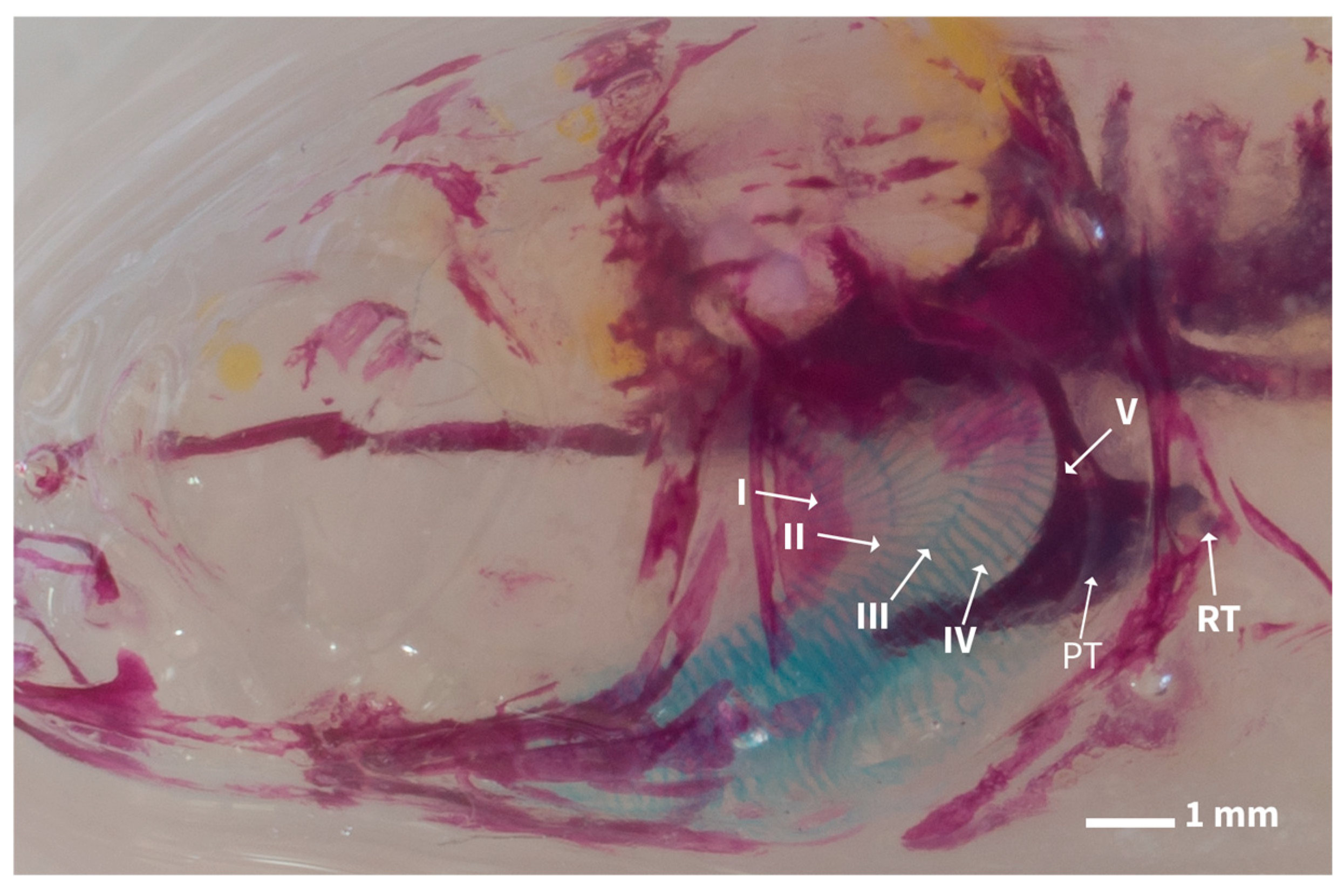
The double-staining technique allowed the natural arrangement of the feeding apparatus to be described. In the head region, the first to fourth branchial arches, including their lamellae and spines, were distinctly visible in blue (Figure 1). Immediately posterior to the fifth ceratobranchial, the pharyngeal teeth were observed, with replacement teeth (RT) located distally on the third pharyngeal tooth row.
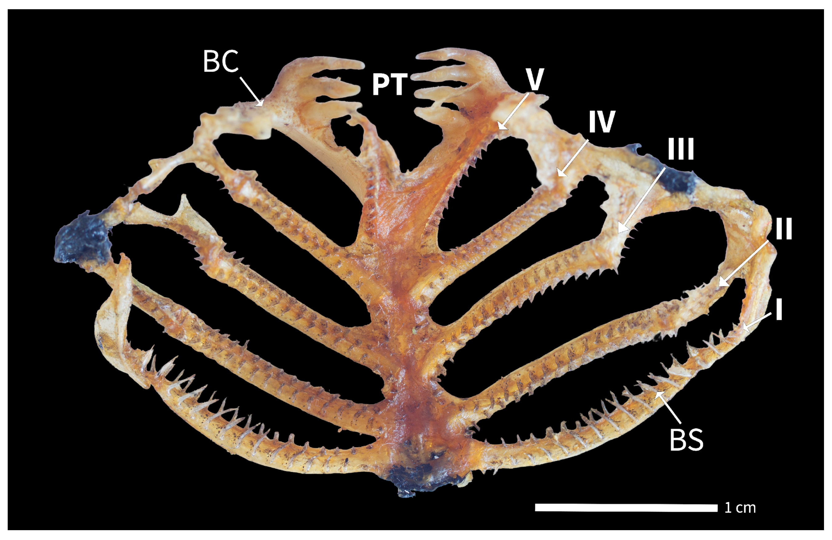
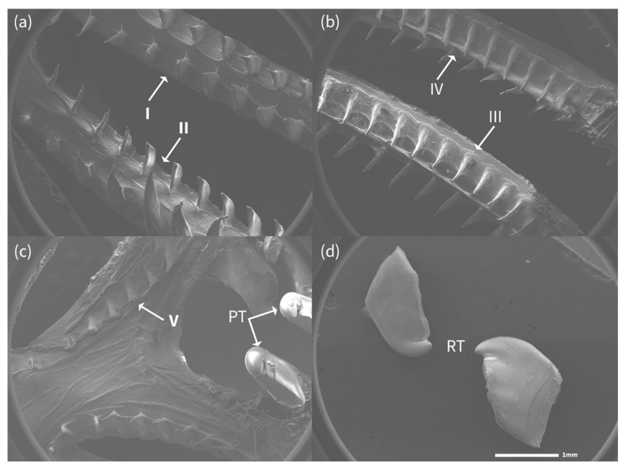
The gill arches of A. lacustris bear short, flattened, triangular spines with a pointed apex. These spines are arranged in two alternating rows along the entire length of each gill arch (Figure 2).
Spines on the first arch are larger and more conspicuous (Figure 3a), and their size gradually decreases on subsequent arches, reaching approximately one-third of the size of those on the first arch by the fifth ceratobranchial.
In the first, second, third, and fourth arches (Figure 3a–c), the alternating arrangement of spines closes the spaces between them, forming a horizontal sieve basket when the arches are opposed, capturing small insects and crustaceans from the benthos. In the fifth ceratobranchial, the arch is modified into an elongated basal arm that supports four long grinding teeth oriented toward the arch axis. Along the main arm of this pharyngeal structure runs a single row of flattened spines, which forms the distal portion of the sieve (Figure 3c).
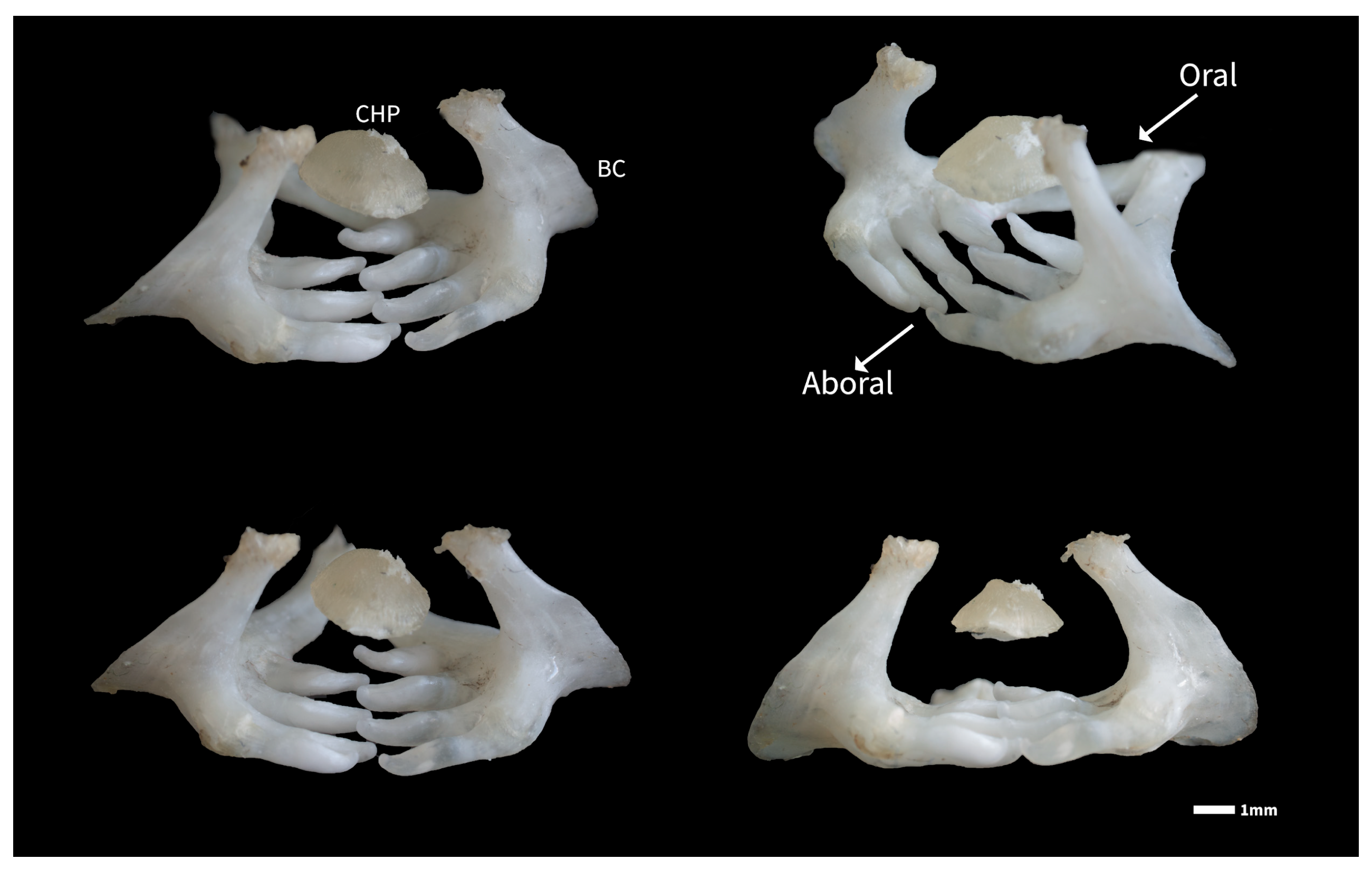
The fifth ceratobranchial is robust and highly calcified. Distally, it bears four strong grinding teeth; medially, it forms a flat, smooth chewing pad; and on the opposite side, it presents a prominent bony crest which is interconnected with the fourth arch (Figure 4).
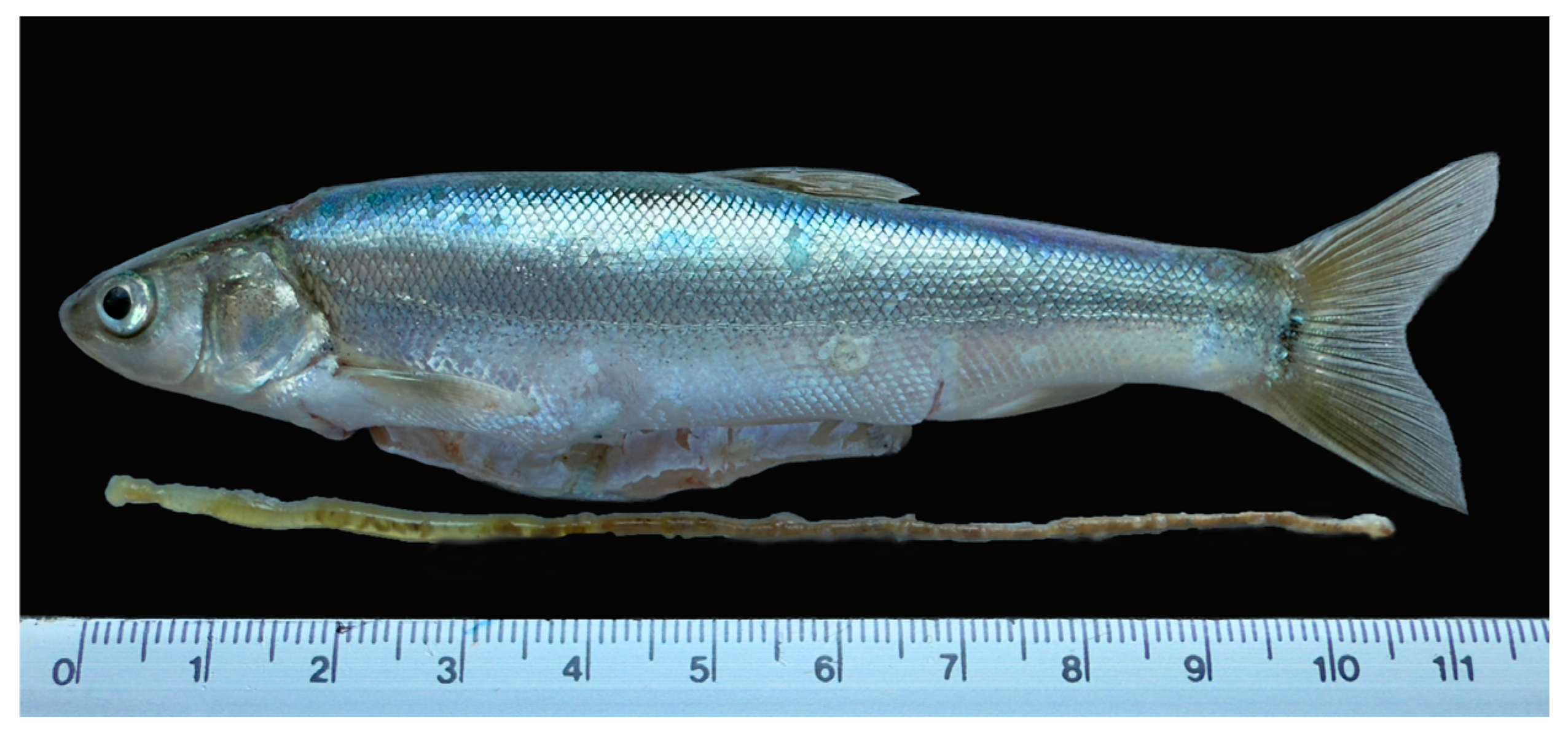
The mean RIL of adult (127.5 ± 9.8 SL) A. lacustris was 0.86 ± 0.10 (mean ± SD; Figure 5).
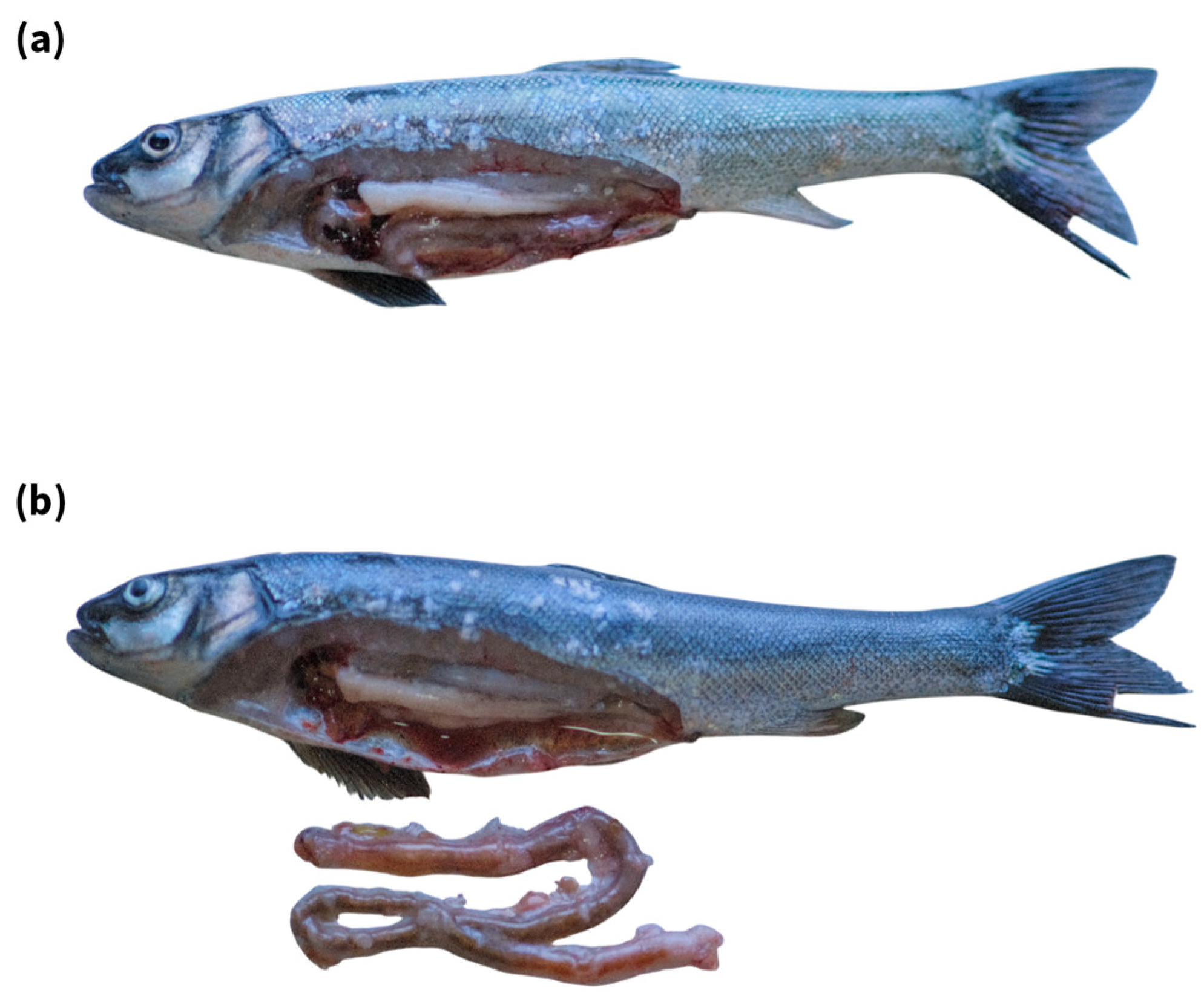
The intestine is located in the abdominal cavity, attached to mesenteric tissue and perivisceral fat, and forms a simple two-loop configuration between the oesophagus and rectum (Figure 6a,b).
Regarding intestinal transit, detailed observations showed that a faecal plug from previously ingested material was expelled in both treatments approximately 20 min after feeding, with no significant difference between diets; this event was, therefore, not included in the analysis. The onset of excretion of the recently consumed diet (balanced diet or Artemia) was identified by the colour of the faecal material. Time to first excretion after feeding the balanced diet was 29.17 ± 2.71 min (mean ± SD), which was significantly (p = 0.0015; T= 4.341; df = 10) longer than after feeding live Artemia (23.67 ± 1.51 min).

4. Discussion

Previous observations showed that Algansea lacustris exhibits an agastric condition and a relatively short intestine throughout its ontogeny. This configuration makes it a particularly useful model for studying digestive function from a structural perspective. The absence of a true stomach and the reduced intestinal length simplify the gross anatomical organisation of the digestive tract, allowing clearer functional interpretations. As documented in other teleosts, particularly cyprinids [9], such anatomical traits are closely associated with trophic ecology. Therefore, understanding this morphological arrangement provides an essential framework for interpreting the feeding habits of A. lacustris, which constitutes the central focus of the present discussion.
Double-staining observations of the branchial and pharyngeal apparatus of A. lacustris provide clear anatomical evidence of trophic specialisation. The first to fourth branchial arches form a sieve structure, which is consistent with the filtering and sorting function of captured prey, facilitating the retention of food items while protecting the respiratory lamella surfaces. This dual role of branchial structures in both respiration and feeding has been widely documented in teleost fishes [20,21,22,23]. The morphology, spacing, and organisation of gill elements are known to be strongly influenced by trophic habits [24,25]. In A. lacustris, the presence of tightly interlocking spines suggests an adaptation for capturing and retaining small, mobile prey rather than for the bulk ingestion of plant material.
The fifth ceratobranchial of A. lacustris is particularly informative. It is robust and highly calcified, with an elongated basal arm supporting four strong grinding teeth and a flat chewing pad, and a prominent bony crest on the opposing surface interconnected to the fourth arch. This configuration is characteristic of cyprinid species adapted to process hard or resistant prey, such as molluscs, benthic invertebrates, or heavily chitinized crustacean and insect larvae [23,25,26]. In contrast to the broad, molariform, or extensively flattened pharyngeal elements typically associated with herbivory or detritivory, A. lacustris exhibits a compact and powerful crushing apparatus. Together with the distal row of flattened spines forming the terminal portion of the branchial sieve, these features indicate a highly integrated system for prey capture, sorting, and mechanical processing of animal food items.
These anatomical findings contrast with earlier classifications of A. lacustris as an omnivorous species with a strong tendency toward algivory; these results were based on a single analysis of the gut contents [12]. Notably, the same author reported a relative intestinal length (RIL) of 2.9 despite examining intestines several hours post-mortem from fishermen’s captures, as inferred from the methodological description [12]. This methodological limitation makes the original study difficult to reproduce and relevant because post-mortem relaxation of soft tissues and repeated handling can increase intestinal pliability, making the gut easier to straighten and inadvertently stretch during measurement, thereby artificially inflating gut length and RIL values, leading to biased trophic interpretations [27,28].
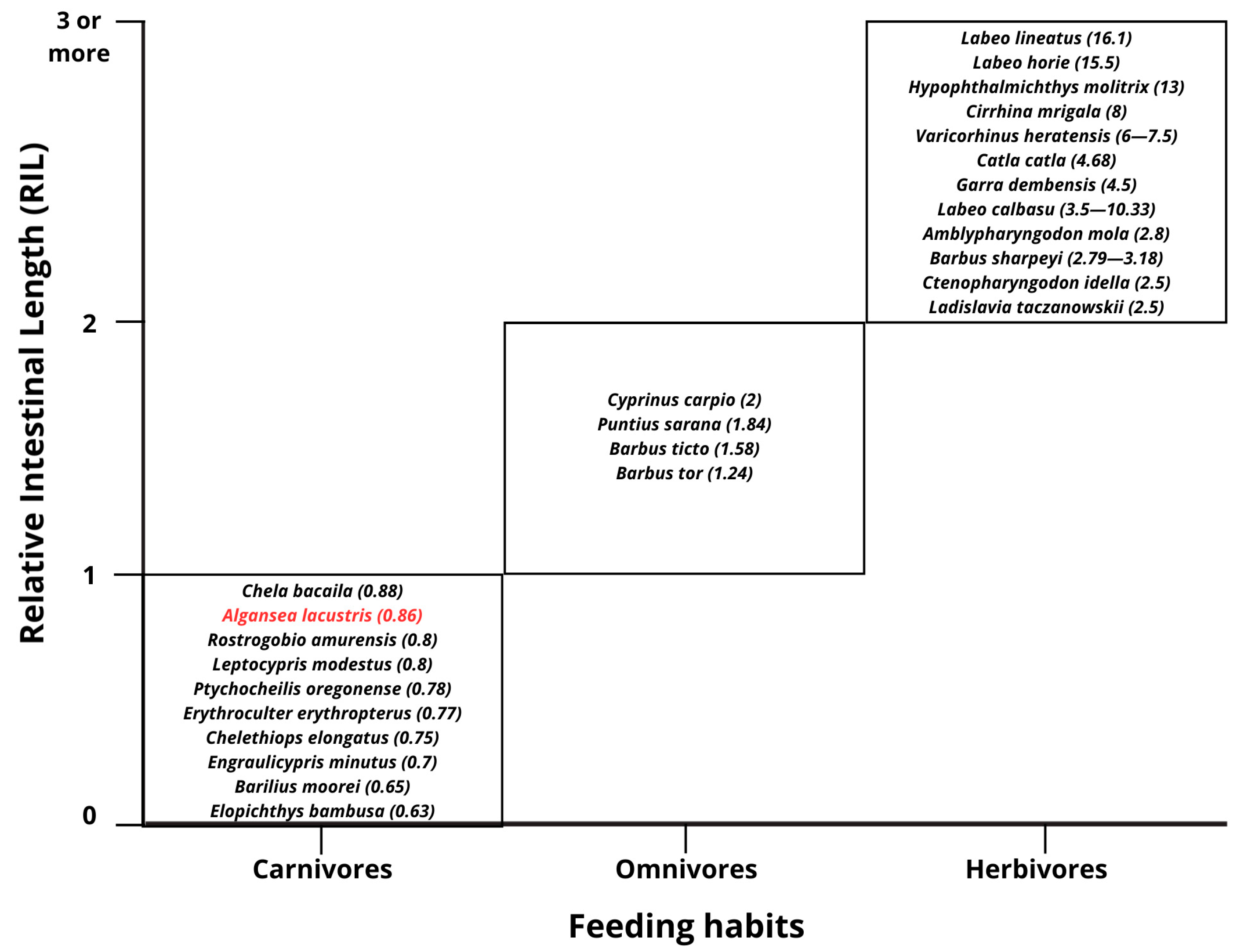
In teleost fishes, relative intestine length (RIL) values below 1 are generally associated with comparatively short digestive tracts and feeding strategies centred on animal prey (e.g., zooplanktivory, molluscivory, and insectivory), a relationship well documented in cyprinids [5,9,29]. In our study, A. lacustris exhibited a short intestine, with a mean RIL of 0.86 ± 0.10, consistent with an invertivorous (animal-prey-oriented) trophic tendency (Figure 7). By contrast, efficient digestion of algae and other plant-derived items, which are often structurally resistant and nutritionally poor, typically involves greater digestive capacity, commonly expressed as a relatively longer intestine (higher RIL) and, frequently, slower passage or increased retention to maximise digestive surface area and processing time [5,10,30,31,32]. Neither pattern is observed in A. lacustris, suggesting that, when detected, filamentous algae are more likely the result of incidental ingestion during benthic prey capture in the wild rather than evidence of targeted herbivory. However, more robust insight and independent confirmation would benefit from future comparative gut microanatomical studies in A. lacustris and other species with similar digestion configurations, as overlapping trophic guilds could exist [5].
Short intestinal transit times (<30 min) observed in A. lacustris further support the relationship between digestive tract morphology, physiological processing rates, and trophic strategy in teleost fishes. Carnivorous and invertivorous species generally exhibit shorter digestive tracts and faster gut passage times than herbivorous or omnivorous taxa, reflecting adaptations for the efficient digestion of animal prey [5,29]. Classical studies have emphasised that gut length and transit time provide functional insight into feeding habits and digestive strategies across teleosts [9,33].
The time to first excretion was significantly longer after ingestion of the balanced formulated diet (29.17 ± 2.71 min) than after feeding live Artemia (23.67 ± 1.51 min), indicating the faster processing of live invertebrate prey. This difference likely reflects variation in physical structure and digestibility between natural prey and the specific diets [34,35]. From an ecological perspective, the rapid processing of live invertebrates supports the view that A. lacustris is well adapted to exploit zooplankton and benthic microinvertebrates. From an applied perspective, these results highlight the importance of tailoring formulated feeds, including protein sources and levels, particle size, and physical characteristics, to better match the digestive capacities of this species under captive conditions. It also implies that almost constant feeding might be appropriate for optimised growth of this species, as is the case for the Mexican pike silverside (Chirostoma estor), another short-intestine agastric teleost [36].

5. Conclusions

In this study, three complementary trophic indicators were used to assess the feeding preferences of A. lacustris under culture conditions. Together, branchial–pharyngeal morphology, relative intestinal length, and intestinal transit time indicate that this species is primarily adapted to exploit small, mobile invertebrate prey. The observed feed transit time also supports its relevance for feeding management in captivity. These findings should, however, be interpreted in light of the indirect trophic proxies obtained under culture conditions and may not fully reflect feeding patterns in the wild or across all developmental stages. Future studies should incorporate controlled feeding trials, field-based dietary analyses, and complementary physiological approaches, including digestive enzyme profiling, gut histology, and microbiome characterisation, to refine the trophic characterisation of this species and support its application in conservation-oriented culture programmes.

Author Contributions

Conceptualization, C.C.M.-C. and C.A.M.-P.; methodology, C.W.R.-P. and A.G.-C.; validation, C.A.M.-P. and M.C.C.-S.; formal analysis, C.C.M.-C. and P.N.-R.; investigation, C.W.R.-P., A.G.-C. and C.C.M.-C.; resources, C.C.M.-C. and M.C.C.-S.; data curation, C.W.R.-P., C.C.M.-C. and P.N.-R.; writing—original draft preparation, C.C.M.-C.; writing—review and editing, C.W.R.-P., M.C.C.-S., P.N.-R., C.A.M.-P., A.G.-C. and C.C.M.-C.; visualisation, C.C.M.-C. and C.A.M.-P.; supervision, C.C.M.-C.; project administration, C.C.M.-C. and P.N.-R.; funding acquisition, C.C.M.-C. All authors have read and agreed to the published version of the manuscript.

Funding

This study was funded by project SECIHTI CBF-2025-G-317.

Institutional Review Board Statement

All experimental procedures involving fish were conducted in accordance with internationally accepted guidelines for the care and use of aquatic organisms. The study was reviewed and approved on 4 April 2025 by the Research, Ethics, and Biosafety Committee of the Universidad Michoacana de San Nicolás de Hidalgo (UMSNH) under approval letter No. 101/2025.

Data Availability Statement

The original contributions presented in this study are included in the article. Further inquiries can be directed to the corresponding authors.

Acknowledgments

The authors would like to thank Ma. Gisela Ríos-Durán for her valuable support in acquiring and processing the photographs used in this study, as well as for her technical assistance, which improved the quality of the visual material. The authors also thank Sibila Concha-Santos, Lucía Leal Cortés and Jesús López for their technical assistance during the development of this work.

Conflicts of Interest

The authors declare no conflicts of interest. The funders had no role in the design of the study, in the collection, analysis, or interpretation of data, in the writing of the manuscript, or in the decision to publish the results.

Abbreviations

The following abbreviations are used in this manuscript:
RILRelative Intestinal Length
SLStandard Length
TLTotal Intestinal Length
RTReplacement teeth

References

  1. Saikia, S.K. Food and feeding of fishes. What do we need to know? Transylv. Rev. Syst. Ecol. Res. 2015, 17, 71. [Google Scholar] [CrossRef]
  2. Ross, L.G.; Martínez-Palacios, C.A.; Aguilar Valdez, M.C.; Beveridge, M.C.M.; Chavez Sanchez, M.C. Determination of feeding mode in fishes: The importance of using structural and functional feeding studies in conjunction with gut analysis in a selective zooplanktivore Chirostoma estor estor Jordan 1880. J. Fish Biol. 2006, 68, 1782–1794. [Google Scholar] [CrossRef]
  3. Castro, L.F.C.; Gonçalves, O.; Mazan, S.; Tay, B.-H.; Venkatesh, B.; Wilson, J.M. Recurrent gene loss correlates with the evolution of stomach phenotypes in gnathostome history. Proc. Biol. Sci. 2014, 281, 20132669. [Google Scholar] [CrossRef] [PubMed]
  4. Day, R.D.; German, D.P.; Manjakasy, J.M.; Farr, I.; Hansen, M.J.; Tibbetts, I.R. Enzymatic digestion in stomachless fishes: How a simple gut accommodates both herbivory and carnivory. J. Comp. Physiol. B 2011, 181, 603–613. [Google Scholar] [CrossRef] [PubMed]
  5. Duque-Correa, M.J.; Clements, K.D.; Meloro, C.; Ronco, F.; Boila, A.; Indermaur, A.; Salzburger, W.; Clauss, M. Diet and habitat as determinants of intestine length in fishes. Rev. Fish Biol. Fish. 2024, 34, 1017–1034. [Google Scholar] [CrossRef]
  6. Krogdahl, Å.; Hemre, G.I.; Mommsen, T.P. Carbohydrates in fish nutrition: Digestion and absorption in postlarval stages. Aquac. Nutr. 2005, 11, 103–122. [Google Scholar] [CrossRef]
  7. Mountfort, D.O.; Campbell, J.; Clements, K.D. Hindgut fermentation in three species of marine herbivorous fish. Appl. Environ. Microbiol. 2002, 68, 1374–1380. [Google Scholar] [CrossRef]
  8. Tanaka, D.; Ohnishi, K.-i.; Watanabe, S.; Suzuki, S. Isolation of cellulase-producing Microbulbifer sp. from marine teleost blackfish (Girella melanichthys) intestine and the enzyme characterization. J. Gen. Appl. Microbiol. 2021, 67, 47–53. [Google Scholar] [CrossRef]
  9. Kapoor, B.; Smit, H.; Verighina, I. The alimentary canal and digestion in teleosts. In Advances in Marine Biology, 1st ed.; Academic Press: San Diego, CA, USA, 1976; pp. 109–239. [Google Scholar]
  10. Martínez-Palacios, C.A.; Ross, L.G. The feeding ecology of the Central American cichlid Cichlasoma urophthalmus (Gunther). J. Fish Biol. 1988, 33, 665–670. [Google Scholar] [CrossRef]
  11. Martínez-Palacios, C.A.; Aguilar-Valdez, M.C.; Strüssmann, C.A.; Ríos-Durán, M.G.; Toledo-Cuevas, E.M.; Navarrete-Ramírez, P.; Fonseca-Madrigal, J.; Martínez-Chávez, C.C.; Ross, L.G. The orobranchial structures in four neotropical silversides (Teleostei: Atherinopsidae) related with feeding habits. Zoomorphology 2019, 138, 511–523. [Google Scholar] [CrossRef]
  12. Rosas-Moreno, M. Datos Biológicos de la Ictiofauna del Lago de Pátzcuaro con Especial Énfasis en la Alimentación de sus Especies. In Memorias del Simposio en Pesquerías de Aguas Continentales, Proceedings of the Simposio en Pesquerías de Aguas Continentales, Tuxtla Gutiérrez, Chiapas, Mexico, 3–5 November 1976; Instituto Nacional de la Pesca: Mexico City, Mexico, 1976; pp. 299–366. [Google Scholar]
  13. Junger, H.; Kotrschal, K.; Goldschmid, A. Comparative morphology and ecomorphology of the gut in European cyprinids (Teleostei). J. Fish Biol. 1989, 34, 315–326. [Google Scholar] [CrossRef]
  14. Martínez-Palacios, C.A.; Ríos-Durán, M.G.; Ambriz-Cervantes, L.; Jauncey, K.J.; Ross, L.G. Dietary protein requirement of juvenile Mexican Silverside (Menidia estor Jordan 1879), a stomachless zooplanktophagous fish. Aquac. Nutr. 2007, 13, 304–310. [Google Scholar] [CrossRef]
  15. Domínguez, O. Algansea lacustris. The IUCN Red List of Threatened Species 2019, e.T191251A1974364. Available online: https://www.iucnredlist.org/species/191251/1974364 (accessed on 4 December 2025).
  16. NOM-062-ZOO-1999; Technical Specifications for the Production, Care, and Use of Laboratory Animals. Official Mexican Standard: Mexico City, Mexico, 2001.
  17. World Organisation for Animal Health (OIE). Aquatic Animal Health Code, 18th ed.; World Organisation for Animal Health: Paris, France, 2015; 279p. [Google Scholar]
  18. American Veterinary Medical Association (AVMA). AVMA Guidelines for the Euthanasia of Animals: 2020 Edition; American Veterinary Medical Association: Schaumburg, IL, USA, 2020; 120p. [Google Scholar]
  19. Darias, M.J.; Lan Chow Wing, O.; Cahu, C.; Zambonino-Infante, J.L.; Mazurais, D. Double staining protocol for developing European sea bass (Dicentrarchus labrax) larvae. J. Appl. Ichthyol. 2010, 26, 280–285. [Google Scholar] [CrossRef]
  20. Langeland, A.; Nøst, T. Gill raker structure and feeding ecology of two sympatric whitefish (Coregonus lavaretus) forms. Environ. Biol. Fishes 1995, 42, 291–300. [Google Scholar] [CrossRef]
  21. Evans, D.H.; Piermarini, P.M.; Choe, K.P. The multifunctional fish gill: Dominant site of gas exchange, osmoregulation, acid-base regulation, and excretion of nitrogenous waste. Physiol. Rev. 2005, 85, 97–177. [Google Scholar] [CrossRef]
  22. Sarkar, I.; Dutta, T.; Sanjay, C.S.; Basu, A.; Tarafdar, S.K.; Majumdar, S. Comparative study of structural variations in gill rakers of different marine and freshwater herbivore and carnivore fishes. J. Entomol. Zool. Stud. 2024, 12, 9386. [Google Scholar] [CrossRef]
  23. Alsafy, M.A.; El-Gendy, S.A.A.; Neveen, E.R.; El-Bakary, N.E.R.; Kamal, B.M.; Derbalah, A.; Roshdy, K. Morphological comparison of the detailed structure of gill rakers from three different feeding habits of marine fish species. Zoomorphology 2023, 142, 87–97. [Google Scholar] [CrossRef]
  24. Mousa, M.A.; Mousa, M.A.; Azab, A.M.; Khalaf-Allah, H.M.; Mohamed, M.A. Comparative studies on the gill rakers of some marine fishes with different feeding habits. Int. J. Dev. 2016, 5, 91–108. [Google Scholar] [CrossRef]
  25. Pasco-Viel, E.; Charles, C.; Chevret, P.; Sémon, M.; Tafforeau, P.; Viriot, L.; Laudet, V. Evolutionary trends of the pharyngeal dentition in Cypriniformes (Actinopterygii: Ostariophysi). PLoS ONE 2010, 5, e11293. [Google Scholar] [CrossRef] [PubMed]
  26. Elsheikh, E.H. Scanning electron microscopic studies of gill arches and rakers in relation to feeding habits of some freshwater fishes. J. Basic Appl. Zool. 2013, 66, 121–130. [Google Scholar] [CrossRef]
  27. German, D.P.; Nagle, B.C.; Villeda, J.M.; Ruiz, A.M.; Thomson, A.W.; Contreras Balderas, S.; Evans, D.H. Evolution of gut size and digestive physiology in North American minnows (Cyprinidae) in relation to diet. Physiol. Biochem. Zool. 2009, 82, 589–607. [Google Scholar] [CrossRef]
  28. Zandonà, E.; Auer, S.K.; Kilham, S.S.; Howard, J.L.; López Sepulcre, A.; Bassar, R.D.; Osorio, A.; Reznick, D.N. Contrasting population and diet influences on gut length of an omnivorous tropical fish, the Trinidadian guppy (Poecilia reticulata). PLoS ONE 2015, 10, e0136079. [Google Scholar] [CrossRef]
  29. Petkar, A.S.; Shaikh, K.M. Morphometric analysis of fish intestines in relation to their dietary preferences. Int. J. Fish. Aquat. Stud. 2025, 13, 202–206. [Google Scholar] [CrossRef]
  30. Horn, M.H.; Messer, K.S. Fish guts as chemical reactors: A model of the alimentary canals of marine herbivorous fishes. Mar. Biol. 1992, 113, 527–535. [Google Scholar] [CrossRef]
  31. German, D.P.; Horn, M.H. Gut length and mass in herbivorous and carnivorous prickleback fishes (Teleostei: Stichaeidae): Ontogenetic, dietary, and phylogenetic effects. Mar. Biol. 2006, 148, 1123–1134. [Google Scholar] [CrossRef]
  32. German, D.P. Do herbivorous minnows have “plug-flow reactor” guts? J. Comp. Physiol. B 2009, 179, 759–771. [Google Scholar] [CrossRef] [PubMed][Green Version]
  33. Horn, M.H.; Kramer, D.L. Gut length and food habits of fishes. Copeia 1974, 4, 931–935. [Google Scholar] [CrossRef]
  34. Cahu, C.; Zambonino, I.J. Substitution of live food by formulated diets in marine fish larvae. Aquaculture 2001, 200, 161–180. [Google Scholar] [CrossRef]
  35. Mohammed, A.A.; Al-Khamis, S. Effects of dietary microalgae inclusion on feed utilization, morphological and chemical characteristics and body health in fish: Advantages and disadvantages. Indian J. Anim. Res. 2024, 58, 1452–1459. [Google Scholar] [CrossRef]
  36. Melo, N.; Konig, I.F.M.; Ríos-Durán, M.G.; Navarrete-Ramírez, P.; Martínez-Palacios, C.A.; Solis Murgas, L.D.; Martínez-Chávez, C.C. Feeding frequency has a determinant role in growth performance, skeletal deformities, and body composition in the Mexican pike silverside (Chirostoma estor), an agastric short-intestine fish (Teleostei: Atheriniformes). Aquaculture 2022, 556, 738766. [Google Scholar] [CrossRef]
Figure 1. Transparent fish head prepared using a double-staining method with alizarin, showing the four branchial arches (I–IV) and the fifth ceratobranchial arch (V) bearing the pharyngeal teeth (PT) and smaller replacement teeth (RT).
Fishes 11 00167 g001
Figure 2. Overview of the gill arches in A. lacustris, showing branchial arches I–IV and the fifth ceratobranchial (V) bearing pharyngeal teeth (PT). The branchial spines (BS) and the bony crest (BC) are also visible, forming a filtration sieve when the arches are brought together.
Fishes 11 00167 g002
Figure 3. Scanning electron micrographs of A. lacustris illustrating gill raker morphology and pharyngeal dentition. (a,b) Branchial arches I–IV. (c) The fifth ceratobranchial arch bearing the pharyngeal teeth (PT). (d) Detail of replacement teeth (RT).
Fishes 11 00167 g003
Figure 4. Different angular views of the pharyngeal teeth: the chewing pad (CHP), and the bony crest (BC) in their natural anatomical position in A. lacustris.
Fishes 11 00167 g004
Figure 5. Gross morphology of the digestive tract of adult A. lacustris, showing a short and straightforward cylindrical intestine (RIL = 0.86 ± 0.10) without accessory digestive organs and with no apparent regional differentiation.
Fishes 11 00167 g005
Figure 6. (a) Ventral view of the intestine within the abdominal cavity of A. lacustris. (b) Intestine after dissection, showing the tract free of mesenteric tissues and perivisceral fat.
Fishes 11 00167 g006
Figure 7. Relative intestinal length (RIL) in cypriniforms and their feeding habits. Based on [9] Kapoor et al. [9]. Algansea lacustris from the present study is highlighted in red.
Fishes 11 00167 g007
Table 1. Incubation times for the double-staining protocol (mm = millimetres) adapted from Darias et al. (2010) [19].
Total Length (mm)31–3940–45
Cartilage staining23 h 40 min23 h 40 min
Bleaching2 h 30 min13 h 30 min
Clearing13 h 20 min20 h 30 min
Bone staining2 h2 h 30 min
Embedding5 min5 min
Disclaimer/Publisher’s Note: The statements, opinions and data contained in all publications are solely those of the individual author(s) and contributor(s) and not of MDPI and/or the editor(s). MDPI and/or the editor(s) disclaim responsibility for any injury to people or property resulting from any ideas, methods, instructions or products referred to in the content.