Article

Skin Ulceration in Farmed Leopard Coral Grouper (Plectropomus leopardus) Was Associated with Vibrio spp. and Photobacterium damselae in China

1
State Key Laboratory of Mariculture Biobreeding and Sustainable Goods, Yellow Sea Fisheries Research Institute, Chinese Academy of Fishery Sciences, Qingdao 266071, China
2
School of Marine Science and Engineering, Qingdao Agricultural University, Qingdao 266109, China
3
Shandong Key Laboratory for Marine Fishery Biotechnology and Genetic Breeding, Qingdao 266071, China
4
Fisheries College, Jimei University, Xiamen 361021, China
5
Yazhou Bay Agriculture and Aquaculture Development Co., Ltd., Sanya Yazhou Harbour Investment Co., Ltd., Sanya 572024, China
6
MOE Key Laboratory of Marine Genetics and Breeding/Key Laboratory of Tropical Aquatic Germplasm of Hainan Province, Sanya Oceanographic Institution, College of Marine Life Sciences, Ocean University of China, 5 Yushan Road, Qingdao 266003, China
7
Wanning Linlan Aquaculture Co., Ltd., Wanning 571528, China
*
Authors to whom correspondence should be addressed.
These authors contributed equally to this work.
Fishes 2026, 11(2), 84; https://doi.org/10.3390/fishes11020084
Submission received: 26 September 2025 / Revised: 19 January 2026 / Accepted: 26 January 2026 / Published: 1 February 2026
(This article belongs to the Special Issue Advances in Pathology of Aquatic Animals)

Abstract

The leopard coral grouper (Plectropomus leopardus), an economically important marine fish, is widely farmed in Hainan Province, China. However, intensive farming has led to frequent disease outbreaks, including viral nervous necrosis, vibriosis, and enteritis, with skin ulceration a widespread and significant concern. To identify the pathogen causing skin ulceration, we collected diseased fish from Hainan and conducted histopathological and ultrastructural analyses, parasite examination, viral metagenomic sequencing, and bacterial isolation and identification. The histopathological analysis and ultrastructural observation showed that the diseased P. leopardus displayed marked pathological alterations, with significant hepatic degeneration and necrosis and splenic structural disorganization. Parasites and viruses were both excluded from being the primary pathogen. A total of 79 bacterial strains were isolated from diseased P. leopardus liver tissue, including Vibrio harveyi (38 strains, 48.1%), Vibrio owensii (17 strains, 21.5%), Vibrio rotiferianus (16 strains, 20.3%), and Photobacterium damselae subsp. damselae (8 strains, 10.1%). Infection experiments suggested that all four bacterial species could induce skin ulceration. This study demonstrates that skin ulceration in P. leopardus is associated with multiple bacterial pathogens, refining the previous view that attributed the disease solely to V. harveyi. These findings provide a foundation for developing vaccines and disease-resistant breeding strategies.
Key Contribution: This study represents a comprehensive analysis of diseased P. leopardus from Hainan Province, China. Analyses of histopathology, ultrastructure, parasites, viruses, and bacteria were conducted. 79 bacterial strains were isolated from diseased liver samples, including Vibrio harveyi, V. owensii, V. rotiferianus, and Photobacterium damselae, thereby suggesting V. harveyi as a pathogen and identifying other pathogenic species.

1. Introduction

The leopard coral grouper (Plectropomus leopardus), an economically important marine fish prized for its vibrant red coloration and superior meat quality, commands a high market value [1,2]. This demand has driven the rapid development of its aquaculture industry in Hainan Province, China, where it is now widely farmed. However, the development of intensive aquaculture models has introduced significant challenges, including issues related to management, disinfection, feeding, and, in particular, disease control [3,4]. These problems have led to decreased survival rates, severely limiting aquaculture industry development [5,6,7].
Among the diseases affecting farmed fish, skin ulceration is a common and detrimental condition reported in numerous species, such as Sebastes schlegeli [8] and Oncorhynchus tshawytscha [9]. It is characterized by pale, ulcerated skin at the lesion site and the exposure of underlying muscle tissue. In severe cases, the muscle tissue deteriorates and sloughs off, revealing exposed bones. The fin rays appear fragmented and incomplete, leading to broom-like branching or splitting. The base of the fin rays exhibits redness, while the overall body coloration darkens [8,9,10]. The etiology of skin ulceration is multifactorial, involving complex interactions among bacterial pathogens [8], environmental stress [11], and host factors like immune status and nutrition [12]. In the context of P. leopardus farming, skin ulcer disease has emerged as a major challenge, causing substantial economic losses. While Vibrio harveyi has been identified as a primary pathogen responsible for this condition [7,13], the potential involvement of other bacterial agents and the complexity of the disease remain inadequately explored.
Vibrio are Gram-negative bacteria widely distributed in marine environments and represent one of the major pathogens in aquaculture [14]. Vibrio spp. lead to fish diseases and significant economic losses [15]. Among them, V. harveyi, Vibrio owensii, Vibrio rotiferianus, and Photobacterium damselae have attracted considerable attention. V. harveyi, one of the most common pathogens in aquaculture [16], is capable of infecting a variety of marine fish and invertebrates, leading to severe disease [17]. Its pathogenic mechanisms are complex and involve multiple virulence factors that play key roles in the infection process, damaging host tissues and physiological functions [18]. Additionally, V. harveyi can enter a viable but non-culturable (VBNC) state to adapt to unfavorable environments, enhancing its survival and transmission capabilities in aquaculture settings [19]. Furthermore, pathogenic mechanisms may vary across different hosts, further complicating disease control and prevention [20]. V. owensii is a bacterium first isolated in Queensland, Australia, from diseased cultured crustaceans Panulirus ornatus and Penaeus monodon in 2009 [21]. It is a marine animal pathogen that primarily infects crustaceans, and it has been detected in lobsters, sea urchins, and Litopenaeus vannamei [21,22,23]. However, its role in diseases involving marine fish such as P. leopardus remains largely unexplored. Similarly, V. rotiferianus is a known pathogen in several marine fish species [24,25,26], suggesting that it is threat to aquaculture. P. damselae, formerly classified under the genus Vibrio, is now placed in the genus Photobacterium, a genus with a close historical relationship with Vibrio [27]. It comprises two subspecies: P. damselae subsp. damselae and P. damselae subsp. piscicida. The former subspecies is generally more associated with causing skin ulcer disease in aquatic hosts [8]. Some studies have found that horizontal gene transfer events in this bacterium lead to variations in virulence among different strains [28]. Given that these bacteria co-inhabit marine ecosystems and have been associated with similar pathologies in other species, it is plausible that they may act as pathogens or opportunistic pathogens in P. leopardus skin ulceration, a possibility largely overlooked in previous studies that have predominantly focused on V. harveyi.
Therefore, this study was designed to systematically investigate the bacterial etiology of skin ulcer disease in farmed P. leopardus in Hainan Province. We hypothesize that a range of bacterial species are capable of causing outbreaks of the disease. This spectrum of pathogens includes Vibrio harveyi and other, previously underreported, species. To test this hypothesis, we utilized optical microscopy, metavirome sequencing, histopathological and ultrastructural analyses, comprehensive bacterial isolation and identification, and infection experiments. The study objectives were as follows: first, rule out the possibility of other pathogens causing the ulcerative clinical signs; second, identify all major bacterial pathogens associated with the ulcers; third, analyze the physiological impacts of the ulcerative lesions on the host; and fourth, assess the virulence of the isolated bacterial strains. This study aims to provide deeper insights into the pathogenic mechanisms of skin ulceration in P. leopardus, challenge the perception that the condition is solely attributed to V. harveyi, and establish a critical foundation for developing targeted and effective disease management strategies.

2. Materials and Methods

2.1. Experimental Fish and Sample Collection

From July to September 2024, P. leopardus specimens showing signs of skin ulceration were collected from several aquaculture areas in Hainan Province, including Wanning (25 fish), Wenchang (17 fish) and Qionghai (12 fish), for preliminary pathogen detection and bacterial isolation. The collected fish had an average length of 10 cm and a weight of 12 g. Criteria for diagnosing skin ulcer disease included skin ulceration, exposed muscle tissue, and damaged or missing fin rays. The livers and spleens from 3 diseased or healthy fish were collected for histopathological observation. These two tissues, along with the kidneys, were used for macroviral metagenomic sequencing analysis; bacteria isolated from the liver were used to analyze bacterial composition.
A total of 1670 apparently healthy leopard coral grouper (average length 10 cm; average weight 12 g) used for subsequent infection experiments were purchased from Wanning Linlan Aquaculture Company Limited (Wanning, China). To prove the fish were pathogen-free, 10 individuals were randomly selected, and their gills, livers, spleens, and kidneys were sampled for pathogen detection. The target pathogens, including bacteria, fungi, parasites, and viruses, were detected. Bacterial pathogens were isolated using 2216E agar and TCBS agar. Fungal and parasitic pathogens were examined through microscopic observation. For viral detection, polymerase chain reaction (PCR) was performed to target iridovirus and nervous necrosis virus (NNV). The results suggested that all the sampled fish were healthy and free of the aforementioned pathogens. The infection experiments were conducted in indoor circular plastic tanks located in Yazhou Bay, Sanya City, Hainan Province. Each tank contained 0.2 m3 of water and was stocked with 20 fish. In the experiments, the fish were maintained in a recirculating aquaculture system (RAS) supplied with disinfected natural seawater. The system underwent twice-daily water exchanges. Key water quality parameters—including temperature, salinity, ammonia nitrogen, nitrite, and hydrogen sulfide levels—were monitored twice daily using thermometers, salinometers, and specific test kits. As the experiment was conducted indoors, temperature and salinity remained stable and within the appropriate range throughout the study period. The specific parameters were maintained as follows: temperature 28–30 °C, salinity 28–32 ‰, ammonia nitrogen < 0.5 mg/L, nitrite < 0.05 mg/L, and hydrogen sulfide < 0.01 mg/L. Prior to the experimental infection, all the healthy fish were subjected to a 7-day acclimation period. During the experiment, the fish were fed once daily at a feeding rate of 1.5% of their body weight. The infection experiments involving the four bacterial species were carried out in four separate batches. During the acclimation, 39, 18, 17, and 9 tanks were used for V. harveyi, V. owensii, V. rotiferianus, and P. damselae subsp. damselae, respectively. All tanks used in the same batch shared a single RAS, which was equipped with an ultraviolet sterilization device to prevent cross-contamination among different experimental groups.

2.2. Pathogen Identification

The pathogen identification methods in this study were adapted from the research by Chunlei Gai et al. (2022) [4]. Firstly, to determine whether the disease was associated with parasites, the gill filaments of diseased fish were placed on a glass slide. After applying physiological saline and covering with a coverslip, the specimens were examined sequentially under an optical microscope at 10× and 20× objectives. Any suspicious structures were further assessed at higher magnifications of 40× and 100×. Secondly, liver, spleen, and kidney tissues from 10 diseased or healthy fish were collected for DNA and RNA metavirome sequencing, aiming to characterize the viral composition in these tissues. All the aforementioned tissues (liver, spleen, and kidney) collected from diseased fish were pooled into two composite samples, which were used for DNA metavirome sequencing and RNA metavirome sequencing, respectively. The same pooling strategy was applied to the tissues obtained from healthy fish.
Thirdly, to research whether skin ulcer disease was associated with bacterial infection, the internal liver bacteria of diseased leopard coral grouper were isolated from 54 diseased fish collected from different regions of Hainan Province. The livers were dissected, and the internal bacteria were separately inoculated onto 2216E agar, TCBS agar (Thiosulfate Citrate Bile Salts Sucrose Agar), and TSA (Tryptose Soya Agar) (Haibo Bio, Qingdao, China) using the streak plate method. The plates were incubated aerobically at 28 °C for 24 h. For discrimination of different colonies isolated from the same fish, the criteria of colony morphology (including shape, color, size, edge, elevation and surface texture) was applied. Following three rounds of streaking and purification, single colonies were picked in a biosafety cabinet and inoculated into 2216E liquid medium (Haibo Bio, Qingdao, China) for sequencing analysis and infection experiment validation.

2.3. Bacterial Identification

Bacterial species were identified through 16S rDNA [29], tox R [30], and ure C [31] gene sequencing, followed by BLAST analysis (https://blast.ncbi.nlm.nih.gov/Blast.cgi, accessed on 24 September 2025) (Table 1). Bacterial DNA was extracted using the TIANamp Bacteria DNA Kit ( Tiangen Bio, Beijing, China) following the manufacturer’s instructions. The amplification process of 16S rDNA included initial denaturation at 95 °C for 5 min, followed by 30 cycles of denaturation at 95 °C for 15 s, annealing at 56 °C for 15 s, extension at 72 °C for 2.5 min, and final extension at 72 °C for 5 min. PCR products were stored at 4 °C. The annealing temperatures for toxR and ureC were 62 °C and 60 °C, respectively. Sequencing was performed by Hainan Nanshan Biotechnology Company Limited (Sanya, China) using the Sanger method (bidirectional sequencing). For the Sanger sequencing data, Sequencing Analysis 5.2 software was employed for sequence quality assessment, and CExpress software (Version 4.0) was used for contig assembly. All the sequences used had a signal value of over 400, with a single and clear peak profile. The results were analyzed using the Basic Local Alignment Search Tool (BLAST) in GenBank.

2.4. Histopathological Examination and Ultrastructural Observation

Liver and spleen tissues were collected from both naturally ulcerated and healthy leopard coral grouper under conditions that were as sterile as possible. The samples were fixed in 4% buffered formalin for 24 h, trimmed, dehydrated, and embedded in paraffin. Sections were cut to a thickness of 4–5 μm using a microtome and stained with hematoxylin and eosin (H&E) [32,33]. The stained sections were photographed to produce pathological slides for histopathological analysis.
For ultramicroscopic pathological analysis, samples were fixed with glutaraldehyde and osmium tetroxide, dehydrated, embedded in resin, sectioned, and stained with uranyl acetate and lead citrate to enhance contrast, followed by observation under a transmission electron microscope (TEM). The magnification of the electron microscope used in ultrastructural pathology ranges from 5000× to 20,000×.

2.5. Viral Metagenomic Sequencing

Viral metagenomic sequencing was performed to characterize the DNA and RNA viral communities in the samples. Briefly, viral particles were concentrated from the samples, followed by nucleic acid extraction (DNA for DNA virome, RNA for RNA virome) and library construction. The libraries were sequenced on an Illumina platform to generate paired-end reads. The subsequent bioinformatic analysis, including quality control, host sequence removal, de novo assembly, viral sequence identification, and taxonomic/functional annotation, was carried out using a standard pipeline. Detailed procedures and the specific software tools employed are thoroughly described in Supplementary Material S1 and S2.

2.6. Infection Experiment

In the infection experiment, the fish were divided into experimental and control groups. A total of 20 fish were infected with each bacterial strain. 39, 18, 17, and 9 tanks were used for V. harveyi, V. owensii, V. rotiferianus, and P. damselae subsp. damselae, respectively. A 7-day acclimation period was implemented prior to the infection trial, and the conditions of the aquaculture system as well as the fish management strategies during the infection experiment were consistent with those applied in the acclimation phase. The bacterial concentrations of V. harveyi (38 strains), V. owensii (17 strains), V. rotiferianus (16 strains), and P. damselae subsp. damselae (8 strains) were adjusted to 7.1 × 10³ CFU/mL, 2.31 × 10⁷ CFU/mL, 6.7 × 10⁶ CFU/mL, and 1.34 × 10⁷ CFU/mL, respectively. Following anesthesia, the experimental groups were intraperitoneally injected with 0.1 mL of different bacterial strains, while the control group received an equivalent volume of sterile physiological saline. Due to the large number of bacteria, this study was conducted only once. Recently deceased fish were promptly sampled, and we reisolated the bacteria applied in the experimental challenge.

3. Results

3.1. Investigation of Skin Ulcer Disease Outbreaks

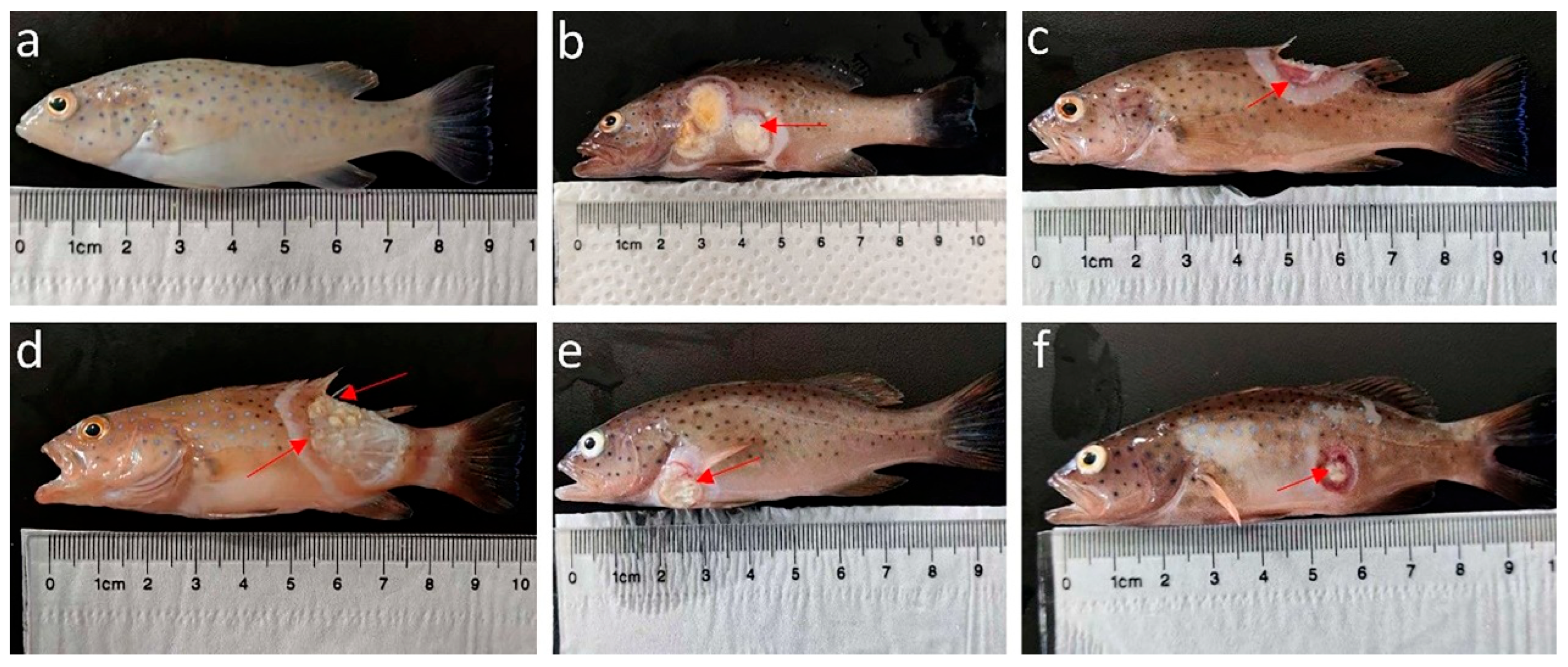
Skin ulcer disease in P. leopardus in Hainan Province typically occurs from June to September. Based on our field observations, during this period, water temperatures in the farming systems reach approximately 30 °C. The healthy fish were active, were responsive to stimuli, and displayed strong feeding behavior, while infected fish exhibited reduced activity, sluggish movement, isolation, delayed responses, and loss of appetite (severe cases may refuse food). In terms of physical appearance, healthy fish displayed smooth body shape, uniform coloration, intact skin/muscles, and undamaged fins (Figure 1a). Infected fish showed uneven body coloration, visible skin ulcers (commonly on the lateral body, Figure 1b,f) and dorsal (Figure 1c), anal (Figure 1d), and pectoral fin (Figure 1e) erosion.

3.2. Histopathological Analysis

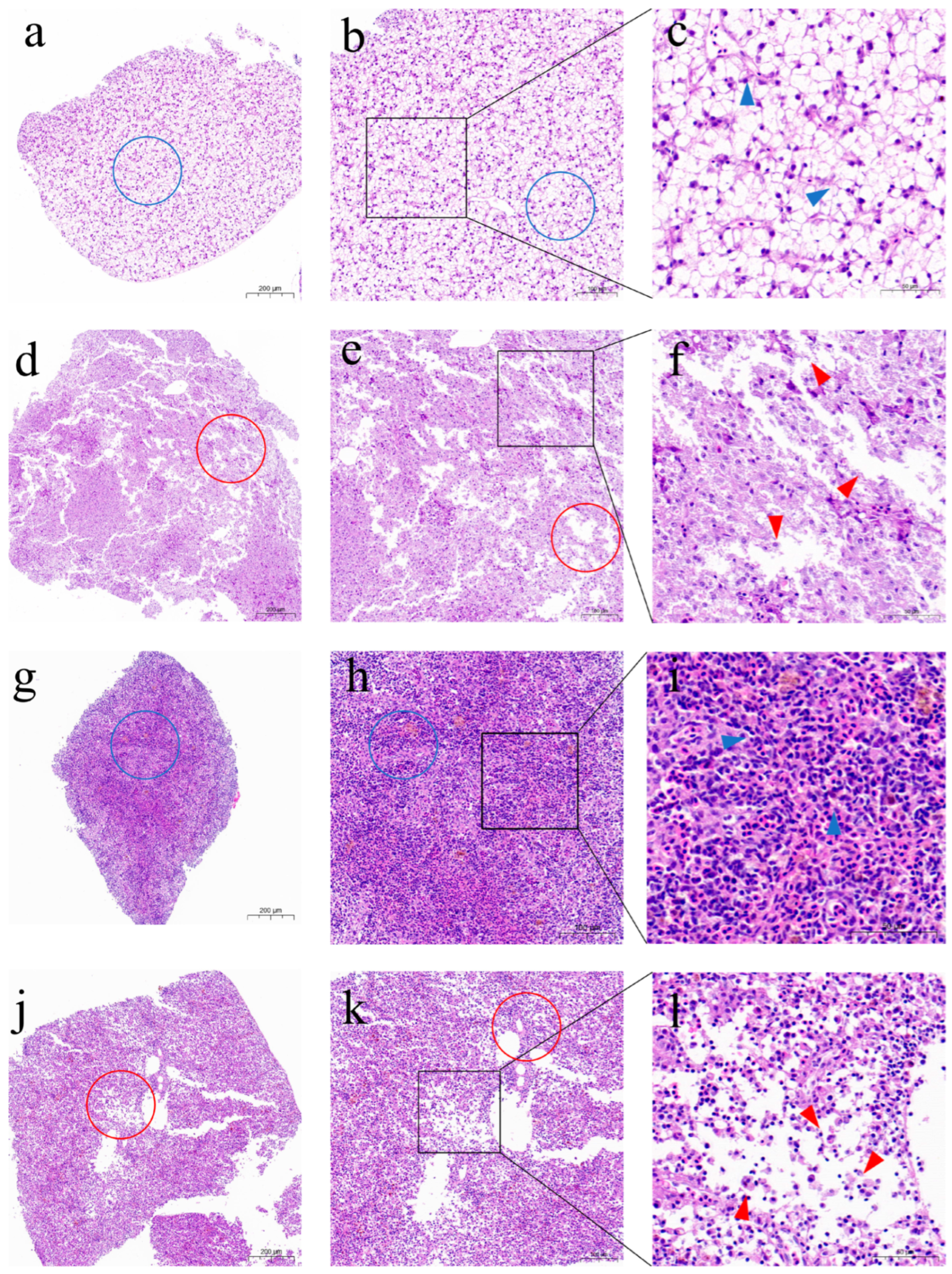
Histopathological analysis of liver sections revealed that healthy P. leopardus exhibited a compact hepatic architecture, with tightly arranged polygonal hepatocytes displaying clear boundaries, evenly distributed cytoplasm, and normal nuclei without abnormal staining regions (Figure 2a–c). The spleen exhibited well-organized structure, distinct demarcation between red pulp and white pulp, and uniform staining, with no signs of congestion, hemorrhage, or inflammation (Figure 2g–i). In contrast, diseased P. leopardus displayed significant hepatic degeneration and necrosis, characterized by loose tissue architecture, irregular hepatocyte morphology, blurred cell boundaries, cytoplasmic vacuolation, and nuclear pyknosis or karyolysis (Figure 2d–f). The spleen also exhibited structural disorganization, disordered cell arrangement, cellular swelling, cytoplasmic vacuolation, and uneven staining indicative of localized necrosis or lesions (Figure 2j–l).

3.3. Ultrastructural Observations

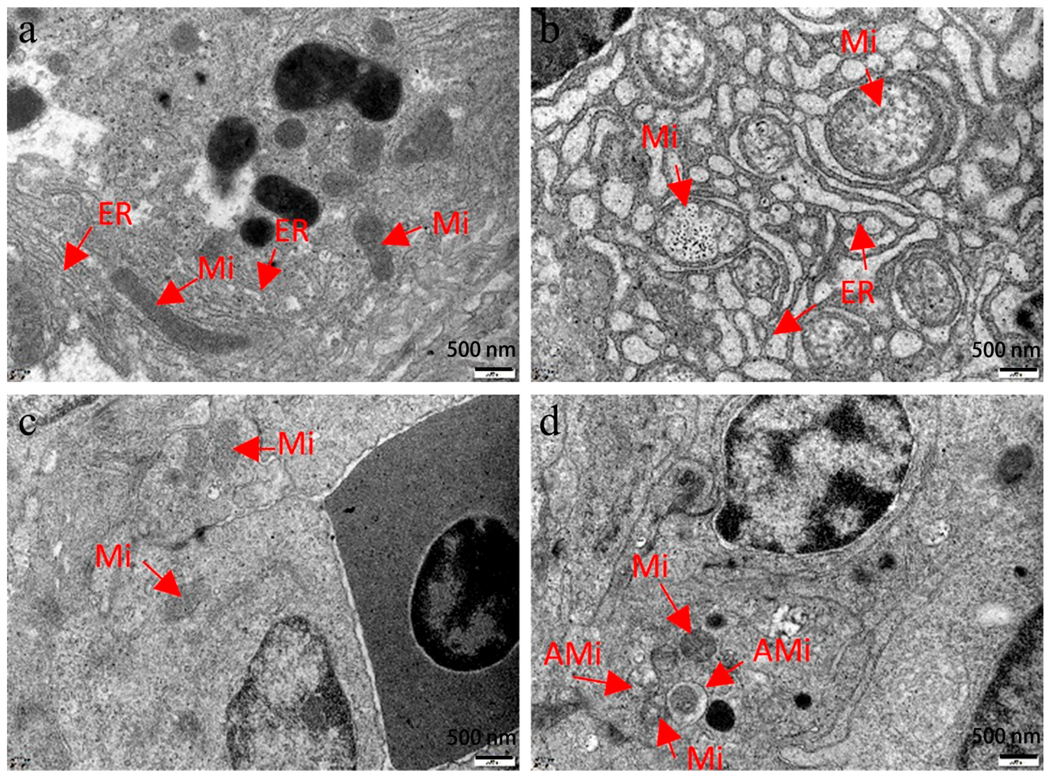
Ultrastructural observation revealed that hepatocytes and splenic cells in healthy P. leopardus exhibited normal morphology and appropriate positioning of organelles, including mitochondria and endoplasmic reticulum (Figure 3a,c). In contrast, diseased individuals with skin ulceration displayed marked pathological alterations: hepatic and splenic mitochondria showed swelling and cristae fragmentation, partial autophagy was observed, and the endoplasmic reticulum exhibited disorganized arrangement (Figure 3b,d). These ultrastructural abnormalities likely contribute to organelle dysfunction, disrupting normal physiological processes in the host.

3.4. Parasite Detection

To determine whether parasites were the causative agents, microscope slides of gill filaments from infected individuals were observed under an optical microscope at magnifications of 10× and 20×; any suspicious structures were further assessed at higher magnifications of 40× and 100×. The results showed no parasites present in the gill filaments of diseased fish. Therefore, the ulcerative clinical signs were not associated with parasitic infection.

3.5. Virus Detection

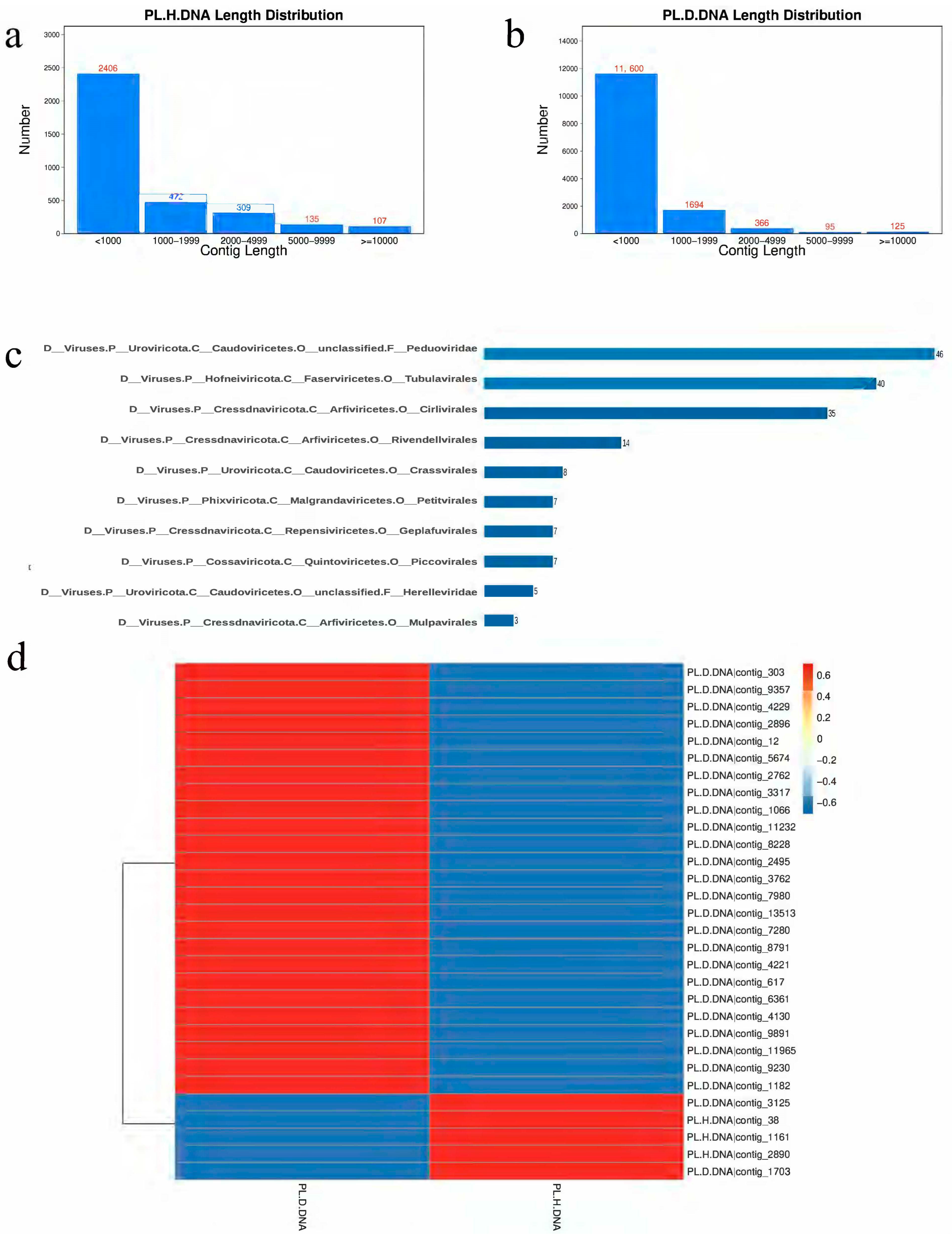
In viral DNA metagenome, a total of 12.2 Gb and 11.6 Gb of raw sequence data was obtained from diseased (PL.D.DNA) and healthy samples (PL.H.DNA), respectively. After the filtration, a total of 21,295,230 and 26,976,044 clean reads from the PL.D.DNA and PL.H.DNA groups, respectively, were obtained and used for assembly into contigs. After removing the host sequence, a total of 17,309 contigs were obtained, and the length distributions of the contigs are displayed in Figure 4a,b. The obtained contigs were compared with the virus database using CheckV and Virsorter2. After comparison and screening, a total of 1245 viral contigs were found, in which the percentage of phage, other viruses (non-bacteriophage viruses), and unassigned contigs was 82.33%, 6.99%, and 10.68%, respectively. A total of 206 viral contigs were annotated to the order level. As shown in Figure 4c, viral taxonomic annotation statistics at the order level revealed that the most abundant were all associated with phages, including Peduoviridae (46 contigs) and Tubulavirales (40 contigs). The viral abundance heatmap showed that the number of high-abundance contigs in diseased individuals was significantly higher than that in healthy samples (Figure 4d). Additionally, the top 30 most abundant viral contigs were predominantly (83.3%) phages such as Tubulavirales and Caudoviricetes. Detailed taxonomic information and rpkm profile for each contig has been supplemented in Supplementary Material S3.
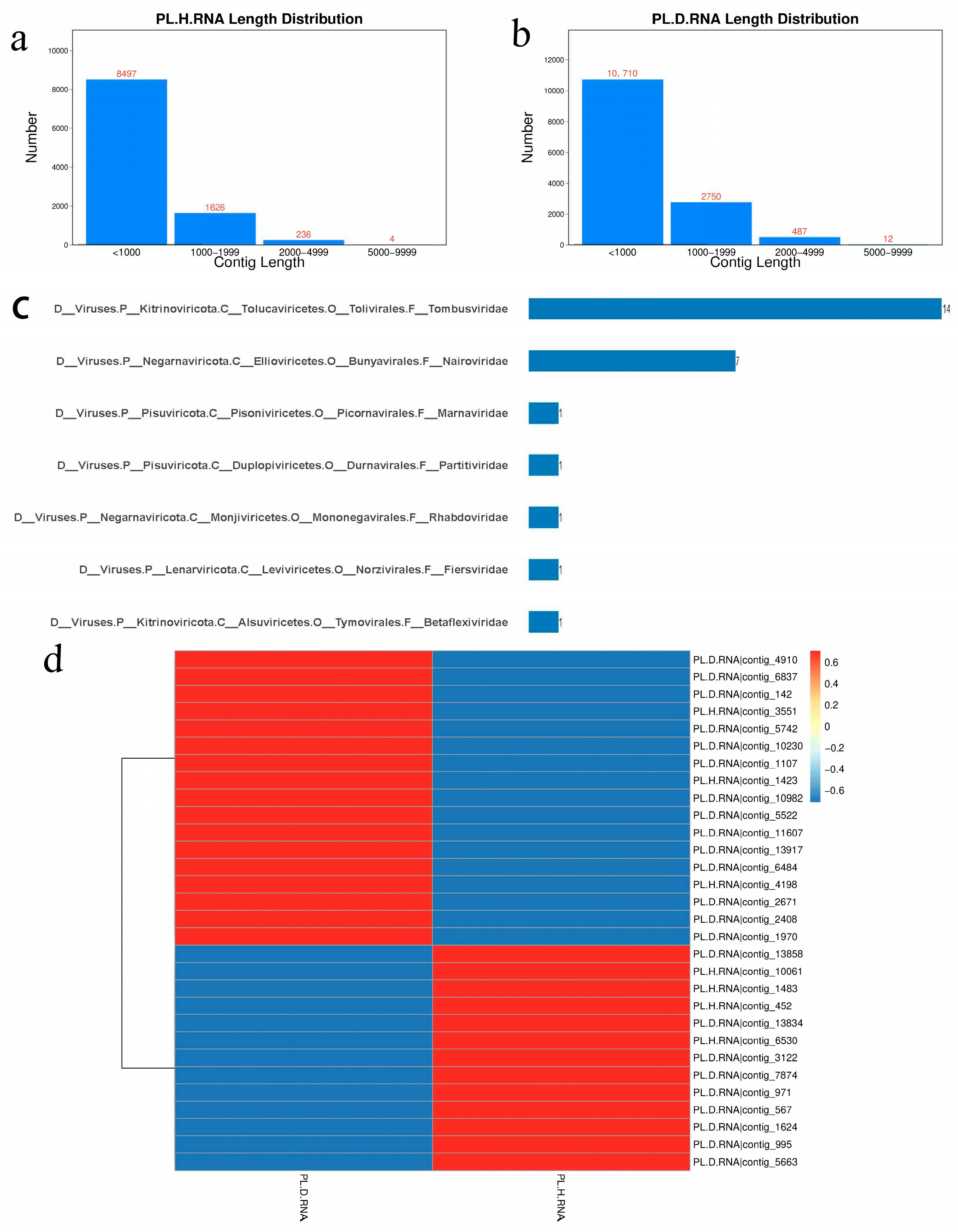
In viral RNA metagenome, a total of 22.9 Gb and 23.2 Gb of raw sequence data was obtained from diseased (PL.D.RNA) and healthy samples (PL.H.RNA), respectively. After the filtration, a total of 39,501,789 and 35,037,146 clean reads from the PL.D.RNA and PL.H.RNA groups, respectively, were obtained and used for assembly into contigs. After removing the host sequence, a total of 24,322 contigs were obtained, and the length distributions of the contigs are displayed in Figure 5a,b. The obtained contigs were compared with the virus database using CheckV. After comparison and screening, a total of 364 viral contigs were found, in which the percentage of single-segmented and multi-segmented virus was 98.08% and 1.92%, respectively. Viral taxonomic annotation statistics (Figure 5c) indicated that the highly represented Tombusviridae family (14 contigs) and Nairoviridae family (7 contigs) were likely environmental viruses and that P. leopardus was not typically their infection target. The viral abundance heatmap (Figure 5d) demonstrated that the number of high-abundance contigs in PL.D.RNA was slightly higher than that in PL.H.RNA, and most (86.7%) of the top 30 abundant viral contigs were classified at the domain level but no further. Detailed taxonomic information and rpkm profile for each contig has been supplemented in Supplementary Material S4.
Combining the above results, no direct pathogen causing ulcer disease in P. leopardus was identified; thus, viral infection can be largely excluded as a potential pathogen of the disease.

3.6. Bacterial Isolation and Identification

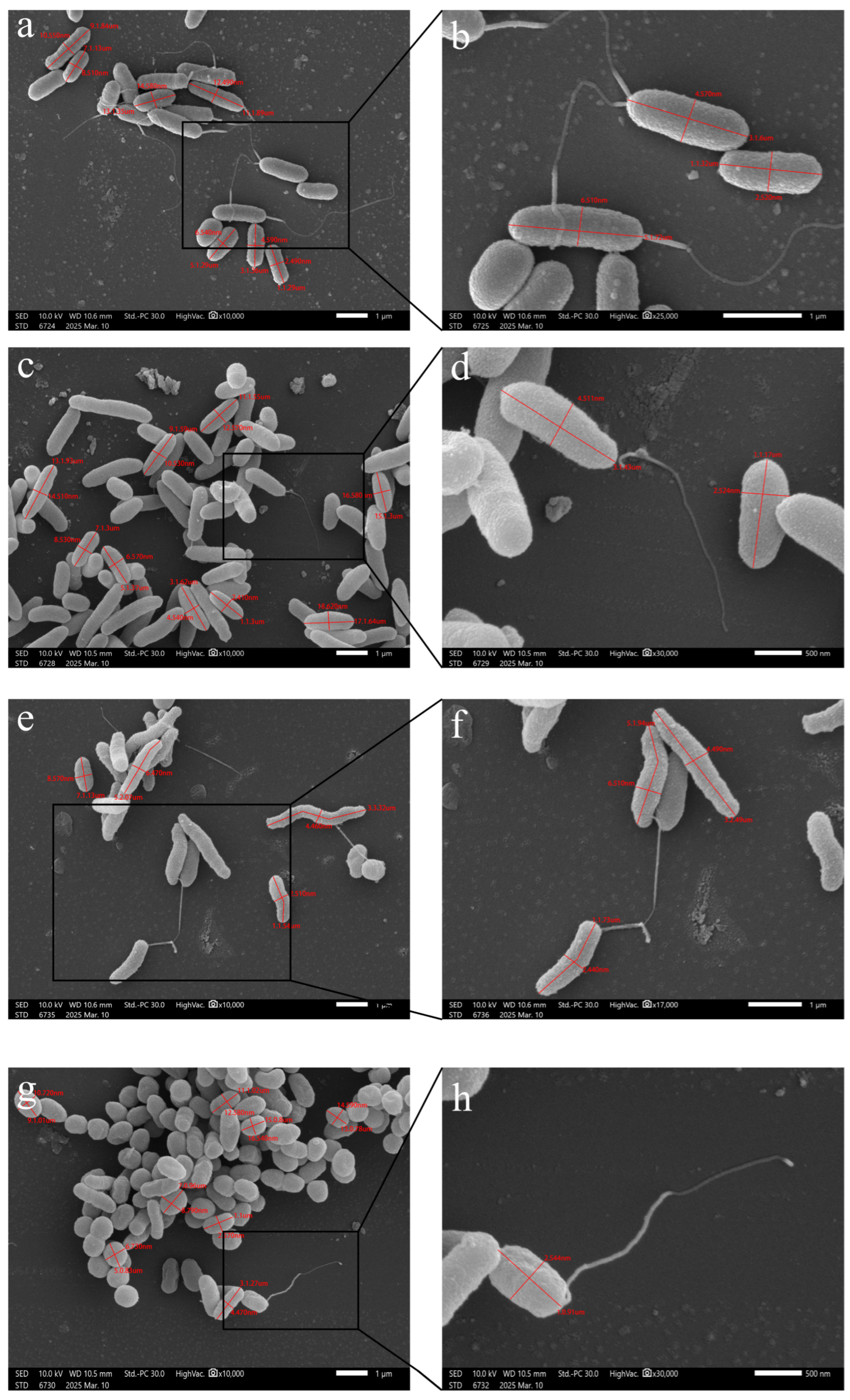
After multiple rounds of isolation and purification, a total of 79 bacterial strains were isolated from the liver tissues of diseased leopard coral grouper. Detailed information on the isolated bacterial strains is shown in Supplementary Material S5. To determine bacterial species, we first amplified the 16S rDNA of these strains. Alignment results indicated the strains belonged to various Vibrio species; however, further validation was required for precise classification. Therefore, the tox R gene was amplified, and subsequent alignment revealed these bacteria as V. harveyi, V. owensii, V. rotiferianus, and P. damselae. To further differentiate the subspecies of P. damselae, the ure C gene was amplified for verification. The results suggested that all isolated P. damselae strains were identified as subsp. damselae. V. harveyi, V. owensii, and V. rotiferianus belong to the genus Vibrio; they share similar morphological characteristics but differ in cell dimensions (Table 2, Figure 6). Statistical analysis of the bacterial isolates revealed that all four pathogenic species were present in the farmed P. leopardus from Wanning, Wenchang, and Qionghai. Although most cases involved single-species infections, co-infections with different bacterial combinations were also observed (Supplementary Material S5). In summary, a total of 79 bacterial strains were isolated from the three regions, comprising V. harveyi (38 strains, 48.1%), V. owensii (17 strains, 21.5%), V. rotiferianus (16 strains, 20.3%), and P. damselae subsp. damselae (8 strains, 10.1%), with the presence of bacterial co-infections noted. Their corresponding sequences, BLAST results and the phylogenetic tree were supplied in Supplementary Material S6.

3.7. Infection Experiments

Infection experiments were conducted in indoor plastic aquaculture tanks located in Yazhou Bay, Sanya City, Hainan Province. In infection experiments, the 79 bacterial strains mentioned above were used to infect healthy leopard coral grouper. The experimental results revealed distinct pathogenicity patterns among the four bacterial species. V. harveyi demonstrated notable virulence, with the VH-2409 strain causing 55% mortality at a dose of 102 CFU per fish (Table 3). Among V. owensii strains, five resulted in mortality exceeding 50%, with the highest rate reaching 75% (VO-2413, Table 4). V. rotiferianus exhibited substantial strain-to-strain variation, with 12 strains showing less than 25% mortality and three strains causing over 40% mortality, peaking at 60% (VR-2408, Table 5). P. damselae subsp. damselae showed the lowest observed mortality, with the PD-2406 strain reaching 20% (Table 6). The control group maintained 0% mortality throughout the experimental period. The detailed daily mortality data have been supplemented in Supplementary Material S7. The infected fish developed clinical signs similar to those observed in naturally diseased fish, including skin ulceration, exposed muscle tissue, and damaged or missing fin rays. V. harveyi, V. owensii, V. rotiferianus, and P. damselae subsp. damselae were also reisolated from the livers of fish that had recently died post-infection. In contrast, healthy fish showed no signs of skin ulceration. According to Koch’s postulates [34], these four bacteria were identified as the pathogens causing skin ulcer disease in leopard coral grouper.

4. Discussion

Skin ulceration has become one of the primary factors hindering the development of the P. leopardus industry. Although studies in other species have indicated that biological factors (bacterial, viral, and parasitic infections), environmental factors, and host immune responses can all lead to skin ulceration [35,36,37], existing research on skin ulceration in P. leopardus primarily attributes the condition to V. harveyi [7,13]. Based on the understanding that the liver is one of the primary target organs of bacterial infection [38,39], bacteria were isolated from the livers of fish exhibiting skin ulceration but from which no parasites or viruses were detected. This led to the isolation of three bacterial species previously unreported in P. leopardus (in addition to V. harveyi).
To identify these three bacterial species, we performed sequencing with specific primers. The 16S rDNA gene is widely used for bacterial identification. However, studies have shown that this gene cannot effectively distinguish between bacteria with high genetic similarity [40], such as V. campbellii, V. owensii, and V. rotiferianus within the V. harveyi clade, where sequence differences in the 16S rDNA gene can be as small as 1% [41]. Therefore, relying solely on the 16S rDNA gene is insufficient for differentiating these bacteria. In this study, we utilized the tox R [30] gene to differentiate Vibrio species and the ure C [31] gene to distinguish between the two subspecies of P. damselae (subsp. damselae and subsp. piscicida). These genes evolve faster than 16S rDNA but are relatively conserved, making them effective for distinguishing highly similar bacterial species.
Through the above methods, we ultimately isolated four pathogenic bacteria (V. harveyi, V. owensii, V. rotiferianus, and P. damselae subsp. damselae) from ulcerated P. leopardus in three different aquaculture regions (Wanning, Wenchang, and Qionghai) in Hainan Province. Subsequent infection experiments suggested that all four pathogens exhibit the category of acute infection, in which the majority of mortality occurs within 1–4 days post-infection. Similarly, the intraperitoneal challenge of V. harveyi induces acute and dose-dependent mortality in hybrid groupers Epinephelus fuscoguttatus (♀) × Epinephelus lanceolatus (♂) and barramundi (Lates calcarifer), with death events restricted to the early post-infection period (≤4 days) [42,43]. In general, Vibrio virulence factors drive pathogenicity by mediating critical steps in the infection cycle, including host adhesion and invasion, in vivo replication, immune evasion, host damage, and dissemination [44]. A large number of studies have shown that many species of Vibrio can lead to a systemic bacterial infection. It is speculated that intraperitoneal injection is more conducive to facilitating the invasion of the Vibrio and triggering such systemic infection. Intraperitoneal injection is a widely employed method for pathogen challenge experiments in aquaculture [45,46]. While this approach offers high reproducibility and moderate reliability, it fails to mimic the natural infection route of the pathogen. In contrast, most V. harveyi outbreaks are considered waterborne or arise from host microbial dysbiosis or immunocompromised individuals with impaired mucosal barriers [47]. Additionally, due to sample size limitations, this study did not include experimental replicates, precluding statistical analysis. Statistical significance and correlation analysis should be investigated in future studies.
The liver serves as a critical organ in modulating immune responses when fish are exposed to bacteria [48]. In this study, histopathological examination revealed that, compared to healthy individuals, the liver structures of naturally diseased P. leopardus appeared loose and disintegrated, with hepatocyte damage, degeneration, necrosis, and indistinct cell boundaries. Some ulcerated individuals displayed abnormal features such as hepatic enlargement and a firm texture. Based on these hepatic lesions, we speculate that, since the liver participates in fatty acid metabolism, hepatocyte damage may lead to deregulated lipid metabolism (e.g., excessive fat accumulation in the liver), potentially resulting in fatty liver disease [49]. Concurrently, given the liver’s vital role in immune defense and regulation, hepatocyte impairment may compromise immune function, diminish the host’s capacity to eliminate pathogens, and increase susceptibility to bacterial, viral, and parasitic infections [50].
The spleen acts as a primary immune and hematopoietic organ in fish. Previous research has indicated that splenic cell damage can reduce lymphocyte proliferation, thereby weakening immune competence [51]. In this study, pathological sections from the spleens of naturally diseased individuals revealed structural loosening, disorganized cell arrangement, cell degeneration, necrosis, and vacuolization compared to healthy individuals. We hypothesize that these pathological changes may lead to clinical signs such as anemia and reduced vitality in farmed fish [52].
In recent years, a growing number of new viruses have been identified across human and veterinary medicine, plant science, and marine science through the application of viral metagenomic approaches [53]. To analyze the potential viruses, this study performed DNA metavirome sequencing and RNA metavirome sequencing using liver, spleen, and kidney tissues collected from both healthy and diseased fish, respectively. For DNA metavirome, the vast majority of the detected viruses were bacteriophages. Consistently, bacteriophages constituted the major component of the Nile tilapia virome [54]. This observation was not unexpected, given the high abundance of bacteriophages in aquatic ecosystems [50,55,56]. It is well-established that bacteriophages in aquatic ecosystems are crucial to modulating the species abundance of archaea and bacteria, thereby exerting regulatory effects on numerous natural bioprocesses [55]. For RNA metavirome, 364 viral contigs were annotated in total, with only 26 contigs assigned to the family taxonomic level and 321 contigs limited to annotation at the domain level. In fact, studies on the RNA viruses in the ocean are relatively scanty [53]. Among the 26 contigs annotated at the family level, 14 were classified into the Tombusviridae family and 7 into the Nairoviridae family, both of which are distant relatives of viruses that infect arthropods and higher plants. As a matter of fact, viruses exist in each tissue. Specifically, viral sequences were identified in all five tested tissues (liver, kidney, spleen, brain, and gills) of Nile tilapia sourced from the four sampling regions, with the viruses showing different distribution patterns in each tissue [54]. In the present study, a mixture of liver, spleen, and kidney tissues was used for metavirome sequencing. While this approach can increase the total yield of viral nucleic acids and thus improve the chance of detecting low-abundance viruses, it also has several notable limitations. Collectively, pooling liver, spleen, and kidney tissues obscures tissue-specific viral tropisms and distribution patterns, introduces host nucleic acid biases that confound viral load quantification, and impedes the correlation of viral contigs with tissue-specific pathological changes, thus limiting the interpretability of virome data.
This study found that, although the four bacterial species—V. harveyi, V. owensii, V. rotiferianus, and P. damselae subsp. damselae—were detected in all three investigated aquaculture regions of Hainan Province, the majority of infections were monomicrobial. Co-infection of multiple bacterial species was observed only in rare instances, suggesting that polymicrobial infection by these four bacteria may occur sporadically. In this study, co-infection involving V. harveyi with V. owensii or V. rotiferianus was observed in the diseased fish. Bacterial co-infection has been widely reported in various aquatic organisms, including teleosts, crustaceans, and mollusks, particularly in intensive aquaculture systems [57]. Consistent with our research, a total of 45 dominant bacterial strains with distinguishable morphological characteristics were isolated from diseased torafugu (Takifugu rubripes) with ulceration, including V. harveyi (27 isolates), V. rotiferianus (3 isolates) and V. owensii (1 isolate), among which 60% were V. harveyi [58]. Their concurrent presence in diseased fish was implied to be linked to epidemic outbreaks with high mortality. The occurrence of such co-infection is closely related to aquaculture environmental conditions and host physiological status. However, the pathogenic mechanisms underlying co-infection by these multiple bacterial isolates remain elusive and warrant further investigation.
In this research, infection was performed through intraperitoneal injection. While this method effectively induces disease occurrence and facilitates observation of pathological progression, it differs from natural infection routes. In natural or aquaculture environments, skin ulcer disease in the leopard coral grouper may be transmitted through pathways such as surface contact with pathogens, water-borne spread, or the ingestion of contaminated food. In contrast, intraperitoneal injections directly introduce pathogens into the body cavity, potentially skipping the initial stages of natural infection (e.g., pathogen adhesion and invasion of the body surface barrier). This may lead to discrepancies in infection progression, pathological response intensity, and tissue damage patterns compared to natural disease occurrence. Thus, the explanatory power of the study’s results regarding natural infection processes is somewhat limited. Future research should explore infection routes closer to natural conditions (e.g., immersion infection, pathogen exposure after surface abrasion) to more accurately simulate the real-world development of the disease. This will provide a more comprehensive basis for deciphering the natural transmission mechanisms and pathological characteristics of the disease.

5. Conclusions

This study identified four bacterial pathogens, V. harveyi (38/79, 48.1%), V. owensii (17/79, 21.5%), V. rotiferianus (16/79, 20.3%), and P. damselae subsp. damselae (8/79, 10.1%), from ulcerated P. leopardus in Hainan Province, China. Infection experiments suggested that most of these species were associated with skin ulceration and mortality, thereby refining the previous paradigm that attributed the disease solely to V. harveyi. Co-occurrence of multiple bacterial pathogens was observed, whereas potential synergistic interactions warrant further experimental testing. The main limitations of this study include its geographic restriction to Hainan Province and the limited sample size, which precluded statistical analysis of virulence differences.

Supplementary Materials

The following supporting information can be downloaded at: https://www.mdpi.com/article/10.3390/fishes11020084/s1, Supplementary Material S1: Detailed procedures and the specific software tools employed in viral DNA metagenomic sequencing; Supplementary Material S2: Detailed procedures and the specific software tools employed in viral RNA metagenomic sequencing; Supplementary Material S3: Detailed taxonomic information and rpkm profile for each contig in viral DNA metagenome; Supplementary Material S4: Detailed taxonomic information and rpkm profile for each contig in viral RNA metagenome; Supplementary Material S5: Detailed information of the isolated bacterial strains; Supplementary Material S6: The bacterial sequences, BLAST results and the phylogenetic tree; Supplementary Material S7: The detailed daily mortality data in the infection experiments.

Author Contributions

Conceptualization, Y.X. and X.L.; methodology, Y.X.; software, Z.B.; validation, L.J., B.Z. and Z.B.; formal analysis, Z.B.; investigation, L.J., X.T. and Y.H.; resources, X.L.; data curation, J.D., Y.Z. and W.W.; writing—original draft preparation, L.W.; writing—review and editing, X.L.; visualization, J.C.; supervision, S.C.; project administration, Y.X.; funding acquisition, Y.X. All authors have read and agreed to the published version of the manuscript.

Funding

This work was supported by the Foundation of the State Key Laboratory of Mariculture Biobreeding and Sustainable Goods (No. BRESG202407), Secondary Sub-projects of Scientific Research and Technology Demonstration Category in the National Modern Agricultural Industrial Park, Wanning City, Hainan Province, Visiting and Training Foundation of Teachers in Ordinary Undergraduate Universities in Shandong Province, Academician Chen Songlin’s Workstation of Hainan Province (YSGZZ2023001), Hainan Province Science and Technology Special Fund (YSPTZX202402), Hainan Province Science and Technology Special Fund (ZDYF2024XDNY278), Fish Innovation Team of Shandong Agriculture Research System (SDAIT-12-06), Shandong Engineering Research Center for Prevention and Control of Aquatic Animal Disease.

Institutional Review Board Statement

All experimental animal protocols were carried out in accordance with the IACUC Committee on the Ethics of Animal Experiments at Qingdao Agricultural University (Institutional Animal Care and Use Committee). Approval Code: 2024-082. Approval Date: 10 May 2024.

Data Availability Statement

The information for the viral DNA metagenome is available in NCBI with accession numbers SRR36305221 and SRR36305222. The information for the viral RNA metagenome is available in NCBI with accession numbers SRR36305219 and SRR36305220. The toxR or ureC sequences of 79 bacterial strains have been deposited in the NCBI database, and the corresponding accession numbers are provided in Supplementary Material S5.

Conflicts of Interest

Author Zemin Bai is from the company Sanya Yazhou Harbour Investment Co., Ltd. Author Bo Zhou is from the company Wanning Linlan Aquaculture. The remaining authors declare that the research was conducted in the absence of any commercial or financial relationships that could be construed as a potential conflict of interest.

References

  1. Amir, A.; Elviana, S.; Kadir, A. Financial analysis of coral trout grouper (Plectropomus leopardus) in Gusung Island, Selayar archipelago regency. IOP Conf. Ser. Earth Environ. Sci. 2019, 343, 012163. [Google Scholar]
  2. Cayetano, C.B.; Asis, E.H.; Dicar, Z.A.; Sajorne, R.E.; Madarcos, K.G.; Alcantara, L.; Cayetano, A.Z.; Palla, H.P.; Creencia, L.A. Trade Dynamics of the Leopard Coral Grouper (Plectropomus leopardus, Lacepède, 1802) in Palawan: A Philippine Perspective on the Impacts of the COVID-19 Pandemic and Coping Strategies. Davao Res. J. 2025, 16, 156–175. [Google Scholar] [CrossRef] [Scilit]
  3. Sundberg, L.R.; Ketola, T.; Laanto, E.; Kinnula, H.; Bamford, J.K.; Penttinen, R.; Mappes, J. Intensive aquaculture selects for increased virulence and interference competition in bacteria. Proc. R. Soc. B Biol. Sci. 2016, 283, 20153069. [Google Scholar] [CrossRef] [Scilit]
  4. Gai, C.; Liu, J.; Zheng, X.; Xu, L.; Ye, H. Identification of Vibrio ponticus as a bacterial pathogen of coral trout Plectropomus leopardus. Front. Cell. Infect. Microbiol. 2022, 12, 1089247. [Google Scholar] [CrossRef] [Scilit]
  5. Wang, J.; Yu, X.; Wu, S.; Jin, C.; Wang, M.; Ding, H.; Song, S.; Bao, Z.; Wang, B.; Hu, J. Identification of candidate SNPs and genes associated with resistance to nervous necrosis virus in leopard coral grouper (Plectropomus leopardus) using GWAS. Fish Shellfish Immunol. 2024, 144, 109295. [Google Scholar] [CrossRef] [Scilit] [PubMed]
  6. Mizuochi, H.; Fujiwara, Y.; Shirakashi, S.; Fujikura, Y.; Ogawa, K. Biology and Pathogenicity of Benedenia akajin Infecting Hatchery-reared Plectropomus leopardus Juveniles. Fish Pathol. 2021, 56, 122–129. [Google Scholar] [CrossRef] [Scilit]
  7. Wang, L.; Zhang, T.; Liu, Y.; Zhang, Z.; Li, K.; Zhu, C.; Chen, S. High-throughput sequencing analysis of the main pathogens of ulcer diseased Plectropomus leopardus. J. Agric. Biotechnol. 2023, 31, 617–628. [Google Scholar] [CrossRef]
  8. Zhang, Z.; Yu, Y.X.; Wang, K.; Wang, Y.G.; Jiang, Y.; Liao, M.J.; Rong, X.J. First report of skin ulceration caused by Photobacterium damselae subsp. damselae in net-cage cultured black rockfish (Sebastes schlegeli). Aquaculture 2019, 503, 1–7. [Google Scholar] [CrossRef] [Scilit]
  9. Chetty, T.; Nowak, B.F.; Walker, S.P.; Symonds, J.E.; Anderson, K. Molecular evidence for stress, inflammation and structural changes in non-specific ulcers in skin of farmed Chinook salmon (Oncorhynchus tshawytscha). Fish Shellfish Immunol. 2023, 137, 108739. [Google Scholar] [CrossRef] [Scilit]
  10. Udomkusonsri, P.; Noga, E.J. The acute ulceration response (AUR): A potentially widespread and serious cause of skin infection in fish. Aquaculture 2005, 246, 63–77. [Google Scholar] [CrossRef] [Scilit]
  11. Noga, E.J. Skin ulcers in fish: Pfiesteria and other etiologies. Toxicol. Pathol. 2000, 28, 807–823. [Google Scholar] [CrossRef] [Scilit] [PubMed]
  12. Law, M. Differential diagnosis of ulcerative lesions in fish. Environ. Health Perspect. 2001, 109, 681–686. [Google Scholar] [CrossRef] [Scilit] [PubMed]
  13. Shiyu, Q.; Sheng, L.; Songlin, C.; Yang, L.; Qian, Z.; Lei, W.; Wenteng, X.; Yu, S. Estimation of genetic parameters of survival against Vibrio harveyi in leopard coral grouper (Plectropomus leopardus). Prog. Fish. Sci. 2024, 45, 15–23. [Google Scholar]
  14. Ina-Salwany, M.Y.; Al-saari, N.; Mohamad, A.; Mursidi, F.A.; Mohd-Aris, A.; Amal, M.N.A.; Kasai, H.; Mino, S.; Sawabe, T.; Zamri-Saad, M. Vibriosis in Fish: A Review on Disease Development and Prevention. J. Aquat. Anim. Health 2019, 31, 3–22. [Google Scholar] [CrossRef] [Scilit]
  15. Nor, N.M.; Yazid, S.H.M.; Daud, H.M.; Azmai, M.N.A.; Mohamad, N. Costs of management practices of Asian seabass (Lates calcarifer Bloch, 1790) cage culture in Malaysia using stochastic model that includes uncertainty in mortality. Aquaculture 2019, 510, 347–352. [Google Scholar] [CrossRef] [Scilit]
  16. Zhu, Z.M.; Duan, C.; Dong, C.F.; Weng, S.P.; He, J.G. Epidemiological situation and phylogenetic relationship of Vibrio harveyi in marine-cultured fishes in China and Southeast Asia. Aquaculture 2020, 529, 735652. [Google Scholar] [CrossRef] [Scilit]
  17. Zhang, X.H.; He, X.X.; Austin, B. Vibrio harveyi: A serious pathogen of fish and invertebrates in mariculture. Mar. Life Sci. Technol. 2020, 2, 231–245. [Google Scholar] [CrossRef] [Scilit]
  18. Pavlinec, Z.; Zupicic, I.G.; Oraic, D.; Lojkic, I.; Fouz, B.; Zrncic, S. Biochemical and molecular characterization of three serologically different Vibrio harveyi strains isolated from farmed Dicentrarchus labrax from the Adriatic Sea. Sci. Rep. 2022, 12, 7309. [Google Scholar] [CrossRef] [Scilit] [PubMed]
  19. Sun, F.; Chen, J.; Zhong, L.; Zhang, X.H.; Wang, R.; Guo, Q.; Dong, Y. Characterization and virulence retention of viable but nonculturable Vibrio harveyi. FEMS Microbiol. Ecol. 2008, 64, 37–44. [Google Scholar] [CrossRef] [Scilit]
  20. Triga, A.; Issa, Z.A.; Smyrli, M.; Fenske, L.; Katharios, P. Virulence and pangenome analysis of Vibrio harveyi strains from Greek and Red Sea marine aquaculture. Aquaculture 2024, 587, 740839. [Google Scholar] [CrossRef] [Scilit]
  21. Cano-Gómez, A.; Goulden, E.F.; Owens, L.; Høj, L. Vibrio owensii sp. nov., isolated from cultured crustaceans in Australia. FEMS Microbiol. Lett. 2010, 302, 175–181. [Google Scholar] [CrossRef] [Scilit]
  22. Sun, J.X.; Liu, L.; Song, J.; Zhan, Y.Y.; Zhang, W.J.; Wang, B.; Zhang, B.J.; Chang, Y.Q. Characterization of two strains of Vibrio sp. from cultured sea urchin, Strongylocentrotus intermedius, in China. Aquac. Rep. 2021, 20, 100667. [Google Scholar] [CrossRef] [Scilit]
  23. Thillaichidambaram, M.; Narayanan, K.; Selvaraj, S.; Sundararaju, S.; Muthiah, R.C.; Figge, M.J. Isolation and characterization of Vibrio owensii from Palk Bay and its infection study against post larvae of Litopenaeus vannamei. Microb. Pathog. 2022, 172, 105751. [Google Scholar] [CrossRef] [Scilit]
  24. Duman, M.; Süzer, B.; Ajmi, N.; Yavaş, Ö.; Saticioğlu, İ.B. Molecular Characterization and Pathogenicity of Vibrio atlanticus and Vibrio rotiferianus Associated With Mortality of Cownose Ray Rhinoptera bonasus. Acta Vet. Eurasia 2024, 50, 242–249. [Google Scholar] [CrossRef] [Scilit]
  25. Liu, C.Y.; Huang, Z.H.; He, H.X.; He, X.; Li, X.S.; Chen, J.P.; Wang, L.Q.; Qin, Q.W.; Yang, M. Isolation and complete genome sequence analysis of Vibrio rotiferianus strain TO-01 from Trachinotus ovatus to reveal its pathogenicity and drug resistance. Aquac. Rep. 2024, 37, 102215. [Google Scholar] [CrossRef] [Scilit]
  26. Chen, Z.; Yao, Z.; Lin, M.; Chang, J. Characterization of pathogenic bacteria Vibrio rotiferianus isolated from Cynoglossus semilaevis Günther. Biotechnol. Bull. 2012, 6, 147–153. [Google Scholar]
  27. Osorio, C.R.; Vences, A.; Matanza, X.M.; Terceti, M.S. Photobacterium damselae subsp damselae, a Generalist Pathogen with Unique Virulence Factors and High Genetic Diversity. J. Bacteriol. 2018, 200, e00002-18. [Google Scholar] [CrossRef] [Scilit]
  28. Rivas, A.J.; Labella, A.M.; Borrego, J.J.; Lemos, M.L.; Osorio, C.R. Evidence for horizontal gene transfer, gene duplication and genetic variation as driving forces of the diversity of haemolytic phenotypes in Photobacterium damselae subsp. damselae. FEMS Microbiol. Lett. 2014, 355, 152–162. [Google Scholar] [CrossRef] [Scilit]
  29. Weisburg, W.G.; Barns, S.M.; Pelletier, D.A.; Lane, D.J. 16S ribosomal DNA amplification for phylogenetic study. J. Bacteriol. 1991, 173, 697–703. [Google Scholar] [CrossRef] [Scilit] [PubMed]
  30. Singh, D.V.; Isac, S.R.; Colwell, R.R. Development of a hexaplex PCR assay for rapid detection of virulence and regulatory genes in Vibrio cholerae and Vibrio mimicus. J. Clin. Microbiol. 2002, 40, 4321–4324. [Google Scholar] [CrossRef] [Scilit]
  31. Osorio, C.R.; Toranzo, A.E.; Romalde, J.L.; Barja, J.L. Multiplex PCR assay for ureC and 16S rRNA genes clearly discriminates between both subspecies of Photobacterium damselae. Dis. Aquat. Organ. 2000, 40, 177–183. [Google Scholar] [CrossRef] [Scilit]
  32. Feldman, A.T.; Wolfe, D. Tissue processing and hematoxylin and eosin staining. Methods Mol. Biol. 2014, 1180, 31–43. [Google Scholar] [CrossRef] [Scilit]
  33. Phrompanya, P.; Panase, P.; Saenphet, S.; Saenphet, K. Histopathology and oxidative stress responses of Nile tilapia Oreochromis niloticus exposed to temperature shocks. Fish. Sci. 2021, 87, 491–502. [Google Scholar] [CrossRef] [Scilit]
  34. Ross, L.N.; Woodward, J.F. Koch’s postulates: An interventionist perspective. Stud. Hist. Philos. Biol. Biomed. Sci. 2016, 59, 35–46. [Google Scholar] [CrossRef] [Scilit]
  35. Anderson, D.P. Environmental factors in fish health: Immunological aspects. In Fish Physiology; Elsevier: Amsterdam, The Netherlands, 1996; Volume 15, pp. 289–310. [Google Scholar]
  36. Kordon, A.O.; Karsi, A.; Pinchuk, L. Innate immune responses in fish: Antigen presenting cells and professional phagocytes. Turk. J. Fish. Aquat. Sci. 2018, 18, 1123–1139. [Google Scholar] [CrossRef] [Scilit] [PubMed]
  37. Sasikumar, R.; Saranya, S.; Lincy, L.L.; Thamanna, L.; Chellapandi, P. Genomic insights into fish pathogenic bacteria: A systems biology perspective for sustainable aquaculture. Fish Shellfish Immunol. 2024, 154, 109978. [Google Scholar] [CrossRef] [Scilit] [PubMed]
  38. Malick, R.C.; Bera, A.K.; Chowdhury, H.; Bhattacharya, M.; Abdulla, T.; Swain, H.S.; Baitha, R.; Kumar, V.; Das, B.K. Identification and pathogenicity study of emerging fish pathogens Acinetobacter junii and Acinetobacter pittii recovered from a disease outbreak in Labeo catla (Hamilton, 1822) and Hypophthalmichthys molitrix (Valenciennes, 1844) of freshwater wetland in West Bengal, India. Aquac. Res. 2020, 51, 2410–2420. [Google Scholar] [CrossRef] [Scilit]
  39. Oh, W.T.; Kim, J.H.; Jun, J.W.; Giri, S.S.; Yun, S.; Kim, H.J.; Kim, S.G.; Kim, S.W.; Han, S.J.; Kwon, J.; et al. Genetic Characterization and Pathological Analysis of a Novel Bacterial Pathogen, Pseudomonas tructae, in Rainbow Trout (Oncorhynchus mykiss). Microorganisms 2019, 7, 432. [Google Scholar] [CrossRef] [Scilit]
  40. Johnson, J.S.; Spakowicz, D.J.; Hong, B.Y.; Petersen, L.M.; Demkowicz, P.; Chen, L.; Leopold, S.R.; Hanson, B.M.; Agresta, H.O.; Gerstein, M.; et al. Evaluation of 16S rRNA gene sequencing for species and strain-level microbiome analysis. Nat. Commun. 2019, 10, 5029. [Google Scholar] [CrossRef] [Scilit]
  41. Sawabe, T.; Ogura, Y.; Matsumura, Y.; Feng, G.; Amin, A.; Mino, S.; Nakagawa, S.; Sawabe, T.; Kumar, R.; Fukui, Y.; et al. Updating the Vibrio clades defined by multilocus sequence phylogeny: Proposal of eight new clades, and the description of Vibrio tritonius sp nov. Front. Microbiol. 2013, 4, 414. [Google Scholar] [CrossRef] [Scilit]
  42. Shen, G.M.; Shi, C.Y.; Fan, C.; Jia, D.; Wang, S.Q.; Xie, G.S.; Li, G.Y.; Mo, Z.L.; Huang, J. Isolation, identification and pathogenicity of Vibrio harveyi, the causal agent of skin ulcer disease in juvenile hybrid groupers Epinephelus fuscoguttatus × Epinephelus lanceolatus. J. Fish Dis. 2017, 40, 1351–1362. [Google Scholar] [CrossRef] [Scilit] [PubMed]
  43. Samsing, F.; Zhang, W.; Zadoks, R.N.; Whittington, R.; Venturini, C.; Giles, C.; Carson, J.; Becker, J.A. Cold temperature stress and damaged skin induced high mortality in barramundi (Lates calcarifer) challenged with Vibrio harveyi. J. Fish Dis. 2023, 46, 751–766. [Google Scholar] [CrossRef] [Scilit] [PubMed]
  44. Darshanee Ruwandeepika, H.A.; Sanjeewa Prasad Jayaweera, T.; Paban Bhowmick, P.; Karunasagar, I.; Bossier, P.; Defoirdt, T. Pathogenesis, virulence factors and virulence regulation of vibrios belonging to the Harveyi clade. Rev. Aquac. 2012, 4, 59–74. [Google Scholar] [CrossRef] [Scilit]
  45. Firmino, J.; Furones, M.D.; Andree, K.B.; Sarasquete, C.; Ortiz-Delgado, J.B.; Asencio-Alcudia, G.; Gisbert, E. Contrasting outcomes of Vibrio harveyi pathogenicity in gilthead seabream, Sparus aurata and European seabass, Dicentrachus labrax. Aquaculture 2019, 511, 734210. [Google Scholar] [CrossRef] [Scilit]
  46. Yang, A.; Li, W.; Tao, Z.; Ye, H.; Xu, Z.; Li, Y.; Gao, Y.; Yan, X. Vibrio harveyi isolated from marine aquaculture species in eastern China and virulence to the large yellow croaker (Larimichthys crocea). J. Appl. Microbiol. 2021, 131, 1710–1721. [Google Scholar] [CrossRef] [Scilit]
  47. Derome, N.; Gauthier, J.; Boutin, S.; Llewellyn, M. Bacterial opportunistic pathogens of fish. In The Rasputin Effect: When Commensals and Symbionts Become Parasitic; Springer: Berlin/Heidelberg, Germany, 2016; pp. 81–108. [Google Scholar]
  48. Causey, D.R.; Pohl, M.A.N.; Stead, D.A.; Martin, S.A.M.; Secombes, C.J.; Macqueen, D.J. High-throughput proteomic profiling of the fish liver following bacterial infection. BMC Genom. 2018, 19, 719. [Google Scholar] [CrossRef] [Scilit]
  49. Li, X.; Han, T.; Zheng, S.; Wu, G. Hepatic Glucose Metabolism and Its Disorders in Fish. Adv. Exp. Med. Biol. 2022, 1354, 207–236. [Google Scholar] [CrossRef] [Scilit]
  50. Gao, B.; Jeong, W.I.; Tian, Z. Liver: An organ with predominant innate immunity. Hepatology 2008, 47, 729–736. [Google Scholar] [CrossRef] [Scilit]
  51. Wang, H.; Feng, S.; Pan, E.; Ji, X.; Zhou, M.; Zhang, S.; Xu, B.; Feng, H.; Yin, J.; Dong, Z. Ferulic acid alleviates long-term avermectin-induced damage to the spleen of carp and restores its inflammatory response and oxidative balance. J. Environ. Sci. 2025, 151, 616–626. [Google Scholar] [CrossRef] [Scilit]
  52. Schuermans, S.; Kestens, C.; Marques, P.E. Systemic mechanisms of necrotic cell debris clearance. Cell Death Dis. 2024, 15, 557. [Google Scholar] [CrossRef] [Scilit]
  53. Alavandi, S.V.; Poornima, M. Viral metagenomics: A tool for virus discovery and diversity in aquaculture. Indian J. Virol. 2012, 23, 88–98. [Google Scholar] [CrossRef] [Scilit] [PubMed]
  54. Ezzat, A.; Abd El Wahed, A.; Ceruti, A.; El Asely, A.M.; Khalifa, M.S.; Winters, A.D.; Truyen, U.; Shaheen, A.A.; Faisal, M. Exploring the Virome of Nile Tilapia (Oreochromis niloticus) Using Metagenomic Analysis. Pathogens 2025, 14, 935. [Google Scholar] [CrossRef] [Scilit] [PubMed]
  55. Suttle, C.A. Marine viruses—Major players in the global ecosystem. Nat. Rev. Microbiol. 2007, 5, 801–812. [Google Scholar] [CrossRef] [Scilit] [PubMed]
  56. Chow, C.E.; Suttle, C.A. Biogeography of Viruses in the Sea. Annu. Rev. Virol. 2015, 2, 41–66. [Google Scholar] [CrossRef] [Scilit]
  57. Kotob, M.H.; Menanteau-Ledouble, S.; Kumar, G.; Abdelzaher, M.; El-Matbouli, M. The impact of co-infections on fish: A review. Vet. Res. 2016, 47, 98. [Google Scholar] [CrossRef] [Scilit]
  58. Wu, F.; Tang, K.; Yuan, M.; Shi, X.; Shakeela, Q.; Zhang, X.H. Studies on bacterial pathogens isolated from diseased torafugu (Takifugu rubripes) cultured in marine industrial recirculation aquaculture system in Shandong Province, China. Aquac. Res. 2015, 46, 736–744. [Google Scholar] [CrossRef] [Scilit]
Figure 1. Comparison of the appearance of healthy P. leopardus (a) and ulcerated ones (bf).
Fishes 11 00084 g001
Figure 2. Histopathological features of liver and spleen in P. leopardus. (a) Liver of a healthy individual showing intact and compact tissue architecture (blue circles) (×50). (b) Tightly arranged hepatocytes in healthy liver (blue circles) (×100). (c) Clear cell boundaries of hepatocytes in healthy liver (blue triangles) (×300). (d) Liver of a naturally diseased individual with disrupted and loose tissue architecture (red circles) (×50). (e) Disorganized hepatocytes with cytoplasmic vacuolation in naturally diseased liver (red circles) (×100). (f) Blurred cell boundaries of hepatocytes in naturally diseased liver (red triangles) (×300). (g) Spleen of a healthy individual displaying organized tissue structure (blue circles) (×50). (h) Densely packed splenic cells in healthy spleen (blue circles) (×100). (i) Well-defined cell boundaries in healthy spleen (blue triangles) (×300). (j) Spleen of a naturally diseased individual exhibiting disintegrated and loose tissue architecture (red circles) (×50). (k) Disordered splenic cells with cytoplasmic vacuolation in naturally diseased spleen (red circles) (×100). (l) Indistinct cell boundaries in naturally diseased spleen (red triangles) (×300).
Fishes 11 00084 g002
Figure 3. Ultrastructural observation of liver and spleen in P. leopardus. (a) Hepatic cells of a healthy individual. (b) Hepatic cells of a diseased individual. (c) Splenic cells of a healthy individual. (d) Splenic cells of a diseased individual. Mi: Mitochondria. AMi: Autophagic mitochondria. ER: Endoplasmic reticulum.
Fishes 11 00084 g003
Figure 4. DNA virome sequencing results. (a) PL.H.DNA length distribution. (b) PL.D.DNA length distribution. (c) Order-level contigs frequency bar chart. (d) Viral abundance heatmap. The heatmap color scale represents log10-transformed viral abundance (RPKM), with red and blue indicating high and low abundance, respectively. PL.H: Healthy P. leopardus. PL.D: Diseased P. leopardus.
Fishes 11 00084 g004
Figure 5. RNA virome sequencing results. (a) PL.H.RNA length distribution. (b) PL.D.RNA length distribution. (c) Family level RNA viral contigs frequency bar chart. (d) Viral abundance heatmap. The heatmap color scale represents log10-transformed viral abundance (RPKM), with red and blue indicating high and low abundance, respectively. PL.H: Healthy P. leopardus. PL.D: Diseased P. leopardus.
Fishes 11 00084 g005
Figure 6. Scanning electron microscopy (SEM) images of four bacterial species. (a) V. harveyi (×10,000). (b) V. harveyi (×25,000). (c) V. owensii (×10,000). (d) V. owensii (×30,000). (e) V. rotiferianus (×10,000). (f) V. rotiferianus (×17,000). (g) P. damselae subsp. damselae (×10,000). (h) P. damselae subsp. damselae (×30,000).
Fishes 11 00084 g006
Table 1. Primer sequences and amplified product sizes used in this study.
Primer NameSequence (5′–3′)Product Size
27FAGAGTTTGATCATGGCTCAG1465 bp
1492RGGTTACCTTGTTACGACTT
toxR-FCCTTCGATCCCCTAAGCAATAC779 bp
toxR-RAGGGTTAGCAACGATGCGTAAG
ureC-FTCCGGAATAGGTAAAGCGG448
ureC-RCTTGAATATCCATCTCATCTGC
Table 2. Morphological characteristics of the four bacterial pathogens.
BacteriumCell ShapeFlagellationLength (μm)Width (μm)
V. harveyiCurved or comma-shaped rodSingle polar flagellum2.5–3.00.5–0.7
V. owensiiSlightly curved or short straight rodSingle polar flagellum1.8–2.20.6–0.8
V. rotiferianusCurved or spiral-shaped elongated rodSingle polar flagellum3.5–4.00.4–0.6
P. damselae
subsp. damselae
Short rodTypically non-flagellated (rarely with a single polar flagellum)1.2–1.50.5–0.7
Table 3. Mortality rates of V. harveyi in infection experiments.
Bacterial StrainStrain IDMortalityStrain IDMortality
Vibrio harveyi
(38 strains)
VH-240115%VH-242010%
VH-240210%VH-242115%
VH-240345%VH-24225%
VH-24040%VH-242325%
VH-240545%VH-242425%
VH-240615%VH-242520%
VH-240735%VH-24265%
VH-240810%VH-242730%
VH-240955%VH-242820%
VH-241030%VH-24295%
VH-241125%VH-243025%
VH-241235%VH-243130%
VH-241325%VH-243235%
VH-24145%VH-243310%
VH-241525%VH-243435%
VH-241615%VH-243520%
VH-241735%VH-24365%
VH-241825%VH-243735%
VH-241935%VH-243815%
ControlSaline0
Table 4. Mortality rates of V. owensii in infection experiments.
Bacterial StrainStrain IDMortalityStrain IDMortality
V. owensii
(17 strains)
VO-240150%VO-241010%
VO-240215%VO-241170%
VO-240345%VO-241270%
VO-240420%VO-241375%
VO-240550%VO-241460%
VO-240645%VO-241550%
VO-240735%VO-241655%
VO-240845%VO-241720%
VO-240935%
ControlSaline0
Table 5. Mortality rates of V. rotiferianus in infection experiments.
Bacterial StrainStrain IDMortalityStrain IDMortality
V. rotiferianus
(16 strains)
VR-240120%VR-240910%
VR-240220%VR-24100%
VR-240325%VR-24110%
VR-240425%VR-24125%
VR-240525%VR-24130%
VR-240640%VR-24140%
VR-240735%VR-24150%
VR-240860%VR-24165%
ControlSaline0
Table 6. Mortality rates of P. damselae subsp. damselae in infection experiments.
Bacterial StrainStrain IDMortalityStrain IDMortality
P. damselaePD-240115%PD-24050%
subsp. damselaePD-240215%PD-240620%
(8 strains)PD-24030%PD-240710%
PD-24040%PD-24085%
ControlSaline0
Disclaimer/Publisher’s Note: The statements, opinions and data contained in all publications are solely those of the individual author(s) and contributor(s) and not of MDPI and/or the editor(s). MDPI and/or the editor(s) disclaim responsibility for any injury to people or property resulting from any ideas, methods, instructions or products referred to in the content.