Article

Phage-Based Approaches for Potential Integration into Bivalve Depuration Systems

1
Centre for Environmental and Marine Studies (CESAM), Department of Biology, University of Aveiro, 3810-193 Aveiro, Portugal
2
Laboratory for Innovation and Sustainability of Marine Biological Resources of the University of Aveiro (ECOMARE), Centre for Environmental and Marine Studies (CESAM), Department of Biology, University of Aveiro, 3810-193 Aveiro, Portugal
*
Author to whom correspondence should be addressed.
Fishes 2026, 11(2), 103; https://doi.org/10.3390/fishes11020103
Submission received: 13 January 2026 / Revised: 3 February 2026 / Accepted: 4 February 2026 / Published: 8 February 2026
(This article belongs to the Section Processing and Comprehensive Utilization of Fishery Products)

Abstract

The rising global demand for bivalves and declining water quality is placing increasing pressure on depuration facilities to ensure product safety and quality, leading to extended processing times and increased energy consumption. Bacteriophages (phages) offer a low-cost, environmentally friendly, and highly specific approach that may enhance depuration efficiency. In this study, we evaluated a phage cocktail targeting Escherichia coli, Aeromonas hydrophila, Salmonella enterica serovar Typhimurium, and Vibrio parahaemolyticus to improve depuration at laboratory-scale using cockles (Cerastoderma edule). Three depuration experiments of 12 h were performed: (i) cockles artificially contaminated with E. coli or V. parahaemolyticus; (ii) cockles inoculated with all four bacteria; and (iii) naturally contaminated cockles. Phages reduced bacterial loads by 1.62 and 1.61 Log colony forming units per gram (CFU/g) for E. coli and V. parahaemolyticus, respectively. In experiments using the four bacterial strains, reductions of ~1.00 Log CFU/g were observed only at higher doses. This phage dose also caused bacterial reduction in naturally harvested animals by 1.00–1.28 Log CFU/g. Our findings suggest that, under phage selective pressure, bacteria may be released from bivalves, probably as result of phage-induced disruption of biofilms, destabilising bacterial colonisation. Therefore, bivalve exposure to phage doses prior to water disinfection can complement the depuration treatment, increasing decontamination efficiency and enhancing food safety.
Key Contribution: Phage application during bivalve depuration leads to a reduction of target bacterial pathogens in bivalves. Phage-mediated bacterial release from the animals may improve depuration efficacy.

1. Introduction

In 2022, the industry of bivalve molluscs represented a 6 billion USD market and accounted for 3% of global exports worldwide [1]. Bivalve molluscs are a valuable and healthy source of protein that present high nutritional quality and can contribute to food security [2]. Additionally, when properly managed, their production can have a beneficial impact on ecosystems and help to mitigate climate change [3,4]. Cockles, clams, and ark shells rank among the most produced bivalve molluscs worldwide, with a combined production of 4.5 million tonnes, surpassed only by oysters [1]. Trends in production show a continuous increase since 2018 to 2022 and are not likely to slow down in years to come [1].
Due to their filter-feeding behaviour, bivalve molluscs concentrate toxins and microorganisms present in their production areas [5,6]. Pristine waterbodies for the farming of bivalve molluscs are increasingly scarce, and this activity is already heavily regulated, with farming areas being highly monitored for microbiological and toxicological aspects [7,8,9]. In Europe, if bivalve molluscs are captured/farmed in transitional waters (e.g., estuaries, coastal lagoons, and deltas), they must be depurated in recirculating water treated with ozone or ultraviolet radiation before being supplied to the market [10]. However, as microorganisms may be strongly adhered to the animal’s tissue [11], the efficacy of treatment is largely reduced, and treatment duration may differ according to microbial loads and animal species [10]. Certain bacterial pathogens, such as Vibrio spp. and Salmonella, are known to resist depuration even after days [10], particularly when internalised in digestive tissues or associated with biofilms on the gills [11,12,13]. Differences in water pumping activity between species (e.g., cockles vs. mussels) further contribute to inconsistent purification outcomes [14]. Several studies have shown that common depuration techniques, such as ozone, chlorine, and ultraviolet radiation, have limited efficacy in eliminating certain pathogens [15]. However, combining these techniques with variations in temperature and salinity has been reported to enhance the reduction of some pathogenic strains, although extended depuration periods are still consistently required [12,16,17]. This will inevitably affect the freshness and safety of the final product once it is made available to supply chains [10]. Despite depuration efforts, foodborne illnesses caused by the consumption of bivalves remain a concern worldwide and a threat to public health [18,19]. This concern is further aggravated by the increase in antimicrobial resistance (AMR) [5]. As AMR bacteria continue to increase globally, complications from consuming contaminated food items [20] are likely to aggravate the clinical outcomes of bacterial food illness. Bacteria such as E. coli, Salmonella serovars, Aeromonas spp., and V. parahaemolyticus are commonly involved in these outbreaks and are often present in bivalves [17,20,21].
As demand for bivalves grows, the need for imports from other countries also increases. In 2023, China, the world’s largest producing country, represented about a quarter of production worldwide, with an annual production of more than 16 million tonnes [1]. However, differences in production and regulatory frameworks have raised concerns regarding imports [1,6]. Imported bivalves may carry higher or unfamiliar pathogen loads [6,22,23], placing additional pressure on depuration facilities.
Despite the monitoring of farming areas and imports, a higher success at guaranteeing the safety of bivalves being traded live often relies on increased depuration times [10], a scenario that negatively affects profitability, as it increases product prices, a trend being recorded in recent years [24]. Extended depuration is not always possible, as apart from the operational costs associated with prolonged holding times, an increase in mortality rates may also occur for certain species [25].
The coexistence of the limitations of depuration [17,26] and AMR pathogens in shellfish [5] highlights the need for new antimicrobial strategies compatible with existing food safety frameworks.
The use of phages has gained renewed interest in recent decades after growing evidence of their successful application in multiple fields [27,28,29,30], including regulatory approvals for human use [31] and certification of several phage formulations for industrial use [32]. Although the current European legal framework does not yet include the use of phages in food safety applications, several phage-based formulations have been approved by the U.S. Food and Drug Administration (FDA) for use in foods, including Listex™ and PhageGuard® (Micreos, Wageningen, The Netherlands) [33]. Recently, the EFSA has included phages within its guidance for the evaluation of substances intended for microbial decontamination of foods of animal origin, specifying detailed phage-specific data requirements related to safety, genetic characterisation, antimicrobial resistance, lysogeny, and efficacy [34].
Phages present unique features, such as their target specificity, offering targeted and conservative approaches with no impact on remaining bacterial communities [35]. Commercial phage-based products are already approved for the control of Listeria monocytogenes, Shigella spp., Salmonella spp., or E. coli in various applications, demonstrating their safety and suitability for food processing environments [36]. Furthermore, while bacteria can evolve to resist phage infection, several studies have reported that resistant variants often present reduced bacterial fitness compared with the parental strain [37,38,39,40]. Such fitness costs may reduce bacterial persistence in bivalve tissues or enhance their removal during depuration [15]. For this reason, the combined use of phages with standard industrial or agricultural practices has been advocated to improve profitability when having to treat bacterial diseases, control bacterial growth, and extend product shelf life [41]. However, in the bivalves depuration industry, there are still a limited number of studies that explore the effects of phage applicability during depuration [42,43,44,45]. Critical questions remain concerning phage stability in depuration water [46], efficacy across different bivalve species [14], the effects of phage dose (multiplicity of infection (MOI)) [43], efficacy of phage cocktails targeting single and multiple bacterial species [47,48], and compatibility with UV [37] or other water disinfection treatments.
The gaps detailed above demonstrate a clear need for studies supporting the use of phages to enhance depuration performance, reduce pathogen loads more efficiently, and offer a practical, cost-effective tool to support food safety in the bivalve industry. Therefore, in the present study, a formulated phage cocktail was used to test its efficacy in the decontamination of artificially (single- and multi-species contaminated) and naturally contaminated bivalve molluscs, using common cockles (Cerastoderma edule) as a case study.

2. Materials and Methods

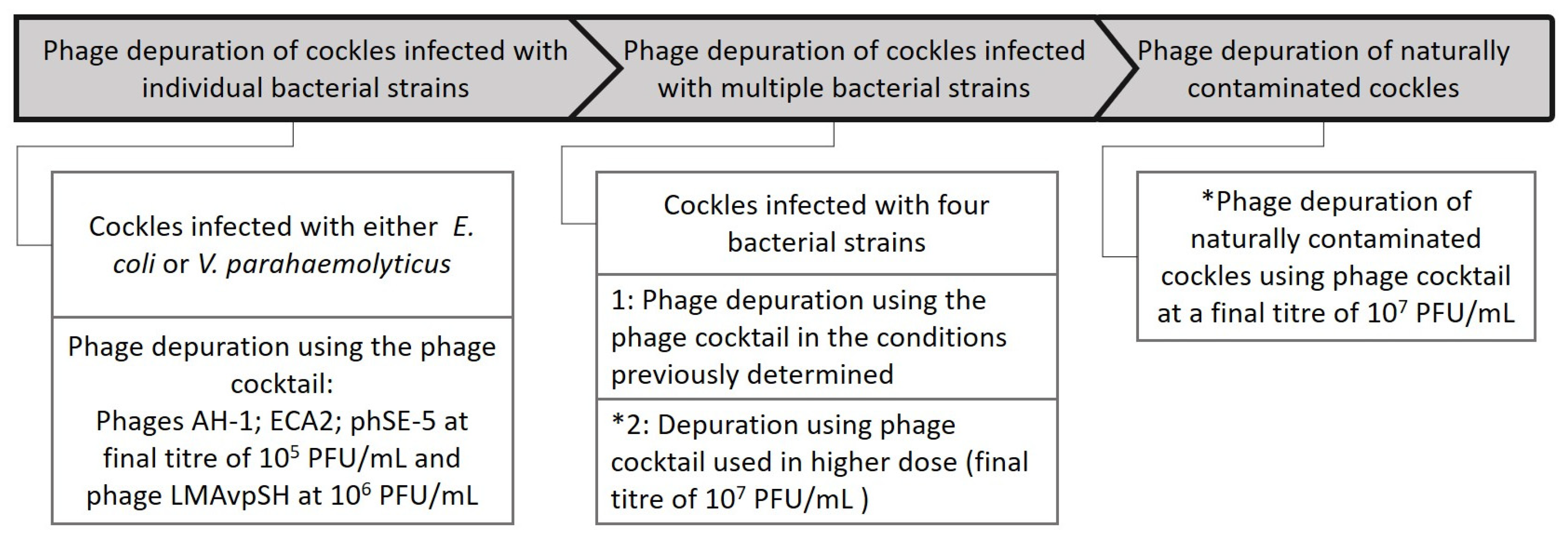
Common cockles are of high socioeconomic importance in Portugal, namely in the region of Aveiro [49], where this study was performed. Commercially depurated cockles were purchased from a local supplier and used to evaluate the effects of the phage cocktail under controlled conditions against single and multiple bacterial species artificially added to the bivalves (Section 2.5 and Section 2.6) (Figure 1). Subsequently, cockles harvested from the wild were also used to evaluate the effectiveness of the cocktail against the bacteria naturally present in mollusc bivalves (Section 2.7) (Figure 1). To monitor bacterial numbers during the experiments, cockle samples were quantified by plating on Tryptic Soy Agar (TSA; Liofilchem, Roseto degli Abruzzi, Italy) or saline TSA (3% w/v NaCl) for marine bacteria as some Vibrio species do not grow in the absence of salt [50]. Selective and differential media were also used in the experiments with multiple bacterial species and with naturally contaminated bivalves to further analyse treatment effects on specific bacterial groups (Section 2.6 and Section 2.7) (Figure 1). Phages were quantified using standard TSA. Bacteria and phages were quantified in the cockles, using the flesh and intra-valvular liquid (FIL), but in the experiments with multiple bacterial species and with naturally contaminated bivalves (Section 2.6.2 and Section 2.7), the determinations were also done in water, with samples being collected from the reservoir water tank (Figure 2 (1)) and in case of bacteria, also from the tank water of each experimental bivalve groups (Figure 2 (8)).

2.1. Bacterial Strains and Growth Conditions

The bacterial strains used in the present study were A. hydrophila (ATCC 7966), E. coli (ATCC 13706) and S. Typhimurium (ATCC 13311), all being acquired from the American Type Culture Collection. Vibrio parahaemolyticus (022C) was isolated from water samples and already used in a previous study [38]. Bacterial cultures were preserved at 4 °C in standard TSA or saline TSA (3% w/v NaCl) for marine bacteria. For each experiment, a single colony was inoculated in Tryptic Soy Broth (TSB, Liofilchem, Roseto degli Abruzzi, Italy) and incubated overnight at 25 °C at 120 rpm, reaching a final density of approximately 109 CFU/mL. Saline TSB (3% w/v NaCl) was used for marine bacteria.

2.2. Phage Purification, Enrichment, and Combination in Cocktail

A polyvalent phage cocktail of four phages: AH-1, ECA2, phSE-5, and LMAVpSH (in short SH) designed to treat specific bacteria known to contaminate bivalves was employed in the present study. The phages used in this cocktail are part of our laboratory collection [38,42,43,44] and are kept at 4 °C in SM buffer (0.1 M NaCl (Sigma-Aldrich, Darmstadt, Germany), 20 mM Tris-HCl (Sigma-Aldrich, Darmstadt, Germany), and 8 mM MgSO4 (Sigma-Aldrich, Darmstadt, Germany), pH 7.5). Phage titres were individually monitored by double agar layer, using soft TSB and standard TSA [51] on their specific hosts: (phage AH-1) A. hydrophila; (Phage ECA2) E. coli; (phage phSE-5) S. Typhimurium; and (phage SH) V. parahaemolyticus. New stocks were produced according to [38] when the titre decreased considerably or if more volume of phage stock was required. Phages were kept separate and were combined in cocktail only when used. To prepare the phage cocktail, phages were individually added to the water tank to a final concentration of 105 plaque forming units per millilitre (PFU/mL) for phages AH-1, ECA2, and phSE-5 and 106 PFU/mL for phage SH, with these concentrations having been previously determined in vitro as the most suitable for infection in vitro studies [28,38,42,43,44].

2.3. Animal Handling and Conditioning

Commercially fresh depurated cockles (30.2 ± 1.36 mm) were selected as biological models, in line with previous studies [43,44]. Specimens were purchased from a local wholesaler (Mar de Sensações Lda. Gafanha da Nazaré, Portugal), after being depurated for 48 h at 15–16 °C in a commercial depuration facility using natural seawater irradiated with UV–C. Subsequently, these cockles were transported to the laboratory and rinsed in sterile synthetic seawater [prepared by dissolving Tropic Marin Pro Reef salt (TropicMarine, Wartenberg, Germany) with freshwater purified by a reverse osmosis unit, according to the instructions provided by the manufacturer, at a salinity of 35 ppt and a temperature of 16 ± 1 °C. After this initial procedure, all specimens were transferred to aerated tanks with sterile synthetic seawater (prepared as described above) at a salinity of 35 ppt and a temperature of 16 ± 1 °C, prior to being transferred to a laboratory scale depuration system [43].
Cockles were also collected from the wild at Canal do Espinheiro (Ria de Aveiro, Portugal) 40°39′16.6″ N 8°41′53.4″ W (RIAV3 area) with a B classification (according to Regulations EC 853/2004 and 2285/2015) in terms of microbiological safety (230–4600 MPN E. coli per 100 g of FIL) (Section 2.8). These organisms were not previously depurated, being transported to the laboratory post capture and rinsed in sterile synthetic seawater as detailed above for previously depurated conspecifics.

2.4. Bacterial Bioaccumulation in Depurated Cockles

A total of 240 commercially depurated cockles were used in this experiment. Specimens were randomly divided in five equal groups (four test groups and one control group) and were placed in glass tanks (100 mm long × 90 mm wide × 150 high) of approximately 1.35 L, operating as a static system, with aeration, and filled with 0.6 L of sterile synthetic seawater. For each test, three different experimental replicate tanks per treatment were used, each one holding 16 cockles. To evaluate the rate of bacterial bioaccumulation in cockles, the water of individual tanks was inoculated with bacteria to a final concentration of 105 CFU/mL. Four different bacterial strains were used: A. hydrophila, E. coli, S. Typhimurium, and V. parahaemolyticus forming the four test groups. In parallel, a group of uninfected animals was kept in the same conditions but without bacterial addition to the water (control group; CTR). Before the beginning of the experiment and every 6 h post exposure, three cockles were randomly selected from each tank and their FIL was pooled to a final weight of 10 g and blended in 90 mL of alkaline peptone water (Liofilchem, Roseto degli Abruzzi, Italy). The sample was homogenised using a Bag Mixer 400 (Interscience, Saint Nom la Brétèche, France), serially diluted ten-fold and an aliquot of each dilution was pour plated using molten TSA. Colony counts were performed after an incubation period of 48 h at 25 °C [44]. Bacterial numbers in the FIL were quantified by plating in TSA or saline TSA for marine bacterium. This experiment was repeated in three different days.
Considering the results retrieved from the bacterial bioaccumulation experiments, which revealed that, within 6 to 12 h, all cockles accumulated bacterial concentrations comparable to those added to the water, a 12 h exposure period was selected for all subsequent experiments.

2.5. Depuration of Artificially Contaminated Cockles with Individual Bacterial Strains

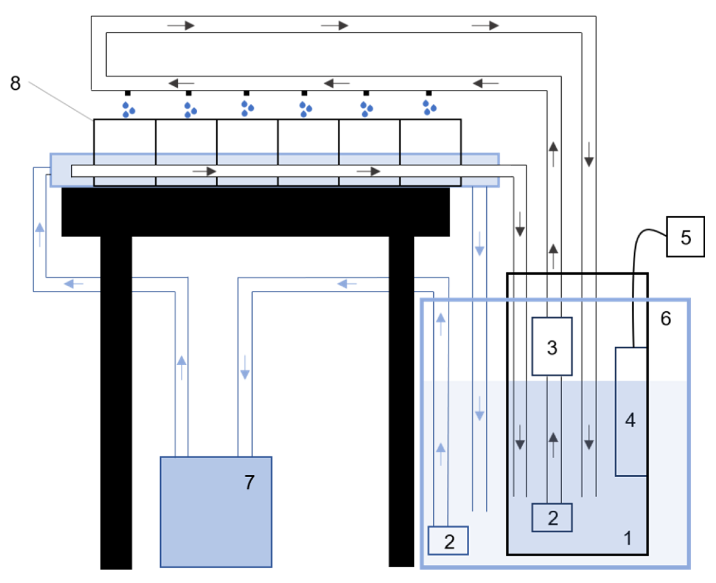
The decontamination experiments were performed using a laboratory scale depuration system (Figure 2) described in detail in previous studies [42,43]. Briefly, each of the four lines of tanks was connected to an independent 16 L water reservoir, allowing the study of independent variables. Water was recirculated by a submerged pump (Eheim Compact+ 3000; Eheim, Deizisau, Germany) and irradiated with 6W UV-C lamp PC-6T5 (TMC, Chorleywood, UK) using a UV filter TMC V2 Vecton 120 Nano (TMC, Chorleywood, UK). To reduce the levels of organic matter in the water, a protein skimmer (RedSea Berlin Air-Lift 60; Red Sea Fish Pharm, Herzliya, Israel) equipped with an air pump (API Rena Air 200; Aquarium Pharmaceuticals, Glen Burnie, MD, USA) was installed in each tank. The reservoir tanks were placed inside a larger 120 L tank that recirculated water through a cooling unit (Hailea HC 500-A; Hailea Aquarium, Guangzhou, China) using a submerged water pump to maintain a cooler temperature in the water tanks during depuration.
Figure 2. Side-view schematic of the laboratory-scale depuration system. 1—14 L water tank; 2—Submerged water pump (Eheim Compact+ 3000; Eheim, Deizisau, Germany); 3—UV filter (TMC V2 Vecton 120 nano; TMC, Chorleywood, UK); 4—Protein skimmer (RedSea Berlin Air-Lift 60; Red Sea Fish Pharm, Herzliya, Israel); 5—Air pump (API Rena Air 2000; Aquarium Pharmaceuticals, Glen Burnie, MD, USA); 6—120 L cooling water tank; 7—cooling unit (Hailea HC 500-A; Hailea Aquarium, Guangzhou, China); and 8—independent tank line. The cooling system functioned as a recirculating water bath, simultaneously cooling the four independent water reservoirs and their tank lines. Arrows indicate the direction of the water flow.
Fishes 11 00103 g002
To understand the effects of phage application during depuration of artificially contaminated cockles, two different experiments were performed using bacteria commonly detected in contaminated bivalves: (1) E. coli, an indicator of faecal contamination used to monitor shellfish farming areas and evaluate the efficiency of depuration procedures; and (2) V. parahaemolyticus, a bacterium implicated in food-poisoning outbreaks and recognised for its resistance to depuration procedures commonly employed by the industry [10].
Commercially depurated animals were infected with either E. coli or V. parahaemolyticus in the abovementioned conditions (Section 2.4) and evenly distributed in three of the tank lines of the laboratory scale depuration system. Four experimental groups were established (Table 1), each one with three replicate tanks: a control group—uninfected commercially depurated cockles being maintained in recirculating water without any treatment; a bacterial control group—infected cockles being maintained in recirculating water without any treatment; an UV-irradiated group—infected cockles being maintained in recirculating water system with UV lamps to mimicking commercial depuration facilities; and a phage treatment group—infected specimens being maintained in recirculating water in the presence of the phage cocktail without UV radiation. In the beginning of the experiment and every 6 h, three cockles were randomly selected from each tank for bacterial quantification, as detailed above (Section 2.4). Additionally, an aliquot of the phage treatment group was plated by double agar layer using the respective host bacteria (E. coli or V. parahaemolyticus) to quantify the titre of phages in the cockles. Colony counts were performed after an incubation period of 48 h at 25 °C and phage plaque counts were performed after incubation for 18 h at 25 °C.

2.6. Depuration of Artificially Contaminated Cockles with a Mixture of the Four Different Bacterial Stains

As multiple bacterial species may naturally be present in bivalves, cockles were simultaneously exposed to all four bacterial strains (A. hydrophila, E. coli, S. Typhimurium, and V. parahaemolyticus) at a final concentration of 105 CFU/mL.

2.6.1. Depuration Using the Phage Cocktail (AH-1, ECA2, phSE-5 and LMAVpSH)

Following infection for 12 h, cockles were transferred to the depuration system and treated under the same conditions as described above (Section 2.6 and Table 1). Additionally, to monitor phage numbers in the recirculating water, a water sample was collected from the reservoir water tank (Figure 2 (1)) connected to the phage tank line after 6 and 12 h of incubation and plated by double agar layer using the four different hosts individually. Bacterial counts were performed after 48 h of incubation at 25 °C and phage plaque counts were performed after incubation for 18 h at 25 °C.

2.6.2. Depuration Using a High Dose (MOI 100) of the Phage Cocktail

As the efficacy of treatment was reduced when the phage cocktail was applied to multiple bacterial strains, an additional experiment was prepared using a phage dose 100 times higher than the initial one (MOI 100). For this experiment, each individual phage was added to the tank water to a final titre of 107 PFU/mL. Colony counts and phage titre in cockles were determined in the same conditions described above (Section 2.6.1). To better understand the effects of this specific treatment, aliquots of water from the reservoir water tank (Figure 2 (1)) of all the four groups were collected and bacterial numbers were monitored by plating in TSA and saline TSA. Additionally, to monitor the effects of treatment on the reduction of a specific bacteria, 100 µL aliquots of bivalve homogenate were also spread plated in selective media, namely: Glutamate starch phenol red agar (GSP agar, Sigma, Germany), Thiosulfate–citrate–bile salts–sucrose agar TCBS (Liofilchem, Italy), and SS agar (Liofilchem, Italy) used to quantify Aeromonas (yellow colonies in GSP), Vibrio (green or yellow colonies in TCBS), E. coli (pink colonies in SS), and S. Typhimurium (dark colonies in SS), respectively. Colony counts were performed after 48 h at 25 °C and phage plaque counts were performed after incubation for 18 h at 25 °C.

2.7. Depuration of Naturally Contaminated Bivalves Using the Phage Cocktail (MOI 100)

To understand if the results from previous experiments performed using commercially depurated cockles artificially infected could be transferred to a real-world application, naturally contaminated conspecifics were used in this experiment. As these bivalves were not artificially infected, they were only divided in three experimental groups: a control group, with uninfected specimens being kept in recirculating water; a UV group, with uninfected specimens being kept in recirculating water treated with UV and; a phage group, with uninfected specimens being treated with the phage cocktail at a final concentration of 107 PFU/mL. Every 6 h interval, tank water and specimens were sampled for quantifying bacteria and phages. Tank water was analysed by collecting an aliquot from each experimental tank reservoir (Figure 2 (1)). Bacteria colony counts in cockles were performed by pour plate in TSA, saline TSA, and by spread plate using the four selective media (Section 2.6.2). Water bacterial colony counts were performed only in TSA and saline TSA. Phages from bivalve samples and water samples of the reservoir tank were quantified by double agar layer using the four different bacterial hosts individually. Bacteria plates were incubated for 48 h at 25 °C and phage plaque counts were performed after 18 h of incubation at 25 °C.

2.8. Statistical Analysis

Statistical analyses were performed using GraphPad Prism 8.4.3 (San Diego, CA, USA). Normality and equality of variances were assessed using the Kolmogorov–Smirnov and Levene tests, respectively. For Section 2.4 and Section 2.5, bacterial concentrations were analysed using two-way ANOVA with treatment (four levels: control, control bacteria, UV-irradiated, and phage treatment) and time (three levels: 0 h, 6 h, and 12 h) as fixed factors. Tukey’s multiple comparison test was applied for post hoc analysis between all groups. Data represent the mean of three independent experiments, each including three tanks per treatment, and test samples were compared with the corresponding control group at each sampling interval. For Section 2.6 and Section 2.7, a single experiment was performed with three tanks per treatment serving as biological replicates. Within each tank and time point, three tanks were sampled as technical replicates, and their mean was used as the biological replicate for statistical analysis. Data were analysed using two-way ANOVA with treatment and time as fixed factors, followed by Tukey’s multiple comparison test between all groups. A p-value < 0.05 was considered statistically significant.
For variations in viral titres in cockles (Section 2.6.1, Section 2.6.2 and Section 2.7), at each time point (6 h and 12 h), a paired design was used, with each tank serving as biological replicate (n = 3). Due to the small sample size, a nonparametric Wilcoxon matched-pairs signed-rank test was performed. All statistical analyses were two-tailed, and p < 0.05 was considered statistically significant.
The single multi-bacterial assay was considered sufficient because prior validation experiments demonstrated high reproducibility of the depuration system, with low variation between independent experiments, and additional independent experiments were therefore not expected to substantially improve statistical robustness. By performing only a single assay, it was possible to minimise the use and sacrifice of animals, in accordance with the 3Rs principle for animal experiments.

3. Results

3.1. Bacteria Bioaccumulation in Depurated Cockles

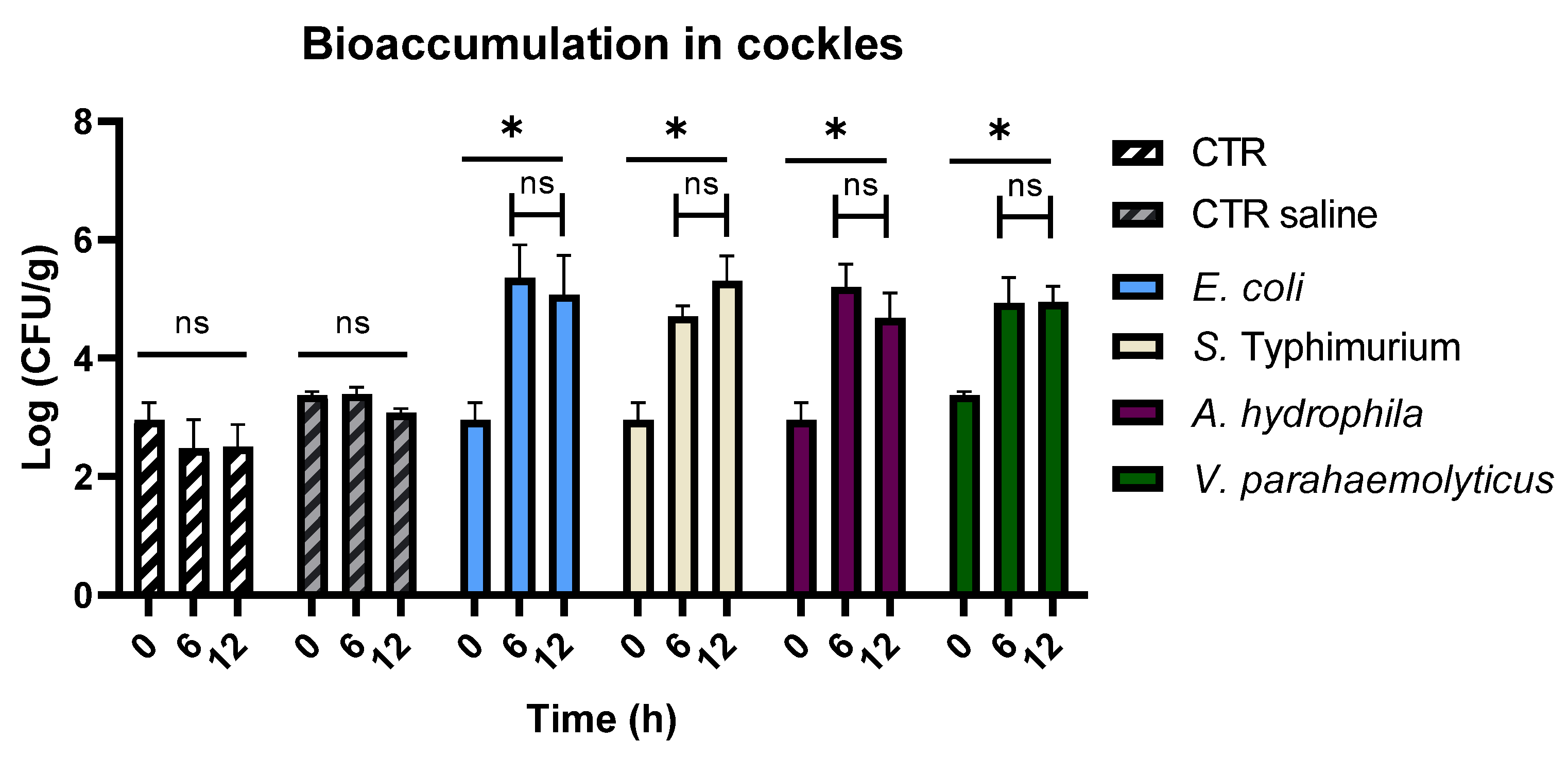
When cockles were kept under aeration in seawater artificially contaminated with each individual bacterial strain tested, bacterial concentrations inside cockles rapidly rose to concentrations similar to those of surrounding water after 6 h (Figure 3). The bacterial concentrations observed in all groups after 6 h were significantly higher than in the uninfected control group (CTR) (p < 0.05). After 12 h of experiment, no significant differences were observed among infected groups (p > 0.05). Furthermore, when comparing bacterial concentrations within infected cockles over time, no significant differences were detected between 6 and 12 h (Figure 3, p > 0.05). Based on these results, a 12 h (overnight) incubation period was selected to facilitate subsequent experiments.

3.2. Depuration of Cockles Artificially Contaminated with Escherichia Coli

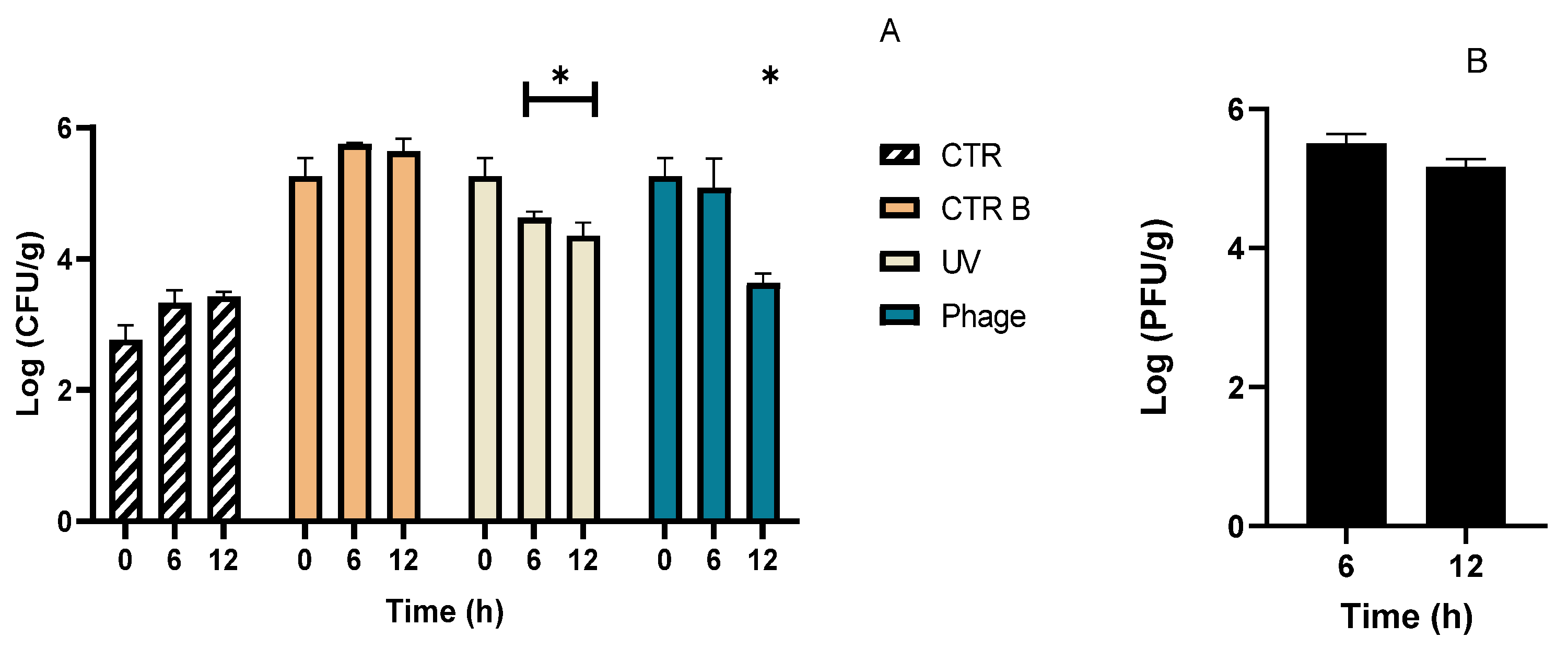
When artificially contaminated cockles were transferred for depuration, the effects of treatment began to be observed after 6 h (Figure 4A). At this time, the concentration of bacteria in the cockles treated with UV irradiation and with phages was significantly lower than the bacterial concentration in the control group (CTR B) (p < 0.05). However, no significant differences were observed when the UV irradiated group was compared with the phage treatment group (p > 0.05). After 12 h of depuration, both the UV irradiated cockles and those from the phage treatment groups remained significantly lower (p < 0.05) than the bacterial control (CTR B), with a reduction of 0.91 and 1.64 Log CFU/g, respectively. Additionally, the phage treatment group showed significantly lower levels than the UV irradiated group (p < 0.05), but did not differ significantly from the uninfected control (CTR) group (p > 0.05). Although no significant differences were detected (p > 0.05), the bacterial concentration in the animal control increased by 0.66 Log CFU/g during the 12 h of experiment. Phage quantification in cockles showed that after 6 h, phage levels were similar to the administered dose, followed by a slight decrease during the remaining treatment (Figure 4B).

3.3. Depuration of Cockles Artificially Contaminated with Vibrio Parahaemolyticus

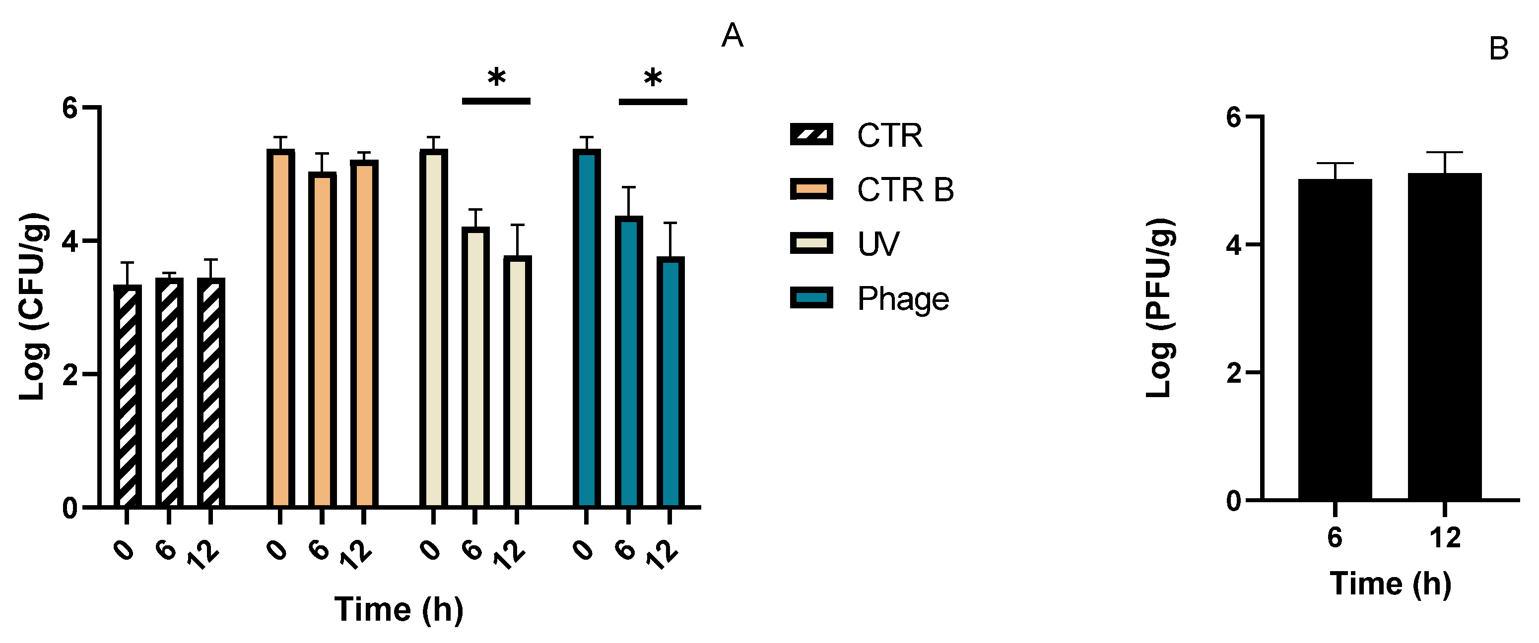
When the same experiment was repeated for cockles artificially contaminated with V. parahaemolyticus, the effects of treatment were observed earlier than in the E. coli experiments (Figure 5A). After 6 h of treatment, bacterial concentrations in both the UV and with the phage cocktail treatments were significantly lower than the infected control group (CTR B) (p < 0.05) with a reduction of 1.16 and 1.00 Log CFU/g, respectively (Figure 5A). After 12 h of depuration, bacterial concentration in treatment groups continued to decrease, reaching reductions of 1.59 and 1.61 Log CFU/g in the UV and phage treatment, respectively. After 12 h, no significant differences (p > 0.05) were observed between the treatment with UV and the phage cocktail nor between both treatments and the uninfected control (CTR) (p > 0.05). Bacterial concentration in specimens of the uninfected control group (CTR) remained constant during the experimental period. After 6 h, phage quantification in the cockles reached 5.02 Log PFU/g and remained stable throughout the 12 h experiment period (Figure 5B).

3.4. Depuration of Cockles Artificially Contaminated with the Mixture of the Four Bacterial Strains

3.4.1. Depuration with Phage Cocktail (AH-1, ECA2, phSE-5, and LMAVpSH)

When cockles were infected with the four different bacterial strains, the efficacy of depuration was reduced (Supplementary Figure S1A,B). After 12 h, a significant decrease (p < 0.05) was observed for the UV irradiated group, with reductions of 0.98 and 0.79 Log CFU/g for plate counts on TSA and saline TSA, respectively. In contrast, the phage treatment group did not show any significant differences in bacterial concentration (p > 0.05), despite the high phage titres (varying between 4.9–5.5 Log PFU/g) detected inside the cockles (Supplementary Figure S1A). Bacterial counts on standard TSA and saline TSA showed only slight differences in concentration (approximately 0.2 Log CFU/g), showing bacterial concentrations in the infected control group (CTR B) varied from 5.97 to 5.77 Log CFU/g when plated on standard TSA and saline TSA, respectively, with no significant differences (p > 0.05). The uninfected control group (CTR) maintained the bacterial concentrations, with a variation of only 0.1 Log CFU/g.
Phage titres in cockles (Supplementary Figure S2A) showed that all phages in the cocktail successfully reached the animals with only slight reductions when compared with the water (Supplementary Figure S2A,B). After 12 h, increases in the titres of phages infecting A. hydrophila and V. parahaemolyticus were observed in both the water and cockles. Phages targeting A. hydrophila increased by 0.2 Log PFU/mL and 0.07 Log PFU/g, while those targeting V. parahaemolyticus increased 0.32 Log PFU/mL and 0.2 Log PFU/g in the water and in the cockles, respectively. Phages infecting S. Typhimurium increased by 0.3 Log PFU/g in cockles, but showed no change in tank water. Conversely, phages infecting E. coli increased by 0.2 PFU/mL in the water, but their titres remained unchanged in cockles. In cockles, phages showed a consistent increase across all tanks sampled but none of the changes were statistically significant when analysed with the nonparametric Wilcoxon matched-pairs signed-rank test (n = 3).

3.4.2. Depuration with Phage Cocktail at MOI 100

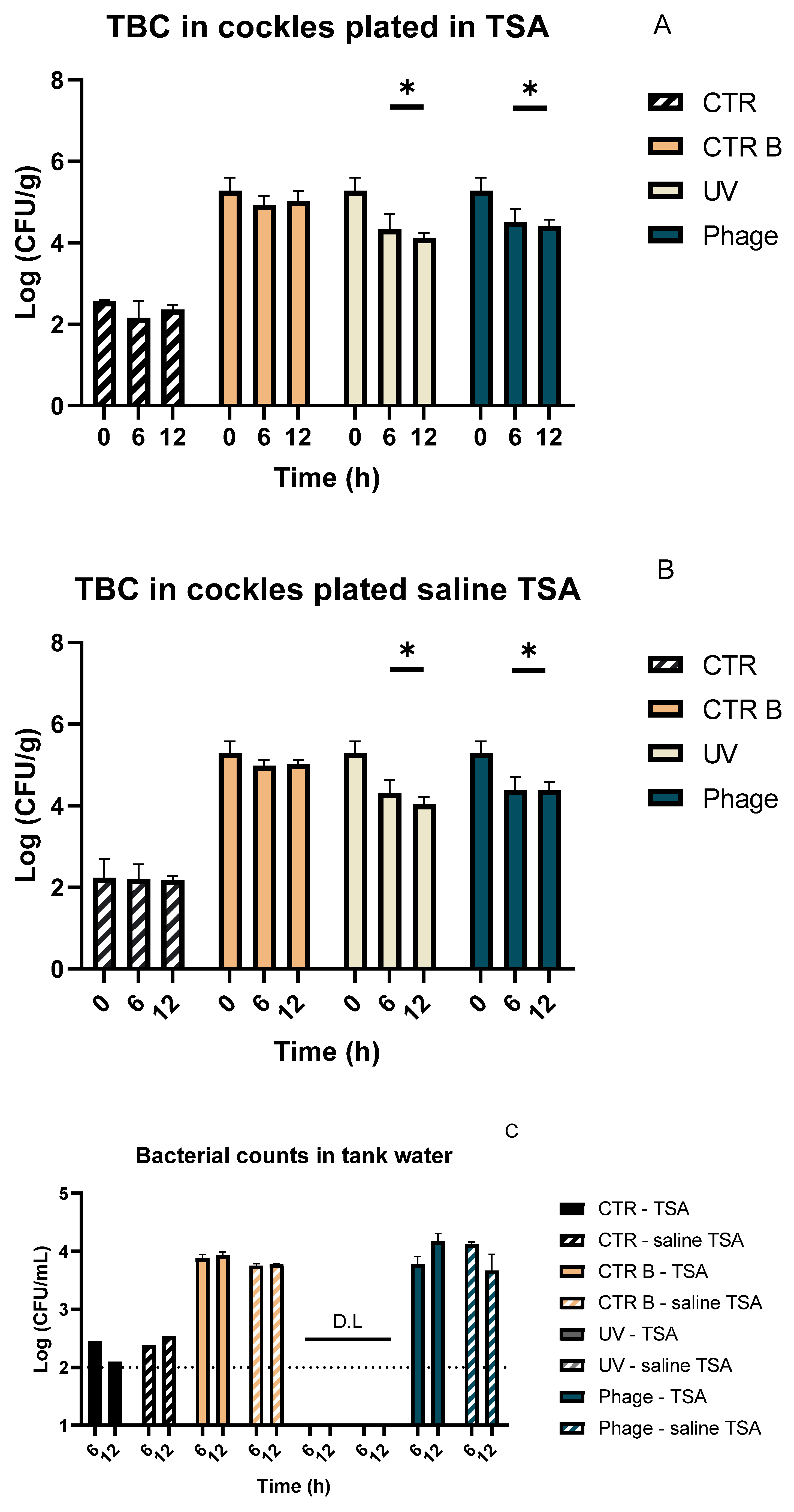
After 12 h of depuration, the UV treatment group showed significantly lower bacterial levels than the infected control group (CTR B) (p < 0.05) with reductions of 1.17 and 1.26 Log CFU/g on TSA and saline TSA, respectively. Treatment with phages showed a significant bacterial decrease (p < 0.05) of approximately 0.88 and 0.91 Log CFU/g on standard TSA and saline TSA, respectively, compared with the infected control (CTR B) after 12 h of depuration (Figure 6A,B). No significant differences were detected between UV and phage treatments after 12 h (p > 0.05). In the uninfected cockle group (CTR), no statistically significant changes (p > 0.05) were observed throughout the experiment. Bacterial counts in selective and differential media revealed decreases in GSP, SS, and TCBS, suggesting a reduction in the corresponding target species: A. hydrophila, E. coli, S. Typhimurium, and V. parahaemolyticus (Supplementary Figure S3A–D). To further understand the effects of the treatment, aliquots of water from the experimental tanks and from the reservoir tank were collected in parallel for analysis after 6 and 12 h (Figure 6C). A lower bacterial concentration in recirculating water was recorded when compared to the levels detected in cockles. However, in water samples from the phage tank group, an increase in bacterial levels of 0.4 Log CFU/mL was recorded between 6 h and 12 h when plated in standard TSA. After 12 h, the bacterial concentration in the phage group was higher than in the infected control group (CTR B), with values of 4.18 Log and 3.94 Log CFU/mL, respectively.
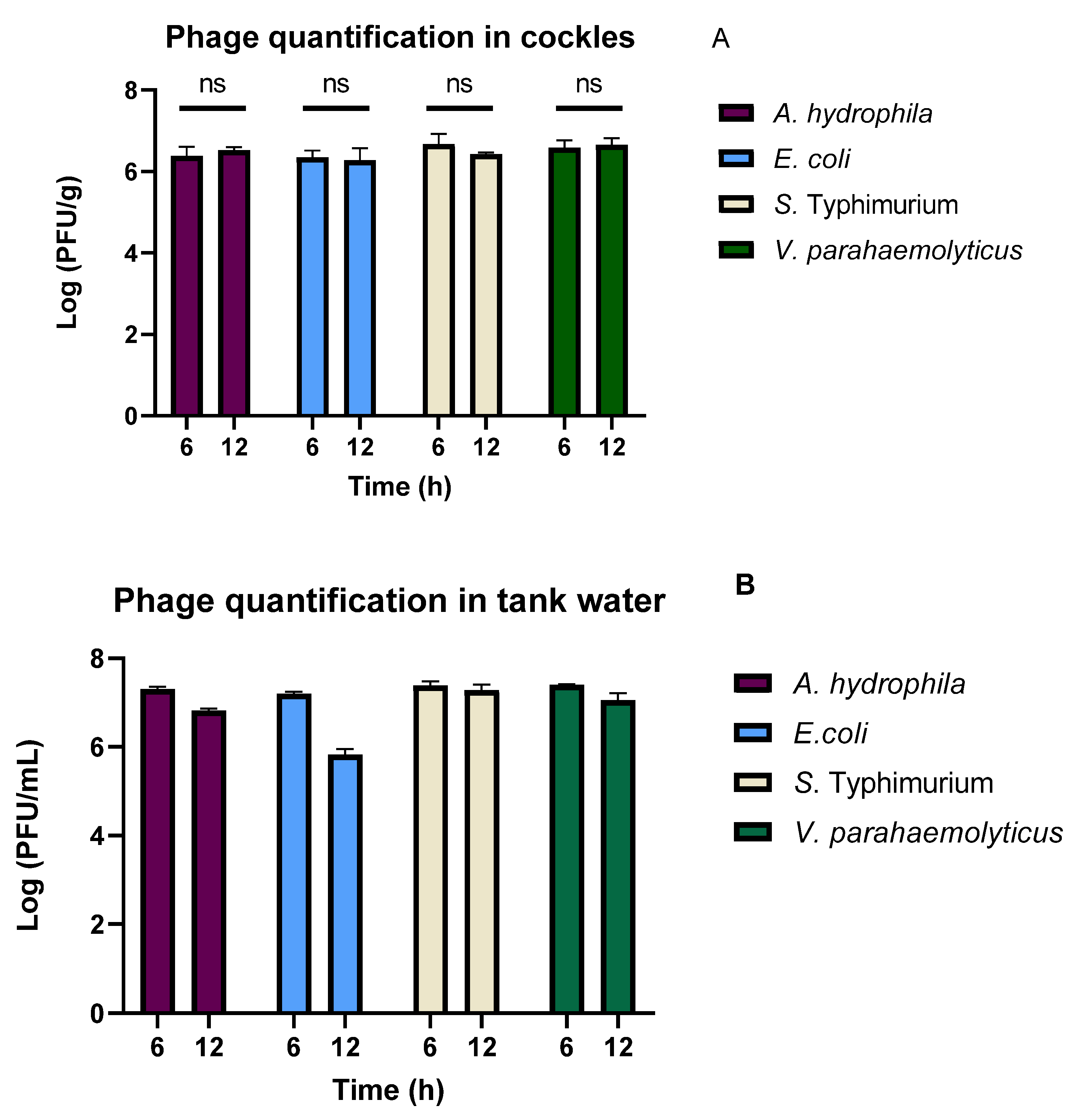
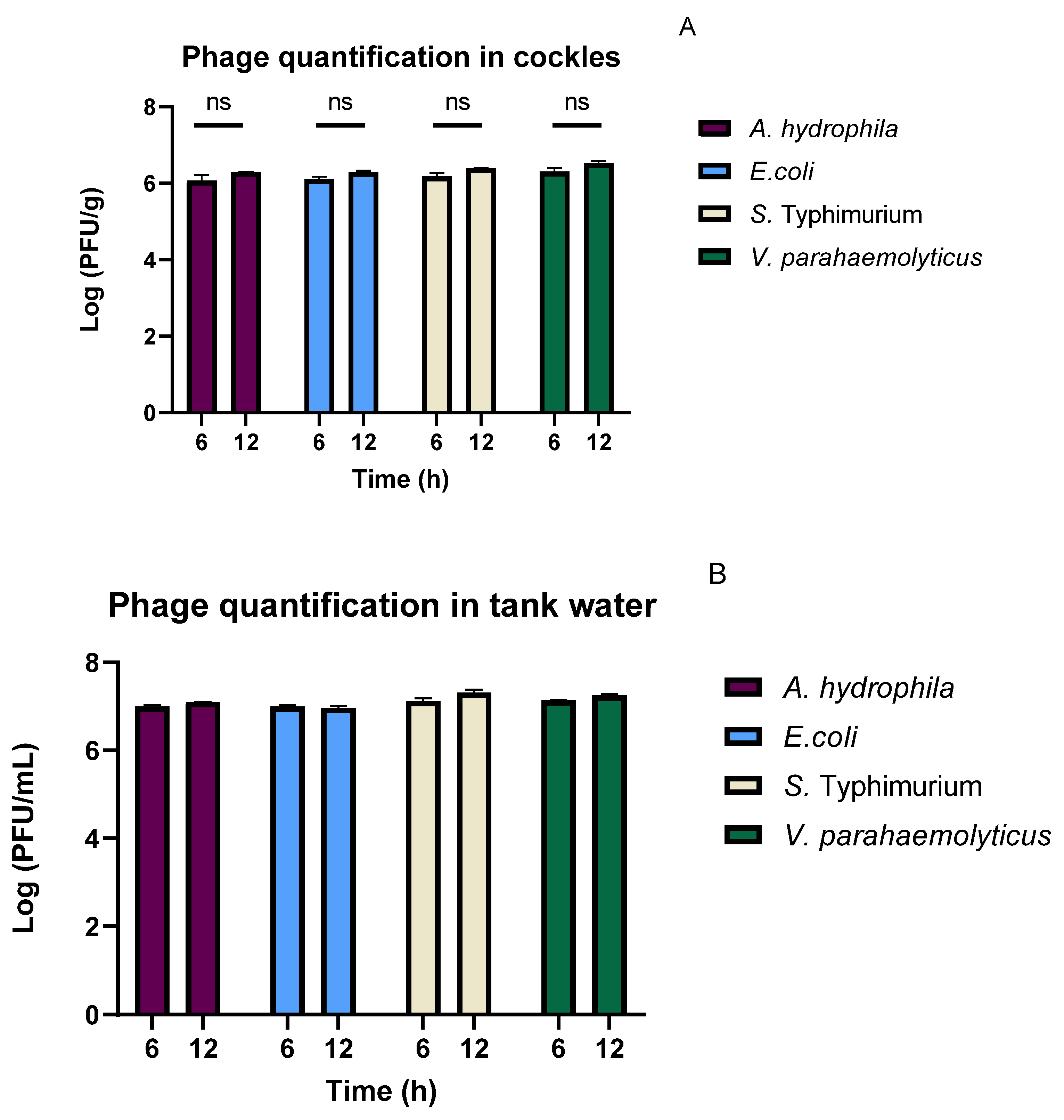
Phage titre quantification in bivalves (Figure 7A) showed that the phages reached the cockles and remained at high levels throughout the experiment. Despite this, a difference of approximately 0.5 Log was observed between the phage concentration in cockles and in tank water (Figure 7B). After 12 h of experiment, the number of phages infecting E. coli decreased in the water; however, phage quantification in bivalves did not reveal any changes. Variations across cockles from all tanks sampled were not statistically significant when analysed with the nonparametric Wilcoxon matched-pairs signed-rank test (n = 3).

3.5. Treatment of Naturally Contaminated Bivalves

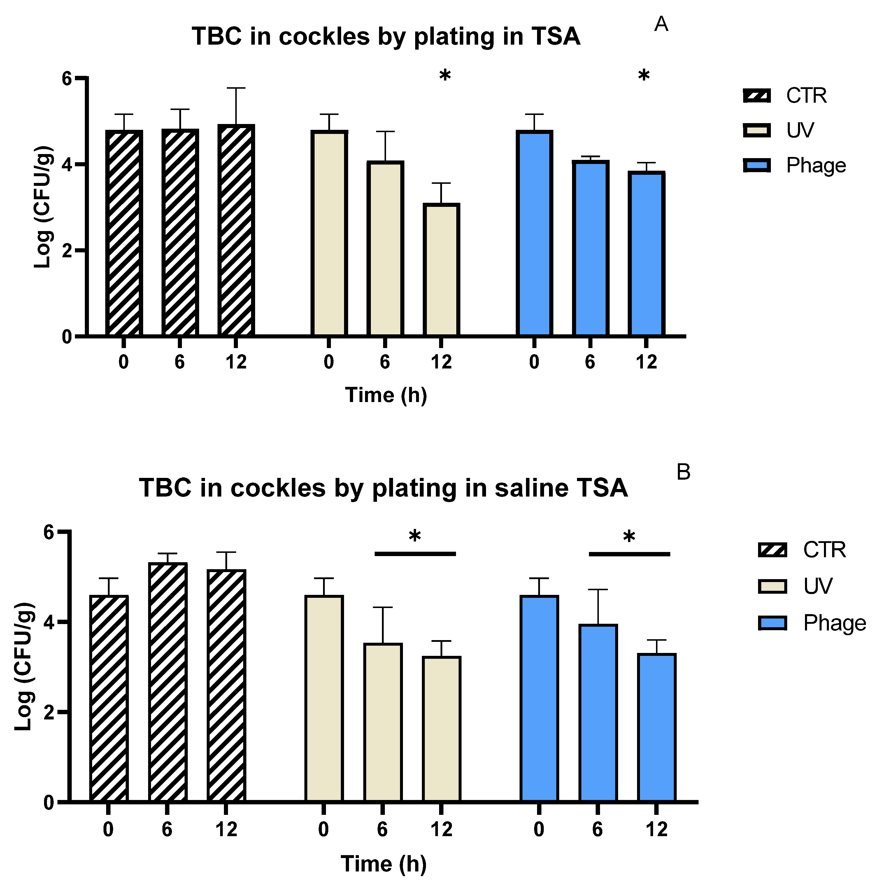
When the same experiment was performed to treat naturally contaminated cockles, a high decrease in bacterial contents was observed after treatment (Figure 8A,B). UV treatment resulted in a significant decrease (p < 0.05) of 0.79 Log CFU/g in bacterial counts after 6 h when plated on saline TSA (Figure 8B). However, bacterial counts on standard TSA did not differ significantly from the control group at this time (p > 0.05). After 12 h, both plate counts continued to decrease and were significantly lower than the control group (p < 0.05), with reductions of 1.69 and 1.12 Log CFU/g for TSA and saline TSA, respectively.
Cockles treated with the phage cocktail also showed a reduction in bacterial load after 6 h. However, at this point, the decrease in bacterial concentration differed significantly (p < 0.05) from the control group only for saline TSA, with a reduction of 0.64 Log CFU/g). After 12 h, reductions of 0.95 and 1.28 Log CFU/g were recorded for standard and saline TSA, respectively, both significantly lower than the control group (p < 0.05). Bacterial counts in specific media showed high variability in bacterial loads particularly for E. coli (Supplementary Materials).
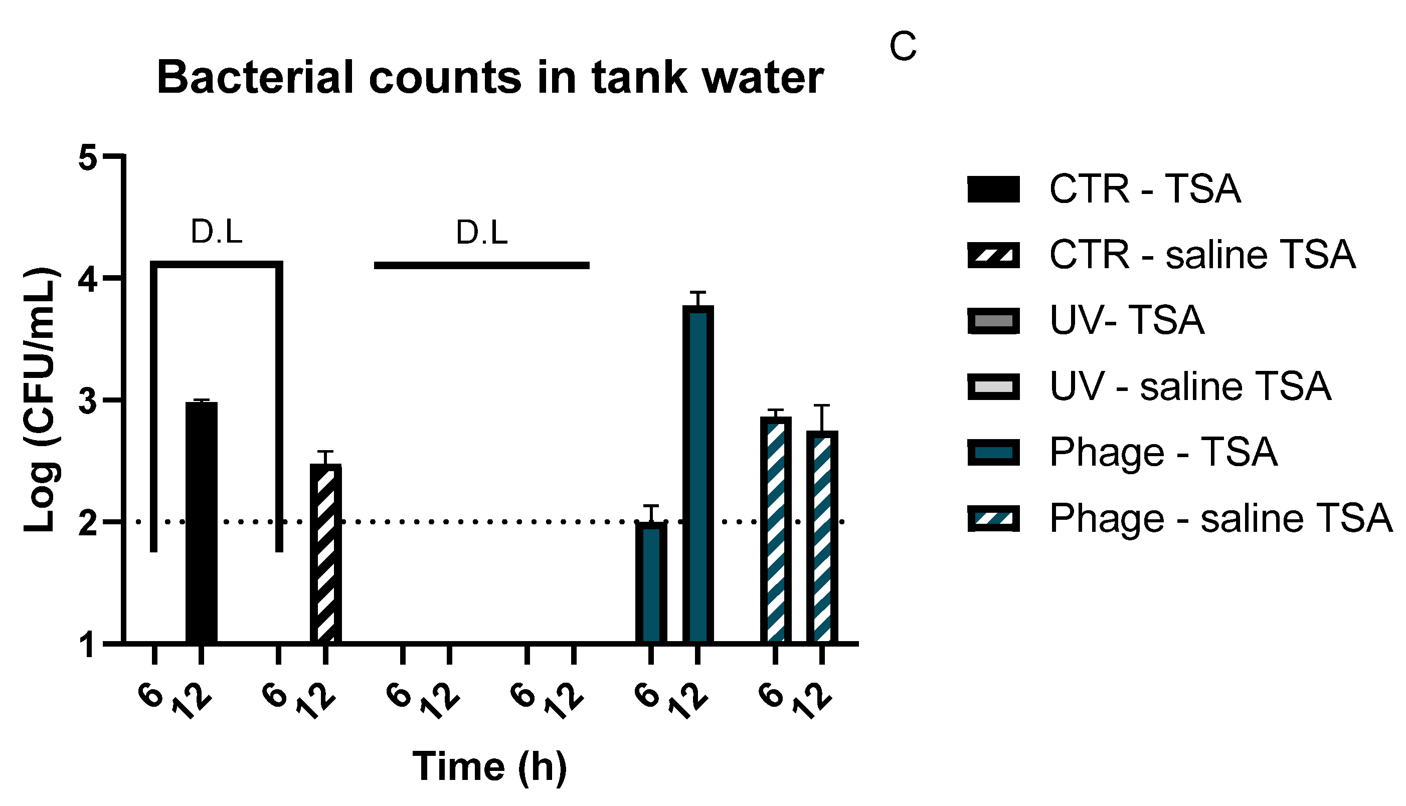
Bacterial quantification in tank water showed an increase in bacterial numbers between 6 and 12 h for the control group and the phage treatment group (Figure 8C). In the phage treatment group, bacterial concentration in water increased from approximately 2.0 to 3.77 Log CFU/mL in standard TSA. In the control group water, bacterial concentrations were below the detection limit after 6 h but subsequently increased to final concentrations of 2.98 and 2.47 log CFU/mL on TSA and saline TSA, respectively. Nonetheless, concentrations remained higher in cockles than in the water.
Phage quantification in cockles and in the tank water (Figure 9A,B) showed that the titres remained high throughout the 12 h of experiment. In cockles, phages showed a consistent increase across all tanks sampled but none of the changes presented statistical significance when analysed with the nonparametric Wilcoxon matched-pairs signed-rank test (n = 3). However, as observed in the previous experiments, a difference of about 1.00 Log was recorded between the titre in water and that inside cockles.

4. Discussion

The ability to enhance bivalve depuration using phages offers a promising technology for depuration facilities by reducing operation costs and improving food safety [15].
In the present study, when phages were used to treat cockles artificially contaminated with individual bacterial strains, bacteria could be efficiently reduced to levels similar to those observed after UV treatment. In the experimental treatment of bivalves contaminated with E. coli, a collection strain (ATCC 13706) was selected because it was considered a strong biofilm former after 24 h of incubation on plastic surfaces [52]. The complex matrices of bacterial biofilms may have delayed the effect of phage treatment, observed only 12 h after the beginning of the experimental trials [53]. Contrarily, for cockles infected with V. parahaemolyticus, differences in bacterial load began to be observed after only 6 h. Similar results were observed when cockles were treated in static water systems using other bacterial species [43,44]. These differences can be the result of contrasting life cycles of the phages employed in the present study, as the Vibrio phage LMAvpSH presents a fast bacterial adsorption, with bacterial decrease being observed after only 2 h of incubation [38]. As Vibrio contaminations can force lengthy depuration treatments [10], the ability of phage LMAvpSH to reduce this bacterium effectively is a promising result for its application.
As bivalves naturally bioaccumulate different bacterial species, understanding the effect of phage use at a multi-bacterial scale is paramount. Therefore, the second step of our experiment was to evaluate the efficacy of the phage cocktail against the four bacterial species initially selected. When cockles were infected with multiple bacterial species and treated under the same conditions as when infected with individual bacterium, the efficacy of the cocktail was lower than that observed against E. coli and V. parahaemolyticus. Despite the high phage titres detected in cockles and tank water, bacterial numbers remained unaffected. The inclusion of several bacterial species in this assay may have resulted in the production of biofilm matrices that hindered phage penetration to infect bacterial cells [53], leading to the slight increase in phage titre, but requiring prolonged exposure to inactivate bacteria [54]. Additionally, as the phage titre did not decrease inside the bivalves, it is possible that phages have been reversibly adsorbed within the biofilm matrix. These adsorbed phages remain detectable, but are unable to successfully infect viable host cells [55]. Another possible explanation may be differences in the extent of bacterial colonisation of bivalves. As multiple bacterial species were used, their colonisation of the gut, gills, and haemolymph [11] may present additional challenges for phage infection. Phage diffusion can be hindered by the natural viscosity and binding properties of bivalve mucus [56]. Nonetheless, even if bacterial inactivation would require prolonged periods of phage exposure, an extended treatment would limit the practical use of phages during the depuration process.
Previous studies suggest that increasing phage dose can promote a faster bacterial inactivation [38,44]. As such, the experiment detailed above was repeated using a higher phage dose (100 times higher). When the phage dose was increased, the results recorded suggest that phages were able to circumvent previous challenges and efficiently reach bacteria, leading to a decrease in bacterial concentration. Moreover, bacterial reduction occurred more rapidly, after only 6 h of treatment. The results recorded suggest that using higher phage doses may provide better outcomes for depuration. Further analysis on selective media (Supplementary Figures S3 and S4) also showed that all the four bacterial species used in this study were reduced by that same treatment. Despite the bacterial reduction inside the cockles (0.88–0.91 Log CFU/g), the bacterial concentration in the tank water remained high (4.18–3.94 Log CFU/mL) in the presence of phages. These results suggest that bacteria in the water are not active enough to be used as hosts for phage infection, probably due to the low concentration of nutrients in the water [57,58]. These results are in agreement with a previous study reporting the use of phages to treat bacteria in low-nutrient marine water systems [58]. Based on these observations, our results suggest that the use of phages in depuration units should be considered only as a complementary strategy to enhance depuration and cannot replace water disinfection as a standalone treatment.
As harvested bivalves contain varying loads of naturally occurring bacterial communities, we performed an experiment using freshly harvested cockles without artificial infection to assess the applicability of this phage cocktail in a real-world scenario. The use of a high phage dose also caused bacterial reduction in specimens collected from the wild, indicating that the broad host range of the phage cocktail extends to naturally occurring bacteria. Furthermore, the analysis of tank water after 6 h of treatment revealed that bacterial loads were higher in phage-treated tanks than in the cockle control groups. These results suggest that, under phage selective pressure, bacteria can be released into the water rather than remaining within the bivalves. This finding may be the result of a phage-induced disruption of bacterial biofilms, destabilising microbial colonisation in the bivalves. As phages lyse bacteria, depolymerase activity disrupts the biofilm matrix, which fosters the release of uninfected bacteria to the water [59,60]. This observation may also be explained by the emergence of phage-resistant bacterial mutants. As phage start to infect the bacterial population, resistant mutants begin to emerge, often presenting metabolic changes that can affect bacterial fitness [61]. These changes can be observed through a slower growth of phage resistant mutants [37,38], loss of virulence [39], and loss of ability to adhere to surfaces [40]. In fact, in a study using phages for the control of Vibrio cholerae, an increase of planktonic cells was observed during the destruction of the biofilm using phage JSF7 [62]. If bacteria are losing their ability to adhere to animal tissues and concentrate in the water, they are more likely to be efficiently destroyed by water disinfection processes, such as UV radiation or ozone, employed in commercial depuration facilities. As natural bacterial concentrations vary among species, environmental conditions, human activities, and geographic region, a large-scale study incorporating animals from multiple regions will be necessary to support industrial implementation of this technology.
Finally, as Gram-negative bacteria may release endotoxins upon lysis, future studies should evaluate the release and concentration of these molecules in bivalves, as well as their potential impact on the safety of the final product [63].

5. Conclusions

The present study demonstrates the potential of a multi-species phage cocktail to enhance industrial depuration processes. Our findings confirm that phages can effectively reduce bacterial concentration in bivalves, and that their selective pressure on natural bacterial communities may be leveraged to improve standard depuration protocols. Specifically, short-term exposure of bivalves to high phage doses prior to water disinfection may enhance the performance of well-established practices, promoting an increased bacterial clearance efficiency. These findings provide a critical proof-of-concept for the integration of phage-based applications into industrial depuration systems and pave the way for future translational applications; however, further testing under industrial conditions is needed to validate and optimise these approaches.

Supplementary Materials

The following supporting information can be downloaded at: https://www.mdpi.com/article/10.3390/fishes11020103/s1, Figure S1: Total bacterial counts in cockles (Cerastoderma edule) during depuration plated in standard TSA (A) and in saline TSA (B). Figure S2: Phage titres in cockles (Cerastoderma edule) (A) and in the depuration system water (B) after 6 and 12 h, using A. hydrophila, E. coli, S. Typhimurium and V. parahaemolyticus as hosts. Figure S3: Bacterial colony counts in cockles (Cerastoderma edule) infected with four different bacterial strains plated in Glutamate starch phenol red agar (GSP) (A); SS agar—E. coli (pink colonies) (B); SS agar—S. Typhimurium (dark colonies) (C) and Thiosulfate–citrate–bile salts–sucrose agar (TCBS) (D). Figure S4: Bacterial colony counts during depuration of freshly harvested cockles (Cerastoderma edule) plated in Glutamate starch phenol red agar (GSP) (A); SS agar—E. coli (pink colonies) (B); SS agar—S. Typhimurium (dark colonies) (C) and Thiosulfate–citrate–bile salts–sucrose agar (TCBS) (D).

Author Contributions

Conceptualisation, J.D., D.T., C.P., R.C. and A.A.; methodology, J.D. and D.T.; validation, J.D., C.P., R.C. and A.A.; formal analysis, J.D.; investigation, J.D. and D.T.; resources, R.C. and A.A.; writing—original draft preparation, J.D.; writing—review and editing, J.D., D.T., C.P., R.C. and A.A.; visualisation, J.D.; supervision, C.P., R.C. and A.A.; project administration, A.A.; funding acquisition, A.A. All authors have read and agreed to the published version of the manuscript.

Funding

This research was funded by FCT—Fundação para a Ciência e Tencologia I.P., under the project CESAM—Centro de Estudos do Ambiente e do Mar, references UID/50017/2025 (https://doi.org/10.54499/UID/50017/2025) and LA/P/0094/2020 (https://doi.org/10.54499/LA/P/0094/2020). This research also received funding from the project Phage@Kiwifruit—Bacteriophages in the control of the kiwifruit canker caused by Pseudomonas syringae pv. actinidiae (Psa) (2024.05721.RESTART), financed by FCT. FCT also supported this work through the attribution of a research contract to Carla Pereira (https://doi.org/10.54499/CEECIND/03974/2017/CP1459/CT0022).

Institutional Review Board Statement

Formal ethical approval was not required for this study because European legislation (Directive 2010/63/EU) does not cover bivalve molluscs such as cockles. All experiments were conducted following standard laboratory animal welfare practices to minimise stress.

Data Availability Statement

Data are contained within the article and Supplementary Materials.

Acknowledgments

The authors acknowledge the help of Ruben Xavier and António Ferro in the assembly of the depuration system. Thanks, are also due to the Department of Biology and the University of Aveiro, where this research was carried out. The authors are also grateful to CESAM and its funding sources. João Duarte was supported in the form of a PhD grant (https://doi.org/10.54499/2021.05519.BD) financed by national funds through the FCT.

Conflicts of Interest

The authors declare no conflicts of interest.

References

  1. Food and Agriculture Organization of the United Nations. The State of World Fisheries and Aquaculture 2024; FAO: Rome, Italy, 2024; ISBN 978-92-5-138763-4. [Google Scholar]
  2. Tamburini, E.; Moore, D.; Castaldelli, G. Global Comparison and Future Trends of Major Food Proteins: Can Shellfish Contribute to Sustainable Food Security? Foods 2025, 14, 2205. [Google Scholar] [CrossRef] [Scilit]
  3. Zhang, H.; Cheong, K.-L.; Tan, K. Bivalves as Climate-Friendly High Quality Animal Protein: A Comprehensive Review. Food Secur. 2025, 17, 739–748. [Google Scholar] [CrossRef] [Scilit]
  4. van der Schatte Olivier, A.; Jones, L.; Vay, L.L.; Christie, M.; Wilson, J.; Malham, S.K. A Global Review of the Ecosystem Services Provided by Bivalve Aquaculture. Rev. Aquac. 2020, 12, 3–25. [Google Scholar] [CrossRef] [Scilit]
  5. Salgueiro, V.; Reis, L.; Ferreira, E.; Botelho, M.J.; Manageiro, V.; Caniça, M. Assessing the Bacterial Community Composition of Bivalve Mollusks Collected in Aquaculture Farms and Respective Susceptibility to Antibiotics. Antibiotics 2021, 10, 1135. [Google Scholar] [CrossRef] [Scilit] [PubMed]
  6. Kijewska, A.; Koroza, A.; Grudlewska-Buda, K.; Kijewski, T.; Wiktorczyk-Kapischke, N.; Zorena, K.; Skowron, K. Molluscs—A Ticking Microbial Bomb. Front. Microbiol. 2023, 13, 1061223. [Google Scholar] [CrossRef] [Scilit] [PubMed]
  7. European Parliament; Council of the European Union. Regulation (EC) No 854/2004 of 29 April 2004 laying down specific rules for the organisation of official controls on products of animal origin intended for human consumption. OJ L 2004, 155, 206–320. [Google Scholar]
  8. European Commission. Commission Regulation (EC) No 2073/2005 of 15 November 2005 on microbiological criteria for foodstuffs. OJ L 2005, 338, 1–26. [Google Scholar]
  9. European Commission. Commission Regulation (EU) 2015/2285 of 8 December 2015 amending Annex II to Regulation (EC) No 854/2004 and Annex I to Regulation (EC) No 2073/2005. OJ L 2015, 323, 2–5. [Google Scholar]
  10. Food and Agriculture Organization (FAO). Bivalve Depuration: Fundamental and Practical Aspects; FAO Fisheries Technical Paper; FAO: Rome, Italy, 2008; pp. 1–161. [Google Scholar]
  11. Destoumieux-Garzón, D.; Canesi, L.; Oyanedel, D.; Travers, M.; Charrière, G.M.; Pruzzo, C.; Vezzulli, L. Vibrio –Bivalve Interactions in Health and Disease. Environ. Microbiol. 2020, 22, 4323–4341. [Google Scholar] [CrossRef] [Scilit] [PubMed]
  12. Koutsoumanis, K.; Allende, A.; Alvarez-Ordóñez, A.; Bolton, D.; Bover-Cid, S.; Chemaly, M.; De Cesare, A.; Herman, L.; Hilbert, F.; Lindqvist, R.; et al. Public Health Aspects of Vibrio spp. Related to the Consumption of Seafood in the EU. EFSA J. 2024, 22, e8896. [Google Scholar] [CrossRef] [Scilit]
  13. Lee, R.J.; Younger, A.D. Developing Microbiological Risk Assessment for Shellfish Purification. Int. Biodeterior. Biodegrad. 2002, 50, 177–183. [Google Scholar] [CrossRef] [Scilit]
  14. Kryger, J.; Riisgard, H.U. Filtration Rate Capacities in 6 Species of European Freshwater Bivalves. Oecologia 1988, 77, 34–38. [Google Scholar] [CrossRef] [Scilit] [PubMed]
  15. Pereira, C.; Costa, P.; Duarte, J.; Balcão, V.M.; Almeida, A. Phage Therapy as a Potential Approach in the Biocontrol of Pathogenic Bacteria Associated with Shellfish Consumption. Int. J. Food Microbiol. 2021, 338, 108995. [Google Scholar] [CrossRef] [Scilit]
  16. Anacleto, P.; Maulvault, A.L.; Chaguri, M.; Pedro, S.; Nunes, M.L.; Rosa, R.; Marques, A. Microbiological Responses to Depuration and Transport of Native and Exotic Clams at Optimal and Stressful Temperatures. Food Microbiol. 2013, 36, 365–373. [Google Scholar] [CrossRef] [Scilit] [PubMed]
  17. Martínez, O.; Rodríguez-Calleja, J.M.; Santos, J.A.; Otero, A.; García-López, M.L. Foodborne and Indicator Bacteria in Farmed Molluscan Shellfish before and after Depuration. J. Food Prot. 2009, 72, 1443–1449. [Google Scholar] [CrossRef] [Scilit]
  18. WHO. Risk Assessment of Vibrio Parahaemolyticus in Seafood: Interpretative Summary and Technical Report; WHO: Geneva, Switzerland, 2011. [Google Scholar]
  19. European Food Safety Authority and European Centre for Disease Prevention and Control. The European Union One Health 2021 Zoonoses Report. EFSA J. 2022, 20, e07666. [Google Scholar] [CrossRef] [Scilit] [PubMed]
  20. Lopatek, M.; Wieczorek, K.; Osek, J. Prevalence and Antimicrobial Resistance of Bacterial Foodborne Pathogens Isolated from Raw Bivalve Molluscs Subjected to Consumption in Poland during a Ten-Year Period. Foods 2022, 11, 3521. [Google Scholar] [CrossRef] [Scilit]
  21. Bonnin-Jusserand, M.; Copin, S.; Le Bris, C.; Brauge, T.; Gay, M.; Brisabois, A.; Grard, T.; Midelet-Bourdin, G. Vibrio Species Involved in Seafood-Borne Outbreaks (Vibrio cholerae, V. parahaemolyticus and V. vulnificus): Review of Microbiological versus Recent Molecular Detection Methods in Seafood Products. Crit. Rev. Food Sci. Nutr. 2019, 59, 597–610. [Google Scholar] [CrossRef] [Scilit]
  22. Baker-Austin, C.; Oliver, J.D.; Alam, M.; Ali, A.; Waldor, M.K.; Qadri, F.; Martinez-Urtaza, J. Vibrio spp. Infections. Nat. Rev. Dis. Prim. 2018, 4, 1–19. [Google Scholar] [CrossRef] [Scilit] [PubMed]
  23. Food and Agriculture Organization of the United Nations. Import Notifications by Causes (2024); Food and Agriculture Organization of the United Nations: Rome, Italy, 2024; pp. 1–8. [Google Scholar]
  24. European Market Observatory for Fisheries and Aquaculture. Products Monthly Highlights; European Market Observatory for Fisheries and Aquaculture: Brussels, Belgium, 2025. [Google Scholar]
  25. Anacleto, P.; Maulvault, A.L.; Nunes, M.L.; Carvalho, M.L.; Rosa, R.; Marques, A. Effects of Depuration on Metal Levels and Health Status of Bivalve Molluscs. Food Control 2015, 47, 493–501. [Google Scholar] [CrossRef] [Scilit]
  26. Zampieri, A.; Carraro, L.; Cardazzo, B.; Milan, M.; Babbucci, M.; Smits, M.; Boffo, L.; Fasolato, L. Depuration Processes Affect the Vibrio Community in the Microbiota of the Manila Clam, Ruditapes philippinarum. Environ. Microbiol. 2020, 22, 4456–4472. [Google Scholar] [CrossRef] [Scilit]
  27. Yu, J.-G.; Lim, J.-A.; Song, Y.-R.; Heu, S.; Kim, G.H.; Koh, Y.J.; Oh, C.-S. Isolation and Characterization of Bacteriophages Against Pseudomonas syringae Pv. Actinidiae Causing Bacterial Canker Disease in Kiwifruit. J. Microbiol. Biotechnol. 2016, 26, 385–393. [Google Scholar] [CrossRef] [Scilit] [PubMed]
  28. Braz, M.; Pereira, C.; Freire, C.S.R.; Almeida, A. Evaluation of the Potential of Phage PhSE-5 to Fight Salmonella Typhimurium in Milk, Liquid Whole Egg, and Eggshell. J. Appl. Microbiol. 2025, 136, lxaf008. [Google Scholar] [CrossRef] [Scilit]
  29. Huang, K.; Nitin, N. Edible Bacteriophage Based Antimicrobial Coating on Fish Feed for Enhanced Treatment of Bacterial Infections in Aquaculture Industry. Aquaculture 2019, 502, 18–25. [Google Scholar] [CrossRef] [Scilit]
  30. Silva, Y.J.; Moreirinha, C.; Pereira, C.; Costa, L.; Rocha, R.J.M.; Cunha, Â.; Gomes, N.C.M.; Calado, R.; Almeida, A. Biological Control of Aeromonas salmonicida Infection in Juvenile Senegalese Sole (Solea senegalensis) with Phage AS-A. Aquaculture 2016, 450, 225–233. [Google Scholar] [CrossRef] [Scilit]
  31. Verbeken, G.; Pirnay, J.-P. European Regulatory Aspects of Phage Therapy: Magistral Phage Preparations. Curr. Opin. Virol. 2022, 52, 24–29. [Google Scholar] [CrossRef] [Scilit]
  32. Rehman, S.; Ali, Z.; Khan, M.; Bostan, N.; Naseem, S. The Dawn of Phage Therapy. Rev. Med. Virol. 2019, 29, e2041. [Google Scholar] [CrossRef] [Scilit] [PubMed]
  33. Estorninho, M.J.; Teixeira, P. Fighting Listeria Monocytogenes with Bacteriophages: Biotechnology for Food Safety. In Blue Planet Law; Garcia, M.d.G., Cortês, A., Eds.; Sustainable Development Goals Series; Springer: Berlin/Heidelberg, Germany, 2023; pp. 265–271. [Google Scholar]
  34. Lambré, C.; Crebelli, R.; de Silva, M.; Grob, K.; Lampi, E.; Milana, M.R.; Pronk, M.; Rivière, G.; Ščetar, M.; Theodoridis, G.; et al. Guidance Document on the Submission of Data for the Evaluation of the Safety and Efficacy of Substances for the Removal of Microbial Surface Contamination of Foods of Animal Origin Intended for Human Consumption. EFSA J. 2026, 24, e9822. [Google Scholar] [CrossRef] [Scilit]
  35. Mandal, S.M.; Roy, A.; Ghosh, A.K.; Hazra, T.K.; Basak, A.; Franco, O.L. Challenges and Future Prospects of Antibiotic Therapy: From Peptides to Phages Utilization. Front. Pharmacol. 2014, 5, 237–244. [Google Scholar] [CrossRef] [Scilit] [PubMed]
  36. Niazi, S.K. Bacteriophage Therapy: Discovery, Development, and FDA Approval Pathways. Pharmaceuticals 2025, 18, 1115. [Google Scholar] [CrossRef] [Scilit] [PubMed]
  37. Duarte, J.; Pereira, C.; Moreirinha, C.; Salvio, R.; Lopes, A.; Wang, D.; Almeida, A. New Insights on Phage Efficacy to Control Aeromonas salmonicida in Aquaculture Systems: An In Vitro Preliminary Study. Aquaculture 2018, 495, 970–982. [Google Scholar] [CrossRef] [Scilit]
  38. Duarte, J.; Trindade, D.; Oliveira, V.; Gomes, N.C.M.; Calado, R.; Pereira, C.; Almeida, A. Isolation and Characterization of Infection of Four New Bacteriophages Infecting a Vibrio parahaemolyticus Strain. Antibiotics 2024, 13, 1086. [Google Scholar] [CrossRef] [Scilit] [PubMed]
  39. Song, L.; Yang, X.; Huang, J.; Zhu, X.; Han, G.; Wan, Y.; Xu, Y.; Luan, G.; Jia, X. Phage Selective Pressure Reduces Virulence of Hypervirulent Klebsiella pneumoniae Through Mutation of the Wzc Gene. Front. Microbiol. 2021, 12, 739319. [Google Scholar] [CrossRef] [Scilit]
  40. Amyx-Sherer, K.; Awasthi, L.C.; Zheng, A.; Johannesman, A.; LeRoux, M.; Reichhardt, C. Two Unrelated Pseudomonas aeruginosa Phages Require the Exopolysaccharide Psl for Infection. npj Biofilms Microbiomes 2025, 11, 211. [Google Scholar] [CrossRef] [Scilit]
  41. Strathdee, S.A.; Hatfull, G.F.; Mutalik, V.K.; Schooley, R.T. Phage Therapy: From Biological Mechanisms to Future Directions. Cell 2023, 186, 17–31. [Google Scholar] [CrossRef] [Scilit]
  42. Pereira, C.; Moreirinha, C.; Lewicka, M.; Almeida, P.; Clemente, C.; Cunha, Â.; Delgadillo, I.; Romalde, J.L.; Nunes, M.L.; Almeida, A. Bacteriophages with Potential to Inactivate Salmonella Typhimurium: Use of Single Phage Suspensions and Phage Cocktails. Virus Res. 2016, 220, 179–192. [Google Scholar] [CrossRef] [Scilit]
  43. Pereira, C.; Moreirinha, C.; Teles, L.; Rocha, R.J.M.; Calado, R.; Romalde, J.L.; Nunes, M.L.; Almeida, A. Application of Phage Therapy during Bivalve Depuration Improves Escherichia coli Decontamination. Food Microbiol. 2017, 61, 102–112. [Google Scholar] [CrossRef] [Scilit] [PubMed]
  44. Duarte, J.; Pereira, C.; Costa, P.; Almeida, A. Bacteriophages with Potential to Inactivate Aeromonas hydrophila in Cockles: In Vitro and In Vivo Preliminary Studies. Antibiotics 2021, 10, 710. [Google Scholar] [CrossRef] [Scilit] [PubMed]
  45. Costa, P.; Pereira, C.; Barja, J.L.; Romalde, J.L.; Almeida, A. Enhancing Bivalve Depuration Using a Phage Cocktail: An in Vitro and in Vivo Study. Food Control 2025, 177, 111442. [Google Scholar] [CrossRef] [Scilit]
  46. Duarte, J.; Pereira, C.; Calado, R.; Almeida, A. Phage Stability Research: A Systematic Review Bridging Methodological Gaps and Paving the Way for Industrial and Clinical Applications. PHAGE 2025, 6, 189–200. [Google Scholar] [CrossRef] [Scilit]
  47. Harcombe, W.R.; Bull, J.J. Impact of Phages on Two-Species Bacterial Communities. Appl. Environ. Microbiol. 2005, 71, 5254–5259. [Google Scholar] [CrossRef] [Scilit]
  48. Costa, P.; Pereira, C.; Romalde, J.L.; Almeida, A. From Isolation to Application: Designing a Multi-Target Phage Cocktail for Bivalve Safety. Microorganisms 2025, 13, 2708. [Google Scholar] [CrossRef] [Scilit] [PubMed]
  49. Direção-Geral de Recursos Naturais, Segurança e Serviços Marítimos. Datapescas Janeiro a Março. 2025. Available online: https://www.dgrm.pt/datapescas-dgrm (accessed on 2 February 2026).
  50. Whitaker, W.B.; Parent, M.A.; Naughton, L.M.; Richards, G.P.; Blumerman, S.L.; Boyd, E.F. Modulation of Responses of Vibrio parahaemolyticus O3:K6 to PH and Temperature Stresses by Growth at Different Salt Concentrations. Appl. Environ. Microbiol. 2010, 76, 4720–4729. [Google Scholar] [CrossRef] [Scilit]
  51. Adams, M.H. Bacteriophages; Interscience Publishers, Inc.: New York, NY, USA, 1959. [Google Scholar]
  52. Brás, A.; Braz, M.; Martinho, I.; Duarte, J.; Pereira, C.; Almeida, A. Effect of Bacteriophages against Biofilms of Escherichia coli on Food Processing Surfaces. Microorganisms 2024, 12, 366. [Google Scholar] [CrossRef] [Scilit] [PubMed]
  53. Abedon, T.; Phage, S. “Delay” towards Enhancing Bacterial Escape from Biofilms: A More Comprehensive Way of Viewing Resistance to Bacteriophages. AIMS Microbiol. 2017, 3, 186–226. [Google Scholar] [CrossRef] [Scilit] [PubMed]
  54. Mayorga-Ramos, A.; Carrera-Pacheco, S.E.; Barba-Ostria, C.; Guamán, L.P. Bacteriophage-Mediated Approaches for Biofilm Control. Front. Cell. Infect. Microbiol. 2024, 14, 1428637. [Google Scholar] [CrossRef] [Scilit] [PubMed]
  55. Visnapuu, A.; Van der Gucht, M.; Wagemans, J.; Lavigne, R. Deconstructing the Phage–Bacterial Biofilm Interaction as a Basis to Establish New Antibiofilm Strategies. Viruses 2022, 14, 1057. [Google Scholar] [CrossRef] [Scilit]
  56. Davies, M.S.; Hawkins, S.J. Mucus from Marine Molluscs. In Advances in Marine Biology; Elsevier: Amsterdam, The Netherlands, 1998; Volume 34, pp. 1–71. ISBN 0120261340. [Google Scholar]
  57. Gerovac, M.; Chihara, K.; Wicke, L.; Böttcher, B.; Lavigne, R.; Vogel, J. Phage Proteins Target and Co-Opt Host Ribosomes Immediately upon Infection. Nat. Microbiol. 2024, 9, 787–800. [Google Scholar] [CrossRef] [Scilit]
  58. Lisac, A.; Podgornik, A. Bacteriophage T4 Propagation in E. coli Exposed to Severe Substrate Limitation. Virol. J. 2025, 22, 304. [Google Scholar] [CrossRef] [Scilit] [PubMed]
  59. Husain, F.M.; Zahra, A.; Ali, A.; Kamthan, M.; Al-Shabib, N.A.; Farooqui, Z.; Ahmad, N.; Albalawi, T.; Alam, P.; Munawar, N. Bacteriophages and Their Enzymes: Allies Against Microbial Biofilms. Pharmaceuticals 2025, 18, 1771. [Google Scholar] [CrossRef] [Scilit] [PubMed]
  60. Gordon, M.; Ramirez, P. Efficacy and Experience of Bacteriophages in Biofilm-Related Infections. Antibiotics 2024, 13, 125. [Google Scholar] [CrossRef] [Scilit] [PubMed]
  61. Zhang, S.; Ahn, J. Phage Therapy as a Novel Alternative to Antibiotics Through Adaptive Evolution and Fitness Trade-Offs. Antibiotics 2025, 14, 1040. [Google Scholar] [CrossRef] [Scilit]
  62. Naser, I.B.; Hoque, M.M.; Abdullah, A.; Bari, S.M.N.; Ghosh, A.N.; Faruque, S.M. Environmental Bacteriophages Active on Biofilms and Planktonic Forms of Toxigenic Vibrio cholerae: Potential Relevance in Cholera Epidemiology. PLoS ONE 2017, 12, e0180838. [Google Scholar] [CrossRef] [Scilit] [PubMed]
  63. Dufour, N.; Delattre, R.; Ricard, J.-D.; Debarbieux, L. The Lysis of Pathogenic Escherichia coli by Bacteriophages Releases Less Endotoxin Than by β-Lactams. Clin. Infect. Dis. 2017, 64, 1582–1588. [Google Scholar] [CrossRef] [Scilit] [PubMed]
Figure 1. Flow diagram depicting the experiments. * Bacterial and phage numbers were also monitored in the recirculating water by analysing aliquots from the reservoir tanks of each group.
Fishes 11 00103 g001
Figure 3. Bacterial bioaccumulation of common cockles (Cerastoderma edule) kept in seawater previously inoculated with bacteria to a final concentration of 105 CFU/mL. CTR: control group—cockles kept in aerated water tanks without the addition of bacteria to the water and plated in standard TSA (CTR) or in saline TSA (CTR saline); E. coli—cockles kept in tanks with E. coli added to the water; S. Typhimurium—cockles kept in tanks with S. Typhimurium added to the water; A. hydrophila—cockles kept in tanks with A. hydrophila added to the water and; V. parahaemolyticus—cockles kept in tanks with V. parahaemolyticus added to the water. Values represent the mean of three experiments and error bars represent standard deviation. * p < 0.05 compared with the control (CTR) group; ns p > 0.05.
Fishes 11 00103 g003
Figure 4. (A) Bacterial concentrations in cockles (Cerastoderma edule) during depuration. Cockles were infected 12 h with E. coli to a final concentration of 105 CFU/g prior to the experiment. CTR: uninfected cockles (control group); CTR B: Infected cockles with no treatment (bacterial control); UV: Infected cockles treated with UV (conventional treatment); Phage: Infected cockles treated with the phage cocktail. (B) Phage titre in cockles (Phage group) quantified by the double agar layer method using E. coli as host. Values represent the mean of three experiments and error bars represent standard deviation. * p < 0.05 compared with the control (CTR B) group.
Fishes 11 00103 g004
Figure 5. (A) Bacterial concentrations in cockles (Cerastoderma edule) during depuration. Animals were infected over 12 h with V. parahaemolyticus to a final concentration of 105 CFU/g prior to the experiment. CTR: uninfected cockles (control group); CTR B: Infected cockles with no treatment (bacterial control); UV: Infected cockles treated with UV (conventional treatment); Phage: Infected cockles treated with the phage cocktail. (B) Phage titre in cockles (Phage group) quantified by the double agar layer method using V. parahaemolyticus as host. Values represent the mean of three experiments and error bars represent standard deviation. * p < 0.05 compared with the control (CTR B) group.
Fishes 11 00103 g005
Figure 6. Total bacterial counts in cockles (Cerastoderma edule) during depuration plated in standard TSA (A) and in saline TSA (B), and total bacterial counts of the recirculating water of each tank line plated on TSA and saline TSA (C). Animals were infected for 12 h with four bacterial species: A. hydrophila, E. coli, S. Typhimurium, and V. parahaemolyticus to a final concentration of 105 CFU/g prior to the experiment. CTR: uninfected group (control group); CTR B: Infected cockles with no treatment (bacterial control); UV: Infected cockles treated with UV (conventional treatment); Phage: Infected cockles treated with the phage cocktail at 107 PFU/mL. Values represent the mean and standard deviation from three experimental replicate tanks. Tick line represents the detection limit of the quantification method and D.L indicates that the values were below the detection limit of the method. The analysis of tank water was performed from the reservoir tank of each tank line of the recirculating system (Figure 2 (1)). * p < 0.05 compared with the infected control (CTR B) group.
Fishes 11 00103 g006
Figure 7. Phage quantification in cockles (Cerastoderma edule) (A) and in the tank water (B) after 6 and 12 h of experiment using A. hydrophila, E. coli, S. Typhimurium, and V. parahaemolyticus as hosts. Phage quantification in cockles was performed for specimens from each tank and values represent the mean and standard deviation. The water analysis was performed once for the reservoir tank supplying the phage tank line in the recirculating system. ns p > 0.05.
Fishes 11 00103 g007
Figure 8. Total bacterial counts in cockles (Cerastoderma edule) during depuration plated in standard TSA (A) and in saline TSA (B) and total bacterial counts of the recirculating water of each tank line plated on TSA and saline TSA (C). Freshly harvested animals were used in this experiment. CTR: freshly harvested cockles with no treatment; UV: freshly harvested cockles treated with UV (conventional treatment); Phage: freshly harvested cockles treated with the phage cocktail at 107 PFU/mL. Values represent the mean and standard deviation from three experimental replicate tanks. Tick line represents the detection limit of the quantification method and D.L indicates that the values were below the detection limit of the method. The analysis of tank water was performed for the main tank of the recirculating system. * p < 0.05 compared with the control (CTR) group.
Fishes 11 00103 g008aFishes 11 00103 g008b
Figure 9. Phage quantification in cockles (Cerastoderma edule) (A) and in the water (B) after 6 and 12 h of experiment using A. hydrophila, E. coli, S. Typhimurium, and V. parahaemolyticus as hosts. Phage quantification in cockles was performed for specimens from each tank and values represent the mean of three replicates and standard deviation. The water analysis was performed once for the reservoir tank supplying the phage tank line in the recirculating system. ns p > 0.05.
Fishes 11 00103 g009
Table 1. Experimental design using the four tank lines depicting the conditions and the analysis performed.
Tank LineNamePre-TreatmentNumber of TanksTest ConditionAnalysis
1CTR—Uninfected cocklesCommercially depurated specimens3Recirculating seawater with no treatmentFIL colony counts performed in standard TSA and saline TSA
2CTR B—Infected cocklesCommercially depurated specimens infected with bacteria for 12 h with E. coli or V. parahaemolyticus to a final concentration of 105 CFU/g3Recirculating seawater with no treatment
3UV—UV-irradiated group3Recirculating seawater irradiated with UV lamps
4Phage—Phage treatment group3Recirculating seawater inoculated with phage cocktailFIL colony counts performed in standard TSA and saline TSA
Phage quantification performed in soft TSB and standard TSA
Disclaimer/Publisher’s Note: The statements, opinions and data contained in all publications are solely those of the individual author(s) and contributor(s) and not of MDPI and/or the editor(s). MDPI and/or the editor(s) disclaim responsibility for any injury to people or property resulting from any ideas, methods, instructions or products referred to in the content.