
Article

Simulation Data-Based Dual Domain Network (Sim-DDNet) for Motion Artifact Reduction in MR Images

by
Seong-Hyeon Kang
1,
Jun-Young Chung
2,
Youngjin Lee
1,* and
for The Alzheimer’s Disease Neuroimaging Initiative
1
Department of Radiological Science, Gachon University, 191, Hambakmoe-ro, Yeonsu-gu, Incheon 21936, Republic of Korea
2
Department of Neuroscience, College of Medicine, Gachon University, 38-13, Dokjeom-ro 3beon-gil, Namdong-gu, Incheon 21565, Republic of Korea
*
Author to whom correspondence should be addressed.
Data used in the preparation of this article were obtained from the Alzheimer’s Disease Neuroimaging Initiative (ADNI) database (https://adni.loni.usc.edu (accessed on 29 June 2023)). As such, the investigators within the ADNI contributed to the design and implementation of the ADNI and/or provided data but did not participate in the analysis or writing of this report. A complete listing of the ADNI investigators can be found at: https://adni.loni.usc.edu/wp-content/uploads/how_to_apply/ADNI_Acknowledgement_List.pdf (accessed on 13 October 2025).
Magnetochemistry 2026, 12(1), 14; https://doi.org/10.3390/magnetochemistry12010014
Submission received: 8 December 2025 / Revised: 14 January 2026 / Accepted: 19 January 2026 / Published: 20 January 2026
(This article belongs to the Special Issue Magnetic Resonances: Current Applications and Future Perspectives)

Abstract

Brain magnetic resonance imaging (MRI) is highly susceptible to motion artifacts that degrade fine structural details and undermine quantitative analysis. Conventional U-Net-based deep learning approaches for motion artifact reduction typically operate only in the image domain and are often trained on data with simplified motion patterns, thereby limiting physical plausibility and generalization. We propose Sim-DDNet, a simulation-data-based dual-domain network that combines k-space-based motion simulation with a joint image-k-space reconstruction architecture. Motion-corrupted data were generated from T2-weighted Alzheimer’s Disease Neuroimaging Initiative brain MR scans using a k-space replacement scheme with three to five random rotational and translational events per volume, yielding 69,283 paired samples (49,852/6969/12,462 for training/validation/testing). Sim-DDNet integrates a real-valued U-Net-like image branch and a complex-valued k-space branch using cross attention, FiLM-based feature modulation, soft data consistency, and composite loss comprising L1, structural similarity index measure (SSIM), perceptual, and k-space-weighted terms. On the independent test set, Sim-DDNet achieved a peak signal-to-noise ratio of 31.05 dB, SSIM of 0.85, and gradient magnitude similarity deviation of 0.077, consistently outperforming U-Net and U-Net++ across all three metrics while producing less blurring, fewer residual ghost/streak artifacts, and reduced hallucination of non-existent structures. These results indicate that dual-domain, data-consistency-aware learning, which explicitly exploits k-space information, is a promising approach for physically plausible motion artifact correction in brain MRI.

1. Introduction

Magnetic resonance imaging (MRI) is a representative noninvasive imaging modality that enables the simultaneous assessment of the structural and functional organization of the human brain and has become an essential tool in both clinical diagnosis and cognitive and behavioral neuroscience [1,2]. A large number of k-space lines must be repeatedly acquired to achieve both high spatial resolution and wide coverage, which inevitably leads to prolonged scanning times. As the acquisition time increased, the likelihood of motion-induced artifacts also increased [3,4,5]. This problem is particularly pronounced in populations with impaired neuromuscular or neurocognitive function, such as patients with neuromuscular disorders, children, and individuals with dementia, who frequently exhibit involuntary tremors, dystonia, and intermittent muscle contractions, in addition to poor cooperation and reduced attention, all of which contribute to more frequent and larger head movements. Periodic motion, such as respiration and cardiac pulsation, disrupts the phase consistency and temporal alignment across phase-encoding lines in k-space, generating ghosting along the phase-encoding direction [6,7]. When nonperiodic motions such as rotation and translation are superimposed, local k-space signal loss and sampling imbalance become more pronounced, giving rise to multiple ghost replicas and streak-type artifacts [8,9,10]. Such motion artifacts reduce the overall image quality by degrading the signal-to-noise ratio (SNR) and contrast-to-noise ratio (CNR), blurring fine anatomical structures, and compromising temporal consistency in dynamic acquisitions, ultimately lowering the accuracy, reproducibility, and reliability of quantitative analyses and diagnostic decisions.
To mitigate these issues, various sensor-based prospective and retrospective motion correction techniques have been developed [11,12,13]. These methods estimate subject motion in real time using external motion sensors or integrated tracking devices during data acquisition and attempt to restore data integrity by adapting gradients or phase-encoding parameters accordingly or by reordering and phase-correcting the acquired k-space samples. Although these approaches can reduce motion at the signal level without requiring additional reconstruction steps, they complicate the MRI workflow and increase equipment and personnel costs because of the need for additional hardware [14,15]. Moreover, for irregular (nonperiodic) motion, phase locking is inherently challenging, and residual errors often remain after correction because of delays between the sensor and scanner and subtle intrashot motion [16,17].
Sensor-free strategies that suppress motion effects at the image processing or reconstruction stage have also been explored. One class of methods aligns repeated or multishot acquisitions and combines them using a reliability-weighted average. Although this approach can reduce blur and ghosting with a relatively simple implementation, its effectiveness is limited when out-of-plane motion in the superior–inferior direction or regionally varying non-rigid motion is present [18,19,20]. Similarly, frequency-domain approaches that employ band-stop filters or shearlet-based representations to selectively attenuate ghosting components have been proposed; however, these techniques are sensitive to filter parameter choices and prone to the loss of fine texture and ringing artifacts [21,22,23]. Motion-compensated compressed sensing (MoCo-CS) first registers the head position across multishot data and then reconstructs the image while suppressing irrelevant components and recovering missing information, thereby simultaneously addressing undersampling and motion. However, its performance depends critically on the accuracy of the registration step, and iterative optimization incurs substantial computational costs [24,25]. Structural low-rank methods such as LORAKS and ALOHA exploit the Hankel structure of k-space data to reduce signal loss and phase inconsistencies; however, they become unstable in the presence of high noise levels or extensive data gaps, and their performance is highly sensitive to hyperparameters such as the chosen rank and regularization weights [26,27,28].
To overcome the limitations of traditional correction techniques, deep learning-based motion artifact suppression methods have recently attracted considerable attention. In particular, U-Net and its variants, such as U-Net++, which are characterized by a multiscale encoder–decoder architecture with skip connections, have become de facto backbones in a wide range of medical image restoration tasks [29,30]. MRI has been successfully applied for undersampled reconstruction, noise reduction, coil combination, and super-resolution of low-resolution images. Similarly, motion artifact reduction has been addressed using U-Net-based architectures, most often in the form of post-processing networks that use reconstructed magnitude images as inputs and aim to suppress ghosting and blurring [31,32].
However, the existing U-Net- and U-Net++-based models designed for motion artifact reduction exhibit several structural limitations. First, most networks operate exclusively on a single-channel magnitude image reconstructed in the image domain and, therefore, cannot directly exploit line-wise phase and magnitude changes or sampling imbalances in k-space, where motion actually occurs. Consequently, ghosting and streak artifacts along the phase-encoding direction tend to be perceived and processed merely as localized blur or texture-like noise; thus, artifacts are not completely removed but instead spread spatially or are attenuated through excessive smoothing [33,34,35]. In addition, the motion simulations used for training data generation are typically limited to simple rigid translations and rotations applied in the image domain, which fail to capture the line-wise phase inconsistencies, signal dropouts, spikes, and complex non-rigid motion patterns that occur during real acquisitions. These data and architectural constraints predispose networks to overfitting a narrow class of artifacts and reduce their generalization performance for clinically common scenarios involving nonrigid head motion and respiration-induced motion [36,37].
Against this background, there is a clear need for a dual-domain architecture that can construct realistic paired datasets using k-space-based motion simulation, which closely reflects the actual MRI acquisition process and jointly exploits complementary information from both the k-space and image domains to suppress motion artifacts [38,39,40]. In this study, we address this need by proposing a simulation-data-based dual domain network (Sim-DDNet). The proposed Sim-DDNet simulates corrupted k-space data from a motion-free reference k-space by embedding various head motion patterns, thereby reproducing the ghosting and streak artifacts observed in real scanners in a physically consistent manner and generating realistic paired datasets with the corresponding motion-free images. Furthermore, Sim-DDNet integrates a k-space branch that processes complex-valued k-space data and an image-domain branch that operates on reconstructed images within a single network, thereby enabling the simultaneous use of information from both domains to suppress motion artifacts. In this study, we employed the widely used image-domain restoration networks U-Net and U-Net++ as baselines and quantitatively compared their performance with that of the proposed Sim-DDNet under an identical simulation-based training and evaluation setup to validate the effectiveness of our method.

2. Materials and Methods

2.1. Motion Artifact Simulation

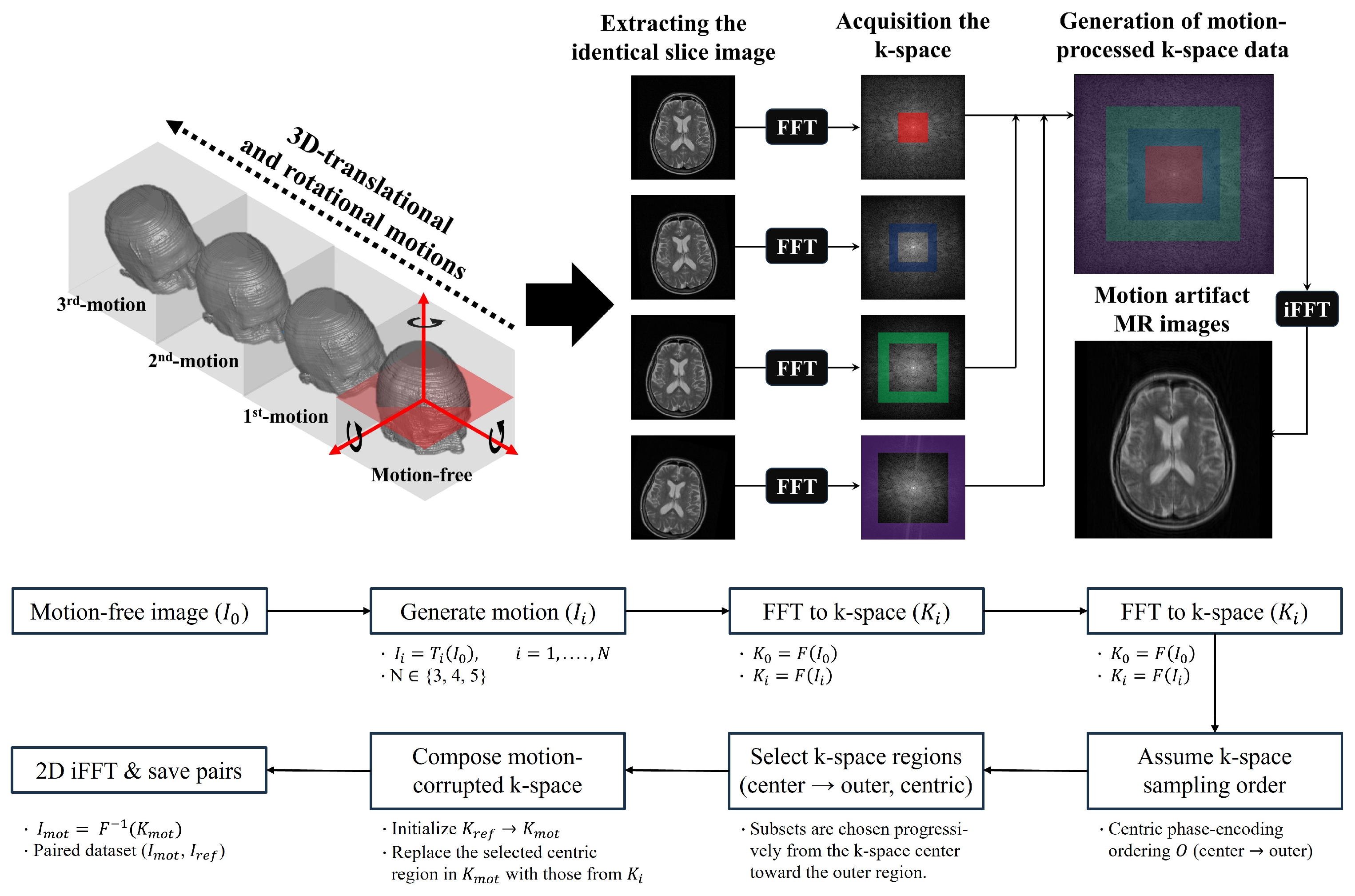
To generate simulation-based motion-artifact MR images, we used the Alzheimer’s Disease Neuroimaging Initiative (ADNI) database (adni.loni.usc.edu). In this study, T2-weighted brain MR images with an in-plane pixel size, slice thickness of 1 mm, and matrix size of 256 × 256 were employed. Motion artifacts in MR images are mainly caused by rigid-body motions such as translation and rotation, and can be simulated by applying rotational and translational operations to the acquired volumetric data. In this study, based on a previously developed simulation scheme, we constructed a motion artifact dataset by simultaneously incorporating z-axis rotation (head rotation) and translational motion for each volume. Figure 1 illustrates the overall simulation procedure used to generate the MR images affected by motion artifacts.
In the proposed simulation method, the volume data were randomly rotated and shifted in the vertical, horizontal, and through-plane directions (x-, y-, and z-axes). Translational shifts and rotations were randomly assigned within ranges of ±10 pixels and ±5°, respectively [41,42,43]. The number of motion events was set to 3–five, and to emulate continuous motion, each subsequent transformation was applied to the volume that had already been transformed in the preceding step. For each motion event, a 2D slice at the same z-axis location was selected, and a two-dimensional FFT was performed to compute the corresponding k-space data for that slice. Subsequently, a subset of phase-encoding lines in the motion-free reference k-space is replaced in the order of motion occurrence with k-space data containing motion, thereby constructing a final k-space dataset that reflects time-varying motion. To define the k-space sampling order, we assumed that a centric (center-out) phase-encoding scheme was used in the fast spin-echo sequence. A motion artifact simulation was performed on the volumetric data acquired from 2000 patients, resulting in 69,283 paired samples. Among these, 49,852, 6969, and 12,462 pairs were used for the training, validation, and testing of the Sim-DDNet model for motion artifact reduction, respectively. All data were split at the subject level (patient-wise split) prior to slice pairing and augmentation, and no subject was shared across training, validation, and test sets.

2.2. Sim-DDNet Model for Motion Artifact Reduction

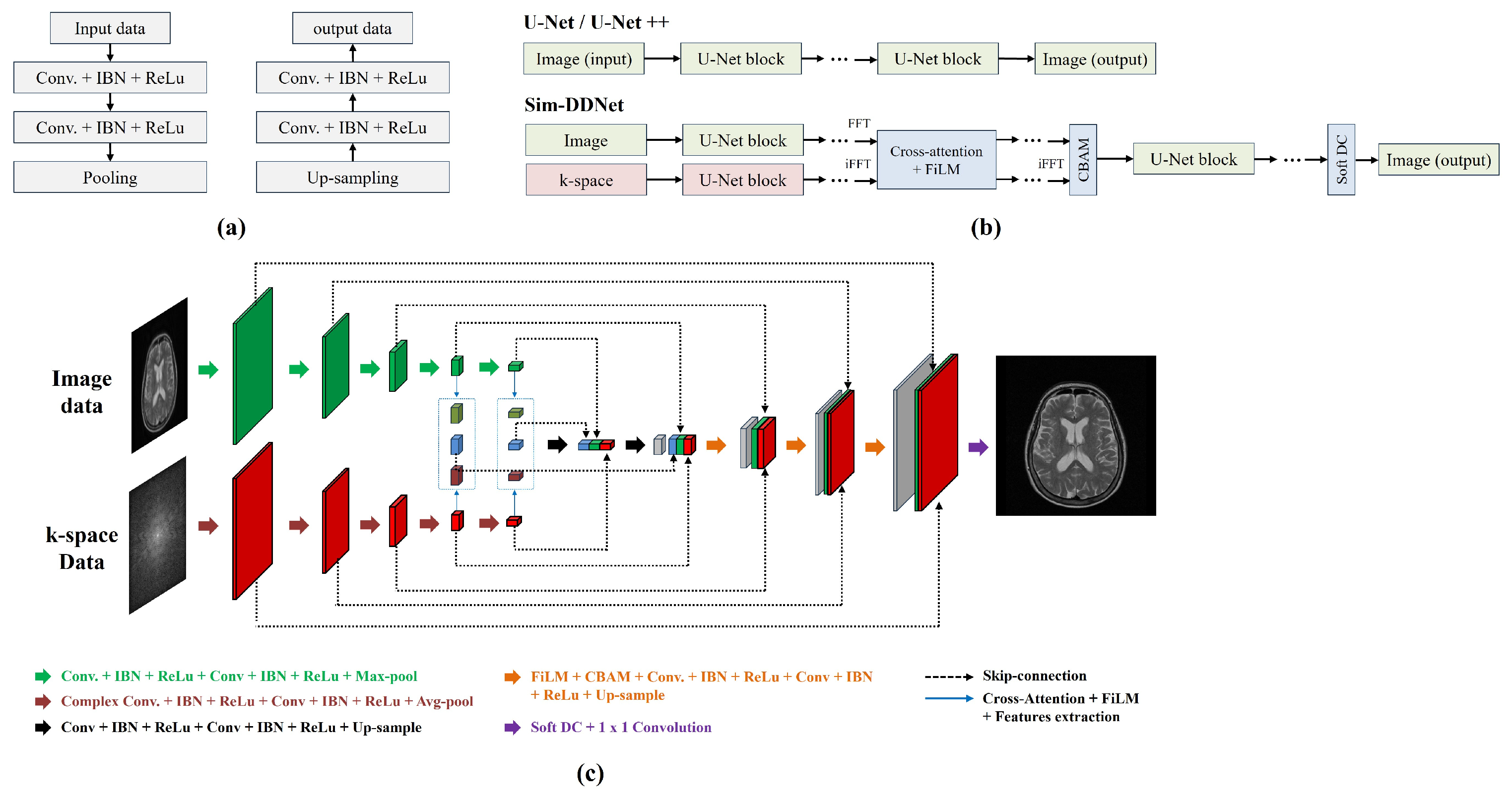
We propose Sim-DDNet, which jointly exploits information from the image and k-space domains to reduce motion artifacts (Figure 2).
The overall architecture follows a U-Net-like encoder–decoder operating on real-valued magnitude images augmented with a parallel complex-valued encoder in k-space, explicit cross-domain fusion mechanisms, and a soft data-consistency layer. On the image side, the network takes a motion-corrupted magnitude image x R 1 × H × W as input and processes it through a five-level encoder–decoder. Each encoder stage consists of two sequential 3 × 3 convolutions, followed by feature normalization and LeakyReLU activation. Between the stages, 2 × 2 max pooling was used for downsampling to produce a hierarchy of feature maps with channel widths of 32, 64, 128, 256, and 512. The decoder mirrors this structure with 2 × 2 transposed convolutions for upsampling and skipping connections from the corresponding encoder stages. At the end of the decoder, a 1 × 1 convolution maps the feature representation onto a single-channel residual image.
Simultaneously, Sim-DDNet builds a k-space branch that processes complex-valued data. The input image is first transformed into k-space using a centered orthonormal 2D Fourier transform, and the resulting complex-valued tensor is then passed through a stack of complex convolutional blocks. Each block comprises two complex 3 × 3 convolutions with complex-valued parametric ReLU activation, and the spatial resolution is reduced across scales, either by complex average pooling or by complex convolutions with a stride of two. This yielded multiscale k-space feature maps aligned in resolution with the image encoder. To interface with the image domain, each complex feature map is converted back to the image domain using an inverse Fourier transform. From this complex image representation, we compute the magnitude and phase, and construct an embedded representation by concatenating the magnitude with the functions of the phase. A 1 × 1 convolution followed by normalization and nonlinear activation projects this embedded representation down to the corresponding encoder width, producing k-space-derived image-domain features that can be directly fused with the image features at each scale.
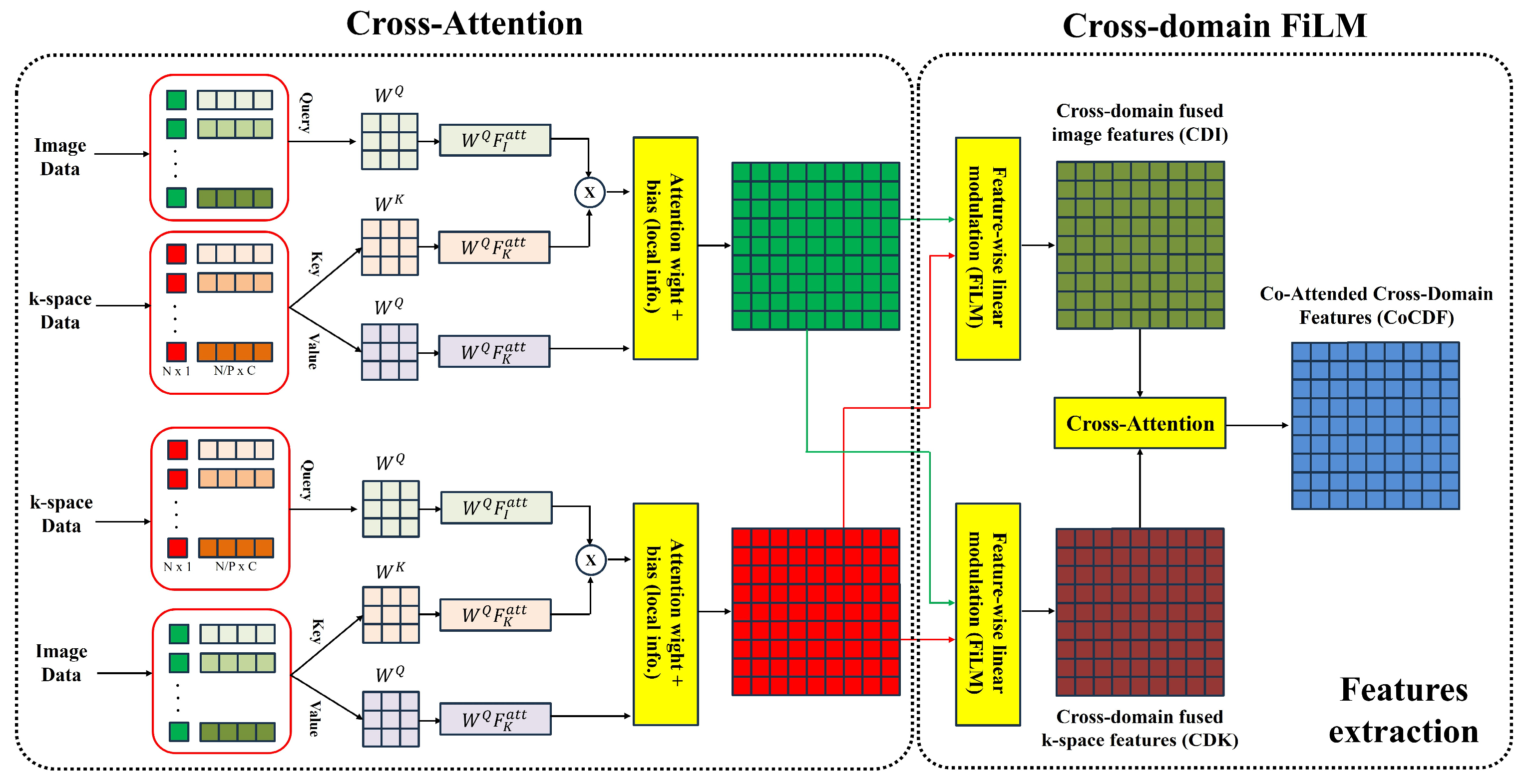
At deeper encoder levels, Sim-DDNet fuses image and k-space information with cross-attention followed by cross-domain FiLM (Figure 3).
For the fourth and fifth encoder stages, bidirectional cross-attention lets image features attend to k-space–derived features and lets k-space features attend to image features. Queries, keys, and values are produced by 1 × 1 convolutions, multi-head scaled dot-product attention is computed over spatial locations, and the attended output is projected back to the original width and added residually. After cross-attention, feature-wise linear modulation operates across domains: global average pooling of the conditioning feature (for example, the k-space stream) followed by a small 1 × 1 network produces per-channel scale and bias that modulate the normalized features in the other domain. This lets the image branch absorb phase and sampling cues from k-space and shapes the k-space branch using anatomical structure from the image domain. The resulting image-domain fused features are called Cross-Domain Fused Image features (CDI), and the corresponding k-space-anchored fused features are called Cross-Domain Fused k-space features (CDK). The ordered pair of cross-attention followed by cross-domain FiLM is called the Co-attended cross domain features (CoCDF).
At the bottleneck, an additional cross-attention maps the k-space-derived representation into the image space. The resulting CDI and CDK are concatenated and fused by a 1 × 1 projection and a convolutional refinement block, forming the deepest decoder input. The decoder then fuses, at every scale, three inputs: the upsampled feature from the previous level, the k-space-derived representation at the same scale, and the image-encoder skip. At the fifth and fourth levels, the upsampled decoder features are first modulated by CDK via CoCDF, which injects phase-related priors into the amplitude-like decoder stream. The triplet (CDI, CDK, and upsampled) is concatenated and passed through a convolutional block attention module (CBAM), and two 3 × 3 convolutions with normalization and activation refine the result. From third to first levels, the concatenated triplet is refined directly with the same convolutional block without additional CoCDF. A final 1 × 1 convolution maps the last decoder features to a residual image r, and the network prediction is expressed as follows:
x r e c o n =   x i n + α r
where α is a learnable residual scaling parameter initialized to 0.1 to stabilize early training and prevent over-correction. To maintain the fidelity of the acquired measurements, SIM-DDNet incorporates a soft data-consistency layer applied to the residual output. The reconstructed image x r e c o n is transformed into k-space, and the coefficients at the measured positions, defined by a binary sampling mask, are softly projected toward the reference k-space data using a proximal mapping of the following form:
Y d c = 1 M Y + M Y + λ K r e f 1 + λ
where Y is the Fourier transform of x recon , K r e f is the acquired k-space, M is the sampling mask, and λ is a spatially varying data-consistency weight. We employ a radially varying λ -map with higher values near the k-space center and lower values toward the periphery, constructed from a Gaussian function of the normalized radius, so that low-frequency components are more strongly constrained by the measurements. The final output image is obtained by applying an inverse Fourier transform to Y d c . By combining a real-valued U-Net-type image pathway, a complex-valued k-space encoder, explicit cross-domain fusion, residual learning, and soft data consistency (DC), Sim-DDNet was designed to leverage complementary information from both domains for robust motion artifact reduction.
For all experiments, the networks were trained end-to-end using the Adam optimizer with an initial learning rate of 1 × 10⁻⁴ and momentum parameters β1 = 0.5 and β2 = 0.999. The models were optimized for 50 epochs with a mini-batch size of six, and the learning rate was adaptively reduced using a validation loss-based scheduling strategy (reduction factor 0.5, patience three epochs). Sim-DDNet, U-Net, and U-Net++ shared the same optimization settings and hyperparameters to ensure a fair comparison.

2.3. Composite Loss Function

During training, the network parameters were optimized using a composite dual-domain loss function that combined complementary error terms in the image and k-space domains. Let x ^ and x denote the reconstructed and reference motion-free images, respectively. The total loss is defined as
L total ( x ^ , x ) = w l 1 L l 1 + w SSIM L SSIM + w perc L perc + w k L k
where w l 1 , w SSIM , w perc , and w k are the scalar weights balancing the contributions of each term. The pixel-wise intensity term L l 1 is given by the mean absolute error as follows:
L l 1 = 1 N i = 1 N x ^ i x i
where N is the number of pixels and x ^ i , x i denote the intensities at pixel i in the reconstructed and reference images, respectively. To encourage structural fidelity, we include a structural-similarity-based term, defined as
L SSIM = 1 SSIM ( x ^ , x )
where SSIM ( x ^ , x ) is the standard structural similarity index computed with a Gaussian window (kernel size 11 × 11 , σ = 1.5 ), as commonly used in image quality assessment. In addition, the perceptual loss L perc is defined in the feature space of a VGG-16 network pretrained on ImageNet. The reconstructed and reference images are replicated across three channels, normalized using the standard RGB statistics, and passed through selected convolutional layers ϕ j . The perceptual loss is then computed as
L perc = j α j ϕ j ( x ^ ) ϕ j ( x ) 1
where α j denotes the relative weight assigned to feature level j . To explicitly regularize the reconstruction in the frequency domain, we introduce a k-space loss L k . Let F denote the centered, orthonormal 2D Fourier transform as follows:
K ^ = F x ^ , K = F x ,
L k = 1 H W p = 1 H q = 1 W w rad ( p , q ) w meas ( p , q ) K ^ p , q K p , q
where H × W is the k-space size; w rad ( p , q ) is a radially varying weight that gradually increases toward the k-space periphery to emphasize the high-frequency components; and w meas ( p , q ) is a measurement-aware weight that assigns higher importance to locations corresponding to the acquired phase-encoding lines. The combination of the image domain and k-space domain discrepancies in L total allows Sim-DDNet to simultaneously preserve the anatomical detail, structural similarity, and frequency-domain consistency during motion artifact reduction.

2.4. Quantitatively Evaluation Factors

To evaluate the performance of the U-Net model for motion artifact reduction, we assessed the similarity between motion-free output images and the corresponding label images. The peak signal-to-noise ratio (PSNR) and structural similarity index (SSIM) were calculated as follows.
P S N R = 10 l o g 10 L 2 M S E ,
M S E = 1 N i = 1 N ( x i y i ) 2
where x i and y i denote the pixel intensities of the reconstructed and reference images, respectively; N is the number of pixels; and L is the dynamic range of the image intensities (e.g., 1 for normalized images).
S S I M x , y = ( 2 μ x μ y + C 1 ) ( 2 μ x y + C 2 ) ( μ x 2 + μ y 2 + C 1 ) ( σ x 2 + σ y 2 + C 2 )
where μ x and μ y are the local means of x and y , σ x 2 and σ y 2 are the corresponding variances, σ x y is the local covariance, and C 1 and C 2 are small stabilization constants. PSNR and SSIM were applied to evaluate the signal intensity and structural similarity of the MR images. These parameters can estimate the overall image restoration performance of the proposed Sim-DDNet. In addition, we computed the gradient magnitude similarity deviation (GMSD) for comparative evaluation. GMSD can evaluate distortion in localized region based on gradient magnitude through high-frequency signals. First, the gradient magnitude similarity (GMS) map between x and y was defined as
G M S i = 2 m x i m y i + T m x 2 i + m y 2 i + T
where m x ( i ) and m y ( i ) denote the gradient magnitudes of x and y at pixel i , and T is a small positive constant. The GMSD was then given by the standard deviation of the GMS map.
G M S D = 1 N i = 1 N ( G M S i G M S ¯ ) 2
where G M S ¯ is the spatial mean of GMS( i ).

3. Results

During brain MR acquisition, motion artifacts occur frequently and impair the visualization of fine structures and the reliability of quantitative analyses, thereby degrading diagnostic accuracy and reproducibility. To address this issue, U-Net-based deep learning models have been proposed; however, most of them operate in a single domain, limiting both the representational capacity and generalization. Moreover, they are typically trained on datasets that assume relatively regular motion, making it difficult to correct artifacts caused by complex, non-periodic head movements commonly observed in clinical practice. In this study, we seek to overcome these limitations by constructing a large-scale paired dataset using a k-space-based motion simulation scheme that faithfully reflects the actual MRI acquisition process and by designing Sim-DDNet, a dual-domain network that simultaneously exploits image- and k-space-domain features. The proposed Sim-DDNet integrates complementary information from both domains via cross-attention and soft data-consistency modules, with the purpose of suppressing motion artifacts in a more physically plausible manner while achieving superior quantitative performance.
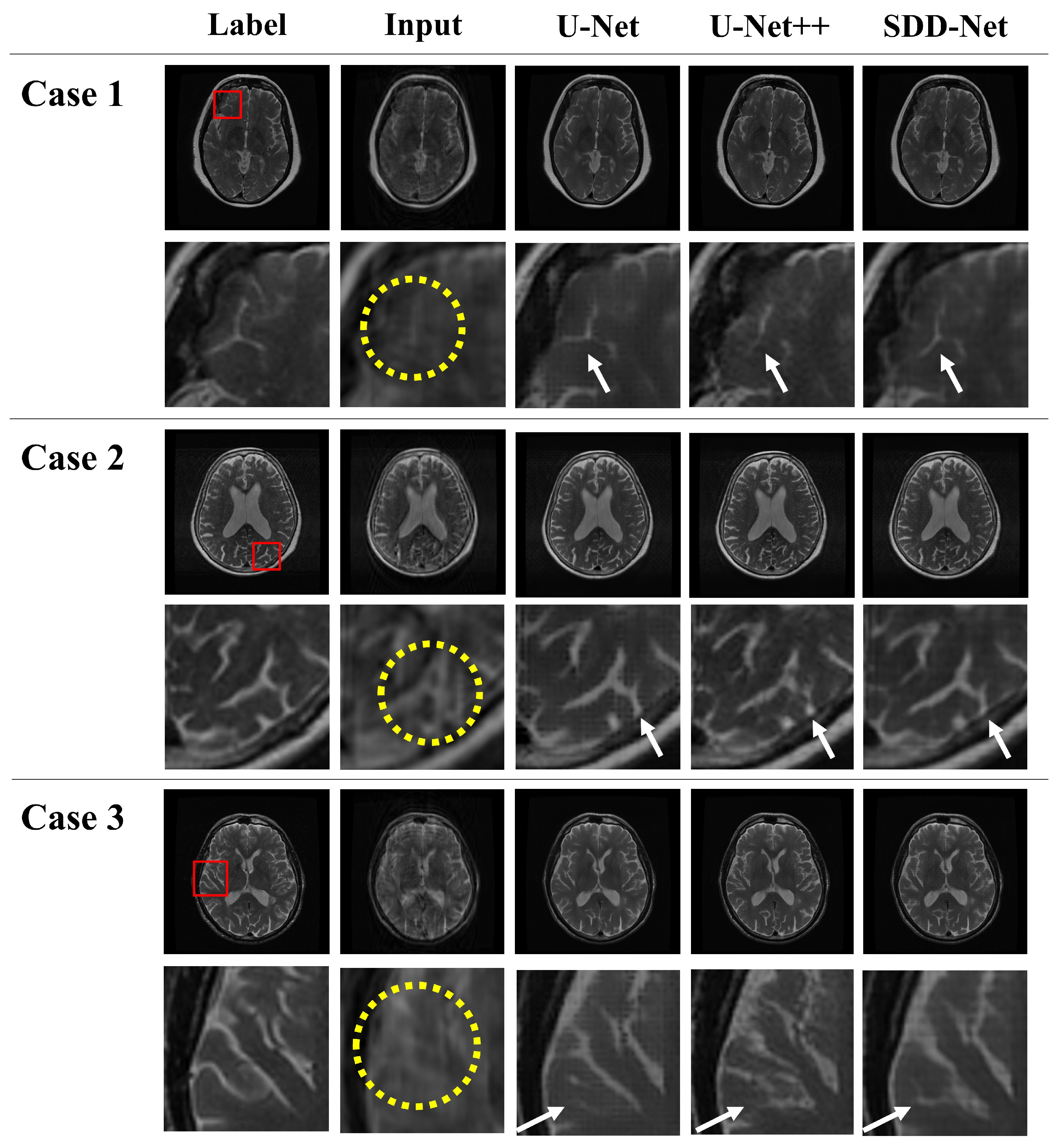
The input in Figure 4 shows motion-corrupted brain MR images generated by applying k-space-based motion simulation to the label (motion-free) images. In the motion-corrupted brain MR images generated by simulation, signal loss due to blurring is commonly observed, as illustrated by the yellow circle in Case 1. In addition, as shown in the yellow circle in Case 2, patient motion can produce ghosting, boundary duplication, and ringing-like patterns along the phase- or frequency-encoding direction, leading to reduced structural sharpness and anatomically implausible repetitive structures. Moreover, because MRI acquires k-space sequentially over time, intermittent rigid motion can interleave k-space lines acquired at pre- and post-motion positions. This inconsistency effectively superimposes two slightly shifted edge components in the reconstructed image, causing edge splitting, which is most evident in high-contrast structures such as the sulci, gray matter, and ventricles, as highlighted by the yellow circle in Case 3. These artifacts vary in their orientation, occurrence frequency, and severity depending on the random parameters used in the simulation.
Figure 4 shows the example results obtained by applying U-Net, U-Net++, and Sim-DDNet to simulated motion artifact images generated from motion-free brain MR images. Visual inspection indicated that all three models substantially reduced the observed motion artifacts. However, each model exhibits distinct limitations, which are highlighted by white arrows that mark regions containing fine anatomical structures. In the case of U-Net, a pronounced blurring effect was observed in most examples. This leads to the loss of fine tissue signals and signal distortion in regions with severe motion artifacts, thereby degrading the clarity and anatomical accuracy of the reconstructed structures. U-Net++ tends to better preserve high-frequency components such as edges and boundaries; however, in some regions, motion artifacts are not sufficiently removed, or fine structural signals are partially missing. In contrast, Sim-DDNet occasionally shows pixel-level distortions or slight signal loss; however, it provides the best visual quality among the three methods in terms of motion artifact suppression, blurring reduction, fine structural signal preservation, and anatomical distortion prevention.
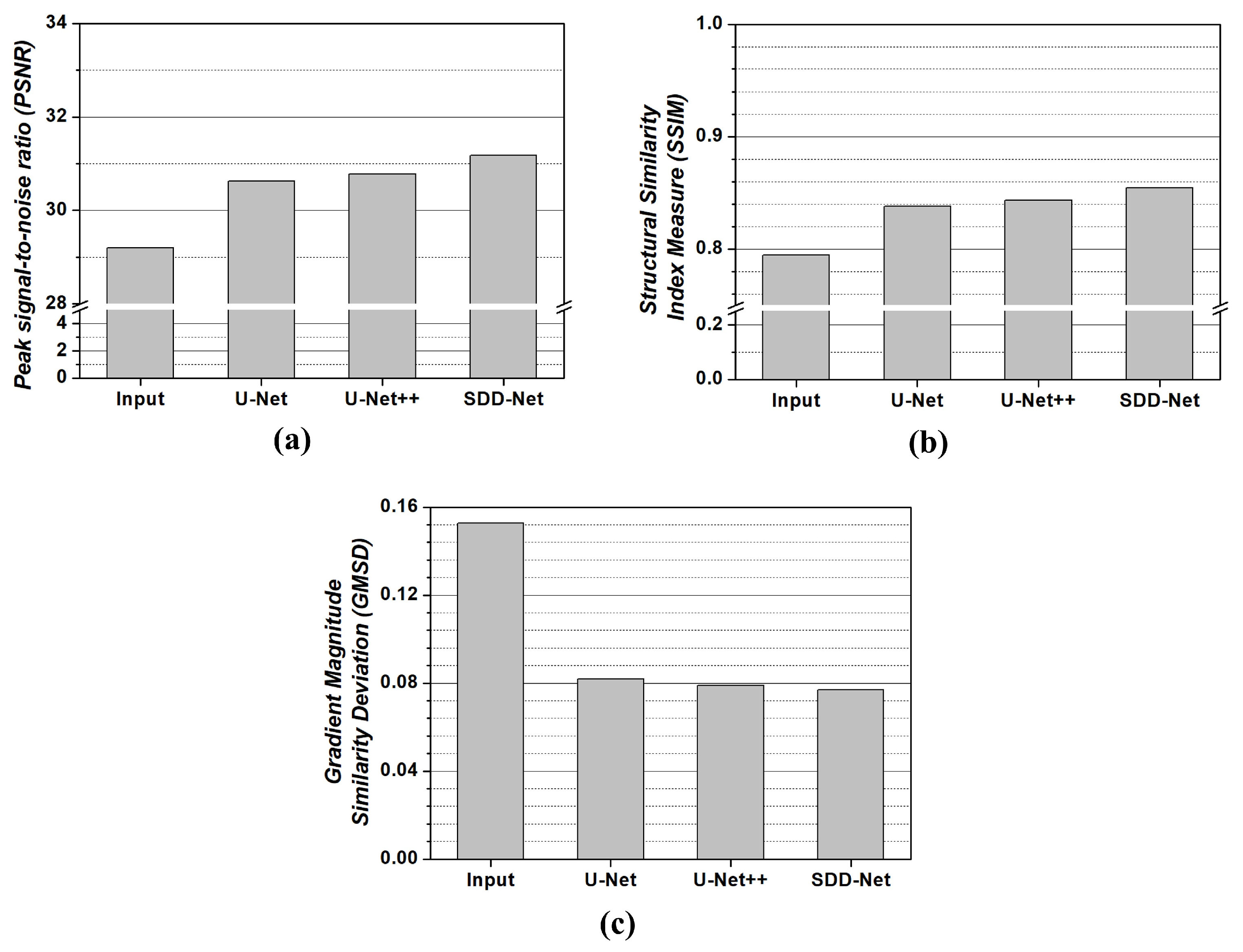
Figure 5 shows the quantitative evaluation results of the motion artifact reduction performance of the trained models. Sim-DDNet achieved the best PSNR, SSIM, and GMSD values among the three models, consistently outperforming U-Net++ and U-Net across all quantitative metrics. Moreover, the evaluation metrics show a monotonic improvement from the input image (with artifacts) using U-Net and U-Net++ to Sim-DDNet. In particular, the PSNR, SSIM, and GMSD values of Sim-DDNet were measured as approximately 31.05, 0.85, and 0.077, respectively, corresponding to approximately 1.06-, 1.07-, and 1.97-fold improvements over the input images.

4. Discussion

We propose Sim-DDNet to jointly leverage features from both the image and k-space domains, and we assume that each component plays a distinct role in motion artifact reduction. Cross-attention is central to cross-domain coupling, enabling the image stream to draw on phase- and sampling-related cues represented in k-space, while allowing the k-space stream to be guided by anatomical structure emphasized in the image domain. Without this mechanism, the two branches would interact more weakly, which may leave more residual ghosting or streaking, particularly under severe or irregular motion [44,45]. Cross-domain FiLM then provides conditional, channel-wise modulation, supporting adaptive feature recalibration across domains; removing FiLM could reduce this adaptivity and manifest as over- or under-correction, diminished robustness to changes in motion severity, and increased oversmoothing of subtle anatomical details [46]. CBAM is used for further refinement by emphasizing salient channels and spatial regions in the fused representation. Although substantial artifact suppression may still be achievable without CBAM, the final reconstructions may appear less well regularized, with residual texture-like artifacts, reduced local contrast, or less consistent preservation of subtle structures [47,48]. In the loss design, the k-space loss directly penalizes frequency-domain inconsistencies that are closely tied to motion-induced corruption, complementing image-domain objectives that may not fully constrain phase-encoding-related artifacts. Omitting this term can yield reconstructions that look plausible in the image domain while retaining frequency-domain mismatch, which in turn may degrade the fidelity of high-frequency details [49]. Finally, soft data consistency acts as a stabilizing constraint by anchoring the reconstruction to the acquired measurements; disabling it may increase susceptibility to over-correction and implausible texture changes, especially in severely corrupted inputs or when artifacts fall outside the training distribution. Collectively, these considerations provide a coherent functional interpretation of the major architectural and loss components in Sim-DDNet [50,51]. Nevertheless, to rigorously delineate the marginal contribution of each component and to elucidate their potential interactions, a systematic ablation study is warranted [52]. In particular, module-wise substitution of cross-attention, FiLM, CBAM, the k-space regularization term, and the soft data consistency constraint, which followed by training and evaluation under an identical protocol, would facilitate quantitative characterization of both the individual effects and cross-component synergies across diverse motion regimes.
Figure 4 demonstrates that the proposed Sim-DDNet achieves an overall superior performance compared with U-Net and U-Net++ in restoring brain MR images affected by motion artifacts. In particular, the conventional U-Net exhibits a pronounced blurring effect and loss of fine signals, which are most prominent in cases 1 and 3. This behavior can be attributed to the fact that U-Net is inherently based on a downsampling–upsampling architecture and pixel-wise loss functions; therefore, it tends to have excessively smooth high-frequency components and fine anatomical structures. Furthermore, because the low-level encoder feature maps are directly connected to the relatively high-level decoder feature maps through simple skip connections, the semantic gap between these representations has not been sufficiently resolved [53,54]. This issue becomes more pronounced in the presence of motion artifacts. Ghost and streak patterns are mixed with true anatomical edge signals in the early encoder features and are passed to the decoder in this entangled form, making it difficult for the network to clearly separate and selectively suppress the artifact components. Consequently, even when motion artifacts are partially reduced, fine structural information is simultaneously lost, and the overall blurring effect is exacerbated [55,56]. To overcome the limitations of the original U-Net, the U-Net++ model is proposed. U-Net++ employs redesigned multiscale skip pathways and a nested (concatenated) skip architecture to reduce the semantic gap between the encoder and decoder and integrate multiresolution information more effectively, thereby providing improved spatial resolution and fine-structure recovery compared with U-Net. However, this design has structural limitations when motion artifacts are present. Because all skip pathways are constructed solely in the image domain, high-frequency ghost and streak patterns are aggregated together with genuine anatomical edges and repeatedly injected into the decoder. Consequently, as observed in Case 1, the network not only fails to sufficiently remove residual motion artifacts in severely corrupted regions but also tends to hallucinate non-existent fine structures by misinterpreting artifact-related patterns as plausible anatomy [57,58].
These limitations are fundamentally difficult to overcome when the expressiveness is extended to a single image domain. By introducing dual-domain representations in both the image and k-space domains, Sim-DDNet explicitly provides the network with the information necessary to distinguish artifact-related signals from anatomical signals, thereby differentiating it from conventional U-Net-based approaches. Instead of relying solely on image-domain features as input, Sim-DDNet simultaneously exploits motion-aware features derived from k-space, encouraging the network to interpret ghost and streak patterns not as generic high-frequency textures but as patterns originating from motion artifacts. Because k-space-derived features are sensitive to phase variations and amplitude imbalances, they provide additional cues for discriminating artifact-related components from true anatomical edges even when both occupy similar high-frequency bands [59,60]. Consequently, the behavior observed in U-Net and U-Net++, where high-frequency components are uniformly smoothed and both artifacts and fine structures are removed together, is mitigated, and the tradeoff between motion artifact suppression and fine structure preservation shifts in a more favorable direction, as illustrated in Figure 4. The cross-attention and FiLM-based feature-wise modulation modules in Sim-DDNet further play a role in indirectly correcting the semantic gap that could not be fully resolved by simple or nested skip connections alone [61]. In conventional U-Net and U-Net++, low-level encoder features containing a mixture of ghost/streak patterns and true edges are passed directly to the decoder, making it easy for the decoder to converge to a solution that simultaneously suppresses or enhances both signals. By contrast, Sim-DDNet is designed such that image-domain features refer repeatedly to k-space-based motion-aware features, thereby providing additional information on whether each boundary signal arises from an artifact or genuine anatomy. This process enables artifacts and anatomical signals to be compared and corrected across distinct representation spaces, which in turn contributes to the simultaneous alleviation of the excessive blurring observed in U-Net and the residual artifacts observed in U-Net++. Finally, by jointly employing a soft data-consistency module and k-space-weighted loss, Sim-DDNet constrains the network to suppress motion artifacts within the bounds of physical consistency with the simulated measured k-space rather than hallucinating visually plausible but unsupported structures. In U-Net++, the combination of dense skip connections and perceptual/SSIM-driven training led to the generation of fine structures that did not agree with actual k-space measurements [62]. In Sim-DDNet, however, the final output is continually compared and constrained against the measured data in k-space. Thus, the parameters are optimized to selectively reduce the ghost and streak components within the range of measurable signals, rather than merely masking artifacts or introducing nonexistent boundaries. Taken together, this combination of dual-domain representation, cross-domain fusion, and data-consistency-aware learning is expected to simultaneously mitigate the blurring, fine structure loss, residual artifacts, and hallucinated structures observed in U-Net and U-Net++, thereby providing a more stable and physically plausible restoration of brain MR images affected by motion artifacts.
In particular, for the PSNR and SSIM, which reflect the overall reconstruction quality, the performance gap between U-Net++ and Sim-DDNet is larger than that between U-Net and U-Net++. This supports the notion that the proposed Sim-DDNet has a substantial potential to compensate for the inherent limitations of both U-Net and U-Net++. Moreover, because GMSD reflects not only the edge sharpness but also the spatial consistency of the gradient magnitude distribution, it is generally expected to favor U-Net++, which tends to preserve high-frequency components more strongly. Nevertheless, the fact that Sim-DDNet, despite being built on the U-Net family, achieved the lowest GMSD value indicates that the proposed model goes beyond simply enhancing the edges. Instead, by leveraging k-space-based motion-aware features and data-consistency constraints, artifact-induced boundaries were effectively suppressed while restoring anatomical edge structures to be more faithful to the reference images.
Nevertheless, this study has several limitations. Although the k-space-based motion artifact simulation was carefully designed to more faithfully reflect the actual brain MR acquisition process and incorporate diverse rotational, translational, and temporal variations, all experiments were ultimately conducted on the datasets generated under this simulation framework. As a result, the performance of Sim-DDNet reported in this work may be partially optimized for the specific simulation conditions and motion patterns defined in this study, and may not fully represent the wide spectrum of irregular motion and complex artifact patterns encountered in real clinical practice. In real-world clinical scans, patient motion can be more irregular and may include non-rigid and temporally varying components. It can also be influenced by scanner- and protocol-specific factors (e.g., parallel imaging, partial Fourier sampling, coil sensitivity profiles, and vendor-dependent reconstruction settings). This limitation is further underscored by the fact that other artifact types, such as zipper artifacts and susceptibility-related artifacts, were not considered. Under such domain shifts, potential failure modes may include residual ghosting/streaking, over smoothing or loss of fine anatomical details, and abnormal texture changes in severely corrupted cases. Therefore, future work should include validation on patient-derived motion-corrupted data acquired on actual MR scanners, as well as evaluation on multicenter datasets obtained under different scanner vendors, imaging protocols, and field strengths (e.g., 1.5 T and 3.0 T), to more systematically assess clinical applicability and generalization capability. In addition, incorporating practical quality-control mechanisms (e.g., reliability/uncertainty estimation) to flag low-confidence outputs and recommend rescanning when necessary will be important for safe clinical deployment.
In this study, U-Net and U-Net++ were selected as the representative image-domain CNN-based reconstruction models to verify the structural advantages and performance gains of Sim-DDNet. This controlled comparison was intended to examine the incremental benefit and structural contribution of explicitly leveraging k-space information within the proposed dual-domain framework, rather than to claim superiority over the latest state-of-the-art methods. Thus, systematic comparisons with other classes of CNN-based models, such as GAN-based reconstruction networks or more advanced encoder–decoder architectures, have not been conducted. Consequently, the extent to which Sim-DDNet maintains a relative advantage over a broader range of deep-learning-based restoration methods can only be discussed to a limited degree. A natural extension of this study would include a more comprehensive set of baselines, such as GAN models using adversarial loss, hybrid networks incorporating self-attention/transformer modules, and self-supervised or unsupervised reconstruction schemes, which should be implemented and tuned under the same simulation and assessment protocol to quantitatively characterize the strengths and weaknesses of Sim-DDNet. Another limitation is the absence of direct quantitative comparisons with the latest state-of-the-art reconstruction methods, including the dual-domain and transformer-based models recently reported in the literature. Because this study focused on analyzing the operational principles and structural contributions of the proposed model within a controlled simulation-based environment, the current evidence is insufficient to position Sim-DDNet precisely with respect to the most recent SOTA approaches, and thus the present results should not be interpreted as a comprehensive SOTA benchmark. Future studies should leverage public benchmark datasets and shared evaluation metrics to perform extensive benchmarking against existing dual-domain networks, k-space image iterative refinement architectures, and transformer-based reconstruction models. In addition, generalizing Sim-DDNet to 3D or temporal extensions is an important step toward handling more realistic clinical scenarios, such as dynamic imaging or long-duration acquisitions. Despite these limitations, the present study demonstrated the potential of combining k-space-based motion simulation with an image-k-space dual-domain reconstruction strategy in the form of Sim-DDNet. The proposed framework provides a foundation upon which future research can build to further bridge the gap between simulation-based evaluations and real clinical data and to more tightly integrate Sim-DDNet with the rapidly evolving landscape of state-of-the-art reconstruction methods.
Beyond these methodological considerations, it is also important to highlight the potential clinical implications of the proposed approach. From a clinical perspective, a robust deep-learning-based post-processing correction method has the potential to improve the usability of MRI examinations affected by motion and to reduce avoidable repeat scans. This is particularly relevant in low- and middle-income countries (LMICs), where MRI availability and scanner capacity may be limited and rescheduling repeat examinations can be challenging. By enhancing image interpretability without additional acquisition time, such a method may help increase scanner throughput and broaden access to diagnostic imaging, especially for motion-prone populations (e.g., pediatric patients, older adults, or individuals who have difficulty remaining still). Nevertheless, before clinical deployment, it is essential to perform external validation on real-world, patient-derived motion-corrupted data and to incorporate quality-control (QC) mechanisms capable of detecting potential failure cases and recommending rescanning when necessary.

5. Conclusions

In this paper, we propose Sim-DDNet, which aims to handle motion artifacts in brain MR images in a more physically plausible manner by combining k-space-based motion simulation with an image-k-space dual-domain reconstruction architecture. Under identical training and evaluation conditions, the proposed Sim-DDNet outperformed the conventional U-Net and U-Net++ in terms of PSNR, SSIM, and GMSD, and qualitative assessment also confirmed overall image quality improvements, including reduced blurring, suppression of residual ghost/streak artifacts, and fewer hallucinated non-existent fine structures. These findings suggest that a dual-domain, data-consistency-aware learning strategy that explicitly exploits k-space information is a promising approach for achieving more stable and physically plausible correction of motion artifacts on brain MR.

Author Contributions

Conceptualization, S.-H.K. and Y.L.; methodology, S.-H.K. and J.-Y.C.; software, S.-H.K.; validation, J.-Y.C. and Y.L.; formal analysis, S.-H.K. and J.-Y.C.; funding acquisition, J.-Y.C. and Y.L.; data curation, S.-H.K.; writing—original draft preparation, S.-H.K.; writing—review and editing, J.-Y.C. and Y.L.; and project administration, Y.L.; investigation, Alzheimer’s Disease Neuroimaging Initiative; data curation, Alzheimer’s Disease Neuroimaging Initiative. All authors have read and agreed to the published version of the manuscript.

Funding

This work was supported by the National Research Foundation of Korea (NRF) grant funded by the Korea government (MSIT) (RS-2025-02303425 & NRF-2022R1A2C2010363).

Institutional Review Board Statement

The study was conducted according to the guidelines of the Declaration of Helsinki and approved by the Institutional Review Board of Gachon University (1044396-202303-HR-031-01).

Informed Consent Statement

A benchmark dataset, the Alzheimer’s Disease Neuroimaging Initiative (ADNI), which was used in our work, obtained informed consent from the participants. More information can be found in the following link: “https://adni.loni.usc.edu/data-samples/ (accessed on 29 June 2023)”.

Data Availability Statement

Data used in the preparation of this article were obtained from the ADNI database (adni.loni.usc.edu). The ADNI was launched in 2003 as a public–private partnership led by Principal Investigator Michael W. Weiner, MD. The primary goal of the ADNI has been to test whether serial magnetic resonance imaging (MRI), positron emission tomography (PET), other biological markers, and clinical and neuropsychological assessments can be combined to measure the progression of mild cognitive impairment (MCI) and early Alzheimer’s disease (AD). For up-to-date information, see www.adni-info.org.

Acknowledgments

Data collection and sharing for this project was funded by the Alzheimer’s Disease Neuroimaging Initiative (ADNI) (National Institutes of Health Grant U01 AG024904) and DOD ADNI (Department of Defense award number W81XWH-12-2-0012). ADNI is funded by the National Institute on Aging, the National Institute of Biomedical Imaging and Bioengineering, and through generous contributions from the following: AbbVie, Alzheimer’s Association; Alzheimer’s Drug Discovery Foundation; Araclon Biotech; BioClinica, Inc.; Biogen; Bristol-Myers Squibb Company; CereSpir, Inc.; Cogstate; Eisai Inc.; Elan Pharmaceuticals, Inc.; Eli Lilly and Company; EuroImmun; F. Hoffmann-La Roche Ltd. and its affiliated company Genentech, Inc.; Fujirebio; GE Healthcare; IXICO Ltd.;Janssen Alzheimer Immunotherapy Research & Development, LLC.; Johnson & Johnson Pharmaceutical Research & Development LLC.; Lumosity; Lundbeck; Merck & Co., Inc.; Meso Scale Diagnostics, LLC.; NeuroRx Research; Neurotrack Technologies; Novartis Pharmaceuticals Corporation; Pfizer Inc.; Piramal Imaging; Servier; Takeda Pharmaceutical Company; and Transition Therapeutics. The Canadian Institutes of Health Research is providing funds to support ADNI clinical sites in Canada. Private sector contributions are facilitated by the Foundation for the National Institutes of Health (www.fnih.org). The grantee organization is the Northern California Institute for Research and Education, and the study is coordinated by the Alzheimer’s Therapeutic Research Institute at the University of Southern California. ADNI data are disseminated by the Laboratory for Neuro Imaging at the University of Southern California.

Conflicts of Interest

The authors declare no conflict of interest.

References

  1. Gould, A.; Chen, Z.; Baylam Geleri, D.; Balu, N.; Zhou, Z.; Chen, L.; Chu, B.; Pimentel, K.; Canton, G.; Hatsukami, T.; et al. Vessel length on SNAP MRA and TOF MRA is a potential imaging biomarker for brain blood flow. Magn. Reson. Imaging 2021, 79, 20–27. [Google Scholar] [CrossRef]
  2. Ye, H.; Zheng, Y.; Li, Y.; Zhang, K.; Kong, Y.; Yuan, Y. RH-BrainFS: Regional heterogeneous multimodal brain networks fusion strategy. Adv. Neural Inf. Process. Syst. 2023, 36, 59286–59303. [Google Scholar]
  3. Havsteen, I.; Ohlhues, A.; Madsen, K.H.; Nybing, J.D.; Christensen, H.; Christensen, A. Are movement artifacts in magnetic resonance imaging a real problem?—A narrative review. Front. Neurol. 2017, 8, 232. [Google Scholar] [CrossRef] [PubMed]
  4. Tamada, D.; Kromrey, M.L.; Ichikawa, S.; Onishi, H.; Motosugi, U. Motion artifact reduction using a convolutional neural network for dynamic contrast enhanced MR imaging of the liver. Magn. Reson. Med. Sci. 2020, 19, 64–76. [Google Scholar] [CrossRef] [PubMed]
  5. Lyu, Q.; Shan, H.; Xie, Y.; Kwan, A.C.; Otaki, Y.; Kuronuma, K.; Li, D.; Wang, G. Cine cardiac MRI motion artifact reduction using a recurrent neural network. IEEE Trans. Med. Imaging 2021, 40, 2170–2181. [Google Scholar] [CrossRef]
  6. Karami-Horestani, A.; Paredes, F.; Martín, F. Hybrid time/phase domain synchronous electromagnetic encoders for near-field chipless-RFID and motion control applications. IEEE Trans. Microw. Theory Tech. 2023, 71, 5457–5469. [Google Scholar] [CrossRef]
  7. Lee, H.; Lee, J.; Park, J.Y.; Lee, S.K. Line scan-based rapid magnetic resonance imaging of repetitive motion. Sci. Rep. 2021, 11, 4505. [Google Scholar] [CrossRef]
  8. Nakamura, M.; Takatsu, Y.; Yoshizawa, M.; Yamamura, K.; Miyati, T. Pulsation artifact reduction using compressed sensitivity encoding in Gd-EOB-DTPA contrast-enhanced magnetic resonance imaging. Radiol. Phys. Technol. 2025, 18, 86–93. [Google Scholar] [CrossRef]
  9. Tripathi, V.R.; Tibdewal, M.N.; Mishra, R. A survey on motion artifact correction in magnetic resonance imaging for improved diagnostics. SN Comput. Sci. 2024, 5, 281. [Google Scholar] [CrossRef]
  10. Yu, H.; Li, F.; Liu, J.; Liu, C.; Li, G.; Wang, J. Spatiotemporal dynamics of periodic and aperiodic brain activity under peripheral nerve stimulation with acupuncture. IEEE Trans. Neural Syst. Rehabil. Eng. 2024, 32, 3993–4003. [Google Scholar] [CrossRef]
  11. Madore, B.; Hess, A.T.; van Niekerk, A.M.J.; Hoinkiss, D.C.; Hucker, P.; Zaitsev, M.; Afacan, O.; Günther, M. External hardware and sensors, for improved MRI. J. Magn. Reson. Imaging 2023, 57, 690–705. [Google Scholar] [CrossRef] [PubMed]
  12. van Niekerk, A.; Berglund, J.; Sprenger, T.; Norbeck, O.; Avventi, E.; Rydén, H.; Skare, S. Control of a wireless sensor using the pulse sequence for prospective motion correction in brain MRI. Magn. Reson. Med. 2022, 87, 1046–1061. [Google Scholar] [CrossRef]
  13. Bschorr, F.; Hüfken, T.; Lobmeyer, T.; Dreyer, F.; Zhao, J.; Anders, J.; Rasche, V. Monitoring physiological signals with NMR-on-a-chip sensors for motion correction in abdominal MRI. IEEE Sens. J. 2024, 25, 2510–2517. [Google Scholar] [CrossRef]
  14. Godenschweger, F.; Kägebein, U.; Stucht, D.; Yarach, U.; Sciarra, A.; Yakupov, R.; Lüsebrink, F.; Schulze, P.; Speck, O. Motion correction in MRI of the brain. Phys. Med. Biol. 2016, 61, R32–R56. [Google Scholar] [CrossRef] [PubMed]
  15. Coutinho, A.M.; Coutu, J.P.; Lindemer, E.R.; Rosas, H.D.; Rosen, B.R.; Salat, D.H. Differential associations between systemic markers of disease and cortical thickness in healthy middle-aged and older adults. Neuroimage 2017, 146, 19–27. [Google Scholar] [CrossRef] [PubMed]
  16. Al Habsi, O.; Sali, S.M.; Meribout, A.; Meribout, M.; Almazrouei, S.; Seghier, M. Hardware acceleration in portable MRIs: State of the art and future prospects. IEEE Access 2025, 13, 186347–186368. [Google Scholar] [CrossRef]
  17. Hu, Y.; Han, C.; Huo, X.; Cao, X.; Chen, Y.; Cao, Z.; Xu, Y.; Tao, L.; Wu, Z. Flexible Sensor for Real-Time Monitoring of Motion Artifacts in Magnetic Resonance Imaging. ACS Sens. 2024, 9, 2614–2621. [Google Scholar] [CrossRef]
  18. Kellman, P.; McVeigh, E.R. Image reconstruction in SNR units: A general method for SNR measurement. Magn. Reson. Med. 2005, 54, 1439–1447. [Google Scholar] [CrossRef]
  19. Morrell, G.R. A phase-sensitive method of flip angle mapping. Magn. Reson. Med. 2008, 60, 889–894. [Google Scholar] [CrossRef]
  20. White, N.; Roddey, C.; Shankaranarayanan, A.; Han, E.; Rettmann, D.; Santos, J.; Kuperman, J.; Dale, A. PROMO: Real-time prospective motion correction in MRI using image-based tracking. Magn. Reson. Med. 2010, 63, 91–105. [Google Scholar] [CrossRef]
  21. Stiller, M.; Jaeckel, K.H.; Weber, M. Effective elimination of subharmonic ghost events from vibroseis data. Geophys. Prospect. 2012, 60, 1095–1113. [Google Scholar] [CrossRef]
  22. Vandeghinste, B.; Goossens, B.; Van Holen, R.; Vanhove, C.; Pižurica, A.; Vandenberghe, S.; Staelens, S. Iterative CT reconstruction using shearlet-based regularization. IEEE Trans. Nucl. Sci. 2013, 60, 3305–3317. [Google Scholar] [CrossRef]
  23. Kutyniok, G.; Lim, W.Q.; Reisenhofer, R. Shearlab 3D: Faithful digital shearlet transforms based on compactly supported shearlets. ACM Trans. Math. Softw. 2016, 42, 5. [Google Scholar] [CrossRef]
  24. Bustin, A.; Menini, A.; Janich, M.A.; Burschka, D.; Felblinger, J.; Brau, A.C.; Odille, F. Motion estimated-compensated reconstruction with preserved-features in free-breathing cardiac MRI. In Proceedings of the International Workshop on Reconstruction and Analysis of Moving Body Organs, Athens, Greece, 17 October 2016; pp. 70–80. [Google Scholar]
  25. Bilen, Ç.; Wang, Y.; Selesnick, I.W. High-speed compressed sensing reconstruction in dynamic parallel MRI using augmented Lagrangian and parallel processing. IEEE J. Emerg. Sel. Top. Circuits Syst. 2012, 2, 370–379. [Google Scholar] [CrossRef]
  26. Ye, J.C. Compressed sensing MRI: A review from signal processing perspective. BMC Biomed. Eng. 2019, 1, 8. [Google Scholar] [CrossRef] [PubMed]
  27. Huang, J.; Wu, Y.; Wang, F.; Fang, Y.; Nan, Y.; Alkan, C. Data-and physics-driven deep learning based reconstruction for fast MRI: Fundamentals and methodologies. IEEE Rev. Biomed. Eng. 2024, 18, 152–171. [Google Scholar] [CrossRef]
  28. Singh, R.; Singh, N.; Kaur, L. Deep learning methods for 3D magnetic resonance image denoising, bias field and motion artifact correction: A comprehensive review. Phys. Med. Biol. 2024, 69, 23TR01. [Google Scholar] [CrossRef]
  29. Ronneberger, O.; Fischer, P.; Brox, T. U-Net: Convolutional networks for biomedical image segmentation. In Proceedings of the International Conference on Medical Image Computing and Computer-Assisted Intervention, Munich, Germany, 5–9 October 2015; pp. 234–241. [Google Scholar]
  30. Zhou, Z.; Rahman Siddiquee, M.M.; Tajbakhsh, N.; Liang, J. Unet++: A nested U-Net architecture for medical image segmentation. In Proceedings of the International Workshop on Deep Learning in Medical Image Analysis, Granada, Spain, 20 September 2018; pp. 3–11. [Google Scholar]
  31. Kang, S.-H.; Lee, Y. Motion artifact reduction using U-Net model with three-dimensional simulation-based datasets for brain magnetic resonance images. Bioengineering 2024, 11, 227. [Google Scholar] [CrossRef]
  32. Kang, S.-H.; Hong, D.-H. Deep learning with U-Net for motion artifact reduction in brain MRI. Vasc. Endovasc. Rev. 2025, 8, 195–200. [Google Scholar]
  33. Zhao, Y.; Ossowski, J.; Wang, X.; Li, S.; Devinsky, O.; Martin, S.P.; Pardoe, H.R. Localized motion artifact reduction on brain MRI using deep learning with effective data augmentation techniques. In Proceedings of the 2021 International Joint Conference on Neural Networks (IJCNN), Shenzhen, China, 18–22 July 2021; pp. 1–9. [Google Scholar]
  34. Zhou, Z.Y.; Li, H.; Liu, J.; Kong, Z.; Huang, T.; Ahn, E.; Lv, Z.; Kim, J.; Feng, D.D. Explicit abnormality extraction for unsupervised motion artifact reduction in magnetic resonance imaging. IEEE J. Biomed. Health Inform. 2024, 29, 3853–3863. [Google Scholar] [CrossRef]
  35. Oh, G.; Jung, S.; Lee, J.E.; Ye, J.C. Annealed score-based diffusion model for MR motion artifact reduction. IEEE Trans. Comput. Imaging 2023, 10, 43–53. [Google Scholar] [CrossRef]
  36. Al-Masni, M.A.; Lee, S.; Yi, J.; Kim, S.; Gho, S.M.; Choi, Y.H.; Kim, D.H. Stacked U-Nets with self-assisted priors towards robust correction of rigid motion artifact in brain MRI. NeuroImage 2022, 259, 119411. [Google Scholar] [CrossRef] [PubMed]
  37. Singh, N.M.; Dey, N.; Hoffmann, M.; Fischl, B.; Adalsteinsson, E.; Frost, R.; Dalca, A.V.; Golland, P. Data consistent deep rigid MRI motion correction. Proc. Med. Imaging Deep. Learn. 2024, 227, 368–381. [Google Scholar]
  38. Mai, Z.; Zhan, Z.; Guo, H.; Huang, Y.; Su, W. DDO-IN: Dual domains optimization for implicit neural network to eliminate motion artifact in magnetic resonance imaging. arXiv 2025, arXiv:2503.08056. [Google Scholar] [CrossRef]
  39. Wang, J.; Yang, Y.; Yang, Y.; Sun, J. Dual domain motion artifacts correction for MR imaging under guidance of k-space uncertainty. In Proceedings of the International Conference on Medical Image Computing and Computer-Assisted Intervention, Vancouver, BC, Canada, 8–12 October 2023; pp. 293–302. [Google Scholar]
  40. Bai, C.; Liu, K.; Chen, S.; Li, Z.; Xie, W.; Bao, Q.; Liu, C. Dual-domain unsupervised network for removing motion artifact related to Gadoxetic acid-enhanced MRI. J. Phys. Conf. Ser. 2022, 2258, 012037. [Google Scholar] [CrossRef]
  41. Shaw, R.; Sudre, C.H.; Varsavsky, T.; Ourselin, S.; Cardoso, M.J. A k-space model of movement artefacts: Application to segmentation augmentation and artefact removal. IEEE Trans. Med. Imaging 2020, 39, 2881–2892. [Google Scholar] [CrossRef]
  42. Usui, K.; Muro, I.; Shibukawa, S.; Goto, M.; Ogawa, K.; Sakano, Y.; Kyogoku, S.; Daida, H. Evaluation of motion artefact reduction depending on the artefacts’ directions in head MRI using conditional generative adversarial networks. Sci. Rep. 2023, 13, 8526. [Google Scholar] [CrossRef]
  43. Wu, Y.; Liu, J.; White, G.M.; Deng, J. Image-based motion artifact reduction on liver dynamic contrast enhanced MRI. Phys. Med. 2023, 105, 102509. [Google Scholar] [CrossRef]
  44. Chen, W.; Gao, Z.; He, Y.; Wanyan, J.; Xiong, C. Dual-path network with dual-domain fusion and cross-attention for MRI reconstruction. Biomed. Signal Process. Control 2025, 110, 108181. [Google Scholar] [CrossRef]
  45. Du, D.; Cui, H.; Li, J.; Zheng, F.; Xia, Y. CAUDA-MI: Cross Attention-Guided Unsupervised Domain Adaptation with Mutual Information for Cardiac MRI Segmentation. In Proceedings of the International Conference on Medical Image Computing and Computer-Assisted Intervention (MICCAI), Daejeon, Republic of Korea, 23–27 September 2025; pp. 46–56. [Google Scholar]
  46. Ren, M.; Dey, N.; Fishbaugh, J.; Gerig, G. Segmentation-renormalized deep feature modulation for unpaired image harmonization. IEEE Trans. Med. Imaging 2021, 40, 1519–1530. [Google Scholar] [CrossRef]
  47. Zang, L.; Lv, C.; Huang, M. Dual-branch collaborative GAN with multi-scale CBAM and anatomical topology coding for enhanced early HCC detection in CT. Sci. Rep. 2025, 15, 40082. [Google Scholar] [CrossRef] [PubMed]
  48. Chen, Y.; He, Z.; Ashraf, M.A.; Chen, X.; Liu, Y.; Ding, X.; Tong, B.; Chen, Y. Performance evaluation of attention-deep hashing based medical image retrieval in brain MRI datasets. J. Radiat. Res. Appl. Sci. 2024, 17, 100968. [Google Scholar] [CrossRef]
  49. Shao, H.-C.; Li, T.; Dohopolski, M.J.; Wang, J.; Cai, J.; Tan, J.; Wang, K.; Zhang, Y. Real-time MRI motion estimation through an unsupervised k-space-driven deformable registration network (KS-RegNet). Phys. Med. Biol. 2022, 67, 135012. [Google Scholar] [CrossRef] [PubMed]
  50. Liu, X.; Pang, Y.; Liu, Y.; Jin, R.; Sun, Y.; Liu, Y.; Xiao, J. Dual-domain faster Fourier convolution based network for MR image reconstruction. Comput. Biol. Med. 2024, 177, 108603. [Google Scholar] [CrossRef]
  51. Zhou, B.; Zhou, S.K. DuDoRNet: Learning a dual-domain recurrent network for fast MRI reconstruction with deep T1 prior. In Proceedings of the IEEE/CVF Conference on Computer Vision and Pattern Recognition, Seattle, WA, USA, 14–19 June 2020; pp. 4273–4282. [Google Scholar]
  52. Liu, X.; Pang, Y.; Jin, R.; Liu, Y.; Wang, Z. Dual-domain reconstruction network with V-Net and K-Net for fast MRI. Magn. Reson. Med. 2022, 88, 2694–2708. [Google Scholar] [CrossRef]
  53. You, C.; Dai, W.; Min, Y.; Staib, L.; Duncan, J.S. Implicit anatomical rendering for medical image segmentation with stochastic experts. In Proceedings of the International Conference on Medical Image Computing and Computer-Assisted Intervention, Vancouver, BC, Canada, 8–12 October 2023; pp. 561–571. [Google Scholar]
  54. Messaoudi, H.; Belaid, A.; Salem, D.B.; Conze, P.H. Cross-dimensional transfer learning in medical image segmentation with deep learning. Med. Image Anal. 2023, 88, 102868. [Google Scholar] [CrossRef]
  55. Jiang, W.; Liu, Z.; Lee, K.H.; Chen, S.; Ng, Y.L.; Dou, Q.; Kwok, K.W. Respiratory motion correction in abdominal MRI using a densely connected U-Net with GAN-guided training. arXiv 2019, arXiv:1906.09745. [Google Scholar]
  56. Zhang, L.; Wu, M.; Wang, L.; Steffens, D.C.; Potter, G.G.; Liu, M. Iterative learning for joint image denoising and motion artifact correction of 3D brain MRI. Med. Image Anal. 2025, 108, 103872. [Google Scholar] [CrossRef]
  57. Mubashar, M.; Ali, H.; Grönlund, C.; Azmat, S. R2U++: A multiscale recurrent residual U-Net with dense skip connections for medical image segmentation. Neural Comput. Appl. 2022, 34, 17723–17739. [Google Scholar] [CrossRef]
  58. Wang, H.; Cao, P.; Yang, J.; Zaiane, O. Narrowing the semantic gaps in U-Net with learnable skip connections: The case of medical image segmentation. Neural Netw. 2024, 178, 106546. [Google Scholar] [CrossRef]
  59. Tu, Z.; Jiang, C.; Guan, Y.; Liu, J.; Liu, Q. K-space and image domain collaborative energy-based model for parallel MRI reconstruction. Magn. Reson. Imaging 2023, 99, 110–122. [Google Scholar] [CrossRef]
  60. Ran, M.; Xia, W.; Huang, Y.; Lu, Z.; Bao, Q.; Liu, Y.; Sun, H.; Zhou, J.; Zhang, Y. MD-Recon-Net: A parallel dual-domain convolutional neural network for compressed sensing MRI. IEEE Trans. Radiat. Plasma Med. Sci. 2021, 5, 120–135. [Google Scholar] [CrossRef]
  61. Ates, G.C.; Mohan, P.; Celik, E. Dual cross-attention for medical image segmentation. Eng. Appl. Artif. Intell. 2023, 126, 107139. [Google Scholar] [CrossRef]
  62. Birnbaum, S.; Kuleshov, V.; Enam, Z.; Koh, P.W.W.; Ermon, S. Temporal FiLM: Capturing long-range sequence dependencies with feature-wise modulations. Adv. Neural Inf. Process. Syst. 2019, 32, 923. [Google Scholar]
Figure 1. Simulation-based motion artifact generation for brain MR images. The upper panel illustrates the generation of motion-corrupted k-space by combining motion-free and motion-affected acquisitions, and the lower panel summarizes the procedure as a flowchart: Starting from a motion-free reference image I 0 , multiple rigid-motion states I i are generated and transformed to k-space via 2D FFT (fast Fourier transform) to obtain K 0 and K i . Assuming a centric region (center-to-outer) sampling order, subsets of k-space regions are selected progressively from the center toward the periphery and used to compose motion-corrupted k-space K m o t . The final motion artifact image I m o t is reconstructed by 2D iFFT (inverse fast Fourier transform) and paired with I 0 for training.
Magnetochemistry 12 00014 g001
Figure 2. Overview of the proposed Simulation data-based dual-domain network (Sim-DDNet): (a) Definitions of the U-Net blocks used in the encoder and decoder. (b) Compact comparison between a baseline U-Net/U-Net++ and Sim-DDNet, highlighting the architectural components added in Sim-DDNet, including dual image/k-space branches, cross-attention, FiLM, and the soft data consistency (DC) module. (c) Detailed architecture of Sim-DDNet illustrating the image-domain and k-space-domain pathways and their feature fusion.
Magnetochemistry 12 00014 g002
Figure 3. Illustration of the cross-attention and FiLM for fusion fuses image and k-space information in SDD-Net.
Magnetochemistry 12 00014 g003
Figure 4. Qualitative motion artifact reduction results on T2-weighted brain MRI from the ADNI dataset (axial 2D slices; matrix size 256 × 256, slice thickness 1 mm) with simulation-based motion artifacts. Representative reconstructions are shown for U-Net, U-Net++, and the proposed Sim-DDNet; white arrows indicate regions containing fine anatomical details.
Magnetochemistry 12 00014 g004
Figure 5. Results of quantitative evaluation for reconstructed MR images with SDD-Net models for motion artifact reduction: (a) peak signal to noise ratio (PSNR), (b) structural similarity index measure (SSIM), and (c) gradient magnitude similarity deviation (GMSD).
Magnetochemistry 12 00014 g005
Disclaimer/Publisher’s Note: The statements, opinions and data contained in all publications are solely those of the individual author(s) and contributor(s) and not of MDPI and/or the editor(s). MDPI and/or the editor(s) disclaim responsibility for any injury to people or property resulting from any ideas, methods, instructions or products referred to in the content.