Article

Morphology, Anatomy, Micromorphology, and Palynology of the Squirrel’s Foot Fern, Davallia mariesii (Davalliaceae)

1
Herbal Medicine Resources Research Center, Korea Institute of Oriental Medicine, Naju 58245, Republic of Korea
2
Department of Biology, Chungbuk National University, 1 Chungdae-ro, Seowon-gu, Cheongju 28644, Republic of Korea
*
Author to whom correspondence should be addressed.
Horticulturae 2023, 9(8), 939; https://doi.org/10.3390/horticulturae9080939
Submission received: 20 July 2023 / Revised: 11 August 2023 / Accepted: 16 August 2023 / Published: 18 August 2023
(This article belongs to the Special Issue Morphology, Palynology and Phytochemicals of Medicinal Plants)

Abstract

:
Davallia mariesii T. Moore ex Baker, a member of the section Trogostolon (Copel.) M. Kato and Tsutsumi (Davalliaceae M.R. Schomb.), is a lithophytic or epiphytic herb that grows on rocks and tree trunks in montane forests. This study analyzed the morphological, anatomical, micromorphological, and palynological characteristics of D. mariesii using a digital slide scanner and a field-emission scanning electron microscope and presented an expanded and updated description. A circumendodermal band was observed in the anatomical structure of the stipe, making D. mariesii the second species in the family Davalliaceae with such a band. The frond anatomical studies revealed that the epidermal cells of the indusium were thicker than those of the epidermis on both sides and that hypostomatic fronds with stomata chambers were present. Diacytic, anisocytic, and tetracytic stomatal complexes were observed on abaxial surfaces. The indusia covered numerous sporangia. Leptosporangium consisted of an apical cap, a basal cap, an annulus, and a stalk. The spore had an ellipsoidal outline, a monolete aperture, and verrucae with colliculate ornamentation. The obtained results provide systematic data for the phylogeny of Davalliaceae and establish a basis for future taxonomic delimitation of other taxa.

1. Introduction

Davallia Sm. (Davalliaceae) is a genus of lithophytic or epiphytic ferns that are widely distributed from the Atlantic Ocean through Africa and southern and eastern Asia to Malaysia, Australia, and the Pacific Islands [1,2,3,4,5].
Morphology-based classifications traditionally divide the Davalliaceae family into from four to ten genera, containing approximately 49–130 species. These classifications are based on the organization of scales, hairs, frond texture, sori, and indusia characteristics [1,5,6]. However, a recent molecular phylogeny of the family using five combined DNA sequence datasets from plastid genes or intergenic spacers has indicated that the Davalliaceae family is divided into seven clades; therefore, phylogeny-based classifications have proposed the classification of Davalliaceae into a single genus, Davallia, with seven sections and approximately 65 species [6].
Davallia has a creeping rhizome covered with peltate scales, and its stems are densely and permanently covered with scales. These scales are often cordate or have peltate bases with serrated margins. Its fronds are monomorphic or dimorphic, with reduced or more dissected fertile blades and various venation segments, such as from bi-pinnate to four-pinnate pinnatifid. Their blades are deltoid- or pentagonal-shaped, leathery, or occasionally thickly herbaceous. The stipe has articulate to short phyllopodia that are terete or slightly winged. The fronds have various indusium, kidney-, or pouch-shaped structures that attach to the edge or side of the base of the sorus. The indusia are extrorse and elongated toward the margins [1,2,5,7].
On the Korean Peninsula, Davallia comprises only one species, D. mariesii T. Moore ex Baker [7]. Furthermore, this species is closely related to D. griffithiana Hook. [≡ Humata griffithiana (Hook.) C.Chr.] in the East Himalayas in Japan and Indochina [8]
The rhizomes of D. mariesii are commonly used in Asia (called “Gusuibu” by China and Taiwan with Drynaria fortunei (Kunze) J.Sm. (Polydiaceae)) as an indigenous herbal medicine, in combination with other herbs for various effects such as liver and kidney strengthening, musculoskeletal pain relief, rheumatoid arthritis progression control, anti-aging diets, and osteogenic activities [9,10]. Moreover, phenolic, flavonoid, and terpenoid components of D. mariesii, including gentistic acid, davallin, and dryocrassol, have been identified [11,12]. In addition, D. mariesii is grown as an ornamental plant and its associated microflora removes indoor air pollutants [13].
Anatomical characteristics of the stipe (petiole), such as the number and shape of vascular bundles, distribution of sclerenchyma, and presence of grooves, are helpful for fern systematics [14,15,16]. Frond (leaf) epidermal characteristics, including epidermal cell shape, stomatal size, stomatal type, and stomatal density, are widely used for the taxonomic analysis of many fern groups using scanning electron microscopy (SEM) [8,17,18,19,20,21]. In addition, spore morphology, such as shape and ornamentation, has been used in the taxonomy and classification of many fern species when exploring the relationships among closely related species [22,23,24,25,26]. Studies on frond anatomy [27,28], scale morphology [29], and spore morphology, including ornamentation [30], have focused on the Davalliaceae family. However, an integrative study comprising the stipe anatomy, frond anatomy, micromorphology, and palynological structure of D. mariesii has yet to be described in detail.
This study aimed to provide a detailed description of the anatomical, micromorphological, and palynological characteristics of the Korean D. mariesii. Moreover, we updated the morphology described in the Flora of Korea, and the taxonomic significance of the findings is discussed while considering previous studies on Davallia.

2. Materials and Methods

2.1. Study Species and Materials

Fertile frond samples of Davallia mariesii were collected during the mature period from five natural populations in South Korea (in Gui-myeon, Wanju-gun, Jeollabuk-do, 35°39′03.5″ N 127°07′05.7″ E, alt. 182 m, YSG_KIOM-2021-304; in Mt. Palbong-san, Hongcheon, Gangwon-do, 37°41′45.7″ N 127°41′58.4″ E, alt. 247 m, SJH_ KIOM-2021-435; in Seongsu-myeon, Imsil-gun, Jeollabuk-do, 35°38′06.7″ N 127°25′16.8″ E, alt. 580 m, SJH_KIOM-2021-476; in Mt. Geumsung-san, Naju, Jeollanam-do, 35°02′35.3″ N 126°42′24.4″ E, alt. 245 m, SJH_KIOM-2021-549; in Haman, Gyeongsangnam-do, 35°15′59.8″ N 128°25′35.1″ E, alt. 59 m, SJH_KIOM-2021-584).
Voucher specimens were stored at the Korean Herbarium of Standard Herbal Resources at the Herbal Medicine Resources Research Center (Index Herbariorum acronym: KIOM). Additional samples were collected from living plants and preserved as liquid specimens in a formalin-acetic acid-alcohol (FAA) solution [31]. Micromorphological, anatomical, and palynological investigations were performed using materials preserved in the FAA solution.

2.2. Morphological Analyses

Field photographs were taken using a digital camera (NIKON D850; NIKON, Tokyo, Japan). The measurements and optical observations of 20 individuals from each of the five collection sites (vouchers) were recorded for a total of 100 individuals. The sizes of the morphological structures were measured using a digital Vernier caliper (CD-15CP; Mitutoyo, Kawasaki, Japan). Fertile fronds (leaves) with sporangia were observed using a stereomicroscope (Olympus SZX16; Olympus, Tokyo, Japan).

2.3. Anatomical Analyses

Transverse sections of the stipe (petiole) and pinnule (subleaflet) were dehydrated in a tertiary butyl alcohol (TBA) series and embedded in paraffin using an automatic tissue processor (Leica EG1150H; Leica Microsystems, Wetzlar, Germany). The tissue blocks were sectioned using a manual rotary microtome (HistoCore MULTICUT; Leica Biosystems, Nussloch, Germany), and 5–8 μm sections were placed on glass slides. Sections were double-stained with Fast-Green FCF and safranin O solutions in an automatic slide stainer. Permanent slides were scanned using a digital slide scanner (3DHistech Pannoramic Desk II DW; 3DHistech Kft., Budapest, Hungary) and images were observed and captured using CaseViewer software (version 2.4.0; 3DHistech Kft., Budapest, Hungary).

2.4. Micromorphological and Palynological Analyses

For the observations using a field-emission scanning electron microscope (FE-SEM), fertile pinnae (leaflets) with sporangia and spores were dehydrated using an ethanol series (70, 90, 95, and 100%) at room temperature for one hour per ethanol concentration. The dehydrated material was immersed in liquid CO2 for critical-point drying (CPD) (SPI-13200JE-AB; SPI Supplies, West Chester, PA, USA), and subsequently mounted on aluminum stubs using a double-sided adhesive conductive carbon disk (05073-BA; SPI Supplies, West Chester, USA). All samples were gold-coated using an ion-sputtering device (208HR; Cressington Scientific Instruments Ltd., Watford, UK) and observed using a low-voltage FE-SEM (JSM-7600F; JEOL, Tokyo, Japan) at an accelerating voltage of 5–10 kV and a working distance of 5–10 mm.
Epidermal cell terminology was based on Wilkinson [32], and spore terminology was based on Wang et al. [30]. Stipe (petiole) and frond (leaf) anatomical terminologies followed those of Sen et al. [33] and Hernández-Hernández et al. [34], respectively.

3. Results

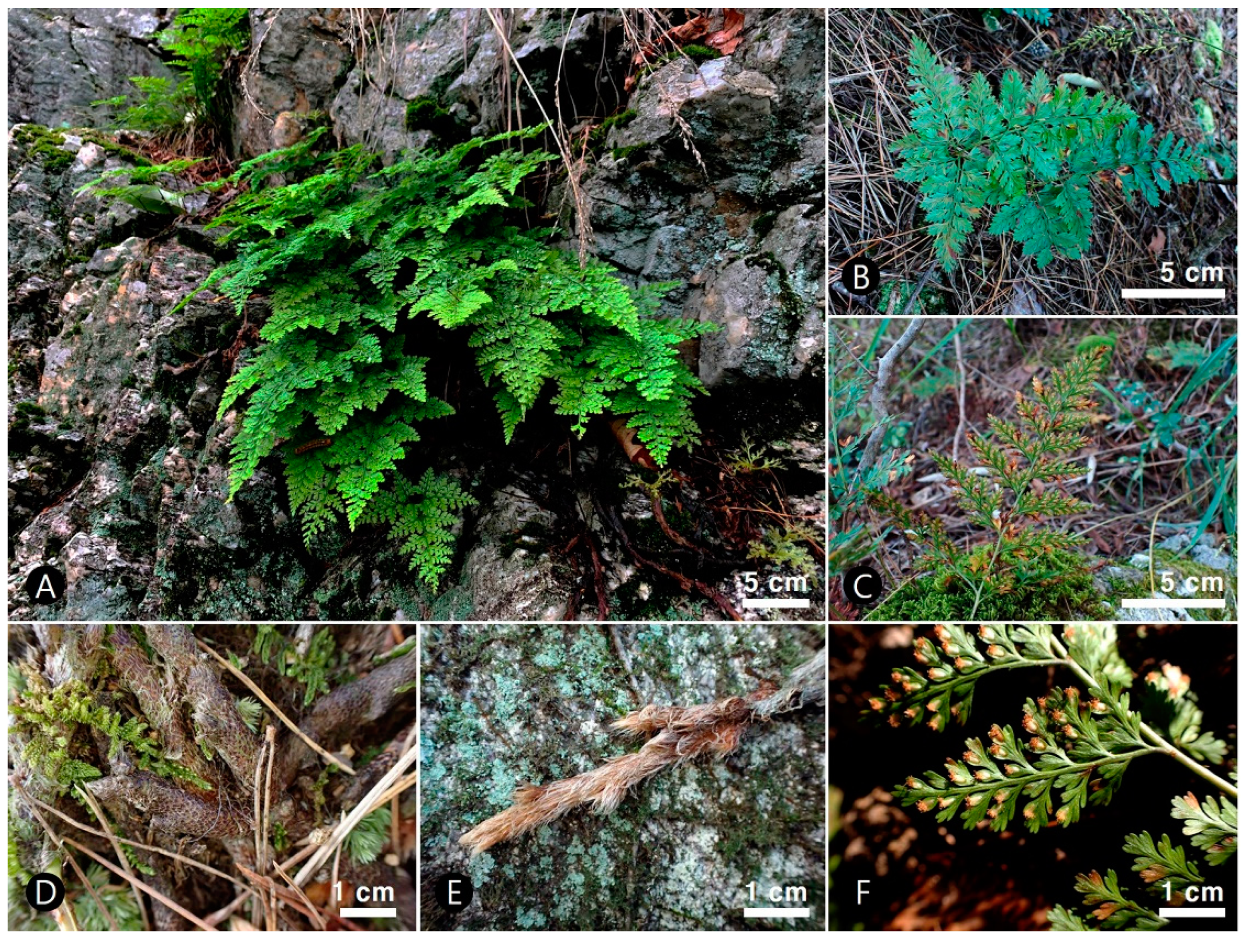
3.1. Morphological Characteristics

Herbs: perennial, usually epipetric (lithophyte) or rarely epiphytic, 9.4–34.0 cm tall. Rhizomes: long creeping, dark brown, 6.1–58 cm × 3.2–9.8 mm, densely scaly; scales persistent, black, dark to light brown or grayish orange, above a considerably broader base, evenly narrowed toward the apex, lanceolate, linear, or sickle-shaped ultimate lobes, peltate, 1.6–9.7 × 0.1–0.8 mm, margins denticulate. Fronds: monomorphic, alternate or opposite, symmetrical or asymmetrical, straight or bent, distantly spaced, petiolate; primary stipe grayish orange or yellowish green, 3.6–15.7 cm × 0.3–2.1 mm, adaxially grooved, sparse fugacious scales; blade rhomboid, triangular or triangular–ovate in outline, 4.5–22.8 × 5.2–26.3 cm, usually 4-pinnate, adaxial surface green or yellowish green, abaxial surface yellowish green, apex obtuse, acute or acuminate, glabrous; rachis winged; primary pinnae alternate or opposite, 5–17 pairs, triangular–ovate, petiolulate, basal pair largest, overlapping or not each pinna; primary upper and middle pinna ovate, oblong or elliptic, 1.0–5.2 × 0.5–2.4 cm; primary basal pinna asymmetry, lanceolate, oblong or ovate, 2.5–13.0 × 1.5–7.5 cm; ultimate segments oblanceolate, narrowly oblong or linear, 1.5–6.0 × 0.5–3.0 mm. Veins: forked, branched, and did not reach the margins. Sori: separate, terminal on veins, usually single on a segment at the forking point of veins, discoid, lateral opening; indusium linear, oblong, or elliptic pouch-shaped, 0.9–2.0 × 0.4–1.0 mm, membranous. Spores: ellipsoidal with monolete apertures (Figure 1; Table 1).

3.2. Anatomical Characteristics
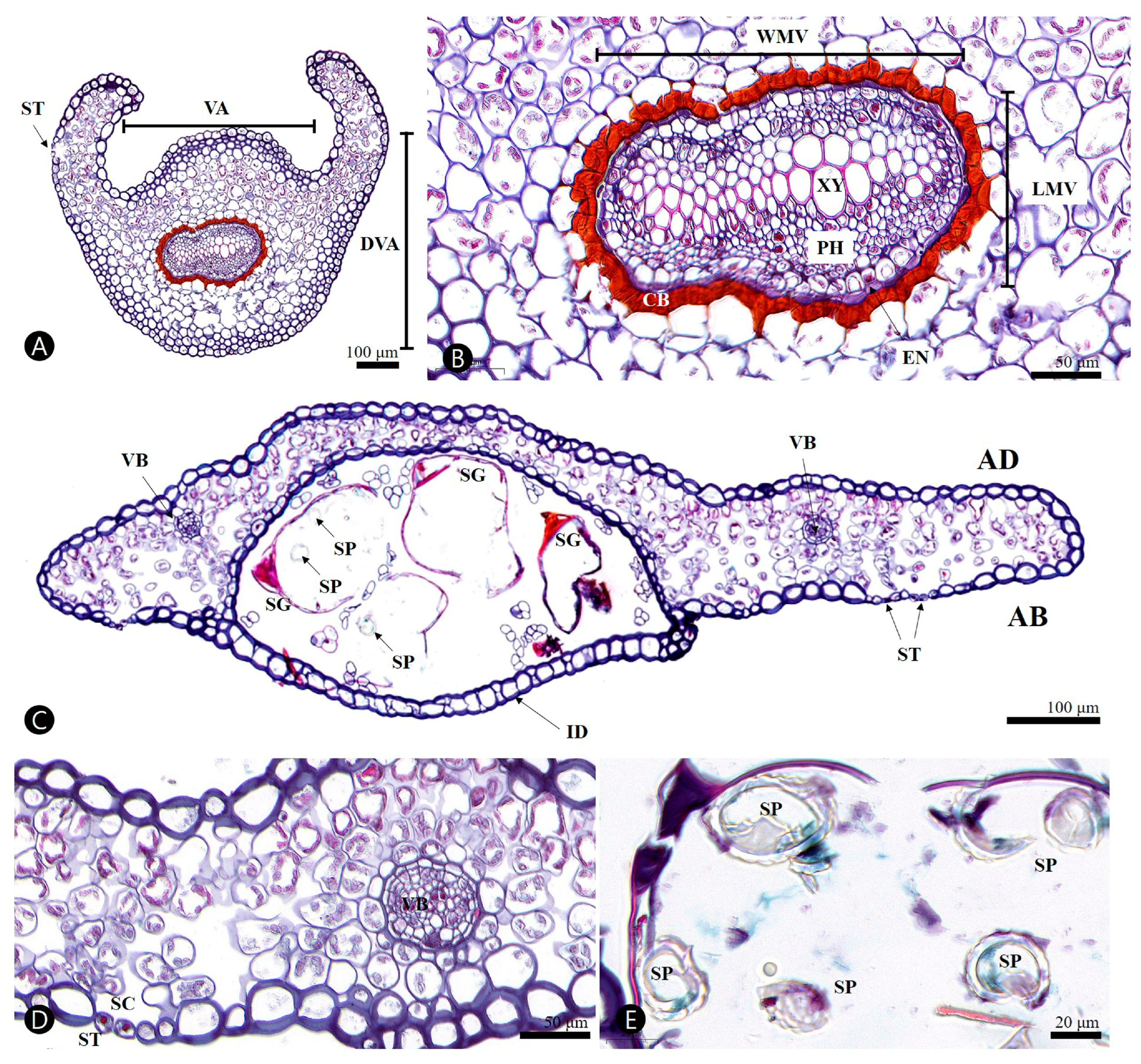
3.2.1. Stipe

The outline was circular, with a wing in the transverse section of the stipe. The size of the stipe (length of the ventral axis (VA) × length of the dorsiventral axis (DVA)) was 468.5–(559.2)–718.0 × 501.2–(585.6)–627.1 μm (Table 2). A thin cuticle layer covered the epidermis and consisted of uniseriate, oval, or rectangular cells (Figure 2A). The thickness of the abaxial side of the epidermal cell in the cross-section was 7.5–(10.5)–18.0 μm, and that of the adaxial side was 11.6–(15.2)–21.3 μm. The angular collenchyma tissue, located immediately under the epidermis, was composed of regular cells. The parenchyma cells of the cortex consisted of from four to seven oval, squashed oval, or almost rectangular layers and were isometrically arranged (Figure 2A). The stipe vascular bundle was open arc-shaped and its size (width of the main vascular bundle (WMV) × length of the main vascular bundle (LMV)) was 171.5–(213.5)–274.6 × 114.1–(133.5)–156.3 μm. The stipe vascular bundle was covered with a thick layer of the circumendodermal band (CB) (Figure 2B). The CB consisted of the continuity of the isodiametric cells formed by the band (ring) and was 6.9–(9.8)–11.7 μm thick (Table 2). The CB consisted of slightly square-shaped cells. Safranin histochemical tests for cell wall thickening in the CB were positive for red, which represented one-quarter to one-half of the cell forming U-shaped thickening (Figure 2B). No individual had CB cell walls with thickened total occlusion of the cell lumina. The endodermis was 2.0–(3.7)–5.4 μm thick. The stipe surface was glabrous. No crystals were observed in any of the stipe cells.

3.2.2. Ultimate Segment (of Pinnule)

The adaxial and abaxial epidermises of the fronds were characterized by one layer of cells covered by a cuticle (Figure 2C). The epidermal thicknesses of abaxial and adaxial were similar [abaxial, 7.1–(13.1)–19.7 μm; adaxial, 11.8–(15.1)–23.0 μm]. The mesophyll contained 3–6 layers of mainly arm-cells that were 61.2–(99.4)–149.6 μm thick and slightly differentiated into palisade and spongy parenchyma (Figure 2C, Table 2). The upper mesophyll in the indusium was arranged more compactly. The epidermal cells of the indusium were thicker than those of the adaxial and abaxial epidermis [15.0–(21.3)–25.1 μm]. Stomata were found only in the abaxial epidermis (hypostomatic fronds), with a stomatal chamber (air space) (Figure 2D). The diameter of vascular bundles was 25.2–(44.3)–70.6 μm (Figure 2D, Table 2). All parts of the adaxial and abaxial epidermis of the fronds were glabrous. No crystals were observed in any of the epidermal cells.

3.3. Micromorphological Characteristics
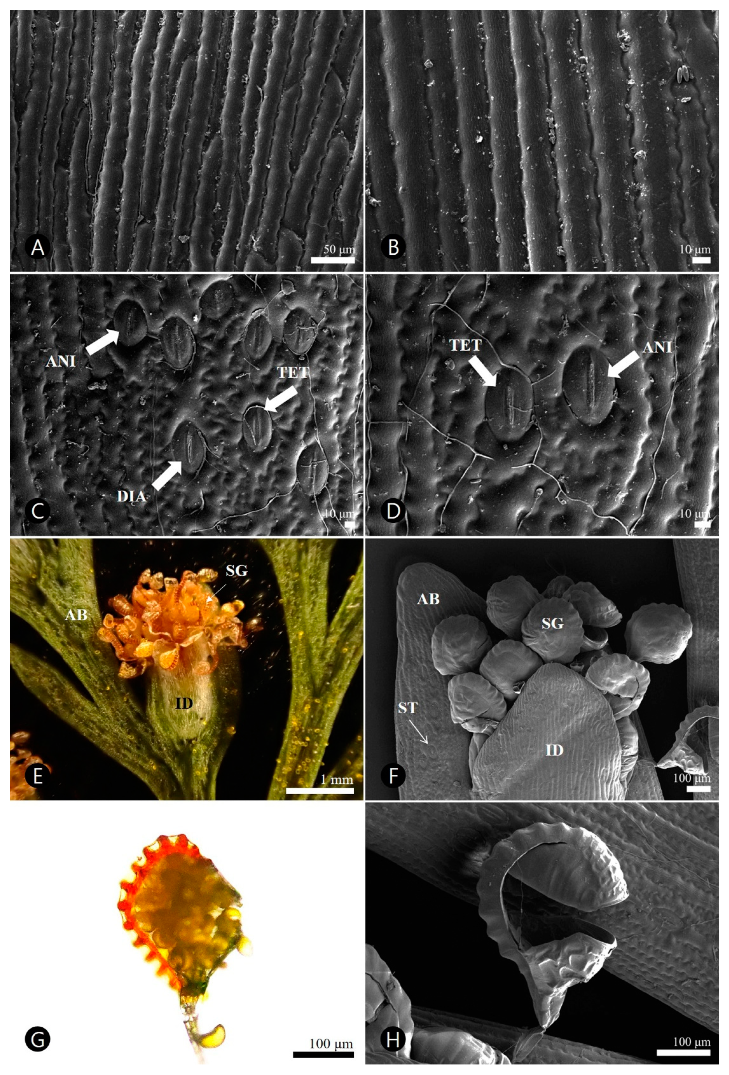
3.3.1. Pinnule

Epidermal cell patterns were described separately for adaxial (AD) and abaxial (AB). Frond epidermal cells on both surfaces were arranged elongately and had striated, convex periclinal walls; however, their anticlinal walls differed (Figure 3, Table 3). AD had straight and slightly undulating anticlinal walls (Figure 3A,B), whereas AB had undulating sinuate walls (Figure 3C,D). Hypostomatic fronds were observed and three types of stomatal complexes were recognized: diacytic, anisocytic, and tetracytic (Figure 3C,D). The stomata were 35.6–(46.3)–51.4 μm long, 25.9–(31.4)–36.6 μm wide, and had an area of 912.5–(1178.5)–1410.9 μm2, respectively (Table 3). The stomatal ledges were lip-shaped.

3.3.2. Sorus

Each pinnule had many separate sori in fertile fronds, which were generally elongated (Figure 3E). The indusium covered numerous sporangia. The epidermal cells of the outer indusium were isodiametrically arranged, straight and elongated in the anticlinal wall, and striated and convex in the periclinal wall (Figure 3F). The leptosporangium consisted of an apical cap, a basal cap, an annulus, and a short stalk (Figure 3G,H). The annulus was arranged longitudinally and consisted of 12–16 thickened cells.

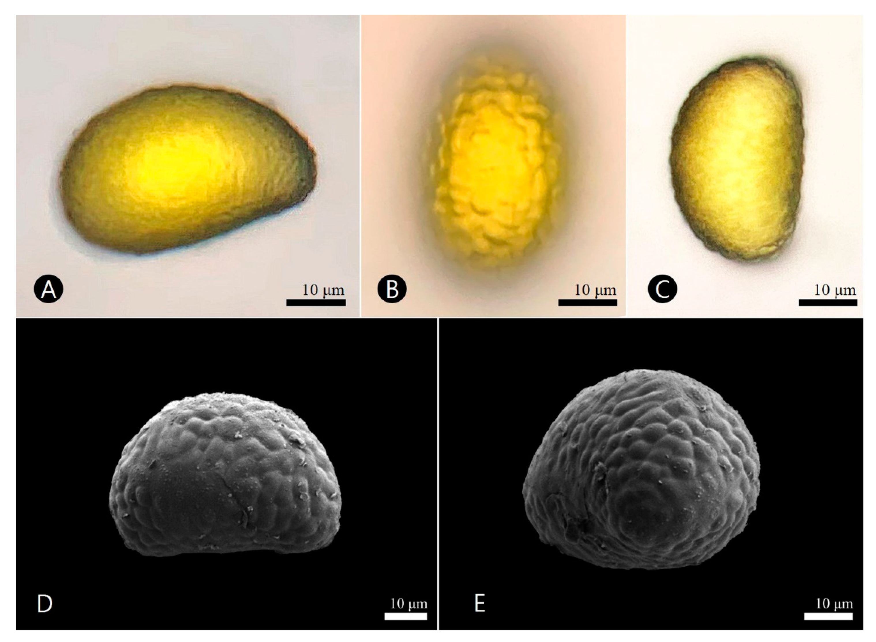
3.4. Palynological Characteristics

The spores were free and their sizes were 34.3–(38.8)–42.2 μm in polar axes, and 39.3–(50.8)–55.0 μm in equatorial axes. The exine was measured to be 2.3–(3.4)–5.1 μm thick (Table 4). The outline of the spore was transversely elliptical–reniform to ellipsoidal, bilaterally symmetrical, aniso-polar, and rounded–elliptical in polar view. The aperture was a monolete, which was a single linear mark designating the dividing axis of the proximal face (Figure 4). The ornamentation was a verrucae–colliculate type; the verrucae were numerous, slightly convex, densely packed, rounded, 2–(3.2)–5 μm diameter with radiating from laesura, forming a polygonal reticulate pattern (Figure 4D,E).

4. Discussion

This study is the first to comprehensively report the characteristics of the medicinal and economically important squirrel’s foot fern, D. mariesii, and provide valuable information regarding its morphological, anatomical, micromorphological, and palynological characteristics.
Morphological characteristics such as the size of fronds and the shape and color of rhizome scales are taxonomically significant for identifying and delimiting species in the genus Davallia [5,35]. D. mariesii was closely related to D. griffithiana (=D. tyermannii (T. Moore) Baker), systematically [8,30,36]. D. mariesii was distinguished by its tri-pinnate or quadri-pinnate fronds (vs. bi-pinnate or tri-pinnate in D. griffithiana), ultimate segments 5–27 × 2–6 mm (vs. 2–5 × 2–3 mm), and pouch-shaped, oblong, longer than wide indusia (vs. semicircular, approximately as wide as long) [35]. Although the present results largely correspond with those reported for the Flora of Korea [7], several differences were found, and additional quantitative data were determined (Table 1). Characteristics such as plant height, rhizome length, scale width, stipes, primary pinna size, ultimate segments, and indusia were updated.
Stipe anatomical characteristics such as vascular bundle shape, number of vascular bundles, the presence or absence of the CB, and adaxial grooves are reflected in the promising results of taxonomic and systematic studies [15,37,38]. In particular, the stipe vascular bundles, which are conserved and stable, are valuable for taxonomy [37,38]. Our results indicated that the openly arc-shaped main vascular bundles of the studied species were similar to those of D. hymenophylloides (Blume) Kuhn (≡Davallodes hymenophylloides (Blume) M. Kato and Tsutsumi) [38].
In 89 species from 28 families of ferns, the second innermost layer of primary tissue next to the innermost endodermis, formed from thick-walled, tannin-rich cells, is present in the stipes, known as the CB [34]. To date, in Davalliaceae, a continuous CB with the total proportion of the cell lumen has only been observed in D. canariensis (L.) Sm. [15,16]. Our study reports an additional Davalliaceae species associated with CB. The degree of CB cell wall thickening in each species ranged from 25% to 100% of the cell lumina. Moreover, interspecific variations have been reported in 10 studied species of the genera Elaphoglossum and Polypodium [34]. In this study, total occlusion of the cell lumina of the CB in D. mariesii was not observed, in contrast to D. canariensis. Thus, the degree of cell wall thickening in the CB may be helpful for identification at the species level in the genus Davallia. However, further studies are required to evaluate the taxonomic significance of these findings.
CBs have been referred to as protective bands [39] and sclerenchymatic rings [15,40,41]. However, previous histochemical tests and developmental investigations revealed that CB cells were negative for lignin and positive for the cytoplasm and nuclei, indicating that CB cells were not lignified sclerenchyma [34,42]. Several studies have shown that CB cells contain numerous tannins [33]. Although this study showed positive results for safranin, more precise histochemical tests and chemical analyses are required to determine its specific molecular composition, including cellulose, tannins, lignin, and suberin.
In D. mariesii, CB may have two possible functions: a protective function similar to that in the endodermis [43] and a biomechanical function [34]. In particular, to resist bending and twisting stresses, the central tissues must be elastic, such as living parenchyma, and not rigid, such as lignified sclerenchyma [34,44]. The CB of D. mariesii, which is an epipetric (lithophyte) species with mid- to large-sized pinnate fronds [4.5–22.8 × 5.2–26.3 cm] and stipes [3.6–15.7 cm × 0.3–2.1 mm], probably experiences higher bending shear forces than other ferns with smaller ones.
Recent micromorphological studies have suggested that the characteristics of the frond epidermis are key features for the classification of Davalliaceae at the genus or species levels [8,17,18,19,20,21]. In particular, the frond (the ultimate segment of the pinnule) micromorpho–anatomical results revealed that the epidermal cell thickness and arrangement of the abaxial and adaxial walls were similar; however, those of the anticlinal walls were different. Most fern leaves have similar anticlinal walls on both epidermal surfaces [8,26,45]. Additional expanded frond cell data are required to evaluate their taxonomic importance in Davallia using the phylogenetic target-sampling strategies. The stomatal characteristics observed in this study disagreed with those from a previous survey. Sen [46] described that Japanese D. mariesii (voucher no. 0035-38 in Naturalis Biodiversity Center, The Netherlands) has polocytic, copolocytic, anomocytic, and staurocutic stomata of 31.5–36 × 22.5–31.5 μm; however, our results revealed that those of Korean D. mariesii have diacytic, anisocytic, and tetracytic stomata with larger size (35.6–51.4 × 25.9–36.6 μm). Understanding the degree of variation in stomatal characteristics of this species requires frond micromorphological studies based on geographical, ecological, and developmental differences.
Palynological data have been extensively used to determine the systematic relationships between fern groups [22,23,24,25,26]. In this study, the spore morphology of D. mariesii was consistent with previous data from the genus Davallia [45,47,48,49,50]. The D. mariesii spore size includes the size variations in the Davallia clade, as described by Wang et al. [30]. All Davallia species have verrucate–colliculate ornamentation patterns [30], including macrofossils [51] such as D. mariesii. The verrucate–colliculate ornamentation type appears to be the most common in the genus and may represent a plesiomorphic state in the genus Davallia. Thus, spore characteristics, especially surface ornamentation in this genus, may be stable and helpful for taxonomy, systematics, paleobotany, and palynology at the generic level.
This study contributes to the knowledge of the anatomical, micromorphological, and palynological diversity of Davalliaceae plants. Nevertheless, further studies are necessary to evaluate species-level variations in the stipe, frond, and spore morphology in this family based on taxonomic and phylogenetic contexts using an expanded and targeted sampling strategy.

5. Conclusions

The present study provides valuable information regarding the morphology, anatomy, micromorphology, and palynology of the medicinally and economically important fern, Davallia mariesii, which is distributed in Korea. In particular, the morphological description of the species has expanded based on a large amount of living materials. This is the first comprehensive study of the stipe and frond anatomy, and the frond and spore micromorphology of D. mariesii in Korea. Our investigations aim to contribute to the comprehensive understanding of the taxonomy, systematics, paleobotany, and palynology of the genus Davallia, including the family Davalliaceae. However, additional molecular frameworks are required to address the challenges of the relationships and taxonomy of this genus. Further studies involving a broader range of Davallia taxa and extensive sampling would enhance our understanding of the taxonomic and systematic implications associated with the stipe, frond, and spore microanatomy.

Author Contributions

Conceptualization, J.-H.S.; methodology, S.Y., G.C. and J.-H.S.; software, S.Y. and J.-H.S.; validation, S.Y. and J.-H.S.; formal analysis, S.Y. and J.-H.S.; investigation, S.Y., G.C. and J.-H.S.; resources, S.Y. and J.-H.S.; data curation, S.Y., G.C. and J.-H.S.; writing—original draft preparation, S.Y. and J.-H.S.; writing—review and editing, J.-H.S.; visualization, S.Y., G.C. and J.-H.S.; supervision, J.-H.S.; project administration, J.-H.S.; funding acquisition, G.C. and J.-H.S. All authors have read and agreed to the published version of the manuscript.

Funding

This research was supported by the Korea Institute of Oriental Medicine, Naju, South Korea (grant numbers KSN1822320) and the National Research Foundation of Korea (NRF) grants funded by the Korea government (MSIT) (NRF-2020R1A2C1100147 and RS-2023-00208589) to J.-H. Song.

Data Availability Statement

All data are fully available without restriction. The plant images and micrographs can be made available upon requests addressed to correspondence (Jun-Ho Song).

Acknowledgments

The authors would like to thank the five anonymous reviewers whose comments and corrections have improved the work.

Conflicts of Interest

The authors declare that they have no competing interest or conflict of interest.

References

  1. Kramer, K.U. Davalliaceae. In The Families and Genera of Vascular Plants: Pteridophytes and Gymnosperms; Kubitzki, K., Ed.; Springer: Heidelberg/Berlin, Germany, 1990; Volume 1, pp. 74–80. [Google Scholar]
  2. Nooteboom, H.P. Davalliaceae of China. Acta Phytotax. Sin. 1996, 34, 162–179. [Google Scholar]
  3. Wu, S.H. Davalliaceae. In Flora Reipublicae Popularis Sinicae; Wu, S.H., Wang, C.H., Eds.; Science Press: Beijing, China, 1999; Volume 6, pp. 161–199. [Google Scholar]
  4. Von Konrat, M.J.; Braggins, J.E.; de Lange, P.J. Davallia (Pteridophyta) in New Zealand, including description of a new subspecies of D. tasmanii. N. Z. J. Bot. 1999, 37, 579–593. [Google Scholar] [CrossRef]
  5. Xing, F.W.; Wang, F.G.; Nooteboom, H.P. Davalliaceae. In Flora of China (Pteridophytes); Wu, Z.Y., Raven, P.H., Hong, D.Y., Eds.; Science Press: Beijing, China; Missouri Botanical Garden Press: St. Louis, UK, 2013; pp. 749–757. [Google Scholar]
  6. Tsutsumi, C.; Chen, C.W.; Larsson, A.; Hirayama, Y.; Kato, M. Phylogeny and classification of Davalliaceae on the basis of chloroplast and nuclear markers. Taxon 2016, 65, 1236–1248. [Google Scholar] [CrossRef]
  7. Sun, B.Y.; Davalliaceae, M.R. Schomb. In Flora of Korea: Pteridophytes and Gymnosperms; Flora of Korea Editorial Committee, Ed.; The National Institute of Biological Resources: Incheon, Republic of Korea, 2015; Volume I, p. 148. [Google Scholar]
  8. Ma, X.D.; Wang, A.H.; Wang, F.G.; He, C.M.; Liu, D.M.; Gerstberger, P.; Xing, F.W. A revised classification of Chinese Davalliaceae based on new evidence from molecular phylogenetics and morphological characteristics. PLoS ONE 2018, 13, e0206345. [Google Scholar] [CrossRef] [PubMed]
  9. Ni, L.J.; Wang, N.N.; Zhang, L.G.; Guo, Y.Z.; Shi, W.Z. Evaluation of the effects of active fractions of Chinese medicine formulas on IL-1beta, IL-6, and TNF-alpha release from ANA-1 murine macrophages. J. Ethnopharmacol. 2016, 179, 420–431. [Google Scholar] [CrossRef] [PubMed]
  10. Lin, Y.T.; Peng, S.W.; Imtiyaz, Z.; Ho, C.W.; Chiou, W.F.; Lee, M.H. In vivo and in vitro evaluation of the osteogenic potential of Davallia mariesii T. Moore ex Baker. J. Ethnopharmacol. 2021, 264, 113126. [Google Scholar] [CrossRef] [PubMed]
  11. Cui, C.B.; Tezuka, Y.; Kikuchi, T.; Nakano, H.; Tamaoki, T.; Park, J.H. Constituents of a fern, Davallia mariesii Moore. I. Isolation and structures of davallialactone and a new flavanone glucuronide. Chem. Pharm. Bull. 1990, 38, 3218–3225. [Google Scholar] [CrossRef] [PubMed]
  12. Cui, C.B.; Tezuka, Y.; Yamashita, H.; Kikuchi, T.; Nakano, H.; Tamaoki, T.; Park, J.H. Constituents of a fern, Davallia mariesii Moore. V. Isolation and structures of davallin, a new tetrameric proanthocyanidin, and two new phenolic glycosides. Chem. Pharm. Bull. 1993, 41, 1491–1497. [Google Scholar] [CrossRef]
  13. Gunasinghe, Y.H.K.I.S.; Rathnayake, I.V.N.; Deeyamulla, M.P. Plant and plant associated microflora: Potential bioremediation option of indoor air pollutants. Nepal J. Biotechnol. 2021, 9, 63–74. [Google Scholar] [CrossRef]
  14. White, R.A. Comparative anatomical studies of the ferns. Ann. Mo. Bot. Gard. 1974, 61, 379–387. [Google Scholar] [CrossRef]
  15. Lin, B.L.; DeVol, C.D. The use of stipe characters in fern taxonomy I. Taiwania 1977, 22, 91–99. [Google Scholar]
  16. Lin, B.L.; DeVol, C.D. The use of stipe characters in fern taxonomy II. Taiwania 1978, 23, 77–95. [Google Scholar]
  17. Lee, C.-S. A taxonomical study of Korean Pteridaceae on the morphology of leaf epidermis. Korean J. Plant Taxon. 1988, 18, 275–290. [Google Scholar] [CrossRef]
  18. Sen, U.; De, B. Structure and ontogeny of stomata in ferns. Blumea 1992, 37, 239–261. [Google Scholar]
  19. Bondada, B.; Tu, C.; Ma, L. Surface structure and anatomical aspects of Chinese brake fern (Pteris vittata; Pteridaceae). Brittonia 2006, 58, 217–228. [Google Scholar] [CrossRef]
  20. Chuang, Y.-Y.; Liu, H.-Y. Leaf epidermal morphology and its systematic implications in Taiwan Pteridaceae. Taiwania 2003, 48, 60–71. [Google Scholar]
  21. Shah, S.N.; Ahmad, M.; Zafar, M.; Ullah, F.; Zaman, W.; Mazumdar, J.; Khuram, I.; Khan, S.M. Leaf micromorphological adaptations of resurrection ferns in northern Pakistan. Flora 2019, 255, 1–10. [Google Scholar] [CrossRef]
  22. Giacosa, J.R.; Morbelli, M.; Giudice, G. Spore morphology and wall ultrastructure of Anemia Swartz species (Anemiaceae) from Argentina. Rev. Palaeobot. Palynol. 2012, 174, 27–38. [Google Scholar] [CrossRef]
  23. Lee, S.J.; Park, C.W. Spore morphology of the genus Dryopteris Adans. (Dryopteridaceae) in Korea. J. Plant Biol. 2014, 57, 302–311. [Google Scholar] [CrossRef]
  24. Giacosa, J.R. Spore morphology and wall ultrastructure of Lomariocycas (Blechnaceae) species from America. Rev. Palaeobot. Palynol. 2019, 269, 55–63. [Google Scholar] [CrossRef]
  25. Yañez, A.; Marquez, G.J.; Morbelli, M.A. Spore morphology and ultrastructure of Dennstaedtiaceae from Paranaense Phytogeographic Province I.: Genus Dennstaedtia. Rev. Palaeobot. Palynol. 2016, 224, 181–194. [Google Scholar] [CrossRef]
  26. Shah, S.N.; Ahmad, M.; Zafar, M.; Hadi, F.; Khan, M.N.; Noor, A.; Malik, K.; Rashid, N.; Iqbal, M. Spore morphology and leaf epidermal anatomy as a taxonomic source in the identification of Asplenium species from Malakand division Pakistan. Microsc. Res. Tech. 2020, 83, 1354–1368. [Google Scholar] [CrossRef] [PubMed]
  27. Phillips, D.A.; White, R.A. Frond articulation in species of Polypodiaceae and Davalliaceae. Am. Fern J. 1967, 57, 78–88. [Google Scholar] [CrossRef]
  28. Ma, X.; He, C.; Wang, F.; Wang, A.; Xing, F. Structural characteristics of leaf epidermis and their systematic significance in Davalliaceae. Plant Sci. J. 2015, 33, 438–447. [Google Scholar]
  29. Tsutsumi, C.; Kato, M. Morphology and evolution of epiphytic Davalliaceae scales. Botany 2008, 86, 1393–1403. [Google Scholar] [CrossRef]
  30. Wang, F.G.; Liu, H.M.; He, C.M.; Yang, D.M.; Xing, F.W. Taxonomic and evolutionary implications of spore ornamentation in Davalliaceae. J. Syst. Evol. 2015, 53, 72–81. [Google Scholar] [CrossRef]
  31. Johansen, D.A. Plant Microtechnique; McGraw-Hill Book Company, Inc.: London, UK, 1940; 530p. [Google Scholar]
  32. Wilkinson, H.P. The plant surface (mainly leaf). In Anatomy of the Dicotyledons, 2nd ed.; Metcalfe, C.R., Chalk, L., Eds.; Clarendon Press: Oxford, UK, 1979; Volume 1, pp. 97–165. [Google Scholar]
  33. Sen, T.; Sen, U.; Holttum, R.E. Morphology and anatomy of the genera Davallia, Araiostegia and Davallodes, with a discussion on their affinities. Kew Bull. 1972, 27, 217–243. [Google Scholar] [CrossRef]
  34. Hernández-Hernández, V.; Terrazas, T.; Mehltreter, K.; Angeles, G. Studies of petiolar anatomy in ferns: Structural diversity and systematic significance of the circumendodermal band. Bot. J. Linn. Soc. 2012, 169, 596–610. [Google Scholar] [CrossRef]
  35. Nooteboom, H.P. Notes on Davalliaceae II. A revision of the genus Davallia. Blumea 1994, 39, 151–214. [Google Scholar]
  36. Liu, H.; Schneider, H. Evidence supporting Davallia canariensis as a Late Miocene relict endemic to Macaronesia and Atlantic Europe. Aust. Syst. Bot. 2013, 26, 378–385. [Google Scholar] [CrossRef]
  37. Noraini, T.; Ruzi, A.R.; Nadiah, N.; Nisa, R.N.; Maideen, H.; Solihani, S.N. Stipe anatomical characteristics in some Davallia (Davalliaceae) species in Malaysia. Sains Malays. 2012, 41, 53–62. [Google Scholar]
  38. Tan, J.M.P.; Banaticla-Hilario, M.C.; Malabrigo, P.; Angeles, M.D.; Buot, I.E. Anatomical examination of the petiole of eupolypods I (Polypodiales). Biodiversitas J. Biol. Divers. 2020, 21, 1767–1777. [Google Scholar]
  39. Russow, E. Vergleichende Untersuchungen betreffend der Histologie (Histographie und Histogenie) der Vegetativen und Sporenbildenden Organe und die Entwicklung der Sporen der Leitbündel-Kryptogamen, mit Berücksichtigung der Histologie der Phanerogamen Ausgehend von der Betrachtung der Marsiliaceae; Mémoires de l’Académie Impériale des Sciences de St. Petersbourg, sér. 7; Académie Impériale des Sciences: Saint Petersburg, Russia, 1872; Volume 19, 207p. [Google Scholar]
  40. Ogura, Y. Comparative Anatomy of the Vegetative organs of the Pteridophytes; Borntraeger: Berlin, Germany, 1972. [Google Scholar]
  41. Moran, R.C. Monograph of the neotropical fern genus Polybotrya (Dryopteridaceae). Ill. Nat. Hist. Surv. Bull. 1987, 34, 1–138. [Google Scholar] [CrossRef]
  42. Hernández-Hernández, V.; Terrazas, T.; Stevenson, D.W.M. Ontogeny of Ctenitis melanosticta and Diplazium expansum fronds with emphasis on the circumendodermal band. Feddes Repert. 2009, 120, 426–442. [Google Scholar] [CrossRef]
  43. Van Fleet, D.S. Histochemistry and function of the endodermis. Bot. Rev. 1961, 27, 165–220. [Google Scholar] [CrossRef]
  44. Niklas, K.J. Research review: A mechanical perspectiveon foliage leaf form and function. New Phytol. 1999, 143, 19–31. [Google Scholar] [CrossRef]
  45. Shah, S.N.; Celik, A.; Ahmad, M.; Ullah, F.; Zaman, W.; Zafar, M.; Malik, K.; Rashid, N.; Iqbal, M.; Sohail, A.; et al. Leaf epidermal micromorphology and its implications in systematics of certain taxa of the fern family Pteridaceae from northern Pakistan. Microsc. Res. Tech. 2019, 82, 317–332. [Google Scholar] [CrossRef]
  46. Sen, T. The evidence of stomatal development on the relationships between Davallia and genera associated with it by recent authors. Ann. Bot. 1986, 58, 663–677. [Google Scholar] [CrossRef]
  47. Huang, T.-C. Spore Flora of Taiwan; Meitai Color Print Co., Ltd.: Taipei, Taiwan, 1981. [Google Scholar]
  48. Moy, C.J. Variations of fern spore ultrastructure as reflections of their evolution. Grana 1988, 27, 39–51. [Google Scholar] [CrossRef]
  49. Tryon, A.F.; Lugardon, B. Spores of the Pteridophyta; Springer: New York, NY, USA, 1991. [Google Scholar]
  50. Sofiyanti, N.; Isda, M.N.; Juliantari, E.; Suriatno, R.; Pranata, S. The inventory and spore morphology of ferns from Bengkalis Island, Riau Province, Indonesia. Biodivers. J. Biol. Divers. 2019, 20, 3223–3236. [Google Scholar] [CrossRef]
  51. Conran, J.G.; Kaulfuss, U.; Bannister, J.M.; Mildenhall, D.C.; Lee, D.E. Davallia (Polypodiales: Davalliaceae) macrofossils from early Miocene Otago (New Zealand) with in situ spores. Rev. Palaeobot. Palynol. 2010, 162, 84–94. [Google Scholar] [CrossRef]
Figure 1. Photographs of the Davallia mariesii T. Moore ex Baker in Korea. (A) Habit, lithophyte. (B) Adaxial side of a fertile frond. (C) Abaxial side of a fertile frond. (D) Rhizomes, under wet conditions. (E) Rhizomes, under dry conditions. (F) Abaxial side of a fertile pinna with sori.
Horticulturae 09 00939 g001
Figure 2. Micrographs of transverse section of the stipe and fertile frond in the Davallia mariesii. (A,B) Stipe. (C) Fertile frond. (D) Blade of fertile frond. (E) Sporangium with spore. AB, Abaxial side; AD, Adaxial side; CB, circumendodermal band; DVA, length of dorsiventral axis; EN, endodermis; ID, Indusium; LMV, length of main vascular bundle; PH, phloem; SC, stomata chamber; SG, Sporangium; SP, spore; ST, stomata; VA, length of ventral axis; VB, vascular bundle; WMV, width of main vascular bundle; XY, xylem.
Horticulturae 09 00939 g002
Figure 3. Stereo (SM), light (LM), and scanning electron (SEM) micrographs of the mature sporophyte in the Davallia mariesii. (A,B) Adaxial side of a fertile frond. (C,D) Abaxial side of a fertile frond with stomata. (E,F) Sorus with indusium. (G,H) Sporangium with spore. (AD,F,H) SEM. (E) SM. (G) LM. AB, Abaxial side; ANI, Anisocytic, DIA, Diacytic; ID, Indusium; SG, Sporangium; ST, stomata; TET, Tetracytic.
Horticulturae 09 00939 g003
Figure 4. Light (LM) and scanning electron (SEM) micrographs of the verrucae-colliculate spore in the Davallia mariesii. (A) Equatorial side with transversely ellipsoidal shape (LM). (B) Proximal side with rounded–elliptical shape (LM). (C) Distal side with transversely elliptical–reniform shape (LM). (D) Equatorial side with rounded verrucae (LM). (E) Equatorial–distal side forming a polygonal reticulate pattern (SEM).
Horticulturae 09 00939 g004
Table 1. Morphological characters of Davallia mariesii T. Moore ex Baker based on our study and Flora of Korea [7].
Our StudyFlora of Korea
Plants9.4–34.0 cm tall-
Rhizome6.1–58 cm × 3.2–9.8 mm3–5 mm in diam.
Scale1.6–9.7 × 0.1–0.8 mm5–8 mm long
Frond8.1–38.5 cm long15–35 cm long
Stipe3.6–15.7 cm × 0.3–2.1 mm5–15 cm long
Blade4.5–22.8 × 5.2–26.3 cm10–20 × 8–15 cm
Pinnae5–17 pairs6–12 pairs
Primary upper and middle pinna1.0–5.2 × 0.5–2.4 cm-
Primary basal pinna2.5–13.0 × 1.5–7.5 cm-
Ultimate segments1.5–6.0 × 0.5–3.0 mm1–2 mm wide
Indusium0.9–2.0 × 0.4–1.0 mm-
Table 2. Anatomical characters of stipe and pinnule of Davallia mariesii.
Davallia mariesii
Stipe
Outline of stipecircular with wing
Length of ventral axis (VA) (μm)468.5–(559.2)–718.0
Length of dorsiventral axis (DVA) (μm)501.2–(585.6)–627.1
Epidermal thickness-abaxial surface (μm)7.5–(10.5)–18.0
Epidermal thickness-adaxial surface (μm)11.6–(15.2)–21.3
Arrangement of cortexisometric
Outline of stipe vascular bundleopen arc
Width of main vascular bundle (WMV) (μm)171.5–(213.5)–274.6
Length of main vascular bundle (LMV) (μm)114.1–(133.5)–156.3
Circumendodermal band typecontinuous ring
Circumendodermal band proportion1/4 to 1/2
Circumendodermal band thickness (μm)6.9–(9.8)–11.7
Endodermis thickness (μm)2.0–(3.7)–5.4
Blade (of ultimate segment)
Epidermal thickness-abaxial surface (μm)7.1–(13.1)–19.7
Epidermal thickness-adaxial surface (μm)11.8–(15.1)–23.0
Indusium epidermal thickness (μm)15.0–(21.3)–25.1
Mesophyll thickness (non-indusium area) (μm)61.2–(99.4)–149.6
Diameter of vascular bundle (μm)25.2–(44.3)–70.6
Table 3. Micromorphological characters of frond epidermal cells of Davallia mariesii.
Adaxial SideAbaxial Side
Epidermal cell arrangementElongatedelongated
Anticlinal cell wallstraight, slightly undulateundulate to sinuate
Periclinal cell wallstriate, convexstriate, convex
Stomata typeAbsentdiacytic, anisocytic, tetracytic
Stomata length (μm)Absent35.6–(46.3)–51.4
Stomata width (μm)Absent25.9–(31.4)–36.6
Stomata area (μm2)Absent912.5–(1178.5)–1410.9
Stomatal ledgeAbsentlip-shaped
Table 4. Palynological characters of spore of Davallia mariesii.
Davallia mariesii
Polar axes (μm)34.3–(38.8)–42.2
Equatorial axes (μm)39.3–(50.8)–55.0
Shapeellipsoidal
Exine thickness (μm)2.3–(3.4)–5.1
Aperturemonolete
Exine ornamentationverrucate colliculate
Disclaimer/Publisher’s Note: The statements, opinions and data contained in all publications are solely those of the individual author(s) and contributor(s) and not of MDPI and/or the editor(s). MDPI and/or the editor(s) disclaim responsibility for any injury to people or property resulting from any ideas, methods, instructions or products referred to in the content.

