Article

Methyl Jasmonate Elicitation for In Vitro Lycorine Accumulation in Three Zephyranthes Species and Comparative Analysis of Tissue-Cultured and Field Grown Plants

1
Cellular Differentiation and Molecular Genetics Section, Department of Botany, Jamia Hamdard, New Delhi 110062, India
2
Plant Production Department, College of Food and Agriculture Sciences, King Saud University, Riyadh 11451, Saudi Arabia
3
Department of Crop Science, Faculty of Agriculture, Universiti Putra Malaysia, Serdang 43400, Malaysia
4
Department of Plant and Soil Sciences, Mississippi State University, 75 B.S. Hood Rd., Mississippi State, MS 39762, USA
*
Author to whom correspondence should be addressed.
Horticulturae 2023, 9(7), 832; https://doi.org/10.3390/horticulturae9070832
Submission received: 8 June 2023 / Revised: 18 July 2023 / Accepted: 18 July 2023 / Published: 21 July 2023
(This article belongs to the Special Issue A New Decade in the Propagation of Horticultural and Medicinal Plants)

Abstract

:
Lycorine is an important Amaryllidaceae alkaloid showing anti-cancerous activity on numerous cell lines; and it also demonstrates antiviral activity against several viruses including SARS-CoV-2 responsible for COVID-19. The in vitro cultivation of plant parts is a good alternative for elicitor mediated secondary metabolite synthesis, mass propagation, and cloning of elite genotypes. In this study, the quantification of lycorine from Zephyranthes candida, Zephyranthes grandiflora, and Zephyranthes citrina was evaluated, and the influence of methyl jasmonate (MJ) treatments on accumulation of lycorine yield was observed in the in vitro cultivated tissues. The influence of methyl jasmonate (MJ) on accumulation of lycorine was investigated for the first time in in vitro regenerated bulb, leaf, and root tissues of Zephyranthues. The efficient in vitro shoot regeneration was achieved in Z. candida (73.9%), Z. grandiflora (85.5%), and Z. citrina (76.5%) on Murashige and Skoog (MS) medium amended with 2.0 mg·L−1 Naphthalene acetic acid + 0.5 mg·L−1 Benzylaminopurine. The regenerants had the highest i.e., 7.46 mean numbers of bulblets per shoot. The best root induction (80.66%) was achieved on 2.0 mg/L Indole butyric acid (IBA). The lycorine content was quantified through High performance thin layer chromatography (HPTLC) in three field grown Zephyranthes species, was highest in Z. candida (1.93 µg g−1 dry wt.), followed by Z. grandiflora (1.87 µg g−1 dry wt.) and Z. citrina (1.62 µg g−1 dry wt.). The yield of lycorine was observed in this order i.e., bulb > leaf > root. The regenerated plants were supplemented with MJ and maximum lycorine yield was noted at 100 µM in bulb tissues of Z. candida (2.74 µg g−1 dry wt.) with an increase in percentage of 39.08. The enhancement could be due to MJ-induced stress as the biochemical attributes and anti-oxidant enzyme activity were high with elevated level of MJ. This enrichment may auger commercial manufacturing and utilization of lycorine in future.

1. Introduction

The plants synthesize primary compounds like carbohydrates, proteins, amino acids for cellular growth, progress, and reproduction; and produce secondary metabolites alkaloids, phenols, flavonoids, terpenes and others for defense purposes against stress, herbivore and pathogen invasion [1]. The alkaloids are primarily produced by flowering taxa; and approximately 20% of the alkaloids with pharmaceutical and industrial values are obtained from these sources. These are by products or subsidiary like medicines, agrochemicals, flavors, fragrances, pesticides, food additives etc. [2]. The genus Zephyranthes of Amaryllidaceae comprises of perennial, bulbous, and plants of ornamental and medicinal importance [3]. According to the world checklist, the genus contains about 90 species, native to western hemisphere and is grown as ornamental in many countries like India, China, Indonesia, Thailand, and Africa [4]. The bulb and leaf extracts are used in Traditional Chinese Medicine (TCM) owing to its anti-inflammatory, antibacterial, antiviral, anticancer, anti-cholinesterase, and antidiabetic abilities [5]. The genus contains a diverse class of compounds commonly called as Amaryllidaceae alkaloids of which tazettine, belladine, homolycorine, crinine, haemanthamine, montanine, lycorine, and galanthamine are popular, showing potent biological activities [6]. Lycorine is one of the novel phenylpropanoid metabolomes found in this genus, formed by a coupling reaction of L-phenylalanine and L-tyrosine through intermediate o-methylnorbelladine—a common precursor of all Amaryllidaceae alkaloids [7]. Lycorine acts as anti-cancerous agent against leukemia, ovarian-, breast-, and lung cancer, multiple myeloma, and colon carcinoma [8]. The lycorine also shows a wide spectrum of antiviral activities like Herpes simplex, Retrovirus, HIV-1, West Nile, Polio, Influenza, Hepatitis C and demonstrates inhibition activity on COVID-19 Corona virus [9]. The natural bioactive compounds are confined to certain genus, species, and varieties, accumulated in specific organs different from the site of synthesis [10]. Notably, the natural accumulation of Amaryllidaceae alkaloids in Zephyranthes is reported to be 0.1–1% of the dry weight, indicating their potential significance. However, the chemical synthesis of these alkaloids is known to be a complex and costly process [11]. The quantitative analytical studies on these alkaloids have been conducted in various genera such as Hippestrum, Crinum, Galanthus, Lycoris, and Narcissus; however, limited and preliminary reports are available specifically for Zephyranthes. These existing reports also have limitations in terms of comprehensiveness and applicability. Therefore, our study aims to address this gap by introducing suitable biotechnological approaches for mass propagation, cloning, and enhanced accumulation of secondary metabolites in Zephyranthes. This innovative approach, focused on the genus Zephyranthes, holds significant promise for the commercial exploitation and sustainable utilization of these valuable alkaloids. To this end, suitable biotechnological approaches need to be introduced for mass propagation, cloning, and higher secondary metabolite accum—ulation in Zephyranthes for commercial application.
Plant tissue culture techniques by using callus [12], suspension [13], hairy root [14], and shoot culture [15] have currently been used as an alternative to enrich the level of secondary metabolite as phyto-geographical variation, difficult extraction procedure, low and organ specific accumulation and extinction threats of field grown plants are linked with traditional methods. Propagation through organs generates clones free from somaclonal variations, which is a reliable target for yielding tissue-specific compounds [16]. The in vitro propagation through shoots was previously reported in several plants of Amaryllidaceae like Narcissus tazzeta L. [17] and Leucojum aestivum L. [18]. The accumulation of secondary metabolites in response to stress may facilitate to meet the demand of pharmacologically useful bioactive compounds. Various biotechnological strategies like medium optimization, addition of precursors, cell wall permeabilization, immobilization and elicitation of cultures have been utilized in enhancing secondary metabolite yield [19]. Elicitation is one of the techniques used in improving secondary metabolites is due to defense response and is widely used in in vitro cultures [20]. An exogenous elicitor administered in trace amount induces stress in culture, changes physiological and biochemical mechanism and activates production of secondary metabolites. Elicitors are classified into biotic (pectin, cellulose, chitin, glycoproteins), abiotic (Cu, Cd, Ag, pH, temperature) and intracellular signaling elements (Methyl jasmonate, MJ; salicyclic acid, SA, and nitric oxide, NO) [21]. Jasmonic acid is a hormone synthesized endogenously to regulate stress and defense mechanism in plants against adverse cellular conditions. Methyl jasmonate, an ester of jasmonic acid, is the most common elicitor used in plant kingdom, followed by phenylpropanoids and phenols constituting about 60% of elicitation process [22]. MJ is easily transported due to low molecular weight and is the only signaling hormone regulates defense related genes directly. MJ enhances secondary metabolite synthesis by triggering signal transduction pathways through transcriptional stimulation of various genes [23]. The MJ induced exogenous stress in in vitro cultures triggers a chain of signal transduction pathways involving reactive oxygen species (ROS) and antioxidant enzymes like superoxide anion (O2•−), hydroxyl radical (•OH), hydrogen peroxide (H2O2), and singlet oxygen (O2) [24]. The optimization of MJ level, growth stage and exposure time is critical for enhancing secondary metabolite yield [21]. MJ treatment is used to enhance secondary metabolites of industrial importance like vincristine, vinblastine and catharanthine from Catharanthes roreus [25], centellosides from Centella asiatica) [26], and ginsenosides from Panax ginsing [27].
The aim of the present study was to quantify the Amaryllidaceae alkaloid lycorine in field grown and tissue culture derived plant parts i.e., bulb, leaf, and root of three Zephyranthes species, Z. candida, Z. grandiflora and Z. citrina. The effect of MJ (an intracellular signaling element) on accumulation and enrichment of lycorine in in vitro cultivated plant source was also evaluated. The biochemical attributes and the antioxidant enzyme activities associated with MJ-induced stress were assessed in tissues showing higher lycorine yield.

2. Materials and Methods

2.1. Plant Material and Sterilization Method

Three species of genus Zephyranthes (Z. grandiflora, Z. candida, Z. citrina) were collected from the herbal garden Jamia Hamdard (28°30′48.0″ N, 77°14′51.4″ E), New Delhi and were identified by Dr. Akhtar H. Malik (Taxonomist, University of Kashmir, India).
The outer scales were removed from bulb segments with the help of a blade and cleaning of bulb segments was done with different commercial detergents and surfactants like Teepol, cetramide, Tween 20, and Sodium hypochlorite each step by step. Before inoculation, the bulb segments in a laminar hood were surface sterilized with 70% ethanol for and 0.2% mercuric chloride, followed by washing with autoclaved distilled water three times. Four different sterilization treatments (ST1-ST4) were used with different combinations and durations of treatment (Supplementary Table S1).
“The bulbs were dissected into 2–3 horizontal segments using a sterilised blade. A single explant was then aseptically transferred to a growth medium in test tubes (Borosil®, Mumbai, India), containing 8–10 mL media per tube. Three replicates (each replicate containing 24 test tubes) of each set of experiments were carried out in a completely randomized experimental design”. The sterilized segments were placed on MS [28] containing macro and micro elements, 0.8% agar, 3% sucrose, pH—5.8, autoclaved at 1.1 kg cm−2 pressure (121 °C) for 15 min and cultures were kept under white fluorescent light (16 h photoperiod; 55 μ molm−2s−1, Philips, Kolkata, India) at 25 ± 2 °C and 70% relative humidity (RH).

2.2. In Vitro Shoot Induction

The bulb parts were inoculated on MS with BAP (Sigma-Aldrich, St. Louis, MO, USA) (0.5, 1.0, 1.5, 2.0, 2.5, 3.0 mg·L−1 ) alone and in combination with 0.5 mg·L−1 NAA. The medium without any PGR was kept as control. The number of bulb-segments regenerated shoots i.e., shoot regeneration frequency was noted after three weeks of culture in three species. The regenerated shoots were isolated and sub cultured on the same PGR concentration used for induction of shoot and bulblet formation. The mean number of bulblets was recorded after third subculture.

2.3. Rooting

The regenerated shoots from three species of Zephyranthes were placed on MS amended with IBA (1.0, 2.0, 3.0 mg·L−1 ) alone or in combination with NAA (0.25 mg·L−1 ) (Sigma-Aldrich, St. Louis, MO, USA). The mean number of shoots forming roots i.e., root induction frequency was calculated after 3 weeks of culture.

2.4. Elicitor Preparation and Dosage

The stock solution of elicitor methyl jasmonate (MJ) (Sigma-Aldrich, St. Louis, MO, USA) was prepared by dissolving the required amount in 90% ethanol and final volume was acquired with distilled water. The in vitro regenerated plants were sub cultured on MS media supplemented with four different concentrations of MJ, T1 = 50, T2 = 100, T3 = 150, and T4 = 200 µM for 15 days. The plants without MJ was considered control (T0). The MJ treated plant parts were collected, washed, and air dried at room temperature.

2.5. Extraction Procedure for High Performance Thin Layer Chromatography (HPTLC)

2.5.1. Stock Solution and Sample Extraction Procedure

One (1.0) mg of Lycorine (Sigma-Aldrich, St. Louis, MO, USA)was dissolved in 1.0 mL of methanol to make a stock solution of 1.0 mg ml−1. From the stock solution, a series of volume 0.3, 0.6, 0.9, 1.2, 1.5, and 1.8 µL were applied on 10 × 10 cm TLC silica plates for standard plot formation. The bulbs, leaves, and roots of 10–15 field grown and tissue culture regenerated (control and MJ treated) plants were collected and shade dried. The dried material was ground into powder with the help of blender. The extraction was done by maceration of 100 mg of powdered sample in 1.0 mL of solvent (methanol: water) (Chempur, Piekary Slaskie, Poland) in the ratio of 9:1 with continuous stirring. The homogenate was centrifuged at 13,000 rpm for 25 min to remove impurities. The extract was concentrated to residue with the help of rotary evaporator at 40 °C, the residue was dissolved in the same solvent and syringe filtered through a 0.45 µm membrane before use.

2.5.2. HPTLC Instrumentation and Chromatographic Conditions

HPTLC was performed on 20 × 10 cm aluminum plates coated with 0.2 µm silica gel layer (Merck SA, Darmstadt, Germany). Plates were washed with methanol before use to remove impurities and oven dried at 100 °C for 5–10 min. Different volumes of sample and standard were applied, with a constant rate of 80 mL/s by constant flow of N2 gas with a band width of 5 mm on sample applicator Linomat v (CAMAG, Muttenz, Switzerland) equipped with 100 µL syringe. After sample application, the plates were dried at room temperature and developed in a CAMAG twin through glass Chamber saturated with mobile phase for 1 h with linear ascending mode up to 90 mm. The mobile phase used was a mixture of solvents chloroform: acetone: ethanol (Sigma-Aldrich, St. Louis, MO, USA) in the ratio of 8:4:1. The developed plates were scanned at wavelength 290 nm with a TLC scanner V (CAMAG, Muttenz, Switzerland) at slit dimension of 6.0 × 0.1 mm, and scanning speed of 20 mm/s. Peak areas of three replicate samples were used for the quantification of lycorine while using the standard peak as reference.

2.6. Biochemical Analysis

2.6.1. Estimation of Sugar

The sugar was estimated by the protocol given by Dey [29], 0.1 g of leaf, bulb, and root was extracted in absolute alcohol at 60 °C and 10 mL of distilled water was added. The addition of 0.5 mL of 5% phenol, 1.0 mL of concentrated H2SO4 to 0.5 mL of aliquot and cooled in an air. The optical density was measured at 485 nm.

2.6.2. Estimation of Proline

The protein estimation was done according to the protocol given by Bates et al. [30]. The tissue i.e., leaf bulb and root of elicitated plant were macerated in 2.0 mL of 3.0% aqueous sulphosalicylic acid. The homogenate was filtered through a Whatman filter paper and the filtrate was added with 1.0 mL of ninhydrin and 1.0 mL of glacial acetic acid, incubated for 1 h at 100 °C in an oven. The reaction mixture was placed in ice bath, 2.0 mL of toluene was added and the mixture was analyzed at 520 nm in a spectrophotometer.

2.6.3. Estimation of Protein

Bradford’s method [31] was applied for protein estimation. 0.25 g of tissue was homogenized in 1.5 mL of phosphate buffer (0.1 M, pH = 7.0) in cold conditions and the homogenate was centrifuged at 25,000 rpm for 10 min and 1.0 mL supernatant was added with 0.5 ml trichloroacetic acid. The supernatant was discarded, and the pellets were washed with acetone, dissolved in 1.0 mL of NaOH (0.1 N). Bradford’s reagent was added to 1.0 mL of aliquot, measured at 595 nm in a spectrophotometer.

2.7. Antioxidant Enzyme Assay

For antioxidant assay determination via catalase (CAT), ascorbate peroxidase (APX) and superoxide dismutase (SOD), 2.0 mg of bulb, leaf, and root were macerated with mortar and pestle in an extraction buffer containing 0.5 mM sodium phosphate, 2.0 mM Ethylenediaminetetraacetic acid (EDTA), 1.0% Polyvinylpyrrolidone (PVP), and 1.0% Triton X 100 at pH 7.0. The extract was centrifuged at 13,000 rpm at 4 °C for 25 min. The supernatant was collected and the following assays were performed separately.
CAT activity was determined by the protocol given by Aebi [32]. To 1000 µL of phosphate buffer (0.5 M, pH = 7.5), 100 µL of H2O2, 200 µL of enzyme extract and 100 µL of EDTA were added in a 3000 µL reaction mixture, run for 3–4 min. The CAT content was measured by the principle of H2O2 reduction measured on spectrophotometer at 240 nm. The molar extinction coefficient of CAT activity is 0.036 mM−1cm−1 and was expressed in EU mg−1 protein min−1.
SOD activity was determined by the protocol given by Dhindsa et al. [33]. To 1000 µL of plant extract, 1500 µL of reaction buffer, 100 µL of sodium carbonate (1.0 M), 200 µL of methionine, 100 µL of Millipore water, 2.25 mM Nitro Blue Tetrazolium (NBT) solution was added in a test tube incubated for 10 min at 25 °C. The SOD activity in the homogenate was determined by principle of photochemical inhibition of NBT. The absorbance was recorded at 560 nm and the 50% color change is referred to as 1 unit expressed in EU mg1 protein min−1.
APX activity was measured by the method given by Nakano and Asada [34]. A reaction mixture of 100 µL plant tissue extract, 100 µL EDTA, 1000 µL sodium buffer (1.0 M, pH 7.0), 1000 µL ascorbate (0.5 mM) was added and the reaction was run for 5 min at 25 °C. The reaction is determined by decrease in ascorbate reduction calculated at 240 nm. The decomposition of 1 µmol of ascorbate is referred to as one unit and the activity is measured in EUmg−1 protein min−1.

2.8. Statistical Analysis

The shoot and root induction and the influence of MJ doses on lycorine, the biochemical differences, the antioxidants enzymes study and the yield of lycorine in Z. grandiflora, Z. candida, Z. citrina were analyzed statistically. The bars in figures indicate standard error of mean and the values shown in tables and figures are the mean ± standard error of three replicates, which were performed at least twice. The presented mean values were separated using Duncan’s Multiple Range Test (DMRT) at p ≤ 0.05.

3. Results

3.1. Disinfectants and Surface Sterilization of Explants

Four different treatments (ST1–ST4) were used for sterilization of Zephyranthes bulbs. Among the various treatments used, ST4 showed 100% survival rate of explants in MS in which 5% sodium hypochlorite proved to be an efficient cleaning agent for sterilization of Zephyranthes bulbs.

3.2. Influence of Plant Growth Regulators (PGRs) on Direct Shoot Regeneration and Bulblet Formation

Significant percentage of shoot formation was achieved on control (without PGR) after two weeks of culture. However, on addition of BAP or in combinations with NAA, an increase in shoot induction percentage with bulblet formation was observed. The highest shoot regeneration percentage was achieved on 2.0 mg·L−1 BAP + 0.5 mg·L−1 NAA. The regeneration percentage was highest in Z. grandiflora (85.5%), followed by Z. candida (73.9%) and Z. citrina (76.5%) shown in Table 1. Upon sub-culturing on same PGR concentrations, enhanced shoot, and bulblet multiplication was noticed as compared to the control. The highest bulblet multiplication was achieved on third subculture with maximum 7.46 bulblets per shoot. The sequential process of shoot regeneration and bulblet formation is shown in Figure 1a–f.

3.3. Auxins and Root Induction

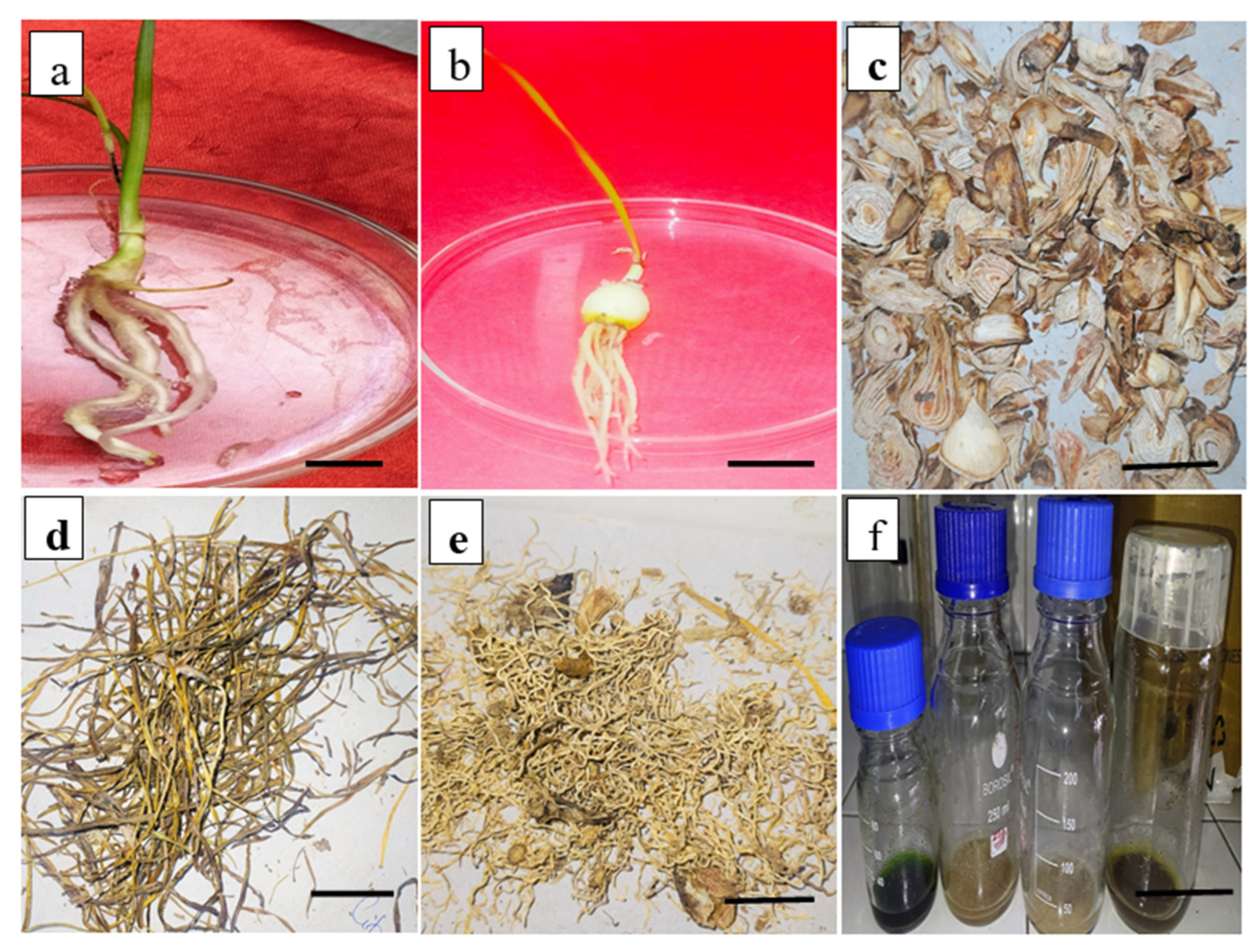
The regenerated plants placed on MS with different IBA doses induced compact white roots after 3 weeks of culture (Figure 2a,b). The maximum root induction frequency (80.66%) was achieved on 2.0 mg·L−1 IBA with 8.32 mean numbers of roots per shoot. IBA with various NAA doses did not improve rooting ability, which is presented in Table 2.
The fully regenerated plants with leaves, roots, and bulblets were placed in different treatments of MJ. The plants were removed from the media, washed with distilled water and the bulb, leaf, and roots were separately shade dried (Figure 2c–f).

3.4. Quantification of Lycorine in Field Grown Z. grandiflora, Z. candida and Z. citrina Plants

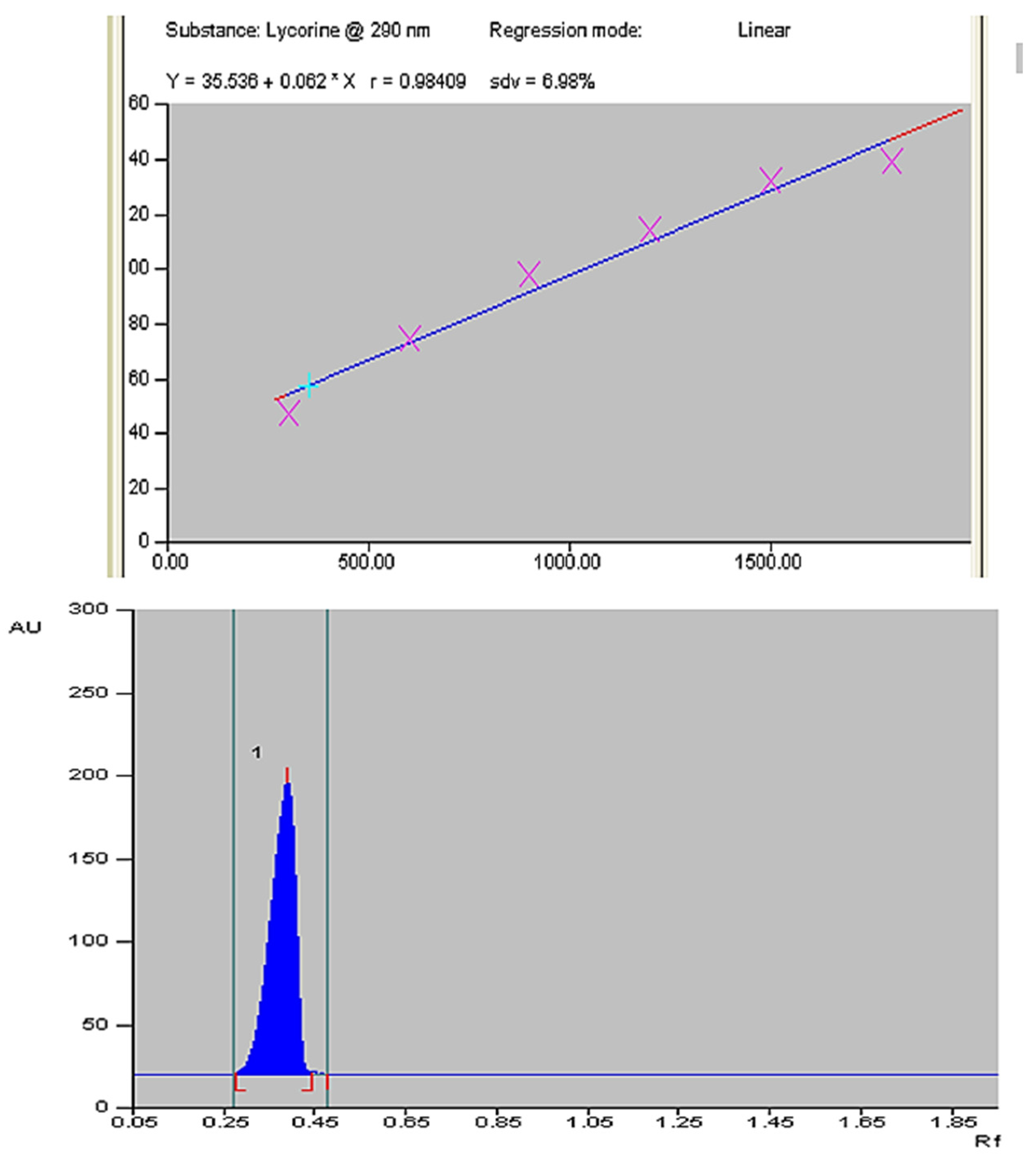
Lycorine was quantified in field grown bulb, leaf, and root of Z. grandiflora, Z. candida and Z. citrina by using HPTLC. The regression equation y = 35.536 + 0.062 × x describes the association between spot area and concentration in μg/spot (Figure 3 top, bottom). The six-point calibration curve shows linear regression correlation coefficient r = 0.984 and displays single sharp, flat, and compact peaks at Rf = 0.30, detected at wavelength 290 nm. The chromatograms of bulb, leaf and root extracts are shown in Figure 4. The HPTLC quantification of lycorine in three species of Zephyranthes is presented in Table 3. It reveals that the maximum level was found in Z. candida bulbs i.e., 1.93 µg g−1 DW, 1.87 µg g−1 DW in Z. grandiflora and 1.62 µg g−1 DW in Z. citrina. The leaf tissues revealed 0.98, 0.83 and 0.65 µg g−1 DW lycorine in Z. candida, Z. grandiflora and Z. citrina respectively. The lowest amount of lycorine was present in roots with 0.50, 0.43 and 0.38 µg g−1 DW in Z. candida, Z. grandiflora and Z. citrina respectively. The accumulation of lycorine follows the order of bulb > leaf > root while the species shows the order as Z. candida > Z. grandiflora > Z. citrina.

3.5. Different Doses of MJ on Lycorine Yield

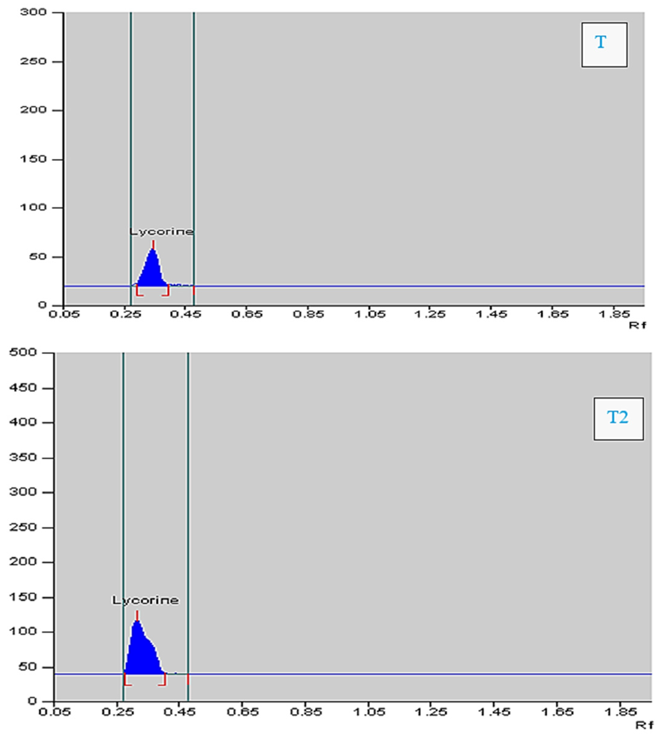
The MJ induced stress in cultured tissues improved the synthesis of lycorine. The yield of lycorine was quantified on MJ treatments i.e.,T1, T2, T3, and T4 (along with control T0). It effectively enhanced lycorine on T1 and T2 but not on T3 and T4 treatments. In all the three species, the highest lycorine was found in T2 i.e., 100 µM. The data are presented in Table 4, Table 5 and Table 6 for Z. candida, Z. grandiflora, and Z. citrina, respectively. The enrichment of lycorine yield was maximum in bulb tissues of Z. candida (2.74 µg g−1 DW) compared to the control, T0 (1.97 µg g−1 DW). In Z. grandiflora and Z. citrina, the lycorine yield was also enhanced (2.15 and 2.08 µg g−1 DW respectively) as compared to the control. The densitograms of control and T2 of leaf and root are shown in Figure 5 and Figure 6. These showed that the lycorine content improved under MJ influence and the maximum lycorine was noted in T2; the yield was1.53, 1.03, 0.86 µg g−1 dry wt. and 0.77, 0.74, 0.66 µgg−1 dry wt. in Z. candida, Z. grandiflora, and Z. citrina, respectively.

3.6. Biochemical Attributes in Response to MJ Treatment

The addition of biotic and abiotic compounds often causes cellular stress, thus the biochemical attributes like sugar, proline, and protein under different dosage of MJ was evaluated in bulb, leaf, and root tissues. The effects of varying concentrations of MeJ were assessed on sugar, proline, and protein; however, the highest levels of these parameters were observed on T4 treatment, at a concentration of 200 µM. More sugar was accumulated with increasing elicitor dose and the maximum was noted in T4 treatment of MJ. Among the species, the highest sugar level (18.06 mg g−1 FW) was observed in Z. candida, followed by Z. grandiflora and Z. citrina (17.56 and 14.04 mg g−1 FW respectively). The maximum proline was also noted in bulbs of Z. candida (7.42 mg g−1 FW) on T4 treatment, in Z. grandiflora and Z. citrina the highest proline content was 5.55 mg g−1 FW and 4.24 mg g−1 FW, respectively. Similar is the protein content i.e., highest accumulation in T4 treatment. The maximum protein accumulation was 6.22, 6.45, and 4.67 mg g−1 FW in bulb tissues of Z. candida, Z. grandiflora and Z. citrina, respectively as shown in Supplementary Tables S2–S10. Therefore, the present study shows variations of biochemicals in different tissues, the highest being in bulb followed by leaf and roots in the order of bulb > leaf > root.

3.7. MJ Treatment and Antioxidant Enzymes Activities

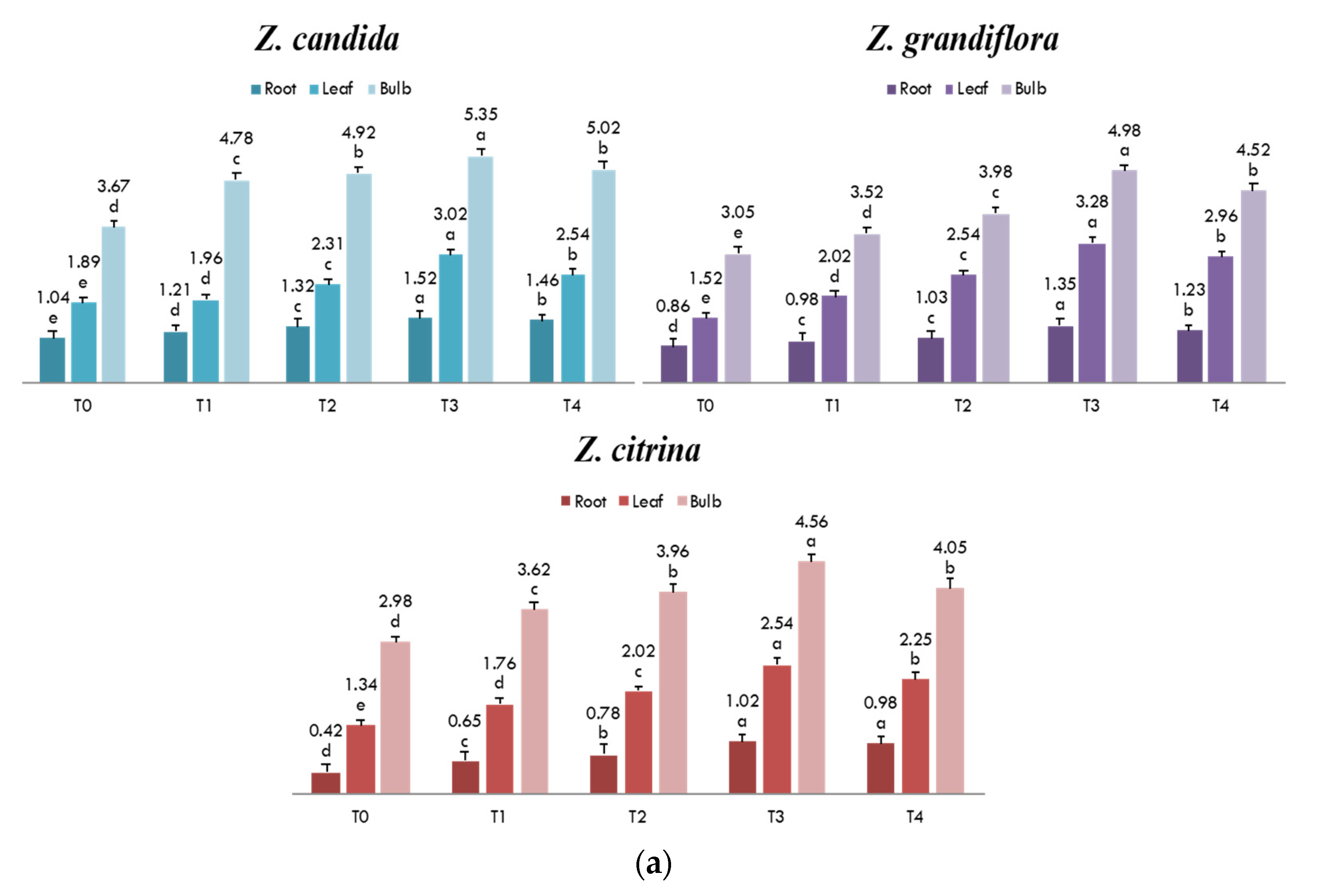
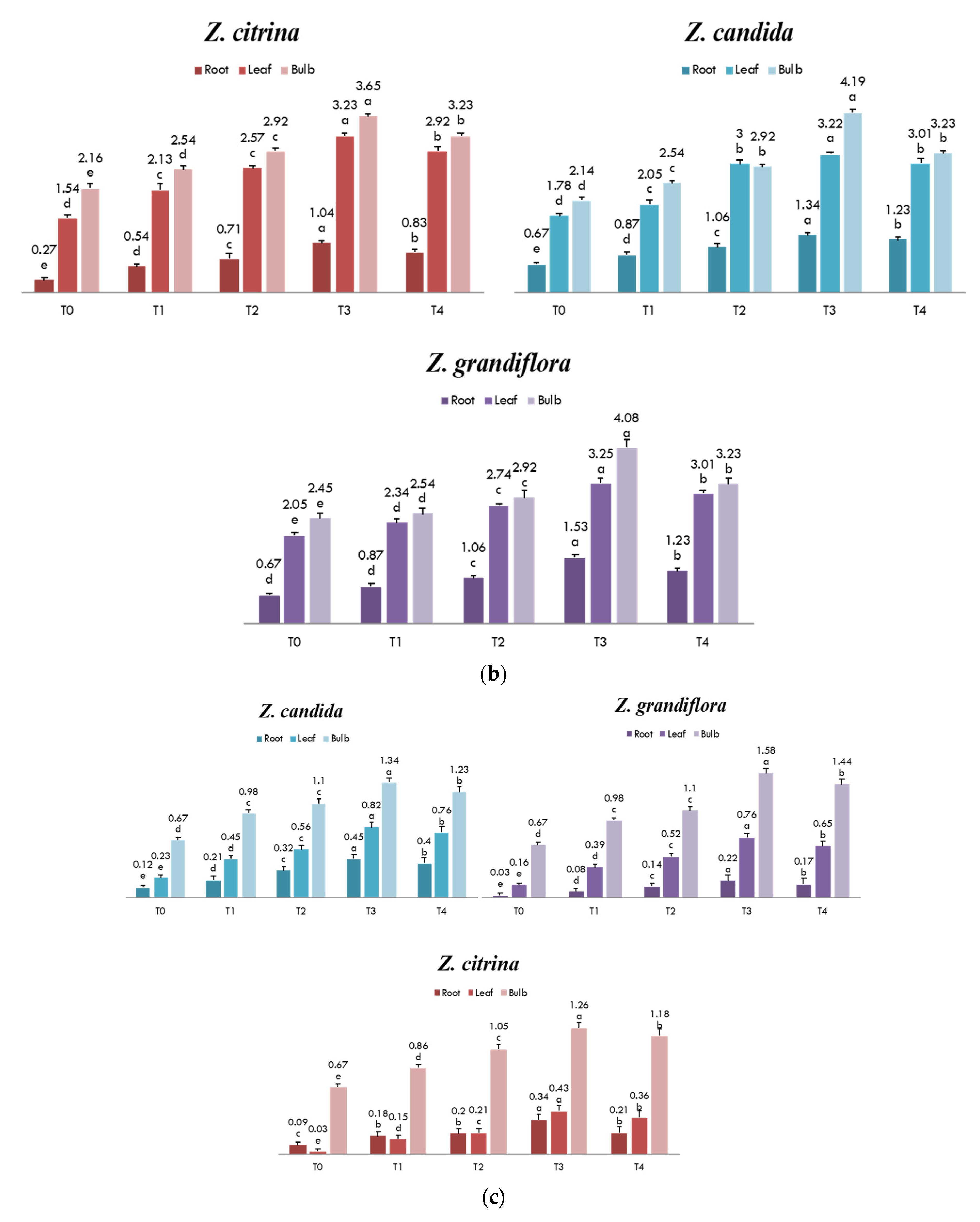
The antioxidant enzyme activities like SOD, CAT, and APX on different MJ treatments were assessed in Z. grandiflora, Z. candida, and Z. citrina which showed an increased enzyme activity at high doses as compared to the control. Increased SOD activity was noted with increasing MJ elicitor level and so were CAT and APX activities. The highest antioxidant activity was noted on T3 as shown in Figure 7a–c, respectively. In all the three Zephyranthes species, the SOD, CAT and APX activities were highest in bulb, followed by leaf and root in bulb > leaf > root order. The SOD and CAT enzyme activity in bulb on T3 dosage were 5.35, 4.98, 4.65 and 4.19, 4.08, 3.65 EU min−1 mg−1 protein, respectively in Z. candida, Z. grandiflora and Z. citrina. The APX activity was highest in Z. grandiflora (1.58 EU min−1mg−1 protein), followed by Z. candida (1.34 EU min−1 mg−1 protein) and Z. citrina (1.26 EU min−1 mg−1 protein).

4. Discussion

In this study, the lycorine content of bulb, leaf, and root of field grown and tissue culture regenerated plants was measured in three different species of Zephyranthes; and the impact of MJ elicitation on lycorine yield was investigated in in vitro derived plant parts. For successful tissue culture propagation, the sterilization steps, the genotype, the selection of explants, explant orientation, PGR concentration, and combinations are considered critical [35,36]. In bulbous monocotyledonous plants, the establishment of callus and rapid synchronous somatic embryogenesis are reported difficult thus the multiplication of organs like bulblets is suggested to be an efficient alternative way for mass propagation of plants and production of bioactive compounds. The sterilization of explants is challenging in geophytic plants since a variety of endophytes present in plant cells cannot easily be eliminated by normal surface sterilization process. In the present study of Zephyranthes, 5% sodium hypochlorite proved to be an effective sterilizing agent with 100% survival rate. Mercuric chloride treatment with moderate exposure time was proved to be an efficient treatment in previous study of Amaryllidaceae [37]. In the present study however, a 5% NaOCl treatment had a significant influence on explants sterilization and similar observation was earlier reported in other plant species [38]. In the present study, a significant percentage of shoot regeneration was achieved on MS without any addition of plant growth regulator. This can be explained by storage ability of bulb, which contains good amount of endogenous plant growth regulators, and in combinations with external factors regulate morphogenetic responses [39]. The present study thus corroborates the findings of other Amaryllidaceae species like Pancratium maritium [40] and Brunsvigia undulate [41] The exogenous addition of BAP improved shoot regeneration frequency and bulblet formation in Zephyranthes and the highest bulblet multiplication was obtained on BAP and NAA amended media. The same PGR (BAP and NAA) combination was noted to have a synergistic influence on cell division, morphogenesis and bulblet formation and was reported in plants like Brunsvigia undulate, Iris sanguine [41], Lilium orientalis cv. “Starfighter” [42]. In Zephyranthes roseus and in other bulbous monocotyledonous plants, the BAP and NAA combinations proved efficient in inducing callus, organogenesis, and plant regeneration as compared to other tested PGRs [43].
The quantification of lycorine in field grown and tissue culture derived parts (bulb, leaf and root) was performed through high-performance thin layer chromatography (HPTLC)—one of the sophisticated instrumental techniques for qualitative and quantitative analysis of herbal drugs. The HPTLC densitograms of standard and samples of lycorine at Rf = 0.30 show a sharp, flat peak which is consistent with earlier validated method [44]. The present study indicated that the maximum lycorine was found in bulb of Z. candida (1.93 µg g−1 DW) followed by Z. grandiflora and Z. citrina. The maximum yield of lycorine was present in this given order bulb > leaf > root, while the tested Zephyranthes species shows lycorine accumulation in Z. candida > Z.grandiflora > Z. citrina order. On addition of MJ as elicitor, the maximum lycorine yield was obtained in bulb of Z. candida, followed by Z. grandiflora and Z. citrina at T2. A lower MJ dosage was proved to be an ideal concentration in enhancing bioactive compounds in other plants such as Changium smyrnioides [45], Mentha × piperita [46]. A lower dose of MJ was illustrated to elicit highest transcript level/biosynthetic genes like ArPAL, ArC4H and Ar4CL in phenylpropanoid alkaloids [47].
To investigate the impact of different doses of MJ on in vitro cultivated plant parts and their subsequent lycorine synthesis in regenerated bulbs, we conducted a biochemical analysis to assess the differences in organ composition of the tested Zephyranthes species. The common stress markers like sugar, proline and protein were high with elevated MJ dosage in all the three species of Zephyranthes. Thus, the addition of MJ induced stress causing conversion of stored sucrose to monosaccharides, facilitating osmotic adjustments of stomata, and acts as signaling elements, plays critical role in osmoprotection, radical scavenging and membrane stability [48]. The increase in sugar has similarly been reported during MJ application in crops like tomato [49]. In this present study, the highest soluble sugar and protein were noted in bulbs of Z. candida and the maximum proline was noted in Z. grandiflora. The difference in biochemical markers in different tissues reveals varied level of stress on different organs, which is considered to be an important criterion for selection of stress tolerant genotypes. Under stress, the proline levels increased due to protein breakdown and is widely distributed osmolyte in plant cells [50]. The accumulation of proline in response to stress is an adaptive response to plants. Beside osmo-protectant, it works as a chelating agent, regulates redox reactions, prevents denaturation of macromolecules and maintains turgidity under low water levels [51]. Similar increase in proline in response to MJ was reported in sugar beet, Beta vulgaris L. [52]. The total soluble protein was also increased in response to MJ. The increase in protein during stress is considered to be one of the stress adapting strategies of plants to prevent oxidative damage to cells [36].
Similarly, the increased activities of enzymes like SOD, CAT, and APX were noted in bulb, leaf, and roots in Z. candida, Z. grandiflora, and Z. citrina with higher dosage of MJ. The increased levels of ROS and H2O2 are the major developments in response to exogenous MJ and their accumulation causes chloroplast perturbations, cellular redox reactions and cell death in other studies. The ROS damage is mitigated by over production and activities of antioxidant enzymes CAT, APX, and SOD; and the balance between ROS generation and antioxidant activity prevents oxidative damage of tissues. The SOD, APX and CAT feature the front line of defense against cellular oxidative injury by superoxide radical’s dismutation into H2O2 and O2, further detoxified by APX and CAT activities. MJ-induced ROS was earlier reported in in vitro cultures of C. officinale and P. ginseng. Similar observation of enhanced SOD with MJ elicitation was reported in Polygonum multiflorum [53], Solanum trilobatum [54], and Withania somnifera [55]. The maximum yield of lycorine may be due to highest expression of biosynthetic regulatory genes C4H (cinnamate-4-hydroxylase) and phenylalanine ammonia-lyase (PAL) in bulb of Amaryllidaceous. Similar report of highest elicitation on alkaloids in bulblets was reported in recent study [47]. Therefore, the comprehensive study suggests that the Amaryllidaceae alkaloids are present in abundance in bulb parts of Zephyranthes.

5. Conclusions

The protocol involves an efficient in vitro organogenesis and bulblet formation in Zephyranthes grandiflora, Zephyranthes citrina and Zephyranthes candida. This is the first quantitative analytical study of HPTLC revealing highest content of lycorine in bulb of Zephyranthes. The MJ elicitation had a significant effect on bulb, leaf and roots. The 100 µM MJ treatment had a notable effect on accumulation of lycorine with maximum yield of 2.74 µg g−1DW in bulbs of Z. candida. The quantification of lycorine in genus Zephyranthes will enhance its scope in pharmacological and industrial applications. The basic process of in vitro propagation and MJ elicitation could be extended to future studies on transcriptomic and putative gene pattern analysis.

Supplementary Materials

The following supporting information can be downloaded at: https://www.mdpi.com/article/10.3390/horticulturae9070832/s1, Table S1: Effects of various surface disinfectants on sterilization of explants; Table S2: Accumulation of sugar content (mg g−1 FW) in bulb, leaf, and root parts of Zephyranthes candida with different methyl jasmonate treatments; Table S3: Accumulation of sugar content (mg g−1 FW) in bulb, leaf, and roots parts of Zephyranthes grandiflora with different methyl jasmonate treatments; Table S4: Accumulation of sugar content (mg g−1 FW) in bulb, leaf, and roots parts of Zephyranthes citrina with different methyl jasmonate treatments; Table S5: Accumulation of proline content (mg g−1 FW) in bulb, leaf, and roots parts of Zephyranthes candida with different methyl jasmonate treatments; Table S6: Accumulation of proline content (mg g−1 FW) in bulb, leaf, and roots parts of Zephyranthes grandiflora with different methyl jasmonate treatments; Table S7: Accumulation of proline content (mg g−1 FW) in bulb, leaf, and roots parts of Zephyranthes citrina with different methyl jasmonate treatments; Table S8: Accumulation of protein content (mg g−1 FW) in bulb, leaf, and roots parts of Zephyranthes candida with different methyl jasmonate treatments; Table S9: Accumulation of protein content (mg g−1 FW) in bulb, leaf, and roots parts of Zephyranthes grandiflora with different methyl jasmonate treatments; Table S10: Accumulation of protein content (mg g−1 FW) in bulb, leaf, and roots parts of Zephyranthes citrina with different methyl jasmonate treatments.

Author Contributions

Conceptualization, R.S. and A.M.; methodology, R.S.; software, R.S.; validation, A.M. and Y.H.D.; formal analysis, M.Q.M.; investigation, Y.B.; resources, A.A.; data curation, B.E.; writing—original draft preparation, R.S.; writing—review and editing, J.M.; visualization, M.H.; supervision, A.M.; project administration, A.M.; funding acquisition, Y.H.D. All authors have read and agreed to the published version of the manuscript.

Funding

The authors acknowledge Researchers Supporting Project number (RSP2023R375), King Saud University, Riyadh, Saudi Arabia.

Data Availability Statement

All data are presented in the article.

Acknowledgments

Authors are thankful to the Department of Botany, Jamia Hamdard for providing laboratory facilities, S. D Sharma (Application Scientist) at CIF, Jamia Hamdard, New Delhi, for assistance in HPTLC facility. The authors acknowledge Researchers Supporting Project number (RSP2023R375), King Saud University, Riyadh, Saudi Arabia.

Conflicts of Interest

The authors declare no conflict of interest.

References

  1. Jan, R.; Asaf, S.; Numan, M.; Lubna; Kim, K.-M. Plant Secondary Metabolite Biosynthesis and Transcriptional Regulation in Response to Biotic and Abiotic Stress Conditions. Agronomy 2021, 11, 968. [Google Scholar] [CrossRef]
  2. Rungsung, W.; Ratha, K.; Dutta, S.; Dixit, A.K.; Hazra, J. Secondary Metabolites of Plants in Drugs Discovery. World J. Pharm. Res. 2015, 4, 604–613. [Google Scholar]
  3. Katoch, D.; Singh, B. Phytochemistry and Pharmacology of Genus Zephyranthes. Med. Aromat Plants 2015, 4, 212. [Google Scholar]
  4. Jin, Z. ChemInform Abstract: Amaryllidaceae and Sceletium Alkaloids. ChemInform 2013, 44. [Google Scholar] [CrossRef]
  5. Syeed, R.; Mujib, A.; Malik, M.Q.; Mamgain, J.; Ejaz, B.; Gulzar, B.; Zafar, N. Mass Propagation through Direct and Indirect Organogenesis in Three Species of Genus Zephyranthes and Ploidy Assessment of Regenerants through Flow Cytometry. Mol. Biol. Rep. 2021, 48, 513–526. [Google Scholar] [CrossRef] [PubMed]
  6. Jin, Z.; Yao, G. Amaryllidaceae and Sceletium Alkaloids. Nat. Prod. Rep. 2019, 36, 1462–1488. [Google Scholar] [CrossRef]
  7. Desgagné-Penix, I. Biosynthesis of Alkaloids in Amaryllidaceae Plants: A Review. Phytochem. Rev. 2021, 20, 409–431. [Google Scholar] [CrossRef]
  8. Khalifa, M.F.; Attia, E.Z.; Fahim, J.R.; Kamel, M.S. An Overview on the Chemical and Biological Aspects of Lycorine Alkaloid. J. Adv. Biomed. Pharm. Sci. 2018, 1, 41–49. [Google Scholar] [CrossRef]
  9. Jin, Y.H.; Min, J.S.; Jeon, S.; Lee, J.; Kim, S.; Park, T.; Park, D.; Jang, M.S.; Park, C.M.; Song, J.H.; et al. Lycorine, a Non-Nucleoside RNA Dependent RNA Polymerase Inhibitor, as Potential Treatment for Emerging SARSvirus Infections. Phytomedicine 2021, 86, 153–440. [Google Scholar] [CrossRef]
  10. Wang, G.-P.; Yu, X.-D.; Sun, Y.-W.; Jones, H.D.; Xia, L.-Q. Generation of Marker- and/or Backbone-Free Transgenic Wheat Plants via Agrobacterium-Mediated Transformation. Front. Plant Sci. 2016, 7, 1324. [Google Scholar] [CrossRef] [Green Version]
  11. Berkov, S.; Osorio, E.; Viladomat, F.; Bastida, J. Chemodiversity, Chemotaxonomy and Chemoecology of Amaryllidaceae Alkaloids. Alkaloids Chem. Biol. 2020, 83, 113–185. [Google Scholar] [CrossRef]
  12. Zafar, N.; Mujib, A.; Ali, M.; Tonk, D.; Gulzar, B.; Malik, M.Q.; Mamgain, J.; Sayeed, R. Cadmium Chloride (CdCl2) Elicitation Improves Reserpine and Ajmalicine Yield in Rauvolfia Serpentina as Revealed by High-Performance Thin-Layer Chromatography (HPTLC). 3 Biotech 2020, 10, 344. [Google Scholar] [CrossRef]
  13. Saiman, M.Z.; Miettinen, K.; Mustafa, N.R.; Choi, Y.H.; Verpoorte, R.; Schulte, A.E. Metabolic Alteration of Catharanthus Roseus Cell Suspension Cultures Overexpressing Geraniol Synthase in the Plastids or Cytosol. Plant Cell Tissue Organ Cult. 2018, 134, 41–53. [Google Scholar] [CrossRef] [PubMed] [Green Version]
  14. Gharari, Z.; Bagheri, K.; Danafar, H.; Sharafi, A. Enhanced Flavonoid Production in Hairy Root Cultures of Scutellaria Bornmuelleri by Elicitor Induced Over-Expression of MYB7 and FNSΠ2 Genes. Plant Physiol. Biochem. 2020, 148, 35–44. [Google Scholar] [CrossRef]
  15. Naik, P.M.; Manohar, S.H.; Praveen, N.; Upadhya, V.; Murthy, H.N. Evaluation of Bacoside A Content in Different Accessions and Various Organs of Bacopa monnieri (L.) Wettst. J. Herbs Spices Med. Plants 2012, 18, 387–395. [Google Scholar] [CrossRef]
  16. Murthy, H.N.; Lee, E.-J.; Paek, K.-Y. Production of Secondary Metabolites from Cell and Organ Cultures: Strategies and Approaches for Biomass Improvement and Metabolite Accumulation. Plant Cell Tissue Organ Cult. 2014, 118, 1–16. [Google Scholar] [CrossRef]
  17. Khonakdari, R.; Rezadoost, M.; Heydari, H.; Mirjalili, R. Effect of Photoperiod and Plant Growth Regulators on in Vitro Mass Bulblet Proliferation of Narcissus tazzeta L. (Amaryllidaceae), a Potential Source of Galantamine. Plant Cell Tissue Organ Cult 2020, 142, 187–199. [Google Scholar] [CrossRef] [PubMed]
  18. Ptak, A.; Simlat, M.; Morańska, E.; Skrzypek, E.; Warchoł, M.; Tarakemeh, A.; Laurain-Mattar, D. Exogenous Melatonin Stimulated Amaryllidaceae Alkaloid Biosynthesis in in Vitro Cultures of Leucojum aestivum L. Ind. Crop. Prod. 2019, 138, 111458. [Google Scholar] [CrossRef]
  19. Malik, M.Q.; Mujib, A.; Gulzar, B.; Zafar, N.; Syeed, R.; Mamgain, J.; Ejaz, B. Kanchan Enrichment of Alliin in Different in Vitro Grown Tissues of Allium Sativum through CdCl2 Elicitation as Revealed by High Performance Thin Layer Chromatography (HPTLC). Ind. Crop. Prod. 2020, 158, 113007. [Google Scholar] [CrossRef]
  20. Halder, M.; Sarkar, S.; Jha, S. Elicitation: A Biotechnological Tool for Enhanced Production of Secondary Metabolites in Hairy Root Cultures. Eng. Life Sci. 2019, 19, 880–895. [Google Scholar] [CrossRef] [Green Version]
  21. Ali, M.; Mujib, A.; Gulzar, B.; Zafar, N. Essential Oil Yield Estimation by Gas Chromatography-Mass Spectrometry (GC-MS) after Methyl Jasmonate (MeJA) Elicitation in In Vitro Cultivated Tissues of Coriandrum sativum L. 3 Biotech 2019, 9, 1–16. [Google Scholar] [CrossRef] [PubMed]
  22. Nabi, N.; Singh, S.; Saffeullah, P. Responses of in Vitro Cell Cultures to Elicitation: Regulatory Role of Jasmonic Acid and Methyl Jasmonate: A Review. Vitr. Cell. Dev. Biol. Plant 2021, 57, 341–355. [Google Scholar] [CrossRef]
  23. Singh, A.; Dwivedi, P. Methyl-Jasmonate and Salicylic Acid as Potent Elicitors for Secondary Metabolite Production in Medicinal Plants: A Review. J. Pharmacogn. Phytochem. 2018, 7, 750–757. [Google Scholar]
  24. Wang, R.; Xu, S.; Wang, N.; Xia, B.; Jiang, Y.; Wang, R. Transcriptome Analysis of Secondary Metabolism Pathway, Transcription Factors, and Transporters in Response to Methyl Jasmonate in Lycoris Aurea. Front. Plant Sci. 2016, 7, 1971. [Google Scholar] [CrossRef] [Green Version]
  25. Zhou, P.; Yang, J.; Zhu, J.; He, S.; Zhang, W.; Yu, R.; Zi, J.; Song, L.; Huang, X. Effects of β-Cyclodextrin and Methyl Jasmonate on the Production of Vindoline, Catharanthine, and Ajmalicine in Catharanthus Roseus Cambial Meristematic Cell Cultures. Appl. Microbiol. Biotechnol. 2015, 99, 7035–7045. [Google Scholar] [CrossRef]
  26. Nguyen, K.V.; Pongkitwitoon, B.; Pathomwichaiwat, T.; Viboonjun, U.; Prathanturarug, S. Effects of Methyl Jasmonate on the Growth and Triterpenoid Production of Diploid and Tetraploid Centella asiatica (L.) Urb. Hairy Root Cultures. Sci. Rep. 2019, 9, 18665. [Google Scholar] [CrossRef] [Green Version]
  27. Wu, X.-H.; Fan, M.-Z.; Li, X.-F.; Piao, X.-C.; Gao, R.; Lian, M.-L. Involvement of Putrescine, Nitric Oxide, and Hydrogen Peroxide in Methyl Jasmonate-Induced Ginsenoside Synthesis in Adventitious Root Cultures of Panax Ginseng C.A. Meyer. J. Plant Growth Regul. 2021, 40, 1440–1449. [Google Scholar] [CrossRef]
  28. Murashige, T.; Skoog, F. A Revised Medium for Rapid Growth and Bioassay with Tobacco Tissue Cultures. Physiol. Plant 1962, 15, 473–497. [Google Scholar] [CrossRef]
  29. Dey, P.M. Oligosaccharides. In Methods in Plant Biochemistry; Elsevier: Amsterdam, The Netherlands, 1990; pp. 189–218. [Google Scholar]
  30. Bates, L.; Waldren, P.P.; Teare, J.D. Rapid Determination of Free Proline of Water Stress Studies. Plant Soil 1973, 39, 205–207. [Google Scholar] [CrossRef]
  31. Bradford, M.M. A Rapid and Sensitive Method for Quantification of Microgram Quantities of Protein, Utilizing the Principle of Protein Dye Binding. Anal. Biochem. 1976, 72, 248–541. [Google Scholar] [CrossRef]
  32. Aebi, H. Catalase in Vitro. In Methods in Enzymology; Elsevier: Amsterdam, The Netherlands, 1984; pp. 121–126. [Google Scholar]
  33. Dhindsa, R.S.; Plumb-Dhindsa, P.; Thorpe, T.A. Leaf Senescence: Correlated with Increased Levels of Membrane Permeability and Lipid Peroxidation, and Decreased Levels of Superoxide Dismutase and Catalase. J. Exp. Bot. 1981, 32, 93–101. [Google Scholar] [CrossRef]
  34. Nakano, Y.; Asada, K. Hydrogen Peroxide Is Scavenged by Ascorbate Specific Peroxidase in Spinach Chloro-Plasts. Plant Cell Physiol. 1981, 22, 867–888. [Google Scholar]
  35. García-Fortea, E.; García-Pérez, A.; Gimeno-Páez, E.; Martínez-López, M.; Vilanova, S.; Gramazio, P.; Prohens, J.; Plazas, M. Ploidy Modification for Plant Breeding Using in Vitro Organogenesis: A Case in Eggplant. Methods Mol. Biol. 2021, 2264, 197–206. [Google Scholar] [CrossRef] [PubMed]
  36. Gulzar, B.; Mujib, A.; Rajam, M.V.; Frukh, A.; Zafar, N. Identification of Somatic Embryogenesis (SE) Related Proteins through Label-Free Shotgun Proteomic Method and Cellular Role in Catharanthus roseus (L.) G. Don. Plant Cell Tissue Organ Cult. 2019, 137, 225–237. [Google Scholar] [CrossRef]
  37. Gangopadhyay, M.; Chakraborty, D.; Dewanjee, S.; Bhattacharya, S. Clonal Propagation of Zephyranthes Grandi-Flora Using Bulbs as Explants. Biol. Plant 2010, 54, 793–797. [Google Scholar] [CrossRef]
  38. Saeed, W.; Naseem, S.; Gohar, D.; Ali, Z. Efficient and Reproducible Somatic Embryogenesis and Micropropa-Gation in Tomato via Novel Structures-Rhizoid Tubers. PLoS ONE 2019, 14, e0215929. [Google Scholar] [CrossRef] [Green Version]
  39. Taha, L.S.; Sayed, S.S.; Farahat, M.M. El-Sayed IM. In Vitro Culture and Bulblets Induction of Asiatic Hybrid Lily’red Alert’. J. Biol. Sci. 2018, 18, 84–91. [Google Scholar] [CrossRef]
  40. Dragassaki, M.; Economou, A.S.; Vlahos, J.C. Bulblet Formation In Vitro and Plantlet Survival Extra Vitrum in Pancratium maritimum L. Acta Hortic. 2003, 616, 347–352. [Google Scholar] [CrossRef]
  41. Rice, L.J.; Finnie, J.F.; Van Staden, J. In Vitro Bulblet Production of Brunsvigia Undulate from Twin-Scales. S. Afr. J. Bot. 2011, 77, 305–312. [Google Scholar] [CrossRef] [Green Version]
  42. Youssef, N.M.; Shaaban, S.A.; Ghareeb, Z.F.; Taha, L.S. In Vitro Bulb Formation of Direct and Indirect Regeneration of Lilium Orientalis Cv. “Starfighter” Plants. Bull. Natl. Res. Cent. 2019, 43, 1–9. [Google Scholar] [CrossRef]
  43. Mujib, A.; Banerjee, S.; Maqsood, M.; Ghosh, P. Organogenesis and Plant Regeneration in Zephyranthes Rosea Lindl.: Histological and Chromosomal Study. Plant Biosyst. 2014, 148, 492–498. [Google Scholar] [CrossRef]
  44. Cai, J.; Ma, Y.; Hu, P.; Zhang, Y.; Chen, J.; Li, X. Elicitation of Furanocoumarins in Changium Smyrnioides Suspension Cells. Plant Cell Tissue Organ Cult. 2017, 130, 1–12. [Google Scholar] [CrossRef]
  45. Krzyzanowska, J.; Czubacka, A.; Pecio, L.; Przybys, M.; Doroszewska, T.; Stochmal, A.; Oleszek, W. The Effects of Jasmonic Acid and Methyl Jasmonate on Rosmarinic Acid Production in Mentha x Piperita Cell Suspension Cultures. Plant Cell Tissue Organ Cult. 2012, 108, 73–81. [Google Scholar] [CrossRef]
  46. Kim, O.T.; Bang, K.H.; Kim, Y.C.; Hyun, D.Y.; Kim, M.Y.; Cha, S.W. Upregulation of Ginsenoside and Gene Expres-Sion Related to Triterpene Biosynthesis in Ginseng Hairy Root Cultures Elicited by Methyl Jasmonate. Plant Cell Tissue Organ Cult. 2009, 98, 25–33. [Google Scholar] [CrossRef]
  47. Tarakemeh, A.; Azizi, M.; Rowshan, V.; Salehi, H.; Spina, R.; Dupire, F.; Arouie, H.; Laurain-Mattar, D. Screen-Ing of Amaryllidaceae Alkaloids in Bulbs and Tissue Cultures of Narcissus Papyraceus and Four Varieties of N. Tazetta. J. Pharm. Biomed. Anal. 2019, 172, 230–237. [Google Scholar] [CrossRef] [PubMed]
  48. Pesaraklu, A.; Radjabian, T.; Salami, S.A. Methyl Jasmonate and Ag+ as Effective Elicitors for Enhancement of Phenolic Acids Contents in Salvia Officinalis and Salvia Verticillata, as Two Traditional Medicinal Plants. South Afr. J. Bot. 2021, 141, 105–115. [Google Scholar] [CrossRef]
  49. Ho, T.-T.; Murthy, H.N.; Park, S.-Y. Methyl Jasmonate Induced Oxidative Stress and Accumulation of Secondary Metabolites in Plant Cell and Organ Cultures. Int. J. Mol. Sci. 2020, 21, 716. [Google Scholar] [CrossRef] [Green Version]
  50. Bhaskara, G.B.; Yang, T.-H.; Verslues, P.E. Dynamic Proline Metabolism: Importance and Regulation in Water Limited Environments. Front. Plant Sci. 2015, 6, 484. [Google Scholar] [CrossRef] [Green Version]
  51. Andi, S.A.; Gholami, M.; Ford, C.M. The Effect of Methyl Jasmonate and Light Irradiation Treatments on the Stilbenoid Biosynthetic Pathway in Vitis Vinifera Cell Suspension Cultures. Nat. Prod. Res. 2018, 32, 909–917. [Google Scholar] [CrossRef]
  52. Fugate, K.K.; Lafta, A.M.; Eide, J.D.; Li, G.; Lulai, E.C.; Olson, L.L.; Deckard, E.L.; Khan, M.F.R.; Finger, F.L. Methyl Jasmonate Alleviates Drought Stress in Young Sugar Beet (Beta vulgaris L.) Plants. J. Agron. Crop. Sci. 2018, 204, 566–576. [Google Scholar] [CrossRef]
  53. Shilpha, J.; Satish, L.; Kavikkuil, M.; Largia, M.; Ramesh, M. Methyl Jasmonate Elicits the Solasodine Pro-Duction and Antioxidant Activity in Hairy Root Cultures of Solanum trilobatum L. Ind. Crop. Prod. 2015, 71, 54–64. [Google Scholar] [CrossRef]
  54. Sivanandhan, G.; Kapil Dev, G.; Jeyaraj, M.; Rajesh, M.; Arjunan, A.; Muthuselvam, M.; Manickavasagam, M.; Selvaraj, N.; Ganapathi, A. Increased Production of Withanolide A, Withanone, and Withaferin A in Hairy Root Cultures of Withania somnifera (L.) Dunal Elicited with Methyl Jasmonate and Salicylic Acid. Plant Cell Tissue Organ Cult. 2013, 114, 121–129. [Google Scholar] [CrossRef]
  55. Abou-Donia, A.H.; Toaima, S.M.; Hammoda, H.M.; Shawky, E. New Rapid Validated HPTLC Method for the Determina-Tion of Lycorine in Amaryllidaceae Plants Extracts. Chromatographia 2007, 65, 497–500. [Google Scholar] [CrossRef] [PubMed]
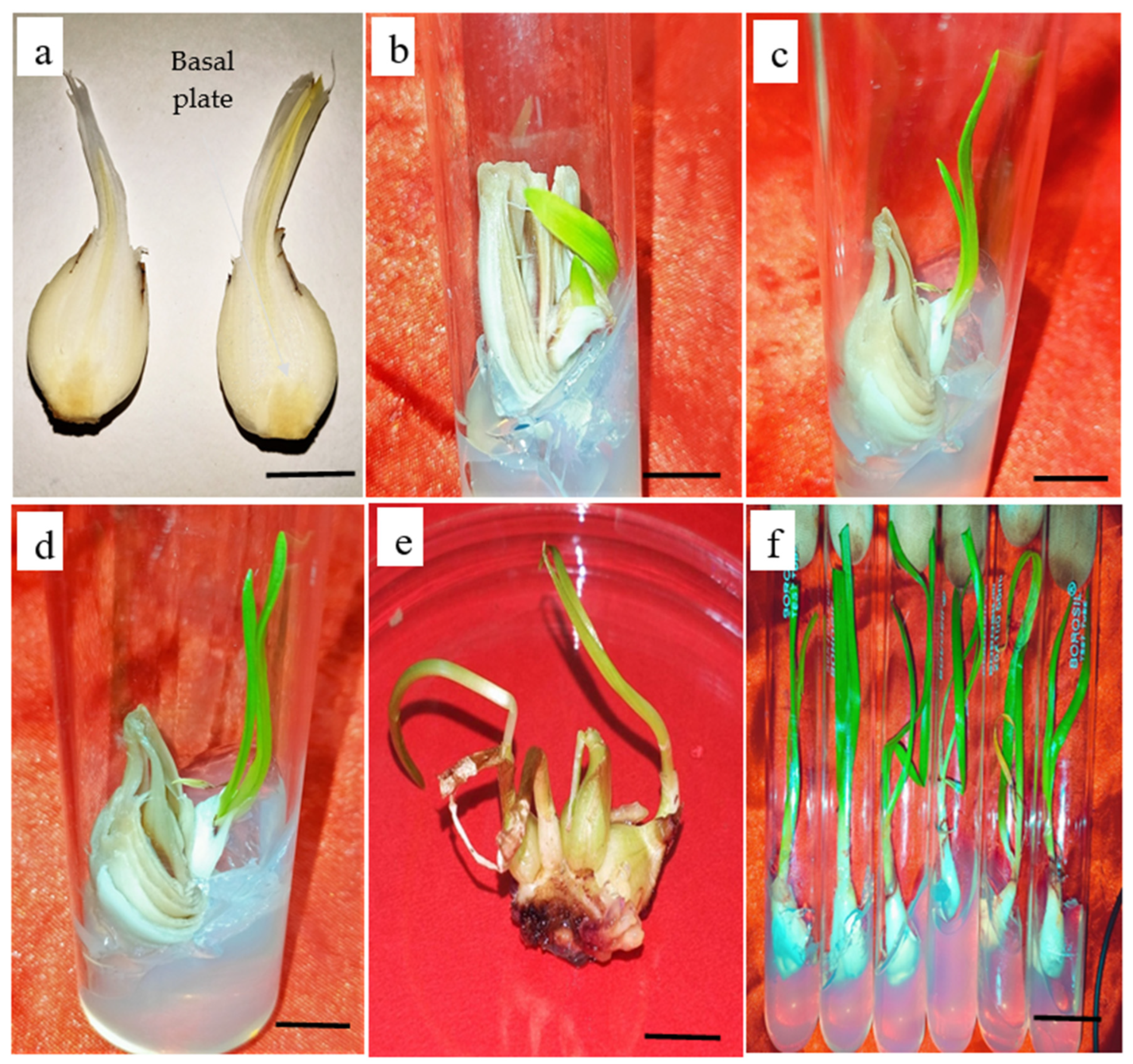
Figure 1. The sequential process of shoot regeneration and bulblet multiplication in Zephyranthes sp. (ac): The bulb segment with attached basal plate showing shoot formation along with bulblet. (df): The multiplication of bulblets after 21 days of subculture showing 4–5 maximum bulblets on basal plate (Bar = 0.2 cm, 0.5 cm).
Horticulturae 09 00832 g001
Figure 2. Root induction on MS medium and extraction of plant parts. (a,b): Induced roots on IBA added medium. The methyl jasmonate elicited (c): bulb (d): leaf, (e): roots, shade dried for 1 month and (f): The methanolic extract of bulb, leaf and root tissues (Bar = 2 cm).
Horticulturae 09 00832 g002
Figure 3. Top: The six-point calibration curve of lycorine with linear regression correlation coefficient r = 0.984, and regression equation y = 35.536 + 0.062 × x where y is the spot area and x is the concentration in µg/spot. Bottom: HPTLC densitograms displaying single, sharp and flat peaks of standard lycorine at Rf = 0.30 measured at wavelength = 290 nm.
Horticulturae 09 00832 g003
Figure 4. HPTLC densitograms of lycorine content in bulb, leaf, and root extracts of Zephyranthes displaying similar peaks at Rf = 0.30.
Horticulturae 09 00832 g004
Figure 5. HPTLC densitograms of lycorinein leaf extracts in control T0 and T2 treatment of MJ.
Horticulturae 09 00832 g005
Figure 6. HPTLC densitograms of lycorine in root extracts in control T0 (represented by T in the machine output) and T2 treatment of MJ.
Horticulturae 09 00832 g006
Figure 7. (a) SOD activity (EU mg−1 protein min- 1) in three different species of Zephyranthes in response to MJ treatments. T0 = Control, T1 = 50, T2 = 100, T3 = 150 and T4 = 200 µM MJ. Values presented are mean ± standard deviation of three experiments. Mean values in the bars followed by different letters are significantly different (at p = 0.05) according to DMRT. The comparison is among different concentrations. (b) CAT activity (EU mg−1 protein min−1) in three different species of Zephyranthes in response to MJ treatments. T0 = Control, T1 = 50, T2 = 100, T3 = 150 and T4 = 200 µM MJ. Values are mean ± standard deviation of three experiments. Mean values in the bars followed by different letters are significantly different (at p = 0.05) according to DMRT. The comparison is among different concentrations. (c) APX activity (EU mg−1 protein min- 1) in three different species of Zephyranthes in response to MJ treatments. T0 = Control, T1 = 50, T2 = 100, T3 = 150 and T4 = 200 µM MJ. Values are mean ± standard deviation of three experiments. Mean values in the bars followed by different letters are significantly different (at p = 0.05) according to DMRT. The comparison is among different concentrations.
Horticulturae 09 00832 g007aHorticulturae 09 00832 g007b
Table 1. Effect of PGRs on direct shoot regeneration (fDSR) frequency from bulb explants in three species of Zephyranthes.
PGRs and ConcentrationsfDSRBmean
BAPNAAZ. grandifloraZ. candidaZ. citrina
--40.2 ± 1.24 f47.7 ± 1.03 e43.2 ± 1.32 d0
0.5-44.3 ± 1.36 e50.3 ± 1.06 d46.5 ± 1.67 d1.01 ± 0.01 e
0.50.550.8 ± 2.21 d55.2 ± 1.22 c49.5 ± 1.90 d1.06 ± 0.01 e
1.0-58.6 ± 2.38 d60.2 ± 1.38 b58.0 ± 2.08 c2.08 ± 0.01 d
1.00.567.5 ± 1.42 c66.3 ± 2.01 b60.8 ± 2.78 c3.03 ± 0.02 c
2.0-75.6 ± 3.44 b67.5 ± 2.78 b68.6 ± 2.80 b5.32 ± 0.04 b
2.00.585.5 ± 3.48 a73.9 ± 3.08 a76.5 ± 3.09 a7.46 ± 0.06 a
2.5-73.6 ± 3.45 b64.0 ± 2.07 b65.6 ± 2.55 b4.87 ± 0.05 c
2.50.547.5 ± 2.54 e58.3 ± 2.54 c43.7 ± 2.21 d2.05 ± 0.04 d
3.0-40.6 ± 1.33 f36.3 ± 1.26 f35.7 ± 1.02 e1.07 ± 0.02 e
3.00.537.5 ± 1.36 g32.5 ± 1.32 f30.8 ± 1.01 f1.66 ± 0.01 e
Data is represented as mean ± SE (n = 3) and was scored after 3 weeks of culture. Mean values in a column followed by different letters are significantly different at p = 0.05 as per Duncan’s Multiple Range test. fDSR frequency of direct shoot regeneration from explants, Bmean represents mean number of bulblet formation on 3rd subculture, irrespective of the species. PGRs = plant growth regulators.
Table 2. Effect of IBA and NAA on root induction and mean number of roots on MS medium in Zephyranthes species.
Auxins Concentrations (mg·L−1) Root Induction Frequency (FRI)Mean Number of Roots
(MR)
IBANAA
1-60.67 ± 1.24 d5.22 ± 0.02 c
10.2568.8 ± 1.08 c5.05 ± 0.04 c
2-80.66 ± 3.43 a8.32 ± 1.28 a
20.2573.55 ± 2.56 b6.03 ± 1.09 b
3-62.05 ± 1.67 d5.43 ± 0.03 c
30.2552.01 ± 1.10 e2.76 ± 0.01 d
Data is represented as mean ± SE (n = 3) and was scored after 3 weeks of culture. Mean values in a column followed by different letters are significantly different at p = 0.05 as per DMRT. FRI = Frequency of root induction, MR = Mean number of roots per shoot.
Table 3. Lycorine (µg g−1 DW) content in bulb, leaf, and roots of field grown Zephyranthes candida, Zephyranthes grandiflora and Zephyranthes citrina.
Parts UsedZ. candidaZ. grandifloraZ. citrina
Bulb1.93 ± 0.04 a1.87 ± 0.04 a1.62 ± 0.03 a
Leaf0.98 ± 0.02 b0.83 ± 0.03 b0.65 ± 0.01 b
Root0.43 ± 0.01 c0.50 ± 0.01 c0.38 ± 0.01 c
Values are mean ± standard deviation of three experiments. Mean values in the bars followed by different letters are significantly different (p = 0.05) as per DMRT.
Table 4. Effect of different MJ treatments on lycorine (µg g−1 DW) content in bulb, leaf, and roots of Zephyranthes candida.
Parts UsedT0T1T2T3T4
Bulb1.97 ± 0.04 d2.33 ± 0.03 c2.74 ± 0.05 a1.53 ± 0.04 b1.45 ± 0.02 b
Leaf1.08 ± 0.02 c1.26 ± 0.01 b1.53 ± 0.05 a1.14 ± 0.02 b1.03 ± 0.02 c
Root0.53 ± 0.01 a0.62 ± 0.01 b0.77 ± 0.01 a0.33 ± 0.01 c0.45 ± 0.01 d
Values are Mean ± SE of three experiments. T0 = Control, T1 = 50, T2 = 100, T3 = 150, and T4 = 200 µM MJ. Mean values in the bars followed by different letters are significantly different (at p = 0.05) according to DMRT.
Table 5. Lycorine (µg g−1 DW) content in bulb, leaf and roots in Zephyranthes grandiflora on different MJ treatments.
Parts UsedT0T1T2T3T4
Bulb1.87 ± 0.04 c1.94 ± 0.03 b2.15 ± 0.04 a1.64 ± 0.02 c1.62 ± 0.01 d
Leaf0.83 ± 0.03 c0.96 ± 0.02 b1.03 ± 0.04 a0.76 ± 0.01 b0.73 ± 0.01 d
Root0.50 ± 0.01 c0.65 ± 0.02 b0.74 ± 0.01 a0.46 ± 0.01 b0.45 ± 0.01 c
Values are Mean ± SE of three experiments. T0 = Control, T1 = 50, T2 = 100, T3 = 150, and T4 = 200 µM MJ. Mean values in the bars followed by different letters are significantly different (at p = 0.05) according to DMRT.
Table 6. Lycorine (µg g−1 DW) content in bulb, leaf and roots in Zephyranthes citrina on different MJ treatments.
Parts UsedT0T1T2T3T4
Bulb1.62 ± 0.03 c1.96 ± 0.04 b2.08 ± 0.05 a1.73 ± 0.04 d1.65 ± 0.04 e
Leaf0.65 ± 0.01 c0.78 ± 0.01 b0.86 ± 0.02 a0.56 ± 0.03 d0.42 ± 0.02 d
Root0.38 ± 0.01 d0.45 ± 0.01 c0.66 ± 0.01 a0.35 ± 0.02 b0.33 ± 0.01 c
Values are Mean ± SE of three experiments. T0 = Control, T1 = 50, T2 = 100, T3 = 150, and T4 = 200 µM MJ. Mean values in the bars followed by different letters are significantly different (at p = 0.05) according to DMRT.
Disclaimer/Publisher’s Note: The statements, opinions and data contained in all publications are solely those of the individual author(s) and contributor(s) and not of MDPI and/or the editor(s). MDPI and/or the editor(s) disclaim responsibility for any injury to people or property resulting from any ideas, methods, instructions or products referred to in the content.