Article

Reactive Oxygen Species Metabolism Modulation on the Quality of Apple Fruits Inoculated with Penicillium expansum under Different Ambient pHs

1
College of Science, Gansu Agricultural University, Lanzhou 730070, China
2
Department of Agricultures, Technologies and Biotechnologies, University of Molise, 86100 Campobasso, Italy
3
College of Food Science and Engineering, Gansu Agricultural University, Lanzhou 730070, China
4
Department of Postharvest Science of Fresh Produce, Agricultural Research Organization, Rishon LeZion 7505101, Israel
*
Authors to whom correspondence should be addressed.
These authors contributed equally to this work.
Horticulturae 2023, 9(5), 538; https://doi.org/10.3390/horticulturae9050538
Submission received: 25 March 2023 / Revised: 21 April 2023 / Accepted: 26 April 2023 / Published: 28 April 2023

Abstract

:
Apple blue mold is a significant postharvest disease caused by Penicillium expansum. pH modification in colonized tissues leads to the production of organic substances, the modulation of enzymes, and then increases fungal pathogenicity. This study evaluated Penicillium expansum-inoculated apple fruits’ quality responding to pH treatments ranging from 2.5 to 8.5 and analyzed the reactive oxygen species (ROS) metabolism modulation in inoculated apple fruits at the same pH. The results showed that the fruit quality of the firmness, total soluble solids, and titratable acid displayed a quick loss at pHs 5.0 and 7.0, compared with 2.5 and 8.5. Similarly, higher disease incidence was observed at pHs 5.0 and 7.0. Apple fruits infected with P. expansum at pHs 2.5 and 8.5 had less content of O2•−, H2O2, and malondialdehyde (MDA); lower enzymatic activity of NADPH oxidase (NOX); and greater cell membrane integrity than those at pHs 5.0 and 7.0. The analysis of the antioxidant enzymatic activities showed upregulation of superoxide dismutase (SOD), catalase (CAT), peroxidase (POD), ascorbate peroxidase (APX), monodehydroascorbate reductase (MDHAR), dehydroascorbate reductase (DHAR), and glutathione reductase (GR) at pHs 2.5 and 8.5 compared with those at pHs 5.0 and 7.0. Similar trends were shown in ascorbic acid and glutathione. These results support the hypothesis that inoculated apple fruits at pHs 2.5 and 8.5 improve resistance to P. expansum by modulating ROS metabolism, compared with pHs 5.0 and 7.0.

1. Introduction

Postharvest losses have a considerable impact on population nutrition and health that contribute considerably to food insecurity. Postharvest disease losses of fruits may occur throughout the postharvest handling process, from harvest to consumption. When assessing postharvest disease losses, it is critical to evaluate fruit quantity and quality loss, as some infections may not produce unsalable fruit but still diminish product value [1]. Environmental parameters such as the local pH significantly improve or slow down the speed of infection. Penicillium expansum acidifies the colonized tissue by generating natural organic acids (such as D-gluconic acid, citric acid, and oxalic acid) that aid fungal colonization and pathogenicity [2]. The same strategy has been reported for Penicillium digitatum, Penicillium italicum [3], and Botrytis cinerea [4]. On the other hand, some pathogens, such as Trichothecium roseum, can alkalize the host tissue environment by releasing ammonia, which enhances their pathogenicity [5]. Colletotrichum gloeosporioides has been reported to turn L-glutamate or glutamine into ammonia and increases the pH of colonized fruit from 5.6 to 8.5 [3,6]. Fungal pH modulation of the host environment regulates gene expression and enzyme activity to improve fungal pathogenicity [7]. In addition, pH change during fungal colonization has a wider effect; it weakens the host’s defenses by increasing intracellular signaling, making cell wall-destroying enzymes, and turning on specific transporters and redox protectant systems [8]. For the successful infection of fruits and vegetables, fungi must overcome the defense mechanism established by the plants. The research work reported by Pitt and Hocking (1997) and Snowdon (1990) [9] suggested that, due to their acidic nature, most fruits are easily spoiled by mold and yeast, with the exception of citrus fruits such as lemons, which have a low pH (as low as 2.2). In contrast, the pH of most vegetables is in the range of 4.8−6.5, so both fungal and bacterial groups may cause their spoilage [10].
Reactive oxygen species (ROS) or oxidative bursts (OB) are recognized as the earliest defense response in plants challenged by pathogens [11]. The cellular level of ROS considerably influences their functions during the interaction between pathogens and plants. During many such interactions, pathogens may come into contact with ROS emanating from the host, where ROS act as signal molecules, and they might be destroyed immediately. However, ROS-caused cell death may result in the death of host cells. This is where dormant pathogens (hem biotrophic or necrotrophic factors) can obtain nutrition and switch to a destructive necrotrophic life mode [12,13]. Nevertheless, when cells are stressed by their environment, making too many ROS is dangerous because it can damage DNA, stop enzymes from working, initiate the PCD pathway, and finally kill the cell [14,15,16]. Pathogens and plants developed an efficient recycling system (enzymatic and non-enzymatic antioxidants) to cope with oxidative stress, reduce the ROS, and restore the ROS balance [11]. To this end, there are antioxidant enzymes, including superoxide dismutase (SOD), peroxidase (POD), and catalase (CAT), and ascorbate glutathione cycle enzymes (AsA-GSH), such as ascorbate peroxidase (APX), monodehydroascorbate reductase (MDHAR), dehydroascorbate reductase (DHAR), and glutathione reductase (GR), and the non-enzymatic antioxidants include ascorbic acid (AsA), glutathione (GSH), carotenoids, tocopherols, and phenolics [17], which are generally deployed to negate undesirable ROS production. Interestingly, host pH modulation by pathogenic fungi includes acidification (pH from 3.6 to 3.0), as observed in P. expansum-colonized apples, and alkalization cases (pH from 5.0 to 8.0), induced by Colletotrichum gloeosporioides in avocado fruit. Alkan et al. [18,19,20] stated that the secretion of ammonia by Colletotrichum gloeosporioides led to the activation of NADPH oxidase activity (NOX) and H2O2 synthesis, as a result, which induces pathogenicity. In addition, Han et al. suggested that the production and scavenging of ROS in plants can be regulated by the environmental pH [18]. Under alkaline stress (pH 11.2), Gongnong No. 1 Medicago sativa L. exhibited enhanced cell membrane damage and malondialdehyde (MDA) concentration [19]. Moreover, Alkan et al. (2009) observed that Colletotrichum coccodes increased the pathogenicity of injured tomato fruit by producing ammonia, activating NOX activity, and initiating H2O2 production [20]. Under acidic pH conditions, rice and tomatoes showed increased APX, CAT, POD, and SOD activity, suggesting the possibility of their regulation by pH [21,22]. However, the effect of environmental pH on ROS metabolism (production and scavenging), as well as quality parameter changes in apples colonized by P. expansum, has still not been elucidated.
The goals of this research were (1) to evaluate quality parameter changes in inoculated apples under different ambient pHs (2.5, 5.0, 7.0, and 8.5); (2) to analyze the malondialdehyde (MDA) content, cell membrane integrity, enzymatic antioxidant activities (NOX, POD, SOD, CAT, MDHAR, DHAR, APX, and GR), and non-enzymatic (GSH, AsA) at the same range of pH; and (3) to elucidate the modulation of the ROS metabolism to keep the quality of the apple fruits inoculated with Penicillium expansum under different ambient pHs.

2. Materials and Methods

2.1. Preparation of Inoculum with Different pH Values

The strain of Penicillium expansum T01 was provided by the Chinese Academy of Sciences Institute of Botany (Beijing, China). The pathogen was grown on potato dextrose agar (PDA) at 25 °C for 7 days. The solution of 0.2 M Na2HPO4.12H2O and 0.1 M C6H8O7.H2O was used to adjust the pH of distilled water at 2.5, 5.0, 7.0, and 8.5. Spore suspension were prepared according to a previously reported method [23]. The spores of Penicillium expansum were extracted and floated in the above prepared different pHs (2.5, 5.0, 7.0, and 8.5) of distilled water with 0.05% Tween-20, and pH 5.0 (our previous study showed that the most favorable pH for P. expansum expansion was 5.0) is considered as the control during the whole experiment. The spore suspension was filtered through four layers of sterile cheesecloth to separate the hyphal fragments. Then, the concentration of spores (1 × 10⁵ spores mL−1) in the suspension was counted using a hemocytometer.

2.2. Fruit Inoculation, Measurement of Disease Area, and Sampling

The apple fruits (the variety of “Fuji”, conventional cultivation) were harvested from Jingtai county, Gansu Province, China, on October 10, 2020, and stored for one month after harvest to the test. The fruits without obvious diseases, mechanical damage, or insect pests were selected, washed with tap water, and then disinfected by soaking them in 0.01 mol L−1 NaClO for 2 min. Then, they were rinsed several times with sterile water to remove any remaining NaClO. The sterilized pipette tips were employed to make a small inoculation hole (1.5 mm in diameter and 5 mm deep) on the equator of each fruit. Each inoculation hole was filled with 5 μL of spore suspension (1 × 10⁵ spores mL−1) with different pH. The infected fruits were kept in a sterile plastic container (25–28 °C, Relative Humidity 70–75%). To keep the pH stable at the inoculation site throughout the sampling period, the inoculation wells were reinjected with buffer solution every 12 h for 7 consecutive days. After 3, 5, and 7 days of inoculation, the diameter of the infected part was measured using an aseptic ruler, and the lesion area was calculated. After 0, 12, 24 h, and 1, 2, 3, 5, 7 days of inoculation, the sampling was conducted by collecting the tissue surrounding (approximately 3 mm) infected parts of the fruits, the samples were immediately frozen in liquid nitrogen and stored at −80 °C for further study. The experiment comprised 3 replicates; 5 apple fruits were included in one replicate, so 480 fruits (5 fruits × 3 replicates × 8 time points × 4 [control (pH 5.0) + 3 treatments (pH 2.5, 7.0, and 8.5) = 480 fruits]) were included in each experiment.

2.3. Determination of Quality Parameters of Apple Fruits Inoculated with P. expansum under Different Ambient pHs

A portable sclerometer (FT327, Fruit Test, Greenwich, Italy) with a conical tip was used to determine the level of firmness of the fruit (12 mm in diameter). A total of four measurements were made on the equatorial surface of the peeled fruit. The firmness of the fruit was calculated by averaging the results of four separate measurements, and the mean was then stated as (N) [24]. For the purpose of estimating the TSS, we used the protocol described by Hossain et al. (2014) [25]. A portable refractometer (N-1α, Atago Ltd., Tokyo, Japan) was used to ascertain the fruit’s refractive index. The total soluble solids were estimated by placing an appropriate quantity (1 mL) of each sample on the prism-plate of the refractometer and recording the resultant reading. The results were expressed as percentages (%). We tested the titratable acidity (TA) by the titration of 50 mL of apple juice using 0.1 mol L−1 NaOH with 2–3 drops of phenolphthalein as an indicator, and the end-point indication of the pH was 8.1. The results were calculated as a percent of malic acid for each replication [26].

2.4. The Generation of Superoxide Anion (O2•−) and Hydrogen Peroxide (H2O2)

A method described by Bao et al. (2014) [27] was adopted to determine the generation rate of superoxide anion (O2•−) The absorption at 530 nm was measured and the generation rate of O2•− was calculated and expressed as μmol kg−1 min−1 for fresh weight (FW). For the H2O2 content measurement, the assay was performed according to the method described by Patterson et al. [28]. The absorbance was measured at 410 nm against a reagent blank at 25 °C. The H2O2 content was expressed as µmol kg−1 for fresh weight. Three replicates were performed for each treatment, and the experiment was repeated twice.

2.5. Cell Membrane Integrity and Malondialdehyde Content

To evaluate the oxidative damage on cell membrane lipids, the content of the MDA was evaluated by following a method reported by Jiang et al. (2020) [29]. Frozen tissue of 3 g was homogenized in 6 mL of pre-cooled trichloroacetic acid (TCA) extraction solution, and then centrifuged at 12,000× g at 4 °C for 10 min. The absorbance of the reaction solution of 2 mL supernatant with 2 mL 0.67% (w/v) 2- thiobarbituric acid (TBA) was determined at 450 nm, 532 nm, and 600 nm after incubating in boiling water for 20 min. The MDA content was calculated according to Equations (1) and (2), and then expressed as µmol kg−1 for fresh weight:
CMDA (μmol/L) = 6.45 × (OD532 − OD600) − 0.56 × OD450
MDA content (μmol/g FW) = CMDA × extract volume (mL)/FW (g kg−1)
Cell membrane integrity was determined by measuring the electrical conductivity of cell leakage using a modified technique [30]. A total of 10.0 g of sample tissue was taken from 5 to 10 mm below the skin of inoculated apple fruit with a cork borer. The sample was washed with deionized water and dried with the filter paper. The conductivity was measured at 0 h and 3 h of incubation in 40 mL of deionized water using a conductivity meter (DDS-307, RIDAO, Shanghai, China) at 25 °C and the data were recorded as C0 and C1, respectively. The sample was then incubated in boiling water at 95 °C for 30 min, allowed to cool, and the conductivity was recorded again as C2. The following formula was used to calculate the cell membrane permeability.
Cell membrane integrity (%) = (C1 − C0)/C2 × 100%

2.6. Enzymatic Activity Assay

2.6.1. Determination of Activities of Enzymes Involved in ROS Production

NADPH oxidase (NOX) activity was determined according to the method reported by Chumyam et al. [31]. The NOX activity was measured using a spectrophotometer at 470 nm at 25 °C. The change in absorbance in 1 min was determined as 103 U Kg−1 for protein.
The SOD activity was determined following instructions on SOD reagent kits (Sino Best Biological Technology Co., Ltd., Shanghai, China). The SOD activity was expressed as 103 U Kg−1 for protein.
The activity of peroxidase (POD) was determined following the instructions on POD reagent kits (Sino Best Biological Technology Co., Ltd., Shanghai, China). The POD activity was expressed as 103 U Kg−1 for protein.
A CAT reagent kit (Sino Best Biological Technology Co., Ltd., Shanghai, China) was used to determine the CAT activity. The CAT activity was measured and reported as the unit of 103 U Kg−1 for protein; U was defined as a 0.01 increase in absorbance per minute at 240 nm. This experiment was carried out in triplicate and repeated three times.

2.6.2. Determination of Key Enzyme Activities Associated with AsA-GSH Cycle

The activity of APX was measured using an APX reagents kits (Suzhou Keming Ltd., Suzhou, China) according to the manufacturer’s instructions. The APX activity was reported as the unit of 103 U Kg−1 for protein; U was defined as a 0.01 increase in absorbance per minute at 290 nm.
The GR activity was measured using GR reagent kits (Suzhou Keming Ltd., Suzhou, China) according to the manufacturer’s instructions. The absorbance was measured for 2 min at 340 nm. The activity of the GR was reported as 103 U Kg−1 for protein; U was defined as a 0.01 increase in absorbance per minute at 340 nm.
The activity of the DHAR was determined using a method described by Han et al. [18], and the DHAR activity was assessed as 103 U Kg−1 for protein. U was defined as a 0.01 increase in absorbance per minute at 290 nm. The following method was used to determine the activity of the MDHAR [32]. The MDHAR activity was evaluated in 103 U Kg−1 protein units; U was defined as a 0.01 increase in absorbance per minute at 340 nm.

2.6.3. Determination of AsA and GSH Contents

The contents of AsA and GSH were determined by using a slightly modified methodology described by Xue et al. [33]. The AsA content was assayed by a spectrophotometer at 265 nm and expressed as µmol kg−1 for fresh weight. The GSH content was assayed at 240 nm using methylglyoxal and glyoxalase I (Sigma, St. Louis, MO, USA) and expressed as nmol kg−1 for fresh weight.

2.7. Statistical Analysis

All the experiments were repeated at least three times and the figures were prepared with GraphPad Prism 8.3.0. The differences between treatment at (p < 0.05) were carried out using SPSS version 17.0 (SPSS, Inc., Chicago, IL, USA).

3. Results

3.1. The Quality of P. expansum-Inoculated Apple Fruit Was Affected by Different Ambient pHs

The ambient pH significantly (p < 0.05) influenced the disease incidence of the P. expansum-infected apple fruits, and the disease incidence was markedly reduced at pHs 2.5 and 8.5. For instance, after inoculation of 5 days, the lesion areas of the apples inoculated with the spore suspension with pHs 5.0 and 7.0 were roughly four times greater than the pHs 2.5 and 8.5 infected areas. Furthermore, inoculating fruit at pH 2.5 resulted in the lowest disease area over inoculation (Figure 1).
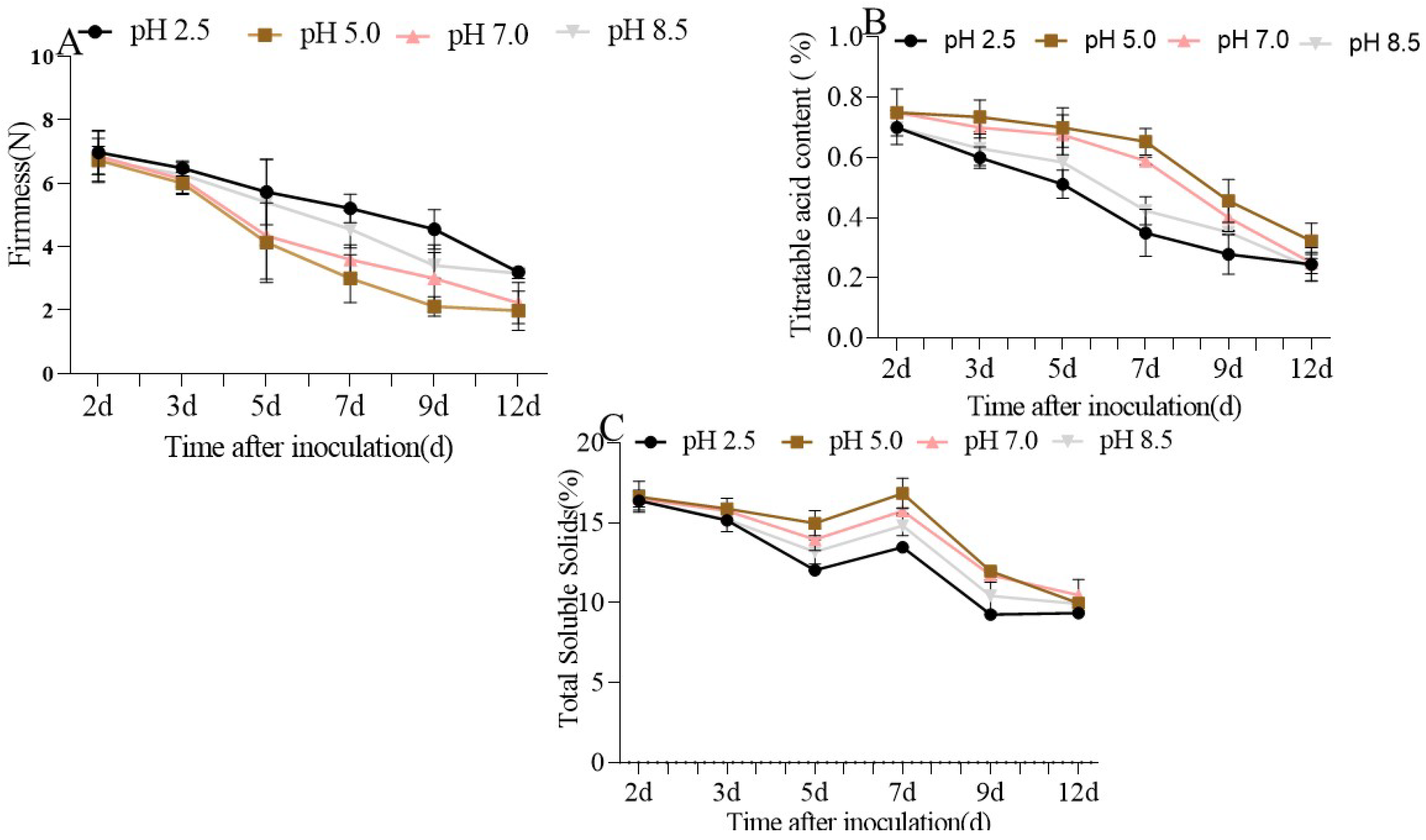
Concerning the quality index, the results showed that fruit firmness gradually decreased during the incubation period across all pH treatments after inoculation. Nonetheless, the inoculated fruits at pHs 2.5 and 8.5 maintained better firmness than those at pHs 5.0 and 7.0. After a period of 9-days inoculation, fruits at pH 2.5 (2.14 N) were firmer than those at pH 5.0 (1.51 N), which were also firmer than those at pH 7.0 (Figure 2A; Table 1). The TSS content in the infected fruits exhibited a declining, rising, and declining pattern during the incubation period after 12 days of inoculation. A peak was observed on the 7th day, and then was reduced in later times during the 12 days of incubation. The apples inoculated with the pH 5.0 spore suspension had much greater TSS content than those at pH 7.0, pH 8.5, and pH 2.5. On the 9th day, the TSS content at pH 5.0 was greater (1.29%) than at pH 2.5 (1.14%), which was also greater than at pH 8.5 (Figure 2B; Table 2). The acid (TA) content decreased all throughout the inoculation period. However, at pHs 5.0 and 7.0, the TA content dropped more quickly than that at pHs 2.5 and 8.5. For instance, the TA content of the contaminated apple fruits under pH 2.5 decreased from the 5th (0.511%) to the 12th (0.245%), while the TA content of the apple inoculated with the pH 5.0 spore suspension followed a similar trend from the 5th (0.70%) to the 12th (0.32%) day (Figure 2C; Table 3).

3.2. Superoxide Anion (O2•−) and Hydrogen Peroxide (H2O2) Production in Apple Fruits Inoculated with P. expansum under Different Ambient pHs

The generation of hydrogen peroxide (H2O2) and superoxide anion (O2•−) is recognized to be involved in plant defense mechanisms and plant development, as well as the abiotic stress response [34]. The analysis of the production rate of O2•− in inoculated fruit under different ambient pHs revealed the highest production at pH 5.0 (control) all throughout the incubation period; from 0 h to 24 h, the results showed a gradual increase in O2•− content. The production of superoxide anion was relatively higher at pH 5.0 (0.21 µmol min−1 kg−1min−1) in the first 24 h (Figure 3A). In addition to the increase observed during the 12–24 h after inoculation in the presence of pH 5.0, superoxide anion production continued to increase between the 2nd and 7th days, up to 0.75 µmol kg−1 min−1 on the 7th day after inoculation (Figure 3A).
The evaluation of the accumulation of hydrogen peroxide indicated a significant increase of 20% (from 12 h to 24 h) after the inoculation process. A further analysis indicated that the accumulation of H2O2 was significantly higher at pH 5.0 (control) (0.22 µmol kg−1 after 3 days) compared to pHs 2.5, 7.0, and 8.5 during the first three days, where we obtained quantities equal to 0.15 µmol kg−1, 0.17 µmol kg−1, and 0.13 µmol kg−1, respectively. Interestingly, from the 5th to 7th day, the H2O2 levels at pHs 5.0 and 7.0 were significantly higher than at pHs 2.5 and 8.5 (Figure 3B).

3.3. Cell Membrane Integrity and MDA Content in P. expansum-Inoculated Apple Fruit under Different Ambient pHs

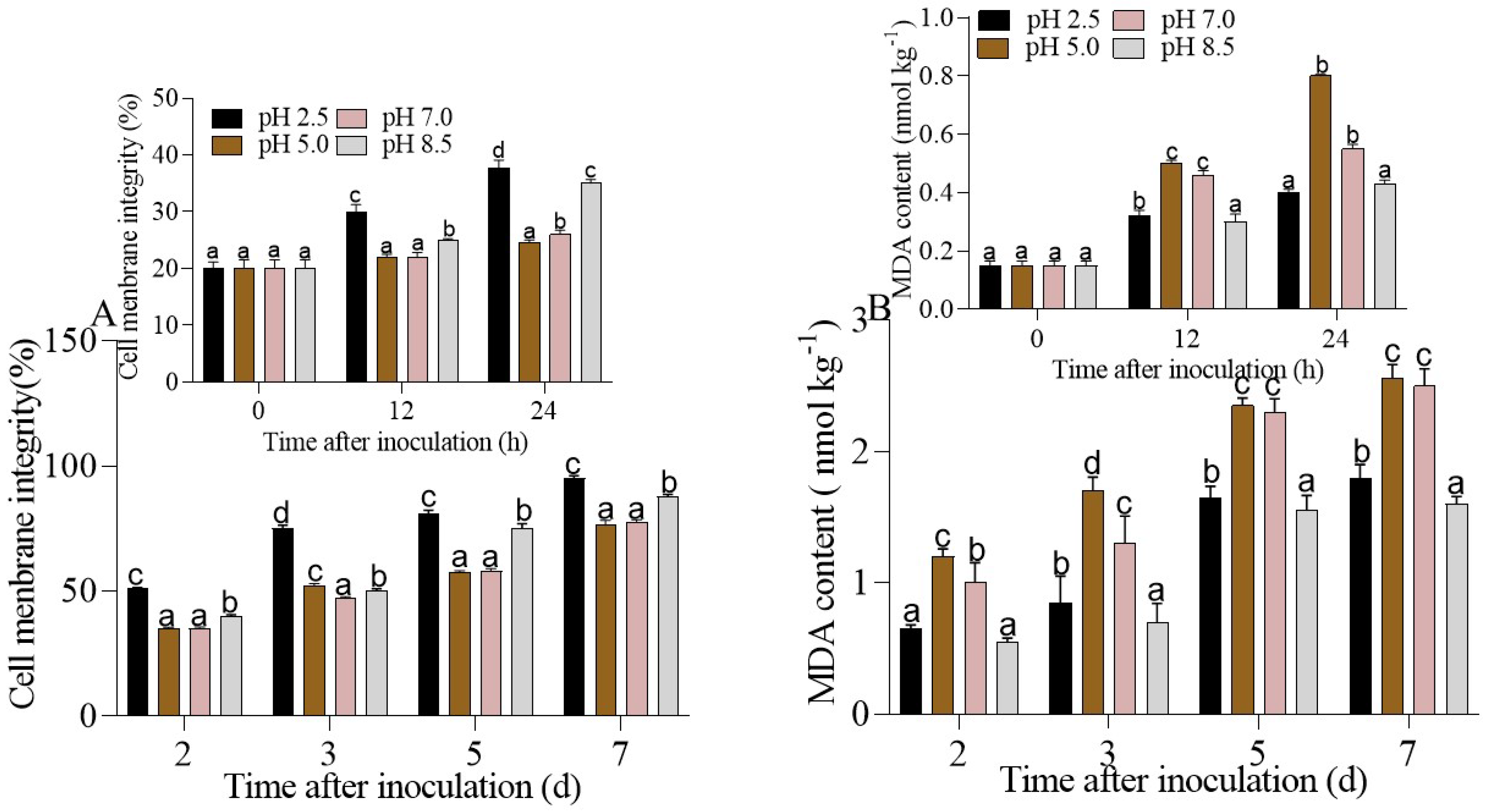
When comparing the membrane integrity of cells at different ambient pHs over the course of 24 h, pHs 5.0 and 7.0 were shown to be much less favorable to cell integrity than pHs 2.5 and 8.5. After 7 days, the percentage of the cell membrane integrity at pH 5.0 (control) was reduced 1.25-fold more than at pH 2.5 (Figure 4A). The lipid peroxidation rates were also calculated by measuring the MDA content. The accumulation of the lipid peroxidation response was very rapid; it was already detected after 12 h of inoculation. This increase was further observed up to the seventh day after inoculation, where the MDA content was lower at pHs 2.5 and 8.5 compared to pHs 5.0 and 7.0, which did not differ between both treatments (Figure 4B). As our previous study [23] showed, the inoculation with P. expansum spore suspension at pH 5.0 accelerated the infection and colonization on apple fruit. Therefore, pH 5.0 leads to a high level of lipid peroxidation and breaks down the integrity of the cell membranes.

3.4. Regulation of Enzymatic Antioxidant Activities in P. expansum-Inoculated Apple Fruits under Different Ambient pHs

3.4.1. NOX, SOD, POD, CAT Regulation in P. expansum-Inoculated Apples Fruits at Different Ambient pHs

Figure 5A demonstrates that the NOX activity rose at pH 5.0 and pH 7.0, whereas at pH 2.5 and pH 8.5, the NOX activity was reduced during the whole incubation period after inoculation. For instance, after 24 h of inoculation, the activities of NOX at pHs 2.5, 7.0, and 8.5 were similar. However, on the 7th day, the NOX activity was the highest at pH 5.0 (control) (6.56 × 10³ U Kg−1), followed by pH 7.0 (6.12 × 10³ U Kg−1), and the NOX activity was quite low at pH 2.5 (2.01 × 10³ U Kg−1) and pH 8.5 (2.25 × 10³ U Kg−1).
SOD contributes to the dismutation of O2•− to produce O2 and H2O2. The SOD activity at pH 2.5 increased considerably at each inoculation time starting at 12 h after inoculation. However, there was limited activation of SOD activity at pHs 5.0, 7.0, and 8.5. This was especially evident at the control of pH 5.0, where the SOD activity was equivalent to 6.72 × 10³ U Kg−1 after 2 days of inoculation and reduced to 4.90 × 10³ U Kg−1 after 7 days of inoculation (Figure 5B).
CAT contributes to the reaction of H2O2 to generate H2O and O2. The CAT activity significantly (p < 0.05) increased at pH 2.5 and pH 8.5 with the extension of the inoculation period, while the levels of the CAT activity at pHs 5.0 and 7.0 were not affected. From the third to the seventh day, the enzymatic activity at pHs 2.5 and 8.5 was nearly two times higher than at pHs 5.0 and 7.0 (Figure 5C).
The POD activity was enhanced when inoculated fruits were treated with buffer solutions at pHs 2.5 and 8.5 between 12 h and 7 days. After 5 days of inoculation, the maximum response was observed at pH 5.0 (3.03 × 10³ U Kg−1), but by the seventh day, the activity dropped significantly at all pH levels (Figure 5D). These findings clearly showed that the activities of SOD, POD, and CAT were upregulated when the pH was too acidic (2.5) or too basic (8.5) and downregulated when the pH was between 5.0 and 7.0. Meanwhile, the NADPH oxidase regulation showed a reverse trend. This could therefore confirm the key role of NOX activity in the mechanism of rebalancing ROS production whenever the plants are under stress. In fact, because the NOX activity was significantly increased at pHs 5.0 and 7.0, the transfer of electrons from the NADPH to the molecular oxygen via the NOX catalytic subunit to generate ROS (O2•− and H2O2) could be increased as well.

3.4.2. Regulation of the AsA-GSH cycle in P. expansum-Inoculated Apple Fruits under Different Ambient pHs

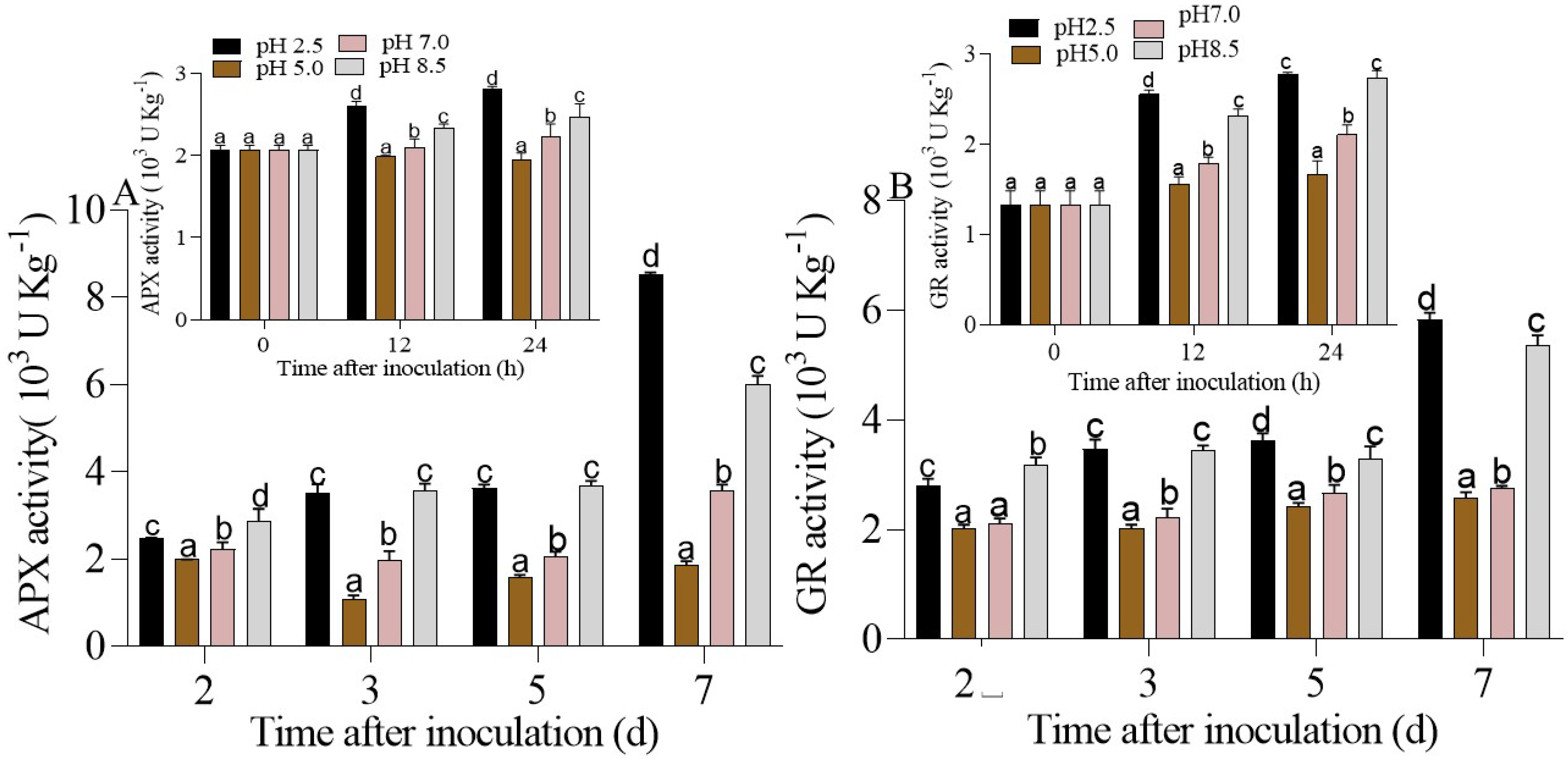
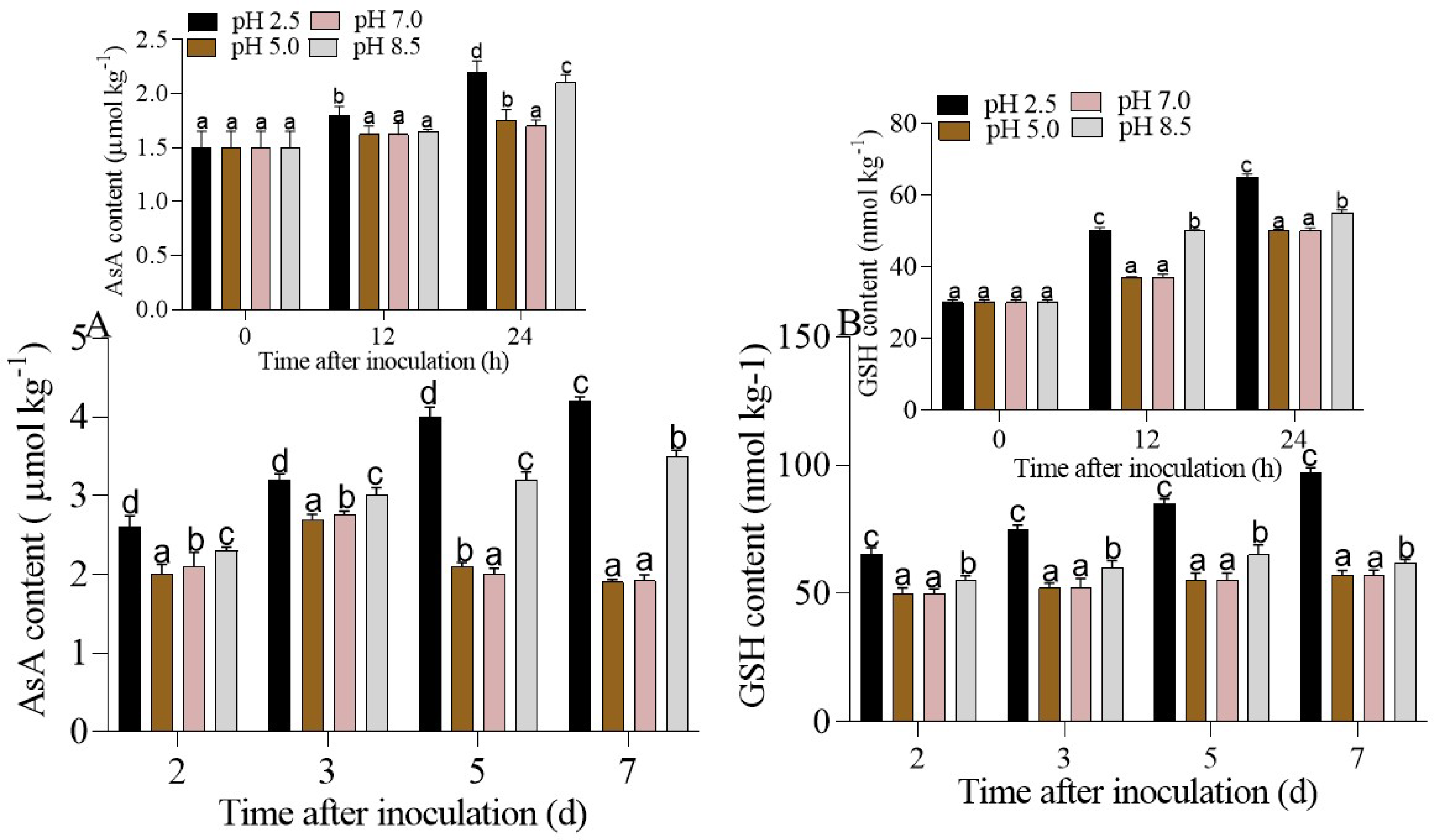
An evaluation of the four enzymes APX, GR, MDHAR, and DHAR after 7-days inoculation revealed dramatically increased activity at pHs 2.5 and 8.5 compared to pHs 5.0 and 7.0. On the 7th day, the APX activity at pHs 5.0 and 7.0 was almost 2.5 times lower than at pH 8.5, and the same trend was observed for the GR activity. Although the activity of MDHAR and DHAR remained intense at pHs 2.5 and 8.5, the results showed that at pH 2.5, the activity slowed down (after 7 days, the MDHAR activity was equal to 5.11 × 10³ U Kg−1, and the DHAR activity was equal to 1.65 × 10³ U Kg−1 (Figure 6C,D) when compared to the activity of other enzymes (APX was equal to 8.45 × 10³ U Kg−1, GR 5.35 × 10³ U Kg−1) at the same pH (2.5) (Figure 6A,B). An analysis of the non-enzymatic antioxidants showed that the contents of the AsA and GSH increased at pH 2.5, followed by pH 8.5, throughout the inoculation period (Figure 7A,B). The present findings might imply that antioxidants are produced in significant amounts by the host plant in a highly acidic or basic pH to lower the amount of oxygen free radicals induced during the interaction between P. expansum and the host to rebalance the environment’s redox system.

4. Discussion

The survival of microorganisms is dependent on their ability to recognize and adapt to changing conditions around their environment. Environmental pH modulation is beneficial because it influences both host resistance and fungi pathogenicity [4]. Although ROS is a byproduct of plant cellular metabolism, it is essential to understand that, apart from pathogen attacks, environmental stresses such as ambient pH also contribute to excessive ROS production, resulting in increasing oxidative stress and inevitably cell death [35]. According to Qiao et al., the overproduction of H2O2 that leads to high MDA content in response to acidic stress was shown to stunt the development of Arabidopsis [36]. Additional research on T. aestivum L. cv. BARI Gom-25 conducted by Bhuyan et al., at highly acidic (pH 4.0) and alkaline (pH 8.5) pHs, discovered elevated levels of MDA, H2O2, and lipid oxidation (LOX) activity, which are all indicators of oxidative stress [37]. Nevertheless, the influence of environmental pH on ROS metabolism in apple fruits infected with P. expansum has not been documented.
In this research, we investigated the change in quality parameters when P. expansum-inoculated apples were exposed to different ambient pHs and how the modulation of ROS metabolism was performed to rebalance the environment. Fruit inoculated with P. expansum under pH 5.0 (control) and pH 7.0 experienced a more rapid decline in quality (rapid loss of TA, TSS content, and firmness over the duration of inoculation with an expanded disease area) than those inoculated under pH 2.5 and pH 8.5 (Figure 1). Moreover, our previous study suggested that the pathogenicity and patulin accumulation induced by P. expansum in apples at pHs 5.0 and 7.0 were greater compared to those at pHs 8.5 and 2.5 [38]; it is therefore possible that, at these pH levels (5.0 and 7.0), P. expansum is able to release other toxins that rapidly destroy fruit quality. The main components of ROS include hydrogen peroxide (H2O2), singlet oxygen (1O2), superoxide anion (O2•−), and hydroxyperoxyl radical (OH). They are extremely toxic at high quantities, and they disrupt normal cellular function by causing substantial damage to protein, DNA, and lipids [39]. However, they act as regulatory signal molecules at a low concentration to modulate the host’s defensive reactions [40,41,42]. The results of this study exhibited P. expansum-infected apples with a higher production of NOX activity at pHs 7.0 and 5.0 compared with pHs 2.5 and pH 8.5 (Figure 5A), leading to significant accumulation of H2O2 and O2•− (Figure 3).
The modification of pH in tissues totally depends on the buffering ability of the host and the original ambient pH. P. expansum can locally acidify tissue to improve colonization [3]; this stress leads to plasma membrane proton pump (H+-ATPase) activation, which combats stress by the influx and efflux of H+ [20]. According to research conducted by Majumdar and Kar, the production of O2•− by the one-electron reduction in O2, which is subsequently dismutated to hydrogen peroxide (H2O2), is mediated by NADPH oxidase (NOX; Respiratory Burst Oxidase Homolog (RBOH)), being itself the triggering element of the ROS circuit [43]. Moreover, it has been shown that NOX and the plasma membrane H+−ATPase are required for SOD to use both apoplastic H+ (extruded from the cytosol by PM H+−ATPase) and O2•− as substrates to generate H2O2 [43]. Then, changes in the pH and excitation pressure cause hazardous free radical production, as well as severe enzymatic activity failures and cellular disruption. The significant production of ROS metabolites at pHs 5.0 and 7.0 subsequently resulted in decreased cell membrane integrity and greater production of MDA at these pHs (Figure 2). These results are in agreement with those of Morales and Munné-Bosch, who postulated that MDA content is used as a lipid peroxidation marker throughout oxidative stress and redox signaling in plant responses to abiotic and biotic stress [44]. Plants are almost certainly subjected to oxidative stress situations throughout their lives.
During their growth period, plants may be subject to oxidative stress caused by unfavorable conditions. Hence, they have established non-enzymatic and enzymatic antioxidative defense systems to effectively remove the excess of ROS [45]. Our results suggested that, when apple fruits were inoculated at pHs 2.5 and 8.5, the CAT, POD, and SOD activities were far more substantial than those at pHs 5.0 and 7.0, where the activities were relatively weak. The metalloenzyme SOD acts as a catalyst in the dismutation reaction of O2•− to create O2 and H2O2, which are two important molecules in the defense against oxidative stress. The thiol groups of enzymes such as the ones involved in the Calvin cycle, Cu/Zn-SOD, and Fe-SOD may be oxidized by H2O2 at high quantities, rendering the enzymes inactive [45]. According to Hernández et al., the elimination of ROS is facilitated by the collaboration of two scavenger enzymes (CAT and POD) with other enzymes of the ascorbate-glutathione cycle [46]. The dismutation of two molecules of H2O2 into water and oxygen is catalyzed by CAT. Based on the nature, duration, and type of stress, the CAT activity can be improved or reduced [47]. As expected, the synergic effect of different environmental pHs and the presence of P. expansum in apple fruit caused a decrease in antioxidant activity at pHs 5.0 and 7.0. This might explain why, in the range of these pHs, the pathogenicity and development of P. expansum were very high, thus causing hypersensitivity and increased production of ROS metabolites, followed by decreased antioxidant activities that try to restore balance in the cell. However, higher antioxidant activities were observed at pHs 2.5 and pH 8.5; although extreme acidity or basicity was not advantageous for P. expansum growth, these ambient pHs remained as potential stress conditions for apple fruits, hence leading to high antioxidant activities. Our results are in accordance to those speculated by Shanan et al., who suggested that the SOD and CAT activity in Rosa hybrida was significantly greater at pH 3.0 than at pH 7.0 [48]. Aside from POD, CAT, and SOD, the AsA-GSH cycle is also critical in countering oxidative damage caused by environmental stress.
To improve plant resistance as well as the activation of enzymes involved in plant development, the production of non-enzymatic antioxidants such as ascorbate and glutathione is essential [49]. In the AsA-GSH cycle, AsA and GSH go through a series of oxidation and reduction reactions. These reactions are accelerated by specific enzymes (APX, GR, MDHAR, and DHAR) [47]. APX uses ascorbic acid (AsA) as a reducing agent to reduce H2O2 to H2O and DHA (H2O2 + AA → 2H2O + DHA). APX is considered as a more efficient scavenger than CAT for H2O2 because of its widespread distribution (in the cytosol and chloroplast). GR uses NADPH as a reductant to convert GSSG into GSH. As a byproduct of regenerating AA from MDHA and DHA, reduced glutathione (GSH) is oxidized (GSSG) [50]. This study’s findings demonstrated that the DHAR, APX, GR, and MDHR activities, as well as the levels of AsA and GSH, were all upregulated in the apple fruits infected with P. expansum at pHs 2.5 and pH 8.5, whereas at pHs 5.0 and pH 7.0, their activities were downregulated (Figure 4 and Figure 5). Further, Bhuyan et al. discovered that the effect of low pH in ginger seedlings induced an increase in antioxidant enzyme activities, such as SOD, CAT, APX, MDHAR, DHAR, and GR [49]. Moreover, wheat cultivars exposed to varying degrees of low pH stress resulted in lower AsA content with increased DHA content, although the APX activity decreased and the MDHAR activity increased [51]. Finally, based on all these findings, we can assert that the colonization and pathogenicity of P. expansum in apple fruit are delayed under acidic or basic conditions, because, at such pHs (2.5 and 8.5), antioxidant activities are enhanced and can efficiently scavenge ROS metabolites and protect the plant cell. The upregulation (pHs 2.5 and 8.5) of non-enzymatic antioxidants (GSH and AsA), as well as antioxidant activities (CAT, SOD, POD, APX, GR MDAHR, and DAHR), indicates avoiding the overaccumulation of ROS at extreme acid or alkaline stress. However, the ROS metabolites were probably low at pHs 2.5 and 8.5, because the high level of antioxidant activity was able to reduce the ROS production.

5. Conclusions

The capacity of pathogens to colonize and proliferate in an environment with high concentrations of ROS demonstrates that they have efficient mechanisms that allow them to protect themselves against harsh environments in the host. In summary, we observed that apples colonized with P. expansum at pHs 5.0 and 7.0 produced H2O2, O2•−, and MDA at high levels with reduced antioxidant activity; meanwhile, the opposite was observed at pHs 2.5 and 8.5, where the non-enzymatic and enzymatic antioxidant activities were enhanced, which therefore repaired the cell damage to maintain the quality of the apple fruits inoculated with P. expansum under different ambient pHs (Figure 8). Our findings also suggest the influence of pH on ROS metabolism in apple fruit contaminated with P. expansum. These studies may contribute to selecting an extreme pH (acidity or alkalinity) to discover relevant phyto protectants to boost antioxidant defense against pathogenic attacks in plants.

Author Contributions

C.J.K.: Conceptualization, Writing—Original Draft. X.Y.: Conceptualization and lesion diameter measurement. H.X.: Project Administration, Writing—Review and Editing, Supervision, Funding Acquisition. Y.B.: Supervision, Funding Acquisition. Z.L.: Validation. J.X.: Enzymatic activity. M.N.: Resources. D.P.: Conceptualization and Supervision. All authors have read and agreed to the published version of the manuscript.

Funding

This research was funded by the Natural Science Foundation of China (32060566) and (31560475).

Data Availability Statement

The datasets used and/or analyzed during the current study are available from the corresponding author upon reasonable request.

Conflicts of Interest

The authors declare that they have no known competing financial interests or personal relationships that could have appeared to influence the work reported in this paper.

References

  1. Kumari, M.; Kamat, S.; Dixit, R.; Pandey, S.; Giri, V.P.; Mishra, A. Microbial Formulation Approaches in Postharvest Disease Management. In Food Security and Plant Disease Management; Elsevier: Amsterdam, The Netherlands, 2021; pp. 279–305. ISBN 978-0-12-821843-3. [Google Scholar]
  2. Prusky, D.; Barad, S.; Luria, N.; Ment, D. PH Modulation of Host Environment, a Mechanism Modulating Fungal Attack in Postharvest Pathogen Interactions. In Post-Harvest Pathology; Prusky, D., Gullino, M.L., Eds.; Springer International Publishing: Cham, Switzerland, 2014; pp. 11–25. ISBN 978-3-319-07700-0. [Google Scholar]
  3. Prusky, D.; Yakoby, N. Pathogenic Fungi: Leading or Led by Ambient PH?: Pathogenic Fungi: Leading or Led by Ambient PH? Mol. Plant Pathol. 2003, 4, 509–516. [Google Scholar] [CrossRef] [PubMed]
  4. Manteau, S.; Abouna, S.; Lambert, B.; Legendre, L. Differential Regulation by Ambient PH of Putative Virulence Factor Secretion by the Phytopathogenic Fungus Botrytis Cinerea. FEMS Microbiol. Ecol. 2003, 43, 359–366. [Google Scholar] [CrossRef] [PubMed]
  5. Wang, Y.; Ji, D.; Chen, T.; Li, B.; Zhang, Z.; Qin, G.; Tian, S. Production, Signaling, and Scavenging Mechanisms of Reactive Oxygen Species in Fruit–Pathogen Interactions. Int. J. Mol. Sci. 2019, 20, 2994. [Google Scholar] [CrossRef] [PubMed]
  6. Vylkova, S. Environmental PH Modulation by Pathogenic Fungi as a Strategy to Conquer the Host. PLoS Pathog. 2017, 13, e1006149. [Google Scholar] [CrossRef]
  7. Prusky, D.; McEvoy, J.L.; Leverentz, B.; Conway, W.S. Local Modulation of Host PH by Colletotrichum Species as a Mechanism to Increase Virulence. Mol. Plant-Microbe Interact. 2001, 14, 1105–1113. [Google Scholar] [CrossRef]
  8. Alkan, N.; Espeso, E.A.; Prusky, D. Virulence Regulation of Phytopathogenic Fungi by PH. Antioxid. Redox Signal. 2013, 19, 1012–1025. [Google Scholar] [CrossRef]
  9. Moss, M.O. Fungi, Quality and Safety Issues in Fresh Fruits and Vegetables. J. Appl. Microbiol. 2008, 104, 1239–1243. [Google Scholar] [CrossRef]
  10. Pitt, J.I.; Hocking, A.D. Primary Keys and Miscellaneous Fungi. In Fungi and Food Spoilage; Springer: Boston, MA, USA, 1997; pp. 59–171. ISBN 978-1-4613-7936-2. [Google Scholar]
  11. Snowdon, A.L. 1: General Introduction and Fruits. In A Colour Atlas of Post-Harvest Diseases and Disorders of Fruits and Vegetables; Wolfe Scientific: London, UK, 1990; ISBN 978-0-7234-0931-1. [Google Scholar]
  12. Alegbeleye, O.; Odeyemi, O.A.; Strateva, M.; Stratev, D. Microbial Spoilage of Vegetables, Fruits and Cereals. Appl. Food Res. 2022, 2, 100122. [Google Scholar] [CrossRef]
  13. Heller, J.; Tudzynski, P. Reactive Oxygen Species in Phytopathogenic Fungi: Signaling, Development, and Disease. Annu. Rev. Phytopathol. 2011, 49, 369–390. [Google Scholar] [CrossRef]
  14. Pilati, S.; Brazzale, D.; Guella, G.; Milli, A.; Ruberti, C.; Biasioli, F.; Zottini, M.; Moser, C. The Onset of Grapevine Berry Ripening Is Characterized by ROS Accumulation and Lipoxygenase-Mediated Membrane Peroxidation in the Skin. BMC Plant Biol. 2014, 14, 87. [Google Scholar] [CrossRef]
  15. Tian, S.; Torres, R.; Ballester, A.-R.; Li, B.; Vilanova, L.; González-Candelas, L. Molecular Aspects in Pathogen-Fruit Interactions: Virulence and Resistance. Postharvest Biol. Technol. 2016, 122, 11–21. [Google Scholar] [CrossRef]
  16. Maheshwari, R.; Dubey, R.S. Nickel-Induced Oxidative Stress and the Role of Antioxidant Defence in Rice Seedlings. Plant Growth Regul. 2009, 59, 37–49. [Google Scholar] [CrossRef]
  17. Overmyer, K.; Brosché, M.; Kangasjärvi, J. Reactive Oxygen Species and Hormonal Control of Cell Death. Trends Plant Sci. 2003, 8, 335–342. [Google Scholar] [CrossRef]
  18. Srivastava, S.; Dubey, R.S. Manganese-Excess Induces Oxidative Stress, Lowers the Pool of Antioxidants and Elevates Activities of Key Antioxidative Enzymes in Rice Seedlings. Plant Growth Regul. 2011, 64, 1–16. [Google Scholar] [CrossRef]
  19. Racchi, M. Antioxidant Defenses in Plants with Attention to Prunus and Citrus Spp. Antioxidants 2013, 2, 340–369. [Google Scholar] [CrossRef]
  20. Alkan, N.; Davydov, O.; Sagi, M.; Fluhr, R.; Prusky, D. Ammonium Secretion by Colletotrichum Coccodes Activates Host NADPH Oxidase Activity Enhancing Host Cell Death and Fungal Virulence in Tomato Fruits. Mol. Plant-Microbe Interact. 2009, 22, 1484–1491. [Google Scholar] [CrossRef] [PubMed]
  21. Han, Z.; Wang, Z.; Bi, Y.; Zong, Y.; Gong, D.; Wang, B.; Li, B.; Sionov, E.; Prusky, D. The Effect of Environmental PH during Trichothecium Roseum (Pers.:Fr.) Link Inoculation of Apple Fruits on the Host Differential Reactive Oxygen Species Metabolism. Antioxidants 2021, 10, 692. [Google Scholar] [CrossRef]
  22. Liu, D.; Liu, M.; Liu, X.-L.; Cheng, X.-G.; Liang, Z.-W. Silicon Priming Created an Enhanced Tolerance in Alfalfa (Medicago sativa L.) Seedlings in Response to High Alkaline Stress. Front. Plant Sci. 2018, 9, 716. [Google Scholar] [CrossRef] [PubMed]
  23. Debnath, B.; Hussain, M.; Irshad, M.; Mitra, S.; Li, M.; Liu, S.; Qiu, D. Exogenous Melatonin Mitigates Acid Rain Stress to Tomato Plants through Modulation of Leaf Ultrastructure, Photosynthesis and Antioxidant Potential. Molecules 2018, 23, 388. [Google Scholar] [CrossRef]
  24. Ju, S.; Yin, N.; Wang, L.; Zhang, C.; Wang, Y. Effects of Silicon on Oryza Sativa L. Seedling Roots under Simulated Acid Rain Stress. PLoS ONE 2017, 12, e0173378. [Google Scholar] [CrossRef]
  25. Luo, S.; Wan, B.; Feng, S.; Shao, Y. Biocontrol of Postharvest Anthracnose of Mango Fruit with Debaryomyces Nepalensis and Effects on Storage Quality and Postharvest Physiology: Biocontrol of Mango with D. Nepalensis. J. Food Sci. 2015, 80, M2555–M2563. [Google Scholar] [CrossRef]
  26. Hossain, M.A.; Rana, M.M.; Kimura, Y.; Roslan, H.A. Changes in Biochemical Characteristics and Activities of Ripening Associated Enzymes in Mango Fruit during the Storage at Different Temperatures. BioMed Res. Int. 2014, 2014, 232969. [Google Scholar] [CrossRef] [PubMed]
  27. Hoehn, E.; Gasser, F.; Guggenbühl, B.; Künsch, U. Efficacy of Instrumental Measurements for Determination of Minimum Requirements of Firmness, Soluble Solids, and Acidity of Several Apple Varieties in Comparison to Consumer Expectations. Postharvest Biol. Technol. 2003, 27, 27–37. [Google Scholar] [CrossRef]
  28. Bao, G.; Bi, Y.; Li, Y.; Kou, Z.; Hu, L.; Ge, Y.; Wang, Y.; Wang, D. Overproduction of Reactive Oxygen Species Involved in the Pathogenicity of Fusarium in Potato Tubers. Physiol. Mol. Plant Pathol. 2014, 86, 35–42. [Google Scholar] [CrossRef]
  29. Patterson, B.D.; MacRae, E.A.; Ferguson, I.B. Estimation of Hydrogen Peroxide in Plant Extracts Using Titanium(IV). Anal. Biochem. 1984, 139, 487–492. [Google Scholar] [CrossRef]
  30. Jiang, H.; Wang, Y.; Li, C.; Wang, B.; Ma, L.; Ren, Y.; Bi, Y.; Li, Y.; Xue, H.; Prusky, D. The Effect of Benzo-(1,2,3)-Thiadiazole-7-Carbothioic Acid S-Methyl Ester (BTH) Treatment on Regulation of Reactive Oxygen Species Metabolism Involved in Wound Healing of Potato Tubers during Postharvest. Food Chem. 2020, 309, 125608. [Google Scholar] [CrossRef]
  31. Ren, Y.; Wang, Y.; Bi, Y.; Ge, Y.; Wang, Y.; Fan, C.; Li, D.; Deng, H. Postharvest BTH Treatment Induced Disease Resistance and Enhanced Reactive Oxygen Species Metabolism in Muskmelon (Cucumis melo L.) Fruit. Eur. Food Res. Technol. 2012, 234, 963–971. [Google Scholar] [CrossRef]
  32. Chumyam, A.; Shank, L.; Faiyue, B.; Uthaibutra, J.; Saengnil, K. Effects of Chlorine Dioxide Fumigation on Redox Balancing Potential of Antioxidative Ascorbate-Glutathione Cycle in ‘Daw’ Longan Fruit during Storage. Sci. Hortic. 2017, 222, 76–83. [Google Scholar] [CrossRef]
  33. Qin, G.; Liu, J.; Cao, B.; Li, B.; Tian, S. Hydrogen Peroxide Acts on Sensitive Mitochondrial Proteins to Induce Death of a Fungal Pathogen Revealed by Proteomic Analysis. PLoS ONE 2011, 6, e21945. [Google Scholar] [CrossRef]
  34. Xue, H.; Sun, Y.; Li, L.; Bi, Y.; Hussain, R.; Zhang, R.; Long, H.; Nan, M.; Pu, L. Acetylsalicylic Acid (ASA) Induced Fusarium Rot Resistance and Suppressed Neosolaniol Production by Elevation of ROS Metabolism in Muskmelon Fruit. Sci. Hortic. 2020, 265, 109264. [Google Scholar] [CrossRef]
  35. Huang, H.; Ullah, F.; Zhou, D.-X.; Yi, M.; Zhao, Y. Mechanisms of ROS Regulation of Plant Development and Stress Responses. Front. Plant Sci. 2019, 10, 800. [Google Scholar] [CrossRef] [PubMed]
  36. Hasanuzzaman, M.; Bhuyan, M.H.M.B.; Parvin, K.; Bhuiyan, T.F.; Anee, T.I.; Nahar, K.; Hossen, M.S.; Zulfiqar, F.; Alam, M.M.; Fujita, M. Regulation of ROS Metabolism in Plants under Environmental Stress: A Review of Recent Experimental Evidence. Int. J. Mol. Sci. 2020, 21, 8695. [Google Scholar] [CrossRef]
  37. Qiao, F.; Zhang, X.-M.; Liu, X.; Chen, J.; Hu, W.-J.; Liu, T.-W.; Liu, J.-Y.; Zhu, C.-Q.; Ghoto, K.; Zhu, X.-Y.; et al. Elevated Nitrogen Metabolism and Nitric Oxide Production Are Involved in Arabidopsis Resistance to Acid Rain. Plant Physiol. Biochem. 2018, 127, 238–247. [Google Scholar] [CrossRef]
  38. Bhuyan, M.H.M.B.; Hasanuzzaman, M.; Mahmud, J.A.; Hossain, M.S.; Alam, M.U.; Fujita, M. Explicating Physiological and Biochemical Responses of Wheat Cultivars under Acidity Stress: Insight into the Antioxidant Defense and Glyoxalase Systems. Physiol. Mol. Biol. Plants 2019, 25, 865–879. [Google Scholar] [CrossRef]
  39. Jimdjio, C.K.; Xue, H.; Bi, Y.; Nan, M.; Li, L.; Zhang, R.; Liu, Q.; Pu, L. Effect of Ambient PH on Growth, Pathogenicity, and Patulin Production of Penicillium Expansum. Toxins 2021, 13, 550. [Google Scholar] [CrossRef]
  40. Foyer, C.H.; Noctor, G. Redox Homeostasis and Antioxidant Signaling: A Metabolic Interface between Stress Perception and Physiological Responses. Plant Cell 2005, 17, 1866–1875. [Google Scholar] [CrossRef]
  41. Tanou, G.; Molassiotis, A.; Diamantidis, G. Induction of Reactive Oxygen Species and Necrotic Death-like Destruction in Strawberry Leaves by Salinity. Environ. Exp. Bot. 2009, 65, 270–281. [Google Scholar] [CrossRef]
  42. Yan, J.; Tsuichihara, N.; Etoh, T.; Iwai, S. Reactive Oxygen Species and Nitric Oxide Are Involved in ABA Inhibition of Stomatal Opening. Plant Cell Environ. 2007, 30, 1320–1325. [Google Scholar] [CrossRef]
  43. Majumdar, A.; Kar, R.K. Orchestration of Cu-Zn SOD and Class III Peroxidase with Upstream Interplay between NADPH Oxidase and PM H+-ATPase Mediates Root Growth in Vigna Radiata (L.) Wilczek. J. Plant Physiol. 2019, 232, 248–256. [Google Scholar] [CrossRef] [PubMed]
  44. Morales, M.; Munné-Bosch, S. Malondialdehyde: Facts and Artifacts. Plant Physiol. 2019, 180, 1246–1250. [Google Scholar] [CrossRef]
  45. Sharma, P.; Jha, A.B.; Dubey, R.S.; Pessarakli, M. Reactive Oxygen Species, Oxidative Damage, and Antioxidative Defense Mechanism in Plants under Stressful Conditions. J. Bot. 2012, 2012, 1–26. [Google Scholar] [CrossRef]
  46. Hernández, J.A.; Ferrer, M.A.; Jiménez, A.; Barceló, A.R.; Sevilla, F. Antioxidant Systems and O2 ·−/H2O2 Production in the Apoplast of Pea Leaves. Its Relation with Salt-Induced Necrotic Lesions in Minor Veins. Plant Physiol. 2001, 127, 817–831. [Google Scholar] [CrossRef]
  47. Sharma, P.; Dubey, R.S. Drought Induces Oxidative Stress and Enhances the Activities of Antioxidant Enzymes in Growing Rice Seedlings. Plant Growth Regul. 2005, 46, 209–221. [Google Scholar] [CrossRef]
  48. Shanan, N. Optimum PH Value for Improving Postharvest Characteristics and Extending Vase Life of Rosa Hybrida Cv. Tereasa Cut Flowers. Asian J. Adv. Agric. Res. 2017, 1, 1–11. [Google Scholar] [CrossRef]
  49. Yin, F.; Liu, X.; Cao, B.; Xu, K. Low PH Altered Salt Stress in Antioxidant Metabolism and Nitrogen Assimilation in Ginger (Zingiber officinale) Seedlings. Physiol. Plant. 2020, 168, 648–659. [Google Scholar] [CrossRef]
  50. Das, K.; Roychoudhury, A. Reactive Oxygen Species (ROS) and Response of Antioxidants as ROS-Scavengers during Environmental Stress in Plants. Front. Environ. Sci. 2014, 2, 53. [Google Scholar] [CrossRef]
  51. Bhuyan, M.; Hasanuzzaman, M.; Mahmud, J.; Hossain, M.; Bhuiyan, T.; Fujita, M. Unraveling Morphophysiological and Biochemical Responses of Triticum aestivum L. to Extreme PH: Coordinated Actions of Antioxidant Defense and Glyoxalase Systems. Plants 2019, 8, 24. [Google Scholar] [CrossRef]
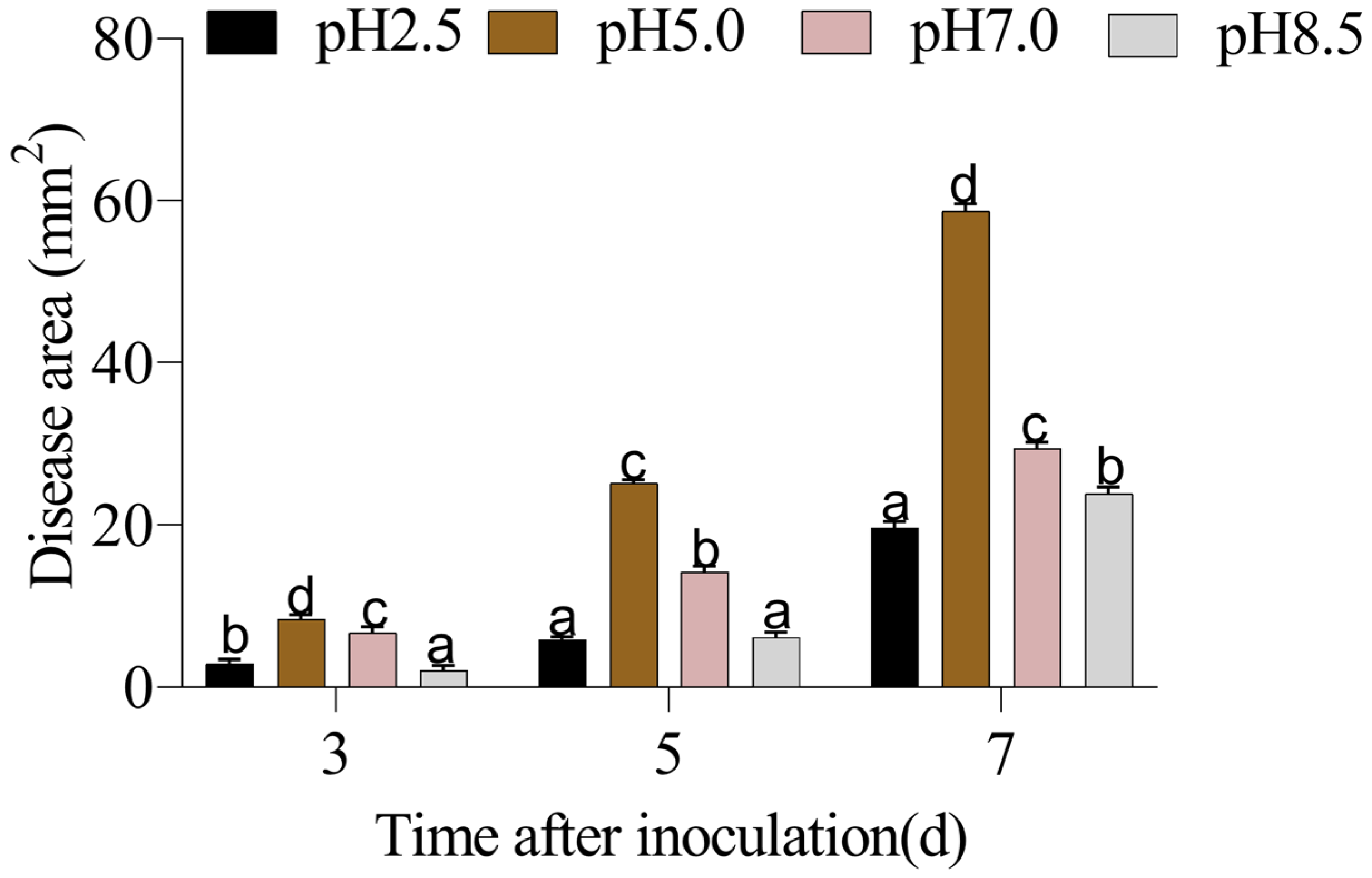
Figure 1. . The effect of ambient pH on the disease development of apple fruits inoculated with P. expansum. Bars represent standard error of the mean. Different letters indicate significant difference (p < 0.05).
Horticulturae 09 00538 g001
Figure 2. The effect of ambient pH on the quality of apple fruits inoculated with P. expansum. (A) Firmness, (B) total soluble solid, (C) titratable acid content. Bars represent standard error of the mean.
Horticulturae 09 00538 g002
Figure 3. The effect of ambient pH on ROS generation of apple fruit inoculated with P. expansum. (A) Superoxide anion (O2•−) production rate, (B) hydrogen peroxide (H2O2) content. Bars represent standard error of the mean. Different letters indicate significant difference (p < 0.05).
Horticulturae 09 00538 g003
Figure 4. The effect of ambient pH on cell membrane integrity (A) and MDA content (B) on apple fruits inoculated with P. expansum. Bars represent standard error of the mean. Different letters indicate significant difference (p < 0.05).
Horticulturae 09 00538 g004
Figure 5. The effect of ambient pH on NADPH oxidase activity (A), SOD activity (B), CAT activity (C), and POD activity (D) on apple fruits inoculated with P. expansum. Bars represent standard error of the mean. Different letters indicate significant difference (p < 0.05).
Horticulturae 09 00538 g005
Figure 6. The effect of ambient pH on the activities of enzyme involved in AsA-GSH cycle on apple fruits inoculated with P. expansum. (A) APX activity; (B) GR activity; (C) MDHAR activity; (D) DHAR activity. Bars represent standard error of the mean. Different letters indicate significant difference (p < 0.05).
Horticulturae 09 00538 g006aHorticulturae 09 00538 g006b
Figure 7. The effect of ambient pH on the contents of metabolites involved in AsA-GSH cycle on apple fruits inoculated with P. expansum. (A) AsA content; (B) GSH content. Bars represent standard error of the mean. Different letters indicate significant difference (p < 0.05).
Horticulturae 09 00538 g007
Figure 8. The possible mechanism of different ambient pH on the postharvest disease in apple fruits inoculated with P. expansum by modulating ROS metabolism.
Horticulturae 09 00538 g008
Table 1. Representative data for firmness.
2 d3 d5 d7 d9 d12 d
pH 2.56.97 (±0.69)6.47 (±023)5.72 (±1.0)5.21 (±0.45)4.55 (±0.62)3.20 (±0.12)
pH 5.06.72 (±0.69)6.00 (±0.35)4.13 (±1.2)3.00 (±0.75)2.12 (±0.30)1.99 (±0.61)
pH 7.06.85 (±0.79)6.12 (±0.35)4.34 (±1.36)3.59 (±0.46)3.00 (±1.06)2.22 (±0.64)
pH 8.56.72 (±0.43)6.27 (±0.37)5.4 (±1.35)4.55 (±0.59)3.42 (±0.4)3.16 (±0.16)
Table 2. The data for TA (titratable acid) content.
2 d3 d5 d7 d9 d12 d
pH 2.50.70 (±0.01)0.60 (±0.04)0.51 (±0.05)0.35 (±0.08)0.27 (±0.07)0.24 (±0.06)
pH 5.00.75 (±0.01)0.73 (±0.06)0.70 (±0.07)0.73 (±0.06)0.65 (±0.04)0.32 (±0.06)
pH 7.00.75 (±0.08)0.70 (±0.04)0.67 (±0.07)0.58 (±0.01)0.40 (±0.04)0.25 (±0.04)
pH 8.50.70 (±0.06)0.63 (±0.06)0.58 (±0.08)0.42 (±0.05)0.35 (±0.01)0.23 (±0.04)
Table 3. The data for TSS (total soluble solid) content.
2 d3 d5 d7 d9 d12 d
pH 2.516.38 (±0.07)15.16 (±0.04)12.03 (±0.04)13.48 (±0.04)9.26 (±0.08)9.35 (±0.07)
pH 5.016.63 (±0.97)15.88 (±0.66)14.98 (±0.78)16.3884 (±0.94)11.98 (±0.09)9.98 (±0.06)
pH 7.016.51 (±0.51)15.76 (±0.08)13.95 (±0.67)15.75 (±0.18)11.73 (±0.44)10.48 (±0.98)
pH 8.516.42 (±0.63)15.22 (±0.78)13.19 (±0.75)14.82 (±0.62)10.43 (±0.98)9.93 (±0.18)
Disclaimer/Publisher’s Note: The statements, opinions and data contained in all publications are solely those of the individual author(s) and contributor(s) and not of MDPI and/or the editor(s). MDPI and/or the editor(s) disclaim responsibility for any injury to people or property resulting from any ideas, methods, instructions or products referred to in the content.