Article

Regeneration of Transgenic Ficus lyrata via Indirect Somatic Embryogenesis and Isolation of Variants for Development of New Cultivars

1
College of Landscape and Horticulture, Southwest Forestry University, Kunming 650224, China
2
Hubei Engineering Research Center for Specialty Flowers Biological Breeding, Jingchu University of Technology, Jingmen 448000, China
3
Mid-Florida Research and Education Center, Department of Environmental Horticulture, Institute of Food and Agricultural Sciences, University of Florida, Apopka, FL 32703, USA
4
Jingmen City’s Shilipai Forest Farm, Jingmen 448000, China
*
Authors to whom correspondence should be addressed.
Horticulturae 2023, 9(5), 530; https://doi.org/10.3390/horticulturae9050530
Submission received: 8 March 2023 / Revised: 17 April 2023 / Accepted: 20 April 2023 / Published: 24 April 2023
(This article belongs to the Special Issue Innovation in Propagation and Cultivation of Ornamental Plants)

Abstract

:
Ficus lyrata is a popular ornamental foliage plant with unique violin- or guitar-shaped green leaves. In our previous study, a grapevine gene VvMybA1 was introduced into F. lyrata via Agrobacterium-mediated transformation, which resulted in the availability of purple-leaved plants. Since VvMybA1 is a transcription factor, the regeneration of transgenic purple-leaved plants might potentially produce variants with multicolored leaves. The objective of this study was to establish a method for regenerating purple-leaved F. lyrata and determine if variants with different coloration or variegation could be isolated from regenerated populations. Leaf explants derived from a completely purple-leaved transgenic plant were cultured on Murashige and Skoog (MS) basal medium supplemented with different concentrations of 6-benzyladenine (BA) and α-naphthalene acetic acid (NAA). Callogenesis occurred in leaf explants, and a subculture of callus-borne explants on the same medium resulted in callus proliferation and the occurrence of somatic embryos. Somatic embryos were more effectively induced from callus pieces cultured on MS medium supplemented with 8.88 μM of BA and 0.27 μM of NAA. More than 30 embryos were induced per callus piece, and the embryos matured and converted to plantlets. MS medium supplemented with 4.92 μM of indolyl-3-butanoic acid (IBA) greatly improved root development. Plantlets were transplanted into soilless substrate and grown in a shaded greenhouse for morphological evaluation. Nine variants with different degrees of coloration and variegation were isolated from the regenerated populations. Our results suggest that the regeneration of transgenic plants that harbor a transcription factor, such as VvMybA1, could be an additional way of isolating novel variants for the development of new cultivars of ornamental plants.

1. Introduction

Ficus lyrata Warb., commonly known as fiddle leaf fig or banjo fig, is native to western Africa, from west Cameroon to Sierra Leone, inhabiting lowland tropical rainforest. Fiddle leaf fig is a member of the family Moraceae and has unique violin- or guitar-shaped, leathery green leaves. It is popular as either an indoor house plant or a garden plant in tropical and subtropical regions [1]. As a house plant, F. lyrata can grow under low-light conditions and tolerate drought stress without dropping old leaves. As a garden plant, it can be planted under shade, producing large violin-shaped leaves, and attracting great attention from visitors. Thus, F. lyrata has become one of the most popular Ficus species used for both indoor and outdoor plantscaping.
Unlike its relatives, such as F. benjamina L. and F. elastica Roxb, which have multiple variegated cultivars, F. lyrata has only one green-leaved cultivar. Ornamental plants are prized for their aesthetic appearance; the flower color and/or leaf variegation are among the most conspicuous and highly valuable features of ornamental plants [2,3]. To produce color-leaved F. lyrate, Zhao et al. introduced a gene VvMybA1 into F. lyrata via Agrobacterium-mediated transformation and generated novel, purple-leaved plants [4]. VvMybA1 is a member of the R2R3 MYB gene family and was isolated from grape plant [5,6,7]. It is a transcription factor regulating anthocyanin biosynthesis through the control of UFGT (UDP-glucose/flavonoid 3-O-glucosyltransferase) gene expression [8,9,10,11]. Among the transgenic plants, some plants had complete purple leaves and few others had green–purple leaves, and the differences were not related to the copy number of VvMybA1 in the plants [4]. Subsequently, the purple-leaved and its parent (green-leaved) plants were analyzed using RNA-Seq, and the results showed that the accumulation of anthocyanin in the purple-leaved plants was attributed to the upregulation of genes encoding UFGT, chalcone isomerase (CHI), UDP rhamnose/anthocyanidin-3-glucoside rhamnosyltransferase (3RT), and chalcone synthase (CHS) [12]. These results indicate that VvMybA1 upregulated not only UFGT but also other gene expressions. It is possible that the upregulation of other genes may result in leaves with different colors and/or novel variegation patterns. Thus, we hypothesized that different degrees of purplish plants or variants could be isolated from plants regenerated from transgenic purple-leaved plants, and the variants, if morphologically unique, could potentially be developed into new cultivars through vegetative propagation, such as stem cutting.
Ficus lyrata was among the first group of ornamental foliage plants that were micropropagated. In the 1970s, Debergh and De Wael [13] regenerated adventitious shoots from leaf explants cultured on MS basal medium [14]. Later, Zhao et al. found that abundant callus and more adventitious shoots were induced by the culture of leaf explants on MS medium containing 4.5 μM of TDZ (N-phenyl-N′-1, 2, 3-thiadiazol-5-yl urea) with 0.5 μM of a-naphthalene acetic acid (NAA) than those cultured on the same medium supplemented with either 4.0 μM of CPPU (N-(2-chloro-4-pyridyl)-N′-phenylurea) or 6-benzylaminopurine (BA) with 0.5 μM of NAA [4]. MS medium has also been used for regenerating other Ficus species through indirect shoot organogenesis, including F. carica [15,16,17], F. religiosa [18,19], and F. pandurate [20]. Among these protocols, BA was the most commonly used cytokinin-like plant growth regulator (PGR) for Ficus regeneration. Thus far, there has been no report on the regeneration of F. lyrata through somatic embryogenesis.
The objectives of this study were to develop an efficient protocol for regeneration of plants from transgenic purple-leaved F. lyrata and to test our hypothesis that novel variants could be isolated from the regenerated populations.

2. Materials and Methods

2.1. Plant Materials

Young leaves (200 cm2) were collected from a purple-leaved transgenic F. lyrata plant (Figure 1A) grown in a shaded greenhouse at the University of Florida’s Mid-Florida Research and Education Center in Apopka, Florida (coordinates: 28°42′06″ N 81°31′54″ W and altitude 25 m). Both upper and lower leaf surfaces were cleaned gently with 75% ethanol and cut into about 10 cm2 pieces. The leaf pieces were washed with running water for 2 h. Under sterile conditions, the leaf pieces were immersed in 75% ethanol for 30 s and washed with sterile distilled water once. They were then immersed in a 25% Clorox (1.5% NaOCl) solution with occasional shaking for about 20 min and washed with sterile distilled water five times. The leaf pieces were further cut into smaller pieces (about 1 cm2) and placed in a sterile Petri dish for further use.

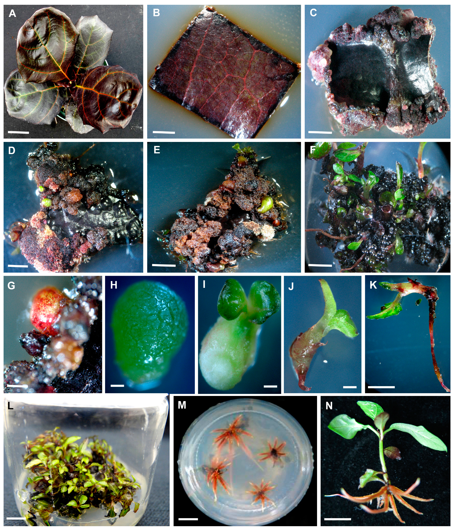
2.2. Initial Culture and Callus Induction

The MS medium (Product ID: M5531, PhytoTech Labs, Shawnee Mission, KS, USA) was supplemented with 3% sucrose and 0.8% agar and used as the basic medium. After the pH was adjusted to 5.8, the medium was aliquoted to six 500 mL glass bottles and autoclaved at 121 °C for 20 min. When the medium temperature dropped to about 50 °C, filter-sterilized stock solutions of BA and NAA were added to the bottles, resulting in the medium with six PGR combinations (4.44 and 8.88 μM BA, each with 0.05, 0.27, and 0.54 μM of NAA). The medium was poured into Petri dishes with about 20 mL each. The disinfected leaf explants were placed on the medium (Figure 1B) with five leaf explants per Petri dish, six dishes per treatment. The Petri dishes were maintained in a culture room under a 12 h photoperiod, provided by cool-white, fluorescent lamps with a light level of 50 μmol m−2 s−1 and a temperature of 25 ± 2 °C. The cultures were monitored daily, and data regarding callus formation and somatic embryo occurrence were taken 50 days after culture initiation.

2.3. Callus Proliferation and Somatic Embryo Occurrence

After 50 days of culture, the explants with callus were subcultured on the fresh medium in 220 mL glass baby food jars (PhytoTech Labs, Shawnee Mission, KS, USA) containing the same combination of PGR as the initial culture medium in Petri dishes. There were four leaf explants per jar and six jars per treatment. The occurrence of somatic embryos was recorded after 50 days of subculture, and embryo conversion rates were calculated.

2.4. Somatic Embryogenesis

The callus mass without somatic embryos was cut into about 1.0 cm3 pieces, and the callus pieces were cultured on the same medium as the initial culture medium in baby food jars. There were 4 callus pieces per jar and 12 jars per treatment. The number of somatic embryos per callus piece was recorded on day 30, 40, and 50 after culture, embryo conversion was determined on day 50, and the embryo conversion rates were calculated. Based on the collected data, a BA and NAA combination that induced the highest numbers of somatic embryos with the highest embryo conversion rate was identified as the appropriate protocol for inducing somatic embryogenesis. The protocol was then used to produce a more bipolar structure or shoots for the following rooting experiment.

2.5. Rooting Medium Selection

The rooting medium was the MS medium mentioned above supplemented with and without 0.2% of activated charcoal (AC). After the pH was adjusted to 5.8, the medium was aliquoted into six 500 mL glass bottles and autoclaved at 121 °C for 20 min. After the medium temperature dropped to 50 °C, a filter-sterilized stock solution of indole-3-butyric acid (IBA) was added to the bottles, resulting in medium with three IBA concentrations (0.49, 2.46, and 4.92 μM) with and without AC. Shoots with a bipolar structure (about 1.5 cm in height) were transplanted into the rooting medium. There were four shoots per jar and 12 jars per treatment. The rooting percentage, root number, and average root length were recorded after 12 days of culture.

2.6. Transplantation and Acclimatization

After 12 days of rooting, seedlings were transplanted into 40-cell plug trays filled with a soilless substrate consisting of 70% peat and 30% perlite. Potted plants were grown in a shaded greenhouse with a maximum photosynthetic photon flux density (PPFD) of 200 μmol m−2 s−1, and a temperature range of 20 to 28 °C with relative humidity varying from 70% to 100%. Two months later, the plugs were transplanted into 15 cm plastic pots filled with a soilless substrate consisting of 90% peat and 10% perlite, fertilized with a Peters 20-20-20 General Purpose (20N-8.7P-16.6 K) fertilizer (Scotts Co., Marysville, OH, USA), and grown in a shaded greenhouse under a PPFD of 350 μmol m−2 s−1.

2.7. Experimental Design and Data Analysis

All experiments were arranged as a completely randomized design with different replications mentioned above. Each culture vessel (Petri dish or glass baby food jar) was considered an experimental unit for in vitro culture. For ex vitro rooting, eight cell plugs were considered an experimental unit, and each was repeated five times. Collected data were subjected to an analysis of variance using SAS (SAS Institute, Cary, NC, USA), and the means were separated using Fisher’s protected least significant differences at the p < 0.05 level.

2.8. Identification of Variants in Regenerated Populations

Three batches of regenerated plants were potted in soilless substrates and grown in the same shaded greenhouse mentioned above. Plant morphological characteristics were closely monitored. Since all regenerated plants had reddish roots, the major emphasis was placed on leaf phenotypes, mainly leaf color and variegation patterns, as outlined by Chen and Henny [21], for the identification of variants.

3. Results

3.1. Callus Induction and Occurrence of Somatic Embryos

Callogenesis occurred about 10 days after the leaf explants were cultured on MS medium regardless of PGR combinations. Calli continuously proliferated with the appearance of a few globular structures (Table 1), which resembled somatic embryos (Figure 1C). Callus growth slowed down after about 50 days. As a result, the explants with Calli were subcultured on fresh medium with the same PGR as the initial culture medium in baby food jars. The subculture resulted in further callus proliferation and increased numbers of somatic embryos (Table 1, Figure 1D), and the conversion of some embryos produced buds (Figure 1E). Among the six PGR combinations, BA at 8.88 μM with 0.27 μM of NAA induced more somatic embryos from callus-borne leaf explants (Table 1).

3.2. Somatic Embryo Induction

To obtain more somatic embryos, callus pieces derived from the subculture were inoculated on fresh medium containing the same PGR. Somatic embryos increasingly appeared from 30 days to 50 days after culture, and over 30 embryos were induced per callus pieces in 50 days (Table 2). Meanwhile, somatic embryo conversion rapidly took place with a conversion rate of up to 60%. As a result, more shoots appeared from callus pieces (Figure 1F). The embryo conversion occurred in an asynchronous fashion, some were at the globular or heart shape stage, others were at the cotyledon stage, and still others appeared as a bipolar structure or seedlings, also known as embling [22]. These structures were easily separated from callus pieces (Figure 1G–K). The medium containing 8.88 μM of BA with either 0.27 μM or 0.54 μM of NAA induced the highest number of somatic embryos and the highest embryo conversion (Table 2). Numerous shoots appeared in baby food jars (Figure 1L). Based on the two parameters, MS medium containing 8.88 μM of BA with either 0.27 or 0.54 μM of NAA is considered the suitable PGR combination for inducing somatic embryos from leaf explants of transgenic purple-leaved F. lyrata.

3.3. In Vitro Rooting

Somatic embryos were able to produce roots (Figure 1K). To speed up the rooting process, bipolar structures or shoots were rooted in the rooting medium. The medium containing 0.49, 2.46, or 4.92 μM of IBA devoid of AC significantly increased the rooting percentage and root numbers (Table 3, Figure 1M,N) after 12 days. The addition of AC, however, reduced the two rooting parameters, but increased the root length (Table 3). Thus, IBA at 0.49, 2.46, or 4.92 μM of IBA were considered suitable for speeding up the rooting of the bipolar structure or shoots.

3.4. Morphology of Regenerated Plants and Isolation of Variants

Three batches of regenerated plants were evaluated. Regenerated seedlings were initially transplanted to cell plug trays (Figure 2A) and were then transplanted to 15 cm pots (Figure 2B). The plants had green or green–purple leaves. After 120 days of growth in a shaded greenhouse, the leaves of most of the plants became purplish (Figure 2C). The first batch had 32 green-leaved plants (Table 4). After 60 days of growth in a shaded greenhouse, the leaves of 18 plants changed to green–purple leaves. After another 60 days of growth, 9 plants showed green–purple leaves, and 23 exhibited purple leaves. The 2nd and 3rd batches had 226 and 239 plants, respectively. About 60% and 57% of them in the first and second batches had green–purple leaves when grown under in vitro conditions, and the rest had green leaves. After 120 days of growth in a shaded greenhouse, green–purple- and purple-leaved plants in the 2nd batch changed to 76 and 150, respectively. A similar trend of color changes happened in the third batch plants as well (Table 4). Such color changes, however, stopped after about 120 days of growth in the shaded greenhouse. Additionally, all regenerated plants had no changes in leaf shape and plant overall growth patterns compared to the non-transgenic plants grown in the same shaded greenhouse.
The regenerated populations did have some variants with novel foliar colorations, which are shown in Figure 2D–L: (1) the leaf edge was darkish purple with the occasional appearance of completely purplish leaves (Figure 2D); (2) dark-purplish leaves with green-colored veins (Figure 2E); (3) completely dark-purplish leaves (Figure 2F); (4) old leaves were completely purplish, but young leaves were reddish purple with greenish veins (Figure 2G); (5) leaves with purple and green variegation (Figure 2H); (6) bright red leaves (Figure 2I); (7) pinkish red leaves (Figure 2J); (8) the majority of the leaves were purplish, but the lower middle of leaves remained green (Figure 2K); and (9) dark red leaves (Figure 2L). The morphological characteristics were largely stable, but some could be affected by seasonal changes. A few of the isolated plants were propagated through stem cuttings, and they had the same phenotypes as the stock plants. These plants have been maintained in a shaded greenhouse for more than five years, and their morphological characteristics remain mostly unchanged.

4. Discussion

This study established an efficient protocol for regeneration of transgenic purple-leaved F. lyrata through indirect somatic embryogenesis. Leaf explants were cultured on MS medium supplemented with 8.88 μM of BA and 0.27 μM of NAA to induce callogenesis in 50 days, and the callus-borne explants were subcultured on the same medium for another 50 days to induce callus proliferation and somatic embryo formation. The callus mass without somatic embryos was cut into 1 cm3 pieces and cultured on the same medium for an additional 50 days to induce embryo proliferation. Over 30 somatic embryos were induced per 1 cm3 piece, which resulted in the occurrence of 18 shoots through embryo conversion. To speed up rooting, shoots cultured on MS medium supplemented with 4.92 μM of IBA induced multiple roots in 12 days. Plantlets were transplanted to cell plug trays filled with a soilless substrate composed of 70% peat and 30% perlite for about two months and were then transplanted to 15 cm or larger sized pots for production in a shaded greenhouse under a PPFD of 350 μmol m−2 s−1.
To the best of the authors’ knowledge, this is the first protocol developed for regeneration of F. lyrata through indirect somatic embryogenesis. Debergh and Da Wael propagated this species through indirect shoot organogenesis, where MS basal medium supplemented with 5 mg L−1 BA and 2.5 mg L−1 IBA was considered an appropriate protocol [13]. The protocol developed by Zhao et al. was MS medium supplemented with 4.5 μM of TDZ with 0.5 μM of NAA [4]. We initially tested the two protocols and found that neither were effective for adventitious shoot production. Thus, we modified their methods by using BA as a cytokinin derivative [13] and NAA as an auxin derivative [4], resulting in the efficient regeneration of plants through indirect somatic embryogenesis. BA has been reported to induce somatic embryogenesis from the calli of coffee [23] and bermudagrass [24] as well as butterfly ginger [25]. BA has also been used for inducing further growth of somatic embryos [25]. BA and NAA, at appropriate concentrations, improved the somatic embryo maturation of rose [26]. As to the selection BA and NAA concentrations, we took into consideration the report of Ji et al. [27], in which it was reported that the adventitious shoots of F. carica were produced by a culture of explants on MS medium supplemented with BA at 1.0 and 2.0 mg L−1 (equivalent to 4.44 and 8.88 μM) and NAA at 0.05 mg L−1 (equivalent to 0.27 μM). Additionally, shoot organogenesis also occurred in the leaf explants of F. religiora cultured on MS medium supplemented with 2.0 mg L−1 BA [28].
More importantly, through the established protocol, nine novel variants were isolated from the regenerated populations. It is known that the parental purple-leaved plant was derived from Agrobacterium-mediated transformation [4]. The transformation produced two types of plants: completely purple-leaved plants and green–purple-leaved plants. In this study, it was a completely purple-leaved plant that was used for regeneration. Thus, the question is how did the variants arise? One possibility is somaclonal variation [29]. Somaclonal variants were isolated from a wide range of ornamental plants [21], including popular foliage plants of Syngonium [30] and Dieffenbachia [31]. The mechanisms underlying somaclonal variation include changes in chromosome numbers, epigenetic effects, and variation at the DNA level [29,32]. Chromosome changes are unlikely because the leaf shape and leaf thickness of the variants were similar to the parental plant, although we have not examined their chromosome numbers cytogenetically. Another possibility could be transformation-induced mutations. The insertion of a foreign gene into the plant genome may disrupt genomic DNA sequences, resulting in changes in plant phenotypes [33]. A third possibility could be the action of the transformed gene VvMybA1. Many genes have been transformed into ornamental plants [34], but a few reports documented the occurrence of morphological variation in regenerated transgenic plants. It is likely that most of the genes transformed were structure genes, which, in general, should lead to stable phenotypes. The gene in this study is a transcription factor which is able to regulate the expression of many other genes in anthocyanin biosynthesis, as documented by Guan et al. [12]. Thus, it is possible that the morphological variation in these variants could be due to the VvMybA1-mediated expression of different genes. Several reports showed that color variation in grape berry skin is associated with molecular variation at VvMybA1 in more than 95% of the analyzed grape cultivars [8,9,10,35]. Further studies will be conducted to explore the molecular mechanisms behind the altered colorations in these variants.
In commercial ornamental plant production, numerous woody plants are propagated by stem cutting [36]. It is an affordable and simple method to propagate true-to-type plants. Similarly, the isolated F. lyrata variants can be propagated by stem cutting to allow propagated plants to maintain the same phenotypes as the stock plants. Thus, these variants could potentially be new cultivars of F. lyrata for the ornamental plant industry.

5. Conclusions

An efficient protocol for regenerating transgenic purple-leaved F. lyrata via indirect somatic embryogenesis was developed in this study. The method is simple: leaf explants were cultured on the same medium for callus and somatic embryo induction, and further culture of a callus piece (1 cm3) led to the occurrence 18 plantlets. Furthermore, the regeneration of transgenic purple-leaved F. lyrata through this protocol resulted in the isolation of nine variants exhibiting novel foliar colors or variegation patterns, which could potentially become new cultivars of F. lyrata. The mechanisms underlying their morphological changes are currently unknown. A possibility could be the VvMybA1-mediated differential expression of anthocyanin biosynthetic genes. Further research is warranted to test this hypothesis.

Author Contributions

Conceptualization, J.C. and L.C.; methodology, S.F. and D.J.; validation, S.F. and D.J.; formal analysis, S.F.; investigation, S.F.; resources, J.C. and L.C.; data curation, S.F. and D.J.; writing—original draft preparation, S.F. and D.J.; writing—review and editing, J.C. and L.C.; supervision, J.C. and L.C.; project administration, L.C.; funding acquisition, S.F. All authors have read and agreed to the published version of the manuscript.

Funding

This research was funded by Hubei Province Central Leading Local Science and Technology Development Special Project “Anthurium andraeanum vermilion and other characteristic flowers for efficient biological breeding and variety development demonstration” with the Project No.: 2022BGE262.

Data Availability Statement

Data are contained within the article.

Acknowledgments

The authors would like to thank Terri A. Mellich for critically reviewing this manuscript.

Conflicts of Interest

The authors declare no conflict of interest.

References

  1. Chen, J.; McConnell, D.B.; Norman, D.L.; Henny, R.J. The foliage plant industry. Hort. Rev. 2005, 31, 47–112. [Google Scholar]
  2. Henny, R.J.; Chen, J. Cultivar development of ornamental foliage plants. Plant Breed. Rev. 2003, 23, 245–290. [Google Scholar]
  3. Chen, J. Ornamental plant research inaugural editorial. Ornam. Plant Res. 2021, 1, 1. [Google Scholar] [CrossRef]
  4. Zhao, J.; Li, Z.T.; Chen, J.; Henny, R.J.; Gray, D.J.; Chen, J. Purple-leaved Ficus lyrate plants produced by overexpressing a grapevine VvMybA1 gene. Plant Cell Rep. 2013, 32, 1783–1793. [Google Scholar] [CrossRef] [PubMed]
  5. Matus, J.T.; Aquea, F.; Arce-Johnson, P. Analysis of the grape MYB R2R3 subfamily reveals expanded wine quality-related clades and conserved gene structure organization across Vitis and Arabidopsis genomes. BMC Plant Biol. 2008, 8, 83. [Google Scholar] [CrossRef]
  6. Li, Z.T.; Dhekney, S.A.; Gray, D.J. Use of the VvMybA1 gene for non-destructive quantification of promoter activity via color histogram analysis in grapevine (Vitis vinifera) and tobacco. Transgenic Res. 2011, 20, 1087–1097. [Google Scholar] [CrossRef]
  7. Li, Z.T.; Kim, K.H.; Jasinski, J.R.; Creech, M.R.; Gray, D.J. Large-scale characterization of promoters from grapevine (Vitis spp.) using quantitative anthocyanin and GUS assay systems. Plant Sci. 2012, 196, 132–142. [Google Scholar] [CrossRef]
  8. Kobayashi, S.; Ishimaru, M.; Hiraoka, K.; Honda, C. Mybrelated genes of the Kyoho grape (Vitis labruscana) regulate anthocyanin biosynthesis. Planta 2002, 215, 924–933. [Google Scholar]
  9. Kobayashi, S.; Ishimaru, M.; Ding, C.K.; Yakushiji, H.; Goto, N. Comparison of UDPglucose:flavonoid 3-O-glucosyltransferase (UFGT) gene sequences between white grapes (Vitis vinifera) and their sports with red skin. Plant Sci. 2001, 160, 543–550. [Google Scholar] [CrossRef]
  10. Niu, S.-S.; Xu, C.-J.; Zhang, W.-S.; Zhang, B.; Li, X.; Lin-Wang, K.; Ferguson, I.B.; Allan, A.C.; Chen, K.-S. Coordinated regulation of anthocyanin biosynthesis in Chinese bayberry (Myrica rubra) fruit by a R2R3 MYB transcription factor. Planta 2010, 231, 887–899. [Google Scholar] [CrossRef]
  11. Liu, J.; Osbourn, A.; Ma, P. MYB transcription factors as regulators of phenylpropanoid metabolism in plants. Mol. Plant 2015, 8, 689–708. [Google Scholar] [CrossRef]
  12. Guan, X.; Wang, W.; Ye, Q.; Xie, Q.; Li, Z.; Chen, Q.; Chen, J. De novo transcriptomic sequencing unraveled the molecular mechanisms of VvMybA1 underlying the alteration of Ficus lyrata leaf color. Acta Physiol. Plant 2019, 41, 16. [Google Scholar] [CrossRef]
  13. Debergh, P.; De Wael, J. Mass propagation of Ficus lyrata. Acta Hortic. 1977, 78, 361–364. [Google Scholar] [CrossRef]
  14. Murashige, T.; Skoog, F. A revised medium for rapid growth and bioassay with tobacco tissue culture. Physiol. Plant 1962, 15, 473–497. [Google Scholar] [CrossRef]
  15. Yakushiji, H.; Mase, N.; Sato, Y. Adventitious bud formation and plantlet regeneration from leaves of fig (Ficus carica L.). J. Pomol. Hortic. Sci. 2003, 78, 874–878. [Google Scholar]
  16. Yancheva, S.D.; Golubowicz, S.; Yablowicz, Z.; Perl, A.; Flaishman, M.A. Efficient Agrobacterium-mediated transformation and recovery of transgenic fig (Ficus carica L.) plants. Plant Sci. 2005, 168, 433–1441. [Google Scholar] [CrossRef]
  17. Soliman, H.I.; Gabr, M.; Abdallah, N.A. Efficient transformation and regeneration of fig (Ficus carica L.) via somatic embryogenesis. GM Crops 2010, 1, 47–58. [Google Scholar] [CrossRef]
  18. Hesami, M.; Daneshvar, M.H.; Yoosefzadeh-Najafabadi, M.; Alizadeh, M. Effect of plant growth regulators on indirect shoot organogenesis of Ficus religiosa through seedling derived petiole segments. J. Genet. Eng. Biotechnol. 2018, 16, 175–180. [Google Scholar] [CrossRef]
  19. Wang, Y.; Sun, N.; Zhang, L. Research on tissue culture technology of linden tree. J. Tianjin Agric. Univ. 2012, 19, 30–33. [Google Scholar]
  20. Zhou, Y.; Ding, L.; Xu, B.; Xia, G.; Cui, Y. Effects of different plant growth regulators on tissue culture of Ficus banyan. J. Zhejiang Agric. For. Univ. 2013, 30, 453–458. [Google Scholar]
  21. Chen, J.; Henny, R.J. Somaclonal Variation: An Important Source for Cultivar Development of Floriculture Crops. In Floriculture, Ornamental and Plant Biotechnology II; Da Silva, J.A.T., Ed.; Global Science Books: London, UK, 2006; pp. 244–253. [Google Scholar]
  22. Bhatia, S.; Bera, T. Somatic Embryogenesis and Organogenesis. In Modern Applications of Plant Biotechnology in Pharmaceutical Sciences; Elsevier: London, UK, 2015; pp. 209–230. [Google Scholar]
  23. Yasuda, T.; Fujii, Y.; Yamaguchi, T. Embryogenic callus induction from Coffea arabica leaf explants by benzyladenine. Plant Cell Physiol. 1985, 26, 595–597. [Google Scholar] [CrossRef]
  24. Chaudhury, A.; Qu, R. Somatic embryogenesis and plant regeneration of turf-type bermudagrass: Effect of 6-benzyladenine in callus induction medium. Plant Cell Tissue Organ Cult. 2000, 60, 113–120. [Google Scholar] [CrossRef]
  25. Verma, M.; Bansal, Y.K. Induction of somatic embryogenesis in endangered butterfly ginger Hedychium coronarium J. Koenig. Indian J. Exp. Biol. 2012, 50, 904–909. [Google Scholar] [PubMed]
  26. Kintzios, S.; Manos, C.; Makri, O. Somatic embryogenesis from mature leaves of rose (Rosa sp.). Plant Cell Rep. 1999, 18, 467–472. [Google Scholar] [CrossRef]
  27. Ji, C.; Xue, C.; Wang, Q. 6-BA and NAA effects on fig tissue culture. North Hort. 2009, 12, 98–99. (In Chinese) [Google Scholar]
  28. Deng, Z.; Li, C.; Wang, L. Tissue culture and rapid propagation of linden tree. Plant Physiol. Comms. 2005, 41, 795–796. (In Chinese) [Google Scholar]
  29. Larkin, P.J.; Scowcroft, W. Somaclonal variation—A novel source of variability from cell cultures for plant improvement. Theor. Appl Genet. 1981, 60, 197–214. [Google Scholar] [CrossRef]
  30. Chen, J.; Henny, R.J.; Devenand, P.S.; Chao, C.T. AFLP analysis of nephthytis (Syngonium podophyllum Schott) selected from somaclonal variants. Plant Cell Rep. 2006, 24, 743–749. [Google Scholar] [CrossRef]
  31. Shen, X.; Chen, J.; Kane, M.E.; Henny, R.J. Assessment of somaclonal variation of Dieffenbachia plants regenerated via indirect shoot organogenesis from leaf explants. Plant Cell Tissue Organ Cult. 2007, 91, 21–27. [Google Scholar] [CrossRef]
  32. Rai, M.K. Somaclonal variation in improvement of agricultural crops: Recent progress. In Agricultural Biotechnology: Latest Research and Trends; Srivastava, D.K., Thakur, A.K., Kumar, P., Eds.; Springer: Singapore, 2021; pp. 129–146. [Google Scholar]
  33. Wilson, A.K.; Latham, J.R.; Steinbrecher, R.A. Transformation-induced mutations in transgenic plants: Analysis and biosafety implications. Biotechnol. Genet. Eng. Rev. 2006, 23, 209–238. [Google Scholar] [CrossRef]
  34. Boutigny, A.L.; Dohin, N.; Pornin, D.; Polland, M. Overview and detectability of the genetic modifications in ornamental plants. Hortic. Res. 2020, 7, 11. [Google Scholar] [CrossRef] [PubMed]
  35. Lijavetzky, D.; Ruiz-García, L.; Cabezas, J.A.; De Andres, M.T.; Bravo, G.; Ibanez, A.; Carreno, J.; Cabello, F.; Ibanez, J.; Martinez-Zapater, J.M. Molecular genetics of berry colour variation in table grape. Mol. Genet. Genom. 2006, 276, 427–435. [Google Scholar] [CrossRef] [PubMed]
  36. Davies, F.T.; Geneve, R.L., Jr.; Wilson, S.B. Hartmann and Kester’s Plant Propagation Principles and Practices, 9th ed.; Pearson Education Inc.: New York, NY, USA, 2017; 1004p. [Google Scholar]
Figure 1. Regeneration of transgenic purple-leaved Ficus lyrata from leaf explants through indirect somatic embryogenesis. (A) The parental plant used for in vitro culture. (B) A leaf explant was inoculated on MS medium. (C) Calli appeared from cut edges of leaf explants. (D,E) Proliferation of Calli after subculture and appearance of somatic embryos. (F) The conversion of somatic embryos resulted in the appearance of many shoot buds after the third subculture. (GK) Somatic embryos at globular (G), heart-shape (H), cotyledon (I), bipolar (J), and seedling (K) stages. (L) Somatic embryo conversion produced many plantlets. (M) Embryo-derived bipolar structures or shoots were rooted in rooting medium. (N) More roots occurred in 14 days in rooting medium. Scale bars in (A) equal 5 cm, (BG) 1.5 mm, (HJ) 0.5 mm, and (KN) 1 mm.
Horticulturae 09 00530 g001
Figure 2. Transplanting and selection of variants from regenerated populations. (A) Rooted plants were potted in cell plug trays. (B) Cell plugs (liners) were transplanted to 15 cm pots. (C) Plants grew vigorously in a shaded greenhouse with purplish leaves. (DL) Novel variants were isolated from the regenerated populations.
Horticulturae 09 00530 g002
Table 1. Callus occurrence and somatic embryo numbers per leaf explant of transgenic purple-leaved F. lyrata initially cultured on MS medium supplemented with different concentrations of BA and NAA for 50 days and also somatic embryo numbers per explant and embryo conversion rates after callus-borne explants were subcultured for 50 days.
PGR (μM)Initial Culture (50 Days)First Subculture (50 Days)
BANAACallus
Occurrence Rate
(%)
Somatic
Embryo No. per Explant
Somatic
Embryo No.
per Explant
Embryo
Conversion Rate
(%)
4.440.0598.75 ± 8.12 a0.6 ± 0.35 bc3.8 ± 1.01 c43.31 ± 3.57 ab
4.440.2792.65 ± 10.56 a0.05 ± 0.05 bc3.7 ± 1.04 c34.62 ± 9.86 b
4.440.5489.81 ± 12.45 a0 ± 0 c2.8 ± 0.54 c33.64 ± 9.81 b
8.880.0597.45 ± 9.65 a0.25 ± 0.10 bc5.85 ± 0.69 bc56.80 ± 2.30 a
8.880.27100 ± 0.00 a1.4 ± 0.38 a10.15 ± 2.26 a54.01 ± 3.99 ab
8.880.5498.89 ± 7.28 a0.7 ± 0.13 b8.35 ± 1.24 ab55.69 ± 6.49 a
Note: The MS medium was supplemented with 3% (w/v) sucrose and 0.8% (w/v) agar with the pH adjusted to 5.8. BA (benzylaminopurine) and NAA (α-naphthalene acetic acid) were added to the medium to achieve the mentioned concentrations. Mean ± standard error (n = 6). Different letters after the means within a column indicate significant differences tested by Fisher’s Protected Least Significant Differences (LSD) at p < 0.05 level.
Table 2. Numbers of somatic embryos induced from callus pieces of transgenic purple-leaved F. lyrata, embryo conversion, and conversion rates after 50 days of culture on MS medium containing different BA and NAA concentrations.
PGR (μM) Somatic Embryos No. per Callus Piece No. of Embryo
Conversion (50 Days)
Conversion Rate (%) in 50 Days
BA NAA 30 Days 40 Days 50 Days
4.440.053.80 ± 0.69 b6.63 ± 1.19 b7.73 ± 1.37 b2.95 ± 0.93 c31.00 ± 5.74 c
4.440.2712.80 ± 4.36 a15.45 ± 5.16 ab18.30 ± 5.83 b10.50 ± 4.34 abc39.00 ± 7.17 bc
4.440.543.55 ± 0.70 b5.35 ± 0.96 b7.60 ± 1.36 b2.85 ± 0.67 c33.00 ± 3.19 c
8.880.0511.30 ± 2.69 ab14.28 ± 3.45 ab18.13 ± 4.28 b9.90 ± 3.00 bc48.00 ± 3.2 ab
8.880.2717.88 ± 3.83 a23.95 ± 4.77 a30.30 ± 5.49 a18.23 ± 3.96 ab56.00 ± 3.48 a
8.880.5418.38 ± 2.93 a24.38 ± 3.69 a30.33 ± 4.44 a18.80 ± 3.15 a60.00 ± 2.46 a
Note: The MS medium was supplemented with 3% (w/v) sucrose and 0.8% (w/v) agar with the pH adjusted to 5.8. BA (benzylaminopurine) and NAA (α-naphthalene acetic acid) were added to the medium, resulting in the mentioned concentrations. Data are mean ± standard error (n = 12). Different letters after the means within a column indicate significant differences tested by Fisher’s Protected Least Significant Differences (LSD) at p < 0.05 level.
Table 3. Rooting percentages, root numbers, and root lengths induced by different concentrations of IBA with or without the addition of activated charcoal after shoots were cultured on MS medium for 12 days.
IBA
(μM)
Activated
Charcoal (%)
Rooting Rate
(%)
Root No.Root Length
(cm)
0.490100.00 ± 0 a7.19 ± 0.51 c0.98 ± 0.08 ab
2.46097.92 ± 2.08 a13.25 ± 1.28 b0.62 ± 0.06 bc
4.92093.75 ± 3.26 ab18.38 ± 1.60 a0.45 ± 0.02 c
0.490.283.33 ± 4.70 bc3.64 ± 0.39 d1.16 ± 0.22 a
2.460.277.08 ± 8.40 c3.40 ± 0.62 d1.01 ± 0.23 ab
4.920.279.17 ± 5.18 c3.42 ± 0.48 d1.10 ± 0.22 a
Note: The MS medium was supplemented with 3% (w/v) sucrose and 0.8% (w/v) agar as well as with or without 0.2% activated charcoal. IBA (indole-3-butyric acid) was added to the medium, resulting in the mentioned concentrations. Data are mean ± standard error (n = 12). Different letters after the means within a column indicate significant differences tested by Fisher’s Protected Least Significant Differences (LSD) at p < 0.05 level.
Table 4. The leaf morphology of three batches of regenerated plants grown in vitro in a culture room or in soilless substrates in a shaded greenhouse for 60 and 120 days.
First Batch Second Batch Third Batch
Cultural
Conditions
Green LeavesGreen–Purple LeavesPurple LeavesGreen LeavesGreen–Purple LeavesPurple LeavesGreen LeavesGreen–Purple LeavesPurple Leaves
In vitro32009013601031360
GH (60 days)1418032102924513262
GH (120 days)0923076150072167
Note: In vitro referred to rooted plants grown in baby food jars in a culture room under a light level of 50 μmol m−2 s−1. GH (60 days) referred to plants grown in plug trays in a shaded greenhouse under PPFDs of 200 μmol m−2 s−1 for 60 days. GH (120 days) referred to plants grown in 15 cm pots in the same shaded greenhouse under PPFDs of 350 μmol m−2 s−1 for 120 days.
Disclaimer/Publisher’s Note: The statements, opinions and data contained in all publications are solely those of the individual author(s) and contributor(s) and not of MDPI and/or the editor(s). MDPI and/or the editor(s) disclaim responsibility for any injury to people or property resulting from any ideas, methods, instructions or products referred to in the content.

