Article

Investigating Cellulose Nanocrystals’ Biocompatibility and Their Effects on Pseudomonas syringae pv. tomato Epiphytic Survival for Sustainable Crop Protection

by
Daniele Schiavi
1,*,
Anna Rita Taddei
2 and
Giorgio Mariano Balestra
1,*
1
Department of Agriculture and Forest Sciences (DAFNE), University of Tuscia, Via S. Camillo de Lellis snc, 01100 Viterbo, Italy
2
High Equipment Centre, Section of Electron Microscopy, University of Tuscia, Largo dell’Università snc, Blocco D, 01100 Viterbo, Italy
*
Authors to whom correspondence should be addressed.
Horticulturae 2023, 9(5), 525; https://doi.org/10.3390/horticulturae9050525
Submission received: 20 March 2023 / Revised: 15 April 2023 / Accepted: 20 April 2023 / Published: 23 April 2023

Abstract

:
Nanotechnology could play a huge role in ensuring safer and greener agriculture in the years ahead by providing sustainable tools to control plant diseases. In this study, the possibility of using cellulose nanocrystals (CNCs) obtained from tomato waste to control the bacterial speck disease’s causal agent was evaluated for the effects on plant development. Biocompatibility was assessed by studying seeds’ germination, leaf area, biomass and nitrogen balance index of tomato seedlings treated with CNC. Since epiphytic survival represents a relevant phase in early and later infections provoked by Pseudomonas syringae pv. tomato (Pst), the CNC’s ability to lower the level of bacterial cells in the plant canopy was evaluated in treated seedlings at 1, 7 and 14 days after being artificially inoculated. Leaflets were collected and washed to quantify the epiphytic bacterial population and observed through electron microscopy. Obtained results indicate that CNCs are non-toxic, compatible nanomaterials, highlighting at the same time their potential in counteracting bacterial speck disease by decreasing the level of epiphytic population after two weeks from inoculation by up to one log unit (3.08 CFU cm−2) compared to the control (3.94 CFU cm−2). Moreover, we were able to demonstrate that it is possible to cut in half the amount of copper without losing effectiveness in controlling the bacteria by mixing it with CNCs, concluding that CNCs could be used to design innovative sustainable plant protection strategies.

Graphical Abstract

1. Introduction

The urge to find alternative sustainable pest control solutions is one of the major challenges in modern agriculture. This is particularly true in plant bacteria management, since copper and its derivatives are progressively being abandoned due to the appearance of copper-tolerant strains and the increasing awareness of the potential environmental damage to arthropods and beneficial microorganisms caused by the overuse of chemicals over the past years [1,2]. Copper is a metal ion that has been used in protection strategies against plant pathogenic fungi and bacteria for many decades. In horticultural crops such as tomato, copper and cupric, compounds have been extensively used to control harmful bacteria such as Xanthomonas and Pseudomonas species, causal agents of bacterial spot and speck disease [3]. Despite still being effective in many cases, in some contexts, copper is gradually losing its control properties due to several reasons: copper can be accumulated in soil and in some cases be dissolved in underground waters, provoking membrane and DNA damage to microorganisms and aquatic animals [4,5]; moreover, copper can display phytotoxicity on plants if its application is not properly performed, addressing the need to monitor its persistence and any leaching phenomenon [6,7]. Another important issue was highlighted by the increasing presence of copper-tolerant or resistant bacterial strains found in tomato crops. The genetic horizontal transfer mechanisms, such as the conjugative transfer of plasmids, have easily provoked a rapid spread of resistance among pathogenic and saprophytic bacteria, rendering most of the traditional plant protection strategies used on tomato obsolete [8,9,10]. Indeed, the European Commission has cut the amount of usable copper in the field from 6 kg per hectare per year (maximum 30 kg per hectare in five years) to 4 kg per hectare per year (maximum 28 kg per hectare in seven years) since the approval of executive regulation 2018/1981 as of 13 December 2018, in which copper was indicated as a candidate for substitution [11].Among the potential innovative strategies applicable in the field, nanotechnology could represent a sustainable way to control bacterial plant pathogens [12,13]. Nanomaterials exhibit a new broad spectrum of biochemical properties in comparison with their bulk counterparts, which can be exploited to develop nanopesticides and nanoferitlizers. Moreover, nanoagrochemicals have been actively investigated for the last decade since the prospective of being used as nanocarriers, granting a controlled release and a lower input of active molecules in the field [14,15,16]. Cellulose-based nanomaterials have already shown interesting applications in several fields, but the study of their potential role in agriculture has just begun [17,18,19]. Since cellulose is the most abundant and renewable polymer on Earth, the synthesis of cellulosic nanomaterial is quite affordable and reproducible. Cellulose nanocrystals (CNC) are rod-shaped nanoparticles obtained from crystalline cellulose fibrils through several chemical, enzymatic or mechanical approaches. CNC size ranges from 50 to 500 nm in terms of length and from 5 to 20 nm in terms of width. Their unique properties, such as low density and thermal expansion coefficient, as well as the high stiffness and elastic modulus and the abundance of hydroxyls groups on the external surface, have made these nanomaterials extremely interesting for developing films, reinforcement phases and delivery nanosystems [20,21,22]. However, CNCs’ antimicrobial properties against plant pathogens have been poorly investigated so far [23,24]. Many research studies have also been conducted on agrofood waste as a source for cellulose extraction and subsequent CNC synthesis [25,26,27,28]. In our previous work, we successfully demonstrated the possibility of isolating CNC (starting from tomato harvesting residues) via a chemical- and enzymatic-mediated protocol, in order to make the whole process more sustainable [29]. At the same time, we were able to investigate CNCs’ antimicrobial mechanisms on the model plant pathogen Pseudomonas syringae pv. tomato (Pst), the causal agent of the bacterial speck disease in tomato, one of the most relevant crops worldwide [30]. Pst is a Gram-negative bacterium characterized by an epiphytic phase, capable of provoking necrosis in the canopy of tomato plants and leading to a decrease in photosynthetic efficiency when symptoms hit the leaves, or to a commercial loss when berries are involved in the infectious cycle [31,32]. Moreover, several cases of emerging copper-resistant populations of Pst and other strictly related Pseudomonas spp. have been reported all around the globe [10,33,34,35]. Although the recent advances in sustainable strategies to control Pst while reducing copper include novel compounds and detection methods [36,37,38,39,40], only few works have considered a nanotechnological approach [41,42,43]. The aim of this work was indeed to study the effects of a CNC as a potential antibacterial compound against Pst in comparison with traditional copper-based treatments. We first described some of the most relevant in vitro modes of action of CNC on Pst cells, pointing out an inhibition on the swimming motility and a reduction of produced biofilm when bacteria where exposed to CNC at 1% w/v [29]. Starting from this evidence, we moved forward to investigate the potential application of CNC on tomato plants, looking at their biocompatibility and then at their effects on Pst epiphytic survival.

2. Materials and Methods

2.1. Materials and Plant Growth Condition

CNC, chemically synthetized from tomato waste in a previous work, were used [29]. All the chemical reagents used in this work were purchased from Sigma-Aldrich, Inc. (Taufkirchen, Germany) and used without further modifications. The bacterial strain Pst DC3000 was kept on agar slants at 4 °C and periodically streaked on King’s B medium and incubated at 27 °C for 48 h before use [44]. Untreated seeds and four-week-old tomato seedlings (cv. Roma) were used in these experiments. The plants were kept in a growth chamber with the following parameters: 16 h-light/8 h-dark photoperiod cycle with 240 µE m−2 s−1 light illumination., 24 °C air temperature, and 65% relative air humidity.

2.2. CNC and Plant Compatibility

In order to figure out the effects of the CNC treatment on the growth of tomato plants, several experiments were conducted. Seed germination was evaluated according to the International Rules for Seed Testing. Tomato seeds were soaked in a 1% w/v CNC water suspension under continuous stirring for 15′, then were allowed to dry on sterile blotting paper at room temperature (RT). Seeds were kept under constant humidity at 24 °C. Sterile deionized water (SDW) was used as control. Each treatment consisted of 3 replicates of 100 seeds. The germination rate was calculated as the percentage ratio between fully germinated seeds (evident root and shoot) and total seeds at 5 and 14 days post treatment (dpt) [45].
Effects on canopy development were recorded on four-week-old plants spray-treated with a 1% w/v CNC water suspension. SDW was used as control. At 1, 7 and 14 dpt, one leaflet per plant was harvested from the same leaf, and its area was calculated using the ImageJ software (version 1.51j8) (NIH, Bethesda, MD, USA) (accessed on Windows 10) (Microsoft, Redmond, WA, USA) [46]. At the same time, leaf chlorophylls and flavonol content was evaluated using a leafclip sensor (Dualex 4 Scientific, FORCE-A, Orsay Cedex, France), by taking 8 measurements per plant from the middle leaves [47]. Nitrogen balance index (NBI) was calculated by finding the ratio between the chlorophylls and flavonols values (expressed as Dualex Units) [48]. At 14 dpt, final root and shoot weight was obtained by letting the plants dry at 40 °C until constant weight was reached.

2.3. Effect of CNC on Pst Epiphytic Survival

To evaluate the leaf colonization by Pst in the presence of CNC in comparison with traditional antibacterial compounds, tomato plants were treated using a 1% w/v CNC water suspension alone and mixed with copper hydroxide at half of a field dose (0.05% w/v), while SDW and copper hydroxide at a field dose (0.1% w/v) were used as negative and positive controls, respectively [49]. The bacterial epiphytic survival was studied by spray-inoculating the plants with a 106 CFU mL−1 suspension made from a fresh Pst culture 24 h after the treatments. Plants were bagged, and air humidity was raised to 80% for the following 24 h. At 1, 7 and 14 days post inoculation (dpi), a bulk of ten leaflets (one per plant) was made and washed in 10 mL of sterile phosphate buffer saline (PBS) using a homogenizer (Stomacher 400 Circulator, Seward Ltd., Worthing, UK) set at 110 rpm for 90 s. For each treatment, several decimal dilutions were obtained in sterile tap water, and 100 µL were plated on sucrose nutritive agar plates. After being incubated for 48 h at 27 °C, developed colonies were counted and divided by the leaf area measured, as previously described. Each treatment consisted of ten plants; for each thesis, two bulks were made and plated three times. Data were expressed as Log10 CFU cm−2.

2.4. SEM Observation

To study the spatial disposition and the interaction among the bacterial population and the sprayed compounds, scanning electron microscope (SEM) observations were performed on the upper pages of the leaves collected 24 h after being inoculated. Samples were pre-fixed for 30 min at 4 °C with 2.5% glutaraldehyde in cacodylate buffer 0.1 M pH 7.3 containing 0.075% ruthenium red and 0.075 M lysine acetate. The fixation of the samples occurred with 2.5% glutaraldehyde in 0.1 M cacodylate buffer pH 7.3 for 2 h at 4 °C after being washed three times in the same buffer. Samples were washed again and post-fixed with 2% osmium tetroxide in cacodylate buffer for 2 h at 4 °C. Specimens were dehydrated in a graded ethanol series after being washed in the same buffer three times. Samples were dried by the critical point method using CO2 in a Balzers Union CPD 020. They were attached to aluminum stubs using a carbon tape and sputter-coated with gold in a Balzers MED 010 unit. The observations were made by a JEOL JSM 6010LA electron microscope.

2.5. Statistical Analysis

Collected data were studied by performing one-way analysis of variance (ANOVA). Differences among means were considered statistically significant when p-values, calculated by Tukey’s HSD post hoc test, were less than 0.05. Each experiment was repeated twice.

3. Results

Looking at the germination rate of tomato seeds, no differences were recorded between CNC treatment and water control. At 5 dpt, both the theses presented a germination rate of 50%, while at 14 dpt, seed germination was almost complete, reaching values of 81 and 82% for CNC and water control, respectively (Figure 1a). No differences were documented on the leaflet expansion either. At 1, 7 and 14 days after the canopy treatment, CNC-sprayed plants showed similar values to the control in terms of leaflet area (Figure 1b). No evidence of detrimental effects by the CNC treatment on the nitrogen metabolism of the leaves were detected by looking at the chlorophyll and flavonol contents, since similar values to the control ones were obtained at the same time points (Figure 1c). Eventually, two weeks after the canopy treatments, no differences were seen in the total dry weight of the plants, since root and shoot biomasses were statistically comparable (Figure 1d).
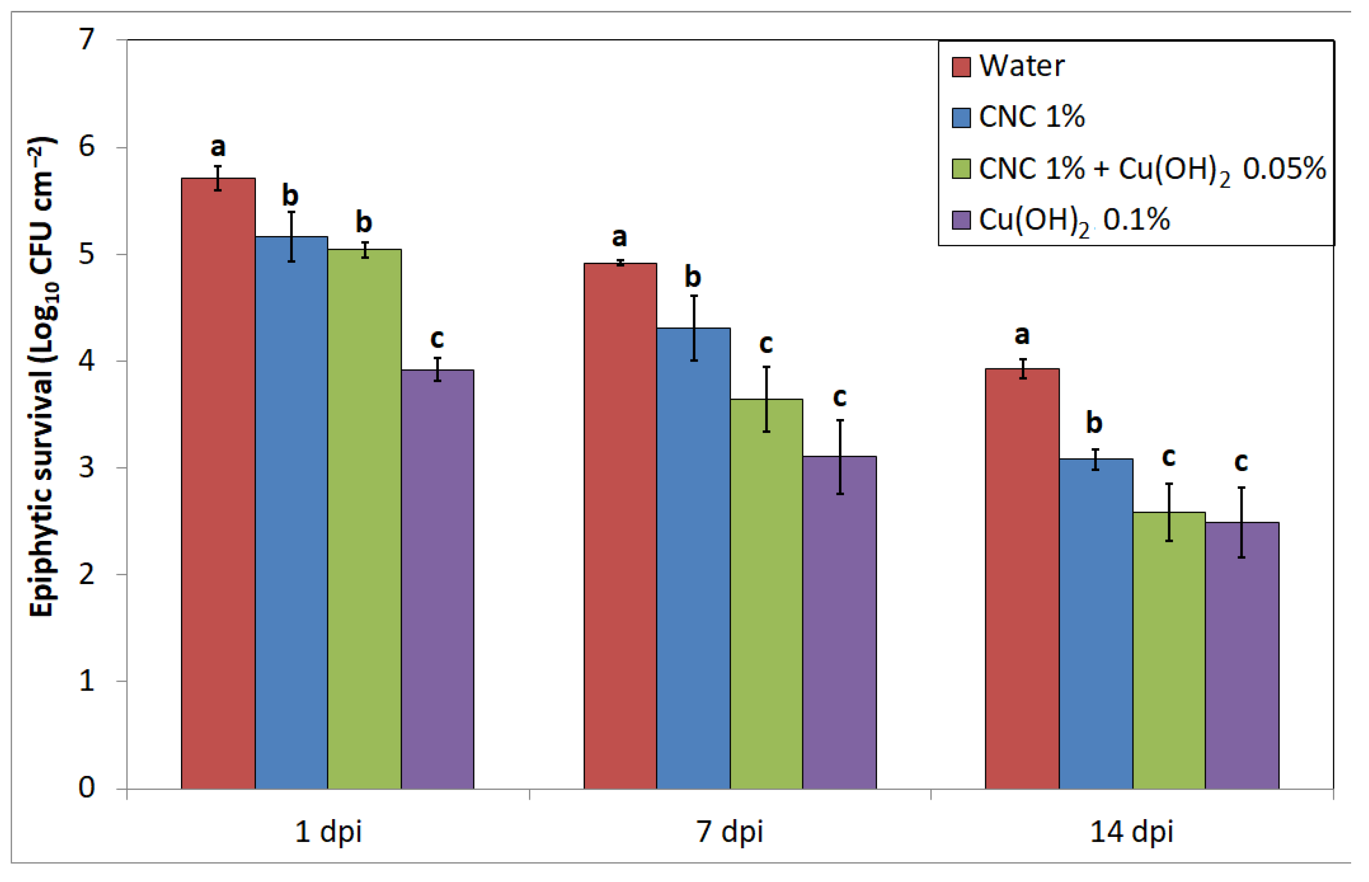
Regarding Pst epiphytic survival, appreciable differences were noted among the treatments in terms of recovered bacterial population from leaves (Figure 2). At 1 dpi, water-treated plant presented the highest number of recovered colonies (5.71 CFU cm−2), while Cu(OH)2 at the field dose showed an expected knockdown effect (3.91 CFU cm−2). CNC alone and mixed with half a field dose of copper hydroxide treatments showed a similar result (5.15 and 5.04 CFU cm−2 respectively), but they were still different from the water control one. At 7 dpi, an overall decrease in the epiphytic populations was observed in each treatment; a statistical difference could still be observed among water control (4.92 CFU cm−2) and CNC alone (4.30 CFU cm−2), while copper hydroxide and CNC with 0.05% Cu(OH)2 had the best effect in terms of bacterial inhibition (3.10 and 3.64 CFU cm−2 respectively). After one week (14 dpi), the previous trend was still evident among the thesis: water control had the highest value in terms of CFU cm−2 (3.94) and CNC at 1% w/v showed a lower significant value (3.08), while CNC mixed with 0.05% Cu(OH)2 and copper hydroxide at the field dose displayed the lowest values (2.58 and 2.49 respectively).
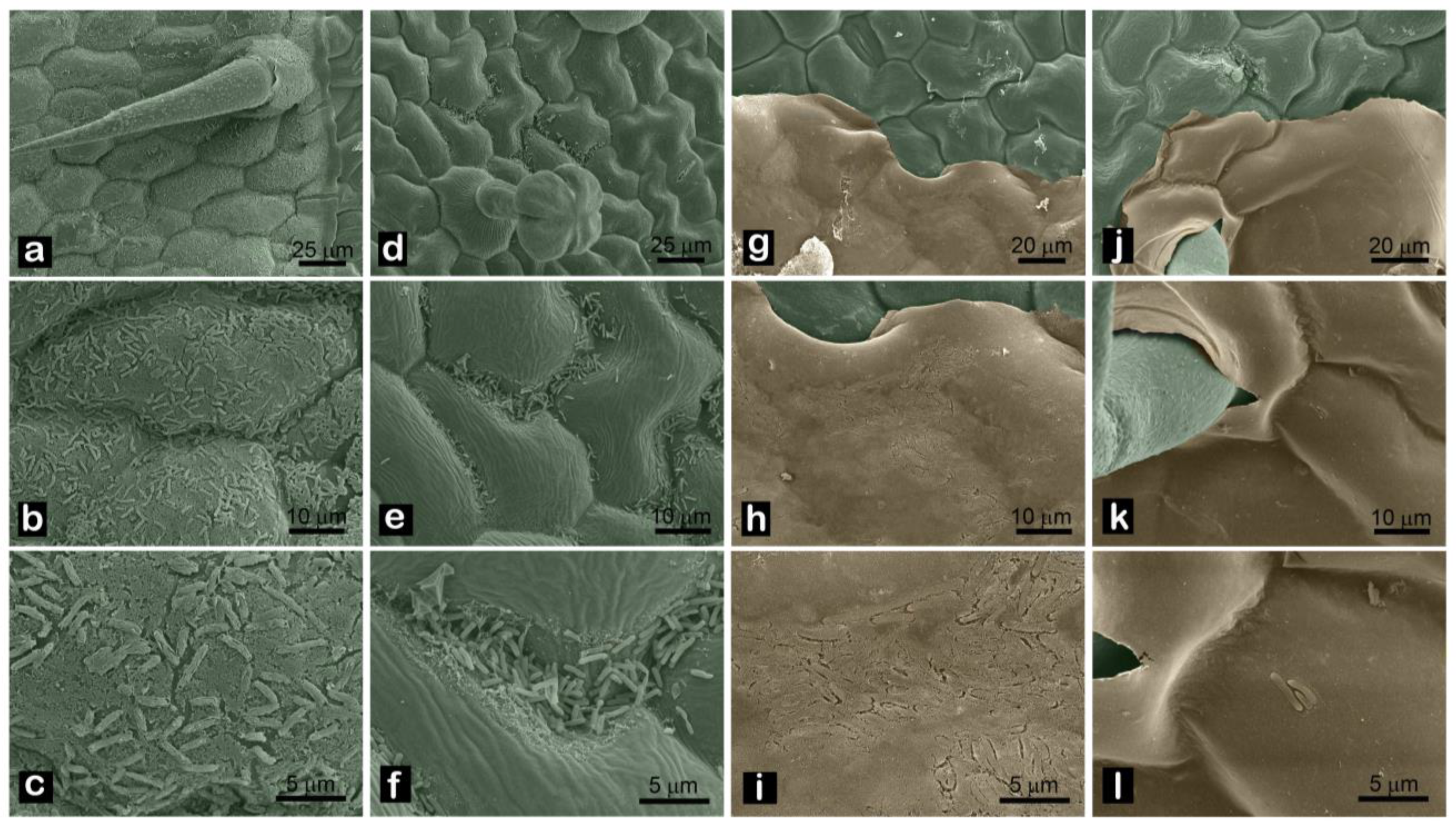
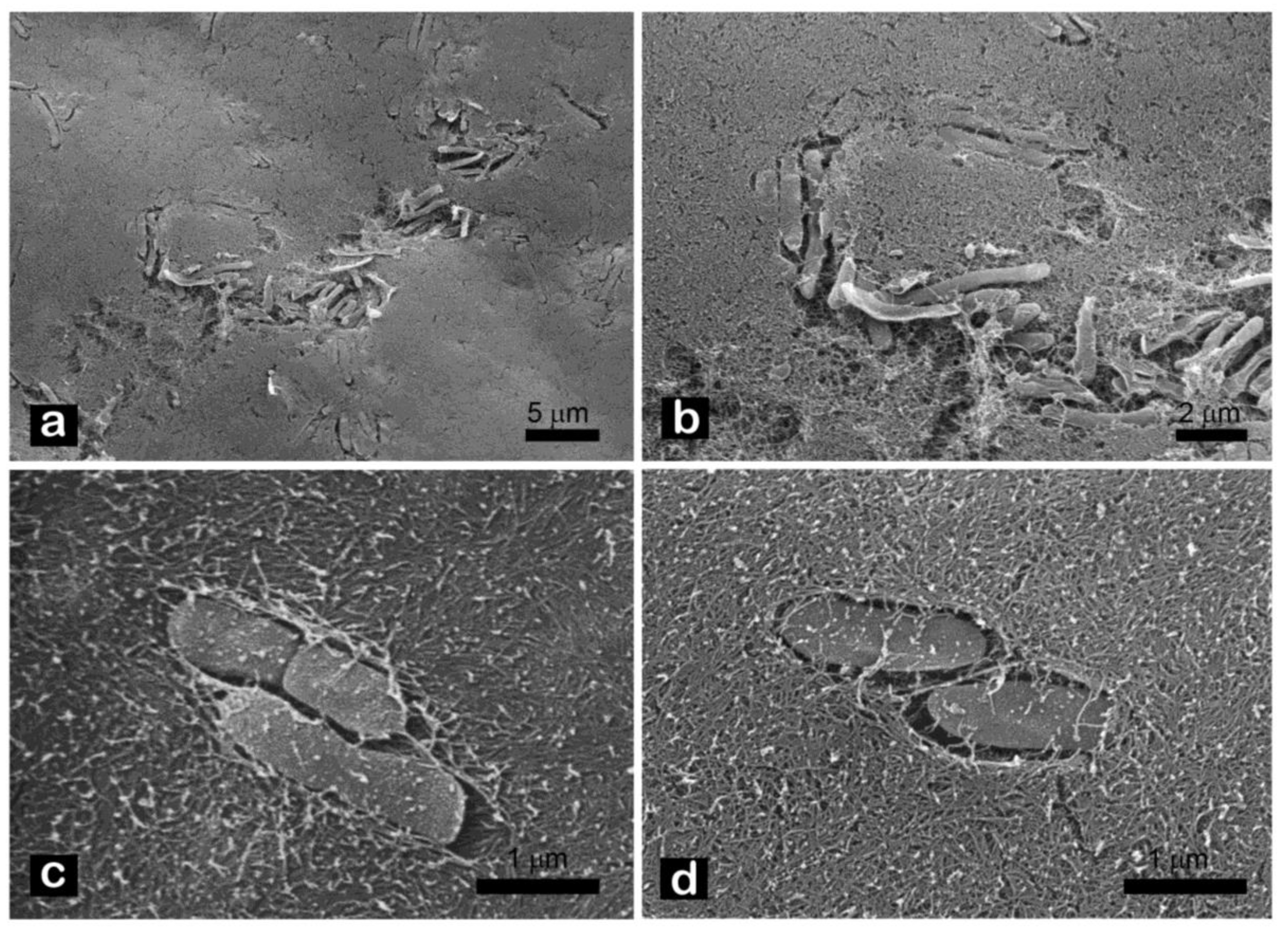
SEM pictures of leaves taken at 1 dpi confirmed the data obtained by the epiphytic survival analysis. In water-treated plants, bacterial cells were well distributed all over the leaf surface and trichomes (Figure 3a–c); on leaves treated with the field dose of copper hydroxide, bacterial cells were scarcely spotted, since their presence was rarefied and mostly limited to the edges of the areoles (Figure 3d–f); on plants treated with CNC alone (Figure 3g–i) or mixed with half a field dose of copper (Figure 3j–l), the evident sign of the nanocrystal layer with bacterial cells embedded in it could be appreciated. More detailed pictures revealed that the Pst cells could be found above and within the CNC matrix spontaneously formed on the leaves (Figure 4).

4. Discussion

CNCs displayed no negative or side effects on the development of the tomato plants. Compatibility towards plants is indeed one of the main concerns when nanomaterials are proposed as innovative fertilizers or pesticides [50,51]. Since nanomaterials present different biological and chemical properties in comparison with their bulk counterparts, it is always recommended to study any possible effects on the metabolism of plants, including the uptake, translocation and accumulation of the nanoparticles, as well as photosynthesis and fertility [52,53]. The obtained results were expected, considering previous research that has already highlighted the lack of toxicity of cellulose and CNCs [54,55]. Although few works have underlined the biocompatibility of CNC towards plants at this time, as far we know, this is the first report of a more complete investigation into CNC application, taking into account seed germination, leaf metabolism and total plant weight [56,57,58]. Soaking seeds in CNC suspension does not affect their ability to fully germinate over time, suggesting that CNCs could be used to treat seeds and to develop coating compounds. Looking at the nitrogen metabolism in treated leaves and the total plant weight, no difference could be seen with the water control over time. NBI is indeed a robust indicator for determining basal metabolism in photosynthetic tissues; in healthy conditions, the plant will use most of the endogenous nitrogen in the synthesis of primarily needed proteins, such as chlorophylls, while in non-optimal conditions, part of the nitrogen will be intended for the secondary pathways, such as the flavonol ones associated with the plant’s response to stresses [59]. Since no sign of phytotoxicity in the chlorophylls’ metabolism was detected, we can conclude that the measured leaf surface and the root and shoot final weight were not affected either, leading to a statistical similarity among the treatments.
Considering the effects of the CNC treatment on Pst epiphytic survival, the obtained results confirmed the previous in vitro findings. While CNCs do not lead to bacteria death, their presence prevents cell swimming and adhesion to surfaces, which are key factors in allowing Pst to colonize the external host tissues [60]. Epiphytic residency represents a fundamental ecological trait in the Pst disease cycle, since the bacteria are able to propagate over winter and maintain a high-density inoculum source by living outside the plants [61,62]. This evidence could be explained by looking at the main interactions between bacteria and CNCs, since they can interfere with the cell motility and they have the ability to stick. CNC’s capability to reduce adhesion was also confirmed on E. coli, while cellulose nanofibers were confirmed to reduce swimming in several bacteria, such as L. monocytogenes, B. cereus and P. cannabina pv. alisalensis, when used at concentrations similar to the ones used in this work [63,64,65,66]. Indeed, the registered results are coherent with other works showing the ability of CNC to reduce bacterial epiphytic survival in treated leaves [56]. Copper hydroxide at the field dose showed the highest reduction on the epiphytic populations, beginning from the first day after the treatment. This could be easily explained by considering the well-known ability of cupric ions to inhibit and kill bacterial cells, as previously shown in vitro [29]. Moreover, we evaluated the possibility of halving the field dose of copper hydroxide by applying it in combination with CNCs. While the effect was similar to the one shown by CNCs at the very beginning, the co-presence of the two compounds was able to lower the Pst epiphytic survival in a way that was comparable to copper hydroxide alone. This effect could be explained by considering the role of CNC in limiting bacterial motility and biofilm formation on the leaf surface, making the cells more inclined to come in contact with copper instead of stepping away or shielding from it. These findings could give future perspectives regarding the possibility of using organic nanomaterials as carriers for cupric compounds, increasing their effectiveness and lowering the amount of total distributed copper [67,68]. Furthermore, the copper-based nanoformulations, as already shown elsewhere, could even target copper-resistant strains [69,70]. The SEM observations allowed us to better understand the physical interaction between CNCs and Pst. Pictures revealed that CNCs at 1% w/v were able to surround the bacteria, entrapping them inside or above the cellulose film and preventing them from moving or getting closer to the plant tissues, making their life on the phylloplane harder. Changes in the topography, surface roughness and wettability can indeed drastically affect the ability of bacteria to colonize the phyllosphere [71,72]. Since for many Pseudomonas spp. the ability to cause disease is directly correlated with the external population level, and controlling the epiphytic survival represents an effective way to prevent plants from getting infected [73,74,75].
The obtained results address further investigations about the effects of CNCs in limiting symptoms displayed and disease development in treated tomato plants. Further research is planned to study in a more detailed way the molecular interaction among plants, CNCs and bacteria, looking at some target genes’ expression involved in plant defense pathways [76].

5. Conclusions

In this work, the effects of CNCs on the model patho-system tomato-Pst was successfully evaluated. CNCs were revealed to be a non-toxic, biocompatible compound that does not affect plant basal functions such as seed germination, leaf development, nitrogen metabolism, and biomass accumulation. Moreover, CNCs exhibited the ability to lower the epiphytic population of Pst, the causal agent of bacterial speck disease in tomatoes. Furthermore, CNCs can be applied with a limited amount of copper, increasing their efficacy in controlling bacteria to the level displayed by the copper hydroxide used in the field. This research highlights the potential of using organic nanomaterials to develop innovative agrochemicals and sustainable crop protection strategies.

Author Contributions

Conceptualization, D.S. and G.M.B.; methodology, D.S., A.R.T. and G.M.B.; validation, G.M.B.; investigation, D.S. and A.R.T.; data curation, D.S.; writing—original draft preparation, D.S.; writing—review and editing, A.R.T. and G.M.B.; supervision, G.M.B. All authors have read and agreed to the published version of the manuscript.

Funding

This research was funded by MUR (Italian Ministry for University and Research) (Law 232/2016, Departments of Excellence) and PON Ricerca e Innovazione 2014–2020: Nanotecnologie chimiche green per la protezione sostenibile delle piante (NEMESI) ARS01_01002CUP: F36C1800018000X.

Data Availability Statement

Not applicable.

Acknowledgments

The authors gratefully acknowledge Linda Felici (@LF_sci.ab) for the great interpretation of the research pictured in the graphical abstract.

Conflicts of Interest

The authors declare no conflict of interest. The funders had no role in the design of the study; in the collection, analyses, or interpretation of data; in the writing of the manuscript; or in the decision to publish the results.

References

  1. La Torre, A.; Iovino, V.; Caradonia, F. Copper in plant protection: Current situation and prospects. Phytopathol. Mediterr. 2018, 57, 201–236. [Google Scholar] [CrossRef]
  2. Lamichhane, J.R.; Osdaghi, E.; Behlau, F.; Köhl, J.; Jones, J.B.; Aubertot, J.N. Thirteen decades of antimicrobial copper compounds applied in agriculture. A review. Agron. Sustain. Dev. 2018, 38, 28. [Google Scholar] [CrossRef]
  3. Abrahamian, P.; Jones, J.B.; Vallad, G.E. Efficacy of copper and copper alternatives for management of bacterial spot on tomato under transplant and field production. Crop. Prot. 2019, 126, 104919. [Google Scholar] [CrossRef]
  4. Keller, A.A.; Adeleye, A.S.; Conway, J.R.; Garner, K.L.; Zhao, L.; Cherr, G.N.; Hong, J.; Gardea-Torresdey, J.L.; Godwin, H.A.; Hanna, S. Comparative environmental fate and toxicity of copper nanomaterials. NanoImpact 2017, 7, 28–40. [Google Scholar] [CrossRef]
  5. Flemming, C.A.; Trevors, J.T. Copper toxicity and chemistry in the environment: A review. Water. Air. Soil Pollut. 1989, 44, 143–158. [Google Scholar] [CrossRef]
  6. Rippa, M.; Battaglia, V.; Cermola, M.; Sicignano, M.; Lahoz, E.; Mormile, P. Monitoring of the copper persistence on plant leaves using pulsed thermography. Environ. Monit. Assess. 2022, 194, 160. [Google Scholar] [CrossRef] [PubMed]
  7. Timilsina, S.; Liao, Y.Y.; Young, M.; Rosskopf, E.N.; Vallad, G.E.; Goss, E.M.; Santra, S.; Jones, J.B.; Hong, J.C.; Paret, M.L. Simulated Leaching of Foliar Applied Copper Bactericides on the Soil Microbiome Utilizing Various Beta Diversity Resemblance Measurements. Microbiol. Spectr. 2022, 10, e0148121. [Google Scholar] [CrossRef]
  8. Cooksey, D.A.; Azad, H.R.; Cha, J.S.; Lim, C.K. Copper resistance gene homologs in pathogenic and saprophytic bacterial species from tomato. Appl. Environ. Microbiol. 1990, 56, 431–435. [Google Scholar] [CrossRef]
  9. Griffin, K.; Gambley, C.; Brown, P.; Li, Y. Copper-tolerance in Pseudomonas syringae pv. tomato and Xanthomonas spp. and the control of diseases associated with these pathogens in tomato and pepper. A systematic literature review. Crop. Prot. 2017, 96, 144–150. [Google Scholar] [CrossRef]
  10. Bender, C.L.; Cooksey, D.A. Indigenous plasmids in Pseudomonas syringae pv. tomato: Conjugative transfer and role in copper resistance. J. Bacteriol. 1986, 165, 534–541. [Google Scholar] [CrossRef]
  11. Tamm, L.; Thuerig, B.; Apostolov, S.; Blogg, H.; Borgo, E.; Corneo, P.E.; Fittje, S.; De Palma, M.; Donko, A.; Experton, C.; et al. Use of Copper-Based Fungicides in Organic Agriculture in Twelve European Countries. Agronomy 2022, 12, 673. [Google Scholar] [CrossRef]
  12. Hofmann, T.; Lowry, G.V.; Ghoshal, S.; Tufenkji, N.; Brambilla, D.; Dutcher, J.R.; Gilbertson, L.M.; Giraldo, J.P.; Kinsella, J.M.; Landry, M.P.; et al. Technology readiness and overcoming barriers to sustainably implement nanotechnology-enabled plant agriculture. Nat. Food 2020, 1, 416–425. [Google Scholar] [CrossRef]
  13. Usman, M.; Farooq, M.; Wakeel, A.; Nawaz, A.; Cheema, S.A.; Rehman, H.; Ashraf, I.; Sanaullah, M. Nanotechnology in agriculture: Current status, challenges and future opportunities. Sci. Total Environ. 2020, 721, 137778. [Google Scholar] [CrossRef] [PubMed]
  14. Balestra, G.M.; Fortunati, E. Nanotechnology-Based Sustainable Alternatives for the Managements of Plant Diseases; Balestra, G.M., Fortunati, E., Eds.; Elsevier: Amsterdam, The Netherlands, 2022; ISBN 978-0-12-823394-8. [Google Scholar]
  15. Fiol, D.F.; Terrile, M.C.; Frik, J.; Mesas, F.A.; Álvarez, V.A.; Casalongué, C.A. Nanotechnology in plants: Recent advances and challenges. J. Chem. Technol. Biotechnol. 2021, 96, 2095–2108. [Google Scholar] [CrossRef]
  16. Prasad, R.; Bhattacharyya, A.; Nguyen, Q.D. Nanotechnology in sustainable agriculture: Recent developments, challenges, and perspectives. Front. Microbiol. 2017, 8, 1014. [Google Scholar] [CrossRef] [PubMed]
  17. Lin, N.; Dufresne, A. Nanocellulose in biomedicine: Current status and future prospect. Eur. Polym. J. 2014, 59, 302–325. [Google Scholar] [CrossRef]
  18. Poulose, A.; Parameswaranpillai, J.; George, J.J.; Gopi, J.A.; Krishnasamy, S.; Dominic, C.D.M.; Hameed, N.; Salim, N.V.; Radoor, S.; Sienkiewicz, N. Nanocellulose: A Fundamental Material for Science and Technology Applications. Molecules 2022, 27, 8032. [Google Scholar] [CrossRef]
  19. Barhoum, A.; Rastogi, V.K.; Mahur, B.K.; Rastogi, A.; Abdel-Haleem, F.M.; Samyn, P. Nanocelluloses as new generation materials: Natural resources, structure-related properties, engineering nanostructures, and technical challenges. Mater. Today Chem. 2022, 26, 101247. [Google Scholar] [CrossRef]
  20. George, J.; Sabapathi, S.N. Cellulose nanocrystals: Synthesis, functional properties, and applications. Nanotechnol. Sci. Appl. 2015, 8, 45–54. [Google Scholar] [CrossRef]
  21. Habibi, Y.; Lucia, L.A.; Rojas, O.J. Cellulose nanocrystals: Chemistry, self-assembly, and applications. Chem. Rev. 2010, 110, 3479–3500. [Google Scholar] [CrossRef]
  22. Fortunati, E.; Luzi, F.; Puglia, D.; Dominici, F.; Santulli, C.; Kenny, J.M.; Torre, L. Investigation of thermo-mechanical, chemical and degradative properties of PLA-limonene films reinforced with cellulose nanocrystals extracted from Phormium tenax leaves. Eur. Polym. J. 2014, 56, 77–91. [Google Scholar] [CrossRef]
  23. Schiavi, D.; Di Lorenzo, V.; Francesconi, S.; Giovagnoli, S.; Camaioni, E.; Balestra, G.M. Waste valorization by nanotechnology approaches for sustainable crop protection: A mini review. IOP Conf. Ser. Mater. Sci. Eng. 2022, 1265, 012009. [Google Scholar] [CrossRef]
  24. De Lima, P.H.C.; Antunes, D.R.; de Lima Forini, M.M.; da Silva Pontes, M.; Mattos, B.D.; Grillo, R. Recent Advances on Lignocellulosic-Based Nanopesticides for Agricultural Applications. Front. Nanotechnol. 2021, 3, 809329. [Google Scholar] [CrossRef]
  25. Fortunati, E.; Luzi, F.; Jiménez, A.; Gopakumar, D.A.; Puglia, D.; Thomas, S.; Kenny, J.M.; Chiralt, A.; Torre, L. Revalorization of sunflower stalks as novel sources of cellulose nanofibrils and nanocrystals and their effect on wheat gluten bionanocomposite properties. Carbohydr. Polym. 2016, 149, 357–368. [Google Scholar] [CrossRef]
  26. Hemmati, F.; Jafari, S.M.; Kashaninejad, M.; Barani Motlagh, M. Synthesis and characterization of cellulose nanocrystals derived from walnut shell agricultural residues. Int. J. Biol. Macromol. 2018, 120, 1216–1224. [Google Scholar] [CrossRef]
  27. García, A.; Gandini, A.; Labidi, J.; Belgacem, N.; Bras, J. Industrial and crop wastes: A new source for nanocellulose biorefinery. Ind. Crops Prod. 2016, 93, 26–38. [Google Scholar] [CrossRef]
  28. Mateo, S.; Peinado, S.; Morillas-Gutiérrez, F.; La Rubia, M.D.; Moya, A.J. Nanocellulose from agricultural wastes: Products and applications—A review. Processes 2021, 9, 1594. [Google Scholar] [CrossRef]
  29. Schiavi, D.; Ronchetti, R.; Di Lorenzo, V.; Vivani, R.; Giovagnoli, S.; Camaioni, E.; Balestra, G.M. Sustainable Protocols for Cellulose Nanocrystals Synthesis from Tomato Waste and Their Antimicrobial Properties against Pseudomonas syringae pv. tomato. Plants 2023, 12, 939. [Google Scholar] [CrossRef] [PubMed]
  30. Bergougnoux, V. The history of tomato: From domestication to biopharming. Biotechnol. Adv. 2014, 32, 170–189. [Google Scholar] [CrossRef]
  31. Xin, X.F.; He, S.Y. Pseudomonas syringae pv. tomato DC3000: A model pathogen for probing disease susceptibility and hormone signaling in plants. Annu. Rev. Phytopathol. 2013, 51, 473–498. [Google Scholar] [CrossRef]
  32. Preston, G.M. Pseudomonas syringae pv. tomato: The right pathogen, of the right plant, at the right time. Mol. Plant Pathol. 2000, 1, 263–275. [Google Scholar] [CrossRef] [PubMed]
  33. Cazorla, F.M.; Arrebola, E.; Sesma, A.; Pérez-García, A.; Codina, J.C.; Murillo, J.; De Vicente, A. Copper resistance in Pseudomonas syringae strains isolated from mango is encoded mainly by plasmids. Phytopathology 2002, 92, 909–916. [Google Scholar] [CrossRef]
  34. Shenge, K.C.; Wydra, K.; Mabagala, R.B.; Mortensen, C.N. Assessment of strains of Pseudomonas syringae pv. tomato from Tanzania for resistance to copper and streptomycin. Arch. Phytopathol. Plant Prot. 2008, 41, 572–585. [Google Scholar] [CrossRef]
  35. McLeod, A.; Masimba, T.; Jensen, T.; Serfontein, K.; Coertze, S. Evaluating spray programs for managing copper resistant Pseudomonas syringae pv. tomato populations on tomato in the Limpopo region of South Africa. Crop Prot. 2017, 102, 32–42. [Google Scholar] [CrossRef]
  36. Moretti, C.; Bocchini, M.; Quaglia, M.; Businelli, D.; Orfei, B.; Buonaurio, R. Sodium Selenate: An Environmental-Friendly Means to Control Tomato Bacterial Speck Disease. Agronomy 2022, 12, 1351. [Google Scholar] [CrossRef]
  37. Mansilla, A.Y.; Albertengo, L.; Rodríguez, M.S.; Debbaudt, A.; Zúñiga, A.; Casalongué, C.A. Evidence on antimicrobial properties and mode of action of a chitosan obtained from crustacean exoskeletons on Pseudomonas syringae pv. tomato DC3000. Appl. Microbiol. Biotechnol. 2013, 97, 6957–6966. [Google Scholar] [CrossRef]
  38. Quattrucci, A.; Ovidi, E.; Tiezzi, A.; Vinciguerra, V.; Balestra, G.M. Biological control of tomato bacterial speck using Punica granatum fruit peel extract. Crop Prot. 2013, 46, 18–22. [Google Scholar] [CrossRef]
  39. Fanelli, V.; Cariddi, C.; Finetti-Sialer, M. Selective detection of Pseudomonas syringae pv. tomato using dot blot hybridization and real-time PCR. Plant Pathol. 2007, 56, 683–691. [Google Scholar] [CrossRef]
  40. Cuppels, D.A.; Louws, F.J.; Ainsworth, T. Development and evaluation of PCR-based diagnostic assays for the bacterial speck and bacterial spot pathogens of tomato. Plant Dis. 2006, 90, 451–458. [Google Scholar] [CrossRef]
  41. Schiavi, D.; Balbi, R.; Giovagnoli, S.; Camaioni, E.; Botticella, E.; Sestili, F.; Balestra, G.M. A green nanostructured pesticide to control tomato bacterial speck disease. Nanomaterials 2021, 11, 1852. [Google Scholar] [CrossRef]
  42. Li, Y.; Yang, D.; Cui, J. Graphene oxide loaded with copper oxide nanoparticles as an antibacterial agent against: Pseudomonas syringae pv. tomato. RSC Adv. 2017, 7, 38853–38860. [Google Scholar] [CrossRef]
  43. Elsharkawy, M.; Derbalah, A.; Hamza, A.; El-Shaer, A. Zinc oxide nanostructures as a control strategy of bacterial speck of tomato caused by Pseudomonas syringae in Egypt. Environ. Sci. Pollut. Res. 2020, 27, 19049–19057. [Google Scholar] [CrossRef] [PubMed]
  44. King, E.O.; Ward, M.K.; Raney, D.E. Two simple media for the demonstration of pyocyanin and fluorescin. J. Lab. Clin. Med. 1954, 44, 301–307. [Google Scholar] [CrossRef]
  45. Orzali, L.; Valente, M.T.; Scala, V.; Loreti, S. Antibacterial Activity of Essential Oils and Trametes versicolor Extract against Clavibacter michiganensis subsp. michiganensis and Ralstonia solanacearum for Seed Treatment and Development of a Rapid In Vivo Assay. Antibiotics 2020, 9, 628. [Google Scholar] [CrossRef] [PubMed]
  46. Easlon, H.M.; Bloom, A.J. Easy Leaf Area: Automated Digital Image Analysis for Rapid and Accurate Measurement of Leaf Area. Appl. Plant Sci. 2014, 2, 1400033. [Google Scholar] [CrossRef]
  47. Cerovic, Z.G.; Masdoumier, G.; Ghozlen, N.B.; Latouche, G. A new optical leaf-clip meter for simultaneous non-destructive assessment of leaf chlorophyll and epidermal flavonoids. Physiol. Plant. 2012, 146, 251–260. [Google Scholar] [CrossRef]
  48. Ben Abdallah, F.; Philippe, W.; Goffart, J.P. Comparison of optical indicators for potato crop nitrogen status assessment including novel approaches based on leaf fluorescence and flavonoid content. J. Plant Nutr. 2018, 41, 2705–2728. [Google Scholar] [CrossRef]
  49. Canzoniere, P.; Francesconi, S.; Giovando, S.; Balestra, G.M. Antibacterial activity of tannins towards Pseudomonas syringae pv. tomato, and their potential as biostimulants on tomato plants. Phytopathol. Mediterr. 2021, 60, 23–36. [Google Scholar] [CrossRef]
  50. Reddy, P.V.L.; Hernandez-Viezcas, J.A.; Peralta-Videa, J.R.; Gardea-Torresdey, J.L. Lessons learned: Are engineered nanomaterials toxic to terrestrial plants? Sci. Total Environ. 2016, 568, 470–479. [Google Scholar] [CrossRef] [PubMed]
  51. Nanotechnology and Plant Sciences: Nanoparticles and Their Impact on Plants; Siddiqui, M.H.; Mohammad, F.; Al-Whaibi, M.H. (Eds.) Springer: Berlin/Heidelberg, Germany, 2015; ISBN 97833-19145020. [Google Scholar]
  52. Ruttkay-Nedecky, B.; Krystofova, O.; Nejdl, L.; Adam, V. Nanoparticles based on essential metals and their phytotoxicity. J. Nanobiotechnol. 2017, 15, 1–19. [Google Scholar] [CrossRef] [PubMed]
  53. Du, W.; Tan, W.; Peralta-Videa, J.R.; Gardea-Torresdey, J.L.; Ji, R.; Yin, Y.; Guo, H. Interaction of metal oxide nanoparticles with higher terrestrial plants: Physiological and biochemical aspects. Plant Physiol. Biochem. 2017, 110, 210–225. [Google Scholar] [CrossRef] [PubMed]
  54. Roman, M. Toxicity of cellulose nanocrystals: A review. Ind. Biotechnol. 2015, 11, 25–33. [Google Scholar] [CrossRef]
  55. Coelho, C.C.S.; Michelin, M.; Cerqueira, M.A.; Gonçalves, C.; Tonon, R.V.; Pastrana, L.M.; Freitas-Silva, O.; Vicente, A.A.; Cabral, L.M.C.; Teixeira, J.A. Cellulose nanocrystals from grape pomace: Production, properties and cytotoxicity assessment. Carbohydr. Polym. 2018, 192, 327–336. [Google Scholar] [CrossRef] [PubMed]
  56. Schiavi, D.; Francesconi, S.; Taddei, A.R.; Fortunati, E.; Balestra, G.M. Exploring cellulose nanocrystals obtained from olive tree wastes as sustainable crop protection tool against bacterial diseases. Sci. Rep. 2022, 12, 6149. [Google Scholar] [CrossRef] [PubMed]
  57. Schiavi, D.; Ronchetti, R.; Di Lorenzo, V.; Salustri, M.; Petrucci, C.; Vivani, R.; Giovagnoli, S.; Camaioni, E.; Balestra, G.M. Circular Hazelnut Protection by Lignocellulosic Waste Valorization for Nanopesticides Development. Appl. Sci. 2022, 12, 2604. [Google Scholar] [CrossRef]
  58. Fortunati, E.; Rescignano, N.; Botticella, E.; La Fiandra, D.; Renzi, M.; Mazzaglia, A.; Torre, L.; Kenny, J.M.; Balestra, G.M. Effect of poly(DL-lactide-co-glycolide) nanoparticles or cellulose nanocrystals-based formulations on pseudomonas syringae pv. tomato (Pst) and tomato plant development. J. Plant Dis. Prot. 2016, 123, 301–310. [Google Scholar] [CrossRef]
  59. Bracke, J.; Elsen, A.; Adriaenssens, S.; Schoeters, L.; Vandendriessche, H.; Van Labeke, M.C. Application of proximal optical sensors to fine-tune nitrogen fertilization: Opportunities for woody ornamentals. Agronomy 2019, 9, 408. [Google Scholar] [CrossRef]
  60. Guttenplan, S.B.; Kearns, D.B. Regulation of flagellar motility during biofilm formation. FEMS Microbiol. Rev. 2013, 37, 849–871. [Google Scholar] [CrossRef]
  61. Babelegoto, N.; Varvaro, L.; Cirulli, M. Epiphytic, and endophytic multiplication of Pseudomonas syringae pv. tomato (Okabel) Young et al. in susceptible and resistant tomato leaves. Phytopathol. Mediterr. 1988, 27, 138–144. [Google Scholar]
  62. Balestra, G.M.; Antonelli, M.; Fabi, A.; Varvaro, L. Effectiveness of natural products for in vitro and in vivo control of epiphytic populations of Pseudomonas syringae pv. tomato on tomato plants. J. Plant Pathol. 1999, 80, 251. [Google Scholar]
  63. D’Orazio, G.; Munizza, L.; Zampolli, J.; Forcella, M.; Zoia, L.; Fusi, P.; Di Gennaro, P.; La Ferla, B. Cellulose nanocrystals are effective in inhibiting host cell bacterial adhesion. J. Mater. Chem. B 2017, 5, 7018–7020. [Google Scholar] [CrossRef]
  64. Noronha, V.T.; Camargos, C.H.M.; Jackson, J.C.; Souza Filho, A.G.; Paula, A.J.; Rezende, C.A.; Faria, A.F. Physical membrane-stress-mediated antimicrobial properties of cellulose nanocrystals. ACS Sustain. Chem. Eng. 2021, 9, 3203–3212. [Google Scholar] [CrossRef]
  65. Sakata, N.; Shiraishi, N.; Saito, H.; Komoto, H.; Ishiga, T.; Usuki, G.; Yamashita, Y.; Ishiga, Y. Covering cabbage leaves with cellulose nanofiber confers resistance against Pseudomonas cannabina pv. alisalensis. J. Gen. Plant Pathol. 2023, 89, 53–60. [Google Scholar] [CrossRef]
  66. Silva, F.; Gracia, N.; McDonagh, B.H.; Domingues, F.C.; Nerín, C.; Chinga-Carrasco, G. Antimicrobial activity of biocomposite films containing cellulose nanofibrils and ethyl lauroyl arginate. J. Mater. Sci. 2019, 54, 12159–12170. [Google Scholar] [CrossRef]
  67. Sathiyabama, M.; Manikandan, A. Application of Copper-Chitosan Nanoparticles Stimulate Growth and Induce Resistance in Finger Millet (Eleusine coracana Gaertn.) Plants against Blast Disease. J. Agric. Food Chem. 2018, 66, 1784–1790. [Google Scholar] [CrossRef] [PubMed]
  68. Jaiswal, M.; Chauhan, D.; Sankararamakrishnan, N. Copper chitosan nanocomposite: Synthesis, characterization, and application in removal of organophosphorous pesticide from agricultural runoff. Environ. Sci. Pollut. Res. 2012, 19, 2055–2062. [Google Scholar] [CrossRef]
  69. Liao, Y.Y.; Strayer-Scherer, A.L.; White, J.; Mukherjee, A.; De La Torre-Roche, R.; Ritchie, L.; Colee, J.; Vallad, G.E.; Freeman, J.H.; Jones, J.B.; et al. Nano-magnesium oxide: A novel bactericide against copper-tolerant xanthomonas perforans causing tomato bacterial spot. Phytopathology 2019, 109, 52–62. [Google Scholar] [CrossRef]
  70. Carvalho, R.; Duman, K.; Jones, J.B.; Paret, M.L. Bactericidal Activity of Copper-Zinc Hybrid Nanoparticles on Copper-Tolerant Xanthomonas perforans. Sci. Rep. 2019, 9, 20124. [Google Scholar] [CrossRef]
  71. Doan, H.K.; Ngassam, V.N.; Gilmore, S.F.; Tecon, R.; Parikh, A.N.; Leveau, J.H.J. Topography-Driven Shape, Spread, and Retention of Leaf Surface Water Impacts Microbial Dispersion and Activity in the Phyllosphere. Phytobiomes 2020, 4, 268–280. [Google Scholar] [CrossRef]
  72. Lindow, S.E.; Brandl, M.T. Microbiology of the Phyllosphere. Appl. Environ. Microbiol. 2003, 69, 1875–1883. [Google Scholar] [CrossRef]
  73. Xin, X.F.; Kvitko, B.; He, S.Y. Pseudomonas syringae: What it takes to be a pathogen. Nat. Rev. Microbiol. 2018, 16, 316–328. [Google Scholar] [CrossRef] [PubMed]
  74. Rouse, D.I.; Nordheim, E.V.; Hirano, S.S.; Upper, C.D. A Model Relating the Probability of Foliar Disease Incidence to the Population Frequencies of Bacterial Plant Pathogens. Phytopathology 1985, 75, 505. [Google Scholar] [CrossRef]
  75. Lamichhane, J.R.; Varvaro, L. Epiphytic Pseudomonas savastanoi pv. savastanoi can infect and cause olive knot disease on Olea europaea subsp. cuspidata. Australas. Plant Pathol. 2013, 42, 219–225. [Google Scholar] [CrossRef]
  76. De Azevedo Souza, C.; Li, S.; Lin, A.Z.; Boutrot, F.; Grossmann, G.; Zipfel, C.; Somerville, S.C. Cellulose-derived oligomers act as damage-associated molecular patterns and trigger defense-like responses. Plant Physiol. 2017, 173, 2383–2398. [Google Scholar] [CrossRef] [PubMed]
Figure 1. Compatibility of CNC on (a) seed germination; (b) leaflet area; (c) leaf content; (d) dry weight. Data are represented as the mean ± standard deviation; different letters stand for significant different values if p < 0.05 after ANOVA and Tukey’s HSD were performed.
Horticulturae 09 00525 g001
Figure 2. Epiphytic survival of Pst on tomato plants treated with water, 1% CNC, 1% CNC + 0.05% Cu(OH)2, 0.1% Cu(OH)2, at 1, 7 and 14 dpi. Different letters stand for significant different values if p < 0.05 after ANOVA and Tukey’s HSD were performed.
Horticulturae 09 00525 g002
Figure 3. SEM images at different magnifications of tomato leaves at 1 dpi with Pst. Leaves were treated with water (ac); 0.1% copper hydroxide (df); 1% CNC (gi); 1% CNC; and 0.05% copper hydroxide (jl). Leaves and bacteria are in pale green; CNCs are in pale red (false colorized).
Horticulturae 09 00525 g003
Figure 4. SEM images at different magnifications ((a): 5 µm; (b): 2 µm) of tomato leaves treated with 1% CNC at 1 dpi. Particulars of bacterial cells embedded within the CNC ((c,d): 1 µm).
Horticulturae 09 00525 g004
Disclaimer/Publisher’s Note: The statements, opinions and data contained in all publications are solely those of the individual author(s) and contributor(s) and not of MDPI and/or the editor(s). MDPI and/or the editor(s) disclaim responsibility for any injury to people or property resulting from any ideas, methods, instructions or products referred to in the content.