Article

Possible Contribution of Corticular Photosynthesis to Grapevine Winter Hardiness

by
Maria A. Sundyreva
1,
Denis V. Yanykin
2,
Mikhail S. Khristin
2,
Ulyana V. Gryaznova
2,
Evgenij O. Lutskiy
1,
Galina A. Semenova
3,
Ilya A. Naydov
2 and
Tatyana V. Savchenko
2,*
1
North Caucasian Federal Scientific Center of Horticulture, Viticulture, Winemaking, Krasnodar 350072, Russia
2
Institute of Basic Biological Problems, Pushchino Scientific Center for Biological Research, Russian Academy of Sciences, Pushchino 142290, Russia
3
Institute of Theoretical and Experimental Biophysics, Russian Academy of Sciences, Pushchino 142290, Russia
*
Author to whom correspondence should be addressed.
Horticulturae 2023, 9(11), 1181; https://doi.org/10.3390/horticulturae9111181
Submission received: 3 October 2023 / Revised: 21 October 2023 / Accepted: 27 October 2023 / Published: 29 October 2023

Abstract

:
Numerous studies show that photosynthesis in non-foliar tissues contributes to plant productivity. Here, we demonstrate that in chlorenchyma tissues of lignified branches of grape vines, photosynthetic activity is maintained during winter and provide evidence that corticular photosynthesis could contribute to the plant’s freeze tolerance. In a collection of grape varieties that varied noticeably in freeze tolerance, a positive correlation between the maximum quantum yield of photosystem II in the wintering vines and the ability to survive harsh winter temperatures was observed. A more detailed comparison of two grapevine varieties differing in freeze tolerance showed that the vines of the more tolerant variety have more abundant corticular chlorenchyma with chloroplasts containing a better developed network of photosynthetic membranes, characterized by a higher photosynthetic pigments content, higher efficiency of both photosystems, and higher mobility of antennae complexes under the changing light intensity. In addition, we found that freezing temperatures induced more damage in vine samples when they were preliminarily treated with a specific inhibitor of photosynthetic electron transfer. The data obtained could be useful in the generation of freeze-tolerant grape varieties.

Graphical Abstract

1. Introduction

The average daily temperature of the land surface ranges from −25 °C to 45 °C, with negative temperatures prevailing for a considerable part of the year in the northern hemisphere [1] (https://earthobservatory.nasa.gov/global-maps/MOD_LSTD_M, accessed on 4 June 2023). Over the course of evolution, perennial land plants have developed strategies that allow them to adjust their growth and development to seasonal variations in temperature by stopping or slowing down the activity of physiological processes during the cold season. However, some plants manage to maintain their physiological activity during winter. Conifers, whose needles maintain photosynthesis in winter, are the most studied example, and extensive research on the physiology of overwintering needles has been carried out to elucidate the molecular mechanisms underlying the extremely high freeze tolerance of the photosynthetic apparatus in conifer needles [2,3,4,5]. Unfortunately, at the same time, the activity of the photosynthetic apparatus in the chlorenchymal tissues of the lignified organs of overwintering perennial plants, trees, and vines remains beyond the scope of the studies focusing on plant freeze tolerance. The presence of photosynthetic activity in lignified trunks and branches, mainly in the chlorenchymal tissues beneath the woody bark (corticular tissues), is already an accepted fact [6,7,8,9,10]. Photosynthesis occurring in these tissues contributes significantly to the plant’s carbon balance by re-fixing the CO2 released during respiration and providing energy and carbon for radial growth of the stem and young leaves [11,12,13,14].
Previously, we demonstrated the exceptional freeze tolerance of the corticular photosynthetic apparatus (CPA) in grapevines, which retain photosynthetic activity even after prolonged incubation at freezing (−20 °C) temperatures [7,15]. The biological feasibility of maintaining photosynthetic activity at low temperatures, when the rate of enzyme-catalyzed dark reactions of photosynthesis is nearly zero, is not yet clear, but this question is of theoretical and practical value. In this article, we demonstrate the photosynthetic activity of CPA in field conditions during winter and provide data indicating a possible contribution of this activity to plant freeze tolerance. The value of the data presented is also determined by the fact that the mechanisms underlying the freeze tolerance in grape plants are still poorly understood.

2. Materials and Methods

2.1. Plant Material and Treatment

The studies were conducted on the first-year lignified vines of Dostoiny, Krasnotop AZOS, Crystal, Zarif, TANA33, and TANA42 varieties obtained from the Anapa ampelographic collection of North Caucasus Federal Scientific Centre for Horticulture, Viticulture and Winemaking (NCFSCHVW), located in the area of Anapskaya village, Krasnodar Region (44.910398, 37.422939). Hybrids TANA33 and TANA42 were also grown in the small-scale experimental field in NCFSCHVW (45.058960, 39.013233). Dostoiny is a late-ripening red wine cultivar, an interspecific hybrid originated from crossing phylloxera-resistant Dzhemete and an unknown variety selected at NCFSCHVW. Krasnostop AZOS is an interspecific, late-ripening red wine cultivar obtained from the crossing phylloxera-resistant Dzhemete × Krasnostop Anapskiy. The Crystal early-ripening white wine cultivar is a complex interspecific hybrid obtained from crossing (Alfeld 100 (Thalloczy Lajos × (V. amurensis × V. vinifera)) × Villard Blanc). The early-ripening red table cultivar Zarif is a freeze-sensitive variety of V. vinifera (Chaush chernyy × Pearl Saba (Csaba gyöngye)).
TANA42 is a freeze-tolerant, late-ripening red wine grape hybrid form of interspecific origin, obtained from crossing Muscat Kuban (Malingre precoce × Black Hamburg) × Saperavi severnyy ((Seedling of Malengr × V. amurensis) × Saperavi). Hybrid TANA33 is a late-ripening red wine form of V. vinifera selection of NCFSCHVW, obtained from crossing two varieties of V. vinifera Antaris (Saperavi × Tsimlyanskiy Chernyy) × Krasnostop Anapskiy (clone of the variety Krasnostop Zolotovsky).
Vines in the Anapa ampelographic collection of NCFSCHVW were planted in 2000–2005, and vines in the small-scale experimental field in NCFSCHVW were planted in 2012. The planting distance between the vines in a row is 2.0 m, and the distance between the rows is 3.5 m in the Anapa ampelographic collection NCFSCHVW. In the small-scale experimental field in Krasnodar, NCFSCHVW vine spacing is 1.5 m, and inter-row spacing is 2.0 m. Inter-row areas in both vineyards are covered with grass. Rows are arranged from north to south. Vines in both vineyards were grafted on the Kober 5BB rootstock. The vine canopy was formed as single high-wire cordon with free-growing branches. The sites are slightly different in terms of climatic conditions: Krasnodar has harsher conditions with lower winter temperatures and sudden temperature spikes/changes, while Anapa has a milder seaside climate. Only fruiting plants at least three years old were used in this work.
To impose the freezing treatment under laboratory conditions, the samples were frozen at −21 °C for 48 h, followed by the slow thawing of tissues at 4 °C for 24 h before the analysis.
For herbicide treatment, vine cuttings were saturated with metribuzin ([4-amino-6-(l, l-dimethyl)-3-(methylthio)-l,2,4-triazin (4H)-one]) [16] solution through incubation with the commercial product Lazurit SP (Avgust crop protection, Moscow, Russia) at the concentration of 1 g/300 mL of water for 2 h at room temperature, according to the manufacturer’s recommendations.
The data on the bud mortality for the grapevine cultivars Crystal, Dostoiny, Krasnostop AZOS, Zarif, TANA42, and TANA33 were obtained in 2006 and 2012 after extremely cold winters, which occur very rarely in the area. Laboratory analyses of Crystal, Dostoiny, Krasnostop AZOS, Zarif, TANA42, and TANA33 were carried out during the winters of 2016–2018 on vines during the period of deep endodormancy. Field measurements of the effective quantum yield (YII) of TANA42 and TANA33 CPA were carried out in 2019 and 2020.
In the laboratory experiments, the necessary measures were taken to avoid moisture loss by the vines by wrapping the cut ends in PET film or immersing them in water during prolonged procedures.

2.2. Weather

Temperature, precipitation, solar radiation, and relative air humidity data were recorded during 2006, 2012, 2019, and 2020 using iMETOS IMT280 field meteorological stations (Pessl Instruments, Weiz, Austria) located at the Anapa amelographic collection and small-scale experimental plot in Krasnodar, NCFSCHVW.

2.3. Variable Chlorophyll Fluorescence Measurements

2.3.1. Field Measurements

Variable chlorophyll fluorescence measurements were carried out under field conditions on first-year lignified branches using a FluorPen FP 100 fluorimeter (Photon Systems Instruments, Drásov, Czech Republic). All measurements were taken at noon. To reach the chlorophyll-containing tissues of inner bark, the outer layer of dead cells (outer bark) was removed with a scalpel from a surface area of a few square millimeters immediately before the measurement. PSII operating efficiency was measured according to the FluorPen FP 100 fluorimeter manual and calculated according to the following equation: Y(II) = (F′m − Fs)/F′m, where Fs is the stationary level, and F′m is the light-induced maximum level of chlorophyll fluorescence in light-adapted samples, as described in [17].

2.3.2. Laboratory Measurements

Chlorophyll fluorescence was measured using a MULTI-COLOR PAM fluorometer (Waltz, Eichenring, Effeltrich, Germany) and a homemade clip for plant sample fixation, as described previously in [8]. To measure maximum photochemical efficiency of PSII, the samples were illuminated with a saturating 200-ms flash (λ = 625 nm, 12,000 μmol photons s−1 m−2), and the values were calculated as Fv/Fm = (Fm − Fo)/Fm, where Fo—initial fluorescence value, and Fm—maximum fluorescence value. In preliminary experiments, we established that the saturating flash is sufficient to reach the Fm.
The effective quantum yield of PSI photochemistry was determined by measuring the differential absorbance at 875–830 nm using a PAM-101/103 (Waltz, Eichenring, Effeltrich, Germany) equipped with an ED-P700DW module and calculated as Y(I) = (Pm′ − P)/Pm, where Pm—the difference signal between the fully reduced and oxidized states of P700, P—the P700 signal recorded just before a saturated pulse, and Pm′—the P700 signal recorded briefly after onset of a saturated pulse.
The low-temperature (77 K) chlorophyll fluorescence spectra of chlorenchyma tissue in lignified stems were obtained in liquid nitrogen on a Hitachi 850 spectrofluorometer (Hitachi, Ltd., Tokyo, Japan), as described previously in [7].
Field measurements of effective quantum yield (YII) of TANA42 and TANA33 CPA were conducted on ten biological replicates. In laboratory experiments, at least five biological replicates of each genotype were analyzed.

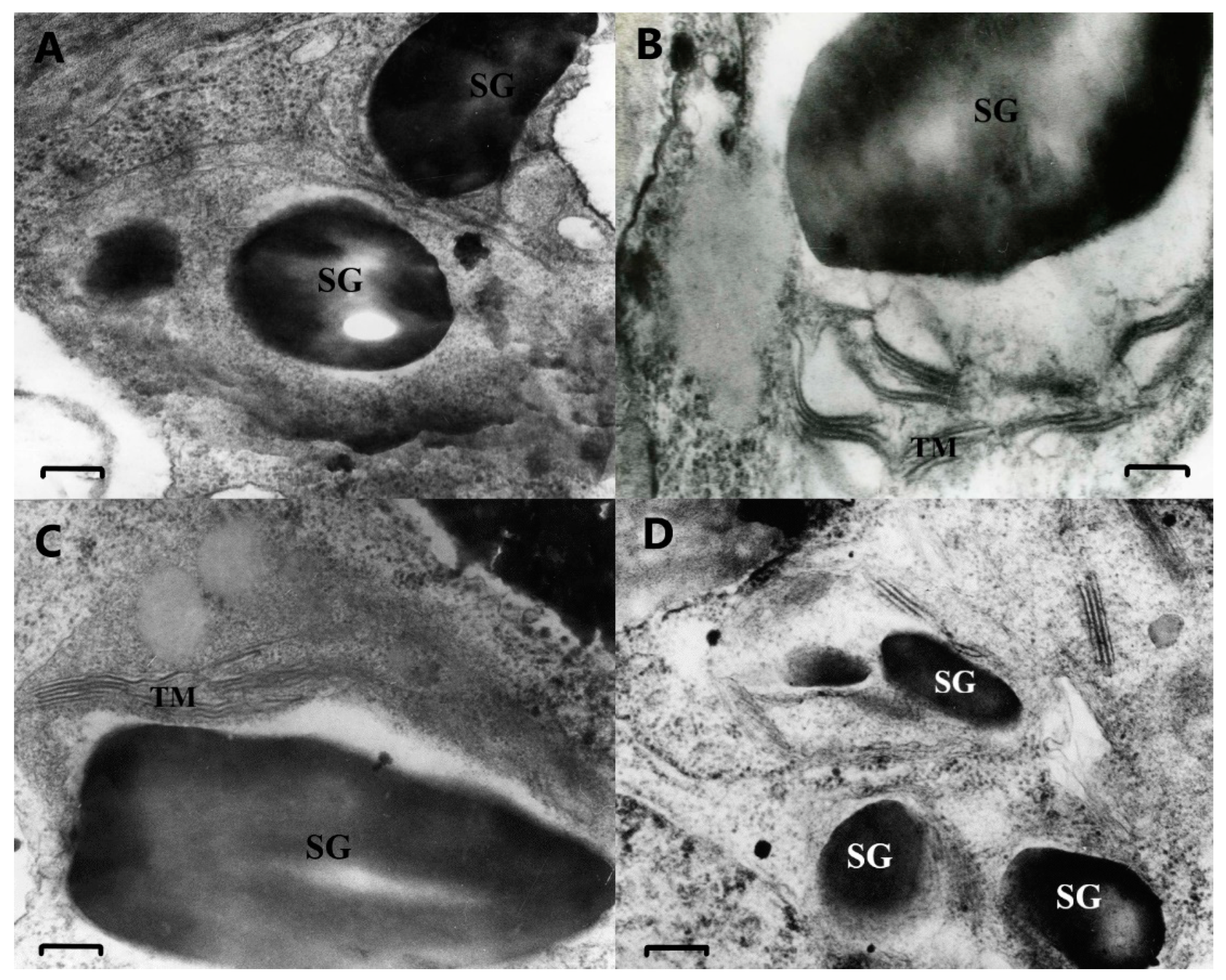
2.4. Microscopy Analysis

2.4.1. Electron Microscopy Analysis

The ultrastructure of chloroplasts in cross-sections of cortex chlorenchyma was examined using transmission electron microscopy (TEM). Vine chlorenchyma tissues were fixed, and the samples were prepared as previously described in [8]. Contrasted ultrathin sections were examined using a JEM-100B electron microscope (Jeol, Tokyo, Japan) at an accelerating voltage of 80 kV.

2.4.2. Fluorescent Microscopy Analysis

Confocal microscopy was performed using Leica TCS SPE (Leica Microsystems CMS GmbH, Mannheim, Germany) with a 10× objective. Fresh cross-sections of lignified vines were used, and chlorophyll and lignin autofluorescence was detected in the 410–750 nm range using excitation by a 405 nm laser.

2.5. Pigment Analysis

To collect corticular chlorenchyma tissues for pigment analysis, the outer bark was removed from the vines with a scalpel; then, the inner bark tissue was quickly cut with a scalpel. The tissues were immediately frozen in liquid nitrogen, then ground with a mortar and pestle, and weighed without thawing. The pigment composition was determined spectrophotometrically in 100% acetone extracts using a Shimadzu UV-1800 spectrophotometer (Shimadzu Corporation, Kyoto, Japan). Complete extraction of the pigments was achieved with four successive cycles of acetone addition, pellet resuspension, sedimentation of cell fragments by centrifugation, and collection of the supernatant until the final precipitate was completely colorless. The collected portions of the supernatant were pooled together, and the pigment content was determined as described in [18].

2.6. Statistical Analysis

All experiments were performed with at least four biological replicates. Data are presented as mean values ± standard errors. Origin (OriginLab Corporation, Northampton, MA, USA) and Microsoft Excel version 2023 (Microsoft, Redmond, WA, USA) software were used to confirm the statistical significance. Statistical significance of the differences for the most experiments was determined by Student’s t-test. The difference was considered statistically significant at p ≤ 0.05 and marked on the graphs and in the tables with an asterisk. Pearson’s correlation coefficient was calculated to confirm significant correlation between variable chlorophyll fluorescence parameters and weather conditions.

3. Results

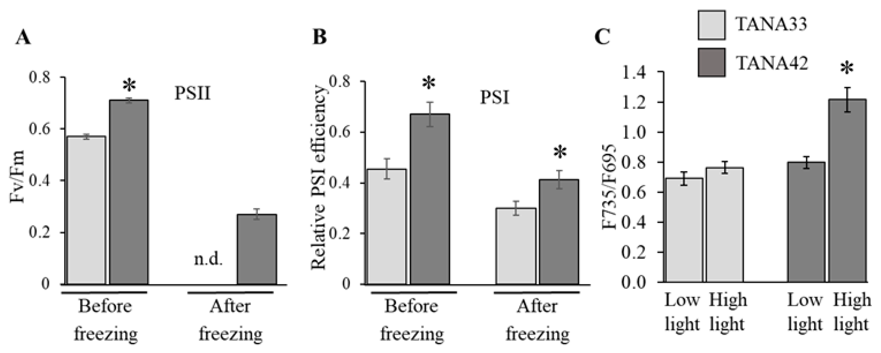
3.1. Analysis of the Y(II) of the Corticular Photosynthetic Apparatus of the Grapevine Varieties Differing in Frost Resistance

Due to severe freezing temperatures during the winters of 2006 and 2012, a significant number of grapevine plants on the experimental plots in Anapa (Krasnodar region, Russia) were damaged. In January 2006, temperatures fell below −10 °C, occasionally reaching as low as −25 °C (Figure 1A). In January and February of 2012, temperatures remained below −10 °C for about two weeks, with occasional temperatures approaching −20 °C (Figure 1B). In addition, at the end of February 2012, the temperature dropped to −10 °C after a long period of positive temperatures, which likely led to the onset of sap movement and the reduction of winter hardiness. As a result, we observed a more severe damage to grapevines that year. The grape varieties on the experimental plots responded differently to severe weather conditions and were damaged to various degrees. The degree of damage was quantified by estimating the percentage of dead buds, i.e., buds that did not sprout in spring. The data on the bud mortality for the grapevine cultivars Crystal, Dostoiny, Krasnostop AZOS, and Zarif in 2006 are presented in (Figure 1C). In 2012, in addition to these four cultivars, two new hybrids, i.e., TANA33 and TANA42, were analyzed (Figure 1D). Among the cultivars analyzed in 2006, Crystal, with a minimal number of dead buds (only 17%), was the most freeze-tolerant, while Zarif was the most sensitive to harsh winter conditions since not a single bud sprouted in spring. Krasnostop AZOS was slightly more tolerant than Dostoiny. The relative tolerance of each variety did not change in 2012, and the data on bud mortality were reproduced. One of the new hybrid forms, TANA42, proved to be extremely tolerant to low temperature since it had the highest number of surviving buds (only 6% of the buds did not sprout in spring). TANA33 displayed moderate tolerance to low temperatures, as one-quarter of its buds did not survive the winter.
It was observed that the vines of the studied varieties differ in the thickness of the chlorenchyma layer under the outer bark and the intensity of the green color of this layer. We performed the measurements of the photosynthetic activity of corticular tissue of grapevine fragments cut during winter 2018–2019 and adapted to the laboratory conditions (dim light, 22 °C). The activity of photosystem II (PSII) was evaluated by means of light-induced chlorophyll fluorescence measurements, as described previously in [7,8]. The analysis showed that the more freeze-tolerant genotypes were characterized by the higher values of PSII activity (maximum quantum yield) in the vine cortex. In the graph (Figure 1E) representing the correlation between the maximum quantum yield values determined in the laboratory and the percentage of dead buds estimated for plants grown in the field, a negative correlation between these parameters can be observed in this sample of plants. The Pearson correlation coefficient (R-value) is equal to 0.845 (p = 0.034), indicating a possible relationship between photosynthetic activity of vine chlorenchymal tissues and freeze tolerance.

3.2. Comparative Analysis of Grapevine Hybrid Forms Differing in Freeze-Tolerance and CPA Activity

3.2.1. Laboratory Study

New hybrids with high commercial potential, i.e., TANA42 and TANA33, which differ markedly in freeze tolerance and thickness of the chlorenchyma layer under the outer bark, were selected for in-depth analysis. Vines of plants grown in Krasnodar and cut during the winters of 2016–2018 were used in the experiments.
Initially, the distribution of chlorenchyma tissues in the vines of TANA42 and TANA33 was studied by fluorescent microscopy (Figure 2). Figure 2A,B show cross-sections of the first-year lignified overwintering vines of TANA33 and TANA42, respectively. Chlorophyll autofluorescence (red) allows clear visualization of the chlorenchyma cells and tissues. The blue color is the autofluorescence of lignin, which is quite abundant in the woody vine. The outer cortex (the phellem) consists of the layers of dead cork cells (bright blue color). The outer cortex layer is followed by the layer of inner cortex, containing living chlorenchyma cells, and the difference in thickness of this layer between TANA42 and TANA33 is obvious. Closer to the center are the medullary rays of parenchyma cells, which contain a significant amount of chlorophyll, as seen in both the fluorescent image and the bright field image (Figure 2). The thickness of these chlorenchyma medullary rays is several times greater in the TANA42 vine. The TANA33 xylem shows wider openings of vascular bundles. In general, the chlorenchyma tissue appears to be more abundant in TANA42.
Next, the ultrastructure of chloroplasts in the cross-sections of cortex chlorenchyma was examined by transmission electron microscopy. Figure 3 shows the representative images of chloroplasts from TANA33 (A) and TANA42 (B) vine chlorenchyma. The shape of plastids from both varieties is irregular. Most of the inner space of the plastids is filled with large starch grains, and bands of thylakoid membranes are localized at the chloroplast periphery, which makes them similar to amylochloroplasts [19]. Despite the general similarity in structure, the differences between the chloroplasts of TANA42 and TANA33 are obvious: The corticular plastids of TANA33 have a less developed internal membrane system. In tissues of the inner cortex of TANA33, plastids without photosynthetic membranes are observed (Figure 3A). In TANA42, corticular chloroplasts with several small starch granules and developed grana containing 4–6 thylakoids are present. Many ribosomes are seen in chloroplasts both with and without photosynthetic membranes, which maybe is an indication of active protein biosynthesis in the wintering vine.
The results of microscopic studies are in good agreement with the data on the pigment composition analysis (Table 1). On a weight basis, the corticular chlorenchyma of TANA42 contains more chlorophyll a and chlorophyll b. At the same time, the total carotenoid content does not differ between the two hybrids. As a result, the chlorophyll/carotenoid ratio is significantly higher in the chlorenchyma of TANA42.
To study the functional characteristics of the photosynthetic apparatus in TANA42 and TANA33 vine chlorenchyma tissues, the maximum quantum yield of PSII and the relative PSI efficiency, which are the parameters routinely used to evaluate functional activity of photosynthetic apparatus, were measured in vines incubated at room temperature and after treatment with freezing temperature under the laboratory conditions (Figure 4A,B). These parameters were significantly higher in TANA42 vines both before and after freezing.
The measurements of the low-temperature chlorophyll fluorescence spectra of vine chlorenchyma were performed to estimate the ratio of fluorescence intensity at λm = 735 nm and at λm = 695 nm, which characterizes the distribution of excitation energy between the two photosystems [20,21]. We previously showed the presence of major peaks in chlorophyll fluorescence emission spectra at 77 K in samples of chlorenchyma tissues of the vine inner cortex. The peaks at λm = 685 and λm = 695 nm are associated mainly with the chlorophyll–protein complex of PSII, and λm = 735 nm is associated with the chlorophyll–protein complex of PSI [7]. It is known that the alteration in light intensity leads to a redistribution of light absorption capacity through the migration of light-harvesting antennae LCHII from PSII to PSI in the process known as “state-transition” [22,23,24]. This regulation of the antenna complex migration allows balancing the excitation energy distribution between the two photosystems and determines the ability to adapt to different light conditions. This parameter was measured in the vines incubated at low (~5 μM photons m−2 s−1) and high (1000 μM photons m−2 s−1) light intensity to reveal the possible redistribution of photosynthetic antennae between PSII and PSI. As can be seen in Figure 4C, the F735/F695 ratio is almost the same in TANA33 vines incubated at low and high light intensities, while in TANA42, this ratio increases significantly after incubation of vines at high light intensity, which may indicate a better adaptability of the photosynthetic apparatus of TANA42 vine to changing light conditions.
Thus, the analyses mentioned above revealed marked differences in the structural and functional characteristics between the chlorenchyma of TANA42 and TANA33 vines.

3.2.2. Field Study

One of the objectives of this study was to analyze the corticular photosynthetic activity in TANA42 and TANA33 growing in the field under different temperature conditions in Krasnodar. We measured the photosynthetic activity, more specifically the effective quantum yield, of TANA42 and TANA33 CPA in situ at noon ± 30 min from early February 2019 to mid-March 2020. From mid-March 2019 to the end of September 2019, no measurements were taken due to dense foliage on growing plants. These data, along with the concurrent temperature measurements, are presented in Figure 5, and the correlative interactions between these parameters are presented in Table 2. The figure shows that the values of effective quantum yield are consistently higher in TANA42. It is evident that all temperature changes affect the effective quantum yield of both hybrid forms. Surprisingly, quite high values of the effective quantum yield are observed even at very low temperatures, near or even below zero, especially in TANA42. As can be seen from Table 2, the values of the effective quantum yield are highly dependent on the weather conditions, and in general, the correlation values with weather parameters are higher for TANA42, suggesting that its photosynthetic apparatus is more responsive to environmental factors. The highest correlation values observed are between the Y(II) value and the air temperature during the measurements: For TANA33, this value was 0.617 (moderate correlation), and for TANA42, it was 0.805 (strong correlation).

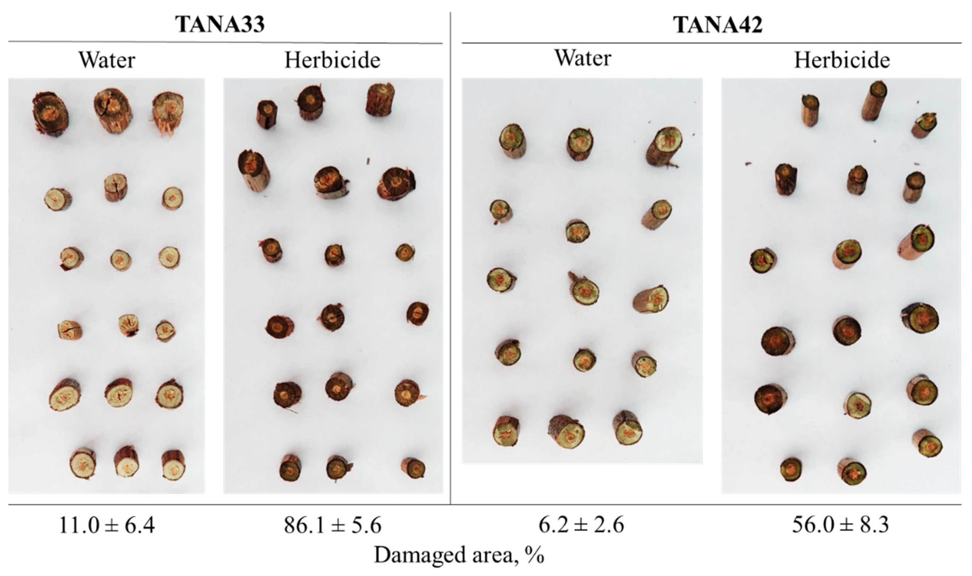
3.3. Effect of Light on the Intensity of Freeze-Induced Injuries

In order to confirm the possible protective effect of corticular photosynthesis under freezing temperatures, we conducted an experiment involving the freezing of vine cuttings simultaneously subjected to illumination in the presence or absence of metribuzin, a specific inhibitor of PS II [16]. The extent of tissue damage due to this treatment was assessed and is shown in Figure 6. Expectedly, TANA33 shows more damage in comparison to TANA42 in both cases with and without the herbicide. Preliminary treatment of both hybrids with metribuzin solution leads to the formation of a larger damaged area.

4. Discussion

In the presented work, the freeze tolerance of grape varieties and hybrids was initially estimated based on the bud death rate in the field under extreme winter weather conditions, which occur very rarely in the area. Further, the activity of corticular photosynthesis in grape varieties differing in freeze tolerance were studied under simulated low-temperature stress conditions in the laboratory. And finally, to confirm the possible connection between corticular photosynthesis and the plant adaptation to sub-optimal temperatures, we again turned to field experiments and revealed the correlations between the values of effective quantum yield of PSII in the corticular chlorenchyma and environmental factors. This points to the involvement of photosynthesis in corticular chlorenchyma in the formation of plant freeze tolerance.
The major factor responsible for the photooxidative damage of photosynthetic apparatus at low temperatures is the imbalance between the inhibited “dark reactions” of photosynthesis (CO2 assimilation and Calvin cycle) and uninhibited “light reactions” (light absorption by photosynthetic antennae and electron transfer through the electron-transport chain of chloroplasts) [4,25,26]. Another important damaging factor occurring at freezing temperatures is associated with the formation of ice crystals, which can physically destroy cell membranes. The corticular photosynthetic apparatus is better protected from both of these damaging factors than are the needles of evergreen plants that are known to maintain photosynthetic activity during winter. In autumn, the lignified plant stems undergo changes to prepare for winter, primarily associated with a decrease in water content and accumulation of reserve compounds and osmolytes. Thus, by the onset of winter frosts, all stem tissues are largely dehydrated, and their cell structures are protected by osmotically active compounds. At the same time, the photosynthetic apparatus in lignified stems is protected from the damaging effects of excessive light by the outer bark, which absorbs a significant part of solar irradiation.
The seasonal variation in the activity of photosynthesis was previously studied mainly on the needles of evergreen plants [27]. By now, several molecular mechanisms responsible for the acclimatization to low-temperature winter conditions are known, including the alterations in the organization of the PSII antenna, induction of chlororespiration, cyclic electron transport, and flavodiiron-mediated O2 photoreduction at the acceptor side of PSI [2,3,4,28]. In the needles of Scots pine, the thylakoid destacking leading to the mixing of PSII with PSI complexes, extreme down-regulation of photosystem II activity, and direct energy transfer from PSII to PSI play a major role in winter acclimation and protection [2]. In general, the problem of functional stability and tolerance of the photosynthetic apparatus under various temperature conditions remains extremely important [29,30]. Very little is known about the structure of the corticular photosynthetic apparatus [8], and nothing is known about its adaptive changes during the preparation for winter. The phenomenon of photosynthesis in lignified branches during the winter period has been described for evergreen plants [31] and common beech Fagus sylvatica [32]. Remarkably, chloroplasts of overwintering pine bark retain higher PSII activity compared to those in needles [31]. To the best of our knowledge, this article is the first report on the photosynthesis in lignified grape vine during the winter period.
In this paper, we show that chlorenchyma tissues in the grapevine cortex remain photosynthetically active during the cold winter period, and this activity is detectable both under laboratory conditions and in the field. Moreover, higher values of the maximum quantum yield of PSII were observed in the more freeze-tolerant cultivars (Figure 1). Measurements of the effective quantum yield of PSII (Y(II)) in the field at different times of the year showed that all temperature changes have a significant impact on this parameter in all studied plants (Figure 5). Surprisingly high values of Y(II) were observed even at very low temperatures. The strongest correlation was observed between Y(II) and temperature during the measurements (Table 2), revealing a significant functional plasticity and adaptability of the corticular photosynthetic apparatus as well as a direct dependence of functional activity on ambient temperature. It was previously shown that the springtime resumption of photosynthesis in overwintering needles depends on water availability and soil and air temperature [33]. The release of photosynthetic dormancy entails multiple changes in chloroplast ultrastructure, depends on the activation of several enzymes associated with CO2 assimilation, and presumably requires a substantial amount of time [4,34]. Rapid changes in the effective quantum yield of PSII in corticular tissues in response to environmental changes may indicate higher responsiveness of corticular photosynthetic apparatus. These adaptive changes may be occurring without deep structural rearrangements and activation of dark reactions of photosynthesis.
We compared two new hybrid forms with high commercial potential and revealed significant differences in the representation of chlorenchyma tissues in the lignified vines (Figure 2), the development of photosynthetic membranes in the chloroplasts of these tissues (Figure 3), and the functional activity of the photosynthetic apparatus and tolerance to freezing temperatures (Figure 4). The more freeze-tolerant variety is characterized by more developed chlorenchyma, represented not only in the inner cortex but also in the medullary rays, the functions of which have previously been only attributed to nutrients storage, tannins deposition, and radial transport of nutrients and water. The more developed photosynthetic membranes and higher chlorophyll content (but not higher carotenoid content) in the freeze-tolerant hybrid TANA42 correspond well to the data showing higher values of functional activity of PSII and PSI as well as higher mobility of light-harvesting antenna complexes. The difference in PSII activity between plants with different levels of freeze tolerance became more obvious after the low-temperature treatment. This is consistent with the information about the greater suppression of PSII in the needles during winter [2]. Thus, corticular PSII activity may serve as an indicator of freeze tolerance in woody plants. It was previously shown that this particularly informative parameter can be used to predict wood density in Eucalyptus saligna [35]. Measurements of variable chlorophyll fluorescence can be performed over a wide temperature range and in all seasons, under both laboratory and field conditions, because the primary photophysical and photochemical processes associated with energy absorption and transformation within PSII are minimally dependent on temperature.
Collectively, this study illustrates the physiological significance of photosynthesis occurring in the chlorenchyma of lignified stems and points to a possible role in the freeze tolerance of overwintering plant organs. It is clear that perennial plants rely on multiple strategies for survival at low temperatures. Thus, even among freeze-tolerant grape varieties, some (such as V. amurensis, capable of withstanding extremely low temperatures down to −40 °C) may rely on the complex mechanisms related to hormonal signaling, metabolic changes, and the activity of cold-responsive regulatory networks [36,37]. Under such extreme conditions, a strategy based on deep dehydration and inhibition of physiological processes during winter appears to be more effective for survival. Maintenance of physiological activity of corticular photosynthetic apparatus is beneficial in winters with low positive temperatures and occasional frosts.
At present, the molecular mechanisms underlying the sustained functioning of corticular photosynthetic apparatus during winter are largely unknown. It was suggested that the photosynthetic acclimation of overwintering evergreens represents specific evolutionary adaptations in cold climatic zones, as exemplified by the boreal forests of the Northern Hemisphere [4]. The evolution of adaptive traits in the corticular photosynthetic apparatus that allow them to tolerate low temperatures must be an even larger evolutionary event than evolutionary changes in the photosynthetic apparatus of conifer needles, given that woody stems are an overwintering organ in a large number of perennial plants. The evolutionary aspects of corticular photosynthesis remains largely unexplored.

5. Conclusions

Non-foliar photosynthesis plays a well-known role in plant growth and productivity [38,39,40]. This article is the first to demonstrate the presence of photosynthetic activity in overwintering vines and highlight the role of this process in low-temperature stress tolerance in perennial plants. The structural and functional characteristics of the corticular photosynthetic apparatus could potentially be used as a marker for selecting productive and freeze-tolerant grape varieties. The corticular photosynthetic apparatus is of great interest as a model of a photosynthetic system extremely resistant to low temperatures.
Future research could focus on the role of corticular photosynthesis in the freeze-tolerance trait formation in other perennial crops. The simple measurements of non-foliar photosynthesis activity can be used in screening for stress-tolerant plants as well as in agronomic practice to assess the degree of damage to plant parts by low-temperature stress.

6. Patents

Patent Savchenko T.V., Sundyreva M.A., Khristin M.S., Klimov V.V., Biryukov S.V. “Method of the grapes frost resistance determining” RU2653016C2, filled 30.09.2016, and issued 4 May 2018.

Author Contributions

T.V.S. and M.A.S. conceived the research; T.V.S., M.A.S., D.V.Y., M.S.K., U.V.G., E.O.L., G.A.S. and I.A.N. carried out the experiments; T.V.S. wrote the manuscript with support from M.A.S. and D.V.Y. All authors have read and agreed to the published version of the manuscript.

Funding

This research was supported by the Ministry of Education and Science of the Russian Federation, theme No 122041100274-6 and FGRE-2022-0004.

Data Availability Statement

The data presented here are available upon request from the corresponding author.

Conflicts of Interest

The authors declare no conflict of interest.

References

  1. Available online: https://earthobservatory.nasa.gov/global-maps/MOD_LSTD_M (accessed on 4 June 2023).
  2. Bag, P.; Chukhutsina, V.; Zhang, Z.; Paul, S.; Ivanov, A.G.; Shutova, T.; Croce, R.; Holzwarth, A.R.; Jansson, S. Direct energy transfer from photosystem II to photosystem I confers winter sustainability in Scots Pine. Nat. Commun. 2020, 11, 6388. [Google Scholar] [CrossRef]
  3. Bag, P.; Shutova, T.; Shevela, D.; Lihavainen, J.; Nanda, S.; Ivanov, A.G.; Messinger, J.; Jansson, S. Flavodiiron-mediated O2 photoreduction at photosystem I acceptor-side provides photoprotection to conifer thylakoids in early spring. Nat. Commun. 2023, 14, 3210. [Google Scholar] [CrossRef] [PubMed]
  4. Oquist, G.; Huner, N.P. Photosynthesis of overwintering evergreen plants. Annu. Rev. Plant Biol. 2003, 54, 329–355. [Google Scholar] [CrossRef]
  5. Krol, M.; Hurry, V.M.; Maxwell, D.P.; Malek, L.; Ivanov, A.G.; Huner, N.P. Low growth temperature inhibition of photosynthesis in cotyledons of jack pine seedlings (Pinus banksiana) is due to impaired chloroplast development. Can. J. Bot. 2002, 80, 1042–1051. [Google Scholar] [CrossRef]
  6. Saveyn, A.; Steppe, K.; Ubierna, N.; Dawson, T.E. Woody tissue photosynthesis and its contribution to trunk growth and bud development in young plants. Plant Cell Environ. 2010, 33, 1949–1958. [Google Scholar] [CrossRef] [PubMed]
  7. Tikhonov, K.G.; Khristin, M.S.; Klimov, V.V.; Sundireva, M.A.; Kreslavski, V.D.; Sidorov, R.A.; Tsidendambayev, V.D.; Savchenko, T.V. Structural and functional characteristics of photosynthetic apparatus of chlorophyll-containing grape vine tissue. Russ. J. Plant Phys. 2017, 64, 73–82. [Google Scholar] [CrossRef]
  8. Yanykin, D.; Sundyreva, M.; Khorobrykh, A.; Semenova, G.; Savchenko, T. Functional characterization of the corticular photosynthetic apparatus in grapevine. Biochim. Biophys. Acta (BBA) Bioenerg. 2020, 1861, 148260. [Google Scholar] [CrossRef]
  9. Aschan, G.; Pfanz, H. Non-foliar photosynthesis—A strategy of additional carbon acquisition. Flora Morphol. Distrib. Funct. Ecol. Plants 2003, 198, 81–97. [Google Scholar] [CrossRef]
  10. Wittmann, C.; Pfanz, H.; Loreto, F.; Centritto, M.; Pietrini, F.; Alessio, G. Stem CO2 release under illumination: Corticular photosynthesis, photorespiration or inhibition of mitochondrial respiration? Plant Cell Environ. 2006, 29, 1149–1158. [Google Scholar] [CrossRef]
  11. Ávila-Lovera, E.; Garcillán, P.P.; Silva-Bejarano, C.; Santiago, L.S. Functional traits of leaves and photosynthetic stems of species from a sarcocaulescent scrub in the southern Baja California Peninsula. Am. J. Bot. 2020, 107, 1410–1422. [Google Scholar] [CrossRef]
  12. Burrows, G.E.; Connor, C. Chloroplast Distribution in the Stems of 23 Eucalypt Species. Plants 2020, 9, 1814. [Google Scholar] [CrossRef] [PubMed]
  13. Chen, X.; Luo, M.; Kang, Y.; Zhao, P.; Tang, Z.; Meng, Y.; Huang, L.; Guo, Y.; Lu, X.; Ouyang, L.; et al. Comparison between the stem and leaf photosynthetic productivity in Eucalyptus urophylla plantations with different age. Planta 2023, 257, 56. [Google Scholar] [CrossRef] [PubMed]
  14. Natale, S.; La Rocca, N.; Battistuzzi, M.; Morosinotto, T.; Nardini, A.; Alboresi, A. Structure and function of bark and wood chloroplasts in a drought-tolerant tree (Fraxinus ornus L.). Tree Physiol. 2023, 43, 893–908. [Google Scholar] [CrossRef]
  15. Savchenko, T.V.; Sundyreva, M.A.; Khristin, M.S.; Klimov, V.V.; Biryukov, S.V. Method of the grapes frost resistance determining. 2018. [Google Scholar]
  16. Ketel, D.H.; Wielen, M.J.W.V.D.; Lotz, L.A.P. Prediction of a low dose herbicide effect from studies on binding of metribuzin to the chloroplasts of Chenopodium album L. Ann. Appl. Biol. 1996, 128, 519–531. [Google Scholar] [CrossRef]
  17. Genty, B.; Harbinson, J.; Cailly, A.; Rizza, F. Fate of excitation at PS II in leaves: The non-photochemical side. Plant Physiol. Biochem. 1996, 86, 28. [Google Scholar]
  18. Lichtenthaller, H.K.; Wellburn, A.R. Chlorophylls and carotenoids: Pigments of photosynthetic biomembranes. Method Enzym. 1987, 148, 350–382. [Google Scholar]
  19. Kufner, R.; Czygan, F.C.; Schneider, L. Changes in pigment content and ultrastructure in the plastids of the needle leaves of Taxus baccata (L.) during their development. Ber. Deut. Bot. Ges. 1978, 91, 325–337. [Google Scholar] [CrossRef]
  20. Krause, G.H.; Weis, E. Chlorophyll fluorescence as a tool in plant physiology: II. Interpretation of fluorescence signals. Photosynth. Res. 1984, 5, 139–157. [Google Scholar] [CrossRef]
  21. Minagawa, J. State transitions—The molecular remodeling of photosynthetic supercomplexes that controls energy flow in the chloroplast. Biochim. Biophys. Acta (BBA) Bioenerg. 2011, 1807, 897–905. [Google Scholar] [CrossRef]
  22. Allen, J.F.; Bennett, J.; Steinback, K.E.; Arntzen, C.J. Chloroplast protein phosphorylation couples plastoquinone redox state to distribution of excitation energy between photosystems. Nature 1981, 291, 25–29. [Google Scholar] [CrossRef]
  23. Bennett, J. Phosphorylation of chloroplast membrane polypeptides. Nature 1977, 269, 344–346. [Google Scholar] [CrossRef]
  24. Murata, N. Control of excitation transfer in photosynthesis I. Light-induced change of chlorophyll a fluoresence in Porphyridium cruentum. Biochim. Biophys. Acta (BBA) Bioenerg. 1969, 172, 242–251. [Google Scholar] [CrossRef]
  25. Egerton, J.J.G.; Banks, J.C.G.; Gibson, A.; Cunningham, R.B.; Ball, M.C. Facilitation of seedling establishment: Reduction in irradiance enhances winter growth of Eucalyptus pauciflora. Ecology 2000, 81, 1437–1449. [Google Scholar] [CrossRef]
  26. Öquist, G.; Ögren, E. Effects of winter stress on photosynthetic electron transport and energy distribution between the two photosystems of pine as assayed by chlorophyll fluorescence kinetics. Photosynth. Res. 1985, 7, 19–30. [Google Scholar] [CrossRef] [PubMed]
  27. Jensen, A.M.; Warren, J.M.; Hanson, P.J.; Childs, J.; Wullschleger, S.D. Needle age and season influence photosynthetic temperature response and total annual carbon uptake in mature Picea mariana trees. Ann. Bot. 2015, 116, 821–832. [Google Scholar] [CrossRef]
  28. Yang, Q.; Blanco, N.E.; Hermida-Carrera, C.; Lehotai, N.; Hurry, V.; Strand, Å. Two dominant boreal conifers use contrasting mechanisms to reactivate photosynthesis in the spring. Nat. Commun. 2020, 11, 128. [Google Scholar] [CrossRef]
  29. Gunderson, C.A.; O’hara, K.H.; Campion, C.M.; Walker, A.V.; Edwards, N.T. Thermal plasticity of photosynthesis: The role of acclimation in forest responses to a warming climate. Glob. Change Biol. 2010, 16, 2272–2286. [Google Scholar] [CrossRef]
  30. Kenchanmane Raju, S.K.; Barnes, A.C.; Schnable, J.C.; Roston, R.L. Low-temperature tolerance in land plants: Are transcript and membrane responses conserved? Plant Sci. 2018, 276, 73–86. [Google Scholar] [CrossRef] [PubMed]
  31. Ivanov, A.G.; Krol, M.; Sveshnikov, D.; Malmberg, G.; Gardestrom, P.; Hurry, V.; Oquist, G.; Huner, N.P. Characterization of the photosynthetic apparatus in cortical bark chlorenchyma of Scots pine. Planta 2006, 223, 1165–1177. [Google Scholar] [CrossRef]
  32. Damesin, C. Respiration and photosynthesis characteristics of current-year stems of Fagus sylvatica: From the seasonal pattern to an annual balance. New Phytol. 2003, 158, 465–475. [Google Scholar] [CrossRef]
  33. Tanja, S.; Berninger, F.; Vesala, T.; Markkanen, T.; Hari, P.; Mäkelä, A.; Ilvesniemi, H.; Hanninen, H.; Nikinmaa, E.; Huttula, T.; et al. Air Temperature Triggers the Recovery of Evergreen Boreal Forest Photosynthesis in Spring. Glob. Change Biol. 2003, 9, 1410–1426. [Google Scholar] [CrossRef]
  34. Monson, R.K.; Sparks, J.P.; Rosenstiel, T.N.; Scott-Denton, L.E.; Huxman, T.E.; Harley, P.C.; Turnipseed, A.A.; Burns, S.P.; Backlund, B.; Hu, J. Climatic influences on net ecosystem CO2 exchange during the transition from wintertime carbon source to springtime carbon sink in a high-elevation, subalpine forest. Oecologia 2005, 146, 130–147. [Google Scholar] [CrossRef] [PubMed]
  35. Johnstone, D.; Tausz, M.; Moore, G.; Nicolas, M. Bark and leaf chlorophyll fluorescence are linked to wood structural changes in Eucalyptus saligna. AoB Plants 2014, 6, plt057. [Google Scholar] [CrossRef] [PubMed]
  36. Fennell, A. Freezing tolerance and injury in grapevines. J. Crop Improv. 2004, 10, 201–235. [Google Scholar] [CrossRef]
  37. Ren, C.; Fan, P.; Li, S.; Liang, Z. Advances in understanding cold tolerance in grapevine. Plant Physiol. 2023, 192, 1733–1746. [Google Scholar] [CrossRef] [PubMed]
  38. Hu, L.; Zhang, Y.; Xia, H.; Fan, S.; Song, J.; Lv, X.; Kong, L. Photosynthetic characteristics of non-foliar organs in main C(3) cereals. Physiol. Plant 2019, 166, 226–239. [Google Scholar] [CrossRef]
  39. Pfanz, H.; Aschan, G.; Langenfeld-Heyser, R.; Wittmann, C.; Loose, M. Ecology and ecophysiology of tree stems: Corticular and wood photosynthesis. Die Nat. 2002, 89, 147–162. [Google Scholar]
  40. Simkin, A.J.; Faralli, M.; Ramamoorthy, S.; Lawson, T. Photosynthesis in non-foliar tissues: Implications for yield. Plant J. 2020, 101, 1001–1015. [Google Scholar] [CrossRef] [PubMed]
Figure 1. Relationship between photosynthetic activity of corticular chlorenchyma and freeze tolerance. Minimum and average temperatures on ampelographic collection plot during extremely cold period of 2006 (A) and 2012 (B); the percentage of dead buds (i.e., buds that did not sprout) in spring of 2006 (C) and 2012 (D) on Anapa ampelographic collection plot, NCFSCHVW. The bars indicate the means of the percentage of dead buds counted on 10 vines; the correlation between the maximum quantum yield values determined in the laboratory on vines grown at Anapa ampelographic collection of NCFSCHVW in 2018 (five biological replicas of each variety) and the percentage of dead buds previously estimated for plants grown in the field (E).
Horticulturae 09 01181 g001
Figure 2. Comparison of structural organization of photosynthetic tissues in grapevine varieties differing in freeze tolerance. Images of chlorenchyma tissue in lignified vine cuts of TANA33 (A) and TANA42 (B) obtained with confocal microscopy for determination of chlorophyll autofluorescence. Vines were grown on the small-scale experimental field in NCFSCHVW in Krasnodar.
Horticulturae 09 01181 g002
Figure 3. Transmission electron microscopy photos of chloroplasts ultrastructure in corticular chlorenchyma of TANA33 (A,C) and TANA42 (B,D) lignified vines. Scale bars are 200 nm. SG, starch grains; TM, thylakoid membranes. Vines were grown on the small-scale experimental field in NCFSCHVW in Krasnodar.
Horticulturae 09 01181 g003
Figure 4. Comparison of functional characteristics of photosynthetic apparatus in TANA33 and TANA42 cortex chlorenchyma. Maximum quantum yield of PSII estimated through the measurements of variable chlorophyll fluorescence parameters (A); effective quantum yield of PSI photochemistry determined by the measuring of the differential absorbance at 875–830 nm (B); the ratio between the intensity of peaks at λm = 735 and λm = 695 nm in chlorophyll fluorescence emission spectra at 77 K (C) of TANA33 (light gray bars) and TANA42 (dark gray bars) vine chlorenchyma. n.d., not detected. Data represent mean values ± SE of five biological replicates. Stars indicate a statistically significant difference between two cultivars at p ≤ 0.05, as assessed by Student’s t-test. Vines were obtained from small-scale experimental field in NCFSCHVW in Krasnodar.
Horticulturae 09 01181 g004
Figure 5. Seasonal changes in effective quantum yield (YII) of TANA42 and TANA33 CPA and mean daily temperatures. Data represent mean values of 10 measurements at each time point with standard errors. Measurements were carried out in small-scale experimental field in NCFSCHVW in Krasnodar in 2019–2020.
Horticulturae 09 01181 g005
Figure 6. Effect of freeze treatment on the formation of necrotic lesions in TANA33 and TANA42 vines soaked in water or herbicide solution. The numbers under the images stand for the means with standard errors (n = 30). Vines were obtained from small-scale experimental field in NCFSCHVW in Krasnodar.
Horticulturae 09 01181 g006
Table 1. Pigments in cortex chlorenchyma of grapevine hybrid plants differing in freeze tolerance.
PigmentTANA33TANA42
Chlorophyll a (μg/g fresh weight)52.2 ± 2.870.8 ± 1.2 *
Chlorophyll b (μg/g fresh weight)24.8 ± 435.4 ± 3.8 *
Chlorophyll a + b (μg/g fresh weight)77 ± 6.4106 ± 5.0 *
Carotenoids (μg/g fresh weight)34.2 ± 1.632.6 ± 1.4
Chl a/Chl b2.25 ± 0.262.09 ± 0.21
Chl/carotenoids2.29 ± 0.29.83.31 ± 0.28 *
Data represent mean values ± SE of five biological replicates. Stars indicate a statistically significant difference between two cultivars at p ≤ 0.05, as assessed by Student’s t-test.
Table 2. Correlation between weather parameters and Y(II) in TANA42 and TANA33 vines.
ParameterTANA33TANA42
Sum of daytime temperatures, °C0.610 *0.790 *
Temperature during measurement, °C0.617 *0.805 *
Average temperature during 12 h before the measurement, °C0.590 *0.752 *
Average temperature during 24 h before measurement, °C0.561 *0.738 *
Average temperature during 48 h before measurement, °C0.4550.669 *
Minimum temperature on the day of measurement, °C 0.599 *0.750 *
Minimum temperature during 24 h before measurement, °C0.599 *0.742 *
Minimum temperature during 48 h before measurement, °C0.585 *0.633 *
Precipitation, mm−0.438−0.514 *
Solar radiation, W/m20.1190.119
Relative air humidity, %−0.014−0.240
Correlations were calculated for 15 Y(II) values obtained on different dates (as presented in Figure 5), each being the mean of at least 10 measurements. Stars indicate the significance of the Pearson’s correlation coefficient at p ≤ 0.05.
Disclaimer/Publisher’s Note: The statements, opinions and data contained in all publications are solely those of the individual author(s) and contributor(s) and not of MDPI and/or the editor(s). MDPI and/or the editor(s) disclaim responsibility for any injury to people or property resulting from any ideas, methods, instructions or products referred to in the content.

