Article

Seasonal Change in Phytochemical Composition and Biological Activities of Carissa macrocarpa (Eckl.) A. DC. Leaf Extract

by
Reshika Ramasar
1,
Yougasphree Naidoo
1,
Yaser Hassan Dewir
2,* and
Antar Nasr El-Banna
3
1
School of Life Sciences, University of KwaZulu-Natal, Westville Campus, Private Bag X54001, Durban 4000, South Africa
2
Plant Production Department, College of Food & Agriculture Sciences, King Saud University, Riyadh 11451, Saudi Arabia
3
Federal Research Centre for Cultivated Plants, Julius Kühn-Institut (JKI), Messeweg 11–12, 38104 Braunschweig, Germany
*
Author to whom correspondence should be addressed.
Horticulturae 2022, 8(9), 780; https://doi.org/10.3390/horticulturae8090780
Submission received: 23 July 2022 / Revised: 10 August 2022 / Accepted: 12 August 2022 / Published: 28 August 2022

Abstract

:
Genus Carissa represents several species that are reported to be of great phyto-medicinal and ethnopharmacological value. However, Carissa macrocarpa is relatively understudied. Furthermore, environmental conditions such as seasonal changes are known to affect the phytochemical composition of medicinal plants. Therefore, this study aimed to investigate the phytochemical composition and biological activity of the leaf extracts of C. macrocarpa in the summer and winter seasons. The phytochemical screening of C. macrocarpa leaves showed positive results for a variety of phytochemicals, such as alkaloids, tannins, phenols, naphthoquinones, flavonoids, saponins, steroids, proteins, carbohydrates, mucilage, gum and resin. The methanolic extract was evaluated for its antibacterial activity against Escherichia coli and Staphylococcus aureus using the agar well diffusion method. The winter leaf extract was distinguished for its potential antibacterial activity against both bacterial strains with inhibition zones (mm) of 8.17 ± 1.04 and 6.83 ± 0.58 at 10 mg/mL. The antioxidant activity of the leaf extracts was evaluated using the 2, 2′-diphenyl-1-picrylhydrazyl (DPPH) assay. The percentage scavenging activities of the different extracts were significantly greater than that of the control. Furthermore, at 15, 30, 60, 120 and 240 µg/mL, the percentage scavenging activities of the winter methanol leaf extract were 74.65, 78.31, 85.45, 90.02 and 95.68%, and those of the summer one were 71.66, 73.57, 84.05, 88.22 and 96.28%, respectively, indicating that the methanol leaf extract had greater percentage scavenging activity in winter than in summer. In winter, the IC50 value of the methanol leaf extract (0.67 µg/mL) was lower than that of ascorbic acid (8.26 µg/mL). It is concluded that winter is the optimal season to harvest leaves of C. macrocarpa for medicinal use. To the best of our knowledge, this is the first report that relates the phytochemical composition and medicinal properties of C. macrocarpa to changes in seasons. The results obtained are promising, and this species should be further explored to decipher its pharmacological worth.

1. Introduction

In many developing countries, plants of medicinal value are being used by billions of people. This is due to the continual inadequate supply of modern, commercial medicines; thus, people prefer to use medicinal plants as they are of low cost and effective [1,2]. Approximately 80% of black South Africans utilize traditional medicine to fulfil their primary health care needs [3]. There are two types of metabolites produced within plants, namely, primary and secondary metabolites [4,5]. Secondary metabolites such as phenolics, flavonoids, alkaloids, lignans and terpenoids are abundant in medicinal plants. Secondary metabolites, also known as phytochemicals, are bioactive compounds that are required for a plant’s survival (under environmental stress) and its protection against insects and predators [6]. Since ancient times, secondary metabolites have been widely utilized in the treatment of different illnesses and disorders, and they are still quite popular today [7]. However, there are many environmental factors that may affect the phytochemical composition of medicinal plants. Among the many factors, there are soil type, change in season, salinity, light, altitude and humidity [8,9,10]. Determining the effect of seasonal change on plant phytochemical composition provides fundamental information on the best time/season to harvest individual plant species (with maximum phytochemical composition) [11,12].
Over the years, the growth and spread of bacterial infections have been controlled; however, many are now emerging due to mutations and drug resistance. Thus, research on and development of new or improved antibacterial medications possibly derived from plants are proposed [13]. Phytochemicals such as phenols, flavonoids, saponins, alkaloids, tannins and steroids exhibit antibacterial activity; hence, they have been of great value in the pharmaceutical industry [14]. Furthermore, phytochemicals are also known to have antioxidant activity [15]. Antioxidants are substances that can lower or prevent oxidative stress caused by reactive oxygen species. This is because antioxidants can neutralize or scavenge reactive oxygen species via hydrogen donation, single-electron transfer and the ability to chelate transition metals [16,17]. Reactive oxygen species (ROS) such as hydroxyl (OH), superoxide (O2), nitric oxide (NO), peroxyl (RO2) and lipid peroxyl (LOO) are free radicals that are naturally produced in the human body as products of cellular metabolism. When they are produced in abundance, they can cause major damage to cellular structures (namely, nucleic acids, proteins and lipids). This can lead to the development of chronic diseases such as diabetes mellitus, cancer, hypertension, cardiac disorders and neurodegenerative diseases [18,19,20,21]. Therefore, the discovery of natural antioxidants from plants must be emphasized. Furthermore, the use of medicinal plants to develop drugs is of great interest as they are more readily available, affordable and effective and have fewer harmful side-effects than synthetic drugs [6,22].
The Apocynaceae family is believed to be one of the largest families in the plant kingdom [23]. The majority of the species belonging to this family are rich in phytochemicals, especially alkaloids, which are known to have great medicinal value [23]. Members of this family are distributed mainly in tropical, subtropical and temperate regions of the world [24]. Carissa macrocarpa (Eckl.) A. DC. syn. C. grandiflora (E.Mey.) A. DC. [25] is an ornamental shrub that is commonly known as the Natal plum. This species belongs to the Apocynaceae family and is indigenous to South Africa, where it is used in traditional medicine [26,27]. Carissa trees are perfect plants for hedges due to their numerous large, y-shaped thorns. Carissa macrocarpa has white–yellow scented flowers that are star-shaped and have red fruits containing considerable amounts of vitamins, carbon, calcium and magnesium [15,26,28]. The fruit of C. macrocarpa is renowned for its several medicinal benefits. They are used to treat anaemia and increase haemoglobin and possess immune-boosting properties [28,29]. On the other hand, the leaves are used by Zulu people to treat diarrhoea in cattle, and they are also used to treat coughs and venereal diseases in humans [29,30,31].
Phytochemical studies on C. macrocarpa plant parts revealed the presence of alkaloids, flavonoids, saponins, triterpenoids, steroids, quinones, tannins, carbohydrates and phenols [27,28,32,33,34]. Studies on C. macrocarpa extracts that have previously been conducted reported pharmacological properties, such as antioxidant and antibacterial activities [28,33,35,36]. However, the impact of seasonal variations on the production of phytochemicals within this species has not been evaluated. Seasonal change plays a vital role in the production of phytochemicals and the overall medicinal potential of a plant. Hence, this study aimed to screen phytochemical composition using qualitative phytochemical tests and to evaluate the in vitro antibacterial and antioxidant properties of C. macrocarpa leaves in summer and winter.

2. Materials and Methods

2.1. Plant-Material Collection

Fresh leaves of C. macrocarpa were harvested from the University of KwaZulu-Natal Westville campus (29.817° S 30.940° E), Durban, South Africa. A voucher specimen was deposited at Bews Herbarium, University of KwaZulu-Natal, Pietermaritzburg campus with accession number 92,175 (voucher number 04).

2.2. Preparation of Plant Material for Extraction

The fresh leaves were collected in summer (December 2020) and winter (July 2021) and dried in an oven at 35 °C for 72 h, until all water molecules that were present completely evaporated. Thereafter, the dried leaves were ground to powder form using a Waring blender (Kenwood Ltd., Havant, UK).

2.3. Extraction of C. Macrocarpa Leaves

Carissa macrocarpa powdered leaves (10 g) (summer) were placed into a round-bottom flask. Thereafter, 100 mL of hexane was placed into the flask, and the latter was then attached to a reflux apparatus. The solvent was heated under reflux for three 3 h sessions to obtain the crude leaf extract. This process was then performed utilizing the same leaf material for chloroform and methanol, respectively. Phytochemicals were extracted using various solvents of different polarities, from non-polar (hexane and chloroform) to more polar solvents (methanol). This was to ensure that a broad polarity range of phytocompounds could be extracted. The resultant crude leaf extracts (hexane, chloroform and methanol) were filtered through Whatman No.1 filter paper (Merck, Darmstadt, Germany). This procedure was conducted again for the extraction of winter leaves.

2.4. Evaporation of Extracts

The extracted material was left in a dark room at room temperature until the solvent completely evaporated. Once solvent evaporated, the crude extract remained at the bottom of the jar. Thereafter, the percentage yields for the 3 different extracts were calculated using the formula below:
E x t r a c t   y i e l d   ( % ) = w e i g h t   o f   d r i e d   e x t r a c t   ( g ) w e i g h t   o f   l e a f   m a t e r i a l   ( g ) × 100

2.5. Phytochemical Screening

The phytochemical screening of the different leaf extracts obtained from C. macrocarpa was conducted utilizing standard qualitative protocols adapted from other studies. Sufficient amounts of the dried extracts (hexane, chloroform and methanol) were dissolved in their respective solvents. These preparations were used for phytochemical testing as detailed below.

2.5.1. Detection of Alkaloids

Mayer’s test [37]: Two drops of Mayer’s reagent (potassium mercuric iodide (K2HgI4) solution) are added along the side of the test tube to 1 mL of each crude extract. A creamy-white (turbid) precipitate confirms the presence of alkaloids.
Wagner’s test [37]: In a test tube containing 1 mL of crude extract, two drops of Wagner’s reagent (aqueous iodine in potassium iodide solution) are added along the side of the test tube. The formation of a reddish brown or yellow precipitate confirms the presence of alkaloids.

2.5.2. Detection of Tannins

Ferric chloride test [38]: In a test tube containing 1 mL of crude extract, 1 mL of 10% of iron (III) chloride (FeCl3) solution is added and mixed. A blue-black or brownish green colour confirms the presence of tannins.

2.5.3. Detection of Phenols

Lead acetate test [39]: In a test tube containing 1 mL of crude extract, 1 mL of 10% of lead acetate solution is added. A white precipitate confirms the presence of phenolic compounds.

2.5.4. Detection of Quinones

Gelatine test [40]: In a test tube containing 1 mL of crude extract, three drops of 1% gelatine solution are added. The formation of white precipitate confirms the presence of tannins, specifically naphthoquinone.

2.5.5. Detection of Flavonoids

Alkaline-reagent test (alkaline hydrolysis) [41]: In a test tube containing 2 mL of crude extract, 2 mL of 5% sodium hydroxide (NaOH) is added. A yellow-coloured solution that becomes decolorized upon the insertion of 1 mL of 50% H2SO4 confirms the presence of flavonoids.
Acid-hydrolysis test [42]: In a test tube containing 1 mL of crude extract, 1 mL of concentrated H2SO4 was added. A yellow-coloured solution that is intense confirms the presence of flavones and flavonols.

2.5.6. Detection of Saponins and Steroids

Saponins

Foam test [43]: In a test tube containing 5 mL of crude extract, 20 mL of distilled water is added. The test tube is then vigorously shaken for 15 min. The appearance of a froth layer is indicative of the presence of saponins (triterpene glycosides). The observed results are recorded as negative if no froth forms, positive for the presence of 1.2 cm high froth, strongly positive for froth formation greater than 2 cm in height and weakly positive for froth formation less than 1 cm in height.
Olive oil test [40]: In a test tube containing 2 mL of crude extract, two drops of olive oil are added. The test tube is vigorously shaken for 5 min. Thereafter, a soluble emulsion confirms the presence of saponins.

Steroids

Salkowski’s test [40]: In a test tube containing 1.5 mL of crude extract, 1 mL of Chloroform is added and thoroughly mixed. Then, with care, 1.5 mL of concentrated H2SO4 is inserted along the side of the test tube. A reddish-brown colour at the interface confirms the presence of a steroid ring.
Liebermann–Burchard test [44]: In a test tube containing 1 mL of crude extract, 1 mL of chloroform is added. The test tube contents are thoroughly mixed. Thereafter, 1 mL of acetic acid is inserted into the test tube. The resultant solution is cooled in ice for approximately 10 min. Then, 1 mL of concentrated sulfuric acid is inserted along the side of the test tube. After a few minutes, the appearance of a reddish-brown ring at the interface confirms the presence of steroids.

2.5.7. Detection of Proteins

Biuret test [42]: In a test tube containing 1 mL of crude extract, 1 mL of 10% NaOH is added. The contents of the test tube are thoroughly mixed, and the addition of 95% ethanol follows. Thereafter, 1 mL of 0.5% copper sulphate is inserted along the side of the test tube. The formation of a purplish-violet or pink-violet colour confirms the presence of a protein.

2.5.8. Detection of Carbohydrates

Molisch’s test [38]: In a test tube containing 3 mL of crude extract, 2 mL distilled water is added. The solution is then filtered. Three drops of Molisch’s reagent are added to the filtrate. Thereafter, 1 mL of concentrated H2SO4 is carefully inserted along the side of the test tube to form a layer without shaking. The resultant solution is allowed to stand for two minutes. The appearance of a purplish ring at the interface is taken as indicative of the presence of carbohydrates.

2.5.9. Detection of Mucilage and Gum

Precipitation test [40]: In a test tube containing 1.5 mL of crude extract, 2 mL of distilled water is added. To this diluted solution, 2 mL of absolute ethanol is added with continuous stirring. The formation of a white or cloudy precipitate confirms the presence of gums or mucilage.
Ruthenium-red test [40]: In a test tube containing 2 mL of crude extract, two drops of ruthenium red are added. The colour change of the solution into a pink-coloured solution confirms the presence of mucilage.

2.5.10. Detection of Resins

Acetone test [42]: In a test tube containing 1 mL of crude extract, 1 mL of acetone is added. The contents of the test tube are thoroughly mixed. Thereafter, 2 mL of distilled water is inserted into this mixture. A turbid solution confirms the presence of resins.

2.6. Antibacterial Activity

In this investigation, two biological strains were used: Gram-negative Escherichia coli (ATCC 25922) and Gram-positive Staphylococcus aureus (ATCC 43300). These bacterial strains were provided by Professor Johnson Lin, Department of Microbiology, University of KwaZulu-Natal, and maintained in 75% glycerol at −80 °C. The methanol crude extracts from the summer and winter leaves of C. macrocarpa were dissolved in 10% DMSO at the different concentrations of 10, 5.2, 2.5, 1.25 and 0.625 mg/mL. The agar well diffusion procedure described by Perez et al. [45] was used to conduct the antibacterial assay (in vitro) on the C. macrocarpa leaf extract (summer and winter), with some modifications. To 10 mL of nutrient broth (Merck, Darmstadt, Germany), a loopful of each bacterial strain stock was inserted. In a test-tube shaker, the cultures were grown overnight for 24 h at 36 ± 1 °C. Thereafter, the bacterial cultures (inoculum) were diluted further with nutrient broth (sterile) to an optical density (OD) of 0.08–0.1 to yield a final concentration of about 1 × 10⁸–1 × 10⁹ bacteria cells/mL. Utilizing Mueller–Hinton agar (MHA), agar plates were prepared. Agar was poured into Petri dishes and allowed to set (solidify) at room temperature. Thereafter, sterile cotton swabs were used to swab the cultures of bacteria onto the agar plates. A six-millimetre-diameter sterile cork borer was used to punch wells in the agar plates. Subsequently, 100 µL of each of the prepared concentrations of the methanol leaf extracts (various concentration) was pipetted into the wells. The plates were then incubated at 36 °C to encourage the growth of bacterial colonies. After 24 h of incubation, the plates were assessed for antibacterial activity by measuring the diameter of the bacterial clearance zone of inhibition (mm). The negative control was 10% DMSO, and the positive controls were 10 µg/mL Gentamicin (for Gram-negative bacteria) and Streptomycin (for Gram-positive bacteria). The tests were carried out in three replicates, and the results were expressed as means ± standard deviations.

2.7. DDPH Scavenging Activity

Summer and winter leaf extracts were used. Stock solutions of 1 mg/mL in methanol of leaf extracts (hexane, chloroform and methanol) were prepared. From these, 15, 30, 60, 120 and 240 µg/mL were prepared for in vitro antioxidant screening. The procedure described by Braca et al. [46] was used to establish the antioxidant activity, such as the 2, 2′-diphenyl-1-picrylhydrazyl (DPPH)-radical-scavenging activity of the crude extracts of leaves of C. macrocarpa. A total volume of 100 µL of each crude extract at standard concentrations (15, 30, 60, 120 and 240 µg/mL) was mixed with 50 µL of 0.3 mM DPPH solution prepared in methanol. For 30 min, the microplate was incubated in the dark at room temperature (24 °C). Subsequently, the absorbance was measured at 517 nm (Synergy HTX Multi-mode reader; BioTek Instruments Inc., Winooski, VT, USA). Ascorbic acid was utilized as the standard drug-positive control.
The potential of the crude extracts to scavenge DPPH radicals were calculated using the following equation:
DPPH   scavenging   activity   ( % ) = ( Abs   control Abs   sample Abs   control ) 100
where Abs control is the absorbance of DPPH and methanol and Abs sample is the absorbance of DPPH radical + sample (sample or standard).
The analysis was performed in three replicates, and all data were reported as means ± standard deviations. The IC50 was used to evaluate the antioxidant activity (in vitro). The IC50 is the concentration of the antioxidant agent that inhibits 50% of the oxidant. The percentage inhibition (scavenging activity) was plotted against the various concentrations (logarithmic scale). The inhibition curves in this graph were used to obtain the IC50. The IC50 values were calculated.

2.8. Statistical Analysis

The results obtained from antioxidant screening were subjected to statistical analyses using SPSS 28 for windows, IBM Corporation, New York, NY, USA. The percentage scavenging values were subjected to Tukey’s honestly significant difference multiple-range post hoc test, and the p-value was obtained. The significant difference was established at p < 0.05.

3. Results and Discussion

3.1. Yield of Extract and Screening of Phytochemicals

The methanol extract of C. macrocarpa leaves obtained the highest percentage yield, and a similar observation was reported by Abbas et al. [33]. The hexane extract had an intermediate percentage yield, followed by the chloroform extract with the lowest metabolite yield (Table 1). The winter extracts (methanol, hexane and chloroform) had higher percentage metabolite yields than the summer extracts (Table 1). In winter, the maximum metabolite yield was obtained from the crude methanol extract (24.3%), while the lowest yield obtained was from the crude chloroform extract (6.5%) (Table 1). In summer, the highest metabolite yield was also obtained from the crude methanol extract (15%), while the lowest yield was obtained from chloroform (4.8%) (Table 1). The results indicate that there were more polar compounds in the leaves of C. macrocarpa. In addition to that, methanol was more effective in extracting phytochemicals from C. macrocarpa leaves than other extraction solvents (Table 1). This is because methanol is a polar solvent and easily interacts with and dissolves polar phytocompounds. Chloroform is a non-polar solvent and would be effective for extracting non-polar compounds such as oils [47].
In this study, secondary metabolites were present in both the summer and winter leaves of C. macrocarpa; however, the presence varied amongst the different extracts (hexane, chloroform and methanol) (Table 2). The results revealed the presence of ten phytochemicals in the methanol leaf extract, which included alkaloids, tannins, phenols, naphthoquinones, flavonoids, saponins, steroids, proteins, carbohydrates, mucilage and gums, and the absence of resin. On the other hand, hexane extracted nine phytochemicals from the leaves, which included alkaloids, tannins, phenols, naphthoquinones, flavonoids, steroids, proteins, mucilage, gums and resin, but not saponins or carbohydrates. The chloroform extract of the leaves tested positive for eight phytochemicals, which included alkaloids, tannins, phenols, saponins, proteins, carbohydrates, mucilage, gums and resin, but tested negative for naphthoquinones, flavonoids and steroids (Table 2). The methanol extract of the leaves tested positive for the majority of the phytochemicals, followed by the hexane extract and then the chloroform extract (Table 2). This could be due to the methanol extract having the highest metabolite yield, while chloroform had the lowest (Table 1).
Several of the above-mentioned phytochemicals found in C. macrocarpa were reported to be present in other Carissa species, such as C. opaca, C. spinarum, C. carandas and C. edulis [34]. A study conducted by Khalil et al. [27] revealed the presence of a variety of phytochemicals in the leaves of C. macrocarpa, namely, saponins, flavonoids, triterpenoids, steroids, tannins, anthraquinones and carbohydrates, and the absence of cardiac glycosides and alkaloids. In addition, the hexane leaf extract did not show any presence of phytochemicals. A similar study conducted by Abbas et al. [33] revealed the presence of alkaloids, steroids and terpenoids in the leaves of C. grandiflora and the absence of flavonoids and tannins. All around the world, carbohydrates are probably the most familiar organic substance [48]. This phytochemical is used in pharmacy for the preparation of tablets (sucrose and lactose), in anti-diarrhoea drugs (pectin), antacids, diuretic drugs (mannitol and sorbitol), etc. [48]. When a plant is injured due to unfavourable conditions such as drought, the cell walls of the plant break down, which causes the formation of gums [49]. Mucilage is a common product of plant metabolism and is formed within the cells of plants [49]. Hence, gums are considered as pathological products, and mucilage is considered as a physiological product of plants [49]. These phytocompounds are used in medicine for their anti-inflammatory and anti-irritant activity in cough suppression. Therefore, the presence of mucilage and gums in the leaves of C. macrocarpa (Table 2) may substantiate its use in traditional medicine to treat coughs [29,30,31]. Furthermore, mucilage and gums are used in several ways in the pharmaceutical industry. Due to their ability to easily dissolve in water, they are used as tablet binders, to coat capsules and as disintegrants (agents added to tablet formations to help to break down the tablet into small particles) [50].
Terpenoids have been found to be useful in the prevention and treatment of several diseases, including cancer. This phytochemical is well known for possessing antimicrobial, antifungal, antiparasitic, anti-inflammatory, and anti-allergenic properties. It also aids in the regulation of immune systems [51,52]. Furthermore, steroids can calm airway inflammation in asthma patients [53] and are effective in reducing cholesterol [54]. Flavonoids and tannins have been reported to possess anti-inflammatory and antimicrobial properties [55,56]; therefore, the leaves of C. macrocarpa could be used for the treatment of wounds due to the presence of flavonoids and tannins. For centuries, alkaloids have been employed for medicinal uses. The most common biological properties of this phytochemical are its toxicity against cells of foreign organisms and anti-asthmatic, anti-inflammatory and anti-anaphylactic properties [57,58]. Furthermore, saponins possess several pharmacological properties (antibacterial, antiviral) and are effective in slowing down or preventing inflammation [59,60]. From this study, it was found that C. macrocarpa leaf extracts exhibited several medicinal properties due to the presence of many different phytochemicals.

3.2. Antibacterial Activity

The antibacterial activity of the methanol leaf extract of C. macrocarpa was evaluated using five different concentrations (0.625, 1.25, 2.5, 5 and 10 mg/mL) against Gram-positive bacteria (Staphylococcus aureus) and Gram-negative bacteria (Escherichia coli). In Table 3, it is evident that the zone of inhibition increased with the increase in the concentration of the extracts. The methanol extract of the leaves inhibited the growth of the two bacterial strains (S. aureus and E. coli) but exhibited modest antibacterial activity compared with the control antibiotics. This may have been due to the small quantities of crude extract used. Future studies should increase the concentration of crude extracts used, which may result in a more favourable positive inhibitory effect. The leaf extracts at the different concentrations showed a greater inhibitory effect against the Gram-positive bacteria (S. aureus) than against the Gram-negative bacteria (E. coli) (Table 3). In summer, the zones of inhibition of the methanol leaf extract at the concentrations of 10, 5 and 2.5 mg/mL were 7.75 ± 1.77, 7.25 ± 0.35 and 6.75 ± 0.35 for S. aureus and 6.75 ± 0.35, 6.75 ± 0.35 and 6.5 ± 0.35 for E. coli, respectively. In winter, the zones of inhibition of the methanol leaf extract at the concentrations of 10, 5 and 2.5 mg/mL were 8.17 ± 1.04, 8.17 ± 1.04 and 7.25 ± 0.35 for S. aureus and 6.83 ± 0.58, 6.83 ± 0.58 and 6.75 ± 0.00 for E. coli, respectively. This is because the impermeable cell wall of Gram-negative bacteria makes them more resistant to certain antibiotics and antibacterial compounds than Gram-positive bacteria [61,62]. The results also revealed that the methanol extracts of C. macrocarpa leaves collected in winter showed greater antibacterial activity than those collected in summer (Table 3). Since E. coli and S. aureus are known to infect the digestive system [63], the findings from this study suggest that the methanol extract of C. macrocarpa leaves could be used to treat the infections caused by these two bacteria.
Studies conducted by Moodley et al. [28] and Abbas et al. [33] reported the antibacterial activities of C. macrocarpa and C. grandiflora. Moodley et al. [28] reported that the pentacyclic triterpenoids extracted from C. macrocarpa leaves and fruits showed antibacterial activity against S. aureus, E. faecium, S. saprophyticus, E. coli, K. pneumonia and P. aeruginosa. Abbas et al. [33] reported that the root, stem and leaf extracts of C. grandiflora showed antibacterial activity against S. aureus and S. epidermidis. The above findings imply that C. macrocarpa can be utilized for treatment against various bacterial strains. Moreover, phytochemicals such as phenols, saponins, flavonoids, tannins and steroids are renowned to be biologically active and thus partially responsible for the antimicrobial activities of plants [34]. Thus, the presence of these phytochemicals in the leaf extracts of C. macrocarpa (Table 2) may substantiate the plant’s use in traditional medicine to treat venereal diseases caused by bacteria [29].

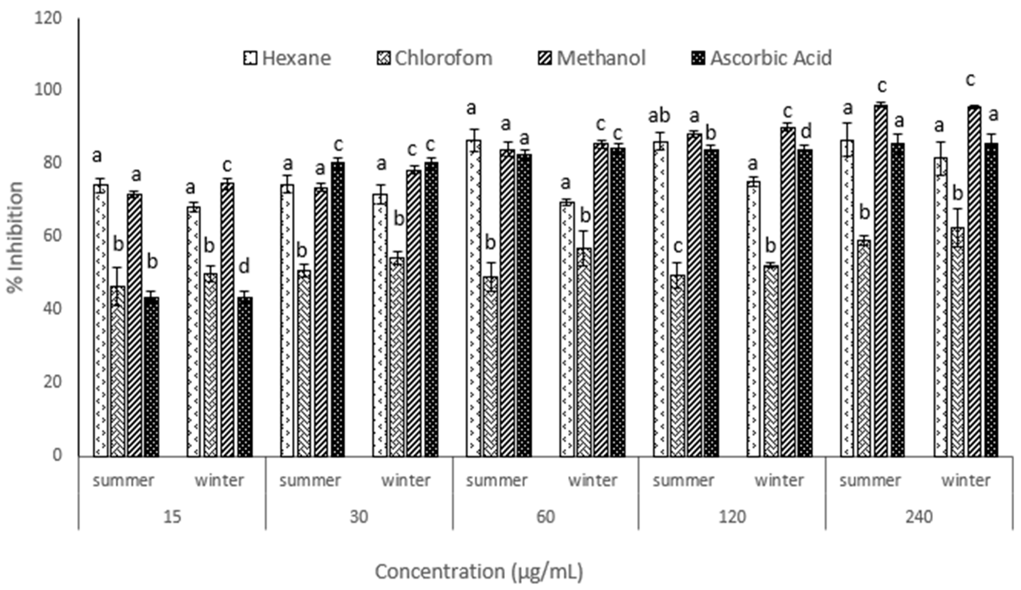
3.3. Antioxidant Activity

The DDPH-radical-scavenging assay was used to determine the antioxidant activity of the crude hexane, chloroform and methanol extracts of the leaves of C. macrocarpa (Figure 1).
For both seasons, the methanol extract of the leaves exhibited the most significant radical-scavenging activity (p < 0.05) at 240 µg/mL, followed by the hexane extract and then the chloroform extract (Figure 1). For each season, the percentage scavenging activity of the DPPH radical increased with the increase in the concentration of the crude methanol leaf extract (Figure 1), which may imply an increased ability to donate hydrogen ions [64]. For the methanol leaf extract, there was a significant difference in the percentage scavenging activities between summer and winter (p < 0.05) at most concentrations. In summer, the percentage scavenging activities of the methanol leaf extract were 71.66, 73.57, 84.05 and 88.22% at 15, 30, 60 and 120 µg/mL, respectively. In winter, the percentage scavenging activities of the methanol leaf extract were 74.65, 78.31, 86.15 and 90.02% at 15, 30, 60 and 120 µg/mL, respectively (Figure 1). Overall, the methanol leaf extract showed a greater percentage of scavenging activity in winter. In summer, the percentage scavenging activities of the hexane, chloroform and methanol extracts at 15 µg/mL were 74.18, 46.64 and 71.66%, respectively, and that of ascorbic was 43.47%. In winter, the percentage scavenging activities of the hexane, chloroform and methanol extracts at 15 µg/mL were 68.27, 49.87 and 74.65%, respectively, and that of ascorbic was 43.47% (Figure 1). The percentage scavenging activities of the different extracts were significantly greater than that of the positive control (p < 0.05), which indicates that the extracts of C. macrocarpa leaves have appreciable antioxidant activity.
The results revealed that in summer, the IC50 values (concentration required for 50% inhibition) of the hexane extract (0.15 µg/mL) and methanol extract (1.72 µg/mL) were lower than that of ascorbic acid (8.26 µg/mL) (Table 4). In winter, the IC50 values of the hexane extract (0.29 µg/mL) and methanol extract (0.67 µg/mL) were also lower than that of ascorbic acid (8.26 µg/mL), whereas the chloroform extract had a greater IC50 value than ascorbic acid (8.26 µg/mL) for summer (44.76 µg/mL) and winter (13.06 µg/mL) (Table 4). A high percentage scavenging activity together with a low IC50 is indicative of strong antioxidant activity. Therefore, hexane and methanol extracts of C. macrocarpa leaves showed strong antioxidant activity. However, the methanol extract of winter leaves showed stronger antioxidant activity than that of summer leaves. While the chloroform extract of winter leaves showed moderate antioxidant activity, that of summer leaves showed low antioxidant activity. The result from this study corresponded to those of a study conducted by [35], whereby the DPPH assay revealed appreciable antioxidant activity in the leaves of C. macrocarpa. A similar study conducted by Abbas et al. [33] revealed that the methanol extracts of C. grandiflora leaves exhibited the highest free-radical-scavenging activity (antioxidant activity), while the hexane fractions exhibited the lowest one. Many phytochemicals possess antioxidant activity, protect our cells against damage caused by oxidative stress and lower the risk of developing certain types of cancer. From previous studies, it is known that flavonoids and phenols exhibit antioxidant properties [51,65]. Flavonoids help to manage diabetes caused by oxidative stress [51]. Phenols were found to possess free-radical-scavenging capabilities and act as food antioxidants [65]. The presence of phenolic compounds in the leaf extracts of C. macrocarpa (Table 2) may have enhanced the scavenging activity of the hexane and methanol extracts [66], thus causing the significantly low IC50 values. In addition, the collective effect of resins, flavonoids, triterpenes and phenols that were present in the leaf extracts (Table 2) may have also positively influenced the percentage inhibition and the IC50 values observed [67,68,69,70,71].
When plants are stressed, they produce more phytochemicals to withstand the unfavourable/harmful conditions [72]. Previous studies were performed on plants under stress conditions, and it was revealed that greater quantities of flavonoids, anthocyanins and mucilage were produced by these plants [73]. Therefore, the observed differences in metabolite yield (%) and antibacterial and antioxidant activities between summer and winter could be due to the differences in the environmental conditions experienced in different seasons. From previous studies, it was suggested that environmental temperatures associated with seasonal change play a vital role in phytochemical composition and biological activity and that it is more pronounced in cold weather [72]. The results from this study indicated that winter provided favourable stimuli (such as low temperatures) to bring about improved phytochemical production and biological activity in the leaves of C. macrocarpa.

4. Conclusions

Carissa macrocarpa leaves are found to contain many bioactive metabolites from various extracts, which implies that the leaves have several medicinal applications. The results from this study suggest that C. macrocarpa extracts possess antibacterial and antioxidant activity; thus, these results are expected to open the possibility of deriving clinically effective drugs from this plant species. It is also found that the winter leaf extracts have potentially higher antibacterial activity and more potent antioxidant activity than the summer leaf extracts, thus inferring that winter is the best season to harvest C. macrocarpa leaves for medicinal use. To the best of our knowledge, this is first report to relate the phytochemical composition and medicinal properties of C. macrocarpa to changes in seasons. It is recommended that further quantitative phytochemical analyses are conducted on this plant in summer and winter. This would improve the understanding of how seasonal changes influence phytochemical composition. This would also elucidate which bioactive compounds are responsible for the potent antioxidant activity in the leaves of C. macrocarpa. The findings from this study add to the existing body of knowledge on South African ethnobotany and aid in better understanding the use and contribution of C. macrocarpa leaves in treating various illnesses.

Author Contributions

Conceptualization, R.R. and Y.N.; methodology, R.R. and Y.N.; formal analysis, R.R., Y.N. and Y.H.D.; investigation and data curation, R.R., Y.N. and Y.H.D.; validation, Y.H.D. and A.N.E.-B.; visualization, A.N.E.-B.; writing—original draft preparation, R.R. and Y.N.; writing—review and editing, Y.N., Y.H.D. and A.N.E.-B., supervision, Y.N. and Y.H.D. All authors have read and agreed to the published version of the manuscript.

Funding

The authors acknowledge Researchers Supporting Project number (RSP-2021/375), King Saud University, Riyadh, Saudi Arabia.

Institutional Review Board Statement

Not applicable.

Informed Consent Statement

Not applicable.

Data Availability Statement

All data are presented within the article.

Acknowledgments

The authors acknowledge Researchers Supporting Project number (RSP-2021/375), King Saud University, Riyadh, Saudi Arabia. Funding by National Research Foundation (NRF-South Africa) is greatly appreciated.

Conflicts of Interest

The authors declare no conflict of interest.

References

  1. Shanley, P.; Luz, L. The Impacts of Forest Degradation on Medicinal Plant Use and Implications for Health Care in Eastern Amazonia. BioScience 2003, 53, 573. [Google Scholar] [CrossRef]
  2. Sheldon, J.W.; Balick, M.J.; Laird, S.A.; Milne, G.M. Medicinal plants: Can utilization and conservation coexist? Adv. Econ. Bot. 1997, 12, 1–104. [Google Scholar]
  3. Mbatha, N.; Street, R.A.; Ngcobo, M.; Gqaleni, N. Sick certificates issued by South African traditional health practitioners: Current legislation, challenges and the way forward: Issues in medicine. South Afr. Med. J. 2012, 102, 129–131. [Google Scholar] [CrossRef] [PubMed]
  4. Svoboda, K.P.; Svoboda, T.G.; Syred, A.D. Secretory Structures of Aromatic and Medicinal Plants: A Review and Atlas of Micrographs; Microscopix Publications: Hoddesdon, UK, 2000. [Google Scholar]
  5. Figueiredo, A.C.; Barroso, J.G.; Pedro, L.G.; Scheffer, J.J. Factors affecting secondary metabolite production in plants: Volatile components and essential oils. Flavour Fragr. J. 2008, 23, 213–226. [Google Scholar] [CrossRef]
  6. Yadav RN, S.; Agarwala, M. Phytochemical analysis of some medicinal plants. J. Phytol. 2011, 3, 10–14. [Google Scholar]
  7. Wakeel, A.; Jan, S.A.; Ullah, I.; Shinwari, Z.K.; Xu, M. Solvent polarity mediates phytochemical yield and antioxidant capacity of Isatis tinctoria. PeerJ 2019, 7, e7857. [Google Scholar] [CrossRef] [PubMed]
  8. Nchabeleng, L.; Mudau, F.N.; Mariga, I.K. Effects of chemical composition of wild bush tea (Athrixia phylicoides DC.) growing at locations differing in altitude, climate and edaphic factors. J. Med. Plants Res. 2012, 6, 1662–1666. [Google Scholar] [CrossRef]
  9. Jayanthy, A.; Prakash, K.U.; Remashree, A.B. Seasonal and geographical variations in cellular characters and chemical contents in Desmodium gangeticum (L.) DC.—An ayurvedic medicinal plant. Int. J. Herb. Med. 2013, 1, 34–37. [Google Scholar]
  10. Odjegba, V.J.; Alokolaro, A.A. Simulated drought and salinity modulate the production of phytochemicals in Acalypha wilkesiana. J. Plant Stud. 2013, 2, 105. [Google Scholar] [CrossRef]
  11. Kale, V.S.; Shitole, M.G. Variable rates of primary and secondary metabolites during different seasons and physiological stages in Datura metel L. J. Maharashtra Agric. Univ. 2010, 35, 371–374. [Google Scholar]
  12. Gololo, S.S.; Shai, L.J.; Agyei, N.M.; Mogale, M.A. Effect of seasonal changes on the quantity of phytochemicals in the leaves of three medicinal plants from Limpopo province, South Africa. J. Pharmacogn. Phytother. 2016, 8, 168–172. [Google Scholar]
  13. Valgas, C.; Souza SM, D.; Smânia, E.F.; Smânia, A., Jr. Screening methods to determine antibacterial activity of natural products. Braz. J. Microbiol. 2007, 38, 369–380. [Google Scholar] [CrossRef]
  14. Babu, H.R.; Savithramma, N. Screening of secondary metabolites of underutilized species of Cyperaceae. Int. J. Pharm. Sci. Rev. Res. 2014, 24, 182–187. [Google Scholar]
  15. Lim, T.K. Carissa macrocarpa. In Edible Medicinal and Non-Medicinal Plants; Springer: Dordrecht, The Netherlands, 2012; pp. 237–239. [Google Scholar]
  16. Halliwell, B.; Gutteridge, J.M. Free Radicals in Biology and Medicine; Oxford University Press: New York, NY, USA, 2015. [Google Scholar]
  17. Erkan, N. Antioxidant activity and phenolic compounds of fractions from Portulaca oleracea L. Food Chem. 2012, 133, 775–781. [Google Scholar] [CrossRef]
  18. Valko, M.; Leibfritz, D.; Moncol, J.; Cronin, M.T.; Mazur, M.; Telser, J. Free radicals and antioxidants in normal physiological functions and human disease. Int. J. Biochem. Cell Biol. 2007, 39, 44–84. [Google Scholar] [CrossRef]
  19. Mayne, S.T. Antioxidant nutrients and chronic disease: Use of biomarkers of exposure and oxidative stress status in epidemiologic research. J. Nutr. 2003, 133, 933S–940S. [Google Scholar] [CrossRef] [Green Version]
  20. Rakesh, S.U.; Patil, P.R.; Mane, S.R. Use of natural antioxidants to scavenge free radicals: A major cause of diseases. Int. J. PharmTech Res. 2010, 2, 1074–1081. [Google Scholar]
  21. Kapadiya, D.B.; Dabhi, B.K.; Aparnathi, K.D. Spices and herbs as a source of natural antioxidants for food. Int. J. Curr. Microbiol. Appl. Sci. 2016, 5, 280–288. [Google Scholar] [CrossRef]
  22. Tadhani, M.B.; Patel, V.H.; Subhash, R. In vitro antioxidant activities of Stevia rebaudiana leaves and callus. J. Food Compos. Anal. 2007, 20, 323–329. [Google Scholar] [CrossRef]
  23. Bhadane, B.S.; Patil, M.P.; Maheshwari, V.L.; Patil, R.H. Ethnopharmacology, phytochemistry, and biotechnological advances of family Apocynaceae: A review. Phytother. Res. 2018, 32, 1181–1210. [Google Scholar] [CrossRef]
  24. El-Taher, A.M.; EL-Gendy, A.G.; Lila, M.I. Morphological and anatomical studies on some taxa of family Apocynaceae. Al-Azhar J. Agric. Res. 2019, 44, 136–147. [Google Scholar] [CrossRef]
  25. The Plant List. Carissa. 2022. Available online: http://www.theplantlist.org/tpl1.1/record/kew-34192 (accessed on 8 August 2022).
  26. Moodley, R.; Koorbanally, N.; Jonnalagadda, S.B. Elemental composition and fatty acid profile of the edible fruits of Amatungula (Carissa macrocarpa) and impact of soil quality on chemical characteristics. Anal. Chim. Acta 2012, 730, 33–41. [Google Scholar] [CrossRef] [PubMed]
  27. Khalil, H.E.; Aljeshi, Y.M.; Saleh, F.A. Authentication of Carissa macrocarpa cultivated in Saudi Arabia; botanical, phytochemical and genetic study. J. Pharm. Sci. Res. 2015, 7, 497. [Google Scholar]
  28. Moodley, R.; Chenia, H.; Jonnalagadda, S.B.; Koorbanally, N. Antibacterial and anti-adhesion activity of the pentacyclic triterpenoids isolated from the leaves and edible fruits of Carissa macrocarpa. J. Med. Plants Res. 2011, 5, 4851–4858. [Google Scholar]
  29. Alsudani AA AA, Z.; Altameme HJ, M. A Taxonomical study of Carissa macrocarpa (Eckl.) A. Dc (Apocynaceae) in Iraq. Rev. Int. Geogr. Educ. 2021, 11, 1328–1341. [Google Scholar]
  30. National Research Council. Lost Crops of Africa: Volume III: Fruits; National Academies Press: Washington, DC, USA, 2008; Volume 3, pp. 262–269. [Google Scholar]
  31. Ibrahim, H.; Abdurahman, E.M.; Shok, M.; Ilyas, N.; Bolaji, R. Preliminary phytochemical and antimicrobial studies of the leaves of Carissa edulis Vahl. In Standardization and Utilization of Herbal Medicines: Challenges of the 21st Century, Proceedings of the 1st International Workshop on Herbal Medicinal Products, Ibadan, Nigeria, 22–24 November 1998; University of Ibadan, Department of Pharmacognosy: Ibadan, Nigeria, 1999; pp. 217–222. [Google Scholar]
  32. Khalil, H.E.; Mohamed, M.E.; Morsy, M.A.; Kandeel, M. Flavonoid and phenolic compounds from Carissa macrocarpa: Molecular docking and cytotoxicity studies. Pharmacogn. Mag. 2018, 14, 304. [Google Scholar] [CrossRef]
  33. Abbas, M.; Rasool, N.; Riaz, M.; Zubair, M.; Abbas, M.; Noor Ul, H.; Hayat, N. GC-MS profiling, antioxidant, and antimicrobial studies of various parts of Carissa grandiflora. Bulg. Chem. Commun. 2014, 46, 831–839. [Google Scholar]
  34. Dhatwalia, J.; Kumari, A.; Verma, R.; Upadhyay, N.; Guleria, I.; Lal, S.; Thakur, S.; Gudeta, K.; Kumar, V.; Chao, J.C.C.; et al. Phytochemistry, pharmacology, and nutraceutical profile of Carissa species: An updated review. Molecules 2021, 26, 7010. [Google Scholar] [CrossRef]
  35. Souilem, F.; Dias, M.I.; Barros, L.; Calhelha, R.C.; Alves, M.J.; Harzallah-Skhiri, F.; Ferreira, I.C. Phenolic profile and bioactive properties of Carissa macrocarpa (Eckl.) A. DC.: An in vitro comparative study between leaves, stems, and flowers. Molecules 2019, 24, 1696. [Google Scholar] [CrossRef]
  36. Souilem, F.; El Ayeb, A.; Djlassi, B.; Ayari, O.; Chiboub, W.; Arbi, F.; Ascizzi, R.; Flamini, G.; Harzallah-Skhiri, F. Chemical composition and activity of essential oils of Carissa macrocarpa (Eckl.) A. DC. Cultivated in Tunisia and its anatomical features. Chem. Biodivers. 2018, 15, e1800188. [Google Scholar] [CrossRef]
  37. Surendra, T.V.; Roopan, S.M.; Arasu, M.V.; Al-Dhabi, N.A.; Sridharan, M. Phenolic compounds in drumstick peel for the evaluation of antibacterial, hemolytic and photocatalytic activities. J. Photochem. Photobiol. B Biol. 2016, 161, 463–471. [Google Scholar] [CrossRef] [PubMed]
  38. Sofowara, A. Medicinal Plants and Traditional Medicine in Africa; Spectrum Books Ltd.: Ibadan, Nigeria, 1993. [Google Scholar]
  39. Tamilselvi, N.; Krishnamoorthy, P.; Dhamotharan, R.; Arumugam, P.; Sagadevan, E. Analysis of total phenols, total tannins and screening of phytocomponents in Indigofera aspalathoides (Shivanar Vembu) Vahl EX DC. J. Chem. Pharm. Res. 2012, 4, 3259–3262. [Google Scholar]
  40. Trease, G.E.; Evans, W.C. Pharmacognosy; Bahiv Tinal: London, UK, 1985. [Google Scholar]
  41. Khandelwal, K. Practical Pharmacognosy; Pragati Books Pvt. Ltd.: Nagpur, India, 2008. [Google Scholar]
  42. Harborne, A.J. Phytochemical Methods a Guide to Modern Techniques of Plant Analysis; Springer Science & Business Media: Dordrecht, The Netherlands, 1998. [Google Scholar]
  43. Bargah, R.K. Preliminary test of phytochemical screening of crude ethanolic and aqueous extract of Moringa pterygosperma Gaertn. J. Pharmacogn. Phytochem. 2015, 4, 7–9. [Google Scholar]
  44. Kumar, R.S.; Balasubramanian, P.; Govindaraj, P.; Krishnaveni, T. Preliminary studies on phytochemicals and antimicrobial activity of solvent extracts of Coriandrum sativum L. roots (Coriander). J. Pharmacogn. Phytochem. 2014, 2, 74–78. [Google Scholar]
  45. Perez, C. Antibiotic assay by agar-well diffusion method. Acta Biol. Med. Exp. 1990, 15, 113–115. [Google Scholar]
  46. Braca, A.; Sortino, C.; Politi, M.; Morelli, I.; Mendez, J. Antioxidant activity of flavonoids from Licania licaniaeflora. J. Ethnopharmacol. 2002, 79, 379–381. [Google Scholar] [CrossRef]
  47. Doss, A.; Mubarack, H.M.; Dhanabalan, R. Antibacterial activity of tannins from the leaves of Solanum trilobatum Linn. Indian J. Sci. Technol. 2009, 2, 41–43. [Google Scholar] [CrossRef]
  48. Ruther, J.; Meiners, T.; Steidle, J.L.M. Allelochemical Reactions Involving Heterotrophic Microorganisms. Biogeochem. Inland Waters 2010, 22, 436. [Google Scholar]
  49. Geetha, B.; Gowda, K.P.; Kulkarni, G.T.; Badami, S. Microwave assisted fast extraction of mucilages and pectins. Indian J. Pharm. Educ. Res. 2009, 43, 260–265. [Google Scholar]
  50. Sungthongjeen, S.; Pitaksuteepong, T.; Somsiri, A.; Sriamornsak, P. Studies on pectins as potential hydrogel matrices for controlled-release drug delivery. Drug Dev. Ind. Pharm. 1999, 25, 1271–1276. [Google Scholar] [CrossRef]
  51. Rahal, A.; Kumar, A.; Singh, V.; Yadav, B.; Tiwari, R.; Chakraborty, S.; Dhama, K. Oxidative stress, prooxidants, and antioxidants: The interplay. BioMed Res. Int. 2014, 2014, 761264. [Google Scholar] [CrossRef] [PubMed]
  52. Rabi, T.; Bishayee, A. Terpenoids and breast cancer chemoprevention. Breast Cancer Res. Treat. 2009, 115, 223–239. [Google Scholar] [CrossRef] [PubMed]
  53. Krishnaiah, D.; Devi, T.; Bono, A.; Sarbatly, R. Studies on phytochemical constituents of six Malaysian medicinal plants. J. Med. Plants Res. 2009, 3, 67–72. [Google Scholar]
  54. Shah, B.A.; Qazi, G.N.; Taneja, S.C. Boswellic acids: A group of medicinally important compounds. Nat. Prod. Rep. 2009, 26, 72–89. [Google Scholar] [CrossRef]
  55. Peter, H.; Pavel, T.; Marie, S. Flavonoids-potent and versatile biologically active compounds interacting with cytochrome P450. Chem. Biol. Interact. 2002, 139, 1–21. [Google Scholar]
  56. Killedar, S.G.; More, H.N. Estimation of tannins in different parts of Memecylon umbellatum Burm. J. Pharm. Res. 2010, 3, 554–556. [Google Scholar]
  57. Ganguly, T.; Sainis, K.B. Inhibition of cellular immune responses by Tylophora indica in experimental models. Phytomedicine 2001, 8, 348–355. [Google Scholar] [CrossRef]
  58. Staerk, D.; Lykkeberg, A.K.; Christensen, J.; Budnik, B.A.; Abe, F.; Jaroszewski, J.W. In Vitro Cytotoxic Activity of Phenanthroindolizidine Alkaloids from Cynanchum vincetoxicum and Tylophora tanakae against Drug-Sensitive and Multidrug-Resistant Cancer Cells. J. Nat. Prod. 2002, 65, 1299–1302. [Google Scholar] [CrossRef]
  59. Just, M.J.; Recio, M.C.; Giner, R.M.; Cuéllar, M.J.; Máñez, S.; Bilia, A.R.; Ríos, J.L. Anti-inflammatory activity of unusual lupane saponins from Bupleurum fruticescens. Planta Med. 1998, 64, 404–407. [Google Scholar] [CrossRef]
  60. Estrada, A.; Katselis, G.S.; Laarveld, B.; Barl, B. Isolation and evaluation of immunological adjuvant activities of saponins from Polygala senega L. Comp. Immunol. Microbiol. Infect. Dis. 2000, 23, 27–43. [Google Scholar] [CrossRef]
  61. Nikaido, H.; Vaara, M. Molecular basis of bacterial outer membrane permeability. Microbiol. Rev. 1985, 49, 1–32. [Google Scholar] [CrossRef] [PubMed]
  62. Rabe, T.; Van Staden, J. Antibacterial activity of South African plants used for medicinal purposes. J. Ethnopharmacol. 1997, 56, 81–87. [Google Scholar] [CrossRef]
  63. Sekirov, I.; Russell, S.L.; Antunes LC, M.; Finlay, B.B. Gut microbiota in health and disease. Physiol. Rev. 2010, 90, 859–904. [Google Scholar] [CrossRef]
  64. Silva, C.D.; Herdeiro, R.S.; Mathias, C.J.; Panek, A.D.; Silveira, C.S.; Rodrigues, V.P.; Renno, M.N.; Falcao, D.Q.; Cerqueira, D.M.; Minto AB, M.; et al. Evaluation of antioxidant activity of Brazilian plants. Pharmacol. Res. 2005, 52, 229–233. [Google Scholar] [CrossRef] [PubMed]
  65. Kasote, D.M.; Katyare, S.S.; Hegde, M.V.; Bae, H. Significance of antioxidant potential of plants and its relevance to therapeutic applications. Int. J. Biol. Sci. 2015, 11, 982. [Google Scholar] [CrossRef] [PubMed]
  66. Srivastava, J.; Kumar, S.; Vankar, P.S. Correlation of antioxidant activity and phytochemical profile in native plants. Nutr. Food Sci. 2012, 42, 71–79. [Google Scholar] [CrossRef]
  67. Kessler, M.; Ubeaud, G.; Jung, L. Anti-and pro-oxidant activity of rutin and quercetin derivatives. J. Pharm. Pharmacol. 2003, 55, 131–142. [Google Scholar] [CrossRef]
  68. Talla, E.; Tamfu, A.N.; Gade, I.S.; Yanda, L.; Mbafor, J.T.; Laurent, S.; Elst, L.V.; Popova, M.; Bankova, V. New mono-ether of glycerol and triterpenes with DPPH radical scavenging activity from Cameroonian propolis. Nat. Prod. Res. 2017, 31, 1379–1389. [Google Scholar] [CrossRef]
  69. Miao, J.; Li, X.; Zhao, C.; Gao, X.; Wang, Y.; Gao, W. Active compounds, antioxidant activity and α-glucosidase inhibitory activity of different varieties of Chaenomeles fruits. Food Chem. 2018, 248, 330–339. [Google Scholar] [CrossRef]
  70. Akwu, N.A.; Naidoo, Y.; Singh, M.; Nundkumar, N.; Lin, J. Phytochemical screening, in vitro evaluation of the antimicrobial, antioxidant and cytotoxicity potentials of Grewia lasiocarpa E. Mey. ex Harv. S. Afr. J. Bot. 2019, 123, 180–192. [Google Scholar] [CrossRef]
  71. Assimopoulou, A.N.; Zlatanos, S.N.; Papageorgiou, V.P. Antioxidant activity of natural resins and bioactive triterpenes in oil substrates. Food Chem. 2005, 92, 721–727. [Google Scholar] [CrossRef]
  72. Iqbal, S.; Bhanger, M.I. Effect of season and production location on antioxidant activity of Moringa oleifera leaves grown in Pakistan. J. Food Compos. Anal. 2006, 19, 544–551. [Google Scholar] [CrossRef]
  73. Kaplan, F.; Kopka, J.; Sung, D.Y.; Zhao, W.; Popp, M.; Porat, R.; Guy, C.L. Transcript and metabolite profiling during cold acclimation of Arabidopsis reveals an intricate relationship of cold-regulated gene expression with modifications in metabolite content. Plant J. 2007, 50, 967–981. [Google Scholar] [CrossRef] [PubMed]
Figure 1. In vitro antioxidant activity (% scavenging) at different concentrations (µg/mL) of crude extracts from summer and winter leaves of C. macrocarpa. Different letters means significant at p < 0.05.
Horticulturae 08 00780 g001
Table 1. Yield percentage of the dried extracts of summer and winter leaves of C. macrocarpa.
SolventYield (Summer)Yield (Winter)
(g)(%)(g)(%)
Hexane0.828.20.989.8
Chloroform0.484.80.656.5
Methanol1.5152.4324.3
Table 2. Qualitative preliminary screening of phytochemicals of C. macrocarpa leaf extracts for summer and winter seasons.
Phytochemical
Constituent
Type of TestWinterSummer
HCMH CM
AlkaloidsWagner’s--+-++++
Meyer’s+++-+++-
TanninsFerric chloride++++++++
PhenolsLead acetate++++++++++
Tannins (naphthoquinone)Gelatine++-++-+
FlavonoidsAlkaline-reagent test+-++-+
Acid-hydrolysis test+-+++-++
SaponinsFoam test--+ --+
Olive oil test-++++-+++
Steroids (terpenoids)Salkowski’s test+-++-+
Lieberman–Bouchard test+-++-+
ProteinsBiuret test++++++
CarbohydratesMolisch’s test-++++-++
Mucilage + gumsPrecipitation test++-++-
Ruthenium-red test-++-++
ResinAcetone test+++-+++-
- Absent, + Present, ++ Intense positive, H = Hexane, C = Chloroform, M = Methanol.
Table 3. Means and standard deviations of the zones of inhibition (mm) of the methanol extract of C. macrocarpa leaves (summer and winter) at different concentrations (mg/mL).
Bacterial StrainConcentrations (mg/mL)Positive Control
1052.51.250.625
Summer
S. aureus7.75 ± 1.777.25 ± 0.356.75 ± 0.357.0 ± 0.007.0 ± 0.0010 ± 1.41
E. coli6.75 ± 0.356.75 ± 0.356.5 ± 0.356.5± 0.006.5 ± 0.0011 ± 1.41
Winter
S. aureus8.17 ± 1.048.17 ± 1.047.25 ± 0.357.0 ± 0.007.0 ± 0.0010 ± 1.32
E. coli6.83 ± 0.586.83 ± 0.586.75 ± 0.006.5± 0.006.5 ± 0.0011 ± 1.00
Table 4. IC50 values showing the antioxidant activity of the various solvent extracts from summer and winter leaves of C. macrocarpa.
SampleIC50 (µg/mL)
SummerWinter
Hexane0.150.29
Chloroform44.7613.06
Methanol1.720.67
Ascorbic acid8.268.26
Publisher’s Note: MDPI stays neutral with regard to jurisdictional claims in published maps and institutional affiliations.

