Article

The Breeding of High-Quality Dandelions by NaCl Induced Callus Variation Combined with a Drosophila Tumor Cell Migration Test

1
Institute of Coastal Agriculture, Hebei Academy of Agriculture and Forestry Sciences, Tangshan 063299, China
2
College of Traditional Chinese Medicine, North China University of Science and Technology, Tangshan 063210, China
*
Authors to whom correspondence should be addressed.
Horticulturae 2022, 8(12), 1167; https://doi.org/10.3390/horticulturae8121167
Submission received: 2 November 2022 / Revised: 29 November 2022 / Accepted: 7 December 2022 / Published: 8 December 2022
(This article belongs to the Special Issue Morphology, Palynology and Phytochemicals of Medicinal Plants)

Abstract

:
Creating high-quality and salt-tolerant plant germplasm is an effective strategy to improve the utilization of saline-alkali land. Salt-induced callus mutation was used to create dandelion germplasm and mutant dandelion calluses were obtained under NaCl concentrations of 0.8%, 1%, and 1.2% with the identification of random amplified polymorphic DNA (RAPD) markers. A new dandelion line, “Binpu 2”, selected from the progenies of dandelion tissue culture plantlets that originated from callus treated under 0.8% NaCl, was evaluated in light of its morphological characteristics, bioactive components, and antitumor functions. Results showed that the plant shape of “Binpu 2”was nearly upright; its cichoric acid content was 6.7 mg/g, which was 39.6% and 36.7% higher than its mother plant and local dandelion cultivar, respectively; its leaf water extracts of 0.2 g/mL had a significant inhibitory effect on cell polarity disruption-induced cell migration without affecting drosophila normal growth, revealed by the strong inhibitory effect on tumor cell migration, the increased level of MMP1 and β-Integrin, and the reduced E-cadherin expression. Our results suggested that “Binpu 2” originated from salt-induced mutant performed better than its mother plant and processed strong antitumor function, which was suitable for amplified cultivation in saline-alkali land for food and medicinal industrial development.

1. Introduction

Soil salinization has become the main factor restricting agricultural development in China [1]. Cultivating salt-tolerant crops have represented the most fundamental and effective method of utilizing saline–alkali land and improving economic profit [2,3]. Studies have shown that plants growing under saline stress could accumulate more nutrients and active substances, and had better nutritional, medicinal, and other economic benefits [4].
Dandelion (Taraxacum mongolicum Hand.-Mazz.), a traditional herb plant used for medication, rubber, cosmetic, feed additives, as well as vegetables in recent years [2,5], could tolerate saline stress up to 1.5% of NaCl [6], which was suitable for cultivation in saline-alkali land. However, poor commercial characters of dandelion in saline-alkali land struggled to satisfy the demands of industrial production. Hence, it was urgent to develop dandelion varieties with good comprehensive performance. At present, most dandelion varieties were bred by natural selection, mainly because dandelion is a self-pollinating plant of with capitulum, leading to the hard application of common hybridization breeding [6]. Furthermore, related molecular breeding in dandelions has been hardly reported, mainly due to the complex genotype and little genome sequence information [7]. Thus, we adopted a new breeding strategy through salt-induced callus mutation to breed a high-quality dandelion variety suitable for cultivation in saline–alkali land.
Because of the medicinal use of dandelion, besides the biomass yield, a good cultivar with highly bioactive compounds and function was also vital. Thus, we evaluated antitumor function at the same time. Cancer is a worldwide fatal disease that seriously threatens human health. More than 90% of cancer patients died from tumor migration, instead of primary tumor overgrowth [8]. Dandelions have traditionally been employed in Chinese, Native American and Arabian traditional medicine for the purpose of treating diseases, including multiple types of cancers [9,10]. The anti-cancer efficacy of dandelion extracts has been previously reported, while their effects on tumor cell migration in vivo were largely unknown.
Natural malignant tumours can occur in Drosophila melanogaster (D. melanogaster). Tumours can be experimentally induced in larvae and adult flies by knocking down fly tumour suppressor genes. D. melanogaster has an important role in chemical genetics, helping to identify the pathways that are affected by pharmaceuticals, facilitating the design of more efficient derivatives and serving as a platform for semi-automated screens for new anticancer drugs [11,12]. Therefore, with less genome redundancy and powerful genetic tools, D. melanogaster was selected as the elegant system to test tumor metastasis.
The current study firstly adopted the method of salt-induced callus mutation combined with functional tests for dandelion breeding. Details of the breeding process were of great significance to enrich modern breeding practices and agriculture developments in saline-alkali land.

2. Materials and Methods

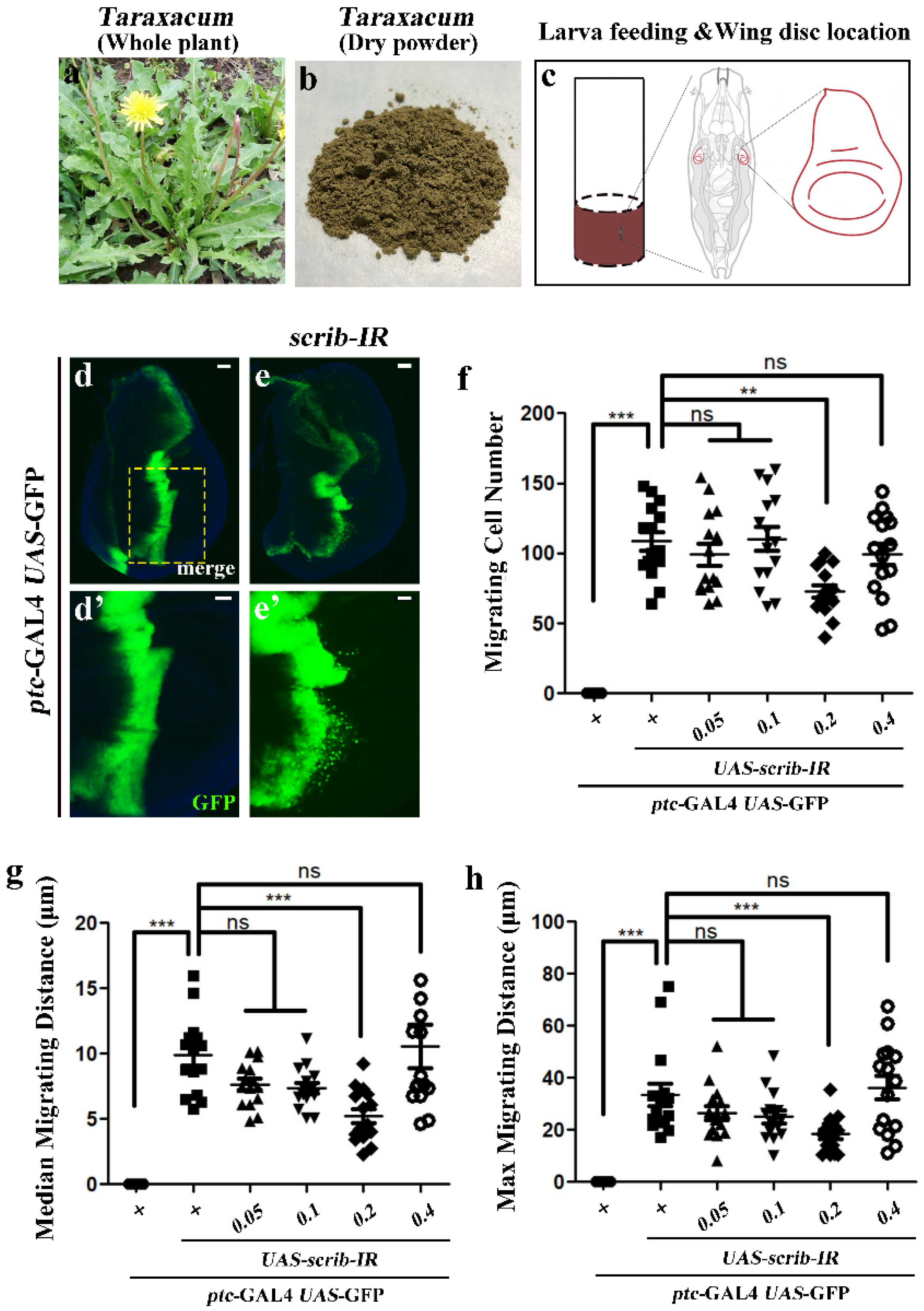
2.1. Dandelion Materials

A wild dandelion resource “Daye” (Taraxacum mongolicum Hand.-Mazz.) was selected as the mother plant due to high biomass yield. This resource was further bred into sister lines “Binpu 1” [13,14] and “Binpu 2” [15] by the Institute of Coastal Agriculture, Hebei Academy of Agriculture and Forestry Sciences, China. “Binpu 1” was noted for its high yield and “Binpu 2” was noted for its strong functions. Their varietal characteristics could be found from above mentioned references. This study mainly focused on the breeding process of “Binpu 2”. The mother plant “Daye” was indoor cultured for 30 days at 25 °C with 16 h of illumination at 2500 lux. Then, the healthy and young leaves were taken as explants for producing callus. Related dandelion materials were shown in Figure S1.

2.2. Production of Dandelion Tissue Culture Plantlet

2.2.1. Induction of Callus with Saline Treatment

Fresh sterilized leaves [6] were cut into 300 approximate pieces of about 1 cm2 and then inoculated with solid MS medium [6] containing 0.5 mg/L of 6-BA and 0.04 mg/L of 2,4-D for 45 days in sterile tissue culture room. Once selected, the callus with the best growth, in order to obtain sufficient experimental materials of callus, was sub-cultured by solid MS medium for 5 times with 21 days for each time. Then, obtained calluses were cut into small pieces of about 0.25 cm2 and treated by solid MS medium containing 85 mM, 136 mM, 170 mM, 205 mM, 256 mM, and 307 mM NaCl, respectively, for 28 days. Each NaCl treatment of solid MS medium contained 30 calluses. Subsequently, one of the best growing callus in each NaCl treatment was picked out into a NaCl-free solid MS medium individually for continuously culturing over 21 days, and then continued to be treated by NaCl treatment of solid MS media for 21 days. Above shift-NaCl process was repeated two times. Lastly, the calluses were transferred to NaCl-free solid MS medium for amplified cultivation over 2 months. Above all works were done in tissue culture room with 24 h of fluorescence illumination at 3000 lux and 25 °C.

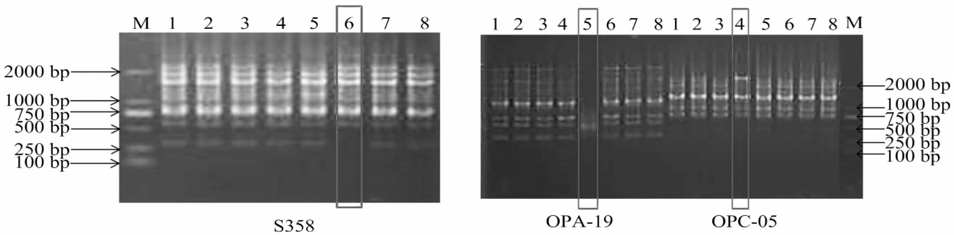
2.2.2. Identification of Salt-Treated Callus

The calluses obtained above were identified by random amplified polymorphic DNA (RAPD) markers. Details of primers, via screening and identification techniques, can be seen from our previous research [14]. DNA extraction was processed by the DNA-secure plant genome DNA extraction kit (Tiangen Biotech Co., Ltd., Beijing, China). In total 41 random primers (Table S1) were used for RAPD identification. The polymerase chain reaction (PCR) amplification procedure involved pre-denaturation for 5 min at 94 °C, followed by 40 cycles of denaturation for 1 min at 94 °C, annealing for 1 min at 38 °C, extension for 2 min at 72 °C, and amplification for 10 min at 72 °C.

2.2.3. Regeneration of Dandelion Tissue Culture Plantlet

The calluses obtained above were placed on a solid MS medium containing 0.2 mg/L of 6-BA and 0.01 mg/L of NAA due to the occurrences of massive adventitious buds and then transferred to a 1/2 solid MS medium with free plant hormone for the continuous culturing process which was carried out 2 times with 21 days of each time. Then, calluses were transferred to a 1/2 solid MS medium containing 0.3 mg/L of NAA for culturing over 21 days. All above processes were finished in a tissue culture room with 24 h of fluorescence illumination at 3000 lux and 25 °C. The resulting tissue seedlings were acclimatized for 35 days to improve the survive rate for the field transplant. Firstly, the seedlings were gradually contacted in an external environment for 3 weeks, and the room was kept ventilated with a temperature at 23 °C, and a relative humidity of 80%. Then, these seedlings were transferred to flowerpots containing vermiculite-nutrient soil-perlite at a ratio of 1:4:3, and were continuously cultured for 2 weeks in field shelter and sprayed with a 1/4 liquid MS medium each day over two weeks. Finally, these tissue culture plantlets were transplanted to the outdoor field and their seeds were harvested separately for amplified field cultivation.

2.3. Determination of Candidate Dandelion Lines

The seeds from dandelion tissue culture seedlings were grown in a seedling tray in greenhouse and then transplanted to an observation nursery in saline-alkali land (Figure S1) when the seedlings grew to three leaves. The soil salt content within 20 cm depth in the observation nursery was checked by AZ 8373 TDS/salinity meter (AZ Instrument Corp., Taiwan, China) [2] and averaged value was around 0.3%. These dandelions plants were cultured for 6 weeks, and various characteristics (such as plant shape, leaf shape, and leaf color, as well as chicoric acid, caffeic acid, and the total flavonoids contents) were repeatedly investigated during the cultivation period. Seeds from these dandelion plants were harvested and propagated separately over 3 generations and finally the plants with stable characteristics were picked out as candidate dandelion lines according to the trait investigation. We finally obtained 6 such dandelion lines as marked by Y1–Y6, which meant these selected lines were originated from the calluses that under NaCl treatment of 85 mM (Y1), 136 mM (Y2), 170 mM (Y3), 205 mM (Y4), 256 mM (Y5), and 307 mM NaCl (Y6), respectively.
Of these characteristics, for determination of compounds contents, 0.5 g of dry dandelion powder was mixed with 25 mL of 50% methanol and ultrasonically extracted for 30 min at 60 °C. Then, the extract was filtered by 0.45 µm membrane for contents check. Contents of chicory acid, caffeic acid and chlorogenic acid were determined by Agilent1200 high performance liquid chromatography (Agilent Technologies Inc., Santa Clara, CA, USA), as described in [5,16]; the total flavonoids were detected by UV-2450 spectrophotometer (Shimadzu Co., Ltd., Kyoto, Japan), as described in [17].

2.4. Anti-Tumor Cell Migration Evaluation

2.4.1. Preparation of Dandelion Extracts

The above obtained dandelion lines, including their mother plant “Daye”, which was cultivated in the field, were harvested at the same time and dried in oven at 60 °C for 2 days. Then, 100 g of dry powder was taken to be extracted with 1000 mL of double-distilled water (ddH2O) at 60 °C for 3 h. The extract that formed was vacuum filtered with a 0.45 µm filter. Then, the extract was frozen at −80 °C and freeze-dried using a lyophilizer, and reconstituted in ddH2O to obtain a final stock concentration of 0.5 g/mL. The extract was added directly to regular food from a 0.5 g/mL aqueous stock to a final concentration of 0.05, 0.1, 0.2, and 0.4 g/mL. For the control food, we used water alone.

2.4.2. Fly Strains

The flies were kept on a cornmeal and agar medium at 25 °C with a 12 h light-dark cycle incubator [18]. Drosophila strains used w1118 (#3605) which was obtained from the Bloomington Drosophila stock center. UAS-scrib-IR (#27424) was obtained from Vienna Drosophila RNAi center [18]. ptc-GAL4 was previously described [19]. For cell migration test, crosses were raised at 25 °C for 2 days, and then shifted to 29 °C for an additional 3 days, The third-instar larvae were dissected.

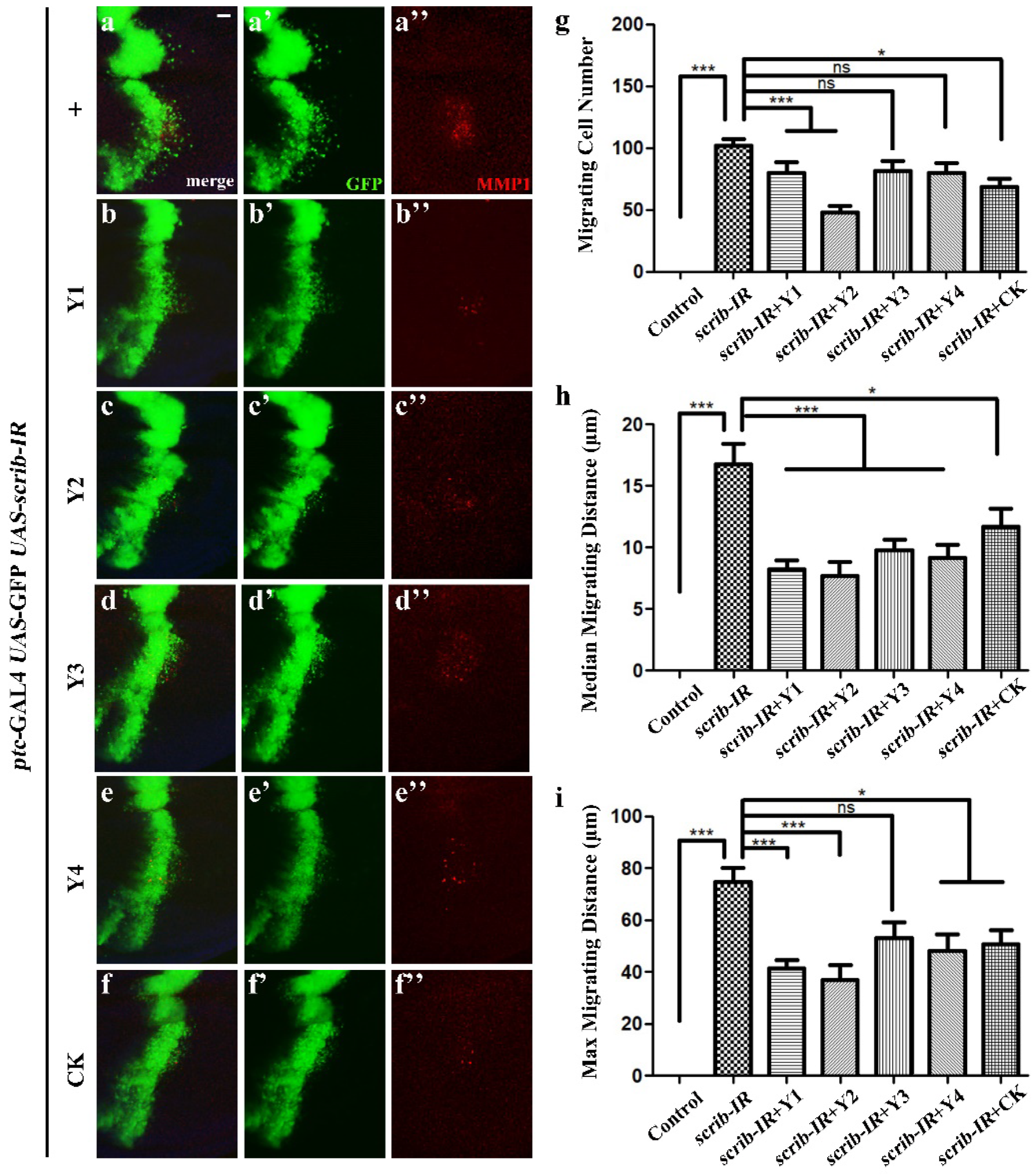
2.4.3. Immunostaining

Larval discs were dissected and then fixed in 4% formaldehyde for 40 min. After several washes with 0.3% (v/v) PBST, the discs were stained with primary antibodies at 4 °C overnight and then secondary antibodies at room temperature for 2 h. The following antibodies were used: mouse anti-MMP1 (1:200, Developmental Studies Hybridoma Bank, DSHB (Iowa City, IA, USA), Cat. #3A6B4-c), mouse anti-β integrin (1:100, DSHB, Cat. #CF.6G11-c), rat anti-DE cadherin (1:100, DSHB, Cat. #DCAD2-c), goat anti-mouse cyanine-3 (1:1000, Life Technologies (Carlsbad, CA, USA), Cat. #A10521), goat anti-rat cyanine3 (1:1000, Life Technologies, Cat. #A10522). Vectashield mounting media (Vector Laboratories, Cat. #H-1500) was used for mounting.

2.5. Data Analysis

The Data were presented as bar graphs or mean value ± standard divisions. The figures were created using the Origin 8.0 software (OriginLab Corporation, Northampton, MA, USA). For statistical significance, one-way ANOVA, along with Bonferroni’s multiple-comparison test and Duncan’s multiple range test, was applied for the drosophila test and dandelion filed evaluation. A p value less than 0.05 was considered significant; ns was not significant, p ≥ 0.05; * was significant, p < 0.05; ** meant p < 0.01; *** meant p < 0.001.

3. Results and Discussion

3.1. Identification of Salt-Induced Calluses

RAPD identification showed that 3 (S358, OPA-19 and OPC-05) out of 41 random primers amplified different bands from that of the control corresponding to NaCl concentrations of 205 mM, 170 mM, and 136 mM, respectively (Figure 1), indicating that high-NaCl-concentration exceeding 136 mM could induce DNA variation, which may cause some different extend of trails variations from their mother plant, such as plant shape, leaf shape, and plant size, etc. [6]. However, dandelions in natural conditions also have chance to produce some morphological variations, and parts of reasons were ascribed to apomixis [6]. No matter what treatment method is used, for dandelion breeders, it is the premise to make trait variation between offspring and mother parent, and then continue to carry out excellent trait screening work for an ideal dandelion line. Our method of NaCl-induced calluses mutation provided the probability for subsequent dandelion breeding works.

3.2. Morphological Evaluation

As the method described for the determination of dandelion lines in the section of materials and method, we obtained 6 candidate dandelion lines and recorded 11 morphological parameters for them. Results showed that these dandelion lines showed some differences in aspects of plant shape, leaf pubescence, leaf vein color, leaf size, etc. (Table 1). Especially, we found two dandelion materials originated from that of NaCl concentrations of 136 mM (Y2) and 170 mM (Y3) were obviously advantaged than other materials in the characteristics of plant shape and whole plant size. Dandelions from Y2 appeared obviously upright growth, while Y3 showed larger size for whole plant than other materials. In addition, among the 11 morphological parameters, the purple at the base of leaf vein was stably inherited and not affected by the environment according to our long-term observations and these materials involved Y2 and Y5.
The plant shape was an important index in directly determining mechanized harvests in the future. The plant shape was divided into three criteria, namely flat growth (angle < 30°), half-upright growth (30° < angle < 75°), and upright growth (angle > 75°), based on the angle between the new leaf and ground. As plant shape of most dandelions was flat or had intimately half-upright growth, leading to that their harvest of fresh leaf was mainly relied on artificial way. Therefore, the relatively upright growth was vital trait for considering the mechanized operation in the future, which could greatly decrease the harvest cost comparing to the manual harvesting. The plant shape of dandelion lines from Y2 and Y3 were divided into upright and half-upright growth, which were named as “Binpu 2” and “Binpu 1” later. In addition, the large plant size of Y3 indicated a potentially high yield. Hence, dandelion materials from Y2 and Y3 were the focus for dandelion breeding.

3.3. Yield and Bioactive Compounds Contents

Dandelions are rich in phenolic acids and flavonoids, which have strong antioxidant and antitumor effects [20]. We further compared leaf yields and the contents of compounds from the two lines (Y2 and Y3). Overall, dandelions of Y3 showed the highest yield and Y2 had the highest compounds content (Table 2). Y2 demonstrated high contents in caffeic acid, chlorogenic acid, and cichoric acid. Of these, the cichoric acid content was 39.6%, 36.7% and 48.9% higher than its mother plant, local dandelion cultivar, and the criterion that stipulated by “the People’s Republic of China Pharmacopoeia 2020”, respectively. Although the total flavonoids content in Y2 were lower than in other resources, considering the relatively high fresh leaf yield, it was still considered to be a satisfied resource for high contents of bioactive compounds.
The dandelion is used as traditional herbal medicine, and generally high contents of bioactive compounds are an important consideration for dandelion quality or function evaluation. Hence, based on the above results, we continued comparing the ability of anti-tumor cell migration among these dandelion lines at the same time.

3.4. Tumor Cell Migration Evaluation

3.4.1. Inhibitory Dose of Dandelion Extract on Tumor Cell Migration

The mobilized UAS/GAL4 expression system was used to generate a cell migration model in Drosophila with third-instar larval wing epithelia. RNA interference (RNAi) mediated the knockdown of the cell polarity gene scribble (scrib) triggered by massive cell migration phenotype (Figure 2d,e) [21,22]. The green fluorescent proteins (GFPs), labeled as migrating cells, were detached from the anterior-posterior (A/P) compartment boundary and moved toward the posterior part of the wing imaginal discs (Figure 2e,e’). After quantifying this phenotype, strong increased levels in the migrating cell number, the median, or the max distance were found (Figure 2f–h). Then, we tested the inhibitory effect of different concentrations of dandelion extract (Figure 2b,c) on ptc > scrib-IR in the Drosophila cell migrating model. The results only showed the extracts at the concentration of 0.2 g/mL obviously inhibited scrib-depleted induced cell invasion without affecting the fly normal growth (Figure 2f–h).

3.4.2. Evaluation of Different Dandelions on Tumor Cell Migration

Based on above results, a 0.2 g/mL extract of different dandelion lines were fed to ptc > scrib-IR larvae. A loss of scrib could induce strong epithelial-mesenchymal transition (EMT) phenotypes, as revealed by the up-regulated expression of matrix metalloprotease1 (MMP1) (Figure 3a) and β-integrin (Figure S2), and a reduction in E-cadherin (a cell adhesion molecule) levels (Figure S2) [21,23,24]. By combining the statistical results of migrating cell number and migrating distances, the increased cell migration of ptc > scrib-IR was mildly suppressed by CK (mother plant) and largely inhibited by Y2 (Figure 3g–i). The up-regulated MMP1 expression was obviously suppressed by Y1, Y2, Y4, or CK, but not by Y3 (Figure 3a–f). In addition, the up-regulation of β-integrin and the reduction in E-cadherin activity were only inhibited by Y2, whereas the other tests had no effects on the β-integrin and E-cadherin alterations induced by a loss of scrib (Figure S2). Collectively, these results proved that dandelion materials from Y2 had a strong inhibitory effect on cell migration, the increased level of MMP1 and β-integrin, and the reduced expression of E-cadherin.

4. Conclusions

We created a series of dandelion progeny materials based on salt-induced callus mutation and tissue culture techniques. Combined with the results of morphological evaluation, a comparison of the main bioactive compounds, as well as an anti-tumor function evaluation of lines of Y2 and Y3, showed satisfied performance in functionality and yield. Of these, Y2 exhibited the highest phenolic acids content and strong ability of anti-tumor cell migration; thus we named Y2 as “Binpu 2”. Its plant shape was upright, the fresh leaf yield was 14.2 t/ha, and the cichoric acid content was 6.7 mg/g. “Binpu 2” was suitable for mechanized harvesting and could be widely used for production in the field of food and medicine.

Supplementary Materials

The following supporting information can be downloaded at: https://www.mdpi.com/article/10.3390/horticulturae8121167/s1, Figure S1: Progenies of dandelion tissue culture plantlets grown on saline-alkali land; Figure S2: Dandelion extract suppresses β-integrin accumulation and reduced E-cadherin level induced by loss of scrib; Table S1: PCR primers information.

Author Contributions

Conceptualization and methodology, Z.W. and C.W.; software, Z.W.; validation, C.W. and R.M.; formal analysis, Z.W. and Z.L.; investigation, W.F., Z.L. and R.M.; resources, C.W. and W.F.; data curation, C.W.; writing—original draft preparation, Z.W.; writing—review and editing, Z.W. and C.W.; visualization, Z.L.; supervision, X.W.; project administration, X.W.; funding acquisition, X.W. All authors have read and agreed to the published version of the manuscript.

Funding

This research was funded by Hebei S & T Innovation Team of Modern Seed Industry of Traditional Chinese Medicine (21326312D-8); HAAFS Science and Technology Innovation Special Project (2022KJCXJX-BHS-4); Hebei Natural Science Foundation (H2022209027); Basic Research Funds of Hebei Academy of Agriculture and Forestry Sciences (2021010202); and Tangshan Talent Funding Project (A202203021).

Data Availability Statement

Not applicable.

Acknowledgments

We thank Lei Xue for technical support and thank the Bloomington and VDRC Fly Centers for fly stocks.

Conflicts of Interest

The authors declare no conflict of interest.

References

  1. Wang, X.; Xue, Z.; Lu, X.; Liu, Y.; Liu, G.; Wu, Z. Salt leaching of heavy coastal saline silty soil by controlling the soil matric potential. Soil Water Res. 2019, 3, 132–137. [Google Scholar] [CrossRef] [Green Version]
  2. Wu, Z.; Xue, Z.; Li, H.; Zhang, X.; Wang, X.; Lu, X. Cultivation of dandelion (Taraxacum erythropodium) on coastal saline land based on the control of salinity and fertilizer. Folia Hortic. 2019, 31, 277–284. [Google Scholar] [CrossRef] [Green Version]
  3. Liu, Y.; Sun, J.; Dai, X.; Zhao, Z.; Wang, X.; Zhang, G. Response of soil bacterial community structure and function under two salt-tolerant plants in a coastal saline soil area of eastern Hebei province of China. Int. J. Phytoremediat. 2021, 24, 842–854. [Google Scholar] [CrossRef] [PubMed]
  4. Wu, Z.; Xue, Z.; Lu, X.; Jia, Y.; Wang, X.; Zhang, X. Salt-tolerance identification and quality evaluation of Abelmoschus manihot (L.) Medik. Can. J. Plant Sci. 2020, 100, 568–574. [Google Scholar] [CrossRef]
  5. Xie, P.; Huang, L.; Zhang, C.; Ding, S.; Deng, Y.; Wang, X. Skin-care effects of dandelion leaf extract and stem extract: Antioxidant properties, tyrosinase inhibitory and molecular docking simulations. Ind. Crop. Prod. 2018, 111, 238–246. [Google Scholar] [CrossRef]
  6. Zhang, X.; Li, Y.; Chen, H.; Shi, W. Selection and identification of salt-tolerant variants of Taraxacum officinale. Chin. J. Biotechnol. 2008, 24, 262–271. (In Chinese) [Google Scholar]
  7. Liu, Q.; Liu, Y.; Xu, Y.; Yao, L.; Liu, Z.; Cheng, H.; Ma, M.; Wu, J.; Wang, W.; Ning, W. Overexpression of and RNA interference with hydroxycinnamoyl-CoA quinate hydroxycinnamoyl transferase affect the chlorogenic acid metabolic pathway and enhance salt tolerance in Taraxacum antungense Kitag. Phytochem. Lett. 2018, 28, 116–123. [Google Scholar] [CrossRef]
  8. Sigstedt, S.C.; Hooten, C.J.; Callewaert, M.C.; Jenkins, A.R.; Romero, A.E.; Pullin, M.J.; Kornienko, A.; Lowrey, T.K.; Slambrouck, S.V.; Steelant, W.F. Evaluation of aqueous extracts of Taraxacum officinale on growth and invasion of breast and prostate cancer cells. Int. J. Oncol. 2008, 32, 1085–1090. [Google Scholar] [CrossRef]
  9. Nassan, M.A.; Soliman, M.M.; Ismail, S.A.; El-Shazly, S. Effect of Taraxacum officinale extract on PI3K/Akt pathway in DMBA-induced breast cancer in albino rats. Biosci. Rep. 2018, 38, BSR20180334. [Google Scholar] [CrossRef] [Green Version]
  10. Menke, K.; Schwermer, M.; Felenda, J.; Beckmann, C.; Stintzing, F.; Schramm, A.; Zuzak, T. Taraxacum officinale extract shows antitumor effects on pediatric cancer cells and enhance mistletoe therapy. Complement. Ther. Med. 2018, 40, 158–164. [Google Scholar] [CrossRef]
  11. Gonzalez, C. Drosophila melanogaster: A model and a tool to investigate malignancy and identify new therapeutics. Nat. Rev. Cancer 2013, 13, 172–183. [Google Scholar] [CrossRef]
  12. Zhang, S.; Guo, X.; Wu, H.; Sun, Y.; Ma, X.; Li, J.; Xu, Q.; Wu, C.; Li, Q.; Jiang, C.; et al. Wingless modulates activator protein-1-mediated tumor invasion. Oncogene 2019, 38, 3871–3885. [Google Scholar] [CrossRef]
  13. Wang, X.; Lu, X. A new dandelion variety “Binpu 1” with high yield, good quality and salt tolerance. Xian Dai Nong Cun Ke Ji 2019, 2, 52. (In Chinese) [Google Scholar]
  14. Chen, G.; Zhang, X. Mechanism for salt tolerance of salt-tolerant mutant ‘BINPU 1’ of Taraxacum mongolicum. Guihaia 2021, 41, 1417–1424. (In Chinese) [Google Scholar]
  15. Zhang, X.; Meng, R.; Feng, W.; Li, Z.; Lu, X.; Pan, X.; Wang, X. A New Taraxacum mongolicum Cultivar ‘Binpu 2’. Acta Hortic. Sin. 2021, 48, 3025–3026. (In Chinese) [Google Scholar]
  16. Wu, Z.; Li, Z.; Xue, Z.; Lu, X.; Wang, X. Optimization of extraction technology for determination of caffeic and chlorogenic acid in dandelion. Banats J. Biotechnol. 2020, 11, 26–37. [Google Scholar] [CrossRef]
  17. Liu, N.; Song, M.; Wang, N.; Wang, Y.; Wang, R.; An, X.; Qi, J. The effects of solid-state fermentation on the content, composition and in vitro antioxidant activity of flavonoids from dandelion. PLoS ONE 2020, 15, e0239076. [Google Scholar] [CrossRef]
  18. Ma, X.; Yang, L.; Yang, Y.; Li, M.; Li, W.; Xue, L. dUev1a modulates TNF-JNK mediated tumor progression and cell death in Drosophila. Dev. Biol. 2013, 380, 211–221. [Google Scholar] [CrossRef] [Green Version]
  19. Wang, X.; Wang, Z.; Chen, Y.; Huang, X.; Hu, Y.; Zhang, R.; Ho, M.S.; Xue, L. FoxO mediates APP-induced AICD-dependent cell death. Cell Death Dis. 2014, 5, e1233. [Google Scholar] [CrossRef] [Green Version]
  20. González-Castejón, M.; Visioli, F.; Rodriguez-Casado, A. Diverse biological activities of dandelion. Nutr. Rev. 2012, 70, 534–547. [Google Scholar] [CrossRef]
  21. Ma, X.; Shao, Y.; Zheng, H.; Li, M.; Li, W.; Xue, L. Src42A modulates tumor invasion and cell death via Ben/dUev1a-mediated JNK activation in Drosophila. Cell Death Dis. 2013, 4, e864. [Google Scholar] [CrossRef] [PubMed] [Green Version]
  22. Ma, X.; Li, W.; Yu, H.; Yang, Y.; Li, M.; Xue, L.; Xu, T. Bendless modulates JNK-mediated cell death and migration in Drosophila. Cell Death Differ. 2014, 21, 407–415. [Google Scholar] [CrossRef] [PubMed] [Green Version]
  23. Zhao, M.; Szafranski, P.; Hall, C.A.; Goode, S. Basolateral junctions utilize warts signaling to control epithelial-mesenchymal transition and proliferation crucial for migration and invasion of Drosophila ovarian epithelial cells. Genetics 2008, 178, 1947–1971. [Google Scholar] [CrossRef] [PubMed] [Green Version]
  24. Zeisberg, M.; Neilson, E.G. Biomarkers for epithelial-mesenchymal transitions. J. Clin. Investig. 2009, 119, 1429–1437. [Google Scholar] [CrossRef]
Figure 1. RAPD identification results. Band M was marker, number 1 to 8 were at NaCl concentrations of 0 mM, 0 mM, 85 mM, 136 mM, 170 mM, 205 mM, 256 mM and 307 mM, respectively.
Horticulturae 08 01167 g001
Figure 2. Dandelion extract inhibited cell polarity disruption-induced cell migration. Light graphs showing whole plant (a) or dry powder (b) of mother plant (CK). (c) Schematic view of larva feeding and the wing discs location. (d,e) Fluorescent images of 3rd instar larval wing discs. Compared with ptc-GAL4 UAS-GFP control (d), knockdown of scrib along the A/P boundary triggered massive cell migration in the wing pouch, which marked by green fluorescent protein (GFP) (e). Statistical analysis of the migrating cell number (f), median migrating distance (g) and max migrating distance (h) in indicated groups (n = 20 for each genotype). The columns from left to right were (1) ptc > GFP/+, (2) ptc > GFP/UAS-scrib-IR, (3)–(6) ptc > GFP/UAS-scrib-IR larve treated with dandelion dry powder aqueous extracts at a concentration of 0.05, 0.1, 0.2 or 0.4 g/mL, respectively. Error bars indicates standard deviations. One-way ANOVA with Bonferroni multiple comparison test was used to compute p-values, *** p < 0.001, ** p < 0.01; ns, no significant difference. Scale bar: 40 µm (d,e), 20 µm (d’,e’).
Horticulturae 08 01167 g002
Figure 3. Dandelion extracts impede cell migration and MMP1 up-regulation. (af) Fluorescence micrographs of third instar larval wing discs were shown, anterior was to the left and dorsal up. The endogenous MMP1 expression was marked by anti-MMP1 antibody (red). Column bar graph of the migrating cell number (g), median migrating distance (h) and max migrating distance (i) as shown in (af) (n = 20 for each genotype), error bars indicate standard deviation. One-way ANOVA with Bonferroni multiple comparison test was used to compute p-values, *** p < 0.001, * p < 0.05; ns, no significant difference. Scale bar: 20 µm (af). CK: mother plant; Y1–Y4, represented the dandelion progenies originated from calluses that treated under NaCl concentration of 85 mM, 136 mM, 170 mM and 205 mM, respectively.
Horticulturae 08 01167 g003
Table 1. Morphological evaluation of dandelion progenies.
IndexY1 *Y2Y3Y4Y5Y6CK
Plant shape<45°75–90°45–75°<30°<45°<30°<45°
Leaf pubescencenoyesnononoonly front sideno
Leaf margin colorgreenlight purplegreengreengreengreengreen
Phyllopodium colorpurplepurplepurplepurplelight purplepurplepurple
Leaf margin toothedyeslight cleftyesyesyesyesyes
Leaf veingreenpurple at lowergreengreenpurple at lowergreengreen
Distance of top leaf lobe to 2nd lobelargersmallermiddlelargerlargerlargerlarger
Leaf crack depthdeepdeepdeepdeepdeepdeepdeep
Leaf flatnessintimate smoothintimate smoothintimate smoothintimate smoothintimate smoothintimate smoothintimate smooth
Leaf length (cm)23.9 ± 1.422.9 ± 1.428.1 ± 1.723.3 ± 1.422.2 ± 1.323.6 ± 1.423.2 ± 1.4
Leaf width (cm)4.7 ± 0.35.3 ± 0.36.1 ± 0.44.8 ± 0.34.5 ± 0.34.8 ± 0.34.7 ± 0.3
* Y1–Y6 were progenies of dandelion tissue culture plantlets originated from calluses treated at 85 mM, 136 mM, 170 mM, 205 mM, 256 mM and 307 mM NaCl, respectively; CK was female parent “Daye”; Data of leaf length or width was shown as mean ± standard deviations (n = 6).
Table 2. Yield and bioactive compounds contents.
Name 1Total Flavonoids (mg/g)Caffeic Acid (mg/g)Chlorogenic Acid (mg/g)Cichoric Acid (mg/g)Fresh Leaf Yield (t/ha)
CK4.51 ± 0.25 c0.07 ± 0.005 c0.28 ± 0.02 c4.8 ± 0.27 b13.2 ± 1.14 ab
Y23.01 ± 0.17 d0.32 ± 0.02 a0.81 ± 0.06 a6.7 ± 0.38 a14.2 ± 1.23 a
Y36.61 ± 0.37 b0.06 ± 0.004 c0.49 ± 0.04 b3.8 ± 0.22 c14.9 ± 1.29 a
Anguo8.72 ± 0.48 a0.1 ± 0.007 b0.26 ± 0.02 c4.9 ± 0.28 b11.6 ± 1.01 b
1 CK, Y2 and Y3 were referred to Table 1; ‘Anguo’ was the local dandelion cultivar; Fresh leaf yield has been converted by actual measured value × loss factor (0.85); Data were expressed as mean ± standard deviations (n = 4), within columns, means followed by the different letters of a, b, c etc. showed significant difference according to Duncan (D) significant difference analysis (p = 0.05).
Publisher’s Note: MDPI stays neutral with regard to jurisdictional claims in published maps and institutional affiliations.

