Article

Development of Loop-Mediated Isothermal Amplification (LAMP) Assay for Specific and Sensitive Detection of Mycocentrospora acerina (Hart.) Causing Round Leaf Spot Disease in Sanqi (Panax notoginseng)

Fujian Key Laboratory for Monitoring and Integrated Management of Crop Pests, Institute of Plant Protection, Fujian Academy of Agricultural Sciences, Fuzhou 350013, China
*
Author to whom correspondence should be addressed.
Horticulturae 2022, 8(11), 1060; https://doi.org/10.3390/horticulturae8111060
Submission received: 2 September 2022 / Revised: 6 November 2022 / Accepted: 10 November 2022 / Published: 11 November 2022
(This article belongs to the Special Issue Horticultural Plants Pathology and Advances in Disease Management)

Abstract

:
Round leaf spot, caused by Mycocentrospora acerina, is one of the most destructive diseases in Sanqi (Panax notoginseng) plantations in China. Accurate and timely detection of M. acerina is critical for developing effective integrated disease management strategies. Therefore, we developed a loop-mediated isothermal amplification (LAMP) assay for detection of M. acerina with primers targeting the internal transcribed spacer (ITS) region of nuclear ribosomal DNA (nrDNA). The LAMP reaction products were visually assessed using SYBR Green I and agarose gel electrophoresis. The ideal reaction temperature and time of LAMP assay were optimized to 64.5 °C and 45 min, respectively. The specificity of the developed LAMP assay was validated using 78 isolates belonging to 26 species, including M. acerina, Mycocentrospora species, and other plant pathogens. The LAMP assay was highly specific for M. acerina. Positive reactions were obtained only with the genomic DNA of M. acerina, and no cross-reaction was obtained with DNA extracted from other species. The detection limit of the LAMP assay for M. acerina was 10 fg genomic DNA per 25-μL reaction mixture. The LAMP assay successfully detected M. acerina in both symptomatic and latently infected leaf samples. The results indicate that the LAMP assay has the potential to be an efficient, highly specific, and sensitive method for diagnosing P. notoginseng round leaf spot disease caused by M. acerina in both the symptomatic and latent stages in the field and might be useful for disease management.

1. Introduction

Sanqi (Panax notoginseng) is a perennial herb which has valuable applications in traditional Chinese medicine, such as promoting blood circulation, preventing blood stasis, reducing swelling and inflammation, relieving pain, and delaying aging [1,2]. After a hundred years of domestication and cultivation, P. notoginseng has been widely planted in Yunnan, Guangxi Province, and other regions in China [3]. The planting cycle of P. notoginseng is usually three to five years. It requires an environment that is warm in winter, cool in summer, partially shaded, and damp. This ecological growth environment often leads to the occurrence and prevalence of various diseases in P. notoginseng planting regions [4,5]. Studies have shown that the main diseases of the aboveground parts of P. notoginseng include black leaf spot, anthracnose, round spot, gray mold, and viruses [6,7,8,9]. Among these diseases, round leaf spot caused by Mycocentrospora acerina is one of the most destructive diseases, which can lead to significant production losses. The disease spreads abruptly and rapidly and can damage all parts of the plant [10]. At the initial stage of the disease, small and faded green spots appear on the back of the leaves. As the disease progresses, these spots gradually expand into round and dark green, which then merge into large spots, leading to the decay and shedding of the leaves [11]. Symptoms on the stem and floral axis top include water-soaked lesion, chlorosis, contraction, and flagging, while the infected stalk base appears chestnut-color. When the weather is dry, the spots are small, round, and brown and present obvious annular striations. Under conditions of high humidity, a thin layer of gray-white mold grows on the surface of the infected part of the plant. Round leaf spot disease in P. notoginseng generally causes a loss of 20–50% in production, and 100% loss has been reported with serious outbreaks, making the disease one of the key factors impinging on the sustainable industrialization of P. notoginseng [10,11].
Accurate identification and detection of pathogens is the basis for effective disease prevention and control. The traditional steps for the identification and detection of Mycocentrospora species involve isolation of the pathogen from the diseased tissue, followed by preliminary identification based on morphological characteristics [12,13]. Such a morphology-based identification is not only difficult but also time-consuming and laborious and cannot meet the demand for rapid and accurate identification and diagnosis of diseases. Polymerase chain reaction (PCR) technologies, such as conventional PCR, nested PCR, and real-time fluorescence quantification PCR, have been successfully applied to the accurate identification and rapid detection of plant pathogens [14,15,16]. Although PCR technologies enhance the accuracy of pathogen identification and the rapidity of detection, they are complex and require expensive equipment, such as precise PCR instruments and gel imaging systems, as well as certain molecular biological technologies. The above disadvantages limit the use and promotion of PCR technologies in poorly resourced districts [17]. Therefore, it is necessary to establish a new technology for rapid and economical detection of plant pathogens.
Loop-mediated isothermal amplification (LAMP), developed by Eiken Chemical Co., Tokyo, Japan, is a simple, accurate, low-cost, and efficient nucleic acid amplification technology [18]. In a LAMP assay, four to six primers are designed according to six to eight specific areas of the target gene, and amplification cannot be performed if any of the primers do not match the target gene, so LAMP has strong specificity [19]. Additionally, LAMP uses a highly active strand displacement DNA polymerase (Bst DNA polymerase) to efficiently amplify the target DNA fragment at an isothermal temperature of 60–65 °C, hence, only a simple water bath or hot block is needed to meet the reaction requirements, the whole reaction is completed within one hour, and the result can be observed with the naked eye [20]. This technology has the advantages of high specificity, minimal equipment requirements, simple operation, high sensitivity, and short reaction times, and it has been widely used in the detection of various plant pathogens [21,22,23]. However, the LAMP assay has not yet been applied to detect M. acerina.
The objective of this study was to establish a new LAMP assay for detecting M. acerina based on the internal transcribed spacer region of nuclear-encoded ribosomal DNA (rDNA-ITS). Moreover, the specificity, sensitivity, and applicability of the developed LAMP assay were evaluated.

2. Materials and Methods

2.1. Isolates and Extraction of DNA

A total of 78 isolates belonging to 26 species (18 from M. acerina, 17 from five fungal species found on P. notoginseng, and 43 from other plant pathogens) were tested in the study (Table 1). All tested isolates were stored in the Institute of Plant Protection, Fujian Academy of Agricultural Sciences. The isolates were preserved on potato dextrose agar (PDA) medium (200 g of potato, 10 g of dextrose, and 15 g of agar per liter of distilled water) in 9-cm Petri dishes at 25 ± 1 °C. Mycelia of fungal isolates were cultured in 250-mL Erlenmeyer flasks containing 100 mL potato dextrose broth (200 g of potato and 10 g of dextrose per liter of distilled water) at 25 ± 1 °C for 5 days. Phytophthora and Pythium isolates were cultured on V8 agar at 25 °C, and mycelia of them were harvested by culturing the isolate in tomato juice broth (50 mL of tomato juice per liter of distilled water) at 20–28 °C (temperature-dependent isolates) for at least 7 days [24]. The mycelia were collected by filtration and lyophilized at −70 °C for 36 h. Genomic DNA was extracted from mycelia of the tested isolates according to the instructions of the Plant Genomic DNA Extraction Kit (Beijing Tiangen Biotechnology Co., Ltd., Beijing, China). The quality and concentration of the extracted genomic DNA were determined using a spectrophotometer (NanoDrop ND1000, Thermo Fisher Scientific, Waltham, MA, USA), and the appropriate dilutions were adjusted and stored at −20 °C for subsequent studies.

2.2. LAMP Primer Design

The rDNA-ITS region was selected as the target for developing LAMP assay primers for detection of M. acerina. The rDNA-ITS sequences of all tested isolates (Table 1) and some Mycocentrospora species closely related to M. acerina in NCBI databases were aligned and compared by DNA Star (5.01) Megalign program to identify conserved and differential regions of M. acerina, and a group of 4 specific primers based on the conserved region of the rDNA-ITS sequences of M. acerina was designed according to the instructions of the LAMP primer software (https://primerexplorer.jp/e, accessed on 20 September 2012, Tokyo, Japan). The LAMP primers contained 2 outer primers (forward outer F3 and backward outer B3) and 2 inner primers (forward inner FIP and backward inner BIP). Primer FIP consisted of F1c (the complementary sequence) and F2, and primer BIP consisted of B1c and B2. The sites of LAMP primers and their complementarity to the target gene (rDNA-ITS) sequences are shown in Figure 1, and information containing primer names, sequences, and lengths is listed in Table 2. Primers were synthesized by Sangon Biotech Co., Ltd. (Shanghai, China).

2.3. Optimization of LAMP Reaction Conditions

The LAMP assay was performed in 0.2 mL micro-centrifuge tubes and used a total volume of 25 μL reaction mixture containing 12.5 μL reaction mix (Lanpu Bio-tech), 0.2 μM each of primer F3 and primer B3, 1.6 μM each of primer FIP and primer BIP, 8 U of Bst DNA polymerase (Lanpu Bio-tech, Co. Ltd., Beijing, China), and 1.0 μL of genomic DNA. The volume of the reaction mixture was made up to 25 μL by adding sterile distilled water (SDW). Before the tube was closed, 2.0 μL of 1000 × SYBR Green I (Solarbio Life Science, Beijing, China) was added to the lid inside of the tube [25]. DNA from M. acerina and SDW were used as positive and negative controls, respectively. In order to establish a fast and efficient LAMP assay, the temperature and time in reaction conditions were optimized. To determine the ideal temperature, the LAMP amplification reaction was first carried out in water baths at 62.5, 63, 63.5, 64, 64.5, 65, and 65.5 °C for 60 min, and then the reaction was terminated at 80 °C for 10 min. To optimize the reaction time, the LAMP assay was performed at ideal temperature for 25, 30, 35, 40, 45, 50, and 55 min, and the process was terminated at 80 °C for 10 min. Each treatment had at least two replicates three times.

2.4. Detection and Confirmation of LAMP Product

When the LAMP reaction was completed, the SYBR Green I pre-added in the inner lid of the tubes was gently centrifuged into the reaction mixture to initiate the color reaction. The reactions were observed with the naked eye under natural and UV light, reactions that exhibited a color change from orange to yellow-green (under natural light) and bright white turbidity (under UV light) were recorded as positive, while reactions recorded as negative appeared orange (under natural light) or clear (under UV light). In parallel, for confirmation of the LAMP assessment based on color change and bright white turbidity, 5 μL of the LAMP products was analyzed using electrophoresis on 2.0% agarose gel stained with ethidium bromide (EB) and observed under the UV transilluminator. Reactions with typical ladder-like band patterns were recorded as positive, and no band appeared in negative reactions. The intensity of DNA bands on agarose gel was analyzed using Quantity One software (Gel Doc XR+, Bio-Rad, Hercules, CA, USA). Each sample was tested in triplicate. Naked eye visualization and agarose gel electrophoresis were used simultaneously to confirm that the LAMP assay amplified the correct target.

2.5. Specificity and Sensitivity of the LAMP Assay

To evaluate specificity, the LAMP assay was carried out with genomic DNA of 78 isolates, including the Mycocentrospora spp. and other non-Mycocentrospora plant pathogens, as listed in Table 1. To analyze the sensitivity of the LAMP assay, genomic DNA from M. acerina was serially diluted to tenfold dilution (100, 10, and 1 pg, 100, 10, and 1 fg, 100, 10, and 1 ag per μL) with SDW, and the LAMP assay was then performed with each diluted DNA sample to determine the detection limit. DNA from M. acerina and SDW were used as positive and negative controls, respectively. The LAMP reaction was conducted under optimized LAMP conditions, and the LAMP assay products were analyzed using naked eye visualization and agarose gel electrophoresis.

2.6. Detection of M. acerina in Leaves by LAMP Assay

To evaluate the feasibility of the LAMP assay as a tool to diagnosing P. notoginseng round leaf spot disease caused by M. acerina in the field, symptomatic and asymptomatic leaves were collected from different P. notoginseng plantations in Fujian and Yunnan Province in China and assayed by the LAMP assay. Total genomic DNA was extracted from leaf tissues using the NaOH method [26,27], and 1.0 μL of extracted DNA was used as a template for the LAMP assay. The LAMP reactions were carried out under optimal conditions, and SDW and purified genomic DNA of M. acerina were used as negative and positive controls, respectively. To confirm the accuracy of the LAMP assay, the target pathogen (M. acerina) in leaf samples was isolated by the traditional tissue-isolation method and was preliminarily identified based on the morphological characteristics.

3. Results

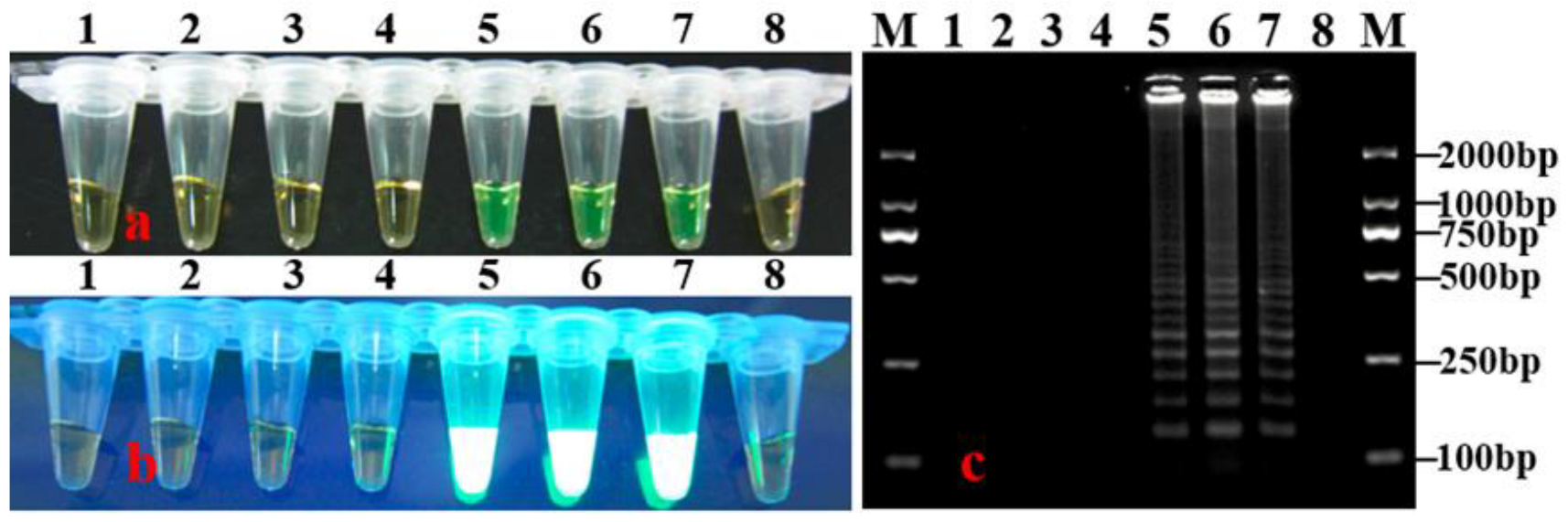
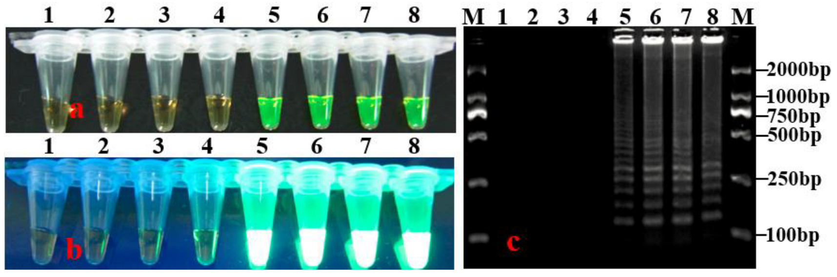
3.1. Optimization of LAMP Reaction Conditions

The genomic DNA of M. acerina was used as the template for the LAMP assay to determine the optimal reaction temperature and time. The tubes containing the reaction mixtures were incubated at various temperatures for a range of times to determine an ideal condition for LAMP assay, and amplification was detected based on color change, bright white turbidity, and ladder-like DNA band patterns on agarose gel. It was revealed that positive LAMP reactions occurred at temperatures of 64–65 °C, and the intensity of ladder-like bands at 64.5 °C was the highest (Figure 2). The results of LAMP reactions performed at 64.5 °C for 25–55 min showed that positive reactions were obtained with times of 40–55 min, and the intensity of ladder-like bands at 45–55 min was higher than those at 40 min, but there was no apparent difference in the intensity of ladder-like bands among 45, 50, and 55 min (Figure 3). Considering the speed of the reaction, 45 min was selected as the optimal reaction time. Overall, the optimum condition for the developed LAMP assay was found to be 64.5 °C for 45 min. Therefore, subsequent LAMP assays were conducted under these conditions.

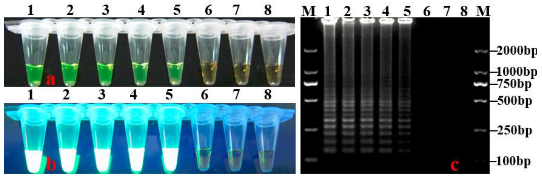
3.2. Specificity and Sensitivity of LAMP Assay

The specificity of the LAMP assay was evaluated using DNA from isolates listed in Table 1. The results showed that positive reactions were observed only for M. acerina isolates, whereas other plant pathogens including some frequent pathogens on P. notoginseng exhibited negative reactions (Table 1; Figure 4). To determine its detection limit, the LAMP assay was performed with a series of tenfold dilutions of M. acerina DNA as a template under optimal reaction conditions. No positive reaction was observed using less than 10 fg of DNA, indicating that the sensitivity of the LAMP assay was 10 fg in a 25-μL reaction mixture (Figure 5). The results of naked eye observation were consistent with those of agarose gel electrophoresis.

3.3. Evaluation of the LAMP Assay by Detecting M. acerina in P. notoginseng Leaf Samples

Genomic DNA extracted from symptomatic and asymptomatic P. notoginseng leaf samples was used as a template to determine whether the LAMP assay could detect M. acerina in diseased leaves. After LAMP reactions, all symptomatic leaves and two of ten asymptomatic leaves showed positive reactions, the amplifications exhibited a yellow-green color change and bright white turbidity, and ladder-like bands appeared in agarose gel electrophoresis (Figure 6). In addition, M. acerina was successfully isolated from all positive leaf samples.

4. Discussion

Round leaf spot caused by M. acerina is a highly destructive disease that threatens P. notoginseng production in China. The control measures of P. notoginseng round leaf spot disease are different from those of diseases caused by oomycetes, but the early symptoms of round leaf spot are similar to those of leaf blight caused by Phytophthora species. Therefore, the accurate detection of plant pathogens at the early stage of disease plays an important role in taking timely and effective management measures for controlling plant disease. In this study, an effective LAMP assay based on rDNA-ITS sequences for specific and sensitive detection of M. acerina in P. notoginseng was developed and evaluated. In order to determine the ideal reaction conditions for the LAMP assay, the amplifications at different temperatures and times were evaluated, and the optimum reaction temperature and time were found to be 64.5 °C for 45 min. Hence, the LAMP assay can be performed in simple heating equipment (such as a water bath or heating block) at an isothermal temperature, which significantly reduces the demand for expensive, specialized, and sophisticated instruments. Therefore, the developed LAMP assay may be suitable for detecting M. acerina in areas lacking adequate laboratory facilities.
The specific detection of pathogens is of great significance for disease prediction and control. The LAMP assay specifically amplified DNA of M. acerina only, and no positives were observed with other isolates. This indicates that the LAMP assay established in this study is highly specific for detecting M. acerina and can effectively distinguish M. acerina from other plant pathogens. It has been previously reported that LAMP has much higher sensitivity than conventional PCR [28,29,30]. In this study, the sensitivity of the LAMP assay for detection of M. acerina DNA was 10 fg per 25-μL reaction mixture, which strongly agrees with previous reports on LAMP methods used for detecting Fusarium oxysporum f. sp. cucumerinum and Colletotrichum gloeosporioides, and the detection limit was about 1000 times higher than conventional PCR [19,25], indicating that the highly sensitive LAMP assay developed in this study was suitable for detecting M. acernia at early stages of round leaf spot. The fact that the detection sensitivity of LAMP is higher than that of conventional PCR may be largely due to the isothermal features, high strand displacement activity of the Bst DNA polymerase, number of copies of the target gene, and high-level tolerance to some inhibitors [31,32].
The utility of the LAMP assay was tested by detecting M. acerina in leaf samples. It is noteworthy that the LAMP assay not only detected M. acerina in all symptomatic leaves but also in some leaves without visible symptoms (two of ten), which indicated that some asymptomatic leaves may have been infected by M. acerina, but the symptoms did not appear at the early stage of the disease, and the LAMP assay can detect M. acerina sensitively and accurately in infected leaf samples at the asymptomatic stage. These results are consistent with previous studies reporting that LAMP can detect pathogens in infected plant tissues [33,34]. To reduce economic losses caused by disease, it is more important to detect pathogens in latent stages than in symptomatic stages. The results demonstrated that the LAMP assay may be useful for Sanqi round leaf spot disease diagnosis at the latent or symptomatic stage in production fields so that growers can improve the control efficiency by taking preventive control measures. Future research is needed to develop sampling plants for use with the LAMP assay, especially at the early stages of the disease when the disease incidence across the field is low, leading to significant sampling challenges since many plants would need to be tested to find positives, especially if symptoms are latent.

5. Conclusions

In this study, we designed LAMP primers targeting the rDNA-ITS sequences, optimized the reaction conditions under various temperatures and times, and evaluated the specificity, sensitivity, and application of the developed LAMP assay. This is the first report on developing a reliable, rapid, efficient, specific, sensitive, and easy-operating LAMP assay for detecting M. acerina. The LAMP assay can be used as a tool for the diagnosis of Sanqi round leaf spot disease caused by M. acerina in the field with a simple heating block or water bath and will provide accurate information in support of precautionary and integrated disease management.

Author Contributions

Conceptualization, C.L. and X.Y.; Methodology, C.L., L.G. and Y.D.; Data curation, C.L., L.G. and X.L.; Software, C.L.; Visualization, X.Y.; Writing-original draft preparation, C.L.; Writing-review and editing, C.L. and X.Y.; Supervision and funding acquisition, C.L. and X.Y. All authors have read and agreed to the published version of the manuscript.

Funding

This study was supported by the Basic R & D Special Business Fund of Fujian Province (2019R1024-8, 2022R1024008), the Agricultural High Quality Development “5511” Collaborative Innovation Project of the Fujian Provincial People’s Government-Chinese Academy of Agricultural Sciences (XTCXGC2021011), and the Science and Technology Innovation Foundation of the Fujian Academy of Agricultural Sciences (CXTD2021002-1).

Data Availability Statement

Not applicable.

Acknowledgments

We would like to thank the Zhouning Fenghong agricultural professional cooperative for providing Sanqi (Panax notoginseng) leaf samples.

Conflicts of Interest

The authors declare that the research was conducted in the absence of any commercial or financial relationships that could be construed as a potential conflict of interest.

References

  1. Wang, T.; Guo, R.X.; Zhou, G.H.; Zhou, X.D.; Kou, Z.Z.; Sui, F.; Li, C.; Tang, L.Y.; Wang, Z.J. Traditional uses, botany, phytochemistry, pharmacology and toxicology of Panax notoginseng (Burk.) F.H. Chen: A review. J. Ethnopharmacol. 2016, 188, 234–258. [Google Scholar] [CrossRef] [PubMed]
  2. Wang, L.D.; Xu, Z.M.; Liang, X.; Qiu, W.R.; Liu, S.J.; Dai, L.L.; Wang, Y.F.; Guo, C.Y.; Qi, X.H.; Wang, J.; et al. Overview of systematic reviews of Panax notoginseng saponins in treatment of acute cerebral infarction. China J. Chin. Mater. Med. 2021, 46, 2963–2971. [Google Scholar]
  3. Guo, H.B.; Cui, X.M.; An, N.; Cai, G.P. Sanchi ginseng (Panax notoginseng (Burkill) F. H. Chen) in China: Distribution, cultivation and variations. Genet. Resour. Crop Evol. 2010, 57, 453–460. [Google Scholar] [CrossRef]
  4. Li, Y.L.; Wang, B.Y.; Chang, Y.F.; Yang, Y.T.; Yao, C.Z.; Huang, X.Q.; Zhang, J.B.; Cai, Z.C.; Zhao, J. Reductive soil disinfestation effectively alleviates the replant failure of Sanqi ginseng through allele chemical degradation and pathogen suppression. Appl. Microbiol. Biotechnol. 2019, 103, 3581–3595. [Google Scholar] [CrossRef]
  5. Li, J.; Yang, Q.L.; Shi, Z.T.; Zang, Z.N.; Liu, X.G. Effects of deficit irrigation and organic fertilizer on yield, saponin and disease incidence in Panax notoginseng under shaded conditions. Agric. Water Manag. 2021, 256, 81–95. [Google Scholar] [CrossRef]
  6. Li, X.; Li, S.; Qiu, B.L.; Zhang, Y.P.; Cui, X.M.; Ge, F.; Liu, D.Q. Thaumatin-like protein genes of Panax notoginseng confers resistance to Alternaria panax. Physiol. Mol. Plant Pathol. 2020, 112, 101537. [Google Scholar] [CrossRef]
  7. Wang, W.Y.; Zhao, C.G.; Chen, Z.G.; Wen, G.S.; Wei, F.G.; Long, T.G.; Li, S.W.; Wang, C.G. Studies on the isolation, identification and in vitro growth rates of the three pathogenic fungi from Panax notoginseng cultivated in Wenshan eparchy. Agric. Sci. Technol. 2015, 16, 1165–1171. [Google Scholar]
  8. Zeng, Z.Y.; Li, Q.Q.; Huo, Y.Y.; Chen, C.J.; Duan, S.S.; Xu, F.R.; Cheng, Y.X.; Dong, X. Inhibitory effects of essential oils from Asteraceae plant against pathogenic fungi of Panax notoginseng. J. Appl. Microbiol. 2021, 130, 592–603. [Google Scholar] [CrossRef]
  9. Han, L.; Zhou, X.; Zhao, Y.T.; Wu, L.X.; Ping, X.R.; He, Y.L.; Peng, S.; He, X.H.; Du, Y.L. First report of Plectosphaerella plurivora causing root rot disease in Panax notoginseng in China. J. Phytopathol. 2020, 168, 375–379. [Google Scholar] [CrossRef]
  10. Wang, H.L.; Wang, F.; Jin, B.H.; Zhang, H.; Yang, K.; Wang, W.P.; Yang, M.; Zhu, S.S.; He, X.H. Sensitivity and fitness analysis of round spot of Panax notoginseng Mycoentrospora acerina to azoxystrobin, prochloraz and difenoconazole. Chin. J. Pestic. Sci. 2019, 21, 273–278. [Google Scholar]
  11. Mao, Z.S.; Long, Y.J.; Bao, Y.Y.; Feng, G.Y.; Zhu, Y.Y.; He, X.H. Analysis of rDNA–ITS sequence of round spot pathogen in Panax notoginsen and indoor toxicity determination of fungicides. J. Chin. Med. Mater. 2020, 43, 831–835. [Google Scholar]
  12. Evenhuis, A.; Verdam, B.; Zadoks, J.C. Splash dispersal of conidia of Mycocentrospora acerina in the field. Plant Pathol. 1997, 46, 459–469. [Google Scholar] [CrossRef]
  13. Gilchrist, L.; Galdames, R.; Chahin, G.; Luchsinger, N.; Diaz, C. First report in chile of Mycocentrospora acerina, causal agent of peony (Paeonia lactiflora) red spot. Plant Dis. 2015, 99, 284. [Google Scholar] [CrossRef] [PubMed]
  14. Abdelmagid, A.; Hafez, M.; Soliman, A.; Adam, L.R.; Daayf, F. First report of Fusarium sporotrichioides causing root rot of soybean in Canada and detection of the pathogen in host tissues by PCR. Can. J. Plant Pathol. 2021, 43, 527–536. [Google Scholar] [CrossRef]
  15. Das, A.K.; Nerkar, S.; Gawande, N.; Thakre, N.; Kumar, A. Scar marker for Phytophthora nicotianae and multiplex PCR assay for simultaneous detection of P. nicotianae and Candidatus Liberibacter asiaticus in citrus. J. Appl. Microbiol. 2019, 127, 1172–1183. [Google Scholar] [CrossRef]
  16. Yao, J.A.; Lan, C.Z.; Huang, P.; Yu, D.Y. PCR detection of Colletotrichum gloeosporioides in Psidium guajava. Australas. Plant Pathol. 2018, 47, 95–100. [Google Scholar] [CrossRef]
  17. Lan, C.Z.; Yao, J.A.; Yan, X.J.; Ruan, H.C.; Yu, D.Y.; Jiang, J.X. Specific and sensitive detection of the guava fruit anthracnose pathogen (Colletotrichum gloeosporioides) by loop mediated isothermal amplification (LAMP) assay. Can. J. Microbiol. 2020, 66, 17–24. [Google Scholar] [CrossRef] [PubMed]
  18. Notomi, T.; Okayama, H.; Masubuchi, H.; Masubuchi, H.; Yonekawa, T.; Watanabe, K.; Amino, N.; Hase, T. Loop-mediated isothermal amplification of DNA. Nucleic Acids Res. 2000, 28, e63. [Google Scholar] [CrossRef] [PubMed] [Green Version]
  19. Nagamine, K.; Watanabe, K.; Ohtsuka, K.; Hase, T.; Notomi, T. Loop-mediated isothermal amplification reaction using a nondenatured template. Clin. Chem. 2001, 47, 1742–1743. [Google Scholar] [CrossRef] [PubMed] [Green Version]
  20. Notomi, T.; Mori, Y.; Tomita, N.; Kanda, H. Loop-mediated isothermal amplification (LAMP): Principle, features, and future prospects. J. Microbiol. 2015, 53, 1–5. [Google Scholar] [CrossRef] [PubMed]
  21. Le, D.T.; Vu, N.T. Progress of loop-mediated isothermal amplification technique in molecular diagnosis of plant diseases. Appl. Biol. Chem. 2017, 60, 169–180. [Google Scholar] [CrossRef]
  22. Zhang, L.; Gleason, C. Loop-mediated isothermal amplification for the diagnostic detection of Meloidogyne chitwoodi and M. fallax. Plant Dis. 2019, 103, 12–18. [Google Scholar] [CrossRef] [PubMed] [Green Version]
  23. Prasannakumar, M.K.; Parivallal, P.B.; Manjunatha, C.; Mahesh, H.B.; Pramesh, D.; Narayan, K.S.; Gopal, V.B.; Priyanka, K.; Puneeth, M.E.; Rangaswamy, K.T. Loop-mediated isothermal amplification assay for pre-symptomatic stage detection of Xanthomonas axonopodis pv. punicae infection in pomegranate. Australas. Plant Pathol. 2020, 49, 467–473. [Google Scholar] [CrossRef]
  24. Zheng, X.B. Methods in Phytophthora, 1st ed.; Chinses Agriculture Press: Beijing, China, 1995; p. 82. [Google Scholar]
  25. Lan, C.Z.; Ruan, H.C.; Yang, X.J.; Yao, J.A.; Jiang, J.X. Development of a loop-mediated isothermal amplification assay for sensitive and specific detection of Fusarium oxysporum f. sp. cucumerinum Owen. Phytoparasitica 2018, 46, 283–293. [Google Scholar] [CrossRef]
  26. Wang, H.; Qi, M.; Cutler, A.J. A simple method of preparing plant samples for PCR. Nucleic Acids Res. 1993, 21, 4153–4154. [Google Scholar] [CrossRef] [PubMed] [Green Version]
  27. Lu, C.; Song, B.; Zhang, H.; Wang, Y.; Zheng, X. Rapid diagnosis of soybean seedling blight caused by Rhizoctonia solani and soybean charcoal rot caused by Macrophomina phaseolina using LAMP assays. Phytopathology 2015, 105, 1612–1617. [Google Scholar] [CrossRef] [Green Version]
  28. Niu, J.H.; Guo, Q.X.; Jian, H.; Chen, C.L.; Yang, D.; Liu, Q.; Guo, Y.D. Rapid detection of Meloidogyne spp. by LAMP assay in soil and roots. Crop Prot. 2011, 30, 1063–1069. [Google Scholar] [CrossRef]
  29. Niu, J.H.; Jian, H.; Guo, Q.X.; Chen, C.L.; Wang, X.Y.; Liu, Q.; Guo, Y.D. Evaluation of loop-mediated isothermal amplification (LAMP) assays based on 5S rDNA-IGS2 regions for detecting Meloidogyne enterolobii. Plant Pathol. 2012, 61, 809–819. [Google Scholar] [CrossRef]
  30. Peng, H.; Peng, D.L.; Hu, X.Q.; He, X.F.; Wang, Q.; Huang, W.K.; He, W.T. Loop-mediated isothermal amplification for rapid and precise detection of the burrowing nematode, Radopholus similis, directly from diseased plant tissues. Nematology 2012, 14, 977–986. [Google Scholar] [CrossRef]
  31. Francois, P.; Tangomo, M.; Hibbs, J.; Bonetti, E.J.; Boehme, C.C.; Notomi, T.; Perkins, M.D.; Schrenzel, J. Robustness of a loop-mediated isothermal amplification reaction for diagnostic applications. FEMS Immunol. Med. Microbiol. 2011, 62, 41–48. [Google Scholar] [CrossRef] [Green Version]
  32. Kaneko, H.; Kawana, T.; Fukushima, E.; Suzutani, T. Tolerance of loop-mediated isothermal amplification to a culture medium and biological substances. J. Biochem. Biophys. Methods 2007, 70, 499–501. [Google Scholar] [CrossRef] [PubMed]
  33. Dai, T.; Lu, C.; Lu, J.; Dong, S.; Ye, W.; Wang, Y.; Zheng, X. Development of a loop mediated isothermal amplification assay for detection of Phytophthora sojae. FEMS Microbiol. Lett. 2012, 334, 27–34. [Google Scholar] [CrossRef] [PubMed] [Green Version]
  34. Feng, W.; Nukaya, A.; Satou, M.; Fukuta, N.; Ishiguro, Y.; Suga, H.; Kageyama, K. Use of LAMP detection to identify potential contamination sources of plant pathogenic Pythium species in hydroponic culture systems of tomato and eustoma. Plant Dis. 2018, 102, 1357–1364. [Google Scholar] [CrossRef] [PubMed]
Figure 1. Design of LAMP primers for detection of M. cerina. The letters (F2, F3, B1c, F1c, B2 and B3) are primer names, the location and sequence of primers are shown in green color, and the direction of amplification is indicated by red arrows.
Horticulturae 08 01060 g001
Figure 2. Optimization of LAMP reaction temperature for detection of M. acerina. (a) LAMP products were visualized by naked eye under natural light (color change, yellow-green indicate positive reaction). (b) LAMP products were visualized by naked eye under UV light (bright white turbidity indicates positive reaction). (c) LAMP products were analyzed using electrophoresis on 2% agarose gel (ladder-like pattern bands indicate positive reaction). Lane 1: negative control (SDW); Lane 2: 62.5 °C; Lane 3: 63 °C; Lane 4: 63.5 °C; Lane 5: 64 °C; Lane 6: 64.5 °C; Lane 7: 65 °C; Lane 8: 65.5 °C; Lane M: DL 2000 DNA marker. The color of tubes 5–7 is yellow-green.
Horticulturae 08 01060 g002
Figure 3. Optimization of LAMP reaction time for detection of M. acerina. (a) LAMP products were visualized by naked eye under natural light (color change, yellow-green indicate positive reaction). (b) LAMP products were visualized by naked eye under UV light (bright white turbidity indicates positive reaction). (c) LAMP products were analyzed using electrophoresis on 2% agarose gel (ladder-like pattern bands indicate positive reaction). Lane 1: negative control (SDW); Lane 2: 25 min; Lane 3: 30 min; Lane 4: 35 min; Lane 5: 40 min; Lane 6: 45 min; Lane 7: 50 min; Lane 8: 55 min; Lane M: DL 2000 DNA marker. The color of tubes 5–8 is yellow-green.
Horticulturae 08 01060 g003
Figure 4. Specificity of LAMP. (a) LAMP products were visualized by naked eye under natural light (color change, yellow-green indicate positive reaction). (b) LAMP products were visualized by naked eye under UV light (bright white turbidity indicates positive reaction). (c) LAMP products were analyzed using electrophoresis on 2% agarose gel (ladder-like pattern bands indicate positive reaction). Lane 1–3: Mycocentrospora acerina; Lane 4: Cylindrocarpon destructans; Lane 5: Alternaria panax; Lane 6: Colletotrichum truncatum; Lane 7: Botrytis cinerea; Lane 8: Negative control (SDW); Lane M: DL 2000 DNA marker. The color of tubes 1–3 is yellow-green.
Horticulturae 08 01060 g004
Figure 5. Sensitivity of the LAMP assay for detection of M. acerina. (a) LAMP products were visualized by naked eye under natural light (color change, yellow-green indicate positive reaction). (b) LAMP products were visualized by naked eye under UV light (bright white turbidity indicates positive reaction). (c) LAMP products were analyzed using electrophoresis on 2% agarose gel (ladder-like pattern bands indicate positive reaction). Lane M: DNA marker DL2000; Lane 1–7, amplified products with a series of tenfold dilutions of M. acerina DNA (100 pg, 10 pg, 1 pg, 100 fg, 10 fg, 1 fg, and 100 ag) in 25 μL LAMP mixtures; Lane 8: Negative control (SDW). The color of tubes 1–5 is yellow-green.
Horticulturae 08 01060 g005
Figure 6. Ability of the LAMP assay to detect M. acerina in Sanqi leaves. (a) LAMP products were visualized by naked eye under natural light (color change, yellow-green indicate positive reaction). (b) LAMP products were visualized by naked eye under UV light (bright white turbidity indicates positive reaction). (c) LAMP products were analyzed using electrophoresis on 2% agarose gel (ladder-like pattern bands indicate positive reaction). Lane M: DL 2000 marker; Lane 1: Positive control (M. acerina genomic DNA); Lane 2–4, 6: Asymptomatic leaves; Lane 5, 7: Leaves with symptoms of round spot disease; Lane 8: Negative control (sterile distilled water). The color of tubes 1, 2, 5, and 7 is yellow-green.
Horticulturae 08 01060 g006
Table 1. Isolates of different plant pathogens used to test the specificity of the LAMP assay.
SpeciesHostLocationNo of IsolatesLAMP Detection *
Agarose GelSYBR Green I
Mycocentrospora acerinaPanax notoginsengFujian8++
M. acerinaP. notoginsengYunnan10++
Cylindrocarpon destructansP. notoginsengFujian3
Alternaria panaxP. notoginsengYunnan5
A. panaxP. notoginsengFujian3
A. solaniSolanum lycopersicumFujian3
Colletotrichum truncatumGlycine maxFujian2
C. orbiculareCucumis sativusFujian3
C. gloeosporioidesCitrus reticulataFujian1
C. musaeMusa nanaFujian1
C. panacicolaP.notoginsengFujian3
Botrytis cinereaS. lycopersicumFujian2
Phytophthora infestansS. lycopersicumFujian2
P. colocasiaeColocasia esculentaFujian5
P. cactorumP. notoginsengFujian3
P. capsiciCapsicum annuumFujian3
P. melonisC.sativusFujian3
P. sojaeG. maxFujian3
P. cryptogeaGerbera jamesoniiFujian1
p. vignaeVigna unguiculataFujian2
Peronophythora litchiLitchi chinensisFujian1
Pythium aphanidermatumC. sativusFujian1
Fusarium solaniS. lycopersicumFujian3
F. oxysporum f. sp. cucumebriumC. sativusFujian2
F. oxysporum f. sp. cubenseM. nanaFujian2
Rhizoctonia solaniS. lycopersicumFujian1
Bipolaria maydisZea maysFujian1
Pestalatiopsis spp.Psidium guajavaFujian1
* Presence (+) or absence (−) is based on the presence of a LAMP product of the expected size.
Table 2. LAMP assay primers for detection of M. acerina in this study.
Primer NameSequence (5′-3′)Length
F35′-GCCTGTTCGAGCGTCATT-3′18
B35′-TCAGCGGGTATCCCTACC-3′18
FIP5′-ACGCCGGCTGCCAATTGTTTTA-CCTCAAGCTCTGCTTGGTG-3′41
BIP5′-CTTCGGAGCGCAGCACATTTTG-TCCGAGGTCAAGAGCGTTAA-3′42
Publisher’s Note: MDPI stays neutral with regard to jurisdictional claims in published maps and institutional affiliations.

