Brief Report

The Feasibility of Using Autofluorescence to Detect Lignin Deposition Pattern during Defense Response in Apple Roots to Pythium ultimum Infection

Tree Fruit Research Laboratory, Agricultural Research Service, United States Department of Agriculture, Wenatchee, WA 98801, USA
Horticulturae 2022, 8(11), 1085; https://doi.org/10.3390/horticulturae8111085
Submission received: 22 September 2022 / Revised: 2 November 2022 / Accepted: 15 November 2022 / Published: 17 November 2022
(This article belongs to the Special Issue Horticultural Plants Pathology and Advances in Disease Management)

Abstract

:
The molecular mechanisms underpinning apple root resistance to infection by Pythium ultimum have not been carefully investigated until recently. A panoramic and high-resolution picture of genome-wide transcriptional networks regulating the defense activation in apple roots was obtained recently from a comprehensive transcriptome analysis. Among the most notable transcriptome changes are the upregulation of phenylpropanoid and flavonoid biosynthesis, transportation of secondary metabolites, and laccase-directed lignin formation. However, assessing cellular responses and anatomical changes in young roots of apple as a woody species of the Rosacea family remains challenging. Taking advantage of the autofluorescence of lignin and the specific staining by Wiesner reagent, the feasibility of using microscopy to detect lignin deposition in apple roots was evaluated. The preliminary results indicated that a combination of brightfield and fluorescence images may provide the opportunity to reveal the location and degree of lignification in sectioned roots. The procedure represents a proof-of-concept of using fluorescence microscopy to examine cellular features relevant to apple root resistance traits. The ability to detect subtle phenotypic variations, such as lignin deposition patterns, is critically needed to bridge the gap between genomic data and macro-level plant resistance phenotypes and to further identify the resistance mechanisms of apple root.

1. Introduction

The molecular regulation of resistance in apple roots to infection from Pythium ultimum has been the subject in our recent research [1,2,3,4,5,6,7,8,9]. With the goal of gene discovery and developing molecular tools for breeding in resistance to apple replant disease (ARD), three consecutive transcriptome analyses uncovered key candidate genes in specific pathways during defense activation in apple root to infection from P. ultimum [1,2,3,10]. Among the most notable transcriptome changes are the consistent upregulation of genes in the phenylpropanoid and flavonoid biosynthesis pathways, transportation of secondary metabolites, and laccase-directed lignin formation [1,2,3]. At the same time, an elite panel of apple rootstock genotypes with contrasting resistance responses to P. ultimum infection has been identified through a systematic phenotyping effort among the progenies of an apple rootstock cross population [5,7,8]. The resistance responses were primarily defined by the genotype-specific plant survival rate and certain microscopic features of tissue necrosis progression. The detailed resistance phenotypes at the anatomical, cellular, and biochemical levels have not been carefully investigated. The capability of detecting subtle cellular changes in apple roots during infection is essential to validate the transcriptome data, associating specific candidate genes with resistance phenotypes, and elucidating the underpinned resistance mechanisms.
Plant defense against invading pathogens is ultimately chemical warfare in nature. Plants are capable of producing a wide variety of antimicrobial secondary metabolites with extreme structural diversity [11,12]. The most widespread groups of secondary metabolites include phenols, terpenoids and alkaloids [13,14,15]. Both preformed antimicrobial compounds (phytoanticipins) and infection-induced antimicrobial metabolites (phytoalexins) have long been associated with plant resistance to fungal, oomycete, and bacterial pathogens [11,16]. Most of these compounds are small molecules and their effective doses can be as low as 10−5–10−4 M [11,12,16]. Between primary and secondary metabolism, there are tradeoffs between available energy, enzymes and metabolic precursors, as well as the transport and storage of the generated secondary metabolites [13]. The outcome of plant–pathogen interactions may depend crucially on the plant’s ability to produce specific secondary metabolites, at proper timing and concentrations, upon pathogenic challenge [17,18].
As an extension to the general phenypropanoid pathway, laccase-directed lignification appeared to be a crucial factor contributing to apple root resistance to P. ultimum infection [1,2,3]. The role of laccase-directed lignin biosynthesis and resulting cell wall fortification to plant disease resistance has been proposed for several decades in different pathosystems [18,19]. More recently, transcriptome and proteome data have verified the direct connection between laccase gene expression and lignification processes in relation to genotype-specific resistance levels in several crops [20,21,22,23,24]. Accumulating evidence indicates that the lignified cell wall serves as a physical barrier against invading phytopathogens as well as other environmental stresses [24,25,26,27]. Our transcriptome data strongly suggested that the upregulated phenylpropanoid pathway, monolignol transportation, and laccase directed lignin biosynthesis constitute a major defense mechanism in apple root to P. ultimum infection [1,2,3,28]. However, an effective and direct detection method for assessing the timing, intensity, and genotype-specific patterns of lignin deposition in apple roots is still lacking.
Lignins are heteropolymers that are covalently associated with polysaccharides in plant cell walls [19,29]. Lignin biosynthesis is the result of oxidative polymerization of three p-hydroxycinnamyl (p-coumaryl, -coniferyl and -sinapyl) alcohols, and is mediated by both laccases and peroxidases [30]. These monolignols are biosynthesized downstream of the general phenylpropanoid pathway [20,30]. The lignification process, including monolignol transport to the apoplast and subsequent oxidation by laccases and/or peroxidases, is crucial for multiple aspects of plant physiology, such as strengthening the plant cell wall and disease resistance [30,31,32,33]. As lignification is a non-reversible process, the pathways of monolignol biosynthesis, polymerization, and lignin deposition are tightly regulated during development and response to stress conditions [25,34,35]. However, in the path system between apple roots and P. ultimum, the role of pathogen-induced lignification and its contribution to resistance have not been studied until recently [1,28]. A specific and straightforward method to detect anatomical and biochemical changes, such as cell wall fortification, is crucially needed to evaluate and dissect resistance traits of apple roots.
Many molecules in plant tissues are autofluorescent, and the two most studied molecules are chlorophyll (orange/red fluorescence) and lignin (blue/green fluorescence) [36,37]. Structural components in lignin molecules, such as phenolic rings and conjugated double bonds, are the important organic fluorophores [36,37]. While lignin can be stained for brightfield observation, its autofluorescent nature offers the advantages of label-free imaging. Therefore, its autofluorescence may be exploited for a quick and fixation-free method to assess the lignin deposition in apple roots during P. ultimum infection. Such specific and in-depth phenotype data at cellular and subcellular levels are pivotal to associate transcriptomic data and macro-level plant resistance phenotypes and to validate the functional roles of specific candidate genes implicated in apple root resistance.

2. Results and Discussion

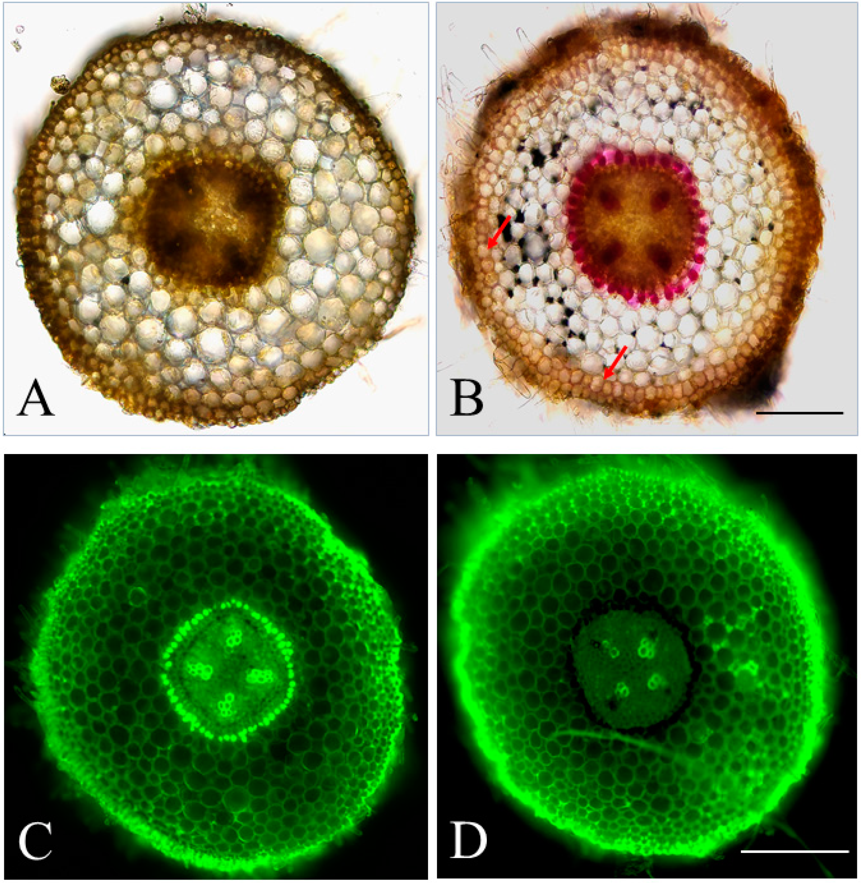
2.1. The Feasibility of Detecting Lignin Deposition Patterns through Autofluorescence

Based on previous transcriptome data, the biosynthesis and transport of secondary metabolites, including monolignols, are clearly the crucial components in apple root defense response to P. ultimum infection [1,2,3]. To validate transcriptome data and unravel resistance mechanisms of apple roots, procedures for detecting the anatomical, cellular, or biochemical changes associated with defense activation are critically needed. In the current study, both brightfield and fluorescence microscope observations were explored to assess the feasibility of detecting lignin deposition (Figure 1). Figure 1A is the brightfield image of hand-sectioned root tissue without Wiesner staining. Figure 1B is the brightfield image of a root section with lignin-specific staining by Wiesner reagent, showing a typical red-color ring outside vascular bundles. The reddish color visible in the cell walls of the outer layer of the cortex tissue (red arrows) is likely the increased deposition of lignin, suggesting a defense response. Two images in the lower half of Figure 1 show the auto-fluorescence of apple root sections. The bottom left panel (Figure 1C) shows a root section without Wiesner staining, exhibiting a high fluorescence intensity outside vascular bundles, within vascular bundles, and along the epidermis. Figure 1D shows the fluorescence image of a section that was stained with Wiesner reagent. The quenched fluorescence signal circling the stele specifically verified the identity of lignin, though the reduced intensity of fluorescence is not readily visible along the outer layer of the cortex under the epidermis. Combining images of brightfield and fluorescence, with assistance from specific staining (such as Wiesner for lignin), it appeared feasible to locate the lignin deposition in young apple root tissue.
The use of fluorescence to probe biological phenomena has been rapidly expanding in cellular and molecular biology more recently [36,38]. Fluorescence is observed when visible light is emitted from a sample that has previously absorbed electromagnetic radiation (i.e., the sample becomes “excited”) with higher energy and shorter wavelengths [38]. Fluorescence microscopy has improved significantly in recent years, and a wide range of synthesized compounds (fluorophores) are available, which allow for the simultaneous imaging of different cellular, subcellular, or molecular components [38]. The more innate form of fluorescence is arguably the intrinsic fluorescence of autofluorescent compounds. Both groups of compounds contain ring structures (aromatic molecules) with pi bonds that require the lower excited energy and the longer wavelength of the exciting light. The autofluorescent natures of these phenolic compounds present an opportunity for their easy and direct detection. Lignin, the natural polymer of coniferyl, sinapyl, or p-coumaryl alcohol, is abundant primarily in woody plant tissues, but can be induced in other sclerenchymatous tissues under certain conditions, such as a defense reaction to pathogen infection [36,37,38]. As a proof of concept, this study attempted to exploit lignin autofluorescence and the lignin-staining properties of the Wiesner reagent to test the feasibility of detecting lignin deposition in apple root tissues. The findings from the current study should set a baseline for a further improved procedure, which can eventually distinguish the subtle variations of lignin deposition patterns during infection or between treatments.

2.2. Upregulated Phenylpropanoid and Flavonoid Biosynthesis Pathways Due to P. ultimum Infection

The primary reason for developing this method of detecting lignin detection is for the systematic observation of upregulated phenylpropanoid and flavonoid biosynthesis, as well as lignin formation in apple root during defense response to infection from P. ultimum. With the advent of revolutionary NextGen sequencing technologies and improved bioinformatic analysis, it is relatively easy to generate huge datasets of genome-wide expression profiling and to identify candidate genes for a targeted biological process [39]. Specific to the pathosystem of apple root infected by P. ultimum, three consecutive transcriptome analyses have provided us with a high-resolution view of the genome-wide transcriptional response [1,2,3,10,40]. Among the most visible transcriptome changes in apple roots during P. ultimum infection are the differentially expressed genes (DEGs) identified, which are mapped to phenylpropanoid and flavonoid biosynthesis pathways [1,2,3]. Table 1 shows the top ten most enriched pathways based on the KEGG pathway analysis. It is probably expected that the transition to the defense mode required activated primary metabolism, such as carbohydrate and protein metabolism, for preparing energy and metabolic precursors. However, the elevated biosynthetic activities in phenylpropanoid and flavonoid biosynthesis pathways represent the specific response to P. ultimum infection in order to produce antimicrobial phenolic compounds. Similar to the overall trend of transcriptome changes due to P. ultimum infection [3], the numbers of DEGs mapped to these two pathways peaked at 48 hpi (hours post inoculation). In addition, “phenylalanine, tyrosine and tryptophan biosynthesis” and “phenylalanine metabolism” were also listed among the top ten most enriched pathways, which have a strong connection to phenylpropanoid and flavonoid biosynthesis pathways through supplying metabolic precursors. Therefore, phenolic compounds from these pathways appear to play a critical role in defense activation towards P. ultimum infection in apple roots.
The identities of these mapped genes are shown in Table 2, which include enzymes functioning at multiple steps in these two biosynthesis pathways [3]. All these identified genes were shown to be upregulated at the critical timepoint of 48 hpi. Most of these genes showed a two to eight-fold increase in transcript abundance relative to mock-inoculated controls. Two genes, MDP0000175949 and MDP0000315857, both encoding beta-glucosidase [EC:3.2.1.21], demonstrated an 85-fold or more increase in transcript abundance due to P. ultimum infection. Notably, five identified members from the same gene family encoding “peroxidase” were mapped to the phenylpropanoid pathway. MDP0000225698 and MDP0000576346, encoding trans-cinnamate 4-monooxygenase [EC:1.14.13.11], were mapped to both phenylpropanoid and flavonoid biosynthesis pathways. The fact that all identified genes showed upregulated expression at 48 hpi strongly suggested the essential role of these pathways to apple root resistance towards P. ultimum infection.
The secondary metabolites from these pathways can function as phytoalexins or lead to cellular alteration, such as cell wall lignification. Individual genes encoding key enzymes in the phenylpropanoid biosynthesis pathway, such as PAL and CAD, are uniformly upregulated in apple roots upon P. ultimum infection [2,3]. Monolignols, including p-coumaryl, -coniferyl and -sinapyl alcohols, are synthesized through the phenylpropanoid pathway [33,42]. Members of several TF (transcription factor) families, such as MYBs, NACs and MYCs, were actively regulated both transcriptionally and post-transcriptionally in response to P. ultimum infection [1,40]. Additionally, transporter-encoding genes, such as those for the ABC transporter or MATE family members, exhibited a quicker, stronger, and more consistent upregulation in the root of a resistant apple rootstock genotype as compared to the chaotic and inconsistent regulation in susceptible genotypes [2]. Overall, transcriptome analysis indicated that the biosynthesis and transport of these phenolic compounds, as well as lignin biosynthesis per se, actively participate in apple root defense activation. Our working hypothesis is that the swiftness and intensity of induced lignin deposition, as well as the accumulation of other antimicrobial phenolic compounds, play a key role in impeding the invasion of P. ultimum and lead to the resistance response in apple root [8]. Testing this hypothesis demands detailed phenotypic analysis such as the extent of cell wall lignification following pathogen infection. However, there are multiple barriers to detect cellular and biochemical changes in apple root cells during defense activation.

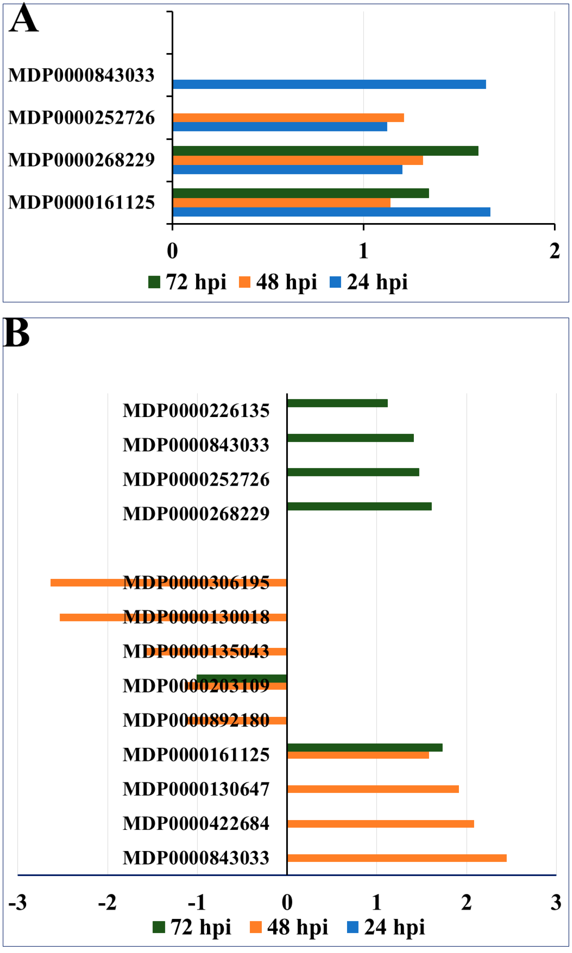
2.3. Contrasting Regulation Patterns of Apple MATE Gene Expression between Apple Rootstock Genotypes

Plant cells, like the cells of most organisms, are capable of removing potentially toxic compounds, including those produced endogenously, such as phenolics, flavonoids, and phytoalexins, from the cytoplasm by either sequestering them in vacuoles or transporting them to the cell wall [43]. Plant MATE (multidrug and toxic compound extrusion) genes encode proteins that function as transporters of these secondary metabolites [44,45]. Multiple MATE genes were identified from transcriptome profiling between a resistant and a susceptible genotype during the defense activation in apple roots [2], which showed the contrasting regulation patterns between the resistant genotype G.935 and the susceptible genotype B.9 during P. ultimum infection (Figure 1). In the root of a resistant G.935, a consistent upregulated pattern for all four identified MATE genes was observed at three timepoints (blue, orange and green bars, respectively), and no downregulated MATE genes were identified (Figure 2A). The expression features in a resistant genotype indicated an early and continuing pattern of elevated expression of MATE genes in response to P. ultimum infection. However, a very chaotic and complicated regulation scenario of nine MATE genes was uncovered in the root of a susceptible genotype B.9 (Figure 2B). First, none of the MATE genes were identified at the earlier stage of defense activation at 24 h post inoculation (hpi). Second, at 48 hpi (orange bars), a chaotic and inconsistent regulation of a dozen MATE genes was observed, i.e., four MATE genes were upregulated and five other members were downregulated. In the later stage, at 72 hpi (green bars), five MATE genes were upregulated and one was downregulated. More interestingly, the same set of MATE genes, which were consistently upregulated since 24 hpi in the root of the resistant G.935, were eventually upregulated at 72 hpi in B.9. Overall, the contrasting expression profiles of MATE genes strongly suggested their crucial impact on the outcome of the interaction between apple root and P. ultimum.

2.4. Epigenetic Regulation of Secondary Metabolism and Lignin Formation in Apple Roots in Response to P. ultimum Infection

In addition to the uncovered regulation at the transcriptional level, microRNA directed post-transcriptional control on phenylpropanoid and flavonoid biosynthesis, as well as lignin biosynthesis, were demonstrated by microRNA profiling and degradome sequencing [1]. Table 3 shows the specific genes encoding two transcription factor families and specific laccase genes were regulated by microRNA directed degradation of corresponding transcripts at 48 hpi [1]. The regulatory roles of NAC and MYB transcription factors on secondary metabolism have been well documented [46,47,48], though the relationships between individual TFs and specific genes remain largely unclear. During the infection of apple root by P. ultimum, multiple members of MYB and NAC transcription factors were targeted by members of microRNA (miR) families (Table 3). Three laccase genes, laccase 3, 5 and 7, were the targets of members of the miR397b. These findings added evidence to the notion that the upregulated phenylpropanoid biosynthesis pathway and subsequent lignin biosynthesis could crucially contribute to apple root resistance towards infection from P. ultimum.
The hidden nature of root systems and the small stature of individual young root branches represent the first tier of difficulties for direct, detailed, and consistent examination of defense responses. Heterogeneity in root development and differentiation stages, even between root branches within one root system, presents another challenge for the accurate and/or comparative evaluation of specific responses between genotypes and treatments [49]. A more practically unique barrier is likely the fact that apple reproduction is self-incompatible, and the apple genome has high-level heterozygosity [50,51]. As a result, seed germination will not produce genetically identical, or true to type, apple plants. For this challenge, a laborious tissue culture-based micropropagation procedure has to be implemented to continuously supply the genetically uniform apple plants for repeated infection assays and more reliable phenotypic analysis [7,8]. Limited root tissue mass from these young apple plants poses a challenge when using other biochemical methodologies, such as metabolomic analysis. In addition, given the fast-growing nature of P. ultimum, it is difficult, if not impossible, to perform a controlled or localized inoculation such as on a specific site of a single root branch. Finally, the low concentration of particular defense metabolites and their specialized distribution patterns present a further hinderance to quantitative analysis. Therefore, it is highly desirable to develop a simple and straightforward method for the instantaneous detection of specific secondary metabolites, such as lignin or other antimicrobial phenolic compounds, in apple roots. For these reasons, we tried to combine fluorescence microscopy and chemical staining to assess the anatomical and biochemical changes associated with apple root resistance to P. ultimum infection.
Systematic and in-depth examination of phenotypic features of apple root defense responses, as compared to massive sequencing information, remains a bottleneck for bridging the gap between genomics data and macro-scale or whole-plant resistance traits. The ability to detect subtle phenotypic variation is a prerequisite for reliably associating specific candidate genes or pathways with the trait of interest. To the best of our knowledge, there is no previous report on using autofluorescence detection as a tool to investigate the cellular changes in young apple roots during interaction with a necrotrophic pathogen. Autofluorescence offers advantages as a method of detecting cellular and subcellular changes. Once the method is established, target metabolites can be detected relatively easily and quickly without introducing artifacts from extensive tissue treatment and/or biochemical procedures. The potentially scaled-up procedure may allow for the detection of phenotypic variation across developmental stages, between genotypes and treatments, even with minimal tissue availability. With a standardized sampling strategy and consistency in sectioning, the resulting microscopy images may allow us to distinguish more subtle lignification patterns under various conditions.
Multiple improvements are required before the quantification of fluorescence intensity from obtained fluorescent images is realistic. Taking the apple root resistance to infection from P. ultimum as an example, the selection of roots with a identical physiological status and/or differentiation stage is extremely challenging if not impossible. This is partly because root growth and differentiation are a continuing process with high plasticity in response to surrounding conditions. The equivalent timing of pathogen exposure on selected root segments (between tested plants) is difficult to control, and yet it can be a source of variation in the lignification level and fluorescence intensity. Additionally, the uniformity or consistency in the thickness of tissue sections within or between selected root segments will almost certainly influence the captured fluorescence intensity. In addition to all of these variables, the degree of lignin fluorescence can also depend on structural variation within the molecule, as well as on the linkages between lignin and carbohydrates [36]. Therefore, the current report is simply a proof-of-concept that takes advantage of auto-fluorescent lignin and its specific staining using Wiesner reagent. Considerable improvements at multiple steps are required to develop a more ideal procedure to investigate the subtle phenotypic variation in lignin deposition during defense activation. Such a novel approach should greatly facilitate the elucidation of the role of cell wall lignification in young and miniature apple roots during defense activation against invading necrotrophic pathogens.

3. Materials and Methods

3.1. Preparation of Apple Plants by Tissue Culture

Tissue culture-based micro-propagation procedures were used to obtain individual apple plants for infection assays and tissue collection as described previously [5]. A micro-propagation process was carried out to generate apple plants with root tissues and to synchronize the developmental stages of both genotypes. A four-week period of root induction in tissue culture medium was followed by one week of root acclimation in soil in a growth chamber. Roots were then ready for P. ultimum inoculation. To minimize transplanting, shock pots were placed within a 10 × 20-inch flat tray and were covered by a transparent 7″ Vented Humidity Dome (Greenhouse Megastore, Danville, IL, USA). All the plant groups were watered identically every other day.

3.2. Wiesner Staining of Lignin and Microscope Images of Sectioned Apple Root Tissue

The Wiesner (phloroglucinol-HCl) staining [52] of hand-sectioned apple root tissues was used to detect the lignin deposition patterns for brightfield imagery. Root sections 80 to 100 μm thick were made by hand using Personna #74-0002. The tissue sections were kept in 4 °C water before staining. At least three plants were used for the selected genotype. For better comparability, the root segments were sectioned using segments at a similar position of 1 cm from the root tip between selected root branches; the resulting root tissue sections represent similar or comparable stages in terms of development and differentiation. Sections were visualized and documented using an Echo Revolve fluorescence Microscope (Discovery Echo, San Diego, CA, USA). The microscope was equipped with a 470/40 excitation filter, 525/50 emission filter, and 495 dichroic mirrors.

4. Conclusions

Tissue lignification has been hypothesized to be an important mechanism of plant disease resistance [18,19]. Our transcriptomic data clearly indicated that upregulated phenylpropanoid synthesis, lignin monomer transportation, and laccase-directed lignin formation may significantly contribute to apple root resistance traits to P. ultimum infection [1,2,3]. However, investigating relevant phenotypic changes at cellular and anatomical levels in young apple roots remains considerably challenging due to existing unique barriers, as discussed earlier. The capability of detecting autofluorescent lignin should provide a new perspective for examining cell wall lignification. This study aimed to explore the feasibility of tracking lignin deposition in young apple roots by comparing brightfield and fluorescence imagery. The microscopic observation method should provide much-needed, real-time cellular features for their connection to the apple root resistance responses. Further improvement in the procedures is needed, such as better standardization on root section quality, root developmental stage, and timing of the infection process. The location and intensity of lignin-originated fluorescence could be exploited to uncover the patterns of its deposition between rootstock genotypes, under various treatments, and at different timepoints during infection.

Funding

This work was supported by the USDA-ARS base fund.

Institutional Review Board Statement

Not applicable.

Informed Consent Statement

Not applicable.

Acknowledgments

The author would like to thank Jordan L Rainbow for language editing and reviewing the manuscript, or their careful review of the paper and constructive suggestions, which helped to improve the quality of this paper significantly. The author thanks Amanda Roelant for her excellent technical assistance.

Conflicts of Interest

The author declares no conflict of interest.

References

  1. Zhu, Y.; Li, G.; Singh, J.; Khan, A.; Fazio, G.; Saltzgiver, M.; Xia, R. Laccase Directed Lignification Is One of the Major Processes Associated with the Defense Response Against Pythium ultimum Infection in Apple Roots. Front. Plant Sci. 2021, 12, 629776. [Google Scholar] [CrossRef] [PubMed]
  2. Zhu, Y.; Shao, J.; Zhou, Z.; Davis, R.E. Genotype-specific suppression of multiple defense pathways in apple root during infection by Pythium ultimum. Hortic. Res. 2019, 6, 10. [Google Scholar] [CrossRef] [PubMed] [Green Version]
  3. Shin, S.; Zheng, P.; Fazio, G.; Mazzola, M.; Main, D.; Zhu, Y. Transcriptome changes specifically associated with apple (Malus domestica) root defense response during Pythium ultimum infection. Physiol. Mol. Plant Pathol. 2016, 94, 16–26. [Google Scholar] [CrossRef] [Green Version]
  4. Zhu, Y.; Fazio, G.; Mazzola, M. Elucidating the molecular responses of apple rootstock resistant to ARD pathogens: Challenges and opportunities for development of genomics-assisted breeding tools. Hortic. Res. 2014, 1, 14043. [Google Scholar] [CrossRef] [Green Version]
  5. Zhu, Y.; Shin, S.; Mazzola, M. Genotype responses of two apple rootstocks to infection by Pythium ultimum causing apple replant disease. Can. J. Plant Pathol. 2016, 38, 483–491. [Google Scholar] [CrossRef]
  6. Zhou, Z.; Tian, Y.; Cong, P.; Zhu, Y. Functional characterization of an apple (Malus × domestica) LysM domain receptor encoding gene for its role in defense response. Plant Sci. 2018, 269, 56–65. [Google Scholar] [CrossRef]
  7. Zhu, Y.; Saltzgiver, M.; Zhao, J. A phenotyping protocol for detailed evaluation of apple root resistance responses utilizing tissue culture micropropagated apple plants. Am. J. Plant Sci. 2018, 9, 2183. [Google Scholar] [CrossRef] [Green Version]
  8. Zhu, Y.; Zhao, J.; Zhou, Z. Identifying an elite panel of apple rootstock germplasm with contrasting root resistance to Pythium ultimum. J. Plant Pathol. Microbiol. 2018, 9, 11. [Google Scholar] [CrossRef]
  9. Zhou, Z.; Zhu, Y.; Tian, Y.; Yao, J.-L.; Bian, S.; Zhang, H.; Zhang, R.; Gao, Q.; Yan, Z. MdPR4, a pathogenesis-related protein in apple, is involved in chitin recognition and resistance response to apple replant disease pathogens. J. Plant Physiol. 2021, 260, 153390. [Google Scholar] [CrossRef]
  10. Zhu, Y.; Shao, J.; Zhou, Z.; Davis, R.E. Comparative transcriptome analysis reveals a preformed defense system in apple root of a resistant genotype of G.935 in the absence of pathogen. Int. J. Plant Genom. 2017, 2017, 8950746. [Google Scholar] [CrossRef]
  11. Dixon, R.A. Natural products and plant disease resistance. Nature 2001, 411, 843. [Google Scholar] [CrossRef] [PubMed]
  12. Grayer, R.J.; Kokubun, T. Plant-fungal interactions: The search for phytoalexins and other antifungal compounds from higher plants. Phytochemistry 2001, 56, 253–263. [Google Scholar] [CrossRef]
  13. Geilfus, C.-M. Plant secondary compounds. In Controlled Environment Horticulture: Improving Quality of Vegetables and Medicinal Plants; Springer International Publishing: Cham, Switzerland, 2019; pp. 19–33. [Google Scholar]
  14. Meyer, J.; Murray, S.L.; Berger, D.K. Signals that stop the rot: Regulation of secondary metabolite defences in cereals. Physiol. Mol. Plant Pathol. 2016, 94, 156–166. [Google Scholar] [CrossRef] [Green Version]
  15. Zhao, N.; Wang, G.; Norris, A.; Chen, X.; Chen, F. Studying plant secondary metabolism in the age of genomics. Crit. Rev. Plant Sci. 2013, 32, 369–382. [Google Scholar] [CrossRef]
  16. Hammerschmidt, R. Phytoalexins: What have we learned after 60 years? Annu. Rev. Phytopathol. 1999, 37, 285–306. [Google Scholar] [CrossRef] [PubMed] [Green Version]
  17. Grayer, R.J.; Harborne, J.B. A survey of antifungal compounds from higher plants, 1982–1993. Phytochemistry 1994, 37, 19–42. [Google Scholar] [CrossRef]
  18. Nicholson, R.L.; Hammerschmidt, R. Phenolic compounds and their role in disease resistance. Annu. Rev. Phytopathol. 1992, 30, 369–389. [Google Scholar] [CrossRef]
  19. Vance, C.; Kirk, T.; Sherwood, R. Lignification as a mechanism of disease resistance. Annu. Rev. Phytopathol. 1980, 18, 259–288. [Google Scholar] [CrossRef]
  20. Miedes, E.; Vanholme, R.; Boerjan, W.; Molina, A. The role of the secondary cell wall in plant resistance to pathogens. Front. Plant Sci. 2014, 5, 358. [Google Scholar] [CrossRef] [Green Version]
  21. Balasubramanian, V.K.; Rai, K.M.; Thu, S.W.; Hii, M.M.; Mendu, V. Genome-wide identification of multifunctional laccase gene family in cotton (Gossypium spp.); expression and biochemical analysis during fiber development. Sci. Rep. 2016, 6, 34309. [Google Scholar] [CrossRef]
  22. Yang, C.; Liang, Y.; Qiu, D.; Zeng, H.; Yuan, J.; Yang, X. Lignin metabolism involves Botrytis cinerea BcGs1-induced defense response in tomato. BMC Plant Biol. 2018, 18, 103. [Google Scholar] [CrossRef] [PubMed] [Green Version]
  23. Arcuri, M.L.; Fialho, L.C.; Nunes-Laitz, A.V.; Fuchs-Ferraz, M.C.P.; Wolf, I.R.; Valente, G.T.; Marino, C.L.; Maia, I.G. Genome-wide identification of multifunctional laccase gene family in Eucalyptus grandis: Potential targets for lignin engineering and stress tolerance. Trees 2020, 34, 745–758. [Google Scholar] [CrossRef]
  24. Lee, M.H.; Jeon, H.S.; Kim, S.H.; Chung, J.H.; Roppolo, D.; Lee, H.; Cho, H.J.; Tobimatsu, Y.; Ralph, J.; Park, O.K. Lignin-based barrier restricts pathogens to the infection site and confers resistance in plants. EMBO J. 2019, 38, e101948. [Google Scholar] [CrossRef] [PubMed]
  25. Vanholme, R.; Demedts, B.; Morreel, K.; Ralph, J.; Boerjan, W. Lignin biosynthesis and structure. Plant Physiol. 2010, 153, 895–905. [Google Scholar] [CrossRef] [PubMed] [Green Version]
  26. Bhuiyan, N.H.; Selvaraj, G.; Wei, Y.; King, J. Role of lignification in plant defense. Plant Signal. Behav. 2009, 4, 158–159. [Google Scholar] [CrossRef] [Green Version]
  27. Xu, L.; Zhu, L.; Tu, L.; Liu, L.; Yuan, D.; Jin, L.; Long, L.; Zhang, X. Lignin metabolism has a central role in the resistance of cotton to the wilt fungus Verticillium dahliae as revealed by RNA-Seq-dependent transcriptional analysis and histochemistry. J. Exp. Bot. 2011, 62, 5607–5621. [Google Scholar] [CrossRef] [Green Version]
  28. Zhu, Y.; Zhou, Z. The genotype-specific laccase gene expression and lignin deposition patterns in apple root during Pythium ultimum infection. Fruit Res. 2021, 1, 12. [Google Scholar] [CrossRef]
  29. Boudet, A.; Lapierre, C.; Grima-Pettenati, J. Tansley review No. 80. Biochemistry and molecular biology of lignification. New Phytol. 1995, 129, 203–236. [Google Scholar] [CrossRef]
  30. Boerjan, W.; Ralph, J.; Baucher, M. Lignin biosynthesis. Annu. Rev. Plant Biol. 2003, 54, 519–546. [Google Scholar] [CrossRef]
  31. Zhao, Q.; Dixon, R.A. Transcriptional networks for lignin biosynthesis: More complex than we thought? Trends Plant Sci. 2011, 16, 227–233. [Google Scholar] [CrossRef]
  32. Chaurasia, P.K.; Yadav, R.S.S.; Yadava, S. A review on mechanism of laccase action. Res. Rev. BioSci. 2013, 7, 66–71. [Google Scholar]
  33. Voxeur, A.; Wang, Y.; Sibout, R. Lignification: Different mechanisms for a versatile polymer. Curr. Opin. Plant Biol. 2015, 23, 83–90. [Google Scholar] [CrossRef] [PubMed]
  34. Giardina, P.; Faraco, V.; Pezzella, C.; Piscitelli, A.; Vanhulle, S.; Sannia, G. Laccases: A never-ending story. Cell. Mol. Life Sci. 2010, 67, 369–385. [Google Scholar] [CrossRef] [PubMed]
  35. Mayer, A.M.; Staples, R.C. Laccase: New functions for an old enzyme. Phytochemistry 2002, 60, 551–565. [Google Scholar] [CrossRef]
  36. Donaldson, L. Autofluorescence in plants. Molecules 2020, 25, 2393. [Google Scholar] [CrossRef]
  37. Pegg, T.J.; Gladish, D.K.; Baker, R.L. Algae to angiosperms: Autofluorescence for rapid visualization of plant anatomy among diverse taxa. Appl. Plant Sci. 2021, 9, e11437. [Google Scholar] [CrossRef]
  38. Lichtman, J.W.; Conchello, J.-A. Fluorescence microscopy. Nat. Method. 2005, 2, 910–919. [Google Scholar] [CrossRef]
  39. Metzker, M.L. Sequencing technologies—The next generation. Nat. Rev. Genet. 2010, 11, 31. [Google Scholar] [CrossRef] [Green Version]
  40. Zhu, Y.; Saltzgiver, M. A systematic analysis of apple root resistance traits to Pythium ultimum infection and the underpinned molecular regulations of defense activation. Hortic. Res. 2020, 7, 62. [Google Scholar] [CrossRef]
  41. Kanehisa, M.; Goto, S. KEGG: Kyoto Encyclopedia of Genes and Genomes. Nucleic Acids Res. 2000, 28, 27–30. [Google Scholar] [CrossRef]
  42. Ma, Q.H.; Zhu, H.H.; Qiao, M.Y. Contribution of both lignin content and sinapyl monomer to disease resistance in tobacco. Plant Pathol. 2018, 67, 642–650. [Google Scholar] [CrossRef]
  43. Eckardt, N.A. Move It on Out with MATEs. Plant Cell 2001, 13, 1477–1480. [Google Scholar] [CrossRef] [PubMed] [Green Version]
  44. Takanashi, K.; Shitan, N.; Yazaki, K. The multidrug and toxic compound extrusion (MATE) family in plants. Plant Biotechnol. 2014, 31, 417–430. [Google Scholar] [CrossRef] [Green Version]
  45. Upadhyay, N.; Kar, D.; Deepak Mahajan, B.; Nanda, S.; Rahiman, R.; Panchakshari, N.; Bhagavatula, L.; Datta, S. The multitasking abilities of MATE transporters in plants. J. Exp. Bot. 2019, 70, 4643–4656. [Google Scholar] [CrossRef] [PubMed]
  46. Bednarek, P.; Schulze-Lefert, P. Role of Plant Secondary Metabolites at the Host-Pathogen Interface. In Annual Plant Reviews, Molecular Aspects of Plant Disease Resistance; Wiley Blackwell: New York, NY, USA, 2009; Volume 34, pp. 220–260. [Google Scholar]
  47. Ko, J.H.; Jeon, H.W.; Kim, W.C.; Kim, J.Y.; Han, K.H. The MYB46/MYB83-mediated transcriptional regulatory programme is a gatekeeper of secondary wall biosynthesis. Ann. Bot. 2014, 114, 1099–1107. [Google Scholar] [CrossRef] [Green Version]
  48. Yang, B.; Li, Y.; Song, Y.; Wang, X.; Guo, Q.; Zhou, L.; Xue, X.; Zhang, C. The R2R3-MYB transcription factor VcMYB4a inhibits lignin biosynthesis in blueberry (Vaccinium corymbosum). Tree Genet. Genomes 2022, 18, 27. [Google Scholar] [CrossRef]
  49. McCully, M.E. Roots in soil: Unearthing the complexities of roots and their rhizospheres. Annu. Rev. Plant Biol. 1999, 50, 695–718. [Google Scholar] [CrossRef]
  50. Janick, J.; Cummins, J.; Brown, S.; Hemmat, M. Apples. Fruit Breed. 1996, 1, 1–77. [Google Scholar] [CrossRef] [Green Version]
  51. Velasco, R.; Zharkikh, A.; Affourtit, J.; Dhingra, A.; Cestaro, A.; Kalyanaraman, A.; Fontana, P.; Bhatnagar, S.K.; Troggio, M.; Pruss, D.; et al. The genome of the domesticated apple (Malus × domestica Borkh.). Nat. Genet. 2010, 42, 833. [Google Scholar] [CrossRef] [Green Version]
  52. Lloyd, S.R.; Schoonbeek, H.-J.; Trick, M.; Zipfel, C.; Ridout, C.J. Methods to study PAMP-triggered immunity in Brassica species. Mol. Plant Microbe Interact. 2014, 27, 286–295. [Google Scholar] [CrossRef]
Figure 1. Brightfield and fluorescent images of hand-cut sections of apple roots. Apple roots were sectioned using a segment of a selected branch one cm behind the root tip. Both brightfield and fluorescent images were captured using an Echo Revolve microscope (ECHO Discovery, San Diego, CA, USA). Magnification of 200×, Scale bar = 80 nm. (A) is the brightfield image of hand-sectioned root tissue without Wiesner staining. (B) is the brightfield image of a root section with lignin-specific staining by Wiesner reagent, showing a typical red-colored ring outside vascular bundles. (C) shows a root section without Wiesner staining, exhibiting a high fluorescence intensity outside vascular bundles, within vascular bundles, and along the epidermis. (D) shows the fluorescence image of a section that was stained with Wiesner reagent.
Horticulturae 08 01085 g001
Figure 2. The regulation pattern of apple MATE genes in roots during P. ultimum infection. (A) The induction trend of identified MATE genes in a resistant genotype of G.935; (B) the regulation pattern of identified MATE genes in a susceptible genotype of B.9. Values on the X axis represents the direction and intensity of the regulation of identified MATE genes; individual MATE genes are shown on the Y. Bars of three colors represent the three time points at 24 hpi (blue), 48 hpi (orange) and 72 hpi (green). The normalized transcript abundance was compared between values from P. ultimum inoculated tissues and those from mock inoculated tissues at the same timepoint within a genotype. Gene ID was identified according to apple genome 3.01a.
Horticulturae 08 01085 g002
Table 1. The top ten most enriched pathways due to the Pythium ultimum infection of apple roots.
Pathways with Mostly Enriched DEGs 24 hpi48 hpi72 hpi 96 hpi
1.Biosynthesis of amino acids18564030
2.Carbon metabolism0443421
3.Glycolysis/Gluconeogenesis038250
4.Phenylpropanoid biosynthesis0252119
5.Flavonoid biosynthesis8171714
6.Methane metabolism5191510
7.Cyanoamino acid metabolism6141412
8.Pyruvate metabolism026200
9.Phenylalanine, tyrosine and tryptophan biosynthesis615138
10.Phenylalanine metabolism0151215
Values are the numbers of differentially expressed genes (or DEGs) identified at various timepoints (hour post inoculation or hpi) after pathogen inoculation. DEGs were identified using the criteria of the two-fold change of transcript abundance by comparing the normalized transcript abundance levels between mock inoculation control and P. ultimum infected apple roots at each timepoint. The genes were assigned to their biologically relevant pathways using the KEGG (Kyoto Encyclopedia of Genes and Genomes, http://www.kegg.jp/) pathway database through the KEGG Automated Annotation Server (KAAS) [Kanehisa et al, 2000]. The analysis of the significant KEGG pathway terms was identified with a p-value < 0.05. hpi (hours post inoculation).
Table 2. Identified genes with annotated function in phenylpropanoid and flavonoid biosynthesis.
Gene IDKEGGAnnotated FunctionFC at 48 hpi
Phenylpropanoid Biosynthesis (ko00940)
MDP0000668828K10775phenylalanine ammonia-lyase [EC:4.3.1.24]3.1
MDP0000175949K01188beta-glucosidase [EC:3.2.1.21]85.8
MDP0000315857 99.1
MDP0000293578K019044-coumarate—CoA ligase [EC:6.2.1.12]2.6
MDP0000225698K00487trans-cinnamate 4-monooxygenase [EC:1.14.13.11]2.6
MDP0000576346 4.6
MDP0000376347K12355coniferyl-aldehyde dehydrogenase [EC:1.2.1.68]7.6
MDP0000438458 5.1
MDP0000123993K00083cinnamyl-alcohol dehydrogenase [EC:1.1.1.195]5.2
MDP0000233961 5.1
MDP0000488361 2.3
MDP0000509183K00430peroxidase [EC:1.11.1.7]5.4
MDP0000215414 2.7
MDP0000233961 4.2
MDP0000818140K12356coniferyl-alcohol glucosyltransferase [EC:2.4.1.111]4.3
MDP0000160216K13065shikimate O-hydroxycinnamoyl transferase [EC:2.3.1.133]4.6
MDP0000630030K09755ferulate-5-hydroxylase [EC:1.14.-.-]7.5
Flavonoid biosynthesis (ko00941)
MDP0000686666K00660chalcone synthase [EC:2.3.1.74]3.4
MDP0000686661 2.9
MDP0000274127K01859chalcone isomerase [EC:5.5.1.6]2.7
MDP0000759336 2.2
MDP0000239947K00475naringenin 3-dioxygenase [EC:1.14.11.9]2.2
MDP0000166375 2.4
MDP0000225698K00487trans-cinnamate 4-monooxygenase [EC:1.14.13.11]2.6
MDP0000576346 4.6
MDP0000127185K05280flavonoid 3′-monooxygenase [EC:1.14.13.21]2.4
MDP0000286933 2.3
MDP0000788934K05277leucoanthocyanidin dioxygenase [EC:1.14.11.19]2.3
MDP0000225491K13081leucoanthocyanidin reductase [EC:1.17.1.3]4.5
DEGs were identified using the criteria of a two-fold change in transcript abundance by comparing the normalized transcript abundance levels between mock inoculation control and P. ultimum infected apple roots at 48 hpi. The genes were assigned to their biologically relevant pathways using the KEGG (Kyoto Encyclopedia of Genes and Genomes, http://www.kegg.jp/) pathway database through the KEGG Automated Annotation Server (KAAS) [41]. The analysis of significant KEGG pathway terms was identified with a p-value < 0.05.
Table 3. Transcription factors in secondary metabolism and laccases genes targeted by microRNAs.
Transcription Factors and Lignin Formation
being Targeted by microRNA Degradation
Example of
Target Genes
Involved miRNA
Family Members
NAC domain-containing proteinHF09293
HF24823miR164 (a, d, h)
HF22809
Transcription factor GAMYBHF16566miR319 (a, c, f)
Transcription factor MYB101HF03499
Transcription repressor MYB4HF00466
Transcription factor MYB26HF08482
Transcription factor MYB3HF13279
Transcription factor MYB15HF16086
Transcription factor MYB44HF21717miR858
Transcription factor MYB1HF24028
Transcription factor MYB102HF29485
Transcription factor MYB7HF05712
Transcription factor GAMYBHF17403miR159 (a–c)
Laccase-3HF40034
Laccase-5HF23917
Laccase-7HF26400miR397 (b)
Publisher’s Note: MDPI stays neutral with regard to jurisdictional claims in published maps and institutional affiliations.

