Article

Optimizing Adventitious Shoot Regeneration in Peach Cultivar and Hybrid Rootstock Genotypes by LED Light Spectrum Modulation


by
Miriam Romero-Muñoz
*,
Gema Fructuoso-Orenes
,
Jose M. Gambín-Sánchez
,
José E. Cos-Terrer
and
Margarita Pérez-Jiménez
Department of Plant Biotechnology, Genomics and Breeding, Instituto Murciano de Investigación y Desarrollo Agrario y Alimentario (IMIDA), 30150 Murcia, Spain
*
Author to whom correspondence should be addressed.
Horticulturae 2026, 12(2), 197; https://doi.org/10.3390/horticulturae12020197
Submission received: 12 January 2026 / Revised: 2 February 2026 / Accepted: 3 February 2026 / Published: 4 February 2026

Abstract

Adventitious shoot regeneration is an essential prerequisite for the application of biotechnological tools such as CRISPR-Cas in woody fruit crops. Nonetheless, many Prunus species exhibit strong recalcitrance to in vitro regeneration. Light quality has emerged as an important environmental factor influencing morphogenic responses under in vitro conditions. In this study, the effect of different LED light spectra on adventitious shoot regeneration was evaluated in three peach-related genotypes: the commercial peach cultivar ‘Siroco 5’ (Prunus persica L.) and the hybrid rootstocks ‘GF677’ and ‘Garnem’ (P. persica × P. dulcis). Callus explants derived from the basal region of in vitro proliferation cultures were exposed for 30 days to five LED light treatments: white (control), blue, red + far-red, mixed (red + far-red + blue), and sequential LED light. Regeneration efficiency was assessed through the frequency of organogenic callus formation (FOC), the number of regenerated shoots per explant, the organogenic rate, and the fresh weight of the regenerated explants. While FOC was consistently high across genotypes and light treatments, shoot regeneration was significantly influenced by both genotype and light spectrum. The hybrid rootstocks exhibited a higher regeneration capacity than the commercial cultivar under most conditions. Red + far-red LED light promoted the highest regeneration efficiency across all of the genotypes, particularly enhancing shoot regeneration and fresh weight in ‘Siroco 5’. These results demonstrate that LED light spectrum modulation, especially red + far-red, is an effective strategy to optimize adventitious shoot regeneration in peach cultivar and hybrid rootstock genotypes, providing a robust basis for future applications in micropropagation and genetic improvement programs.

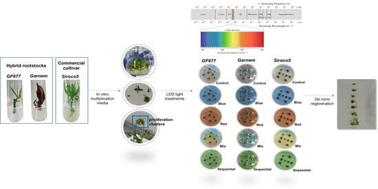
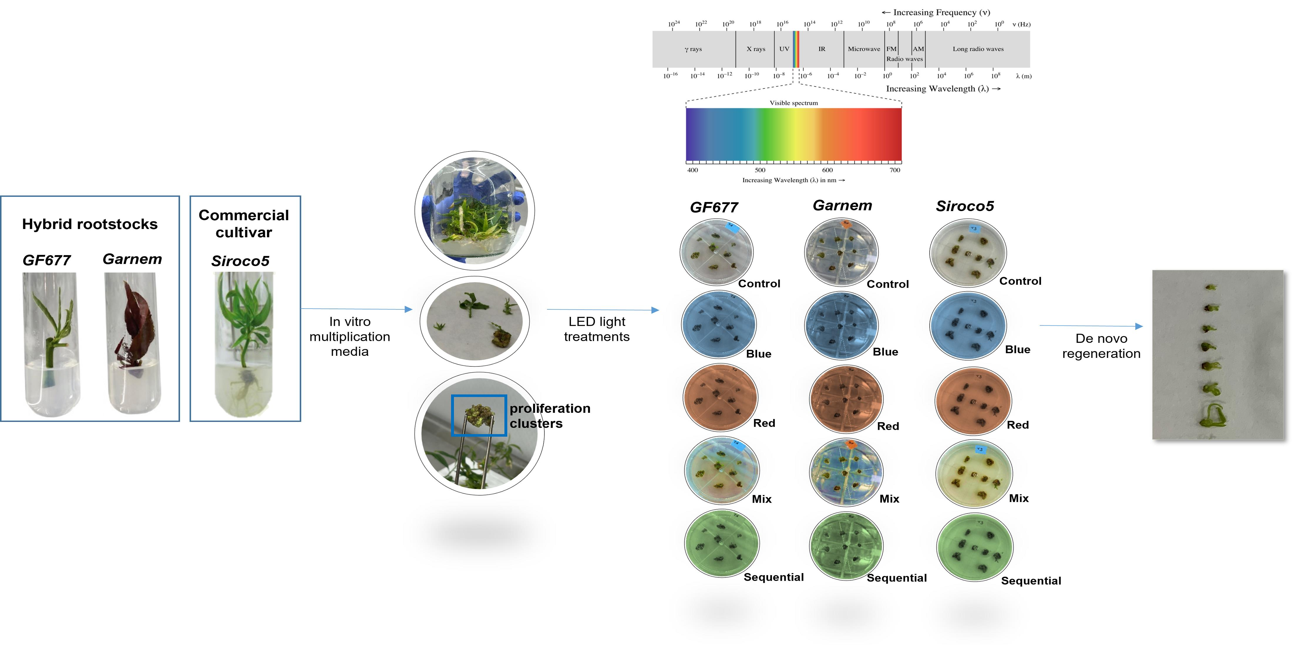
Graphical Abstract

1. Introduction

Woody fruit species of the genus Prunus are of major agronomic relevance worldwide; however, they are also widely recognized for being recalcitrant to in vitro regeneration. This characteristic poses a major limitation for the application of biotechnological tools in breeding and genetic improvement programs. Notably, peach (Prunus persica L. Batsch) has been identified as one of the most challenging species within the genus for in vitro culture, exhibiting strong genotype dependency, low regeneration frequencies, and frequent physiological disorders such as tissue necrosis and phenolic oxidation during the phases of culture establishment and regeneration [1,2,3]. Conventional breeding in peach and other stone fruit species is hindered by long juvenile periods, high heterozygosity, and extended selection cycles, which considerably slow the development of new cultivars capable of addressing emerging challenges such as climate change, biotic stresses, and sustainability requirements [4]. In this context, advanced biotechnological approaches—including genetic transformation and genome editing technologies, namely CRISPR-Cas systems—offer promising alternatives to accelerate genetic improvement [5]. Nonetheless, the successful implementation of these technologies critically depends on the availability of efficient and reproducible in vitro regeneration systems capable of generating complete plants from modified cells or tissues [6,7]. Among the various regeneration pathways, adventitious organogenesis has become a particularly relevant strategy for woody species. This process involves the formation of shoots or roots from somatic tissues through cellular dedifferentiation followed by redifferentiation, occurring without the involvement of a zygotic embryogenesis stage [8,9]. In Prunus species, adventitious regeneration is predominantly achieved via an indirect pathway, whereby organ formation originates from previously induced callus tissue [10]. Although this approach is often associated with limited regeneration efficiency across many genotypes, it enables greater cellular manipulation and has thus been extensively adopted in protocols aimed at genetic improvement and transformation [11,12]. The efficiency of adventitious organogenesis is influenced by multiple factors, including genotype, explant source, culture medium composition, plant growth regulators, and environmental conditions. In Prunus, regeneration capacity is strongly genotype-dependent, with hybrid rootstocks such as ‘GF677’ and ‘Garnem’ (P. persica × P. dulcis) generally demonstrating higher morphogenic competence than commercial peach cultivars [13,14,15]. Explant selection is also crucial, as juvenile and highly meristematic tissues typically exhibit enhanced regenerative potential [16,17]. Recent studies further indicate that callus derived from the basal region of in vitro proliferation cultures displays superior regenerative capacity compared with other tissue sources, underscoring its suitability for regeneration-based protocols in Prunus [10,15]. In addition to chemical and biological factors, physical environmental conditions play a decisive role in in vitro morphogenesis.
Light not only functions as the primary energy source for photosynthesis but also operates as a key regulatory signal that governs plant development through specific photoreceptors sensitive to distinct wavelengths. Variations in light spectrum, intensity, and photoperiod can markedly influence callus induction, shoot regeneration, and overall plantlet quality in tissue culture systems [18,19]. Red and far-red light are primarily perceived through phytochromes, whereas blue and ultraviolet light are detected by cryptochromes and phototropins, thereby activating signaling pathways that modulate morphogenesis and physiological responses (reviewed by [19]). The advent of light-emitting diode (LED) technology has created new opportunities for precise regulation of light quality in plant tissue culture. Compared to traditional fluorescent or high-pressure sodium lamps, LEDs provide several advantages, such as low heat emission, high energy efficiency, and the capability to deliver narrow and customizable wavelength spectra [20,21]. Consequently, LED lighting has been increasingly employed to investigate and optimize morphogenic responses in vitro across a wide range of plant species, including woody fruit crops. Prior studies have demonstrated that specific LED spectra, particularly red, blue, or their combinations, can enhance shoot regeneration, biomass accumulation, and physiological quality in species such as kiwifruit (Actinidia deliciosa), pear (Pyrus communis), plum (P. salicina), cherry rootstocks (P. cerasus × P. canescens), and apple rootstocks [10,22,23,24,25,26].
To date, the effects of LED light spectra on adventitious shoot regeneration in peach (P. persica L.), including peach-related rootstocks (P. persica × P. dulcis), have not been thoroughly investigated, despite existing studies in other Prunus species and woody fruit crops. Given the strong genotype dependence observed in Prunus regeneration and the increasing importance of both hybrid rootstocks and commercial cultivars in modern fruit production, it is essential to evaluate how different peach-related genotypes respond to controlled light spectrum modulation during in vitro regeneration. In this context, several aspects of peach in vitro regeneration remain unresolved, notably whether light spectrum modulation can improve adventitious shoot regeneration from organogenic calli derived from the basal region of in vitro proliferation clusters in peach, and whether responses differ between recalcitrant commercial cultivars and more regeneration-competent hybrid rootstocks. Additionally, the potential influence of far-red radiation, through alterations in the red to far-red ratio, on shoot redifferentiation in peach has yet to be assessed. Finally, it remains unclear whether continuous exposure to specific spectral compositions or the use of alternating (sequential) LED regimes differentially impact regeneration efficiency in this species. Addressing these gaps is critical for developing robust, reproducible, and broadly applicable regeneration protocols for peach cultivars and hybrid rootstocks that support micropropagation and future biotechnological applications, such as genetic transformation and CRISPR-Cas genome editing. Therefore, the present study aims to evaluate the effects of different LED light treatments on adventitious shoot regeneration in three peach-related genotypes: the commercial peach cultivar ‘Siroco 5’ (P. persica L.) and the hybrid rootstocks ’GF677’ and ‘Garnem’ (P. persica × P. dulcis). By analyzing organogenic callus formation, shoot regeneration efficiency, and fresh weight under controlled in vitro conditions, this study seeks to contribute to the optimization of regeneration protocols for peach cultivars and hybrid rootstocks.

2. Materials and Methods

2.1. Plant Material

The experiment was conducted utilizing 4-year-old peach trees cultivated at the experimental field station of the Instituto Murciano de Investigación y Desarrollo Agroalimentario y Medioambiental (IMIDA, Murcia, Spain). Nodal segments were obtained from current-year, actively growing shoots of the peach scion cultivar ‘Siroco 5’ and the hybrid peach × almond rootstocks ‘GF677’ and ‘Garnem’. These segments were collected and transferred to the tissue culture laboratory.

2.2. In Vitro Establishment and Proliferation Cultures

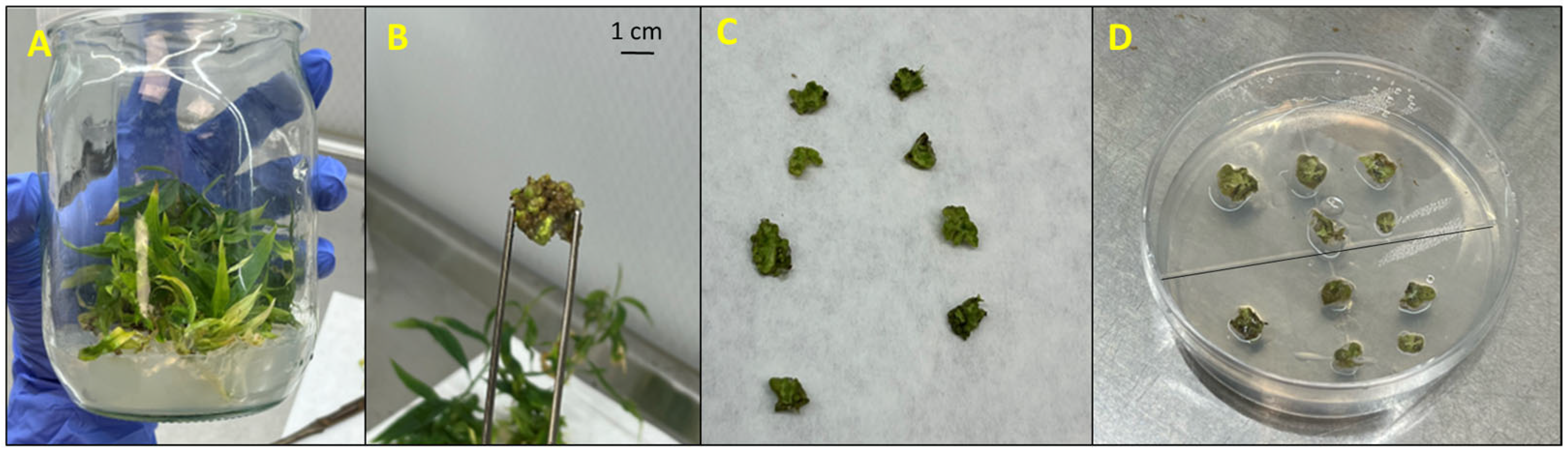
The nodal segments were meticulously washed under running tap water and surface-sterilized by immersion in a 2% (v/v) sodium hypochlorite solution supplemented with 0.1% (v/v) Tween 20 for 2 h under continuous agitation, according to the methodology of [15,17]. Following sterilization, the explants were transferred to a laminar flow hood, where the basal cut was renewed to optimize nutrient uptake. Each explant was cultured individually in a test tube containing solid Murashige and Skoog (MS) medium supplemented with vitamins (Duchefa Biochemie, Haarlem, The Netherlands; 100 mg L−1 myo-inositol, 2.0 mg L−1 glycine, 0.5 mg L−1 nicotinic acid, 0.5 mg L−1 pyridoxine-HCl, and 0.1 mg L−1 thiamine-HCl), 3% (w/v) sucrose, and 0.7% (w/v) plant propagation agar (Pronadisa, Madrid, Spain). The pH of the medium was adjusted to 5.8 using 0.1 N KOH and sterilized by autoclaving at 121 °C (1.1 kg cm−2) for 16 min. The explants were maintained under these conditions for up to four weeks, during which shoot emergence was observed, and an approximate explant survival rate of 80% was recorded. Once shoot emergence was observed, the shoots were transferred to a multiplication medium consisting of MS basal salts supplemented with 1 mg L−1 6-benzylaminopurine (BAP), 0.1 mg L−1 indole-3-butyric acid (IBA), 3% (w/v) sucrose, and 0.7% (w/v) plant propagation agar. Cultures were maintained at 25 ± 1 °C under a 16 h photoperiod with an irradiance of approximately 45 µmol m−2 s−1 provided by LED tubes (spectral range 380–780 nm). After three subcultures, organogenic calli were obtained from the base of proliferation clusters (Figure 1A,B).

2.3. Explant Preparation and De Novo Regeneration

Once proliferation clusters were established (Figure 1A,B), callus tissue was carefully excised under sterile conditions and transversely sectioned into thin slices (approximately 3 mm thick) (Figure 1C), in accordance with the methodology described by [10]. Each callus was subdivided into five slices, and two calli (a total of ten slices) were placed within each Petri dish (Figure 1D). After isolation, the calli preserved their structural integrity and uniform appearance upon transfer to the regeneration medium (Figure 1D). The plates were subsequently sealed with Parafilm M (Amcor, Zürich, Switzerland) to ensure aseptic conditions. Explants representing each of the genotypes (‘Siroco 5’, ‘GF677’, and ‘Garnem’) were cultured on a regeneration medium composed of MS basal salts supplemented with vitamins, 3% (w/v) sucrose, 0.7% (w/v) agar, 2 mg L−1 BAP, and 0.1 mg L−1 IBA, following the protocol outlined by [15]. The pH was adjusted to 5.8 using 1 N KOH or 1 N HCl prior to autoclaving at 121 °C (1.1 kg cm−2) for 16 min.

2.4. Experimental Design and Light Treatments

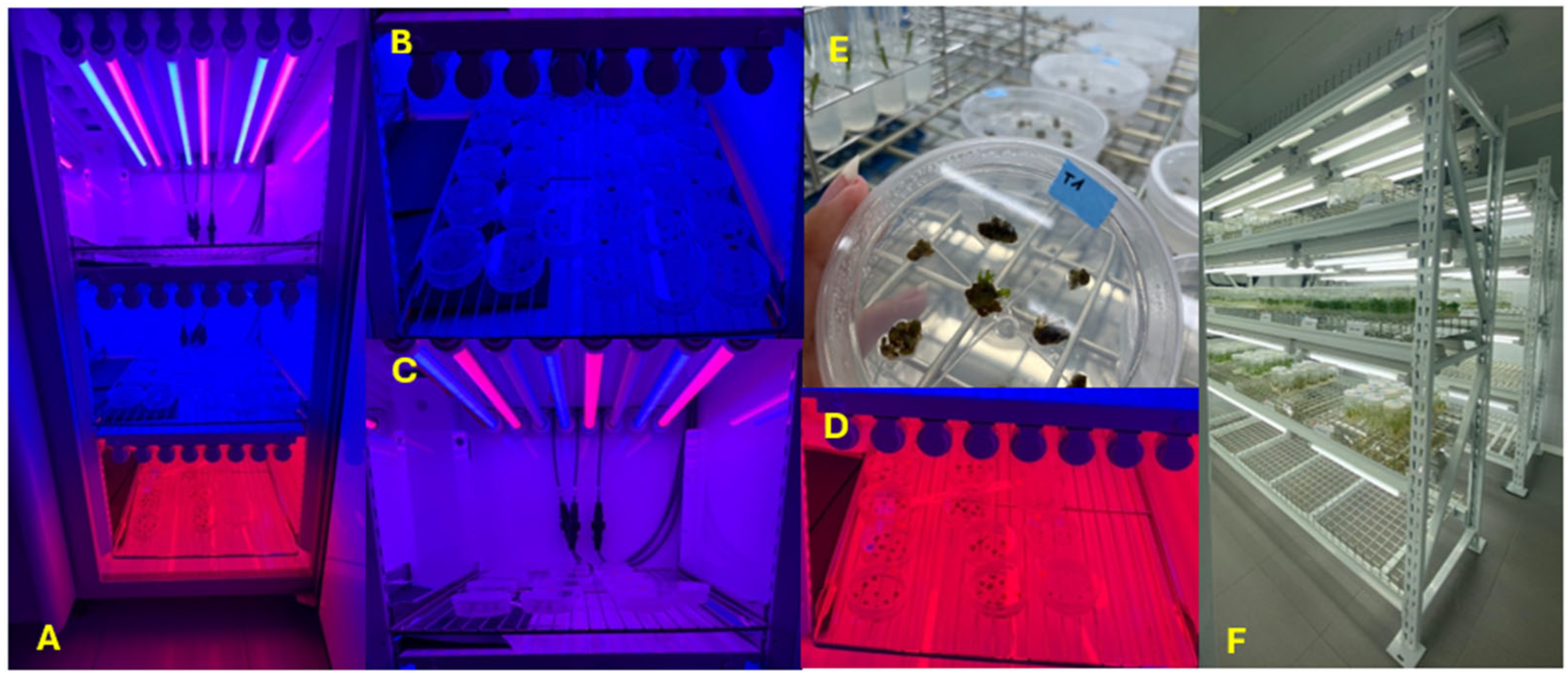
A factorial experimental design was established to evaluate the influence of light spectrum on adventitious shoot regeneration. Three genotypes (‘GF677’, ‘Garnem’, and ‘Siroco 5’) were combined with five LED light treatments (Figure 2). The control group was cultured under white LED light (control; 380–780 nm). A second group was cultured in 55% red (600–700 nm) + 45% far-red light (700–800 nm) (red + far-red). The third group was subjected to 100% blue light (blue; 400–500 nm): A fourth group received a combination of 42% red, 34% far red, and 24% blue light (mixed; 400–800 nm). The final group involved sequential light conditions, alternating between blue, red-far red, and mixed light every 10 days. The photosynthetically active photon flux density (PPFD, 400–700 nm) measured at the culture level was 74.68 μmol m−2 s−1 under white light (control), 52.58 μmol m−2 s−1 under blue light (B), 93.64 μmol m−2 s−1 under red + far-red light (R + FR), and 133.8 μmol m−2 s−1 under the combined blue + red + far-red treatment (B + R + FR). The spectral composition and photon flux density parameters were quantified for each of the LED treatments and are summarized in Table 1.
A total of 25 Petri dishes per genotype were used, corresponding to five replicates per light treatment and genotype (n = 5 dishes per genotype × light treatment). The white-light control treatment was conducted within a growth chamber equipped with white LED lamps (Aralab 1200 PLH LED, Aralab, Madrid, Spain) (Figure 2). Cultures subjected to the blue, red + far-red, mixed, and sequential LED treatments were maintained in a second growth chamber (Aralab 1200 PLH LED, Aralab, Madrid, Spain) (Figure 2). All of the treatment replicates were kept under identical environmental conditions (25 ± 1 °C with a 16 h light/8 h dark photoperiod). The duration of the experiment was 30 days, during which the explants remained on the same regeneration medium without distinct induction or regeneration phases.

2.5. Data Collection and Statistical Analysis

A total of 150 calli (50 per genotype) were distributed across Petri dishes (two calli per dish) and utilized as subsamples for evaluation, whereas the Petri dish was regarded as the experimental unit for statistical analyses (n = 5 dishes per genotype × light treatment). Explants were periodically monitored during the 30-day culture period. At the conclusion of the experiment, the number of regenerated shoots was recorded for each of the original calli by summing the shoots produced across the five slices derived from the same callus. The number of calli-producing shoots was recorded per Petri dish. The organogenic rate was expressed as the number of regenerated shoots per original callus. The percentage of regenerating explants was calculated as the frequency of organogenic calli (FOC, %), considering an original callus as regenerating when at least one of its derived slices produced shoots. FOC was calculated at the Petri dish level as the proportion of regenerating calli (0, 1, or 2 calli per dish). Additionally, shoot fresh weight was measured as an additional growth parameter. For statistical purposes, callus-level values were aggregated at the Petri dish level to yield a single observation per replicate dish. Data were initially analyzed for normality and homogeneity of variance. A two-way analysis of variance (ANOVA) was performed to assess the effects of genotype and light treatment. Mean separation was conducted using Duncan’s multiple range test at a significance level of p ≤ 0.05. Furthermore, a principal component analysis (PCA) was performed to explore the relationships among treatments. All of the statistical analyses were performed using SPSS software (IBM SPSS Statistics v25.0, Chicago, IL, USA).

3. Results

3.1. Adventitious Shoot Regeneration Under Different LED Light Spectra

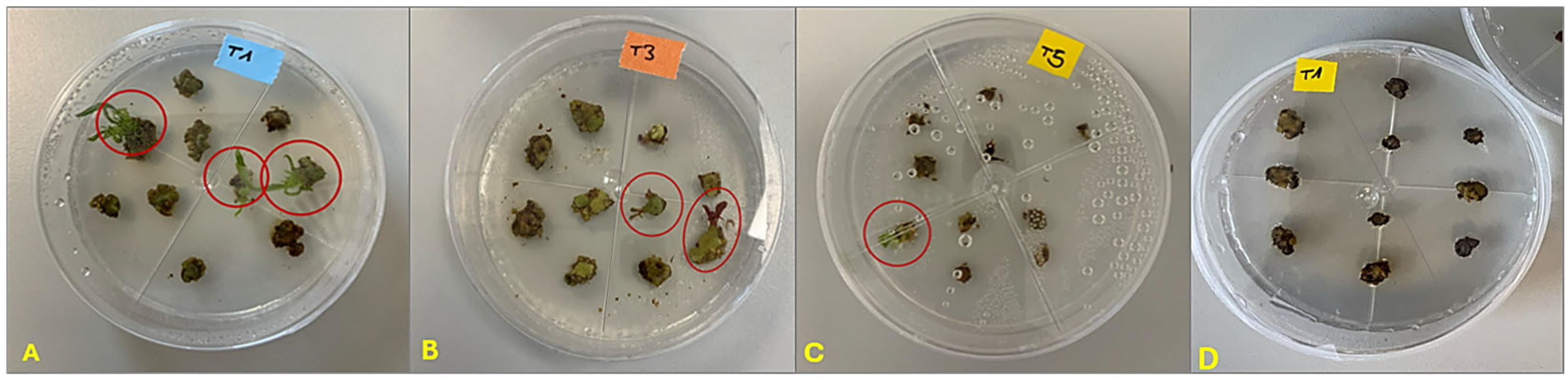
Organogenic calli derived from the basal region of in vitro-grown shoots exhibited a compact and nodular morphology, with a dense structure and well-defined contours (Figure 1B). The calli showed an intense green coloration, indicative of metabolically active tissue with a high organogenic competence, and no visible signs of necrosis, hyperhydricity, or oxidative browning were observed (Figure 1B,C). Following the transfer of callus slices to the regeneration medium, the first regenerated shoots appeared approximately six days after culture initiation across all light treatments (Figure 3). During this early stage, the hybrid rootstocks ‘GF677’ and ‘Garnem’ displayed a faster and more uniform regenerative response (Figure 3A,B). In contrast, ‘Siroco 5’ showed a delayed and limited shoot regeneration during the first week of culture (Figure 3C), together with a higher incidence of tissue browning and necrosis, particularly under white light conditions (Figure 3D). After 30 days of culture, shoot regeneration was detected in all of the genotypes under the different light spectra evaluated. The total number of regenerated shoots per callus was subsequently recorded and used to calculate regeneration efficiency parameters.
Statistical analysis revealed no significant effects of genotype, light treatment, or their interaction on FOC (p > 0.05), indicating that the frequency of shoot-regenerating calli was not significantly affected by either the applied light spectrum or the genotype under study. Despite the absence of statistically significant differences, LED light treatments generally resulted in equal or higher FOC values compared with the white light control in most cases across the evaluated genotypes (Table 2). This trend was particularly evident in the hybrid rootstock ‘Garnem’ and, to a lesser extent, in ‘GF677’, which exhibited high FOC values, often close to 100%, under several LED spectra. In contrast, the commercial cultivar ‘Siroco 5’ demonstrated greater variability among treatments, with comparatively lower FOC values under control and sequential light conditions.

3.2. Effect of Genotype and Light Spectrum on Organogenic Rate

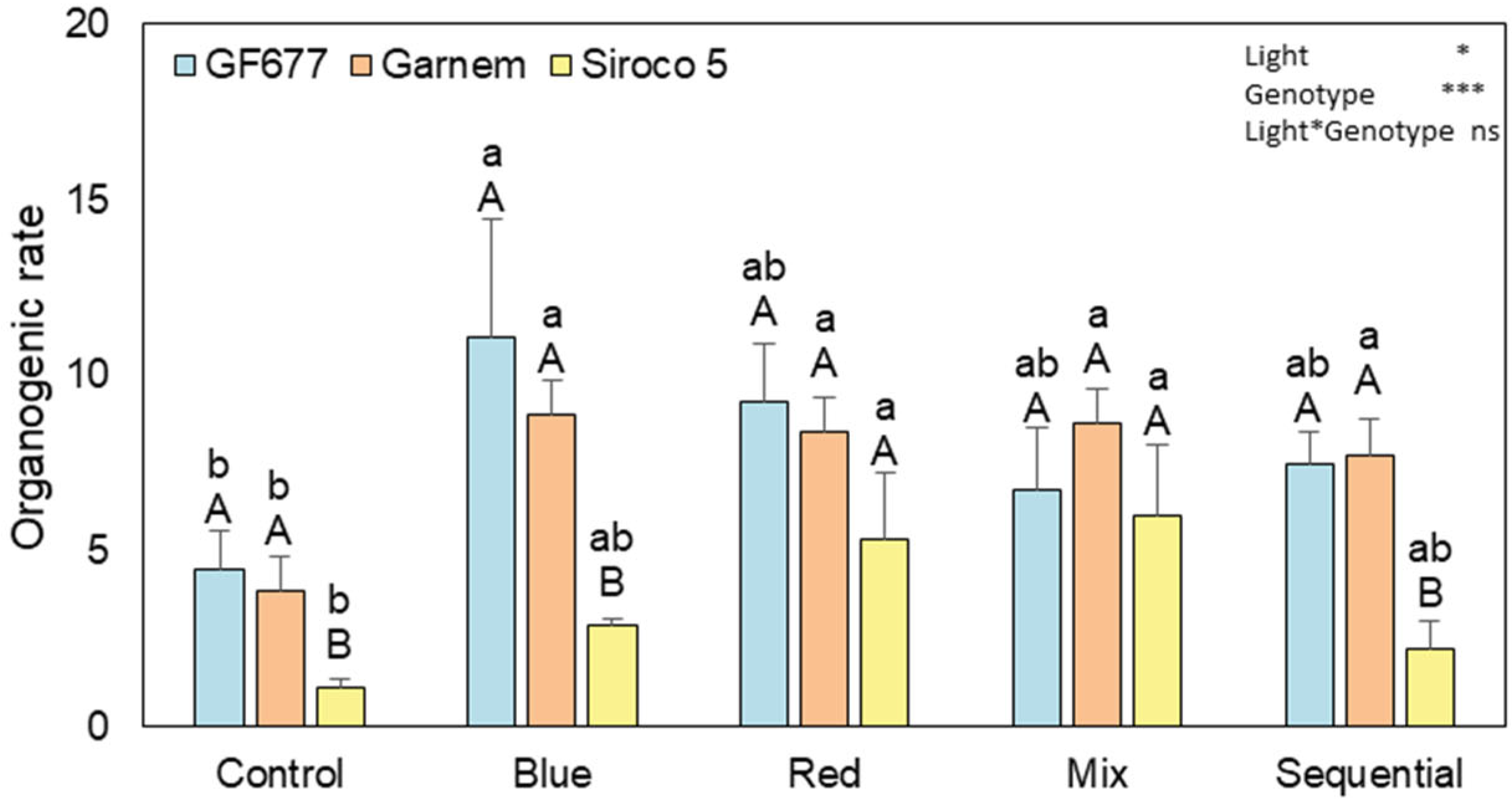
The organogenic rate, expressed as the number of regenerated shoots per callus, was significantly influenced by both genotype and LED light spectrum (p < 0.05). No significant interaction between these factors was observed (Figure 4). Overall, the hybrid rootstocks ‘GF677’ and ‘Garnem’ demonstrated higher organogenic rates than the commercial cultivar ‘Siroco 5’ under most light treatments. Under white light (control), ‘GF677’ and ‘Garnem’ exhibited similar organogenic rates, both significantly exceeding those observed in ‘Siroco 5’. A similar pattern was observed under blue light, where both hybrid rootstocks achieved their peak organogenic values and showed significantly higher regeneration rates than ‘Siroco 5’, with no marked differences detected between ‘GF677’ and ‘Garnem’. For ‘Siroco 5’, the highest organogenic rates were recorded under red + far-red and mixed LED light treatments, under which no significant differences among genotypes were observed, and a significant increase in shoot regeneration was evident compared to white, blue, and sequential light treatments. Conversely, under sequential light conditions, ‘Siroco 5’ again displayed significantly lower organogenic rates than the hybrid rootstocks.

3.3. Number of Regenerated Shoots per Callus and Shoot Fresh Weight

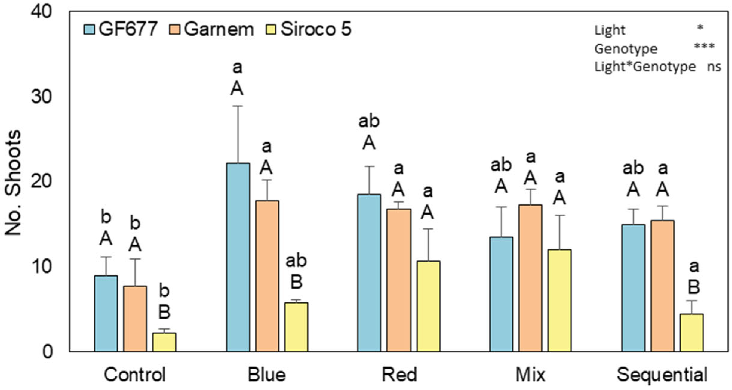
The number of regenerated shoots per callus followed trends that were similar to those observed for the organogenic rate (Figure 5). Both genotype and LED light spectrum had significant independent effects on shoot regeneration (p < 0.05), whereas their interaction was not significant. Overall, the hybrid rootstocks ‘GF677’ and ‘Garnem’ produced a higher number of shoots per callus than the commercial cultivar ‘Siroco 5’ under most light treatments. Under white light (control), all of the genotypes exhibited the lowest regeneration values, with ‘Siroco 5’ producing significantly fewer shoots than the hybrid rootstocks. A marked increase in shoot regeneration was observed under blue light, where ‘GF677’ and ‘Garnem’ reached their highest values and produced significantly more shoots per callus than ‘Siroco 5’. In contrast, the highest number of regenerated shoots in ‘Siroco 5’ was recorded under red + far-red and mixed LED light treatments, with which no significant differences among genotypes were detected. Under sequential light conditions, shoot regeneration declined again in ‘Siroco 5’, whereas the hybrid rootstocks maintained comparatively higher regeneration levels.
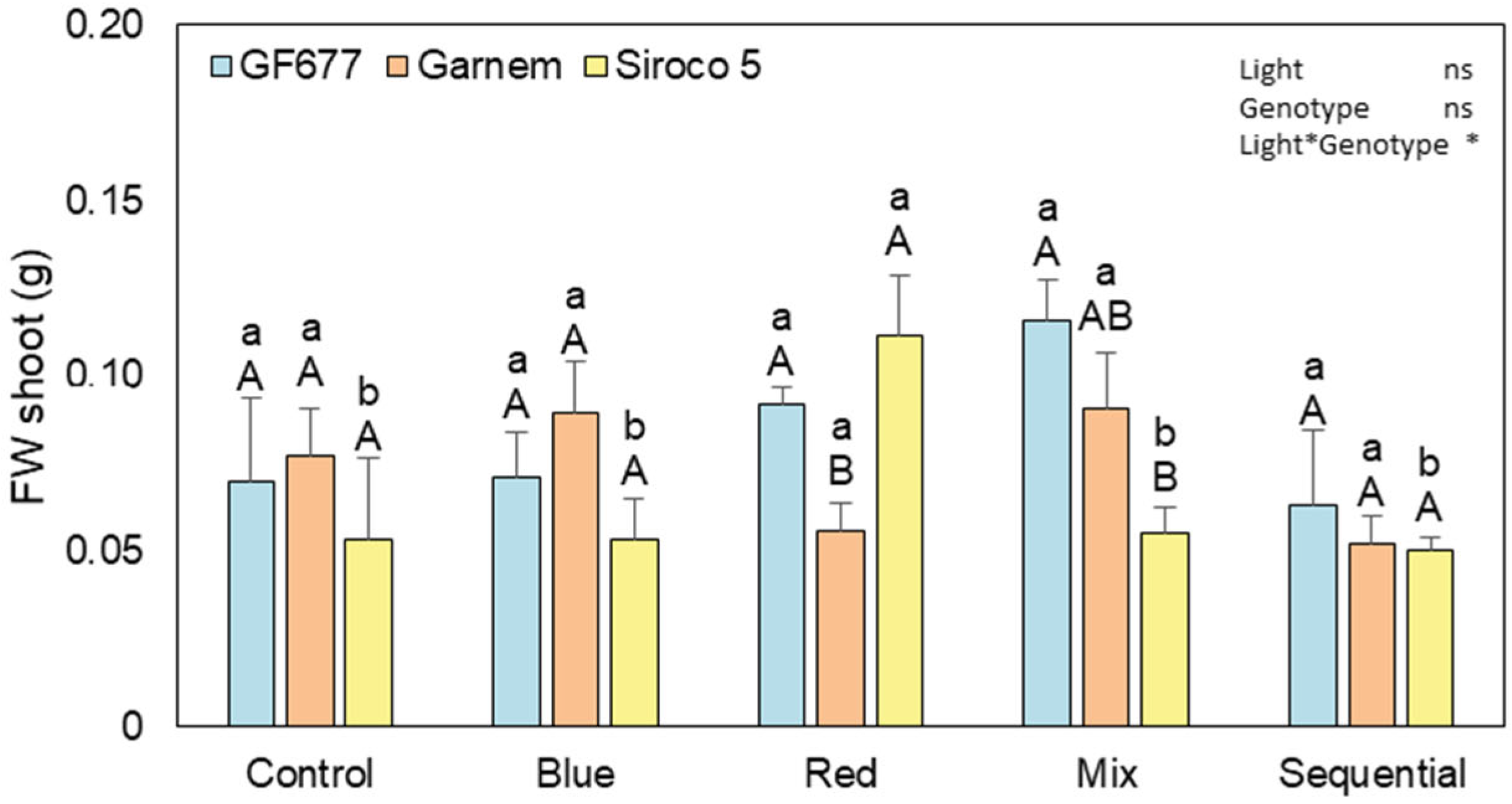
Shoot fresh weight was significantly influenced by the interaction between genotype and LED light spectrum (p ≤ 0.05), whereas no significant independent effects of genotype or light treatment were observed (Figure 6). When comparing genotypes within each light treatment, no significant differences were noted under white (control) or blue light. Under red + far-red light, ‘Siroco 5’ and ‘GF677’ exhibited significantly higher shoot fresh weights than ‘Garnem’. Under mixed light conditions, ‘GF677’ demonstrated significantly higher shoot fresh weight compared to ‘Siroco 5’, whereas ‘Garnem’ showed intermediate values. No significant differences among genotypes were detected under sequential light. When examining light treatments within each genotype, ‘GF677’ achieved its highest shoot fresh weight under mixed light, with lower values under white, blue, and sequential light. In ‘Garnem’, shoot fresh weight was greater under blue and mixed light than under red + far-red light, whereas control and sequential treatments showed intermediate values. For the commercial cultivar ‘Siroco 5’, the maximum shoot fresh weight was recorded under red + far-red light, being significantly higher than that observed under the other light treatments.

3.4. Score Principal Component Analysis

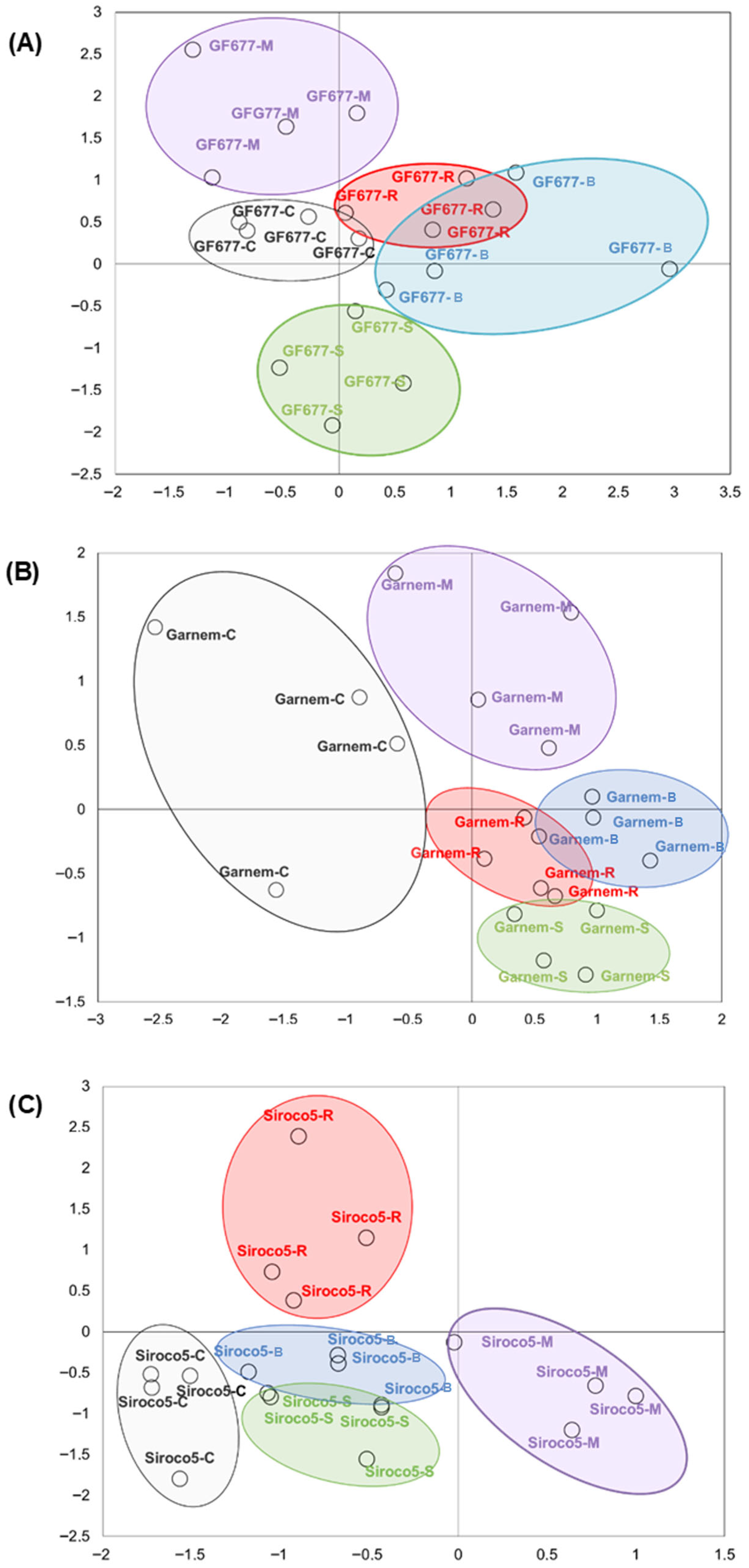
A principal component analysis (PCA) was conducted to obtain a comprehensive overview of the responses of the three evaluated genotypes to the different LED light treatments and to visualize similarities and differences among treatments based on the measured regeneration parameters. The PCA biplot demonstrated distinct genotype-dependent clustering patterns, indicating unique multivariate responses to light spectrum modulation in ‘GF677’, ‘Garnem’, and ‘Siroco 5’ (Figure 7). In the hybrid rootstock ‘GF677’ (Figure 7A), the mixed, control, and sequential light treatments each formed well-defined and clearly separated clusters within the PCA space, indicating distinct multivariate responses under these light conditions. Conversely, the blue and red + far-red light treatments exhibited significant overlap, grouping closely together and suggesting similar responses for this genotype under these spectra. For the hybrid rootstock ‘Garnem’ (Figure 7B), the control treatment distinctly clustered apart from the remaining light treatments, indicating clear differentiation within the PCA space. The mixed light treatment also constituted an independent cluster, whereas the red + far-red and blue light treatments grouped more closely, reflecting comparable responses under these spectra. The sequential light treatment formed a smaller and more compact cluster, partially overlapping with red + far-red and blue treatments. Regarding the commercial cultivar ‘Siroco 5’ (Figure 7C), the PCA revealed a markedly different pattern from that observed in the hybrid rootstocks. The red + far-red light treatment generated a distinctly separate cluster, well differentiated from the other treatments, whereas the control, blue, mixed, and sequential light treatments clustered more closely together, indicating more similar multivariate responses to these conditions for this genotype.

4. Discussion

Peach (Prunus persica L.) has long been regarded as one of the most recalcitrant woody fruit species for in vitro regeneration, largely due to its strong genotype dependency and low regenerative capacity under standard culture conditions. Advances in regeneration and genetic engineering technologies for P. persica contribute to the genetic improvement of this species and serve as valuable tools for molecular research in this crop and other related species. The findings of this study confirm these characteristics and further demonstrate that genotype and light environment are both important factors influencing differences in adventitious shoot regeneration efficiency in peach-related materials. The frequency of organogenic callus (FOC) remained consistently high across all of the genotypes and LED light treatments, with no statistically significant differences observed among the spectra. Conversely, significant differences among genotypes and light treatments were identified during the shoot regeneration phase, supporting the premise that morphogenic competence in Prunus is chiefly expressed during redifferentiation and shoot development. This observation aligns with prior reports indicating that, in woody species, variation in regeneration efficiency is predominantly associated with organ differentiation and shoot development, rather than earlier callus-related stages, which are recognized as major bottlenecks in regeneration protocols for woody species [12,27,28]. Clear genotype-dependent responses were documented throughout the regeneration process, with the hybrid peach × almond rootstocks ‘GF677’ and ‘Garnem’ consistently outperforming the commercial cultivar ‘Siroco 5’ in terms of organogenic rate and number of regenerated shoots. This behavior corroborates earlier research that indicates the presence of higher in vitro regeneration capacities in hybrid rootstocks relative to commercial peach cultivars, which often display increased recalcitrance and tissue necrosis, as well as diminished regenerative efficiency [2,13,14]. The observed reduction in regeneration capacity in ‘Siroco 5’, especially under control (white) and sequential light conditions, underscores the relevance of genetic background as a determining factor in in vitro response. These results reinforce the notion that commercial cultivars, primarily selected for their agronomic and fruit quality traits, may exhibit lower morphogenic plasticity under in vitro conditions compared to hybrid rootstocks developed with propagation efficiency as a priority. Additionally, light conditions significantly influenced the in vitro morphogenic responses of cultured tissues. Beyond morphogenic effects, the light environment has been demonstrated to modulate physiological traits associated with plantlet quality in woody species, including stomatal characteristics and gas exchange parameters, which are critical for photosynthetic performance and successful establishment during acclimatization [29].
The current findings align with earlier efforts to establish relationships between light spectra and the regulation of morphogenesis [22], although available information for woody species remains relatively limited [30]. In this investigation, modulation of the light spectrum exerted a marked influence on adventitious shoot regeneration, underscoring the importance of the light environment as a key modulatory factor in in vitro morphogenesis. Among the evaluated treatments, red + far-red LED light was associated with higher adventitious shoot regeneration values, particularly in the commercial cultivar ‘Siroco 5’, where it markedly increased both shoot number and biomass accumulation (Figure 5 and Figure 6). From a practical standpoint, this finding is especially relevant, as it suggests that, under the tested growth chamber conditions, targeted manipulation of light quality may help mitigate genotype-related limitations in regeneration. The stimulatory effect of red + far-red light observed in this study concurs with prior reports in woody species describing positive effects of red wavelengths on shoot regeneration, biomass production, and morphogenic responses, which are typically associated with phytochrome-mediated developmental regulation [31,32]. Similar enhancements in shoot growth and biomass under red-light-based spectra have also been documented in controlled production systems for Cirsium setidens, thereby reinforcing the general role of red wavelengths in promoting aboveground growth responses across diverse plant systems [33]. Additionally, blue LED light supported high regeneration efficiency, particularly in the hybrid rootstock ‘GF677’, although its effects appeared more genotype-dependent and were not always statistically distinguishable from those observed under red + far-red light. Similar genotype-specific responses to blue light have been reported in other woody fruit species and are generally linked to cryptochrome- and phototropin-mediated regulation of cell division and differentiation [20].
The contrasting responses observed between hybrid rootstocks and the commercial cultivar under blue light further emphasize the interaction between genetic background and light perception pathways in shaping in vitro morphogenic outcomes. In contrast, white light was associated with lower regeneration performance across genotypes, suggesting that broad-spectrum illumination may be less effective than targeted wavelength application for optimizing adventitious shoot regeneration in Prunus tissue culture systems (Table 1). The mixed-light treatment produced intermediate to high regeneration responses depending on the genotype, indicating that wavelength combinations can support morphogenesis but do not necessarily surpass the effectiveness of a well-defined spectrum such as red + far-red light. This fact underscores the significance of genotype-specific variations in light perception and subsequent signal integration during in vitro culture, ultimately modulating morphogenic competence and regeneration efficiency under specific light environments [19,34]. Numerous studies on woody fruit species have indicated that red–blue mixed light outperforms monochromatic treatments, presumably due to the complementary functions of red and blue wavelengths in regulating photosynthesis, cell division, and morphogenesis [33,35]. Moreover, sequential light treatment did not demonstrate clear benefits and was generally linked to decreased regeneration efficiency and reduced shoot biomass. These findings suggest that continuous exposure to a specific spectral composition may be more effective than alternating light conditions for promoting adventitious shoot regeneration in peach-related genotypes under the tested conditions. Such insight is particularly relevant for protocol simplification, as constant lighting regimes are more straightforward to implement and standardize in routine tissue culture applications. Considering that the employed LED treatments differed not only in spectral composition but also in photosynthetically active photon flux density (PPFD), as detailed in Table 1, the observed regeneration responses should be interpreted as genotype-specific performance within integrated light environments, rather than as direct causal effects attributable to individual wavelengths. Variations in light responses among different genotypes are extensively documented. In Prunus, dark incubation of explants has been shown to significantly enhance adventitious shoot regeneration frequency [13,29,36] compared to cultures maintained under continuous light. Conversely, other studies suggest that monochromatic light promotes calli multiplication [20], and the combination of multiple spectra can induce de novo organogenesis [10,37]. In fact, explants from various plant species complete regeneration by responding to different photoreceptors under diverse light spectra [10,20,38], and identical spectral conditions can either facilitate or inhibit processes related to adventitious shoot regeneration, depending on the genotype [39]. In this context, the bi-plot derived from principal component analysis (PCA) served as an exploratory multivariate visualization tool to summarize genotype-specific responses to LED light environments, revealing clustering patterns predominantly associated with red + far-red and mixed light treatments, especially in ‘Siroco 5’. The distinct separation of treatments in the bi-plot highlights the consistency of the observed responses and reinforces the identified response patterns. From an applied standpoint, the identification of red LED light (red + far-red) as a broadly effective treatment across both peach cultivar and hybrid rootstock genotypes constitutes a significant step forward for the optimization of in vitro regeneration protocols. The efficient and reproducible regeneration of adventitious shoots is a fundamental requirement for micropropagation and the successful application of biotechnological tools such as genetic transformation and genome editing in woody crops. The findings presented here establish a robust experimental basis for enhancing regeneration efficiency in peach-related materials and support the development of more resilient and transferable protocols for future genetic improvement initiatives.

5. Conclusions

This study demonstrates that adventitious shoot regeneration in peach-related materials exhibits clear genotype-dependent variations and fluctuates under different LED light environments. Conversely, the frequency of organogenic callus remains consistently high and does not differ statistically among the various light treatments. Hybrid peach × almond rootstocks (‘GF677’ and ‘Garnem’) exhibited greater regenerative capacity compared to the commercial cultivar ‘Siroco 5’, thereby confirming the pronounced genotype dependency of in vitro regeneration in Prunus. Among the evaluated treatments, red + far-red light was consistently associated with enhanced shoot regeneration efficiency and increased shoot fresh weight across the tested genotypes. Additionally, this was linked to a partial mitigation of the recalcitrant response that is characteristic of the commercial cultivar. These findings underscore the potential value of targeted LED light spectrum modulation as a practical strategy for optimizing adventitious shoot regeneration protocols in peach cultivars and hybrid rootstock genotypes, under the specific light spectra and photon flux density conditions assessed in this research. Consequently, this study provides a valuable foundation for future applications in woody fruit crop biotechnology.

Author Contributions

Conceptualization, M.P.-J. and J.E.C.-T.; methodology, G.F.-O., M.R.-M. and J.M.G.-S.; software, M.R.-M., J.E.C.-T. and M.P.-J.; validation, M.R.-M., J.E.C.-T. and M.P.-J.; formal analysis, G.F.-O. and M.R.-M.; investigation, M.P.-J. and G.F.-O.; resources, M.P.-J. and J.E.C.-T.; data curation, M.R.-M.; writing—original draft preparation, M.R.-M. and G.F.-O.; writing—review and editing, M.R.-M., J.E.C.-T. and M.P.-J.; visualization, M.R.-M. and J.E.C.-T.; supervision, M.R.-M. and J.E.C.-T.; project administration, M.P.-J.; funding acquisition, M.P.-J. All authors have read and agreed to the published version of the manuscript.

Funding

This work was financed by the European Regional Development Fund ERDF50464MG_HB.

Data Availability Statement

The raw data supporting the conclusions of this article will be made available by the authors on request.

Acknowledgments

We thankfully acknowledge the help of Carmen López Sierra with the laboratory work and Andrés Paredes for revising the English.

Conflicts of Interest

The authors declare no conflicts of interest.

References

  1. Prakash Narayan, J. Plant Tissue Culture: Theory and Practice; Bhojwani, S., Razdan, M.K., Eds.; Elsevier: Amsterdam, The Netherlands, 1996; ISBN 0-444-81623-2. [Google Scholar]
  2. Pérez-Jiménez, M.; López-Soto, M.B.; Cos-Terrer, J. In Vitro Callus Induction from Adult Tissues of Peach (Prunus persica L. Batsch). Vitro Cell. Dev. Biol.-Plant 2013, 49, 79–84. [Google Scholar] [CrossRef]
  3. Pritchard, H.W.; Sershen; Tsan, F.Y.; Wen, B.; Jaganathan, G.K.; Calvi, G.; Pence, V.C.; Mattana, E.; Ferraz, I.D.K.; Seal, C.E. Regeneration in Recalcitrant-Seeded Species and Risks from Climate Change. In Plant Regeneration from Seeds; Elsevier: Amsterdam, The Netherlands, 2022; pp. 259–273. [Google Scholar] [CrossRef]
  4. Van Nocker, S.; Gardiner, S.E. Breeding Better Cultivars, Faster: Applications of New Technologies for the Rapid Deployment of Superior Horticultural Tree Crops. Hortic. Res. 2014, 1, 14022. [Google Scholar] [CrossRef]
  5. Jaganathan, D.; Ramasamy, K.; Sellamuthu, G.; Jayabalan, S.; Venkataraman, G. CRISPR for Crop Improvement: An Update Review. Front. Plant Sci. 2018, 9, 985. [Google Scholar] [CrossRef]
  6. Gentile, A.; Monticelli, S.; Damiano, C. Adventitious Shoot Regeneration for Agrobacterium-Mediated Transformation in Prunus. Acta Hortic. 2003, 616, 331–334. [Google Scholar] [CrossRef]
  7. Savadi, S.; Mangalassery, S.; Sandesh, M.S. Advances in Genomics and Genome Editing for Breeding next Generation of Fruit and Nut Crops. Genomics 2021, 113, 3718–3734. [Google Scholar] [CrossRef] [PubMed]
  8. De Klerk, G.J.M.; Van Den Dries, N.; Krens, F.A. Hyperhydricity: Underlying Mechanisms. Acta Hortic. 2017, 1155, 269–275. [Google Scholar] [CrossRef]
  9. George, E.; Hall, M.; De Klerk, G.-J. Plant Propagation by Tissue Culture, 3rd ed.; Springer: Dordrecht, The Netherlands, 2008. [Google Scholar]
  10. López-Sierra, C.; Cos-Terrer, J.; Rómero-Muñoz, M.; Pérez-Jiménez, M. Source of Explant and Light Spectrum Influence in Adventitious Shoot Regeneration of Prunus salicina Lindl. (Japanese Plum). Plants 2025, 14, 2230. [Google Scholar] [CrossRef] [PubMed]
  11. Desai, P.; Desai, S.; Rafaliya, R.; Patil, G. Plant Tissue Culture: Somatic Embryogenesis and Organogenesis. In Advances in Plant Tissue Culture; Academic Press: New York, NY, USA, 2022; pp. 109–130. [Google Scholar] [CrossRef]
  12. Ricci, A.; Mezzetti, B.; Navacchi, O.; Burgos, L.; Sabbadini, S. In Vitro Regeneration, via Organogenesis, from Leaves of the Peach Rootstock GF677 (P. persica × P. amygdalus). Acta Hortic. 2023, 1359, 81–85. [Google Scholar] [CrossRef]
  13. Escalettes, V.; Dosba, F. In Vitro Adventitious Shoot Regeneration from Leaves of Prunus spp. Plant Sci. 1993, 90, 201–209. [Google Scholar] [CrossRef]
  14. Liu, X.; Pijut, P.M. Plant Regeneration from in Vitro Leaves of Mature Black Cherry (Prunus serotina). Plant Cell Tissue Organ Cult. 2008, 94, 113–123. [Google Scholar] [CrossRef]
  15. Pérez-Jiménez, M.; Carrillo-Navarro, A.; Cos-Terrer, J. Regeneration of Peach (Prunus persica L. Batsch) Cultivars and Prunus persica × Prunus dulcis Rootstocks via Organogenesis. Plant Cell Tissue Organ Cult. 2012, 108, 55–62. [Google Scholar] [CrossRef]
  16. Tian, L.; Wen, Y.; Jayasankar, S.; Sibbald, S. Regeneration of Prunus salicina Lindl (Japanese Plum) from Hypocotyls of Mature Seeds. Vitro Cell. Dev. Biol.-Plant 2007, 43, 343–347. [Google Scholar] [CrossRef]
  17. Pérez-Jiménez, M.; Cantero-Navarro, E.; Pérez-Alfocea, F.; Le-Disquet, I.; Guivarc’h, A.; Cos-Terrer, J. Relationship between Endogenous Hormonal Content and Somatic Organogenesis in Callus of Peach (Prunus persica L. Batsch) Cultivars and Prunus persica × Prunus dulcis rootstocks. J. Plant Physiol. 2014, 171, 619–624. [Google Scholar] [CrossRef]
  18. Zhang, L.; Meng, S.; Liu, Y.; Han, F.; Xu, T.; Zhao, Z.; Li, Z. Advances in and Perspectives on Transgenic Technology and CRISPR-Cas9 Gene Editing in Broccoli. Genes 2024, 15, 668. [Google Scholar] [CrossRef]
  19. Fan, C.; Manivannan, A.; Wei, H. Light Quality-Mediated Influence of Morphogenesis in Micropropagated Horticultural Crops: A Comprehensive Overview. BioMed Res. Int. 2022, 2022, 4615079. [Google Scholar] [CrossRef] [PubMed]
  20. Han, J.; Li, Y.; Zhao, Y.; Sun, Y.; Li, Y.; Peng, Z. How Does Light Regulate Plant Regeneration? Front. Plant Sci. 2024, 15, 1474431. [Google Scholar] [CrossRef]
  21. Bula, R.J.; Morrow, R.C.; Tibbitts, T.W.; Barta, D.J.; Ignatius, R.W.; Martin, T.S. Light-Emitting Diodes as a Radiation Source for Plants. HortScience 1991, 26, 203–205. [Google Scholar] [CrossRef]
  22. Gupta, S.D.; Jatothu, B. Fundamentals and Applications of Light-Emitting Diodes (LEDs) in in Vitro Plant Growth and Morphogenesis. Plant Biotechnol. Rep. 2013, 7, 211–220. [Google Scholar] [CrossRef]
  23. Muleo, R.; Morini, S. Effect of Light Quality on Regeneration from Callus of Actinidia deliciosa. Acta Hortic. 1990, 280, 155–158. [Google Scholar] [CrossRef]
  24. Nacheva, L.; Dimitrova, N.; Berova, M. Effect of LED Lighting on the Growth of Micropropagated Pear Plantlets (Pyrus communis L. OHF 333). Acta Hortic. 2022, 1337, 9–16. [Google Scholar] [CrossRef]
  25. Sarropoulou, V.; Sperdouli, I.; Adamakis, I.D.; Grigoriadou, K. The Use of Different LEDs Wavelength and Light Intensities for in Vitro Proliferation of Cherry Rootstock: Influence on Photosynthesis and Photomorphogenesis. Plant Cell Tissue Organ Cult. 2023, 152, 317–330. [Google Scholar] [CrossRef]
  26. Tomazini Scolaro, A.M.; De Martin, M.S.; Vieira, R.L.; Schveitzer, B.; de Souza, E.L.; Borges, E.M. Use of Light-Emitting Diodes on the In Vitro Rooting of Apple Tree Rootstocks. Int. J. Plant Biol. 2024, 16, 12. [Google Scholar] [CrossRef]
  27. Sabbadini, S.; Ricci, A.; Limera, C.; Baldoni, D.; Capriotti, L.; Mezzetti, B. Factors Affecting the Regeneration, via Organogenesis, and the Selection of Transgenic Calli in the Peach Rootstock Hansen 536 (Prunus persica × Prunus amygdalus) to Express an RNAi Construct against PPV Virus. Plants 2019, 8, 178. [Google Scholar] [CrossRef] [PubMed]
  28. Ricci, A.; Sabbadini, S.; Prieto, H.; Padilla, I.M.; Dardick, C.; Li, Z.; Scorza, R.; Limera, C.; Mezzetti, B.; Perez-Jimenez, M.; et al. Genetic Transformation in Peach (Prunus persica L.): Challenges and Ways Forward. Plants 2020, 9, 971. [Google Scholar] [CrossRef]
  29. Espinosa, A.; Pijut, P.; Michler, C. Adventitious Shoot Regeneration and Rooting of Prunus Serotina in Vitro Cultures. HortScience 2006, 41, 193–201. [Google Scholar] [CrossRef]
  30. Maharjan, B.K.; Islam, M.T.; Muzaffar, A.; Tschaplinski, T.J.; Tuskan, G.A.; Chen, J.-G.; Yang, X. Woody Plant Transformation: Current Status, Challenges, and Future Perspectives. Plants 2025, 14, 3420. [Google Scholar] [CrossRef]
  31. Hamant, O. Plants Show Us the Light. Trends Plant Sci. 2021, 26, 97–99. [Google Scholar] [CrossRef]
  32. Massa, G.D.; Kim, H.-H.; Wheeler, R.M.; Mitchell, C.A. Plant Productivity in Response to LED Lighting. HortScience 2008, 43, 1951–1956. [Google Scholar] [CrossRef]
  33. Lee, J.H.; Kim, E.A.; Sunwoo, Y.; Bin Cha, E.; Joo, S.; Lee, S.; Lee, J.G.; Nam, S.Y. Light Spectrum Strategies to Optimize Yield and Physiological Performance of Korean Thistle (Cirsium setidens (Dunn) Nakai) in a Closed-Type Plant Production System. J. People Plants Environ. 2025, 28, 799–823. [Google Scholar] [CrossRef]
  34. Long, Y.; Yang, Y.; Pan, G.; Shen, Y. New Insights Into Tissue Culture Plant-Regeneration Mechanisms. Front. Plant Sci. 2022, 13, 926752. [Google Scholar] [CrossRef] [PubMed]
  35. Ait Hammou, R.; Harrouni, C.; Ben El Caid, M.; Hallouti, A.; Baroud, S.; Daoud, S. Establishment of Argan Tree Plantlets (Argania spinosa (L.) Skeels) Grown from Generative and Vegetative Propagation under Different Watering Regimes at the Nursery Stage. Biocatal. Agric. Biotechnol. 2022, 44, 102457. [Google Scholar] [CrossRef]
  36. Canli, F.; Tian, L. Assessment of Regeneration and Transient Expression Factors for Agrobacterium-Mediated Transformation of Prunus salicina Lindl. Eur. J. Hortic. Sci. 2009, 74, 66–72. [Google Scholar] [CrossRef]
  37. Loshyna, L.; Bulko, O.; Kuchuk, M. Adventitious Regeneration of Blackberry and Raspberry Shoots and the Assessment of the LED-Lighting Impact. Zemdirbyste-Agriculture 2022, 109, 49–54. [Google Scholar] [CrossRef]
  38. Hew, W.; Thien, V.; Rahman, Z.; Pang, W.; Subramaniam, S.; Tan, S. Effects of the Spectrum of Different Light-Emitting Diodes (LEDs) on the Morphological and Physiological Condition of Kaempferia parviflora Plants. S. Afr. J. Bot. 2024, 172, 227–235. [Google Scholar] [CrossRef]
  39. Barceló-Muñoz, A.; Barceló-Muñoz, M.; Gago-Calderon, A. Effect of LED Lighting on Physical Environment and Microenvironment on In Vitro Plant Growth and Morphogenesis: The Need to Standardize Lighting Conditions and Their Description. Plants 2022, 11, 60. [Google Scholar] [CrossRef]
Figure 1. Preparation and arrangement of callus explants for adventitious shoot regeneration. In vitro proliferation culture used as the source of callus (A); excision of callus tissue from the basal region of proliferation clusters (B); transversely sectioned callus slices (C); culture of callus slices on Petri dishes for de novo regeneration (D).
Horticulturae 12 00197 g001
Figure 2. Experimental growth chambers and LED light treatments were applied during the in vitro culture of the evaluated Prunus genotypes. Growth chamber (A); blue LED light treatment (B); mixed LED light treatment (C); red LED light treatment (red + far-red) (D); white LED light treatment (E); and white light growth room (F).
Horticulturae 12 00197 g002
Figure 3. Morphogenic responses of organogenic calli after exposure to different in vitro culture conditions. (A) ‘GF677’ calli showing active shoot regeneration; (B) ‘Garnem’ calli with active shoot regeneration; (C) ‘Siroco 5’ calli displaying minimal regeneration; and (D) ‘Siroco 5’ calli exhibiting complete necrosis with no regeneration. Red circles highlight areas of shoot formation.
Horticulturae 12 00197 g003
Figure 4. Organogenic rate of in vitro-cultured calli from the three evaluated peach-related genotypes (‘GF677’, ‘Garnem’, and ‘Siroco 5’) exposed to different light spectrum treatments. All of the data are expressed as mean ± standard error of the mean (SE), n = 5. Different lowercase letters indicate significant differences among light treatments, whereas different uppercase letters indicate significant differences among the evaluated genotypes within the same light treatment (p ≤ 0.05). Asterisks indicate the level of statistical significance of main effects in the two-way ANOVA. Significance levels are coded as * (p < 0.05), and *** (p < 0.001).
Horticulturae 12 00197 g004
Figure 5. Number of in vitro regenerated shoots in the three evaluated peach-related genotypes (‘GF677’, ‘Garnem’, and ‘Siroco 5’) exposed to different light spectrum treatments. All of the data are expressed as mean ± standard error of the mean (SE), n = 5. Different lowercase letters indicate significant differences among light treatments, whereas different uppercase letters indicate significant differences among the evaluated genotypes within the same light treatment (p ≤ 0.05). Asterisks indicate the level of statistical significance of main effects in the two-way ANOVA. Significance levels are coded as * (p < 0.05), and *** (p < 0.001).
Horticulturae 12 00197 g005
Figure 6. Shoot fresh weight (g) of in vitro regenerated shoots in the three evaluated peach-related genotypes (‘GF677’, ‘Garnem’, and ‘Siroco 5’) exposed to different light spectrum treatments. All of the data are expressed as mean ± standard error of the mean (SE), n = 5. Different lowercase letters indicate significant differences among light treatments, whereas different uppercase letters indicate significant differences among the evaluated genotypes within the same light treatment (p ≤ 0.05). Asterisk indicates the level of statistical significance of main effects in the two-way ANOVA. Significance levels are coded as * (p < 0.05).
Horticulturae 12 00197 g006
Figure 7. Bi-plot showing the score values of a principal component analysis (PCA) for the three evaluated genotypes: (A) ‘GF677’, (B) ‘Garnem’, and (C) ‘Siroco 5’, exposed to different light spectrum treatments. Circles indicate groups of scores clustering together in the PCA. Abbreviations: C (control), B (Blue), R (red + far-red), M (mixed), S (sequential).
Horticulturae 12 00197 g007
Table 1. Spectral composition and photon flux density characteristics of the LED light treatments applied during in vitro culture.
LightPPFD (400–700) µmol m−2 s−1PFD-Blue (400–500)
µmol m−2 s−1
PFD Red (600–700)
µmol m−2 s−1
PFD Far-Red (700–800)
µmol m−2 s−1
White74.6819.1819.961.95
Blue52.5848.354.022.29
Red + FR93.640.9692.6874.60
Mixed133.8044.7388.9360.39
Note. Abreviations: PPFD (photosynthetically active photon flux density); PFD (photon flux density).
Table 2. Frequency of organogenic callus (FOC) formation in the three peach-related genotypes: the commercial peach cultivar ‘Siroco 5’ and the peach × almond hybrids ‘GF677’ and ‘Garnem’.
GenotypeFOC (%)
ControlBlueRed + Far-RedMixSequential
GF67770.0 ± 20.0100.0 ± 0.090.0 ± 10.070.0 ± 14.480.0 ± 12.2
Garnem80.0 ± 20.0100.0 ± 0.0100.0 ± 0.090.0 ± 10.090.0 ± 10.0
Siroco570.0 ± 12.280.0 ± 12.2100.0 ± 0.0100.0 ± 0.070.0 ± 12.2
Note. All data are expressed as mean ± standard error of the mean (SE), n = 5.
Disclaimer/Publisher’s Note: The statements, opinions and data contained in all publications are solely those of the individual author(s) and contributor(s) and not of MDPI and/or the editor(s). MDPI and/or the editor(s) disclaim responsibility for any injury to people or property resulting from any ideas, methods, instructions or products referred to in the content.