Article

Comparative Assessment of Phytochemical Content and Antioxidant Activities in Different Parts of Pyrus ussuriensis Cultivars

1
Department of Plant Science and Technology, Chung-Ang University, Anseong 17546, Republic of Korea
2
Forest Bioresources Department, National Institute of Forest Science, Suwon 16631, Republic of Korea
3
National Instrumentation Center for Environmental Management, Seoul National University, Seoul 08826, Republic of Korea
4
Research & Business Development Institute, Agricultural Corporation, Jeju 63101, Republic of Korea
5
Natural Product Institute of Science and Technology, Anseong 17546, Republic of Korea
*
Author to whom correspondence should be addressed.
Horticulturae 2025, 11(2), 184; https://doi.org/10.3390/horticulturae11020184
Submission received: 24 December 2024 / Revised: 28 January 2025 / Accepted: 7 February 2025 / Published: 8 February 2025

Abstract

:
Pyrus ussuriensis, also known as Ussurian pear, is a deciduous tree from the Rosaceae family. This study examined the phytochemical profiles and antioxidant activities of different parts (1-year-old stem, 2-year-old stem, and leaves) of Pyrus ussuriensis Maxim. and P. ussuriensis var. ovoidea. The analysis included measurements of total polyphenol content (TPC), total flavonoid content (TFC), and evaluations of 2,2-diphenyl-1-picrylhydrazyl (DPPH) and 2,2′-azinobis-(3-ethylbenzothiazoline-6-sulfonic acid) (ABTS+) radical scavenging activities. Quantitative analyses were conducted using liquid chromatography–tandem mass spectrometry (LC-MS/MS) and high-performance liquid chromatography (HPLC/UV). The analysis with one-way ANOVA indicated significant differences in TPC and TFC across tissues, with the stems exhibiting the highest TPC and the leaves showing the highest TFC in both P. ussuriensis and P. ussuriensis var. ovoidea. Among the examined structures, the stems exhibited the highest TPC, whereas the leaves exhibited the highest TFC in both P. ussuriensis and P. ussuriensis var. ovoidea. P. ussuriensis var. ovoidea displayed stronger antioxidant activity than P. ussuriensis. LC-MS/MS detected 18 phytochemicals, with chlorogenic acid being the most abundant, especially in leaf extracts, as confirmed using HPLC/UV analyses. This cultivar-based comparison highlights a tissue-specific distribution of bioactive compounds, with leaves primarily contributing to high flavonoid content and stems to polyphenolic content. Collectively, these findings provide valuable insights into the rich phytochemical diversity and antioxidant potential of P. ussuriensis cultivars, highlighting their versatility in various fields.

Graphical Abstract

1. Introduction

Pyrus ussuriensis, commonly known as the Ussurian or Harbin pear, is a deciduous tree from the Rosaceae family [1]. This tree is native to Korea, China, and Japan, where it grows near villages and in mountainous regions [2]. Typically, P. ussuriensis grows to a height of 10 to 15 m, forming a rounded crown with spreading branches. Its fruit has distinctive morphological features, including a short peduncle and pedicel, small size, round or oblate shape, yellow or brown skin, and a diameter ranging from 2 to 6 cm, with a short pedicle measuring 1 to 2 cm. The flowers, arranged in clusters of five to seven, have a diameter of 3 to 3.5 cm, with a peduncle measuring 1 to 3 cm. The leaves are uneven, rounded, or egg-shaped; 5 to 10 cm long, with pointed tips; and are hairless on both sides, with needle-like teeth along the margins. The bark is grayish-brown and becomes rough and fissured as the tree matures [3].
There are various cultivars of this species. Among them, the Korean variety P. ussuriensis is known for its egg-shaped or oval fruits, whereas the Chinese variety, P. ussuriensis var. ovoidea Rehder, produces more round-shaped fruits [4,5]. These Asian pear varieties are prized for their sweet, fleshy interiors and are widely cultivated for their resistance to drought and fire blight [6]. In traditional Chinese medicine, P. ussuriensis has been used as a natural remedy, particularly for its diuretic and cough-relieving properties [7]. These therapeutic properties are attributed to the bioactive compounds found in the species, particularly polyphenols [8]. Polyphenols are known for their wide range of biological activities, including anticancer [9], antitumor [10], antibacterial [11], and antiviral [12] effects. These properties are primarily linked to the antioxidant potential of these compounds [13]. However, the concentration of these bioactive compounds is higher in certain plant structures other than the fruit, such as the stems, leaves, and roots [14].
Due to its resilience, P. ussuriensis is often used as a rootstock for grafting less cold-tolerant pear cultivars [15]. Its resistance to cold conditions and some diseases makes it a valuable resource in pear breeding programs. While the fruits of P. ussuriensis are generally too astringent for fresh consumption, they are commonly used in preserves, jams, and occasionally in the production of pear brandy. Additionally, the Ussurian pear is valued in landscape design for its spring blossoms and vibrant fall foliage, making it a popular choice for parks and gardens in cold regions.
Current studies on P. ussuriensis as a species primarily focus on the bioactivity of the P. ussuriensis cultivar, with limited studies addressing P. ussuriensis var. ovoidea, a distinct cultivar. Moreover, there is a gap in the literature regarding comparative analyses between these two cultivars, particularly in terms of the quantification and characterization of their phytochemical profile. The lack of such studies hinders a comprehensive understanding of the potential differences in bioactive compounds between the cultivars and their implications for medicinal or agricultural applications. Hence, this study aimed to explore the variations in phytochemical profiles across different plant structures to better understand their potential as sources of bioactive compounds. Colorimetric assays, including total polyphenol content (TPC) and total flavonoid content (TFC), were used to quantify major phytochemical classes, while antioxidant activities were evaluated using 2,2-diphenyl-1-picrylhydrazyl (DPPH) and 2,2′-azinobis-(3-ethylbenzothiazoline-6-sulfonic acid) (ABTS+) radical scavenging activities. Advanced analytical techniques, such as liquid chromatography–tandem mass spectrometry (LC-MS/MS) and high-performance liquid chromatography (HPLC), were employed to identify and quantify specific compounds, providing deeper insights into the chemical composition of these plant structures. By determining which structures harbor the highest concentrations of bioactive compounds, this research highlights their potential applications in functional foods, pharmaceuticals, or natural product-based industries, contributing to the broader understanding of phytochemicals and their distribution.

2. Materials and Methods

2.1. Plant Materials

All analyses were conducted using progenies of Pyrus ussuriensis Maxim. (Natural Monument No. 408) and P. ussuriensis var. ovoidea Rehder (Natural Monument No. 497). In 2017, grafting was performed to produce progenies with the same genetic characteristics as the natural monuments. Ussurian pear (P. ussuriensis Maxim.) was grafted onto the rootstock of the sand pear (P. pyrifolia), whereas P. ussuriensis var. ovoidea Rehder was propagated by re-grafting onto the seed-propagated rootstock sourced from a natural monument. Both cultivars were collected from the field of the Forest Bioresources Department at the National Institute of Forest Science in Suwon, Korea. The plants were identified and authenticated by Dr. J. Ku from the same institute (Figure 1). The examined plant parts from P. ussuriensis var. ovoidea Rehder included 1-year-old stems (1PUR), 2-year-old stems (2PUR), leaves (LPUR), and unripe fruits (FPUR). Similarly, for P. ussuriensis Maxim., we examined 1-year-old stems (1PUM), 2-year-old stems (2PUM), and leaves (LPUM).

2.2. Instruments, Chemicals, and Reagents

LC-MS/MS chromatographic analyses were conducted using an LC system equipped with a Thermo Vanquish UHPLC apparatus. Phytochemical quantification was performed using an HPLC system (Agilent Technology 1290 Infinity II, MA, USA), which included a pump, an auto-sampler, and a UV detector. The MTops EAM-MS heating mantle used for extraction was acquired from Misung Scientific Equipment Co., Ltd. (Yangju, Republic of Korea), while the Eyela digital rotary evaporator was acquired from Sunil Eyela Ltd. (Seongnam, Republic of Korea). Ethanol (EtOH) was purchased from Samchun Chemicals (Pyeongtaek, Republic of Korea). The HPLC solvents, water, and acetonitrile (ACN) were supplied by Honeywell (Burdick and Jackson, Muskegon, MI, USA), and trifluoroacetic acid (TFA) was obtained from J. T. Baker (Phillipsburg, PA, USA). Standard compounds, quercetin, tannic acid, chlorogenic acid (1), cynaroside (2), 3,4-dicaffeoylquinic acid (3), apigetrin (4), 4,5-dicaffeoylquinic acid (5), and luteolin (6) were generously provided by the Natural Product Institute of Science and Technology (www.nist.re.kr; accessed on 9 September 2024), Anseong, Republic of Korea (Figure 2).

2.3. Crude Extraction

Fresh samples of both cultivars were air-dried at 37 °C for 14 days in the dark. Ten grams of each sample (1PUR, 2PUR, LPUR, FPUR, 1PUM, 2PUM, and LPUM) was then subjected to reflux extraction in a 1:20 ratio with 200 mL 95% EtOH in a heating mantle for 5 h. EtOH was used in this study as it is generally regarded as safe (GRAS), making the extract fit for further biological experimentations as it is not toxic. This procedure was repeated three times and was filtered using Whatman qualitative filter paper No. 1 (Rahway, NJ, USA). The filtrates were then collected and concentrated in vacuo using an Eyela rotary evaporator (Sunil Eyela Ltd., Seongnam, Republic Korea). The concentrated extracts were stored for further use. The extraction yields of 1PUR, 2PUR, LPUR, FPUR, 1PUM, 2PUM, and LPUM were 1.5, 1.1, 3.4, 2.5, 1.4, and 2.9 g, respectively.

2.4. Total Polyphenol Content (TPC) Assay

The TPCs of the extracts (1PUR, 2PUR, LPUR, FPUR, 1PUM, 2PUM, and LPUM) were measured according to a previous study [16]. In the wells of a 96-well microplate, 60 μL of each extract was dispensed. Following this, 100 μL of a 7.5% sodium carbonate (Na2CO3) solution and 40 μL of Folin–Ciocalteu reagent (Sigma-Aldrich, St. Louis, MO, USA) were added to the wells. The microplate was vortexed using a Micromixer MX4 (FINEPCR, Gunpo, Republic of Korea) and kept in the dark at room temperature for 30 min to allow the reaction to occur. The absorbance was then measured at 760 nm using an Epoch microplate reader (BioTek, Winooski, VT, USA). A standard curve (y = 0.0090x + 0.133) created from the serial dilutions (200–12.5 µg/mL) of tannic acid was used to calculate the TPC, which was expressed as mg of tannic acid equivalents (TAEs) per mL of extract. To express the TPC in terms of the extract’s dry weight, the estimated concentration (mg/mL) was divided by the extract’s weight and multiplied by the dilution factor to yield mg TAE per gram (g).

2.5. Total Flavonoid Content (TFC) Assay

Similarly, the TFC of the extracts was measured according to a previous study [16]. To determine the TFC, 100 μL of each extract (1PUR, 2PUR, LPUR, FPUR, 1PUM, 2PUM, and LPUM) was dispensed into a 96-well microplate, followed by the addition of 100 μL of a 2% aluminum chloride hexahydrate (AlCl3·6H2O) (Sigma-Aldrich, St. Louis, MO, USA) solution. The plate was briefly vortexed and left at room temperature for 10 min. After incubation, the absorbance was measured at 430 nm using an Epoch microplate reader (BioTek, Winooski, VT, USA). A standard curve (y = 0.0783x + 0.0153) created from the serial dilutions (200–12.5 µg/mL) of quercetin was used to calculate the TFC, which was expressed as mg of quercetin equivalents (QEs) per mL of extract. To express the TPC in terms of the extract’s dry weight, the estimated concentration (mg/mL) was divided by the extract’s weight and multiplied by the dilution factor to yield mg QE per gram (g).

2.6. DPPH Radical Scavenging Assay

A 0.2 mM DPPH working solution was prepared by diluting the original DPPH stock solution with 95% ethanol. In a 96-well plate, 200 µL of the DPPH working solution and 10 µL of the extracts from 1PUR, 2PUR, LPUR, FPUR, 1PUM, 2PUM, and LPUM were added to each well. Triplicates were performed for accuracy. The solutions were mixed thoroughly using a microplate shaker and then incubated in the dark for 30 min. The absorbance was measured at 514 nm (OD514), with ascorbic acid used as a standard for comparison. The percentage of DPPH radical scavenging activity was calculated using the following formula:
DPPH radical scavenging activity (%) = (Blank OD514 − Sample OD514)/Blank OD514 × 100

2.7. ABTS+ Radical Scavenging Assay

The ABTS+ stock solution was prepared and diluted with water to create the ABTS+ working solution. In a 96-well plate, 200 µL of the ABTS+ working solution and 10 µL of the extracts from 1PUR, 2PUR, LPUR, FPUR, 1PUM, 2PUM, and LPUM were added to each well. The samples were prepared in triplicate to ensure accuracy. After mixing the solutions thoroughly in a microplate shaker, they were incubated in the dark for 30 min. Absorbance was then measured at 734 nm (OD734) using ascorbic acid as a standard for comparison. The ABTS+ radical scavenging activity was calculated using the following equation:
ABTS+ radical scavenging activity (%) = (Blank OD734 − Sample OD734)/Blank OD734 × 100

2.8. Liquid Chromatography–Tandem Mass Spectrometry (LC-MS/MS)

The analysis was performed using an LC system with a Thermo Vanquish UHPLC apparatus, equipped with a Waters Cortex T3 column (150 mm × 2.1 mm with a particle size of 1.6 μm). The column temperature was maintained at 45 °C. The mobile phase consisted of acetonitrile (eluent B: 0.1% HCOOH) and water (eluent A: 0.1% HCOOH). The flow rate was set at 0.25 mL/min with a gradient elution. Mass spectrometric analysis was conducted using a Triple TOF 5600+ system (AB SCIEX, Framingham, MA, USA) with a heated electrospray ion source (H-ESI). The mass spectrometer was operated in both positive and negative ion modes, generating survey full-scan MS spectra (m/z 100–1500) with a quadrupole system at a resolution of 70,000. The spray voltage was set to 3.5 kV for positive ion mode. The top 18 most intense precursor ions were selected for MS2 fragmentation, performed at a resolution of 17,500. Additional MS parameters included a capillary temperature of 320 °C, sheath gas at 50 AU, sweep gas at 0 AU, and auxiliary gas at 10 AU.

2.9. High-Performance Liquid Chromatography (HPLC)

Each standard component was dissolved in 1 g of HPLC-grade methanol (MeOH) to create a stock solution with a concentration of 1 mg/mL. For the extracts, 30 g of each concentrated extract was diluted in 1 mL of the same solvent to achieve a final concentration of 30 mg/mL. All solutions were filtered through a 0.45 µm polyvinylidene difluoride (PVDF) filter. Quantitative analysis of the extracts (1PUR, 2PUR, LPUR, FPUR, 1PUM, 2PUM, and LPUM) was performed using a reverse-phase HPLC system with a YMC Pro Pack C18 column (25 cm × 4.6 mm, 5 μm). The samples were analyzed using a 10 μL injection volume, and the absorbance was measured at 254 nm. The flow rate was set to 1 mL/min, and the column was maintained at 30 °C. The mobile phase for gradient elution consisted of 0.1% trifluoroacetic acid (TFA) in water (A) and acetonitrile (ACN) (B). The gradient program was as follows: 87% A from 0 to 10 min, 70% A from 20 to 25 min, 30% A at 40 min, 0% A from 41 to 45 min, and 13% B from 50 to 60 min.
Standard stock solutions were serially diluted to obtain concentrations ranging from 500 to 31.25 µg/mL, which were then used to prepare the working solutions for the calibration curve. Calibration functions for the extracts (1PUR, 2PUR, LPUR, 1PUM, 2PUM, and LPUM) were determined based on the peak areas (Y) and the corresponding concentrations (X, μg/10 μL). The results are presented as mean values ± standard deviation (SD) (n = 5).

2.10. Statistical Analaysis

The results were expressed as mean ± standard deviation (SD), and all analyses were performed in triplicate. Data were analyzed using one-way analysis of variance (ANOVA) followed by Tukey’s post hoc test. Statistical analyses were conducted with the GraphPad Prism 8.0.2 software (GraphPad Software, Boston, MA, USA). p-values < 0.05 were considered statistically significant. Pearson’s correlation analyses were conducted using the MetScape plugin (Version 3.1.3) for Cytoscape (Version 3.10.2; https://cytoscape.org/; accessed on 13 August 2024), and a correlation coefficient network was created.

3. Results

3.1. TPC and TFC Assays

Table 1 summarizes the results of the TPC and TFC assays. Overall, the TPC was higher than the TFC for all samples. The 2-year-old stems of both cultivars (2PUR and 2PUM) had the highest TPC. For P. ussuriensis var. ovoidea Rehder, the leaves (LPUR) had the second-highest TPC, whereas the second-highest TPC for P. ussuriensis Maxim. was observed in the 1-year-old stems (1PUM). The extracts from 1PUR and LPUM had the second lowest TPC within their respective groups, with FPUR containing the least amount of TPC overall. Notably, P. ussuriensis Maxim. extracts had higher TPC than P. ussuriensis var. ovoidea Rehder. In contrast, TFC was generally higher in the leaves, followed by the 2-year-old stem extracts and the 1-year-old stem extracts for both cultivars. Additionally, lignified plant structures such as the stems contained higher levels of polyphenols.

3.2. DPPH and ABTS+ Assays

DPPH and ABTS+ radical scavenging assays were used to evaluate the antioxidant activity of the extracts (Figure 3). Generally, all extracts showed lower IC50 values in the DPPH assay compared to the ABTS+ assay, indicating higher antioxidant activity. Lower IC50 values indicate higher radical scavenging rates. The highest DPPH radical scavenging activity was observed in FPUR (0.57 mg/mL), followed by the stems of 2PUM (0.85 mg/mL) and 1PUR (0.70 mg/mL). These were followed by LPUM (0.90 mg/mL) and 1PUM (1.02 mg/mL) for P. ussuriensis Maxim. and 2PUR (0.75 mg/mL) and LPUR (1.09 mg/mL) for P. ussuriensis var. ovoidea Rehder. In the ABTS+ assay, FPUR (0.69 mg/mL) still exhibited the highest antioxidant activity. This was followed by LPUM (0.80 mg/mL), 1PUR (1.05 mg/mL), 2PUM (0.85 mg/mL), and 1PUM (1.18 mg/mL) for P. ussuriensis Maxim. and 2PUR (1.34 mg/mL) and LPUR (1.67 mg/mL) for P. ussuriensis var. ovoidea Rehder. Despite FPUR having the lowest TPC and TFC among the samples, it demonstrated the highest antioxidant activity in both assays. This might be due to other bioactive ingredients or synergistic mechanisms that affect the antioxidant properties of fruits.

3.3. LC-MS/MS and HPLC/UV Analyses

The phytochemicals responsible for the observed antioxidant activity were identified via LC-MS/MS analysis (Figure 4). Tandem MS was used to determine the mass-to-charge ratio (m/z) of the compounds. The primary peaks were identified by consulting a web-based database and a spectrum library. Eighteen phytochemicals were identified using negative ion mode analysis (Table 2). However, only six of these compounds could be quantified using HPLC/UV. Operating the mass spectrometer in both positive and negative ion modes allow for extensive chemical detection and characterization. Acidic chemicals, such as polyphenols, benefit from negative ion mode because they ionize more efficiently via proton loss, whereas positive mode is better suited for basic compounds or those that form adducts. Furthermore, examining both modes confirms ionization behavior, offers additional structural information, and assures that no compounds are missed. This strategy exhibits methodological rigor while also increasing the analysis’s trustworthiness.
The phytochemicals present in the extracts were quantified using HPLC. Six standard compounds were analyzed and demonstrated good separation and retention times (Table 3 and Figure 5).
Among the six standard compounds analyzed, chlorogenic acid (1) was the most abundant, followed by 4,5-dicaffeoylquinic acid (5) and cynaroside (2). Apigetrin (4) ranged from trace amounts up to 18.39 mg/g DW, whereas 3,4-dicaffeoylquinic acid (3) ranged from trace amounts to 9.07 mg/g (Table 4).
In contrast, luteolin (6) was detected only in one sample at a very low amount (0.27 mg/g). Among the samples, the leaves of both cultivars had the highest total phytochemical content compared to their stems and FPUR. Specifically, LPUR (79.6 mg/g) had the highest total content, followed by LPUM (67.84 mg/g), 1PUM (12.70 mg/g), FPUR (9.46 mg/g), 1PUR (1.39 mg/g), 2PUM (10.40 mg/g), and 2PUR (0.88 mg/g). The high total content in LPUR and LPUM was attributed to their high chlorogenic acid content (28.53 mg/g and 25.5 mg/g, respectively). FPUR also had a higher total phytochemical content than other extracts, primarily due to a decent amount of chlorogenic acid. The high antioxidant activity of FPUR can be linked to the amount of chlorogenic acid detected in this plant part. For LPUR, both 4,5-dicaffeoylquinic acid (20.21 mg/g) and cynaroside (20.21 mg/g) were present in high amounts, significantly contributing to its total content. In contrast, LPUM contained lower amounts of these compounds (3.31 mg/g and 16.90 mg/g, respectively). While these compounds were found in higher concentrations in the leaf samples, they were only present in trace amounts in the stems and fruits. The HPLC chromatograms of the representative P. ussuriensis extracts are shown in Figure 6.
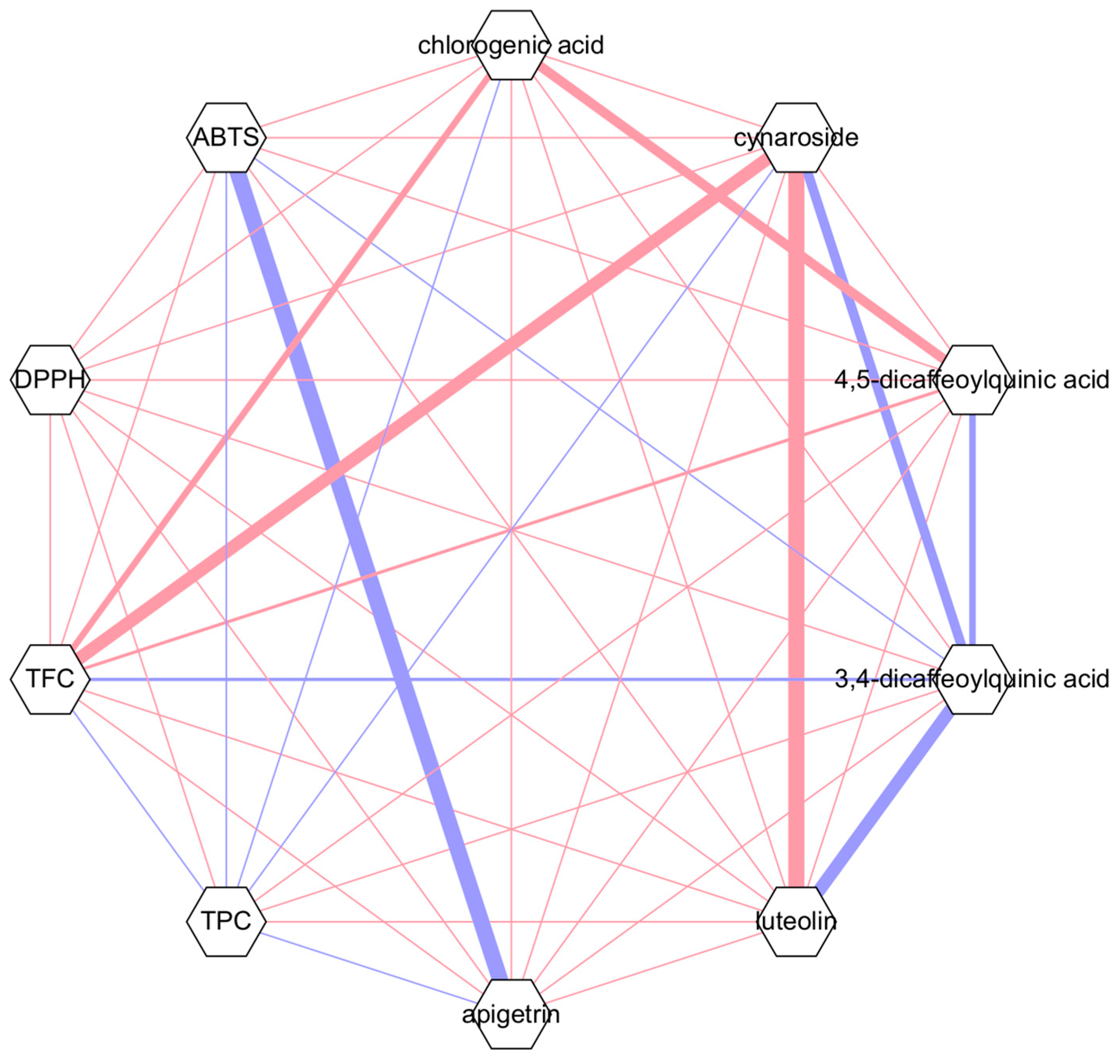
Pearson’s correlation analysis was conducted to examine the relationships between TPC, TFC, DPPH, and ABTS+ as well as the compounds quantified using HPLC (Figure 7). The Pearson correlation coefficients were used to evaluate how the concentrations of bioactive compounds influenced the assayed endpoints and to highlight similarities and differences among the various plant components. Although chlorogenic acid was the most abundant phytochemical detected, it did not correlate positively with TFC, DPPH, or ABTS+. However, it did exhibit a weak positive correlation with TPC. Conversely, apigetrin (4), despite its lower concentration, demonstrated a strong positive correlation with ABTS+.

4. Discussion

Notable variations in polyphenol and flavonoid content were observed across different structures of the two P. ussuriensis cultivars (1PUR, 2PUR, LPUR, FPUR, 1PUM, 2PUM, and LPUM) in the TPC and TFC assays. The results reveal significant differences, with TPC being consistently higher than TFC across all samples. This aligns with the general understanding that polyphenolic compounds are more abundant than flavonoids in many plant tissues [17]. Interestingly, the stem extracts of both cultivars (2PUM and 2PUR) exhibited the highest TPC, indicating that stems are particularly rich in polyphenolic compounds. This is likely due to the presence of polyphenols in lignins, which play a structural and protective role in more lignified parts such as stems and can react with the Folin–Ciocalteu reagent [18]. The significant differences observed in TFC and TPC among the extracts further emphasize the differential distribution of these phytochemicals, showing tissue-specific accumulation trends.
Furthermore, the higher TPC in P. ussuriensis var. ovoidea Rehder extracts compared to P. ussuriensis Maxim. suggests that this variety may have a greater potential for antioxidant activities, which are often associated with polyphenol content. Regarding TFC, the trend where leaf extracts had the highest flavonoid content, followed by stem and fruit extracts, is consistent with the role of flavonoids in protecting leaves from UV radiation and pathogens [19]. This also aligns with previous studies on a related species, Malus domestica, where flavonoid accumulation was shown to be higher in leaves exposed to sunlight, suggesting an adaptive response to environmental stressors [20]. This pattern highlights the differential allocation of secondary metabolites within the plant, likely related to the specific functional requirements of each plant structure [21].
The antioxidant activity, as assessed via the DPPH and ABTS+ assays, supports the differences observed in TPC and TFC. The lower IC50 values in the DPPH assay compared to the ABTS+ assay across all extracts suggest a stronger radical scavenging ability in the DPPH system. This finding contrasts with previous studies, which indicated that the ABTS+ assay is generally more sensitive than the DPPH assay due to its quicker response kinetics and greater sensitivity to antioxidants [22]. Notably, the highest DPPH radical scavenging activity was observed in 2PUM, 1PUR, and FPUR. This suggests that these parts of the plant might contain specific phytochemical compounds that are particularly effective at neutralizing free radicals. The presence of these compounds and their differences in structure likely influence their effectiveness in scavenging free radicals [23].
The variation in antioxidant activity among different plant structures and between the two cultivars underscores the complex interplay between the types and concentrations of polyphenol and flavonoid compounds present. The high activity observed in 2PUM and 1PUR in the DPPH assay indicates that these tissues might be rich in compounds effective against a broader range of free radicals, which is a characteristic feature of the DPPH assay [24].
Phytochemical analyses using LC-MS/MS identified 18 compounds in the negative ionization mode. However, only six were quantifiable via HPLC/UV. This highlights the diversity of phytochemicals present in the extracts. Particularly, chlorogenic acid, which is widely known for its antioxidant properties [25,26], was the most abundant compound across all samples. Chlorogenic acid (1), along with other quantified compounds such as 3,4-dicaffeoylquinic acid (3), 4,5-dicaffeoylquinic acid (5), and cynaroside (2), likely plays a significant role in the observed antioxidant activities [27,28]. Based on the results of the HPLC analysis, all extracts have chlorogenic acid (1), a compound known to scavenge free radicals by providing hydrogen atoms or electrons to the reactive oxygen species, thereby neutralizing them [29]. Although other extract had higher chlorogenic acid (1) content, FPUR had the strongest antioxidants activity. Other compounds found in the other extracts like 2PUM and 1PUR might have antagonistic effects with chlorogenic acid (1) in neutralizing the DPPH and ABTS+ radicals [30]. A previous study on the “Sanhyang” cultivar of P. ussuriensis in Korea reported chlorogenic acid (1) and cynaroside (2) levels of 11.9 mg/g and 9.7 mg/g, respectively [31]. These values are lower than those observed in this study, except for cynaroside (2) in LPUM (3.31 mg/g). Another study reported lower levels of chlorogenic acid (1) in P. ussuriensis fruits [32], which could be attributed to variations in the plant structures used for analysis [33].
The variation in compound content among different plant structures was significant. The leaves of both cultivars had the highest total content of quantified compounds, which correlates with their strong antioxidant activity, particularly in the ABTS+ assay [34]. The higher chlorogenic acid content in LPUM and LPUR compared to other plant structures underscores its significant role in the antioxidant potential of the leaves. The detection of these compounds in trace amounts in the stems, except for relatively higher levels of 3,4-dicaffeoylquinic acid (3) in 2PUM and 1PUM, suggests a highly tissue-specific distribution of bioactive compounds [35,36].
Comparing the extracts of both cultivars reveals that P. ussuriensis Maxim generally has higher TPC and exhibits stronger antioxidant activity, especially in stems and leaves. This suggests that P. ussuriensis is more suitable for applications requiring high antioxidant activity. Conversely, P. ussuriensis var. ovoidea Rehder showed notable antioxidant activity, particularly in its 1-year-old stems, which are rich in specific polyphenolic compounds.
The differences in phytochemical profiles and antioxidant activities between the two cultivars could be attributed to genetic factors, environmental conditions, or a combination of both [37,38]. These findings underscore the importance of carefully selecting specific plants and cultivars for targeted applications in food, pharmaceutical, or cosmetic industries where antioxidant properties are valued [39]. Future research could look into the effects of these phytochemicals on specific health outcomes or their application in complex biological systems.

5. Conclusions

This study investigated the phytochemical profiles and antioxidant capacities of various parts of P. ussuriensis Maxim. and P. ussuriensis var. ovoidea Rehder. The results indicated that the leaf extracts of both cultivars exhibited the highest TFC, whereas the TPC was highest in the 2-year-old stems of both cultivars. Although P. ussuriensis var. ovoidea Rehder generally showed higher TPC and stronger antioxidant activity, P. ussuriensis demonstrated unique strengths, particularly in its thorn extracts. These findings suggest that different cultivars may be better suited for specific applications, enabling a more targeted approach for developing products from these plants. Additionally, LC-MS/MS identified 18 phytochemicals, with chlorogenic acid (1) being the most abundant, especially in leaf extracts determined via HPLC/UV analysis. Furthermore, the high TFC of the LPUR and LPUM can be attributed to their high luteolin (6) content. On the other hand, the high TPC in the 2PUR and 2PUM might be due to some compounds that are not quantified in this study. The high TPC in these samples cannot be precisely attributed to specific compounds as total tannin content was not measured in the stems and leaves of the examined cultivars. This study emphasizes the strong antioxidant activity and significant phytochemical diversity present in the two P. ussuriensis cultivars. The results underscore the potential of these plants as sources of natural antioxidants, with specific structures such as the leaves and stems showing particular promise. P. ussuriensis is among the largest and oldest pear trees in Korea, highlighting its role as a valuable biological resource with significant conservation value.

Author Contributions

HPLC/PDA, TPC, and TFC analysis as well as radical scavenging activity, N.P.U.; sampling and resources, J.K. and S.J.N.; LC-ESI/MS analysis, D.-H.L.; supervision, writing—review, and editing, S.L. All authors have read and agreed to the published version of the manuscript.

Funding

This research was supported by a grant from the National Institute of Forest Science (FG0802-2020-01-2024), Suwon, Republic of Korea.

Data Availability Statement

Data is contained within the article.

Acknowledgments

The authors would like to thank the Forest Bioresources Department at the National Institute of Forest Science for providing the pear samples used in this study.

Conflicts of Interest

The authors declare no conflicts of interest.

References

  1. Cho, M.; Kim, Y.; Kim, S.; Park, J. The complete chloroplast genome of Korean Pyrus ussuriensis Maxim. (Rosaceae): Providing genetic background of two types of P. ussuriensis. Mitochondrial DNA Part B 2019, 4, 2424–2425. [Google Scholar] [CrossRef] [PubMed]
  2. Ahn, Y.; Lee, S.; Lee, S.; Kang, K. Distribution and ecological characteristics of native Iris odesanensis in Mt. Naeyon. J. Environ. Sci. Int. 2006, 15, 1103–1107. [Google Scholar] [CrossRef]
  3. Wuyun, T.; Amo, H.; Xu, J.; Ma, T.; Uematsu, C.; Katayama, H. Population structure of and conservation strategies for wild Pyrus ussuriensis Maxim. in China. PLoS ONE 2015, 10, e0133686. [Google Scholar] [CrossRef]
  4. Bokszczanin, K.L.; Przybyla, A.A.; Schollenberger, M.; Gozdowski, D.; Madry, W.; Odziemkowski, S. Inheritance of fire blight resistance in Asian Pyrus species. Open J. Genet. 2012, 2, 109–120. [Google Scholar] [CrossRef]
  5. Katayama, H.; Amo, H.; Wuyun, T.; Uematsu, C.; Iketani, H. Genetic structure and diversity of the wild Ussurian pear in East Asia. Breed. Sci. 2016, 66, 90–99. [Google Scholar] [CrossRef] [PubMed]
  6. Reighard, G.L. Evaluation of commercial potential of Asian pear cultivars in South Carolina. HortScience 1995, 30, 793. [Google Scholar] [CrossRef]
  7. Feng, Y.; Cheng, H.; Cheng, Y.; Zhao, J.; He, J.; Li, N.; Wang, J.; Guan, J. Chinese traditional pear paste: Physicochemical properties, antioxidant activities, and quality evaluation. Foods 2023, 12, 187. [Google Scholar] [CrossRef]
  8. Kar, A.; Bhattacharjee, S. Bioactive polyphenolic compounds, water-soluble vitamins, in vitro anti-inflammatory, anti-diabetic and free radical scavenging properties of underutilized alternate crop Amaranthus spinosus L. from Gangetic plain of West Bengal. Food Biosci. 2022, 50, 102072. [Google Scholar] [CrossRef]
  9. Bhosale, P.B.; Ha, S.E.; Vetrivel, P.; Kim, H.H.; Kim, S.M.; Kim, G.S. Functions of polyphenols and its anticancer properties in biomedical research: A narrative review. Transl. Cancer Res. 2020, 9, 7619–7631. [Google Scholar] [CrossRef]
  10. Moar, K.; Yadav, S.; Pant, A.; Deepika; Maurya, P.K. Anti-tumor effects of polyphenols via targeting cancer-driving signaling pathways: A review. Indian J. Clin. Biochem. 2024, 39, 470–488. [Google Scholar] [CrossRef] [PubMed]
  11. Manso, T.; Lores, M.; De Miguel, T. Antimicrobial activity of polyphenols and natural polyphenolic extracts on clinical isolates. Antibiotics 2021, 11, 46. [Google Scholar] [CrossRef]
  12. Chojnacka, K.; Skrzypczak, D.; Izydorczyk, G.; Mikula, K.; Szopa, D.; Witek-Krowiak, A. Antiviral properties of polyphenols from plants. Foods 2021, 10, 2277. [Google Scholar] [CrossRef]
  13. Pandey, K.B.; Rizvi, S.I. Plant polyphenols as dietary antioxidants in human health and disease. Oxidative Med. Cell. Longev. 2009, 2, 270–278. [Google Scholar] [CrossRef]
  14. Prabakaran, M.; Kim, S.; Sasireka, A.; Chandrasekaran, M.; Chung, I.-M. Polyphenol composition and antimicrobial activity of various solvent extracts from different plant parts of Moringa oleifera. Food Biosci. 2018, 26, 23–29. [Google Scholar] [CrossRef]
  15. Hancock, J.; Lobos, G. Pears. In Temperate Fruit Crop Breeding: Germplasm to Genomics, 1st ed.; Hancock, J.F., Ed.; Springer: Dordrecht, The Netherlands, 2008; pp. 265–298. [Google Scholar]
  16. Lee, C.-D.; Ku, J.; Lee, S.; Lee, S. Quantification of phytochemicals in Cephalotaxus harringtonia: Insights into plant tissue-specific allocation. Horticulturae 2024, 10, 1286. [Google Scholar] [CrossRef]
  17. Singla, R.K.; Dubey, A.K.; Garg, A.; Sharma, R.K.; Fiorino, M.; Ameen, S.M.; A Haddad, M.; Al-Hiary, M. Natural polyphenols: Chemical classification, definition of classes, subcategories, and structures. J. AOAC Int. 2019, 102, 1397–1400. [Google Scholar] [CrossRef]
  18. da Silva, P.R.; de Lima, M.d.C.A.; Souza, T.P.; Sandes, J.M.; Lima, A.d.C.A.d.; Neto, P.J.R.; dos Santos, F.A.B.; Alves, L.C.; da Silva, R.M.F.; Rocha, G.J.d.M.; et al. Lignin from Morinda citrifolia leaves: Physical and chemical characterization, in vitro evaluation of antioxidant, cytotoxic, antiparasitic and ultrastructural activities. Int. J. Biol. Macromol. 2021, 193, 1799–1812. [Google Scholar] [CrossRef] [PubMed]
  19. Ferreyra, M.L.F.; Serra, P.; Casati, P. Recent advances on the roles of flavonoids as plant protective molecules after UV and high light exposure. Physiol. Plant. 2021, 173, 736–749. [Google Scholar] [CrossRef]
  20. Hasan, Z.Y.M. Total phenolic, flavonoid contents, and antioxidant activities of different parts of Malus domestica L. in Iraq. Iraqi J. Pharm. Sci. 2024, 33, 174–189. [Google Scholar] [CrossRef]
  21. Dong, D.; Shi, Y.; Mou, Z.; Chen, S.-Y.; Zhao, D.-K. Grafting: A potential method to reveal the differential accumulation mechanism of secondary metabolites. Hortic. Res. 2022, 9, uhac050. [Google Scholar] [CrossRef] [PubMed]
  22. Lee, K.J.; Oh, Y.C.; Cho, W.K.; Ma, J.Y. Antioxidant and anti-inflammatory activity determination of one hundred kinds of pure chemical compounds using offline and online screening HPLC assay. Evid.-Based Complement. Altern. Med. 2015, 2015, 165457. [Google Scholar] [CrossRef] [PubMed]
  23. Cesari, L.; Mutelet, F.; Canabady-Rochelle, L. Antioxidant properties of phenolic surrogates of lignin depolymerisation. Ind. Crops Prod. 2019, 129, 480–487. [Google Scholar] [CrossRef]
  24. Baliyan, S.; Mukherjee, R.; Priyadarshini, A.; Vibhuti, A.; Gupta, A.; Pandey, R.P.; Chang, C.-M. Determination of antioxidants by DPPH radical scavenging activity and quantitative phytochemical analysis of Ficus religiosa. Molecules 2022, 27, 1326. [Google Scholar] [CrossRef] [PubMed]
  25. Wang, L.; Pan, X.; Jiang, L.; Chu, Y.; Gao, S.; Jiang, X.; Zhang, Y.; Chen, Y.; Luo, S.; Peng, C. The biological activity mechanism of chlorogenic acid and its applications in food industry: A review. Front. Nutr. 2022, 9, 943911. [Google Scholar] [CrossRef] [PubMed]
  26. Huang, J.; Xie, M.; He, L.; Song, X.; Cao, T. Chlorogenic acid: A review on its mechanisms of anti-inflammation, disease treatment, and related delivery systems. Front. Pharmacol. 2023, 14, 1218015. [Google Scholar] [CrossRef] [PubMed]
  27. Nguyen, A.N.T.; Vu, T.T.T.; Do, T.H.T.; Nguyen, T.H.; Van Le, H.; Pham, H.K.T.; Truong, P.C.H.; Pham, D.P.; Tran, M.H. Identification of phenolic compounds from Vietnamese artichoke (Cynara scolymus L.) leaf and their antioxidant activities. Nat. Prod. Sci. 2024, 30, 39–112. [Google Scholar] [CrossRef]
  28. Kim, J.S.; Lim, J.H.; Cho, S.K. Effect of antioxidant and anti-inflammatory on bioactive components of carrot (Daucus carota L.) leaves from Jeju Island. Appl. Biol. Chem. 2023, 66, 34. [Google Scholar] [CrossRef]
  29. Liang, N.; Dupuis, J.H.; Yada, R.Y.; Kitts, D.D. Chlorogenic acid isomers directly interact with Keap 1-Nrf2 signaling in Caco-2 cells. Mol. Cell. Biochem. 2019, 457, 105–118. [Google Scholar] [CrossRef] [PubMed]
  30. Silva, F.; Veiga, F.; Cardoso, C.; Dias, F.; Cerqueira, F.; Medeiros, R.; Paiva-Santos, A.C. A rapid and simplified DPPH assay for analysis of antioxidant interactions in binary combinations. Microchem. J. 2024, 202, 110801. [Google Scholar] [CrossRef]
  31. Kim, H.; Kim, T.H.; Jeon, B.; Bang, M.H.; Kim, W.J.; Park, J.W.; Chung, D.-K. Evaluation of the physiological activity on the skin and identification of the active ingredient of leaf extract from Sanhyang Sandolbae (Pyrus ussuriensis) as a new variety. J. Korean Soc. Food Sci. Nutr. 2020, 49, 35–45. [Google Scholar] [CrossRef]
  32. Boby, N.; Abbas, M.A.; Lee, E.; Im, Z.-E.; Hsu, W.H.; Park, S.-C. Protective effect of Pyrus ussuriensis Maxim. extract against ethanol-induced gastritis in rats. Antioxidants 2021, 10, 439. [Google Scholar] [CrossRef] [PubMed]
  33. Brahem, M.; Renard, C.M.; Eder, S.; Loonis, M.; Ouni, R.; Mars, M.; Le Bourvellec, C. Characterization and quantification of fruit phenolic compounds of European and Tunisian pear cultivars. Food Res. Int. 2017, 95, 125–133. [Google Scholar] [CrossRef]
  34. Dibacto, R.E.K.; Tchuente, B.R.T.; Nguedjo, M.W.; Tientcheu, Y.M.T.; Nyobe, E.C.; Edoun, F.L.E.; Kamini, M.F.G.; Dibanda, R.F.; Medoua, G.N. Total polyphenol and flavonoid content and antioxidant capacity of some varieties of Persea americana peels consumed in Cameroon. Sci. World J. 2021, 2021, 1–11. [Google Scholar] [CrossRef] [PubMed]
  35. Kim, D.; Choi, S.; Park, J.; Kim, K.; Oh, J.-E. Phenolic compounds in the freshwater environment in South Korea: Occurrence and tissue-specific distribution. Sci. Total Environ. 2023, 905, 166914. [Google Scholar] [CrossRef] [PubMed]
  36. Wu, J.; Song, Z.; Cai, N.; Wang, Q.; Xiao, X.; Yang, X.; He, Y.; Zou, S. Pharmacokinetics, tissue distribution, and excretion of six bioactive components from total glucosides picrorhizae rhizoma, as simultaneously determined by a UHPLC-MS/MS method. J. Chromatogr. B 2023, 1227, 123830. [Google Scholar] [CrossRef] [PubMed]
  37. Zhang, J.; Tian, J.; Gao, N.; Gong, E.S.; Xin, G.; Liu, C.; Si, X.; Sun, X.; Li, B. Assessment of the phytochemical profile and antioxidant activities of eight kiwi berry (Actinidia arguta (Siebold & Zuccarini) Miquel) varieties in China. Food Sci. Nutr. 2021, 9, 5616–5625. [Google Scholar] [CrossRef]
  38. Akullo, J.O.; Kiage-Mokua, B.N.; Nakimbugwe, D.; Ng’ang’a, J.; Kinyuru, J. Phytochemical profile and antioxidant activity of various solvent extracts of two varieties of ginger and garlic. Heliyon 2023, 9, e18806. [Google Scholar] [CrossRef] [PubMed]
  39. Lekshmi, S.G.; Sethi, S.; Pooja, B.K.; Nayak, S.L.; Menaka, M. Ornamental plant extracts: Application in food colouration and packaging, antioxidant, antimicrobial and pharmacological potential—A concise review. Food Chem. Adv. 2023, 3, 100529. [Google Scholar] [CrossRef]
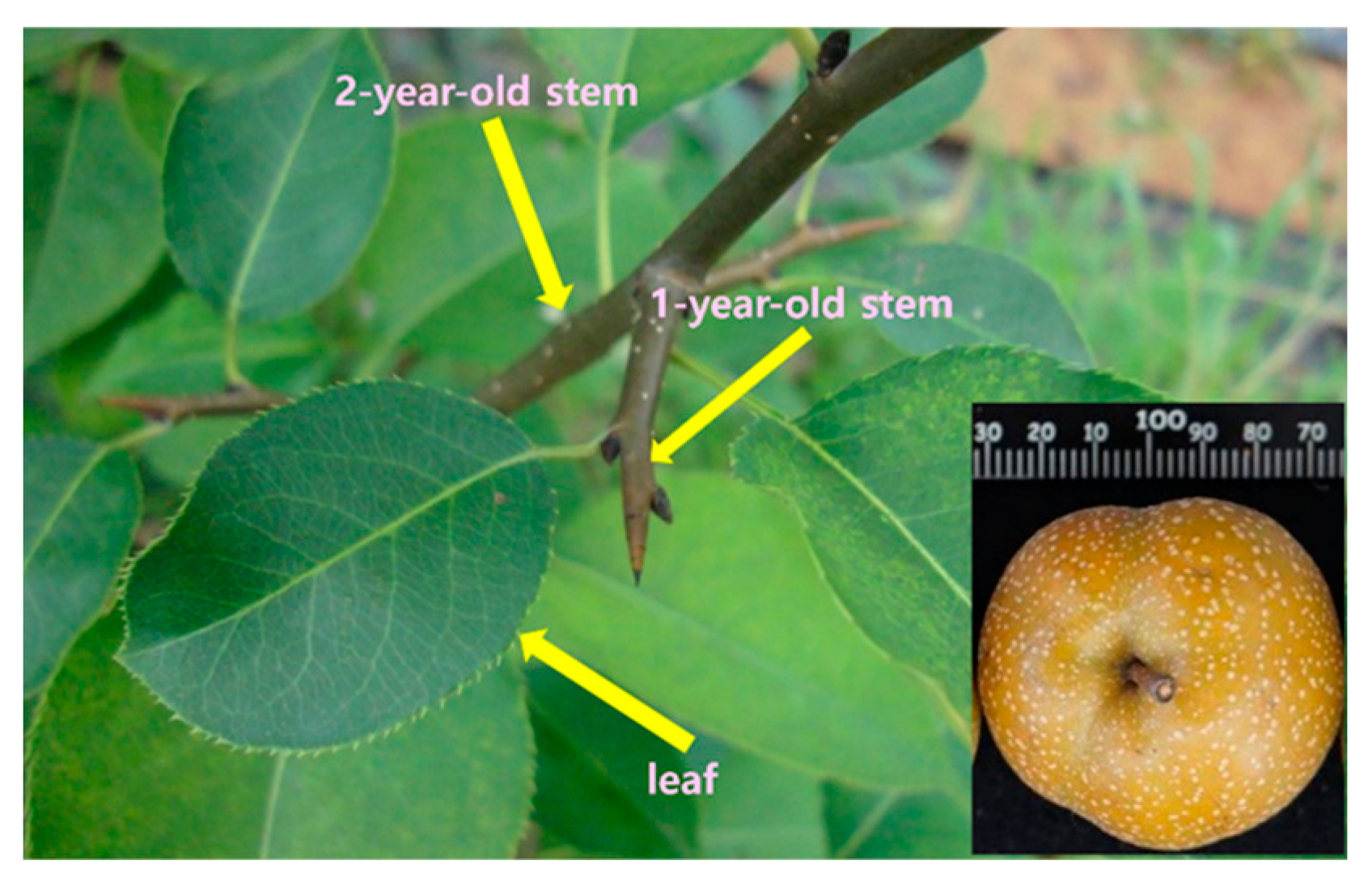
Figure 1. P. ussuriensis var. ovoidea Rehder and the parts analyzed.
Horticulturae 11 00184 g001
Figure 2. Chemical structures of chlorogenic acid (1), cynaroside (2), 3,4-dicaffeoylquinic acid (3), apigetrin (4), 4,5-dicaffeoylquinic acid (5), and luteolin (6).
Horticulturae 11 00184 g002aHorticulturae 11 00184 g002b
Figure 3. Dose-response curves (A,B) and IC50 values (C,D) in DPPH and ABTS+, respectively. Different lowercase letters indicate significant differences at p < 0.05. AA: ascorbic acid.
Horticulturae 11 00184 g003
Figure 4. UV chromatogram (A) and base peak chromatogram (B) of LPUR analyzed in negative ionization mode.
Horticulturae 11 00184 g004
Figure 5. HPLC chromatogram of chlorogenic acid (1), cynaroside (2), 3,4-dicaffeoylquinic acid (3), apigetrin (4), 4,5-dicaffeoylquinic acid (5), and luteolin (6).
Horticulturae 11 00184 g005
Figure 6. HPLC chromatograms of LPUM (A), LPUR (B), and FPUR (C) showing chlorogenic acid (1), cynaroside (2), 3,4-dicaffeoylquinic acid (3), apigetrin (4), 4,5-dicaffeoylquinic acid (5), and luteolin (6).
Horticulturae 11 00184 g006
Figure 7. Pearson’s correlation coefficient network (r ≥ |1.00|) among phytochemical concentrations and antioxidant activities. Red and blue lines indicate positive and negative correlations, respectively.
Horticulturae 11 00184 g007
Table 1. TPC and TFC of different P. ussuriensis samples.
SampleTPC (mg TAE/g)TFC (mg QE/g)
1PUR60.7 ± 3.29 b0.45 ± 0.22 c
2PUR82.9 ± 11.3 ab0.78 ± 0.06 c
LPUR79.6 ± 9.08 ab13.5 ± 0.82 a
FPUR41.5 ± 2.14 c0.30 ± 0.12 d
1PUM81.1 ± 6.77 ab0.52 ± 0.09 c
2PUM94.7 ± 20.9 a1.05 ± 0.04 c
LPUM76.5 ± 13.3 ab6.72 ± 0.73 b
TAE: tannic acid equivalent; QE: quercetin equivalent. Different lowercase letters indicate significant differences at p < 0.05.
Table 2. Proposed identity of the compounds detected via LC-MS/MS analysis (negative mode).
tR (min)Molecular FormulaMolecular WeightTentative Identification
7.55C16H18O9354.1Neochlorogenic acid
10.82C16H18O9354.1Chlorogenic acid
13.41C16H18O9354.11-Caffeoylquinic acid
13.88C16H18O8338.14-Coumaroylquinic acid
15.80C17H20O9368.13-Feruloylquinic acid
16.30C16H18O8338.11-Coumaroylquinic acid
17.03C21H22O10434.16-Caffeoylarbutin
18.17C17H20O9368.1Chlorogenic acid methyl ester
19.94C21H22O10464.1Hirsutrin
20.21C21H20O11448.1Cynaroside
21.17C21H20O12516.13,4-Dicaffeoylquinic acid
21.67C21H20O10432.1Apigetrin
21.94C25H24O12516.14,5-Dicaffeoylquinic acid
22.24C22H22O11462.1Isoscoparin
23.41C25H24O12516.1Phellopterin
24.51C15H10O6516.1Luteolin
26.37C15H10O5286.1Apigenin
26.98C16H12O6300.1Disometin
Table 3. Calibration data for chlorogenic acid (1), cynaroside (2), 3,4-dicaffeoylquinic acid (3), apigetrin (4), 4,5-dicaffeoylquinic acid (5), and luteolin (6).
CompoundtR (min)Regression EquationCoefficient of Determination (r2)
18.19y = 7.9045x + 100.640.9979
220.70y = 10.729x + 328.350.9916
321.56y = 5.4865x + 154.180.9836
422.67y = 7.4078x + 251.790.9864
523.04y = 5.3829x + 91.650.9981
629.01y = 16.394x + 513.670.9893
Table 4. Content of chlorogenic acid (1), cynaroside (2), 3,4-dicaffeoylquinic acid (3), apigetrin (4), 4,5-dicaffeoylquinic acid (5), and luteolin (6) in the different P. ussuriensis samples.
SampleContent (mg/g)
123456Total
1PUR1.23 ± 0.01 ea tr 0.16 ± 0.00 eb NDtrND1.39
2PUR0.69 ± 0.04 ftr0.19 ± 0.02 etrtrtr0.88
LPUR28.53 ± 0.03 a20.21 ± 0.02 a1.22 ± 0.04 d8.06 ± 0.00 b20.21± 0.03 a0.27 ± 0.0079.5
FPUR9.41 ± 0.11 ctr0.05 ± 0.00 ftrtrtr9.46
1PUM2.96 ± 0.00 d tr9.07 ± 0.01 a0.67 ± 0.01 c trND 12.70
2PUM2.09 ± 0.02 dtr7.57 ± 0.09 b0.74 ± 0.02 ctrND10.40
LPUM25.55 ± 0.06 b3.31 ± 0.01 b3.69 ± 0.01 c18.39 ± 0.04 a16.90 ± 0.04 btr67.84
a tr: trace; b ND: not detected; different lowercase letters indicate significant differences at p < 0.05.
Disclaimer/Publisher’s Note: The statements, opinions and data contained in all publications are solely those of the individual author(s) and contributor(s) and not of MDPI and/or the editor(s). MDPI and/or the editor(s) disclaim responsibility for any injury to people or property resulting from any ideas, methods, instructions or products referred to in the content.