Article

Valorisation of Sunflower Crop Residue as a Potentially New Source of Bioactive Compounds

by
Ivona Veličković
1,*,
Stevan Samardžić
2,
Marina T. Milenković
3,
Miloš Petković
4 and
Zoran Maksimović
2
1
Institute of Botany and Botanical Garden “Jevremovac”, Faculty of Biology, University of Belgrade, Studentski trg 16, 11000 Belgrade, Serbia
2
Department of Pharmacognosy, Faculty of Pharmacy, University of Belgrade, Vojvode Stepe 450, 11221 Belgrade, Serbia
3
Department of Microbiology and Immunology, Faculty of Pharmacy, University of Belgrade, Vojvode Stepe 450, 11221 Belgrade, Serbia
4
Department of Organic Chemistry, Faculty of Pharmacy, University of Belgrade, Vojvode Stepe 450, 11221 Belgrade, Serbia
*
Author to whom correspondence should be addressed.
Horticulturae 2025, 11(2), 206; https://doi.org/10.3390/horticulturae11020206
Submission received: 31 December 2024 / Revised: 31 January 2025 / Accepted: 13 February 2025 / Published: 15 February 2025

Abstract

:
Reducing agricultural waste through reuse has become one of the most important strategies to minimise impact on the environment—an emerging global issue. Sunflower ranks fourth in the world in the production of vegetable oilseeds and therefore generates large amounts of agricultural waste. The aim of this study was to investigate the phytochemical composition and bioactivity of sunflower crop residues in order to open up new opportunities for waste management. TPC and TFC were determined spectrophotometrically, while the dominant compounds were identified by LC-DAD-ESI-MS as ent-kaur-16-en-19-oic acid (KA) and 6Ac-7OH-dimethylchromone (DMC). Both compounds were present in higher concentrations in the ethyl acetate fraction (245.5 and 16.8 mg/g, respectively) than in the ethanol extract. None of the tested samples showed antimicrobial effects in the microdilution test. DMC showed remarkable antioxidant activity by DPPH, ABTS, FRAP and TRC in vitro assays, while both compounds proved to be promising enzyme inhibitory agents, being particularly efficient in inhibiting anti-neurodegenerative enzymes (IC50 values of DMC and KA were 1.20/1.37 mg/mL and 1.44/1.63 mg/mL for AChE/BChE, respectively) and tyrosinase. The results presented indicate that sunflower crop residues are a good candidate for the extraction of bioactive compounds with potential application in the food, pharmaceutical and cosmetic industries.

Graphical Abstract

1. Introduction

The growing world population, which is estimated to reach 9.8 billion people by 2050, poses increasing demands for foods, energy and resources [1]. Therefore, as the largest food producer, agriculture faces two major challenges—providing the growing population with sufficient food and reducing the amount of agricultural waste [1,2]. To overcome these issues, the conventional agricultural practise, which assumes a linear use of resources, should be replaced by a circular economy that creates a closed-loop system by minimising dependence on external resources and thus mitigating the harmful effects of disposed waste on the environment [1,3]. The challenges of the transition from one economic approach to another have been recognised by the European Commission, which adopted the first Circular Economy Action Plan in 2015, and by many scientists who are working intensively on the valorisation of agricultural by-products of plant origin and new ways of reusing them [3].
The sunflower (Helianthus annuus L., Asteraceae) is an annual crop that is native to North America but cultivated worldwide [4,5,6]. It is one of the most important crops on the world market, as it is estimated to grow on 23 million hectares in 60 countries [7]. Sunflower is the third most produced oilseed, the fourth most produced vegetable oil and the third most produced oilseed meal among protein feed sources [8,9]. Sunflower is also used for ornamental purposes, and its cultivation as an ornamental plant has recently attracted increasing attention.
H. annuus is also used in ethnomedicine for its curative effects on various health problems such as heart disease, respiratory infections, coughs and colds [4]. Sunflower is a rich source of bioactive compounds including flavonoids, tannins, terpenes, alkaloids, saponins, steroids, fixed oils and proteins, with pronounced pharmacological activity including antioxidant, anti-inflammatory, antimicrobial, hypoglycaemic and antitumour effects, etc. [4]. Previous studies on the bioactivity potential of sunflower have focused on the aerial parts of sunflower, especially the leaves and seeds [5,10,11,12,13,14,15]. Thus, there is a lot of literature data on the antioxidant and antimicrobial properties of these plant parts [12,13,14,15]. In addition, sunflower leaves have shown antidiabetic effects [10,11].
Sunflower is the third largest crop in Serbia, grown on 182,000 hectares with an average yield of 2.5 t seeds/ha. Sunflower seeds are generally used for the production of refined sunflower oil, which reaches 173,924 t annually and generates significant amounts of agricultural waste: hulls (754 t), oil cake (226,101 t), slurry (53,834 t), phospholipids (106 t), soapstock (1270 t), spent bleaching earth (713 t), oily perlite (157 t) and deodorised distillate (696 t) [16,17]. Recent studies have shown that this type of waste material could be used for the production of meat functional foods and natural cosmetics due to the presence of considerable amounts of bioactive compounds [18,19].
Although sunflower harvest residues as raw material could have a significant impact on the circular economy, they are still underutilised. Therefore, the aim of this work was to valorise them as a potential new source of bioactive compounds with added value. To achieve the set goal, the chemical composition of the ethanol extract from sunflower harvest residues and its ethyl acetate fraction was evaluated. In addition, the ethanol extract, its ethyl acetate fraction and the main compounds were analysed for their bioactivity by antioxidant, antimicrobial and enzyme inhibitory assays.

2. Materials and Methods

2.1. Chemicals and Reagents

Silica gel on TLC Al foils were procured from Sigma-Aldrich Chemie GmbH (Steinheim, Germany). Methanol (HPLC-grade), ethanol (HPLC-grade), dimethyl sulphoxide (p.a. grade), di-sodium hydrogen phosphate dodecahydrate and sodium carbonate anhydrous were purchased from VWR Chemicals (Lutterworth, Leicestershire, UK). Glacial acetic acid, concentrated hydrochloric acid and ethanol 96% v/v (Ph. Eur.) were purchased from Zorka Pharma (Šabac, Serbia) and sodium acetate trihydrate from Centrohem (Stara Pazova, Serbia). TCI Europe N.V. (Zwijndrecht, Belgium) supplied quercetin hydrate, 2,2′-azino-bis(3-ethylbenzothiazoline)-6-sulphonic acid (ABTS) and butylated hydroxytoluene (BHT). 2,2-diphenyl-1-picrylhydrazyl (DPPH), α-glucosidase from bakers’ yeast (EC 3.2.1.20, type I), α-amylase from porcine pancreas (EC 3.2.1.1, type VI-B), acetylcholinesterase (AChE) from Electrophorus electricus (EC 3.1.1.7, type VI-S), butyrylcholinesterase (BChE) from equine serum (EC 3.1.1.8), tyrosinase from mushroom (EC 1.14.18.1), p-nitrophenyl-α-D-glucopyranoside, 5,5′-dithio-bis(2-nitrobenzoic acid) (DTNB), acetylthiocholine iodide (ATCI), butyrylthiocholine iodide (BTCI), 3,4-dihydroxy-L-phenylalanine (L-DOPA), acarbose, galanthamine hydrobromide, kojic acid, Folin–Ciocalteu reagent, gallic acid, iron (II) sulphate heptahydrate, iron (III) chloride hexahydrate, potassium hexacyanoferrate (III), Lugol solution and silica gel 60 (0.015–0.040 mm) for column chromatography were procured from Merck KGaA (Darmstadt, Germany). Thermo Fisher Scientific (New York, NY, USA) provided potassium per-oxydisulphate, sodium sulphate anhydrous (p.a. grade), trichloroacetic acid, hexane (p.a. grade), ethyl acetate (p.a. grade) and acetonitrile (HPLC-MS grade). Celite® 545, deuterated chloroform and 1% starch solution were acquired from Carl Roth GmbH (Karlsruhe, Germany) and Sephadex® LH-20 from Cytiva (Marlborough, MA, USA). L-ascorbic acid and 2,4,6-Tris(2-pyridyl)-s-triazine (TPTZ) were obtained from Fluka Chemie AG (Buchs, Switzerland), while formic acid (LC-MS grade) was from Honeywell, Fluka (Seelze, Germany). Sodium chloride anhydrous was sourced from Superlab (Belgrade, Serbia).

2.2. Preparation of Sunflower Ethanol Extract and Its Ethyl Acetate Fraction

Glišić et al. [18] prepared the ethanol extract from harvest residues of sunflower (Helianthus annuus L.), especially the stems. The plant material was collected from fields in the autonomous province of Vojvodina, Serbia in October 2021. After collection, the material was shade-dried in a well-ventilated area and ground with a mechanical grinder. The extraction process was carried out in a high-capacity stainless steel extractor (60 L). First, 3 kg of the plant material was extracted with hexane at 40 °C for 1 h to release it from waxes, the ratio of plant to solvent being 1:6 (m/m). After hexane extraction, the plant material was allowed to dry completely before being subjected to a second extraction with 96% (v/v) ethanol at 45 °C for 1 h, using the same ratio of plant to solvent. Ethanol as a polar solvent with low molecular weight is convenient for extraction of phenolic compounds from plant material and further application in bioassays. The resulting extracts were filtered, and the solvents were evaporated under reduced pressure using a DLAB RE 200 Pro industrial rotary evaporator (DLAB Scientific Co., Ltd., Beijing, China). The ethanol extract, a dark green, semi-solid material with a characteristic odour, was obtained with a yield of 2.55%. It was stored at +4 °C until further analysis.
To prepare the ethyl acetate fraction, a liquid–liquid extraction was carried out in a separatory funnel. The ethanol extract (5.152 g) was suspended in 500 mL of water and extracted with three portions of ethyl acetate (3 × 250 mL). The organic layers were combined, dried over anhydrous sodium sulphate and evaporated to dryness. The resulting ethyl acetate fraction was subjected to a subsequent bioactivity test together with the ethanol extract.

2.3. Isolation of Main Compounds from Sunflower Ethanol Extract

The ethanol extract (2 g) of the sunflower harvest residues was subjected to a multi-stage separation process. The extract was dissolved in a minimal amount of ethanol and adsorbed onto Celite® using a rotary vacuum evaporator. The resulting mixture was applied as a thin layer to the upper part of a silica gel column (height = 5.5 cm, diameter = 4 cm, particle size = 15–40 μm), which was prepared according to Pedersen et al. (2001) [20]. Elution was first carried out with hexane, followed by mixtures of hexane and ethyl acetate (Hex: EtOAc, 95:5 → 50:50, v/v). Eleven fractions (A1–A11, 40 mL each) were collected and analysed by silica gel TLC. Due to similarities in composition, fractions A6–A8 were combined and evaporated to dryness, yielding 107 mg residue. This residue was further separated on a Sephadex LH-20 column (height = 33 cm, radius = 1 cm) with ethanol as eluent. Fifteen fractions (B1–B15, 10 mL each) were obtained. Fraction B9 (44.7 mg) was further purified by silica gel TLC with a mobile phase consisting of hexane and ethyl acetate (8:2, v/v). Compound 1 (Rf = 0.94) was not visible under UV light at 254 or 365 nm but developed a purple colour after the plate was sprayed with 10% sulphuric acid in ethanol and heated at 105 °C for 5 min. The zone was scraped off, extracted twice with ethyl acetate (2 × 20 min) and evaporated to dryness. This yielded 23.21 mg of whitish crystals (compound 1). Fractions B10 and B11 were combined and a precipitate formed during evaporation of the solvent. The supernatant was carefully removed to isolate 4.2 mg of whitish crystals (compound 2). This compound showed intense yellow fluorescence under UV light at 365 nm (TLC system; stationary phase—silica gel; mobile phase—hexane/ethyl acetate, 8:2, v/v; Rf = 0.39). To obtain sufficient amounts for the bioactivity tests, the described procedure was repeated several times. The UV and MS spectra of the isolated compounds were measured with the Agilent LC-MS System 1260/6130 (Agilent Technologies, Waldbronn, Germany). The UV spectra were recorded in the range of 190–640 nm, and the MS spectra were acquired in the m/z range of 100–1000 in negative ion mode with a fragmentor voltage of 100 V. 1H and 13C NMR spectra were generated on a Bruker Ascend™ 400 spectrometer (Bruker Scientific LLC, Billerica, MA, USA, at 400 MHz and 101 MHz, respectively, using deuterochloroform (CDCl₃) as solvent and tetramethylsilane (TMS) as internal standard. The chemical shifts (δ) were expressed in ppm and the coupling constants (J) in Hz. The identity of the compounds was confirmed by comparing their spectral characteristics with previously published literature data.

2.4. Evaluation of Total Phenol and Flavonoid Content

The total phenolic content (TPC) in the ethyl acetate fraction was estimated using the Folin–Ciocalteu method as described by Ahmad et al. [21] and expressed as mg gallic acid equivalents per gram dry fraction (mg GAE/g). The total flavonoid content (TFC) was determined according to the modified protocol of Chatatikun and Chiabchalard [22] following Sembring et al. [23], with results that were expressed as mg quercetin equivalents per gram of dry fraction (mg QE/g).

2.5. LC-DAD-ESI-MS Analysis

Liquid chromatography coupled with diode array detection and electrospray ionisation mass spectrometry (LC-DAD-ESI-MS) was performed using an Agilent 1260/6130 LC-MS system (Agilent Technologies, Waldbronn, Germany). The system included a quaternary pump, a column compartment, a DAD detector, an ESI ion source and a single quadrupole mass detector. This system was used to evaluate the effects of ethyl acetate liquid–liquid extraction on the concentrations of chlorogenic acid and isolated compounds 1 and 2. The analytical method used was described by Glišić et al. [18]. Samples were dissolved in ethanol to reach a concentration of 10 mg/mL, filtered through 0.22 µm membrane filters (Macherey-Nagel, Düren, Germany) and injected with a volume of 5 µL. The mobile phase consisted of 0.1% aqueous formic acid (A) and acetonitrile (B). The elution profile was as follows: 10–90% B (0–25 min), 90% B (25–27 min) and 90–10% B (27–30 min). The flow rate was 0.35 mL/min. The separation was carried out on a reversed-phase Zorbax SB-Aq column (3 × 150 mm, 3.5 µm particle size, Agilent Technologies) at a temperature of 25 °C. Chromatograms were recorded at 210, 270, 320 and 350 nm. Fragmentation was performed with an ESI ion source in negative ion mode at a fragmentation voltage of 100 V. The parameters of the spray chamber were set to a gas temperature of 350 °C, a drying gas flow of 10 L/min, nebulizer pressure of 40 psig and a capillary voltage of 3500 V. The sample components were identified by comparing their retention times and UV and mass spectra with those of a commercially available standard (chlorogenic acid; Carl Roth GmbH & Co. KG, Karlsruhe, Germany) or isolated compounds (see Section 3.1) obtained under identical chromatographic conditions. Quantification was performed using the external standard method. The calibration curves for compound 1 (y = 4423.5x + 383.1; R2 = 0.9988; concentration range: 0.084–2.940 mg/mL; LoD = 0.148 mg/mL; LoQ = 0.449 mg/mL) and compound 2 (y = 57,358.0x + 89.3; R2 = 1.0000; concentration range: 0.005–0.250 mg/mL; LoD = 0.002 mg/mL; LoQ = 0.006 mg/mL) were prepared at 210 nm and 270 nm, respectively. The previously created calibration curve for chlorogenic acid was used [18].

2.6. Evaluation of Antioxidant Activity

The following assays were used to evaluate antioxidant activity: (a) free radical scavenging assays—DPPH and ABTS; (b) ferric reducing assays—FRAP and TRC. The DPPH assay was performed according to the protocol of Prieto [24] for microplate readers, the ABTS assay as proposed by Xiao et al. [25], the FRAP assay as described by Benzie and Devaki [26] and the TRC assay according to the procedure of Mokrani et al. [27].
The results for DPPH and ABTS were expressed in terms of IC50 values, concentration that scavenges 50% of the free radicals. The results of the FRAP assay were expressed as mg Fe2+ equivalents and those of the TRC assay as mg L-ascorbic acid equivalents per g dry weight. The results of the antioxidant assays were compared with those of known antioxidants, L-ascorbic acid and butylated hydroxytoluene (BHT).

2.7. Evaluation of Enzyme Inhibitory Activity

The α-amylase inhibition assay was performed using a modified Caraway–Somogyi iodine/potassium iodide protocol as suggested by Zengin et al. [28]. Different concentrations (25 µL) of the sample and 0.5 mg/mL amylase (50 µL) solution in 6 mM NaCl in phosphate buffer (pH 6.8) were pre-incubated in 96-microtitre plates for 15 min at 37 °C. The reaction was initiated by the addition of substrate, a 0.2% starch solution. After incubation for a further 20 min at 37 °C, aliquots of 25 µL of 1 M HCl were added to stop the reaction, while aliquots of 100 µL of I/KI solution were added to visualise the reaction. The absorbance was measured at 630 nm using a Multiscan Sky Thermo Scientific Plate Reader (Thermo Fisher Scientific, WLM, MA, USA). Acarbose was used as a positive control in both this and the following assay.
The α-glucosidase inhibitory activity was evaluated according to Wan et al. [29]. The mixture of sample (120 µL) and 0.6 U/mL enzyme (25 µL) was pre-incubated at 37 °C for 15 min. Then, 3.5 mM p-nitrophenyl-α-D-glucopyranoside (PNPG) in phosphate buffer (pH 6.8) was added, and the incubation continued for a further 20 min. The reaction was stopped by adding 0.2 M sodium carbonate and the absorbance was measured at 405 nm.
The cholinesterase inhibitory activity was determined using Ellman’s reagent according to a slightly modified method by Aktumsek et al. [30]. Different concentrations of the sample (50 µL) were mixed with 0.026 U/mL acetyl-/butyrylcholinesterase (25 µL) and the staining reagent 3 mM DTNB (125 µL) in 50 mM TRIS-HCl buffer solution (pH 8) and incubated at 37 °C for 15 min. An aliquot of 25 µL of the substrate acetyl-/butyrylcholine iodide (ATCI/BTCI) was added. After a further incubation of 20 min at 37 °C, the absorbance was measured at 405 nm. Galantamine was used as a positive control.
Tyrosinase inhibitory activity was performed according to Moonrungesee et al. [31]. The mixture of sample (50 µL), enzyme (mushroom tyrosinase 50 µL) and substrate (L-DOPA) in 0.1 M phosphate buffer (pH 6.8) was incubated at 25 °C for 30 min. The absorbance was then measured at 475 nm. Kojic acid was used as a positive control.
The results of the enzyme inhibition assays were expressed as IC50 values (mg/mL), which represent the concentration of the sample that reduces the enzyme activity by 50%.

2.8. Evaluation of Antimicrobial Activity

The following microorganisms were used to test the antimicrobial activity: Gram-positive bacterial strains—Staphylococcus aureus ATCC 6538, Enterococcus faecalis ATCC 29212 and Bacillus subtilis ATCC 6633; Gram-negative bacterial strains—Escherichia coli ATCC 8739, Klebsiella pneumoniae NCIMB 9111, Salmonella abony NCTC 6017 and Pseudomonas aeruginosa ATCC 9027; and a yeast strain—Candida albicans ATCC 10231. The antimicrobial tests were carried out in accordance with the CLSI guidelines (2015). In brief, the bacterial cultures were grown overnight in Mueller–Hinton broth, while the yeast strain was grown in Sabouraud dextrose broth (HiMedia, Thane, India) at 37 °C. Aliquots (100 µL) of broth containing test microorganisms at a concentration of 10⁶ CFU/mL for bacteria and 107 CFU/mL for yeast were transferred to 96-well microtitre plates. The samples to be tested were prepared by dissolving them in dimethyl sulphoxide (DMSO) and then diluted in the appropriate broth so that the final DMSO concentration was 0.5% per well. Sample concentrations ranged from 62.5 to 1000 µg/mL for the extract, from 31.25 to 500 µg/mL for the ethyl acetate fraction and from 15.63 to 250 ug/mL for the isolated compounds.
The microtitre plates were incubated at 37 °C for 24 h after the addition of the test samples. The minimum inhibitory concentration (MIC) was defined as the lowest concentration that showed visible inhibition of microbial growth. All tests were performed in duplicate. For each microbial strain, the wells containing only bacterial or yeast cells in the corresponding broth (200 µL) were used as positive controls.

2.9. Statistical Analysis

The data were analysed using SPSS software (version 20, Chicago, IL, USA). A one-way ANOVA followed by Tukey’s post hoc test and a t-test for independent samples were performed. A p-value < 0.05 was considered statistically significant. The results are presented as mean ± standard deviation of independent measurements.

3. Results

3.1. Main Compounds Isolated from Sunflower Ethanol Extract

A combination of chromatographic techniques, including silica gel dry column vacuum chromatography, Sephadex LH-20 column chromatography and silica gel TLC, was used to isolate compounds 1 and 2 from the ethanol extract of sunflower stalks. These compounds were obtained in sufficient quantities for bioactivity assays. The compounds were identified by comparing the experimentally obtained spectral data with literature values, which showed good agreement. Compound 1 was identified as ent-kaur-16-en-19-oic acid [32,33,34,35], while compound 2 was identified as 6-acetyl-7-hydroxy-2,3-dimethylchromone [36,37,38].
The spectral data of the isolated compounds can be found below.
Compound 1 (ent-kaur-16-en-19-oic acid) (Figure S1 in Supplementary Materials). UV λmax 200 nm; (Figure S2 in Supplementary Materials); ESI-MS m/z 301 [M-H]- (Figure S3 in Supplementary Materials); 1H NMR (400 MHz, CDCl3) δ 4.80 (1H, s, H-17), 4.74 (1H, s, H-17), 2.64 (1H, br s, H-13), 2.18–2.14 (1H, m, H-3), 2.06 (2H, br s, H-15), 2.00–1.97 (1H, m, H-14), 1.90–1.79 (3H, m, H-1, H-6), 1.62–1.47 (8H, m, H-2, H-7, H-11, H-12), 1.24 (3H, s, H-18), 1.13–1.05 (4H, m, H-3, H-5, H-9, H-14), 0.95 (3H, s, H-20), 0.81–0.78 (1H, m, H-1) (Figure S4 in Supplementary Materials); 13C NMR (101 MHz, CDCl3) δ 182.63 (C-19), 155.90 (C-16), 103.01 (C-17), 57.02 (C-5), 55.11 (C-9), 48.96 (C-15), 44.24 (C-4), 43.85 (C-13), 43.66 (C-8), 41.28 (C-7), 40.71 (C-1), 39.70 (C-10), 39.66 (C-14), 37.90 (C-3), 33.12 (C-12), 28.96 (C-18), 21.84 (C-6), 19.10 (C-2), 18.43 (C-11), 15.63 (C-20) (Figure S5 in Supplementary Materials).
Compound 2 (6-acetyl-7-hydroxy-2,3-dimethylchromone) (Figure S6 in Supplementary Materials). UV λmax 272 nm (Figure S7 in Supplementary Materials); ESI-MS m/z 231 [M-H] (Figure S8 in Supplementary Materials); 1H NMR (400 MHz, CDCl3) δ 13.19 (1H, s, 7-OH), 8.23 (1H, s, H-5), 6.65 (1H, s, H-8), 2.66 (3H, s, H-12), 2.36 (3H, s, H-14), 2.10 (3H, s, H-13) (Figure S9 in Supplementary Materials); 13C NMR (101 MHz, CDCl3) δ 204.01 (C-11), 181.88 (C-4), 169.79 (C-7), 168.12 (C-9), 145.37 (C-2), 132.09 (C-3), 129.00 (C-5), 116.38 (C-10), 116.21 (C-6), 99.97 (C-8), 26.60 (C-12), 20.14 (C-13), 17.36 (C-14) (Figure S10 in Supplementary Materials).
According to HPLC analysis, the purity of compounds 1 and 2 was estimated to be over 93% and 97%, respectively (Figures S11 and S12 in Supplementary Materials).

3.2. Chemical Composition of Sunflower Samples

The liquid–liquid extraction of the ethanol extract (EES) from sunflower harvest residues yielded 1.44 g of the ethyl acetate fraction (EFS), which corresponds to an extraction efficiency of 28.01%. The spectrophotometric analysis revealed significantly higher values of the chemical parameters in the EFS compared to the EES (Table 1). In particular, the total phenolic content (TPC) was 19.20 mg GAE/g in the EFS, exceeding the 15.83 mg GAE/g quantified in the EES, as previously reported by Glišić et al. [18]. A similar trend was observed for total flavonoid content (TFC), with 8.98 mg QE/g in EES, as determined in the study by Glišić et al. [18], and 15.29 mg QE/g in EFS, as determined in the current study.
LC-DAD-ESI-MS analysis revealed a 2.4-fold reduction in chlorogenic acid concentration in EFS compared to EES. In contrast, compound 1 (ent-kaur-16-en-19-oic acid) and compound 2 (6-acetyl-7-hydroxy-2,3-dimethylchromone) showed a significant enrichment in EFS, with an increase of 4.1-fold and 4.2-fold, respectively. These results indicate that ethyl acetate effectively enriches the relatively non-polar compounds 1 and 2, while reducing the content of the relatively polar chlorogenic acid (Table 1). Remarkably, the concentration of compound 1 in EFS reached 245.50 mg/g, indicating that sunflower stalks are a promising source of this bioactive compound. The comparison of EES and EFS chromatograms, highlighting the peaks of compounds 1 and 2, is provided in the Supplementary Materials (Figures S13 and S14).

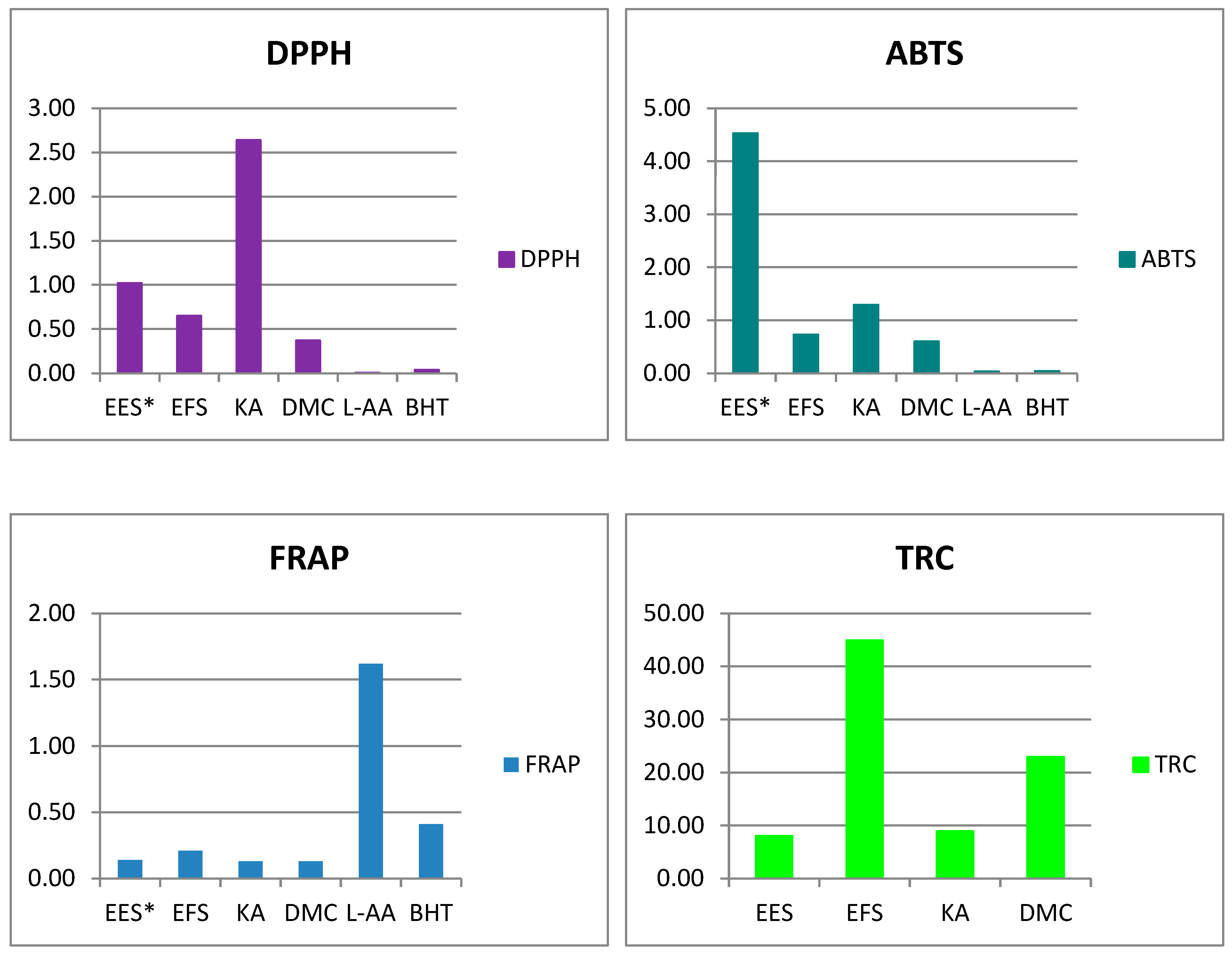
3.3. Antioxidant Activity of Sunflower Ethanol Extract, Its Ethyl Acetate Fraction and Isolated Compounds

The antioxidant activity was evaluated using four in vitro tests, and the results obtained are shown in Figure 1 and Table 2.
The ethanol extract from the sunflower crop residues in our previous study [18] showed significantly lower free radical scavenging activity than the ethyl acetate fraction in the current study, both in the DPPH (IC50 0.65 mg/mL) and ABTS assays (IC50 0.73 mg/mL). 6-Acetyl-7-hydroxy-2,3-dimethylchromone (DMC) was the most efficient in neutralising DPPH and ABTS free radicals (IC50 values 0.37 and 0.60 mg/mL in DPPH and ABTS assays, respectively) in contrast to ent-kaur-16-en-19-oic acid (KA), whose IC50 value in the DPPH assay was more than twice that of the crude ethanol extract.
The FRAP values ranged from 0.13 µmol Fe2+/g for the isolated compounds (KA and DMC) to 0.21 µmol Fe2+/g for the ethyl acetate fraction. Similarly, the ethyl acetate fraction showed the highest total reduction capacity with 44.88 mg L-AAE/g, followed by DMC (22.92 mg L-AAE/g). The ethanol extract of sunflower residue was found to be the least potent antioxidant in the TRC assay (8.03 mg L-AAE/g), and similar activity was observed for KA.
However, L-ascorbic acid and BHT, which were used as reference compounds, were significantly better antioxidants.

3.4. Enzyme Inhibitory Activity of Sunflower Ethanol Extract, Its Ethyl Acetate Fraction and Isolated Compounds

The enzyme inhibitory properties were tested using enzymes relevant for the treatment of diabetes (α-amylase and α-glucosidase), neurodegenerative diseases (acetyl-/butyrylcholinesterase) and skin conditions (tyrosinase) (Table 3). The ethanol extract from sunflower harvest residues showed the lowest enzyme inhibitory activity in all assays used, with IC50 values above 50 mg/mL, with the exception of inhibition of α-glucosidase (IC50 = 48.79 mg/mL). This was followed by the ethyl acetate fraction, which showed the lowest efficacy in inhibiting acetylcholinesterase (IC50 > 50 mg/mL). The ethyl acetate fraction showed twice the higher potential to inhibit α-glucosidase (IC50 = 20.62 mg/mL) than the α-amylase. It was also active in the inhibition of butyrylcholinesterase and tyrosinase with IC50 34.54 mg/mL and 40.69 mg/mL, respectively. The isolated compounds were found to be potent inhibitors of all the enzymes analysed except KA, which showed no inhibitory effect on α-amylase. However, both KA (IC50 = 0.04 mg/mL) and DMC (IC50 = 0.05 mg/mL) inhibited α-glucosidase even more strongly than acarbose, which was used as a reference compound (IC50 = 0.37 mg/mL). In addition, DMC (IC50 = 0.92 mg/mL) inhibited α-amylase with an IC50 value twice as high as acarbose. The isolated compounds showed a significantly higher potential to inhibit the anti-neurodegenerative enzymes acetyl-/butyrylcholinesterase than the positive control galantamine. In addition, both compounds showed antityrosinase inhibitory activity (IC50 = 0.26 mg/mL and IC50 = 0.61 mg/mL, for KA and DMC, respectively) comparable to that of kojic acid (IC50 = 0.34 mg/mL) used as a positive control.

3.5. Antimicrobial Activity of Sunflower Ethanol Extract, Its Ethyl Acetate Fraction and Isolated Compounds

In the concentration range tested, all samples analysed were inactive against the microorganisms tested (Table 4).

4. Discussion

Although the chemical compositions of sunflower oil and extracts from various parts of the plant have been reported, information on the constituents of sunflower stalk from crop residues is lacking.
Mehmood et al. [39] optimised the extraction conditions for bioactive compounds from the sunflower head and obtained a TPC value ranging from 4.64 mg GAE/g to 13.55 mg GAE/g, which is in agreement with our results. According to Akpor and Olaolu [12], the TPCs in sunflower methanol, n-hexane and ethyl acetate leaf extract were between 5.58 and 6.66 mg tannic acid equivalents per g, which is significantly lower compared to our results. The reason for this discrepancy is probably due to the different reference substances used for the presentation of the results. Gai et al. [5] investigated the phenolic profile of 80% methanol extracts of the aerial parts of sunflowers harvested at five growth stages—stem extension, visible bud, early, mid and late flowering stages. The reported extraction yields were without significant differences between the analysed growth stages (26.2–32.2%) and were comparable to our results. The TPC content varied from 17.6 mg GAE/g to 29.3 mg GAE/g, being highest at the mid-flowering stage, which is slightly higher than the values of the current study. Salwa [14] found similar amounts of phenolics in the leaf, bark and flower extract (35.149, 17.462 and 24.884 mg GAE/g, respectively). These results are also confirmed by a recent study by Sharma and Alam [15], who investigated TPC in the extracts of sunflower leaves obtained with solvents of different polarity. The concentration of phenols varied from 14.71 mg GAE/g in petroleum ether to 44.66 mg GAE/g in methanol extract. The extraction yield and the number of phenolic compounds obtained are influenced by the choice of solvent and the experimental conditions, such as extraction time, temperature and solvent/solid ratio. Polar solvents improve extraction from plant waste due to their better affinity for phenols. In addition, some low-molecular-weight solvents such as water, methanol, ethanol and acetone have a stronger swelling effect, which facilitates penetration into the plant cells and extraction of the matrix-bound phenols [40].
Several authors have previously demonstrated the presence of flavonoids in sunflower extracts [4,10,11,12,41,42]. Salwa [14] reported a total flavonoid content in ethanol extracts of sunflower leaves, bark and flowers between 2.598 and 10.917 mg QE/g. Slightly higher values were determined for various extracts of sunflower leaves (12.33–18.96 mg QE/g) in the study by Sharma and Alam [15], which is consistent with the results presented here.
Gai et al. [5] identified several phenolic compounds in the aerial parts of sunflower during various growth stages, such as neochlorogenic acid, chlorogenic acid and caffeoylquinic acids. The concentrations of chlorogenic acid varied from 6.42 to 12.30 mg/g, being highest in the mid-flowering stage. The presence of KA has already been confirmed in various sunflower organs, including leaves [35], receptacles [33], flowers [43] and stems [44]. In contrast, DMC has only been detected in sunflower seeds in one study [38]. Besides sunflower, DMC has also been isolated from parts of other Asteraceae species, such as the roots of Tithonia diversifolia [36] and the flower buds of Tussilago farfara [37].
From sunflower agro-industrial waste, the antioxidant activity of sunflower oilcake and seed husks has mainly been reported so far [45,46,47,48]. However, there is also literature data on the antioxidant activity of different parts of the sunflower. Onoja and Anaga [10] investigated the antioxidant activity of the methanol leaf extract of sunflower using the DPPH and FRAP assays. They found a dose-dependent increase in antioxidant potential in both assays, which is consistent with our results. The same authors investigated the antioxidant properties of sunflower leaf extract and its fractions and showed the highest antioxidant activity in the fraction consisting of saponins, terpenes and glycosides [11]. According to [49], sunflower floret discs showed high antioxidant activity in the DPPH, ABTS and FRAP assays (13,244.5, 26,492.10 and 18,662.7 µM Trolox/100 g, respectively). The antioxidant effect of sunflower heads was also confirmed by Mehmood et al. [39], who attributed it to phenolic compounds. Akpor and Olaolu [12] presented results for the free radical scavenging activity of sunflower ethyl acetate, n-hexane and methanol leaf extracts that increased in a dose-dependent manner. The highest potential to scavenge free radicals was found for the ethyl acetate extract, which was even more active in the DPPH test than the antioxidant reference compounds (rutin, vitamin E and C). The antioxidant activity of the aerial parts of the sunflower differed in the five growth stages and was highest in the DPPH, ABTS and FRAP tests in the middle of the flowering period [5]. The results correlated strongly with the phenolic concentration determined with the Folin–Ciocalteu assay and as the sum of the quantified phenolic compounds with HPLC-DAD. Similar correlations were reported by Salwa [14], who investigated the antioxidant potential of sunflower leaf, flower and bark extract using DPPH. The IC50 values in the DPPH assay ranged from 50.60 to 62.83 µg/mL for different sunflower leaf extracts, with the methanol extract having the highest value, which was also the richest in phenols and flavonoids [15].
Both isolated compounds, KA and DMC, were present in the ethyl acetate fraction in fourfold higher concentration than in the ethanol extract. However, KA was a less potent antioxidant than DMC and even the ethanol extract, suggesting that the antioxidant potential of the sunflower harvest residues shown may be at least partly due to DMC and other flavonoid compounds. Khan et al. [50] previously reported a low antioxidant activity of KA isolated from the petroleum ether extract of Wedelia chinensis using the TRC assay, while Jung et al. [51] found a lack of activity in the radical scavenging assay against ONOO and NO at the concentration tested. In contrast, several authors confirmed a promising antioxidant activity of the different chromones [52,53,54,55].
Unlike the results of the current study, some authors have previously demonstrated the antimicrobial properties of sunflower and KA. Akpor and Olaolu [12] investigated the antibacterial properties of sunflower leaf extracts against Bacillus subtilis, Escherichia coli, Pseudomonas aeruginosa, Staphylococcus aureus and Klebsiella pneumoniae using the agar diffusion method. Of the extracts tested, only the methanol extract was effective against all microorganisms, while the hexane extract was inactive against S. aureus and K. pneumoniae, and the ethyl acetate extract was inactive against S. aureus. Similar results for the antibacterial activity of sunflower methanol leaf extract were obtained by Sharma and Alam [15]. They also demonstrated antifungal activity against Fusarium oxysporum and Aspergillus niger. Based on the results of Amirul’s [13] literature review, sunflower possesses significant antibacterial activity, especially its seeds, which affected S. aureus, Streptococcus uberis, Propionibacterium acnes, P. aeruginosa, E. coli, B. subtilis and M. luteus.
KA, isolated from sunflower stems, inhibited the growth of S. aureus and Mycobacterium smegmatis with an MIC of 12.5 µg/mL and 6.25 µg/mL, respectively [44]. Móricz et al. [35] confirmed the antibacterial activity of KA from sunflower leaves against B. subtilis, Aliivibrio fischeri and Xanthomonas euvesicatoria. In addition, KA from the flowers of H. tuberosus showed moderate activity against Enterococcus faecium [56]. Some authors have previously investigated the antibacterial properties of diterpenoid compounds, including KA, derived from various plants. According to Abrão et al. [57], who estimated the antibacterial properties of Copaifera langsdorffii diterpenes by the microdilution method, Gram-positive bacteria were more sensitive than Gram-negative ones. The MICs for KA varied from 5.0 to 100 µg/mL. The most sensitive bacteria tested were Streptococcus pyogenes and S. pneumoniae (MIC 5 µg/mL), followed by S. dysgalactiae and Staphylococcus epidermidis (MIC 8 µg/mL). S. aureus was also affected, while E. coli was resistant to the mixture of ent-kaurene derivatives containing 25% KA, confirming the previously reported findings [58]. According to Soares et al. [59], KA affected the growth of several multidrug-resistant strains of S. aureus, S. capitis, S. epidermidis, S. haemolyticus and E. faecalis with MICs between 12.5 µg/mL and 25 µg/mL. The antibacterial efficacy of KA against S. aureus (MRSA) and E. faecium was also confirmed by Çiçek et al. [60]. In a study by Khan et al. [50], KA inhibited the growth of five Gram-positive and nine Gram-negative bacteria by the disc diffusion method. The discrepancy between the results obtained in this study for antibacterial activity and those of the above-mentioned publications could be explained by the differences between the bacterial strains used, the concentrations tested and the methods. With regard to Gram-negative strains, our results, which show no sensitivity, are in agreement with those reported in the literature [57,58].
However, many authors confirmed the lack of activity of KA against the yeast Candida albicans, which is consistent with our results [44,58,61,62].
Not many studies focused on the enzyme inhibitory potential of sunflower harvest residues. However, the methanol seed extracts of seven sunflower lines were found to be potent inhibitors of tyrosinase (52.94–60.43 mg kojic acid equivalents/g) and acetyl-/butyrylcholinesterase (1.74–2.49/1.97–2.92 mg galantamine equivalents/g), while they were less potent in inhibiting α-amylase and α-glucosidase [6]. Polyiam and Thukhammee [63] also confirmed the high anti-neurodegenerative potential of sunflower protein concentrates by inhibiting acetylcholinesterase, monoamine oxidase and γ-aminobutyric acid transaminase. This disagreement with our results could be due to differences between plant organs, phenophase and the way the results were presented. Nevertheless, low antityrosinase activity of sunflower agricultural residues has been previously reported [3], which is consistent with our results.
To our knowledge, this is the first report on the enzyme inhibitory activity of DMC. On the other hand, several authors have previously investigated the ability of KA to inhibit enzymes. KA (22.39/93.49 µM for AChE/BChE) as well as other tested ent-kaurene diterpenoids extracted from Aralia cordata successfully inhibited AChE/BChE [51]. However, the results obtained were lower than those of the reference compounds, eserine (0.003 µM) and galanthamine (1.09 µM). Similar findings were also reported for KA from sunflower leaves by Móricz et al. [35]. Moreover, KA was the most promising antiacetylcholinesterase agent among the diterpenoids tested [64]. Khan et al. [50] confirmed the ability of KA to inhibit AChE/BChE, and the IC50 values for both enzymes were very close to each other (34.82 and 33.70 µg/mL for AChE and BChE, respectively), which is consistent with the results presented here. KA also showed significant antidiabetic effects in the in vivo study conducted by [65].

5. Conclusions

In this study, the chemical composition and bioactivity of the ethanol extract, its ethyl acetate fraction and the main compounds from sunflower residues after harvest were analysed. The ethyl acetate fraction contained significantly higher amounts of phenols, flavonoids and major constituents, KA and DMC. Based on the results presented, it could be concluded that DMC contributes to the antioxidant activity and together with KA to the enzyme inhibitory activity. The isolated compounds were found to be particularly potent inhibitors of anti-neurodegenerative enzyme (AChE/BChE) with significantly lower IC50 values than the reference compound. These findings indicate that it is justified to consider post-harvest sunflower residues as a potentially raw source of bioactive compounds. However, before further implementation, a few limiting factors should be considered, including extraction optimisation, toxicity etc.

Supplementary Materials

The following supporting information can be downloaded at: https://www.mdpi.com/article/10.3390/horticulturae11020206/s1, Figure S1. Structure of ent-kaur-16-en-19-oic acid. Figure S2. UV spectrum of ent-kaur-16-en-19-oic acid. Figure S3. MS spectrum of ent-kaur-16-en-19-oic acid. Figure S4. 1H NMR spectrum of ent-kaur-16-en-19-oic acid. Figure S5. 13C NMR spectrum of ent-kaur-16-en-19-oic acid. Figure S6. Structure of 6-acetyl-7-hydroxy-2,3-dimethylchromone. Figure S7. UV spectrum of 6-acetyl-7-hydroxy-2,3-dimethylchromone. Figure S8. MS spectrum of 6-acetyl-7-hydroxy-2,3-dimethylchromone. Figure S9. 1H NMR spectrum of 6-acetyl-7-hydroxy-2,3-dimethylchromone. Figure S10. 13C NMR spectrum of 6-acetyl-7-hydroxy-2,3-dimethylchromone. Figure S11. HPLC chromatogram of ent-kaur-16-en-19-oic acid. Figure S12. HPLC chromatogram of 6-acetyl-7-hydroxy-2,3-dimethylchromone. Figure S13. Comparative view of LC-DAD chromatograms of the sunflower ethanol extract (blue line) and its ethyl acetate fraction (red line) recorded at 210 nm. KA – ent-kaur-16-en-19-oic acid. Figure S14. Comparative view of LC-DAD chromatograms of the sunflower ethanol extract (blue line) and its ethyl acetate fraction (red line) recorded at 270 nm. 6Ac-7OH-DMC – 6-acetyl-7-hydroxy-2,3-dimethylchromone.

Author Contributions

Conceptualization, I.V., S.S. and Z.M.; methodology, I.V., S.S., M.T.M., M.P.; formal analysis, I.V., S.S., M.T.M. and M.P.; investigation, I.V., S.S., M.T.M. and M.P.; resources, Z.M.; data curation, I.V. and S.S.; writing—original draft preparation, I.V. and S.S.; writing—review and editing, I.V., S.S., M.T.M., M.P. and Z.M.; visualisation, I.V.; supervision, Z.M.; project administration, I.V., S.S. and Z.M.; funding acquisition, Z.M. All authors have read and agreed to the published version of the manuscript.

Funding

This research was funded by the Science Fund of the Republic of Serbia, Grant No. 7752847, Value-Added Products from Maize, Wheat and Sunflower Waste as Raw Materials for Pharmaceutical and Food Industry–PhAgroWaste and by the Ministry of Science, Technological Development and Innovation, Republic of Serbia through two grant agreements with the University of Belgrade Faculty of Pharmacy, No. 451-03-65/2024-03/200161 and No. 451-03-66/2024-03/200161.

Data Availability Statement

The raw data supporting the conclusions of this article will be made available by the authors on request.

Conflicts of Interest

The authors declare no conflicts of interest.

References

  1. Haque, F.; Fan, C.; Lee, Y.Y. From waste to value: Addressing the relevance of waste recovery to agricultural sector in line with circular economy. J. Clean. Prod. 2023, 415, 137873. [Google Scholar] [CrossRef]
  2. Rani, G.M.; Pathania, D.; Umapathi, R.; Rustagi, S.; Huh, Y.S.; Gupta, V.K.; Kaushik, A.; Chaudhary, V. Agro-waste to sustainable energy: A green strategy of converting agricultural waste to nano-enabled energy applications. Sci. Total Environ. 2023, 875, 162667. [Google Scholar]
  3. Chiocchio, I.; Mandrone, M.; Tacchini, M.; Guerrini, A.; Poli, F. Phytochemical profile and in vitro bioactivities of plant-based by-products in view of a potential reuse and valorization. Plants 2023, 12, 795. [Google Scholar] [CrossRef]
  4. Tasneem Bashir, T.B.; Zia-ur-Rehman Mashwani, Z.U.R.M.; Kulsoom Zahara, K.Z.; Shakeela Haider, S.H.; Shaista Tabassum, S.T.; Mudrikah, M. Chemistry, pharmacology and ethnomedicinal uses of Helianthus annuus (sunflower): A review. Pure App. Biol. 2015, 4, 226–235. [Google Scholar] [CrossRef]
  5. Gai, F.; Karamać, M.; Janiak, M.A.; Amarowicz, R.; Peiretti, P.G. Sunflower (Helianthus annuus L.) plants at various growth stages subjected to extraction—Comparison of the antioxidant activity and phenolic profile. Antioxidants 2020, 9, 535. [Google Scholar] [CrossRef] [PubMed]
  6. Abdalla, A.A.; Yagi, S.; Abdallah, A.H.; Abdalla, M.; Sinan, K.I.; Zengin, G. Phenolic profile, antioxidant and enzyme inhibition properties of seed methanolic extract of seven new Sunflower lines: From fields to industrial applications. Process Biochem. 2021, 111, 53–61. [Google Scholar] [CrossRef]
  7. Seiler, G.J.; Jan, C. Chapter 1, Basic Information. In Genetics, Genomics and Breeding of Sunflower; Hu, J., Seiler, G., Kole, C., Eds.; Science Publishers: Enfield, NH, USA, 2010; pp. 1–50. [Google Scholar]
  8. Seiler, G.J.; Qi, L.L.; Marek, L.F. Utilization of sunflower crop wild relatives for cultivated sunflower improvement. Crop Sci. 2017, 57, 1083–1101. [Google Scholar] [CrossRef]
  9. Pilorgé, E. Sunflower in the global vegetable oil system: Situation, specificities and perspectives. OCL 2020, 27, 34. [Google Scholar] [CrossRef]
  10. Onoja, S.O.; Anaga, A.O. Evaluation of the antidiabetic and antioxidant potentials of methanolic leaf extract of Helianthus annuus L. on alloxan-induced hyperglycemic rats. Comp. Clin. Pat. 2013, 23, 1565–1573. [Google Scholar] [CrossRef]
  11. Onoja, S.O.; Anaga, A.O. Bioassay-guided fractionation, antihyperglycemic and antioxidant properties of the methanol leaf extract of Helianthus annuus. Int. J. Pharm. Phytochem. Res. 2015, 7, 340–346. [Google Scholar]
  12. Akpor, O.B.; Olaolu, T.D. Antibacterial and antioxidant potentials of leave extracts of Helianthus annuus. Potravin. Slovak J. Food Sci. 2019, 13, 1026–1033. [Google Scholar] [CrossRef] [PubMed]
  13. Amirul, R. Literature Review: Study of Antibacterial Activity of Sunflower (Helianthus annuus L.) Extract and Its Phytochemical Profiles. JNHM 2020, 3, 29–37. [Google Scholar]
  14. Salwa, M. Antioxidant activity of sunflower (Helianthus annuus L.) ethanolic extract with DPPH method and determination of total phenolic and flavonoid levels. J. Nutraceuticals Herb. Med. 2021, 4, 31–42. [Google Scholar] [CrossRef]
  15. Sharma, S.K.; Afroz, A.L.A.M. Phytochemical screening, antimicrobial, and antioxidant properties of Helianthus annuus and Hyophila involuta: A comparative account. Not. Sci. Biol. 2024, 16, 11557. [Google Scholar] [CrossRef]
  16. Kovačević, V. Korišćenje Poljoprivredne Biomase za Energetske Potrebe u Srbiji; UNDP, Ministarstvo Energetike Republike Srbije: Belgrade, Serbia, 2018. [Google Scholar]
  17. Statistical Office of The Republic of Serbia. Statistical Yearbook of the Republic of Serbia; Statistical Office of The Republic of Serbia: Belgrade, Serbia, 2018.
  18. Glišić, M.; Bošković Cabrol, M.; Čobanović, N.; Starčević, M.; Samardžić, S.; Veličković, I.; Maksimović, Z. The effects of sunflower and maize crop residue extracts as a new ingredient on the quality properties of pork liver pâtés. Foods 2024, 13, 788. [Google Scholar] [CrossRef] [PubMed]
  19. Lukić, M.; Ćirić, A.; Božić, D.D.; Stanković, J.A.; Medarević, Đ.; Maksimović, Z. Extracts from Wheat, Maize, and Sunflower Waste as Natural Raw Materials for Cosmetics: Value-Added Products Reaching Sustainability Goals. Pharmaceutics 2024, 16, 1182. [Google Scholar] [CrossRef] [PubMed]
  20. Pedersen, D.S.; Rosenbohm, C. Dry column vacuum chromatography. Synthesis 2001, 2001, 2431–2434. [Google Scholar] [CrossRef]
  21. Ahmad, I.; Yanuar, A.; Mulia, K.; Mun’im, A. Application of Ionic Liquid as a Green Solvent for Polyphenolics Content Extraction of Peperomia Pellucida (L) Kunth Herb. J. Young Pharm. 2017, 9, 486–490. [Google Scholar] [CrossRef]
  22. Chatatikun, M.; Chiabchalard, A. Phytochemical Screening and Free Radical Scavenging Activities of Orange Baby Carrot and Carrot (Daucus Carota Linn. ) Root Crude Extracts. J. Chem. Pharm. Res. 2013, 5, 97–102. [Google Scholar]
  23. Sembiring, E.; Elya, B.; Sauriasari, R.; Sauriasari, R. Phytochemical Screening, Total Flavonoid and Total Phenolic Content and Antioxidant Activity of Different Parts of Caesalpinia Bonduc (L.) Roxb. Pharmacogn. J. 2018, 10, 123–127. [Google Scholar] [CrossRef]
  24. Prieto, J. Procedure: Preparation of DPPH Radical, and Antioxidant Scavenging Assay. DPPH Microplate Protoc. 2012, 2012, 7–9. [Google Scholar]
  25. Xiao, F.; Xu, T.; Lu, B.; Liu, R. Guidelines for Antioxidant Assays for Food Components. Food Front. 2020, 1, 60–69, 838. [Google Scholar] [CrossRef]
  26. Benzie, I.F.F.; Devaki, M. The Ferric Reducing/Antioxidant Power (FRAP) Assay for Non-Enzymatic Antioxidant Capacity: Concepts, Procedures, Limitations and Applications. In Measurement of Antioxidant Activity & Capacity; John Wiley & Sons Ltd.: Hoboken, NJ, USA, 2018; pp. 77–106. ISBN 978-1-119-13538-8. [Google Scholar]
  27. Mokrani, A.; Krisa, S.; Cluzet, S.; Da Costa, G.; Temsamani, H.; Renouf, E.; Mérillon, J.-M.; Madani, K.; Mesnil, M.; Monvoisin, A.; et al. Phenolic Contents and Bioactive Potential of Peach Fruit Extracts. Food Chem. 2016, 202, 212–220. [Google Scholar] [CrossRef] [PubMed]
  28. Zengin, G.; Sarikurkcu, C.; Aktumsek, A.; Ceylan, R.; Ceylan, O. A Comprehensive Study on Phytochemical Characterization of Haplophyllum myrtifolium Boiss. Endemic to Turkey and Its Inhibitory Potential against Key Enzymes Involved in Alzheimer, Skin Diseases and Type II Diabetes. Ind. Crops Prod. 2014, 53, 244–251. [Google Scholar] [CrossRef]
  29. Wan, L.-S.; Min, Q.-X.; Wang, Y.-L.; Yue, Y.-D.; Chen, J.-C. Xanthone Glycoside Constituents of Swertia kouitchensis with α-Glucosidase Inhibitory Activity. J. Nat. Prod. 2013, 76, 1248–1253. [Google Scholar] [CrossRef]
  30. Aktumsek, A.; Zengin, G.; Guler, G.O.; Cakmak, Y.S.; Duran, A. Antioxidant Potentials and Anticholinesterase Activities of Methanolic and Aqueous Extracts of Three Endemic Centaurea L. Species. Food Chem. Toxicol. 2013, 55, 290–296. [Google Scholar] [CrossRef]
  31. Moonrungsee, N.; Shimamura, T.; Kashiwagi, T.; Jakmunee, J.; Higuchi, K.; Ukeda, H. Sequential Injection Spectrophotometric System for Evaluation of Mushroom Tyrosinase-Inhibitory Activity. Talanta 2012, 101, 233–239. [Google Scholar] [CrossRef]
  32. Panizo, F.M.; Rodriguez, B. Some diterpenic constituents of the sunflower (Helianthus annuus L.). Anal. Quim. 1979, 75, 428–430. [Google Scholar]
  33. Nakano, M.; Fukushima, M.; Azuma, H. Isolation and chemical characterization of antimicrobial compounds from sunflower (Helianthus annuus L.). Food Hyg. Saf. Sci. (Shokuhin Eiseigaku Zasshi) 1995, 36, 22–28. [Google Scholar] [CrossRef]
  34. Lyu, J.H.; San Lee, G.; Kim, K.H.; Kim, H.W.; Cho, S.I.; Jeong, S.I.; Hong-Jun, K.; Young-Seung, J.; Ho-Kyoung, K.; Ruxana, T.S.; et al. ent-kaur-16-en-19-oic Acid, isolated from the roots of Aralia continentalis, induces activation of Nrf2. J. Ethnopharmacol. 2011, 137, 1442–1449. [Google Scholar] [CrossRef]
  35. Móricz, Á.M.; Ott, P.G.; Yüce, I.; Darcsi, A.; Béni, S.; Morlock, G.E. Effect-directed analysis via hyphenated high-performance thin-layer chromatography for bioanalytical profiling of sunflower leaves. J. Chromatogr. A. 2018, 1533, 213–220. [Google Scholar] [CrossRef] [PubMed]
  36. Kuo, Y.H.; Lin, B.Y. A new dinorxanthane and chromone from the root of Tithonia diversifolia. Chem. Pharm. Bull. 1999, 47, 428–429. [Google Scholar] [CrossRef]
  37. Wu Di, W.D.; Zhang Mian, Z.M.; Zhang Chao Feng, Z.C.; Wang Zheng Tao, W.Z. Chromones from the flower buds of Tussilago farfara. Biochem. Syst. Ecol. 2008, 36, 219–222. [Google Scholar]
  38. Fei, Y.H. Chemical constituents from seeds of Helianthus annuus. Chin. Tradit. Herb. Drugs 2014, 24, 631–634. [Google Scholar]
  39. Mehmood, A.; Zhao, L.; Ishaq, M.; Safdar, B.; Wang, C.; Nadeem, M. Optimization of total phenolic contents, antioxidant, and in-vitro xanthine oxidase inhibitory activity of sunflower head. CyTA-J. Food 2018, 16, 957–964. [Google Scholar] [CrossRef]
  40. Zuorro, A.; Maffei, G.; Lavecchia, R. Effect of solvent type and extraction conditions on the recovery of phenolic compounds from artichoke waste. Chem. Eng. 2014, 39, 463–468. [Google Scholar]
  41. Fuentes-Gandara, F.; Torres, A.; Fernández-Ponce, M.T.; Casas, L.; Mantell, C.; Varela, R.; Martínez de la Ossa-Fernández, E.J.; Macias, F.A. Selective fractionation and isolation of allelopathic compounds from Helianthus annuus L. leaves by means of high-pressure techniques. J. Supercrit. Fluids 2019, 143, 32–41. [Google Scholar] [CrossRef]
  42. Kusmiati, E.B.N.; Indriati, R.; Mellova, A. Antibacterial and antioxidant activity test of crude lutein Extracted from sunflower (Helianthus annuus L.). AIP Conf. Proc. 2021, 2331, 050001. [Google Scholar]
  43. Pyrek, J.S. New pentacyclic diterpene acid: Trachyloban-19-oic acid from sunflower. Tetrahedron 1970, 26, 5029–5032. [Google Scholar] [CrossRef]
  44. Mitscher, L.A.; Rao, G.S.R.; Veysoglu, T.; Drake, S.; Haas, T. Isolation and identification of trachyloban-19-oic and (-)-kaur-16-en-19-oic acids as antimicrobial agents from the prairie sunflower, Helianthus annuus. J. Nat. Prod. 1983, 46, 745–746. [Google Scholar] [CrossRef]
  45. Szydłowska-Czerniak, A.; Trokowski, K.; Szłyk, E. Optimization of extraction conditions of antioxidants from sunflower shells (Helianthus annuus L.) before and after enzymatic treatment. Ind. Crops Prod. 2011, 33, 123–131. [Google Scholar]
  46. Rodríguez, M.; Nolasco, S.; Izquierdo, N.; Mascheroni, R.; Madrigal, M.S.; Flores, D.C.; Ramos, A.Q. Microwave-assisted extraction of antioxidant compounds from sunflower hulls. Heat Mass Transf. 2019, 55, 3017–3027. [Google Scholar] [CrossRef]
  47. De’Nobili, M.; Bernhardt, D.C.; Basanta, M.F.; Rojas, A.M. Sunflower (Helianthus annuus L.) seed hull waste: Composition, antioxidant activity, and filler performance in pectin-based film composites. Front. Nut. 2021, 8, 777214. [Google Scholar] [CrossRef]
  48. Hallouch, O.; Ibourki, M.; Devkota, K.P.; Gharby, S. Proximate Composition, Antioxidant Activity, Lipids and Elemental Profiling of Argan, Almond, Sesame, Nigella, Soybean and Sunflower Oil Press Cakes Reveal a Great Potential of Valorization. Waste Biomass. Valor. 2024; under review. [Google Scholar]
  49. Ye, F.; Liang, Q.; Li, H.; Zhao, G. Solvent effects on phenolic content, composition, and antioxidant activity of extracts from florets of sunflower (Helianthus annuus L.). Ind. Crops Prod. 2015, 76, 574–581. [Google Scholar] [CrossRef]
  50. Khan, M.A.R.; Islam, M.A.; Biswas, K.; Al-Amin, M.Y.; Ahammed, M.S.; Manik, M.I.N.; Islam, K.M.M.; Kader, M.A.; Khurshid Alam, A.H.M.; Zaman, S.; et al. Compounds from the Petroleum Ether Extract of Wedelia chinensis with Cytotoxic, Anticholinesterase, Antioxidant, and Antimicrobial Activities. Molecules 2023, 28, 793. [Google Scholar] [CrossRef] [PubMed]
  51. Jung, H.A.; Lee, E.J.; Kim, J.S.; Kang, S.S.; Lee, J.H.; Min, B.S.; Choi, J.S. Cholinesterase and BACE1 inhibitory diterpenoids from Aralia cordata. Arch. Pharmacol. Res. 2009, 32, 1399–1408. [Google Scholar] [CrossRef] [PubMed]
  52. Hashem, F.A. Phenolic compounds of Erucaria microcarpa Boiss. and their effect as scavengers for singlet oxygen. J. Herbs. Spices Med. Plants 2007, 12, 27–41. [Google Scholar] [CrossRef]
  53. Lee, S.; Do, S.G.; Kim, S.Y.; Kim, J.; Jin, Y.; Lee, C.H. Mass spectrometry-based metabolite profiling and antioxidant activity of Aloe vera (Aloe barbadensis Miller) in different growth stages. J. Agricult. Food Chem. 2012, 60, 11222–11228. [Google Scholar] [CrossRef] [PubMed]
  54. Kuroda, M.; Uchida, S.; Watanabe, K.; Mimaki, Y. Chromones from the tubers of Eranthis cilicica and their antioxidant activity. Phytochemistry 2009, 70, 288–293. [Google Scholar] [CrossRef] [PubMed]
  55. Lucini, L.; Pellizzoni, M.; Pellegrino, R.; Molinari, G.P.; Colla, G. Phytochemical constituents and in vitro radical scavenging activity of different Aloe species. Food Chem. 2015, 170, 501–507. [Google Scholar] [CrossRef]
  56. Jantaharn, P.; Mongkolthanaruk, W.; Senawong, T.; Jogloy, S.; McCloskey, S. Bioactive compounds from organic extracts of Helianthus tuberosus L. flowers. Ind. Crops Prod. 2018, 119, 57–63. [Google Scholar] [CrossRef]
  57. Abrão, F.; De Araújo Costa, L.D.; Alves, J.M.; Senedese, J.M.; De Castro, P.T.; Ambrósio, S.R.; Cássio, R.; Veneziani, S.; Kenupp Bastos, J.; Crispim Tavares, D.; et al. Copaifera langsdorffii oleoresin and its isolated compounds: Antibacterial effect and antiproliferative activity in cancer cell lines. BMC Comp. Altern. Med. 2019, 15, 443. [Google Scholar] [CrossRef]
  58. Arciniegas, A.; Pérez-Castorena, A.L.; Meléndez-Aguirre, M.; Ávila, J.G.; García-Bores, A.M.; Villaseñor, J.L.; Romo de Vivar, A. Chemical composition and antimicrobial activity of Ageratina deltoidea. Chem. Biodiver. 2018, 15, e1700529. [Google Scholar] [CrossRef]
  59. Soares, A.C.F.; Matos, P.M.; Silva, K.F.D.; Martins, C.H.; Veneziani, R.C.; Ambrósio, S.R.; Herbert, J.D.; Raquel, A.S.; Heleno, V.C. Antimicrobial potential of natural and semi-synthetic ent-kaurane and ent-pimarane diterpenes against clinically isolated gram-positive multidrug-resistant bacteria. J. Braz. Chem. Soc. 2019, 30, 333–341. [Google Scholar] [CrossRef]
  60. Çiçek, S.S.; Wenzel-Storjohann, A.; Girreser, U.; Tasdemir, D. Biological activities of two major copaiba diterpenoids and their semi-synthetic derivatives. Rev. Bras. Farmacogn. 2020, 30, 18–27. [Google Scholar] [CrossRef] [PubMed]
  61. Zgoda-Pols, J.R.; Freyer, A.J.; Killmer, L.B.; Porter, J.R. Antimicrobial diterpenes from the stem bark of Mitrephora celebica. Fitoteraia 2002, 73, 434–438. [Google Scholar] [CrossRef] [PubMed]
  62. Boeck, P.; Sá, M.M.; Souza, B.S.D.; Cercená, R.; Escalante, A.M.; Zachino, S.A.; Valdir Cechinel, F.; Yunes, R.A. A simple synthesis of kaurenoic esters and other derivatives and evaluation of their antifungal activity. J. Braz. Chem. Soc. 2005, 16, 1360–1366. [Google Scholar] [CrossRef]
  63. Polyiam, P.; Thukhammee, W. A Comparison of Phenolic, Flavonoid, and Amino Acid Compositions and In Vitro Antioxidant and Neuroprotective Activities in Thai Plant Protein Extracts. Molecules 2024, 29, 2990. [Google Scholar] [CrossRef] [PubMed]
  64. Santos, G.F.D.; Pereira, R.G.; Boaventura, M.A.; Macias, F.A.; Lima, G.D.S.; Coelho, A.; Jose, M.G.; Molinillob, A.C.; Takahashi, J.A. Structure-activity relationship study of diterpenes for treatment of Alzheimer’s Disease. Química Nova 2017, 40, 1045–1050. [Google Scholar] [CrossRef]
  65. Ragasa, C.Y.; Alimboyoguen, A.B.; Urban, S.; Raga, D.D. A bioactive diterpene from Smallanthus sonchifolius. Nat. Prod. Commun. 2008, 3, 1934578X0800301016. [Google Scholar] [CrossRef]
Figure 1. Antioxidant activity of ethanol extract, ethyl acetate fraction and isolated compounds from sunflower waste material. Abbreviations: EES—ethanol extract of sunflower stalks; EFS—ethyl acetate fraction of the ethanol extract of sunflower stalks; KA—ent-kaur-16-en-19-oic acid; DMC—6Ac-7OH-dimethylchromone; BHT—butylated hydroxytoluene; L-AA—L-ascorbic acid; DPPH and ABTS are presented as IC50 (mg/mL), FRAP as µmol Fe2+/g and TRC as mg L -AAE/g; * DPPH, ABTS and FRAP for EES are reported from the study of our collaborators on the project, Glišić et al. [18], and are presented here for comparison purposes.
Horticulturae 11 00206 g001
Table 1. Content of total phenolics, total flavonoids and individual compounds in sunflower ethanol extract and its ethyl acetate fraction.
Parameter/CompoundEES *EFS
TPC (mg GAE/g)15.83 ± 0.30 a19.20 ± 0.83 b
TFC (mg QE/g)8.98 ± 0.34 a15.29 ± 1.67 b
Chlorogenic acid (mg/g)2.40 ± 0.06 a1.00 ± 0.01 b
Ent-kaur-16-en-19-oic acid (mg/g)59.36 ± 1.09 a245.51 ± 2.00 b
6Ac-7OH-Dimethylchromone (mg/g)3.99 ± 0.05 a16.79 ± 0.11 b
Results are expressed as the mean ± standard deviation (n = 3). Different superscript letters in the same row indicate statistically significant differences between values (p < 0.05). Abbreviations: TPC—total phenolic content; TFC—total flavonoid content; EES—ethanol extract of sunflower stalks; EFS—ethyl acetate fraction of the ethanol extract of sunflower stalks; GAE—gallic acid equivalent; QE—quercetin equivalent; 6Ac-7OH-dimethylchromone—6-acetyl-7-hydroxy-2,3-dimethylchromone. * TPC, TFC and the content of chlorogenic acid in EES are reported from the study of our collaborators on the project, Glišić et al. [18], and are presented here for comparison purposes.
Table 2. Antioxidant activity of ethanol extract, ethyl acetate fraction and isolated compounds from sunflower waste material.
Sample/ControlDPPH
IC50 (mg/mL)
ABTS
IC50 (mg/mL)
FRAP
µmol Fe2+/g
TRC
mg L-AAE/g
EES *1.02 ± 0.01 b4.53 ± 1.20 a0.14 ± 0.01 d8.03 ± 0.54 c
EFS0.65 ± 0.02 c0.73 ± 0.03 b,c0.21 ± 0.01 c44.88 ± 2.16 a
KA2.64 ± 0.19 a1.29 ± 0.13 b0.13 ± 0.03 d8.93 ± 0.80 c
DMC0.37 ± 0.00 d0.60 ± 0.01 b,c0.13 ± 0.03 d22.92 ± 1.47 b
L-AA0.006 ± 0.000 e0.03 ± 0.00 c1.62 ± 0.00 ant
BHT0.038 ± 0.000 e0.04 ± 0.00 c0.41 ± 0.03 bnt
Results are expressed as the mean ± standard deviation (n = 3). Different superscript letters in the same column indicate statistically significant differences between values (p < 0.05). Abbreviations: EES—ethanol extract of sunflower stalks; EFS—ethyl acetate fraction of the ethanol extract of sunflower stalks; KA—ent-kaur-16-en-19-oic acid; DMC—6Ac-7OH-dimethylchromone; BHT—butylated hydroxytoluene; L-AA—L-ascorbic acid; L-AAE—L-ascorbic acid equivalents; nt—not tested. * DPPH, ABTS and FRAP are reported from the study of our collaborators on the project, Glišić et al. [18], and are presented here for comparison purposes.
Table 3. Enzyme inhibitory activity of ethanol extract, ethyl acetate fraction and isolated compounds from sunflower waste material.
Sample/Controlα-AMYα-GLUAChEBChE TyR
IC50 (mg/mL)
EES>5048.79 ± 3.98 a>50>50>50
EFS48.57 ± 5.52 a20.62 ± 2.58 b>5034.54 ± 1.54 a40.69 ± 8.77 a
KA/0.04 ± 0.00 c1.44 ± 0.45 b1.63 ± 0.13 c0.26 ± 0.08 b
DMC0.92 ± 0.08 b0.05 ± 0.00 c1.20 ± 0.09 b1.37 ± 0.22 c0.61 ± 0.03 b
Acarbose0.40 ± 0.04 b0.37 ± 0.01 cntntnt
Galantaminentnt9.66 ± 0.92 a9.87 ± 1.26 bnt
Kojic acidntntntnt0.34 ± 0.07 b
Results are expressed as the mean ± standard deviation (n = 3). Different superscript letters in the same column indicate statistically significant differences between values (p < 0.05). Abbreviations: EES—ethanol extract of sunflower stalks; EFS—ethyl acetate fraction of the ethanol extract of sunflower stalks; KA—ent-kaur-16-en-19-oic acid; DMC—6Ac-7OH-dimethylchromone; nt—not tested.
Table 4. Antimicrobial activity of ethanol extract, ethyl acetate fraction and isolated compounds from sunflower waste material.
SampleS. aureusE. faecalisB. subtilisE. coliK. pneumoniaeS. abonyP. aeruginosaC. albicans
MIC * (µg/mL)
EES>1000>1000>1000>1000>1000>1000>1000>1000
EFS>500>500>500>500>500>500>500>500
KA>250>250>250>250>250>250>250>250
DMC>250>250>250>250>250>250>250>250
* MIC—minimal inhibitory concentration (µg/mL).
Disclaimer/Publisher’s Note: The statements, opinions and data contained in all publications are solely those of the individual author(s) and contributor(s) and not of MDPI and/or the editor(s). MDPI and/or the editor(s) disclaim responsibility for any injury to people or property resulting from any ideas, methods, instructions or products referred to in the content.

