Article

Dendrobium huoshanense In Vitro Culture and Selenium Metabolism: Speciation Mechanisms

1
Laboratory of Pharmaceutical Plant Cell Culture Research, School of Biological Engineering, Dalian Polytechnic University, Dalian 116034, China
2
Fujian Provincial Key Laboratory of Haixia Applied Plant Systems Biology, Center for Genomics, College of Life Science, Fujian Agriculture and Forestry University, Fuzhou 350002, China
3
College of Plant Science & Technology, Huazhong Agricultural University, Wuhan 430070, China
4
Hangzhou Institute of Advanced Technology, Hangzhou 310018, China
*
Authors to whom correspondence should be addressed.
These authors contributed equally to this work.
Horticulturae 2025, 11(10), 1263; https://doi.org/10.3390/horticulturae11101263
Submission received: 16 September 2025 / Revised: 17 October 2025 / Accepted: 17 October 2025 / Published: 19 October 2025

Abstract

Selenium-enriched Dendrobium huoshanense C. Z. Tang et S. J. Cheng is a precious medicinal herb that combines traditional therapeutic value with modern nutritional benefits. However, its wild populations primarily inhabit special habitats like cliffs and rock crevices, resulting in limited yield and low selenium content. This study optimized an in vitro selenium-enriched cultivation system for D. huoshanense, investigating the regulatory mechanisms of selenium on physiological metabolism by modulating exogenous selenium concentrations, and determining the spatiotemporal distribution and speciation of selenium in plantlets. The results showed the optimal medium composition was as follows: MS + IBA (0.1 mg/L) + NAA (0.6 mg/L) + 7% agar + 30% sucrose + 100 g/L banana homogenate + 3 mg/L sodium selenite (pH 5.8). Under these conditions, roots served as the primary selenium accumulation sites at 30 and 60 days of cultivation. After 90 days, selenium redistribution occurred from storage organs (roots) to metabolically active organs (leaves). Organic selenium constituted 83.70% of total selenium, comprising 44.90% selenoproteins, 29.20% selenopolysaccharides, and 9.60% other organic forms. The contents of selenomethionine (SeMet), methylselenocysteine (MeSeCys), and selenocysteine (SeCys2) were 0.63 ± 0.04, 0.20 ± 0.11, and 0.28 ± 0.06 mg/kg, respectively. Using plant tissue culture technology, we successfully cultivated selenium-enriched D. huoshanense, and investigated its growth metabolism, selenium translocation mechanisms, and selenium speciation. These findings provide theoretical foundations for developing selenium-enriched medicinal materials and have significant implications for enhancing the medicinal value of D. huoshanense.

1. Introduction

Dendrobium huoshanense C. Z. Tang et S. J. Cheng, a perennial herb belonging to the Orchidaceae family, is one of the most representative medicinal species within the Dendrobium genus and has been widely used in health preservation and wellness applications [1]. The dried stems contain abundant bioactive constituents such as flavonoids, polysaccharides, and alkaloids, which contribute to its remarkable pharmacological properties, including immunomodulatory, antitumor, antioxidant, and hypoglycemic effects [2,3,4,5]. Recent studies reveal additional therapeutic potential against rheumatoid arthritis, atherosclerosis, and gut microbiota modulation. The booming health industry has elevated its status as a premium material for nutraceuticals and functional foods, driving escalating market demand [6].
However, slow growth rate, habitat destruction, and overexploitation have created supply–demand imbalances in natural populations. Establishing in vitro propagation systems presents a viable solution to alleviate wild resource depletion while ensuring seedling supply through growth-cycle acceleration [7]. For instance, the seed germination pathway achieved a germination rate of 58% on 1/2 MS medium supplemented with 0.5 mg/L NAA, 200 g/L potato juice, and 30 g/L sucrose; the PLB pathway reached an induction rate of 50% using 1/2 MS medium containing 0.1 mg/L 2,4-D, 1.0 mg/L KT, and 100 mL/L coconut water; and the multiple-shoot induction route achieved 86% efficiency with 1/2 MS + 1.0 mg/L 6-BA + 0.2 mg/L NAA + 100 mL/L coconut water. In the rooting and seedling strengthening stage, the optimal combination (1.4 mg/L NAA + 0.7 mg/L IBA + 90 g/L banana homogenate) resulted in 100% rooting with robust plantlets [8]. Although the tissue culture and rapid propagation system of D. huoshanense has been established, there are still many deficiencies. For example, there are longer cultivation cycles, lower proliferation coefficients, limited plant survival rates, and unstable accumulation levels of functional components. These issues somewhat restrict large-scale production and industrial application [9]. Therefore, there is an urgent need to further optimize the medium formulation and exogenous factor regulation strategies to improve rapid propagation efficiency and enhance the quality and content of medicinal active ingredients.
Selenium is an essential trace element with crucial physiological functions, including antioxidation, immunomodulation, and detoxification, playing pivotal roles in preventing ocular diseases [10] and diabetes mellitus [11]. However, selenium-deficient soils prevail across most Chinese regions, resulting in inadequate selenium accumulation in crops and medicinal plants [12], thereby failing to meet human nutritional requirements. Current dietary selenium supplementation primarily involves selenium-enriched agricultural products, fortified foods, and selenium supplements, with natural organoselenium compounds in selenium-biofortified crops being preferred due to their high bioavailability and low toxicity [13]. Selenium in biofortified products exists in two primary forms: organic and inorganic selenium, which exhibit significant differences in human absorption and transport mechanisms. Inorganic selenium demonstrates higher toxicity and potential harm, whereas organic selenium shows superior bioavailability, safety profile, and absorption efficiency [14]. The phytotransformation of inorganic selenium into organic forms for dietary intake represents a safe and effective supplementation strategy [15]. Nevertheless, commercial selenium-enriched products typically declare total selenium content without specifying organoselenium proportions or speciation. Therefore, developing novel selenium-biofortified medicinal plants holds significant scientific merit and application potential. Speciation analysis of organoselenium compounds is essential for guiding safe and scientific selenium supplementation in populations [16,17].
Selenium-enriched medicinal plants represent a distinctive class of herbal resources that combine traditional therapeutic activities with enhanced pharmacological functions mediated by selenium-polysaccharides and selenium-proteins [18]. Previous studies have extensively examined the effects of exogenous selenium on the physiological and biochemical characteristics of various crops, including rice [19], tomato [20], Lycium barbarum [21], and wheat [22]. However, little is known about how selenium influences the physiological metabolism and biochemical properties of D. huoshanense. In this study, plant tissue culture technology was applied to establish an optimized in vitro selenium-enriched culture system for D. huoshanense. By supplementing the medium with controlled concentrations of inorganic selenium, we aimed to evaluate its effects on plant growth performance and physiological–biochemical responses, while elucidating the accumulation dynamics and conversion pathways of selenium species under in vitro conditions. The results will provide theoretical insights and technical support for the development of high-value selenium-enriched medicinal materials and offer new perspectives for the functional improvement of selenium-biofortified herbal plants.

2. Materials and Methods

2.1. Plant Materials

The Dendrobium huoshanense plantlets were propagated in vitro from aseptic shoot cultures maintained in the Laboratory of Pharmaceutical Plant Cell Culture Research, Dalian Polytechnic University.

2.2. Optimization of the Tissue Culture System for D. huoshanense

2.2.1. Influence of Different Culture Media on D. huoshanense Development

D. huoshanense plantlets (in vitro, aseptic seedlings, 0.5–1.0 cm in height, either rootless or with minimal fibrous roots) were aseptically transferred to sterilized solid media [Murashige and Skoog (MS), 1/2MS, Knudson C (KC), Gamborg B5 (B5), and Chu’s N-6 (N6)], each supplemented with 30 g/L sucrose, 7.0 g/L agar, 0.15 mg/L indole-3-butyric acid (IBA), and 0.4 mg/L 1-naphthaleneacetic acid (NAA). Plantlets were aseptically removed from pre-culture flasks, rinsed with sterile distilled water, and transferred onto sterilized solid media using sterile forceps under a laminar flow hood (Shanghai, China, Shanghai bluepara instruments Co., Ltd., SW-CJ-IF). Each medium treatment consisted of 10 flasks containing 5 plantlets, maintained at 25 °C under 1500 lux illumination with a 16 h photoperiod. After 60 days of culture, growth parameters, including average plant height, root number, and root length, were measured to evaluate the effects of basal medium types. The experiment was conducted with three biological replicates.

2.2.2. Effects of pH Variation on the Growth of D. huoshanense

Based on the culture characteristics of orchids and the natural habitat conditions of D. huoshanense [23,24], which often grows in slightly alkaline rock crevices, a range of pH values was selected for the experiment. The optimized basal medium (MS) was adjusted to pH 5.4, 5.8, 6.2, 6.6, and 7.2, and D. huoshanense plantlets were cultured according to Section 2.2.1 for 60 days. Growth parameters, including average plant height, root number, and root length, were subsequently measured to evaluate the effects of pH variation on growth performance.

2.2.3. Effects of Plant Growth Regulators on Rooting and Seedling Vigor in D. huoshanense

The optimized basal medium (MS, pH 5.8) was supplemented with apple homogenate, banana homogenate, or potato homogenate (each at 100 g/L), pH-adjusted and sterilized prior to inoculating D. huoshanense plantlets according to Section 2.2.1. Growth performance was analyzed to determine the optimal organic supplement. The plant homogenate preparation method was as follows: After grinding the plants with a blender (Foshan, China, Medi group stock limited, WBL2501B), there were accurately weighed, added directly to the culture medium, and then sterilized together using high-pressure steam (121 °C, 20 min). It should be noted that apples (Liaoning, China, Red Fuji apple) and bananas (Hainan, China, Chengmai Banana) were not peeled, potatoes (Liaoning, China, yellow-skinned, yellow-fleshed potato) were peeled, and apples were cored, and water was not added. A three-factor (NAA, IBA, and organic supplements), three-level L9(34) orthogonal array design (Table 1) was employed. Plantlets were cultured for 60 days following protocol 2.2.1, with average plant height, root number, and root length measured to optimize the tissue culture medium formulation.

2.3. Physiological and Metabolic Responses of D. huoshanense Under Exogenous Selenium Gradient Regulation

2.3.1. Effects of Sodium Selenite Concentration on Growth, Physiological Characteristics, Selenium Accumulation, and Polysaccharide Biosynthesis in Tissue-Cultured D. huoshanense

The optimized tissue culture medium for D. huoshanense was supplemented with sodium selenite at concentrations of 0.0 (control cultures), 0.3, 3.0, 7.0, and 20.0 mg/L. Uniform plantlets (approximately 2.0 cm in height) were selected and cultured according to protocol 2.2.1 for 90 days. After growth analysis, whole plants were collected, rinsed with distilled water, and surface-dried with filter paper. Samples were then deactivated at 105 °C for 2 h, followed by drying to constant weight at 65 °C (approximately 48 h). The dried material was ground for determination of selenium and polysaccharide contents. This experimental design enabled comprehensive evaluation of sodium selenite effects on growth parameters, selenium bioaccumulation, polysaccharide production, and physiological metabolism in tissue-cultured D. huoshanense.

2.3.2. Determination of Selenium Content in D. huoshanense

Sample pretreatment: Precisely weigh 0.3 g of D. huoshanense powder (sieved through 0.15 mm mesh) to digest for 12 h with 8 mL of nitric acid and then introduce 2 mL of hydrogen peroxide. Digest sequentially in a graphite digestion system under a fume hood at 100 °C (45 min), 150 °C (3 h), and 180 °C (2 h). Open lids for acid evaporation at 130 °C until a clear digestate is obtained (≈1 mL residual). After cooling, dilute to 25 mL with distilled water and filter through a 0.22 μm membrane prior to analysis [25].
Standard preparation: Precisely pipette 10 mL of 100 mg/L selenium stock solution and dilute to 100 mL as an intermediate standard. Prepare working standards (0.0, 0.1, 0.5, 1.0, 2.5, 5.0 mg/L) by diluting 0.0–25.0 mL aliquots to 50 mL with 2% nitric acid. Analyze to establish a calibration curve for quantitative determination.
Total selenium analysis: Quantify selenium content in selenium-enriched D. huoshanense using ICP-OES with instrument parameters specified in Table 2.

2.3.3. Determination of Polysaccharide Content in D. huoshanense

A kit (boxbio® AKSU078C, Beijing, China, Boxbio Science & Technology Co., Ltd.) was used to measure polysaccharides. The method was the phenol-sulfuric acid method. The instructions in the manual were followed. A total of 50 mg of dry D. huoshanense powder (sieved through a 0.2 5 mm mesh) was weighted accurately, and all procedures were performed according to the manufacturer’s guidelines (n = 3). The standard curve equation was obtained using glucose as the standard was y = 1.0499x + 0.0299, and the correlation coefficient between the glucose mass (mg) and absorbance value was R2 = 0.9923, indicating that the equation was highly accurate and could be used for subsequent calculation of polysaccharide content. We used the TU-1810 ultraviolet–visible spectrophotometer (Shanghai, China, Shanghai Spectroscopic Instrument Co., Ltd.) to measure absorbance.

2.3.4. Determination of Physiological Indices in D. huoshanense

Commercial assay kits from Solarbio® (Beijing, China, Beijing Solarbio Science & Technology Co., Ltd.) were employed for all biochemical analyses: BC0170 (Superoxide Dismutase, SOD), BC0200 (CAT, Catalase), BC0090 (POD, Peroxidase), PC0010 (protein), and BC0990 (chlorophyll). During the measurement, 0.5 g of fresh leaves of D. huoshanense was taken and placed in a pre-cooled mortar, and the extraction solution from each reagent kit was added. All procedures were performed according to the manufacturer’s guidelines. The results were calculated (n = 3). We used the TU-1810 ultraviolet–visible spectrophotometer (Shanghai, China, Shanghai Spectroscopic Instrument Co., Ltd.) to measure absorbance.

2.4. Spatiotemporal Distribution Patterns of Selenium in D. huoshanense

The optimized tissue culture medium was supplemented with an optimal concentration of sodium selenite (MS, IBA 0.1 mg/L, NAA 0.6 mg/L, 7 g/L agar, 30 g/L sucrose, and 100 g/L banana homogenate, pH 5.8, sodium selenite 3 mg/L). Following protocol 2.2.1, D. huoshanense plantlets were cultured and harvested at 30, 60, and 90 days post-inoculation. Selenium content in roots, stems, and leaves was quantified according to Section 2.3 to elucidate spatiotemporal accumulation patterns of selenium in micropropagated D. huoshanense.

2.5. Speciation Analysis of Selenium in Tissue-Cultured Plantlets of D. huoshanense

2.5.1. Determination of Organic and Inorganic Selenium Content

A total of 1.0 g of dried sample powder (sieved through a 0.15 mm mesh) was accurately weighted. Total selenium content was determined by ICP-OES according to Section 2.3, while inorganic selenium was quantified using solid-phase extraction atomic fluorescence spectrometry (AFS) with the operational parameters detailed in Table 3. Organic selenium content was calculated by subtracting inorganic selenium from total selenium content [26].

2.5.2. Extraction and Quantification of Selenium-Containing Polysaccharides

Precisely weigh 5.0 g of dried sample powder (sieved through a 0.25 mm mesh), add distilled water at a 1:40 (w/v) ratio, and perform ultrasonic extraction for 50 min. Centrifuge at 4000 r/min for 20 min, and collect the supernatant. Repeat this extraction process three times with the residue. Combine all supernatants, concentrate, and precipitate with 4 volumes of ethanol. Dry the obtained precipitate to isolate selenopolysaccharides, whose selenium content can be determined according to Section 2.3.2.

2.5.3. Quantification of Selenoproteins

Aliquot 2.0 g of dried sample powder into six beakers, each containing 30 mL of water. After homogenization, adjust pH to 10.0 using 1.0 mol/L NaOH and incubate at 40 °C for 40 min. Centrifuge at 4000× g for 15 min and collect supernatants. Adjust each supernatant to pH 3.2, 3.4, 3.6, 3.8, 4.0, and 4.2, respectively, with 1.0 mol/L HCl, followed by centrifugation (4000× g, 15 min). Add 5 mL of Coomassie Brilliant Blue solution, and the pH showing minimal absorbance corresponds to the protein isoelectric point.
Precisely weigh 4.0 g of sieved (0.25 mm) sample powder, mix with distilled water (1:40 w/v), and homogenize. Alkalinize with 1.0 mol/L NaOH and incubate at controlled temperature with constant pH monitoring. Centrifuge (4000× g, 15 min), then adjust the supernatant to the predetermined isoelectric point using 1.0 mol/L HCl. After 30 min of precipitation, centrifuge (4000× g, 15 min), wash the pellet with 5 × water volume, and repeat centrifugation. Dry the final precipitate and analyze for selenium content following Section 2.3.2.

2.5.4. Analysis of Selenoamino Acid Content

Standard solutions were prepared by dissolving 5.0 mg of selenomethionine (SeMet) powder in 0.1% HCl (v/v) to 10 mL, followed by serial dilution to obtain 0, 20, 40, and 60 μg/L working standards. Selenocysteine (SeCys2) and methylselenocysteine (MeSeCys) standards were prepared identically. All solutions were stored at 4 °C in darkness.
For selenoamino acid analysis, 0.50 g of sieved (0.15 mm) sample powder was enzymatically digested with 20 mg of protease in 10.00 mL of Tris-HCl buffer (pH 7.5) through the following steps: (1) 37 °C ultrasonic extraction (1 h); (2) orbital shaking (24 h, 37 °C), followed by centrifugation (4000× g, 10 min, 4 °C). The supernatant was filtered (0.22 µm aqueous membrane) prior to speciation analysis.
HPLC-ICP-MS analysis employed a Hamilton PRP X-100 column (Shanghai, China, Hamilton (Shanghai) Laboratory Equipment Co., Ltd. 250 × 4.1 mm, 10 μm) with 77 Se isotope monitoring (Beijing, China, Agilent Technologies (China) Co., Ltd). Operational parameters (Table 4 and Table 5) and authentic standards (SeMet, SeCys2, MeSeCys) retention times enabled peak identification. Quantification was achieved via external calibration using standard peak areas.

2.6. Statistical Analyses

Analysis of variance (ANOVA) and multivariate analysis of variance (MANOVA) were performed using SPSS software 19.0. Data are presented as mean ± standard deviation (mean ± SD). Differences were considered statistically significant at p < 0.05. Graphical representations were generated using Origin software 8.0. For data obtained from a one-factor experimental design, one-way analysis of variance (ANOVA) was applied to assess the significant differences in the mean values of the indicators. For data obtained from a multi-factor experimental design, multivariate analysis of variance (MANOVA) was used to determine the significant differences in the mean values of multiple factors. Post hoc comparisons were conducted using the Duncan test to identify specific differences between groups.

3. Results

3.1. Investigation of Factors Influencing Rooting and Seedling Vigor in D. huoshanense

3.1.1. Effects of Culture Medium Types on Growth Performance of D. huoshanense

Figure 1 demonstrates the effects of different basal media on D. huoshanense growth. Plantlets cultured in MS medium exhibited vibrant green leaves and superior clumping morphology (Figure S1). The recorded growth parameters (mean ± SD)—plant height (2.53 ± 0.65 cm), root number (4.0 ± 1.0), and root length (1.2 ± 0.3 cm)—significantly surpassed those in other media (p < 0.05). These results confirm MS medium as the optimal basal medium for D. huoshanense cultivation.

3.1.2. Impact of pH Variation on In Vitro Growth of D. huoshanense

pH represents a critical parameter in plant tissue culture. As shown in Figure 1, D. huoshanense exhibited optimal growth at medium pH 5.4–6.2, with the most vigorous growth observed at pH 5.8, characterized by robust stems and enhanced nutrient uptake. Conversely, cultures at pH 6.6–7.0 developed fewer and shorter roots, indicating nutritional deficiency (Figure S2). Quantitative analysis revealed maximum growth parameters at pH 5.8: plant height (3.1 ± 0.36 cm), root number (3.33 ± 0.58), and root length (1.57 ± 0.40 cm). These values were significantly higher than those at pH 5.4/6.2 (intermediate) and pH 6.6/7.0 (minimum) (p < 0.05). These results demonstrate that pH 5.8 constitutes the optimal condition for root development and seedling vigor in D. huoshanense micropropagation.

3.1.3. Effects of Exogenous Additives on the Growth of D. huoshanense

This study evaluated apple, banana, and potato homogenates as organic supplements for D. huoshanense cultivation (Figure 1). Plantlets cultured in MS medium supplemented with banana or potato exhibited superior root development and clumping morphology (Figure S3), indicating enhanced growth promotion. Banana supplementation yielded maximal growth parameters: plant height (2.4 ± 0.36 cm), root number (3.33 ± 0.58), and root length (1.93 ± 0.35 cm). The growth-enhancing effects of banana homogenate may derive from its: (1) abundant carbohydrates, (2) potential endogenous phytohormones, and (3) bioactive secondary metabolites. These results establish banana homogenate as the optimal organic supplement for D. huoshanense micropropagation.

3.1.4. Orthogonal Array Analysis of Phytohormone Combinations on Seedling Growth

Phytohormones play crucial regulatory roles in plant development. Our investigation of hormone combinations on D. huoshanense growth is presented in Figure 1 and Figure S4. M2 medium produced plantlets with well-developed root systems, green leaves, and optimal growth status, followed by M4, which showed taller shoots but shorter roots. M1, M3, and M7 groups exhibited comparatively weaker growth performance. Figure 1 demonstrates that all nine hormone combinations stimulated rooting, though with significant variations in root number, length, and plant height. M2 and M4 treatments yielded the tallest plantlets with abundant roots, while M2 and M9 produced the longest roots. These results suggest that optimal auxin concentrations promote root development and seedling vigor in D. huoshanense, while supraoptimal levels exert inhibitory effects.
Range analysis (Table S1) revealed factors NAA and banana homogenate as most the influential for plant height and root length (banana homogenate > NAA > IBA), with optimal levels of IBA (0.6 mg/L), NAA (0.6 mg/L), and banana homogenate (100 g/L). For root number, the factors NAA and banana homogenate remained dominant (banana homogenate > NAA > IBA) with optimal levels of IBA (0.1 mg/L), NAA (0.6 mg/L), and banana homogenate (100 g/L). ANOVA (Tables S2–S4) confirmed banana homogenate’s significant effects on growth parameters. The optimized medium formulation was determined as follows: MS basal medium supplemented with IBA (0.1 mg/L), NAA (0.6 mg/L), 7 g/L agar, 30 g/L sucrose, and 100 g/L banana homogenate (pH 5.8).

3.2. Physiological and Metabolic Responses to Graded Selenium Supplementation

3.2.1. Concentration-Dependent Effects of Sodium Selenite on Growth, Polysaccharide Biosynthesis, and Selenium Accumulation in D. huoshanense

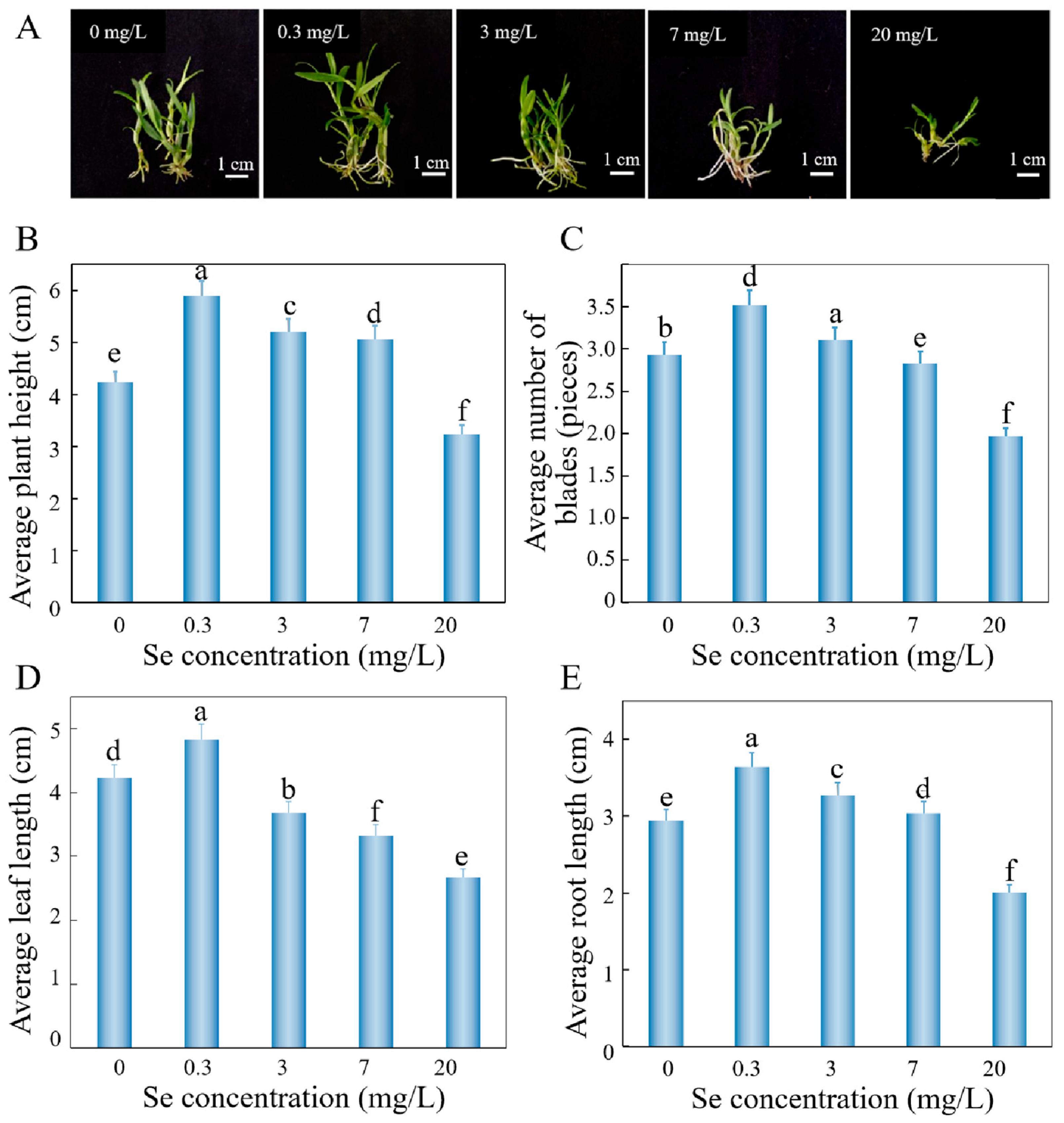
To investigate the effects of exogenous inorganic selenium supplementation on D. huoshanense, different concentrations of sodium selenite were added to the optimized culture medium. Growth parameters, polysaccharide content, and selenium accumulation were analyzed (Figure 2). At 0.3 mg/L sodium selenite, plantlets exhibited optimal growth, with maximum values for plant height (5.89 ± 0.08 cm), leaf number (3.52 ± 0.07), leaf length (4.83 ± 0.28 cm), and root length (3.64 ± 0.05 cm), representing increases of 39.2%, 20.1%, 14.1%, and 23.8%, respectively, compared to the control. At 7 mg/L sodium selenite, all growth metrics declined, and at 20 mg/L sodium selenite, they reached minimal values (3.24 ± 0.13 cm, 1.96 ± 0.15, 2.67 ± 0.57 cm, and 2.01 ± 0.01 cm), corresponding to reductions of 23.4%, 33.1%, 36.9%, and 31.6%, respectively, indicating near growth arrest. These results demonstrate that low sodium selenite concentrations (≤0.3 mg/L) enhance D. huoshanense growth, whereas excessive sodium selenite (≥7 mg/L) suppresses development.
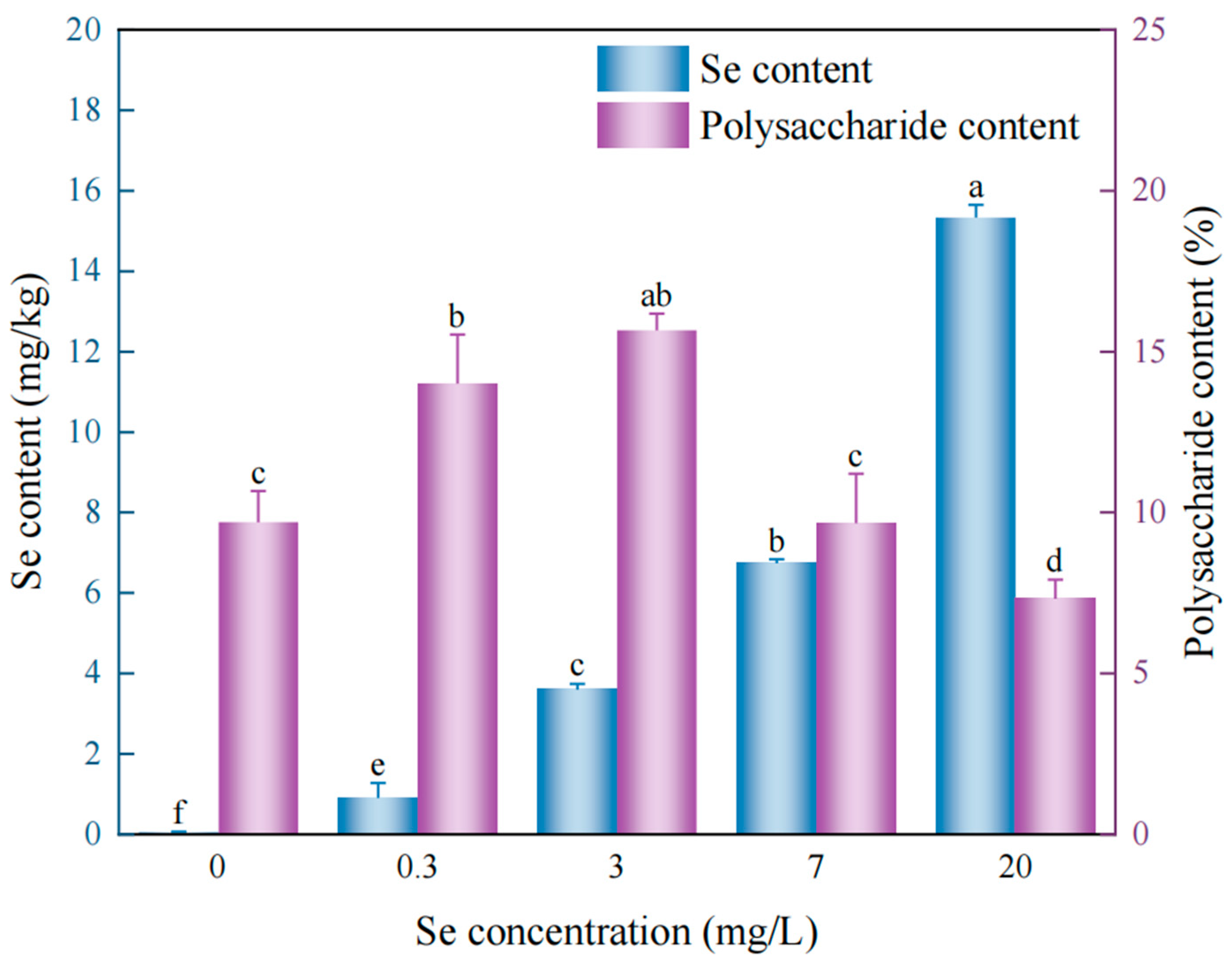
Figure 3 illustrates the effects of sodium selenite on selenium and polysaccharide content in D. huoshanense plantlets. Selenium accumulation increased with sodium selenite concentration, ranging from 0.91 ± 0.37 mg/kg (0.3 mg/L sodium selenite) to 15.33 ± 0.32 mg/kg (20 mg/L sodium selenite). Polysaccharide content followed a biphasic trend, peaking at 15.66% (3 mg/L sodium selenite), a 61.6% increase over the control (p < 0.05). Beyond 3 mg/L, polysaccharides declined significantly, reaching a 24.4% reduction (p < 0.05) at 20 mg/L sodium selenite. Optimal sodium selenite (3 mg/L) enhanced polysaccharide biosynthesis, whereas excessive sodium selenite (≥7 mg/L) suppressed it. Thus, while higher sodium selenite boosts selenium accumulation, 3 mg/L sodium selenite was identified as the optimal trade-off—maximizing both growth and polysaccharide yield in D. huoshanense.

3.2.2. Impact of Sodium Selenite Gradients on Physiological Parameters in D. huoshanense

Given the significant impact of sodium selenite on D. huoshanense growth, we analyzed physiological indices including oxidative stress markers, soluble protein, and chlorophyll content under varying sodium selenite concentrations (Figure 4). Superoxide dismutase (SOD) activity exhibited a biphasic response (0–20 mg/L sodium selenite), peaking at 3 mg/L sodium selenite (103.7 ± 15.16 U/g, 20.8% increase vs. control, p < 0.05), followed by 0.3 mg/L (92.77 ± 3.40 U/g), with minimum activity at 20 mg/L (55.0 ± 4.03 U/g) (Figure 4A). Peroxidase (POD) activity showed similar trends, maximized at 0.3 mg/L sodium selenite (566.37 ± 47.00 U/(g·min), 28.4% increase, p < 0.05), while 3–20 mg/L showed no statistical difference, with the lowest activity at 20 mg/L (418.9 ± 41.34 U/(g·min)) (Figure 4B). Catalase (CAT) activity peaked at 0.3 mg/L sodium selenite (18.07 ± 1.72 U/(g·min), 41.09% increase) and 3 mg/L (17.57 ± 1.23 U/(g·min), 38.0% increase) (both p < 0.05), but decreased by 32.7% at 20 mg/L (Figure 4C). These results demonstrate that 0.3–3 mg/L sodium selenite optimally enhances antioxidant enzyme activities in D. huoshanense seedlings.
The soluble protein content in D. huoshanense seedlings under varying sodium selenite treatments is presented in Figure 4D. Soluble protein content followed a biphasic pattern, with low sodium selenite concentrations (0.3 mg/L) inducing significant accumulation (1.91 ± 0.07 mg/g)—a 45.8% increase versus control (p < 0.05). Protein content declined at concentrations > 3 mg/L, reaching 0.87 ± 0.14 mg/g (33.6% reduction, p < 0.05) at 20 mg/L sodium selenite.
Figure 4E,F demonstrates the effects of sodium selenite on chlorophyll content in D. huoshanense. At 0.3 and 3 mg/L sodium selenite, chlorophyll a levels significantly increased to 0.65 ± 0.04 mg/g (+30%) and 0.63 ± 0.02 mg/g (+26%) versus control (both p < 0.05). Chlorophyll b peaked at 0.3 mg/L sodium selenite (+40%, p < 0.05), while 3 mg/L sodium selenite showed a non-significant increase. At 20 mg/L sodium selenite, chlorophyll a and b dropped to minimal levels (23.9% and 56%, respectively), indicating severe photosynthetic inhibition.
Integrated analysis of growth performance, selenium/polysaccharide accumulation, and physiological indices established 3 mg/L sodium selenite as the optimal concentration for D. huoshanense micropropagation.

3.3. Spatiotemporal Dynamics of Selenium Uptake and Accumulation in D. huoshanense

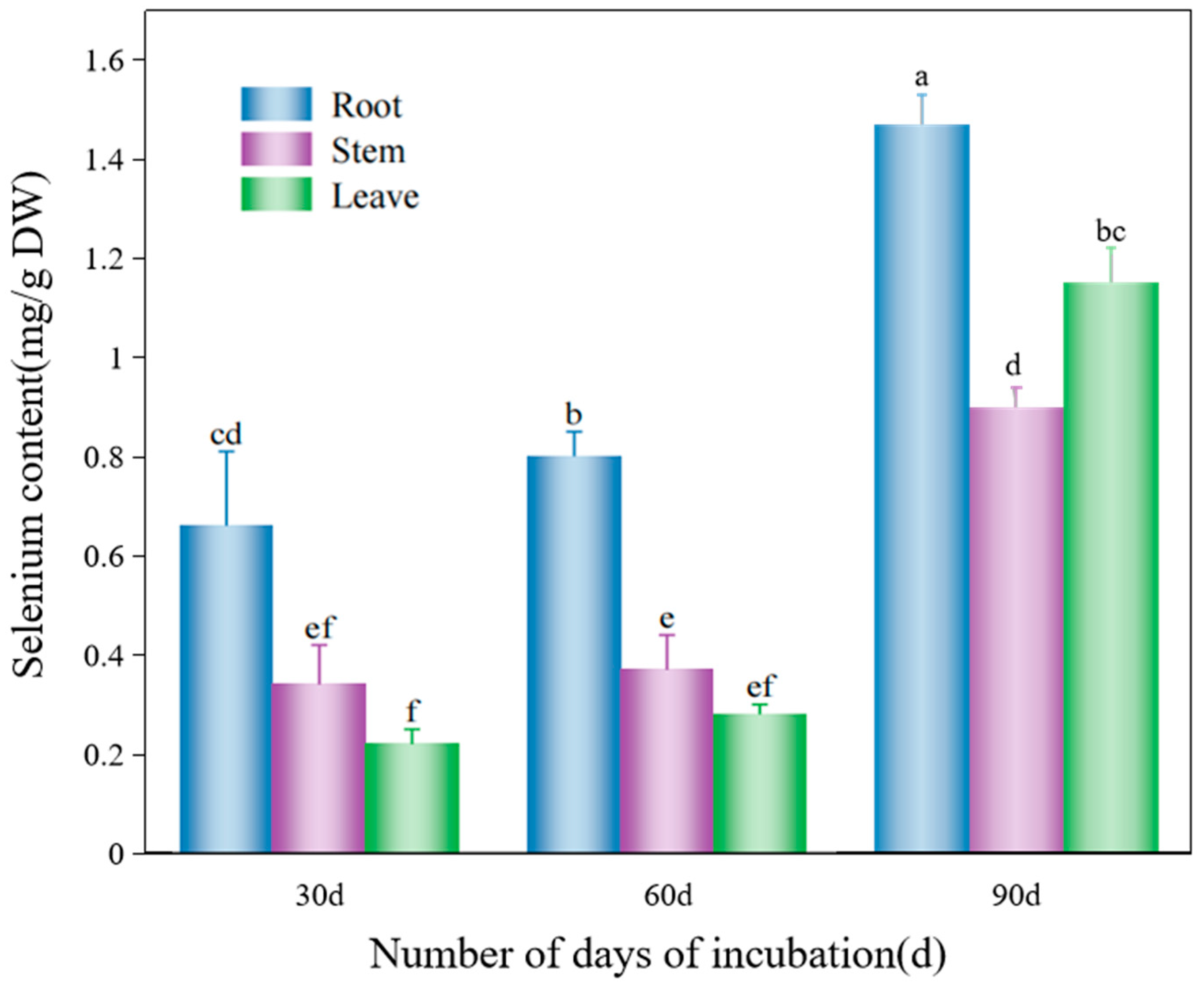
This study quantified selenium accumulation in roots, stems, and leaves of D. huoshanense at different culture periods (Figure 5). Selenium content increased progressively across organs: at 30 days, concentrations were 0.66 ± 0.15 mg/kg (roots) > 0.34 ± 0.08 mg/kg (stems) > 0.22 ± 0.03 mg/kg (leaves). By 60 d, levels rose to 0.80 ± 0.05 mg/kg (roots), 0.37 ± 0.07 mg/kg (stems), and 0.28 ± 0.02 mg/kg (leaves), maintaining root > stem > leaf distribution. At 90 d, a dramatic shift occurred: 1.47 ± 0.06 mg/kg (roots) > 1.15 ± 0.07 mg/kg (leaves) > 0.90 ± 0.04 mg/kg (stems). The Se allocation pattern transitioned from root-dominant (30–60 d) to root-to-leaf redistribution (90 d). These results demonstrate the following: (1) roots as primary Se sinks during early growth (≤60 d), and (2) subsequent phloem-mediated Se remobilization to photosynthetically active leaves (90 d).

3.4. Chemical Speciation of Selenium in D. huoshanense

3.4.1. Quantification of Selenium Species in Selenium-Enriched D. huoshanense

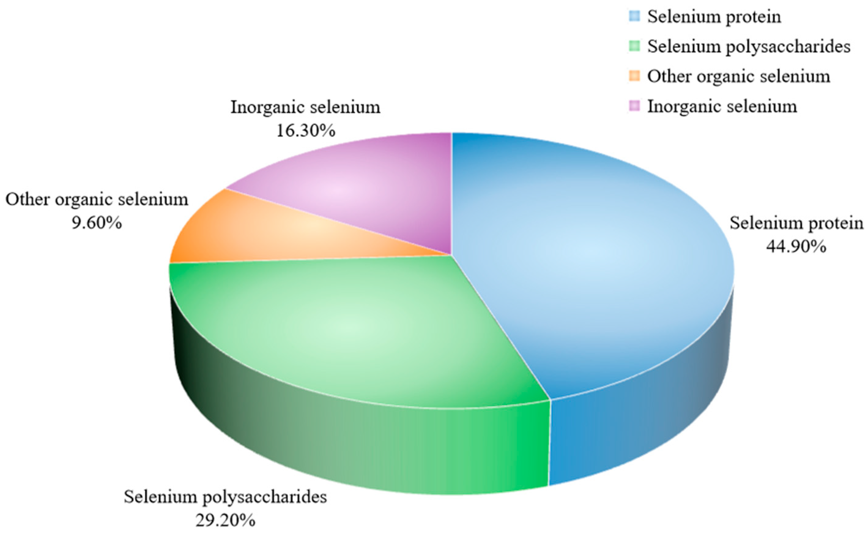
Figure 6 presents the selenium speciation analysis in D. huoshanense (83.70%), comprising selenoproteins (44.90%), selenopolysaccharides (29.20%), and other organic forms (9.60%). These results demonstrate the following: (1) organic speciation as the primary Se form, and (2) efficient biotransformation of inorganic selenium to organic selenium in D. huoshanense. The preferential accumulation in proteins (1.54-fold higher than polysaccharides) suggests selenoproteins serve as the dominant selenium reservoir in this medicinal orchid.

3.4.2. Distribution Profile of Selenoamino Acids in Selenium-Biofortified D. huoshanense

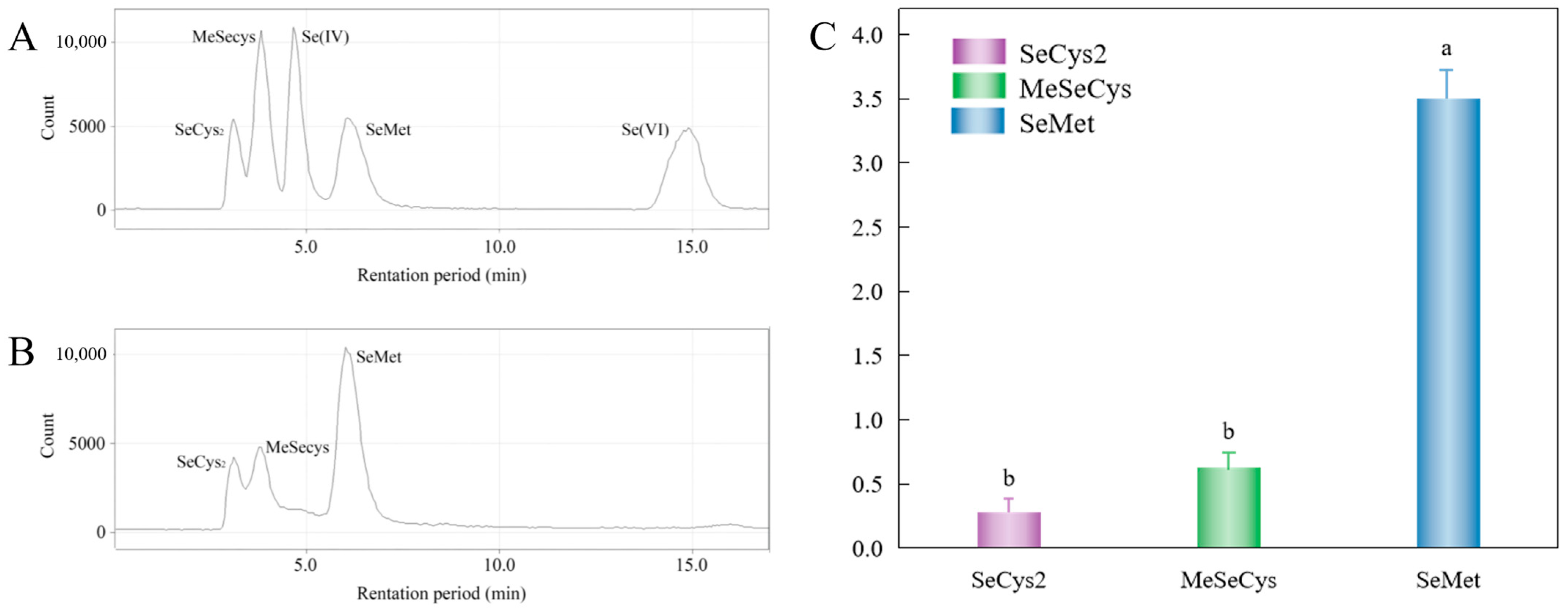
Quantitative analysis of selenoamino acids (SeMet, MeSeCys, and SeCys2) in selenium-enriched D. huoshanense was performed using HPLC-ICP-MS (Figure 7). Under 3 mg/L sodium selenite treatment, concentrations were 0.63 ± 0.04 mg/kg (SeMet) > 0.28 ± 0.06 mg/kg (SeCys2) > 0.20 ± 0.11 mg/kg (MeSeCys). The predominance of SeMet (2.25-fold higher than MeSeCys) demonstrates the following: efficient bioconversion of inorganic selenium to organoselenium compounds, and selenomethionine as the primary metabolic end-product in D. huoshanense.

4. Discussion

4.1. Synergistic Effects of Phytohormones and Organic Supplements on Rooting and Seedling Vigor in D. huoshanense Plantlets

Currently, although significant progress has been made in the tissue culture technology of D. huoshanense, there are still several shortcomings. Firstly, the growth period of tissue-cultured seedlings is too long, typically lasting 7–8 months [27]. Secondly, the tissue-cultured seedlings are relatively weak and have a low number of roots [8]. In addition, the optimization of hormone ratios is insufficient, making it difficult to balance the propagation efficiency of the tissue-cultured seedlings with the accumulation of medicinal components [28]. In this study, low concentrations of IBA and NAA effectively promoted rooting and robust growth in D. huoshanense plantlets, whereas higher levels inhibited growth, consistent with previous findings on orchid tissue culture [29]. Auxins demonstrate biphasic effects in vitro: lower concentrations stimulate cell division and rhizogenesis, whereas higher levels may induce ethylene biosynthesis or hormonal imbalance, leading to growth suppression. Furthermore, IBA at concentrations lower than NAA optimized root development, likely due to IBA’s superior competence in root primordia induction, while NAA preferentially enhanced root elongation [30].
Organic supplements are widely employed in plant tissue culture as natural growth regulators and nutritional adjuvants. Various supplements (kiwifruit juice, pear homogenate, potato extract, and coconut water) have demonstrated efficacy in enhancing Asparagus officinalis growth [31]. This study confirms banana homogenate’s significant promotion of D. huoshanense proliferation and rhizogenesis, and similar reports of this effect exist in Dendrobium Alya [32]. The growth-enhancing effects likely derive from banana homogenate’s rich composition: soluble sugars, amino acids, vitamins, and phytohormones (e.g., auxins and cytokinins), which provide carbon sources while mimicking endogenous hormonal regulation of cell division and organogenesis. It should be noted that although natural homogenates are effective in promoting plant growth, their lack of standardization may lead to variability among experiments. Further studies are needed to standardize their preparation and evaluate their reproducibility.

4.2. Physiological and Metabolic Response Mechanisms to Graded Selenium Supplementation in D. huoshanense

Sodium selenite treatment significantly influences the effect of selenium application on D. huoshanense plantlets in vitro. At a selenium concentration of 0.3 mg/L, the growth of D. huoshanense was most enhanced; at 3 mg/L, the polysaccharide content peaked; whereas at 20 mg/L, although the selenium content in D. huoshanense reached its maximum, it exerted detrimental effects on the plantlets, leading to a decline in physiological and biochemical indices. In summary, supplementing with 0.3–3 mg/L sodium selenite enhances selenium accumulation and polysaccharide biosynthesis without impeding growth, while also alleviating plant senescence and improving stress resistance. Research on Cardamine violifolia has also found that [33] low concentrations of exogenous selenium promoted seedling growth, whereas high concentrations exhibited significant inhibitory effects on both young and mature plants. Regarding polysaccharide accumulation in D. huoshanense, low sodium selenite concentrations stimulated polysaccharide synthesis, similar to reports on enhanced mushroom [34] polysaccharide production under low selenium levels and its suppression at high concentrations. A dose-response relationship exists between selenium concentration and both the plant antioxidant defense system and photosynthetic characteristics. In this study, low sodium selenite concentrations effectively activated the plant antioxidant defense network, with superoxide dismutase (SOD) activity peaking at 3 mg/L treatment. As a key enzyme for scavenging O2− in plants, enhanced SOD activity helps maintain intracellular Reactive Oxygen Species (ROS) homeostasis, thereby mitigating oxidative damage [35]. Meanwhile, POD and CAT activities reached their maxima at 0.3 mg/L treatment. POD exerts dual roles in plant stress response by catalyzing H2O2-dependent oxidation: it scavenges ROS during early stress and later facilitates chlorophyll degradation to reduce photo-oxidative damage. CAT, as a H2O2-specific decomposing enzyme, significantly strengthens cell membrane resistance to peroxidation when its activity increases. When sodium selenite concentration exceeded 7 mg/L, all three antioxidant enzymes exhibited significant activity reduction, indicating that high-level selenium stress surpassed the threshold of plant cellular antioxidant defense capacity, resulting in the collapse of the ROS scavenging system [36]. Regarding primary metabolism, soluble protein content demonstrated similar selenium concentration-dependent dynamics. As crucial osmoregulatory substances and nutrients, elevated soluble protein levels not only enhanced cellular water retention capacity but also provided structural foundations for maintaining biomembrane stability and enzymatic activity [37]. In D. huoshanense treated with 0.3 mg/L sodium selenite, soluble protein content increased markedly, whereas at 20 mg/L, rapid depletion occurred, suggesting high selenium stress either inhibited protein synthesis or accelerated degradation pathways.
Photosynthetic pigments exhibited higher sensitivity to sodium selenite concentrations. Moderate selenium treatment may enhance photosynthesis through the following mechanisms: (1) activating magnesium chelatase to promote chlorophyll biosynthesis; (2) improving the electron transport efficiency of photosystem II; (3) maintaining the structural integrity of thylakoid membranes. Results demonstrated that 0.3 mg/L sodium selenite treatment significantly increased both chlorophyll a and b contents in D. huoshanense, which is similar to the findings of Zhong et al. in Landoltia punctata [38]; however, high selenium treatment (20 mg/L) reduced chlorophyll a and b contents compared to the control, potentially due to: (1) selenium-induced membrane lipid peroxidation damaging chloroplast structure; (2) competitive occupation of magnesium binding sites by selenium; (3) enhanced activity of chlorophyll-degrading enzymes.
This study identified 3 mg/L sodium selenite as the critical threshold concentration for D. huoshanense, at which multiple physiological parameters achieved optimal equilibrium. Beyond this concentration, the beneficial effects of selenium transitioned into detrimental effects, a shift closely associated with selenium’s dual mechanism of action at the cellular level: At optimal concentrations, selenium enhances antioxidant enzyme (e.g., glutathione peroxidase, GPx) activity by incorporating into their active centers; whereas excess selenium displaces sulfur in proteins, causing structural abnormalities and functional impairment of enzymes.

4.3. Spatiotemporal Dynamics and Organ-Specific Allocation Mechanisms of Selenium Accumulation in D. huoshanense

This study systematically elucidated the spatiotemporal distribution patterns of selenium accumulation in D. huoshanense across different cultivation periods. At 30 days of cultivation, selenium was predominantly accumulated in roots, showing a characteristic distribution pattern of root > stem > leaf. This pattern likely stems from roots’ preferential retention of selenite as primary absorption organs [39], coupled with temporary selenium immobilization in roots through cell wall binding or vacuolar sequestration [40] to mitigate their potential toxicity to aerial parts [41]. When cultivation extended to 60 days, selenium content increased significantly in all organs while maintaining the original distribution pattern, indicating that transmembrane transport and long-distance translocation of selenium require temporal accumulation. Notably, substantial upward migration only occurred after 60 days, suggesting potential selenium transformation or storage processes in roots. After 90 days of cultivation, leaf selenium content exhibited exponential growth, ultimately establishing a new distribution hierarchy of root > leaf > stem. The explosive accumulation in leaves may relate to selenium’s involvement in photosynthesis or antioxidant metabolism (e.g., selenomethionine incorporation into proteins). Stems, functioning primarily as transport conduits, likely lack efficient selenium retention mechanisms, explaining their consistently lower content. This suggests D. huoshanense may employ phased translocation strategies analogous to Stanleya pinnata and Astragalus bisulcatus: initial root-dominated storage followed by xylem–phloem coordinated redistribution to metabolically active leaves [42].

4.4. Chemical Speciation and Biotransformation Pathways of Selenium in Selenium-Enriched D. huoshanense

In selenium-enriched D. huoshanense obtained in this study, organic selenium accounted for 83.70% of total selenium content, demonstrating its highly efficient metabolic capacity for inorganic-to-organic selenium conversion [43]. Notably, selenoproteins constituted a significantly higher proportion than selenopolysaccharides in organic selenium components. This distribution pattern aligns with selenium-enriched tea plants [44], further supporting that selenoproteins may represent the predominant selenium speciation in plants. Biochemically, the chemical similarity between selenium and sulfur provides the fundamental basis for this conversion. Plants likely assimilate absorbed inorganic selenium into selenoamino acids (e.g., selenocysteine and selenomethionine) via sulfur metabolic pathways, subsequently incorporating them into protein structures [45]. This conversion not only reduces free selenium toxicity but may also enhance the plant’s antioxidant defense system through bioactive selenoprotein formation, as confirmed by HPLC-ICP-MS analysis of selenium-enriched D. huoshanense. SeMet showed the highest content in selenium-enriched D. huoshanense, serving as the primary organic selenium storage form that potentially regulates plant growth and development while conferring enhanced antioxidant activity [46]. Although MeSeCys occurred at lower concentrations [47,48], its distinctive anticancer properties offer unique advantages for anticancer drug development. This diversity in selenium speciation enables multifunctional applications of D. huoshanense.

5. Conclusions

In this study, we successfully established an optimized selenium-enriched in vitro culture system for D. huoshanense, which effectively enhanced rooting and seedling vigor. The optimal formulation was determined as MS basal medium (pH 5.8) supplemented with 0.1 mg/L IBA, 0.6 mg/L NAA, 30 g/L sucrose, 7 g/L agar, and 100 g/L banana homogenate. A graded sodium selenite experiment revealed that low selenium concentrations (≤0.3 mg/L) promoted plant growth, while 3 mg/L represented the optimal balance between growth, polysaccharide accumulation, and antioxidant performance; higher levels (≥7 mg/L) exhibited inhibitory effects. Selenium distribution analysis demonstrated that Se was primarily accumulated in roots during the early culture stage (≤60 days), followed by redistribution from roots to leaves at 90 days, forming a root > leaf > stem pattern. Chemical speciation analysis indicated that organic selenium dominated (83.70% of total selenium), mainly in the forms of selenoproteins (44.90%) and selenopolysaccharides (29.20%). Furthermore, HPLC–ICP–MS confirmed the presence of three key selenoamino acids—SeMet, SeCys2, and MeSeCys—with SeMet as the major species, suggesting efficient inorganic-to-organic selenium transformation in D. huoshanense. Overall, these findings provide a scientific and practical framework for selenium biofortification in medicinal orchids and support the development of high-value selenium-enriched D. huoshanense products. Future studies should explore the long-term stability, environmental safety, and ecological impacts of selenium-enriched cultivation systems, as well as the potential applications of this strategy in sustainable medicinal plant production.

Supplementary Materials

The following supporting information can be downloaded at: https://www.mdpi.com/article/10.3390/horticulturae11101263/s1. Table S1: Orthogonal Table Extreme Variance Analysis. Table S2: Analysis of variance significance of mean plant height results. Table S3: Analysis of variance significance of mean root results. Table S4: Analysis of variance significance of mean root length results. Figure S1: Comparison of the growth status of D. huoshanense test tube seedlings under different medium treatments. Figure S2: Comparison of the growth status of D. huoshanense test-tube seedlings under different pH treatments. Figure S3: Comparison of the growth status of D. huoshanense test-tube seedlings under different treatments of exogenous additives. Figure S4: Comparison of different treatments on the growth status of D. huoshanense test tube seedlings.

Author Contributions

Conceptualization, Y.Q. and Z.Z.; data curation, Y.W. (Yulai Wu) and C.A.; formal analysis, Y.W. (Yulai Wu), C.A. and Y.W. (Yanjie Wang); funding acquisition, Z.Z.; investigation and methodology, Y.W. (Yulai Wu), C.L., C.A. and Y.W. (Yanjie Wang); project administration, Y.Q. and Z.Z.; software, Y.W. (Yulai Wu), C.A., Y.S. and Y.W. (Yanjie Wang); supervision, Y.Q. and Z.Z.; writing—original draft, Y.W. (Yanjie Wang), Y.W. (Yulai Wu), C.L., B.W. and Y.S.; writing—review and editing, Y.Q. and Z.Z. All authors have read and agreed to the published version of the manuscript.

Funding

This work was supported by Dalian’s Science and Technology Innovation Fund (grant no. 2020JJ27SN107).

Data Availability Statement

The data will be made available upon request.

Acknowledgments

We thank all colleagues at our lab for providing valuable discussions and technical assistance.

Conflicts of Interest

The authors declare no conflicts of interest.

Abbreviations

SeMetSelenomethionine
MeSeCysMethylselenocysteine
SeCys2Selenocysteine
NAA1-Naphthaleneacetic acid
IBAIndole-3-butyric acid
SODSuperoxide dismutase
PODPeroxidase
CATCatalase
ROSReactive oxygen species
MSMurashige and Skoog medium
KCKnudson C medium
B5Gamborg B5 medium
N6Chu’s N-6 medium

References

  1. Sarsaiya, S.; Jain, A.; Shu, F.; Jia, Q.; Gong, Q.H.; Wu, Q.; Shi, J.S.; Chen, J.S. Unveiling the potential of dendrobine: Insights into bioproduction, bioactivities, safety, circular economy, and future prospects. Crit. Rev. Biotechnol. 2025, 25, 1268–1286. [Google Scholar] [CrossRef]
  2. Wan, J.; Gong, X.; Wang, F.; Wen, C.; Wei, Y.; Han, B.; Ouyang, Z. Comparative analysis of chemical constituents by HPLC–ESI–MSn and antioxidantactivities of Dendrobium huoshanense and Dendrobium officinale. Biomed. Chromatogr. 2022, 36, e5250. [Google Scholar] [CrossRef]
  3. Hao, Q.; Jiang, L.; Ma, J.; Wang, H.K.; Liu, Y.; Xu, Q.C.; Li, S.Z.; Han, S.; Zheng, Q.S.; Fan, X.C.; et al. Dendrobium huoshanense C. Z. Tang and S. J. Cheng can be prepared as a food with the ability to prevent and treat hyperuricaemia. Front. Nutr. 2025, 12, 1518014. [Google Scholar] [CrossRef]
  4. Xu, H.J.; Liu, Z.; Xu, W.; Zhang, Y.F. Beneficial in vitro effects of polysaccharide and non-polysaccharide components of Dendrobium huoshanense on gut microbiota of rats with type 1 diabetes as opposed to metformin. Molecules 2024, 29, 2791. [Google Scholar] [CrossRef]
  5. Zhang, X.Q.; Ge, R.P.; Wu, J.; Cai, X.; Deng, G.H.; Lv, J.H.; Ma, M.Z.; Yu, N.J.; Yao, L.; Peng, D.Y. Structural characterization and improves cognitive disorder in ageing mice of a glucomannan from Dendrobium huoshanense. Int. J. Biol. Macromol. 2024, 269, 131995. [Google Scholar] [CrossRef]
  6. Gao, L.L.; Wang, F.; Hou, T.T.; Geng, C.Y.; Xu, T.; Han, B.X.; Liu, D. Dendrobium huoshanense C.Z. Tang et S.J. Cheng: A review of its traditional uses, phytochemistry, and pharmacology. Front. Pharmacol. 2022, 13, 920823. [Google Scholar] [CrossRef]
  7. Teixeira da Silva, J.A.; Cardoso, J.C.; Dobránszki, J.; Zeng, S.J. Dendrobium micropropagation: A review. Plant Cell Rep. 2015, 34, 671–704. [Google Scholar] [CrossRef] [PubMed]
  8. Zhou, Y. Research on the In Vitro Rapid Propagation of Dendrobium huoshanense. Master’s Thesis, Guizhou University, Guiyang, China, May 2024. [Google Scholar] [CrossRef]
  9. Jin, R.; An, C.; Wang, B.R.; Liu, C.B.; Fan, X.P.; Zhang, Z.Z. Bioreactor scale-up and bioactivity evaluation of Psammosilene tunicoides hairy roots. Asian J. Agric. Biol. 2025, 4, e2025067. [Google Scholar] [CrossRef]
  10. Omid, A.; Somayeh, S.; Azita, H.; Foad, A.; Esmaeil, Y.R.; Shadab, S. Effects of selenium supplementation on serum C reactive protein level: A systematic review and meta-analysis of randomized controlled clinical trials. Obes. Med. 2020, 17, 100182. [Google Scholar] [CrossRef]
  11. Huang, Z.; Rose, A.H.; Hoffmann, P.R. The role of selenium in inflammation and immunity: From molecular mechanisms to therapeutic opportunities. Antioxid. Redox Signaling 2012, 16, 705–743. [Google Scholar] [CrossRef]
  12. Tan, L.C.; Nancharaiah, Y.V.; van Hullebusch, E.D.; Lens, P.N.L. Selenium: Environmental significance, pollution, and biological treatment technologies. Biotechnol. Adv. 2016, 34, 886–907. [Google Scholar] [CrossRef]
  13. Gu, L.Y.; Zhao, W.Z.; Hu, J.C.; Jye, M.W.; Yu, Z.P. Selenium-Containing Peptides from Foodstuff: Preparation, Bioavailability, Bioactivities, and Future Perspectives. J. Agric. Food Chem. 2025, 73, 24501–24516. [Google Scholar] [CrossRef]
  14. Li, H.A.; Liu, H.Q.; Tang, X.R.; Deng, Z.Y.; Li, H.Y. From Soil to Table: A Comprehensive Review of Selenium-Fortified Foods. Compr. Rev. Food Sci. Food Saf. 2025, 24, e70250. [Google Scholar] [CrossRef]
  15. Tangjaidee, P.; Swedlund, P.; Xiang, J.Q.; Yin, H.Q.; Quek, S.Y. Selenium-enriched plant foods: Selenium accumulation, speciation, and health functionality. Front. Nutr. 2023, 9, 962312. [Google Scholar] [CrossRef]
  16. Xie, M.H.; Sun, X.Y.; Li, P.; Shen, X.C.; Fang, Y. Selenium in cereals: Insight into species of the element from total amount. Compr. Rev. Food Sci. Food Saf. 2021, 20, 2914–2940. [Google Scholar] [CrossRef]
  17. Hachemi, M.A.; Cardoso, D.; De Marco, M.; Geraert, P.A.; Briens, M. Inorganic and organic selenium speciation of selenoyeasts used as feed additives: New insights from elemental selenium determination. Biol. Trace Elem. Res. 2023, 201, 5839–5847. [Google Scholar] [CrossRef]
  18. Cheng, K.X.; Sun, Y.; Liu, B.W.; Ming, J.J.; Wang, L.L.; Xu, C.F.; Xiao, Y.Y.; Zhang, C.; Shang, L.C. Selenium modification of natural products and its research progress. Foods 2023, 12, 3773. [Google Scholar] [CrossRef] [PubMed]
  19. Xu, H.Z.; Yan, J.P.; Qin, Y.; Xu, J.M.; Shohag, M.J.I.; Wei, Y.N.; Gu, M.H. Effect of different forms of selenium on the physiological response and the cadmium uptake by rice under cadmium stress. Int. J. Environ. Res. Public Health 2020, 17, 6991. [Google Scholar] [CrossRef] [PubMed]
  20. Puccinelli, M.; Malorgio, F.; Terry, L.A.; Tosetti, R.; Rosellini, I.; Pezzarossa, B. Effect of selenium enrichment on metabolism of tomato (Solanum lycopersicum) fruit during post-harvest ripening. J. Sci. Food Agric. 2018, 99, 2463–2472. [Google Scholar] [CrossRef]
  21. Yang, W.Q.; Jiang, T.T.; Wang, Y.Q.; Wang, X.J.; Wang, R. Combined transcriptomics and metabolomics analysis reveals the effect of selenium fertilization on Lycium barbarum fruit. Molecules 2023, 28, 8088. [Google Scholar] [CrossRef] [PubMed]
  22. Lei, H.L.; Zhou, M.G.; Li, B.; Fu, Y.A.; Shi, Z.Y.; Ji, W.Q.; Zhang, R.; Wang, Z.H. Humic acid chelated selenium is suitable for wheat biofortification. J. Sci. Food Agric. 2023, 103, 4887–4898. [Google Scholar] [CrossRef]
  23. Zhang, H.B.; He, D.; Li, X.L.; Dun, B.C.; Wu, D.; Huang, G.Y. The establishment of rapid propagation system of ‘RED SUN’ Phalaenopsis aphrodite. Sustainability 2022, 14, 15305. [Google Scholar] [CrossRef]
  24. Zhang, Y.; Wang, S.W.; Li, N.Q.; Jin, Q.; Anbazhakan, R.; Dai, Y.F.; Xiang, Z.X.; Gao, J.Y. Ecological specificity of fungi on seedling establishment in Dendrobium huoshanense: A narrow distributed medicinal orchid. Mycorrhiza 2025, 35, 41. [Google Scholar] [CrossRef] [PubMed]
  25. Tie, M.; Zang, S.L.; Zhang, W.; Li, J.; Sun, T.B.; Li, H.W. Study on the Method of Using ICP-MS to Determine Se in the Edible Fungi. Spectrosc. Spectr. Anal. 2006, 26, 551–553. [Google Scholar]
  26. Zhao, Y.Q.; Zheng, J.P.; Yang, M.W.; Yang, G.D.; Wu, Y.N.; Fu, F.F. Speciation analysis of selenium in rice samples by using capillary electrop hores is inductively coupled plasma mass spectrometry. Talanta 2011, 84, 983–988. [Google Scholar] [CrossRef]
  27. Xu, M.Y. Optimization of Industrial Seedling Technology System of Dendrobium houshanense. Master’s Thesis, Nanjing Agricultural University, Nanjing, China, June 2023. [Google Scholar] [CrossRef]
  28. Zhang, F.W. Study on the Tissue Culture and Temperature Stress Response Mechanisms of Dendrobium huoshanense. Master’s Thesis, Nanjing Normal University, Nanjing, China, March 2022. [Google Scholar] [CrossRef]
  29. Lee, P.L.; Chen, J.T. Plant regeneration via callus culture and subsequent in vitro flowering of Dendrobium huoshanense. Acta Physiol. Plant 2014, 36, 2619–2625. [Google Scholar] [CrossRef]
  30. Liu, Z.K.; Min, C.; Dong, H.; Zhang, Z.S. Improvement of adventitious root formation in Sedum aizoon L. and the production of flavonoids. S. Afr. J. Bot. 2021, 137, 483–491. [Google Scholar] [CrossRef]
  31. Klanrit, P.; Lila, K.; Netsawang, P.; Siangsanor, P.; Thanonkeo, P.; Thanonkeo, S. Effect of organic additives on the micropropagation of Asparagus officinalis. Horticulturae 2023, 9, 1244. [Google Scholar] [CrossRef]
  32. Nambiar, N.; Tee, C.S.; Maziah, M. Effects of organic additives and different carbohydrate sources on proliferation of protocorm like bodies in Dendrobium alya Pink. Plant Omics 2012, 5, 10–18. [Google Scholar]
  33. Rao, S.; Yu, T.; Cong, X.; Zhang, W.W.; Zhu, Z.Z.; Liao, Y.L.; Ye, J.B.; Cheng, S.Y.; Xu, F. Effects of selenate applied at two growth stages on the nutrient quality of Cardamine violifolia. Sci. Hortic. 2021, 288, 110352. [Google Scholar] [CrossRef]
  34. Xu, M.M.; Zhu, S.; Li, Y.R.; Xu, S.; Shi, G.Y.; Ding, Z.Y. Effect of selenium on mushroom growth and metabolism: A review. Trends Food Sci. Technol. 2021, 118, 328–340. [Google Scholar] [CrossRef]
  35. Lanza, M.; dos Reis, A.R. Roles of selenium in mineral plant nutrition: ROS scavenging responses against abiotic stresses. Plant Physiol. Biochem. 2021, 164, 27–43. [Google Scholar] [CrossRef]
  36. Li, L.X.; Qiao, Z.; Cai, J.Y.; Liang, Y.; Lin, Y.; Wei, G.L.; Hou, X.L.; Miao, J.H.; Wei, K.H. Effects of selenium on growth and biochemical characters of tissue culture seedlings of Sophora tonkinensis. Pharmacogn. Mag. 2023, 19, 772–781. [Google Scholar] [CrossRef]
  37. Ozturk, M.; Unal, B.T.; García-Caparrós, P.; Khursheed, A.; Gul, A.; Hasanuzzaman, M. Osmoregulation and its actions during the drought stress in plants. Physiol. Plant 2020, 172, 1321–1335. [Google Scholar] [CrossRef]
  38. Zhong, Y.; Li, Y.; Cheng, J.J. Effects of selenite on chlorophyll fluorescence, starch content and fatty acid in the duckweed Landoltia punctata. J. Plant Res. 2016, 129, 997–1004. [Google Scholar] [CrossRef]
  39. Jiang, Y.; Zeng, Z.H.; Bu, Y.; Ren, C.Z.; Li, J.Z.; Han, J.J.; Tao, C.; Zhang, K.; Wang, X.X.; Lu, G.X.; et al. Effects of selenium fertilizer on grain yield, Se uptake and distribution in common buckwheat (Fagopyrum esculentum Moench). Plant Soil Environ. 2015, 61, 371–377. [Google Scholar] [CrossRef]
  40. Wang, M.K.; Peng, Q.; Zhou, F.; Yang, W.X.; Dinh, Q.T.; Liang, D.L. Uptake kinetics and interaction of selenium species in tomato (Solanum lycopersicum L.) seedlings. Environ. Sci. Pollut. Res. 2019, 26, 9730–9738. [Google Scholar] [CrossRef]
  41. Wu, H.Z.; Zhang, D.X.; Wu, X.M.; Tian, X.S.; Hu, G.; Liu, S.L.; Jie, X.L.; Wang, D.C. Uptake and transport of selenium in a soil-tea plant–tea Infusion system: A study of typical tea plantations in a selenium-rich area of China. Forests 2024, 15, 914. [Google Scholar] [CrossRef]
  42. Freeman, J.L.; Marcus, M.A.; Fakra, S.C.; Devonshire, J.; McGrath, S.P.; Quinn, C.F.; Pilon-Smits, E.A.H. Selenium Hyperaccumulator plants Stanleya pinnata and Astragalus bisulcatus are colonized by Se-resistant, Se-excluding wasp and beetle seed herbivores. PLoS ONE 2012, 7, e50516. [Google Scholar] [CrossRef] [PubMed]
  43. Both, E.B.; Stonehouse, G.C.; Lima, L.W.; Fakra, S.C.; Aguirre, B.; Wangeline, A.L.; Xiang, J.Q.; Yin, H.Q.; Jókai, Z.; Soós, A.; et al. Selenium tolerance, accumulation, localization and speciation in a Cardamine hyperaccumulator and a non-hyperaccumulator. Sci. Total Environ. 2020, 703, 135041. [Google Scholar] [CrossRef] [PubMed]
  44. Wei, K.; Wei, Y.; Zhou, P.; Zhu, J.X.; Peng, L.L.; Cheng, L.Z.; Wang, Y.F.; Wei, X.L. Preparation, characterization, and antioxidant properties of selenium-enriched tea peptides. Foods 2023, 12, 4105. [Google Scholar] [CrossRef]
  45. Zhang, X.; He, H.; Xiang, J.Q.; Yin, H.Q.; Hou, T. Selenium-containing proteins/peptides from plants: A review on the structures and functions. J. Agric. Food Chem. 2020, 68, 15061–15073. [Google Scholar] [CrossRef]
  46. Fang, Y.; Catron, B.; Zhang, Y.F.; Zhao, L.Y.; Caruso, J.A.; Hu, Q.H. Distribution and in Vitro availability of selenium in seleniumcontaining storage protein from selenium-enriched rice utilizing optimized extraction. J. Agric. Food Chem. 2010, 58, 9731–9738. [Google Scholar] [CrossRef] [PubMed]
  47. Torres, S.; Cerutti, S.; Raba, J.; Pacheco, P.; Silva, M.F. Preconcentration of seleno-amino acids on a XAD resin and determination in regional olive oils by SPE UPLC-ESI-MS/MS. Food Chem. 2014, 159, 407–413. [Google Scholar] [CrossRef] [PubMed]
  48. Rao, S.; Gou, Y.Y.; Yu, T.; Cong, X.; Gui, J.Y.; Zhu, Z.Z.; Zhang, W.W.; Liao, Y.L.; Ye, J.B.; Cheng, S.Y.; et al. Effects of selenate on Se, flavonoid, and glucosinolate in broccoli florets by combined transcriptome and metabolome analyses. Food Res. Int. 2021, 146, 110463. [Google Scholar] [CrossRef] [PubMed]
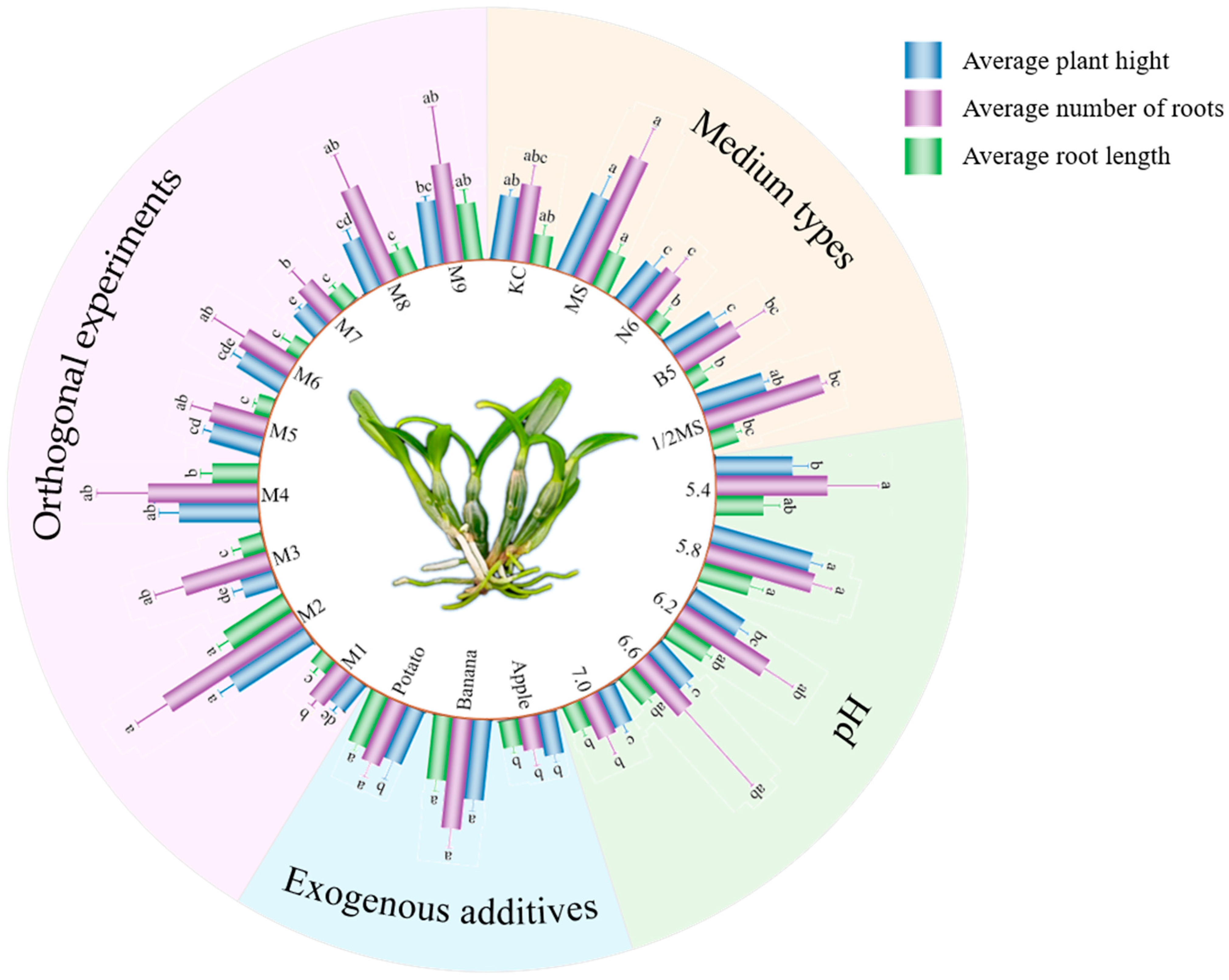
Figure 1. Effects of different cultivation conditions (medium types, pH, exogenous additives, and orthogonal experiments) on average plant height, average root number, and average root length of D. huoshanense. M is an abbreviation of the medium of used in the orthogonal experiment. Different letters indicate statistically significant differences (p < 0.05) according to one-way ANOVA.
Horticulturae 11 01263 g001
Figure 2. Effects of different sodium selenite concentrations on growth in D. huoshanense. (A) Comparison of the growth status; (B) Comparison of the average plant height; (C) Comparison of the average number of blades; (D) Comparison of the average leaf length; (E) Comparison of the average root length. Different letters indicate statistically significant differences (p < 0.05) according to one-way ANOVA.
Horticulturae 11 01263 g002
Figure 3. Effects of different concentrations of sodium selenite treatment on selenium content and polysaccharides of D. huoshanense test tube seedlings. Different letters indicate statistically significant differences (p < 0.05) according to one-way ANOVA.
Horticulturae 11 01263 g003
Figure 4. Effects of sodium selenite treatment at different concentrations on physiological indices of D. huoshanense. (A) SOD; (B) POD; (C) CAT; (D) Soluble protein; (E) Chlorophyll a; (F) Chlorophyll b. Different letters indicate statistically significant differences (p < 0.05) according to one-way ANOVA.
Horticulturae 11 01263 g004
Figure 5. Selenium content in the rhizome and leaves of test tube seedlings of D. huoshanense at different incubation times. Different letters indicate statistically significant differences (p < 0.05) according to one-way ANOVA.
Horticulturae 11 01263 g005
Figure 6. Distribution of selenium in selenium-enriched D. huoshanense.
Horticulturae 11 01263 g006
Figure 7. Distribution of selenoamino acids in selenium-enriched D. huoshanense. (A) Chromatograms of five selenium-containing compounds; (B) Chromatogram of seleno-substituted amino acids of Se-enriched D. huoshanense; (C) Comparison of seleno-substituted amino acid content of Se-enriched D. huoshanense. Different letters indicate statistically significant differences (p < 0.05) according to one-way ANOVA.
Horticulturae 11 01263 g007
Table 1. Orthogonal design of culture media with different combinations of IBA, NAA, and exogenous additives.
Medium NumberIBA (mg/L)NAA (mg/L)Exogenous Additives (g/L)
M10.10.10
M20.10.6100
M30.11.5200
M40.60.1100
M50.60.6200
M60.61.50
M71.50.1200
M81.50.60
M91.51.5100
Table 2. ICP-OES instrument operating parameters.
Parameter NameValue
RF Power1200 W
Plasma Gas Flow Rate12 L/min
Carrier Gas Flow Rate0.80 L/min
Auxiliary Gas Flow Rate0.40 L/min
Peristaltic Pump Speed1.0 L/min
Nebulizer Gas Flow Rate0.5 L/min
Argon Pressure0.65 MPa
Normal Sampling Time15 s
Fast Pump Flow Rate4.0 L/min
Rinse Time5 s
Integration Time1 s
Detection Wavelength196.026 nm
Number of Replicates3
Table 3. Atomic fluorescence spectrometer (with selenium hollow cathode lamp) instrument operating parameters.
Parameter NameSpecification
SAX Strong Anion Exchange Column1000 mg/6 mL
Negative High Voltage300 V
Lamp Current80 mA
Furnace Height8 mm
Carrier Gas Flow Rate400 mL/min
Shield Gas Flow Rate1000 mL/min
Table 4. HPLC working parameters.
ParameterSpecification
ColumnHamilton PRPX-100 analytical column, 250 mm × 4.1 mm, 10 μm
Guard ColumnHamilton PRPX-100 guard column, 20 mm × 2.1 mm, 10 μm
Mobile Phase1.05 g citric acid monohydrate + 20 mL methanol, diluted to 1000 mL with water
Flow Rate0.8 mL/min
pH5.3
Injection Volume100 µL
Table 5. ICP-MS operating parameters.
ParameterOperating Condition
RF Power1400 W
Nebulizer Gas Flow Rate1.05 L/min
Helium Flow Rate2.0 L/min
Integration ModePeak Area
Disclaimer/Publisher’s Note: The statements, opinions and data contained in all publications are solely those of the individual author(s) and contributor(s) and not of MDPI and/or the editor(s). MDPI and/or the editor(s) disclaim responsibility for any injury to people or property resulting from any ideas, methods, instructions or products referred to in the content.