Article

Bacillus subtilis Fermentation Augments the Anti-Inflammatory and Skin Moisture Improvement Activities of Tetragonia tetragonoides through the Upregulation of Antioxidant Components

Department of Medical Laboratory Science, College of Health Science, Dankook University, Cheonan-si 31116, Chungnam, Republic of Korea
*
Author to whom correspondence should be addressed.
Fermentation 2023, 9(9), 800; https://doi.org/10.3390/fermentation9090800
Submission received: 31 July 2023 / Revised: 22 August 2023 / Accepted: 28 August 2023 / Published: 30 August 2023
(This article belongs to the Special Issue Fermentation in Cosmetics)

Abstract

:
This study demonstrates that the fermentation of Tetragonia tetragonioides (T. tetragonioides) by Bacillus subtilis (B. subtilis) subsp. spizizenii enhances its antioxidant, anti-inflammatory, and skin-moisturizing activities. Fermented T. tetragonioides extracts (FTEs) showed a significant increase (p < 0.05) of approximately 1.3 to 3.07 times in their total polyphenol content (TPC), total flavonoid content (TFC), and vanillic acid content compared to pre-fermentation T. tetragonioides extracts (TEs). Additionally, the 2,2-diphenyl-1-picrylhydrazyl (DPPH)- and 2,2′-azino-bis(3-ethylbenzothiazoline-6-sulfonic acid) (ABTS) radical-scavenging activities were significantly higher (p < 0.05) in FTEs than in TEs, showing an increase of about 1.25 to 1.64 times. The anti-inflammatory effects, evaluated through the inhibition of nitric oxide (NO) in RAW 264.7 cells stimulated by lipopolysaccharide (LPS), revealed that FTEs exhibited significant (p < 0.05) NO inhibition activity at less than half the concentration of TEs. It is particularly noteworthy that the FTE at 200 μg/mL significantly suppressed the expression of the cyclooxygenase-2 (COX-2), inducible nitric oxide synthase (iNOS), and tumor necrosis factor-alpha (TNF-α) proteins. In HaCaT cells, FTEs substantially (p < 0.001) increased the mRNA expression of filaggrin (FLG), hyaluronan synthase (HAS)-1, and HAS-3, indicating improved skin protection and moisturization. In conclusion, this study confirms that T. tetragonioides’ antioxidant, anti-inflammatory, and skin-moisturizing activities are enhanced by B. subtilis fermentation, suggesting the potential of FTEs as a cosmeceutical ingredient.

1. Introduction

The skin is constantly exposed to various environmental factors, rendering it directly vulnerable to oxidative stress. The primary factor contributing to oxidative stress is exposure to solar ultraviolet (UV) radiation; other factors include pollution, chemical oxidants, microorganisms, and respiratory or inflammatory reactions [1]. In particular, prolonged exposure to ultraviolet radiation induces photodamage in the skin through reactive oxygen species (ROS), which compromises the enzymatic and non-enzymatic antioxidant defense systems [2]. ROS encompass highly reactive species, such as singlet oxygen (1O2) and hydroxyl (⋅OH), as well as hydrogen peroxide (H2O2), peroxyl (ROO⋅), alkoxyl radicals (RO⋅s), organic hydroperoxides (ROOHs), and hypochloride (HOCl) [3]. They can be generated within cells and tissues through various processes involving high-energy radiation, photochemical reactions, and several enzyme reactions [4]. These ROS participate in skin-aging processes by disrupting the skin’s antioxidant defense, initiating lipid peroxidation reactions, oxidizing proteins, oxidizing DNA, promoting the degradation of collagen and hyaluronic acid through abnormal cross-linking and cleavage, and influencing melanin production [5,6].
The skin tissue is composed of the epidermis, dermis, and subcutaneous fat layers. The epidermis contains keratinocytes, melanocytes, and other cells, while the dermis consists of fibrous and matrix components, with collagen accounting for 90% of the dermal layer [7]. The epidermis, which plays a role in maintaining skin barrier function through preventing water loss, can become damaged by ultraviolet radiation, leading to decreased skin moisture, and the formation of dry, wrinkled, and rough skin. Therefore, adequate hydration through sufficient moisturization is crucial to maintaining healthy skin [8,9]. In addition, the activity of hyaluronan synthase (HAS), an enzyme responsible for the production of hyaluronic acid (HA) in the stratum corneum, should be maintained to preserve the moisturizing effect of HA [10]. Furthermore, the action of ROS generated from prolonged exposure to ultraviolet radiation stimulates the activity of matrix metalloproteinases (MMPs) known as collagenases, which impair collagen synthesis in the dermis and promote skin elasticity loss and wrinkle formation [11,12]. Consequently, there is a pressing need for the development of natural ingredients that can suppress the skin aging and moisture loss caused by UV radiation.
Tetragonia tetragonioides (T. tetragonioides), also known as New Zealand spinach or sea spinach, is a perennial plant belonging to the Aizoaceae family. It grows to a height of 40–60 cm and has succulent stems with numerous branches that spread horizontally or grow obliquely from the ground, eventually reaching a height of around 50 cm. The plant forms rounded clusters of roots that attach to the ground and spread in all directions. T. tetragonioides is found in New Zealand, China, Japan, South Asia, Australia, South America, and other regions. It has a strong vitality and can thrive in harsh and arid environments, such as gravel fields and rocky crevices with limited moisture. The plant is commonly used in salads and can also be cooked as a vegetable. In traditional medicine, T. tetragonioides has been used for its cooling, detoxifying, dispersing, and anti-inflammatory properties in the treatment of dysentery, sepsis, edema, and fever. Various compounds have been identified in this plant, including β-carotene, diterpenes, flavones, cerebrosides, oxalic acid, sterylglucoside, and polysaccharides. It has been reported to possess anti-inflammatory and anti-ulcer activities [13,14,15,16,17,18,19]. Despite the diverse physiological activities and associated effects of the various compounds in T. tetragonioides, studies substantiating its potential as a cosmetic ingredient are limited.
Fermentation is a processing method that can influence the composition and flavor of natural plants and enhance their storage stability. It involves enzymatic action, derived from fermented microorganisms, which breaks down the nutrients of natural ingredients into easily digestible and absorbable forms [20]. Various microorganisms, such as Bacillus sp., lactic acid bacteria, Aspergillus oryzae, and Saccharomyces sp., are used in the fermentation of natural plants. Among them, Bacillus sp. is widely used in the production of traditional Korean fermented plants and is recognized as a probiotic strain [21]. However, compared to the research on the nutritional components of T. tetragonioides itself, the studies on the changes in its composition and physiological activities before and after fermentation are still limited. Therefore, it is necessary to explore effective and environmentally friendly fermentation, as well related processing methods, in order to apply T. tetragonioides as a cosmeceutical ingredient.
In the present study, it was demonstrated that the anti-inflammatory and skin-barrier-improving activities of T. tetragonioides can be enhanced via fermentation by Bacillus subtilis (B. subtilis). Based on this result, it is thought that the utilization of T. tetragonioides as a therapy or cosmeceutical can be expanded.

2. Materials and Methods

2.1. Preparation of T. tetragonioides 70% EtOH Extracts

We purchased T. tetragonioides from Jeju san ya cho (SJDBL2581, Jeju, Republic of Korea). For the experiment, T. tetragonioides was powdered and soaked in 70% ethanol at a ratio of 10 times the weight of the sample. The soaking was performed at room temperature (RT) for 24 h and repeated three times in preparation for extraction. The extract was filtered using No. 1 and 3 filter paper (Hyundai Micro Co., Ltd., Seoul, Republic of Korea), concentrated, and then freeze-dried.

2.2. Preparation of TEs and FTEs

We utilized the Gram-positive bacterium B. subtilis subsp. spizizenii (KACC 17047) obtained from the Korea Agricultural Culture Collection (KACC). The bacterium was cultured in a broth medium (Kisanbio Co., Seoul, Republic of Korea) consisting of 0.5 g/L yeast extract, 0.5 g/L proteose peptone, 0.5 g/L casamino acid, 0.5 g/L dextrose, 0.5 g/L soluble starch, 0.3 g/L dipotassium phosphate, 0.05 g/L magnesium sulfate, and 0.3 g/L sodium pyruvate, with a pH of 7.2. These microorganisms were cultured at 30 °C and used for the experiment. The samples used in the experiment were divided into TEs and FTEs. Firstly, to dissolve the extracts, distilled water (DW) was added at a ratio of 10 times the weight of the T. tetragonioides 70% ethanol extracts. The T. tetragonioides extracts (TEs) were supplemented with medium containing 0.6% glucose, 0.3% yeast extract, and 0.1% soytone. In contrast, fermented T. tetragonioides extracts (FTE) were supplemented with medium and inoculated with B. subtilis subsp. Spizizenii at a concentration of 1.5 × 10⁶ CFU/mL. The samples were then fermented for 3 days at 130 rpm and 37 °C. Subsequently, the fermented solution was filtered using No. 1 and 3 filter paper (Hyundai Micro Co., Ltd.) and freeze-dried for use in the experiment. The final yields for the TEs and FTEs were measured at 7.50% (w/w) and 8.00% (w/w), respectively (Figure 1).

2.3. Determination of Total Phenolic Content (TPC) and Total Flavonoid Content (TFC)

The total phenolic content (TPC) was determined via the addition of 60 μL of Folin–Ciocâlteu’s phenol reagent (Sigma-Aldrich Co., St. Louis, MO, USA) to a 60 μL aliquot of the sample solution that had previously been diluted two-fold. The mixture was allowed to react for 3 min at RT. Then, 60 μL of 10% Na2CO3 (Sigma Co.) was added, and the reaction was allowed to proceed for 40 min at RT. The absorbance was measured at 700 nm. Gallic acid (Sigma Co.) was used as the standard, and a standard curve was constructed to quantify the total phenolic content. The total flavonoid content (TFC) was determined via the addition of 20 μL each of 10% aluminum nitrate (Sigma Co.) and 1 M potassium acetate (Sigma Co.) to a 100 μL aliquot of the sample solution. Then, 860 μL of 80% ethanol was added, and the mixture was allowed to react for 40 min at RT. The absorbance was measured at 415 nm. Quercetin (Sigma Co.) was used as the standard, and a standard curve was constructed to calculate the total flavonoid content.

2.4. High-Performance Liquid Chromatography (HPLC) Analysis

The analysis of vanillic acid content (Sigma-Aldrich Co., St Louis, MO, USA), as a marker component of the TEs and FTEs, was performed via HPLC. For analysis, approximately 10 mg of TE and FTE was dissolved in 1 mL of DW. After the solution was cooled to RT, it was filtered through a filter and used for the experiment. The instruments used in the analysis comprised the Waters 2690 (Waters corporation, Milford, MA, USA) HPLC system equipped with an autosampler; the analysis conditions are shown in Table 1.

2.5. Free Radical Inhibition Activity

The 2,2-diphenyl-1-picrylhydrazyl (DPPH) radical-scavenging activity of the TEs and FTEs was determined following the method of Blois [22]. An aliquot of 200 μL of 0.15 mM DPPH solution was added to 800 μL of the sample solution. The mixture was left at RT for 30 min, and the absorbance was measured at 517 nm. The radical-scavenging activity of each sample was calculated as the concentration of the sample required to reduce the absorbance of the control group, which did not contain the sample, by one half. The reduction concentration (RC)50 value was obtained and compared to the positive control group, ascorbic acid. The 2,2′-azino-bis(3-ethylbenzothiazoline-6-sulfonic acid) (ABTS)-radical-scavenging activity of the TEs and FTEs was measured via a modified method described by Re et al. [23]. A mixture of 7 mM ABTS and 2.45 mM potassium persulfate (Sigma Co.) was prepared and left in a dark place at RT for 24 h. Then, it was diluted with DW to obtain an absorbance value of 0.70 (±0.02) at 732 nm. A volume of 990 μL of this diluted solution was taken, and 10 μL of the sample solution was added. After incubation at RT for 1 min, the absorbance was measured at 732 nm. The ABTS radical-scavenging activity was calculated as the RC50 value.

2.6. Cell Culture

The mouse-derived macrophage cell line RAW 264.7 and human keratinocyte cell line HaCaT were obtained from the Korean Cell Line Bank (KCLB, Seoul, Republic of Korea). The cells were cultured in Dulbecco’s Modified Eagle Medium (DMEM, Gibco-BRL, Rockville, MD, USA) supplemented with 10% fetal bovine serum (FBS, Gibco-BRL), 100 μg/mL penicillin (Gibco-BRL), and 100 μg/mL streptomycin (Gibco-BRL). The cells were maintained at 37 °C in a humidified atmosphere with 5% CO2. The subculturing of the cells was performed every 2–3 days.

2.7. Cell Viability

The cell viability was measured using the 3-(4,5-dimethylthiazol-2-yl)-2,5-diphenyltetrazolium bromide (MTT) assay. RAW 264.7 and HaCaT cells were seeded in 96-well plates at densities of 1 × 10⁵ cells/well and 1 × 10⁴ cells/well, respectively. After incubation for 24 h, the medium was removed, and various concentrations of the test samples were added to the wells in 200 μL of DMEM. The cells were then incubated for an additional 24 h at 37 °C in a 5% CO2 incubator. Following this, MTT solution (5.0 mg/mL) was added, and the cells were further incubated for 4 h under the same culture conditions. The resulting formazan crystals were dissolved through the addition of 100 μL of dimethyl sulfoxide (DMSO) to each well, followed by 10 min of shaking. The absorbance was measured at the wavelength of 550 nm.

2.8. Measurement of Nitric Oxide (NO) Inhibition

The amount of nitric oxide (NO) generated by lipopolysaccharide (LPS, Sigma Co.) was measured via a modified version of the method developed by Green et al. [24]. RAW 264.7 cells were seeded in 96-well plates at a density of 1 × 10⁵ cells/well. After incubation for 24 h, the medium was removed, and the cells were treated with LPS (100 ng/mL) in the presence of various concentrations of the test samples (final concentrations: 0, 25, 50, 100, and 200 μg/mL) and a positive control, dexamethasone (20 μM). The cells were then incubated for an additional 24 h at 37 °C in a 5% CO2 incubator. After incubation, the culture supernatant was mixed with an equal volume of Griess reagent (Sigma Co.) and incubated for 10 min. The absorbance was measured at a wavelength of 540 nm. A standard curve was prepared for sodium nitrate (Sigma Co.) and used to calculate the extent of NO inhibition.

2.9. Western Blot Analysis

The RAW 264.7 cells were seeded in a 6-well plate at a density of 5 × 10⁵ cells/mL, and incubated for 24 h. After the removal of the medium, the cells were treated with LPS at a concentration of 100 ng/mL in a serum-free medium. For the FTEs and dexamethasone treatment, they were added to the LPS-containing medium and incubated for an additional 24 h. After the removal of the medium, the cells were washed with PBS and lysed with a lysis buffer. The lysates were then incubated at 4 °C for 30 min and centrifuged at 13,000 rpm for 15 min, for the collection of the protein supernatant. The protein samples were subjected to 8% sodium dodecyl sulfate–polyacrylamide gel electrophoresis (SDS-PAGE) and transferred onto a nitrocellulose membrane. The membrane was blocked with a 5% skim milk solution at RT for 1 h, followed by overnight incubation with the primary antibody at 4 °C. The next day, the membrane was washed with Tris-buffered saline Tween 20 (TBST), incubated with the secondary antibody at RT for 2 h, washed again with TBST, and subjected to an ECL reaction through the use of an ECL kit. Finally, the protein bands were visualized on X-ray film.

2.10. RNA Isolation and Reverse Transcription-Polymerase Chain Reaction (RT-PCR)

HaCaT cells were seeded in a 6-well plate, at a density of 5 × 10⁵ cells/well, in a medium containing 10% FBS, and incubated for 24 h. The medium was then replaced with a serum-free medium, and the cells were treated with FTEs at different concentrations (10, 50, and 100 µg/mL). After 24 h, the cells were harvested, and the RNA was isolated with TRIzol reagent (Molecular Research Center, Inc., Cincinnati, OH, USA). The concentration of the isolated RNA was determined using a NanoDrop spectrophotometer (Thermo Scientific). For reverse transcription, 1 µg of the isolated RNA was mixed with 1 μL of oligo dT (100 pM) and DEPC-treated water to a final volume of 12 μL. The mixture was then incubated at 70 °C for 10 min. Reverse transcription (RT) was performed according to the instructions provided with the RT premix kit (Bioneer, Daejeon, Republic of Korea). PCR amplification for the filaggrin (FLG), hyaluronan synthases (HAS)-1, HAS-3, and GAPDH genes was carried out with the use of 2 μL of the cDNA product obtained from each reverse transcription reaction, following the instructions provided with the PCR premix kit (Bioneer). The primer sequences are provided in Table 2. PCR was performed over 30 cycles, through the use of a PCR thermal cycler (Takara MP-300, Takara Bio Inc., Kusatsushi, Japan), with denaturation at 94 °C for 30 s, annealing at 56 °C for 30 s, and extension at 72 °C for 45 s. The amplified cDNA products obtained from the RT-PCR were analyzed via electrophoresis on a 1.2% agarose gel containing ethidium bromide.

2.11. Statistical Analysis

The experimental results are presented as the mean ± standard error of mean and were analyzed using SPSS Statistics software (ver. 25, IBM Co., Armonk, NY, USA). ANOVA was performed, and the statistical significance of the mean values was analyzed via Duncan’s multiple range test at the p < 0.05 level.

3. Results

3.1. Total Phenolic, Flavonoid, and Vanillic Acid Contents of TEs and FTEs

The TPC of the non-fermented TEs and FTEs was determined to be 14.96 ± 0.54 GAE mg/g and 20.45 ± 1.45 GAE mg/g, respectively. The TFC increased after fermentation, with a value of 3.19 ± 1.57 QE mg/g for TEs and 9.09 ± 0.56 QE mg/g for FTEs (Figure 2A,B). The quantitative analysis of vanillic acid, a phenolic compound, showed an increase in its content after fermentation, with values of 34.87 ± 1.57 μg/g for the TEs and 107.11 ± 1.45 μg/g for the FTEs (Figure 2C).

3.2. Antioxidant Activity Analysis

It is essential to confirm the effect of fermentation on the changes in the TPC of T. tetragonioides after fermentation, as TPC changes in plants are associated with antioxidant activity. Therefore, the scavenging ability of the TEs and FTEs was evaluated against two representative radicals: the DPPH radical and the ABTS radical (Figure 3). The RC50 values for the DPPH radical-scavenging ability of the TEs and FTEs were determined to be as 113.57 ± 14.18 μg/mL and 69.24 ± 11.21 μg/mL, respectively, indicating an increased radical-scavenging ability after fermentation. The RC50 value for the ABTS radical-scavenging ability was determined to be as 291.36 ± 17.98 μg/mL for the TEs and 232.14 ± 2.68 μg/mL for the FTEs, showing an enhanced radical-scavenging ability for both radicals after fermentation. Ascorbic acid, used as the positive control, had RC50 values of 0.81 ± 0.22 μg/mL and 4.98 ± 0.22 μg/mL for the two radical-scavenging abilities, respectively.

3.3. Effect of TEs and FTEs on NO Production via LPS Stimulation

To investigate the anti-inflammatory effects of TEs and FTEs, their effects on NO production induced by LPS were examined in RAW 264.7 cells. Firstly, the cytotoxicity of TEs and FTEs was measured, and it was confirmed that the cell viability was above 90% at all the concentrations tested (0, 6.25, 12.5, 25, 50, 100, and 200 μg/mL), indicating no toxicity (Figure 4A). The cells were treated with LPS and various concentrations of TE or FTE, and the NO production was measured. Dexamethasone at 20 μM, a commonly used anti-inflammatory agent, was used as the positive control. As shown in Figure 4B, the TE exhibited a significant (p < 0.05) inhibitory effect on NO production at a concentration of 200 μg/mL, while the FTE showed a significant (p < 0.05) inhibition of NO production starting from the lower concentration of 100 μg/mL.

3.4. The FTEs Inhibit LPS-Stimulated COX-2, iNOS, and TNF-α Expression

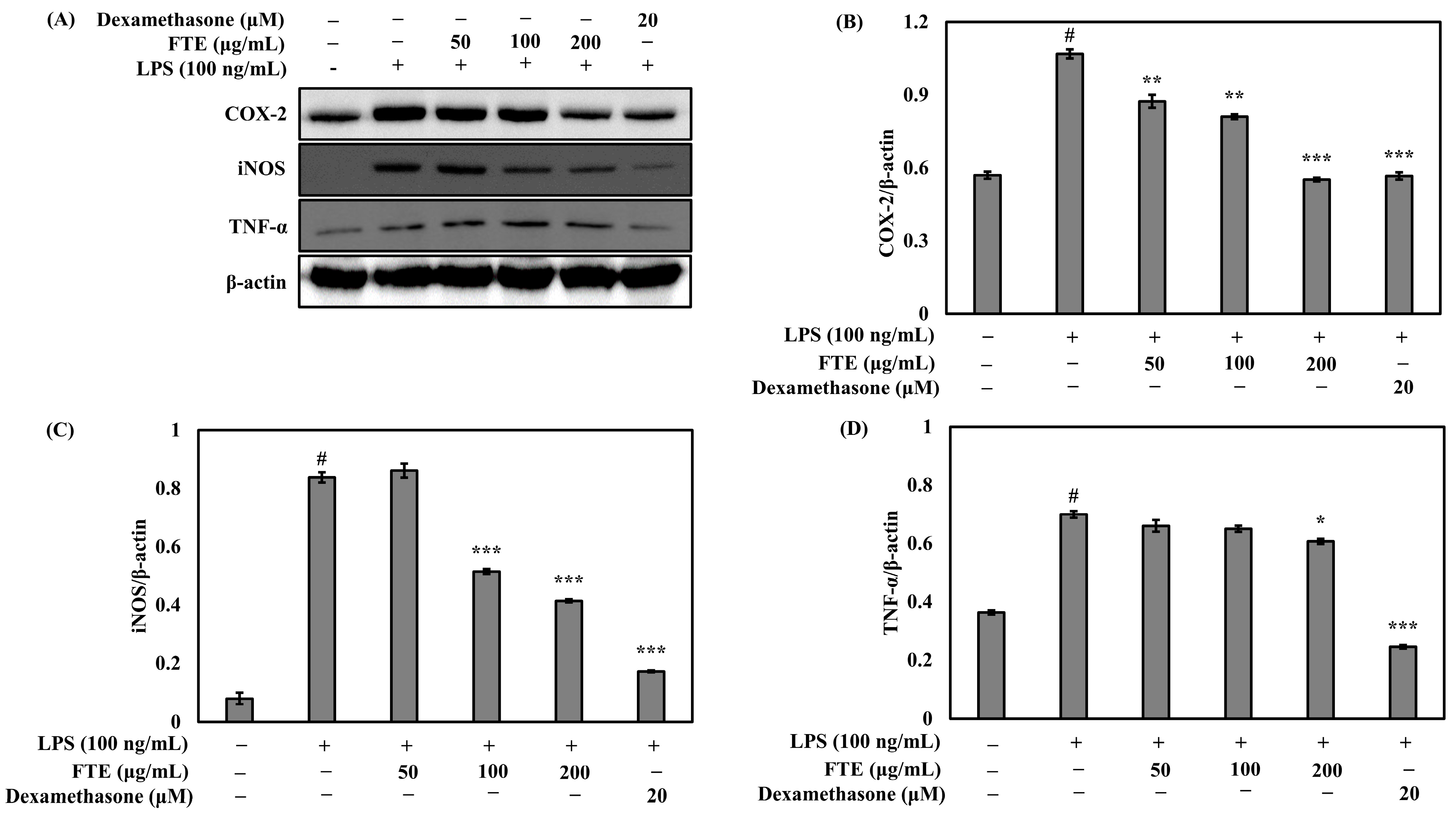
The anti-inflammatory effects of FTEs, and their association with the regulation of cyclooxygenase-2 (COX-2), inducible nitric oxide synthase (iNOS), and tumor necrosis factor-α (TNF-α) expression were investigated. Normally, in unstimulated cells, the proteins COX-2, iNOS, and TNF-α are not detectable. However, in response to immune stimulants, such as LPS, the genes for COX-2, iNOS, and TNF-α are strongly induced. As observed in Figure 5, the FTE potently inhibited COX-2, iNOS, and TNF-α protein expression at a concentration of 200 μg/mL. These results indicate that FTEs suppress the production of NO by inhibiting the expression of COX-2, iNOS, and TNF-α at the protein level.

3.5. Effects of FTEs on Cell Viability and Skin Moisture Protection Activity

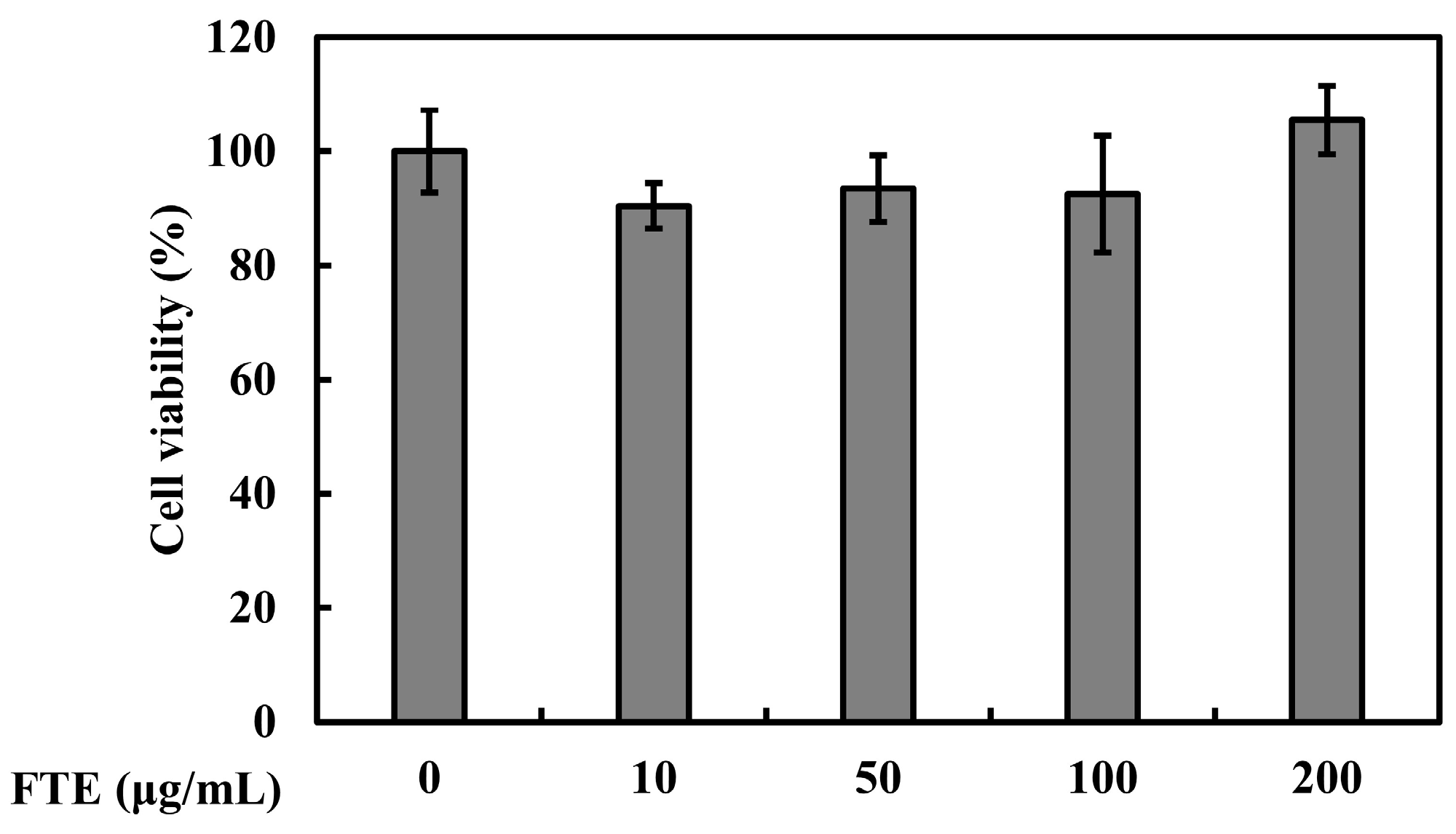
Before measuring the skin-protective activity of FTEs in HaCaT, MTT analysis was conducted to evaluate the cell toxicity of FTEs. The results for the cell viability demonstrated that FTEs did not induce cell death in HaCaT cells (Figure 6). To confirm the skin-protective and skin-moisturizing effects of FTEs, the mRNA expression levels of FLG, HAS-1, and HAS-3 were detected via RT-PCR. As shown in Figure 7, the mRNA expression levels of FLG, HAS-1, and HAS-3 were significantly (p < 0.001) increased by the FTEs.

4. Discussion

To enhance the utilization of various biomasses, including plants, fermentation processes using microorganisms have been frequently attempted. Seo et al. [25] examined the potential utilization of fermented mulberry leaf extract by inoculating it with B. subtilis, aiming to increase its protein content. Similarly, Kim et al. [26] investigated the changes in antioxidant activity induced by inoculating green tea with B. subtilis, exploring the potential application of the product as a microbial fermented tea. In this study, B. subtilis, commonly used in plant fermentation, was used to inoculate T. tetragonioides, to allow us to observe the functional components, as well as the antioxidant, anti-inflammatory, and skin-moisturizing activities, before and after fermentation.
Flavonoids, carotenoids, phenolic compounds, and vitamins A, C, and E are representative antioxidant components found in plants. They exhibit strong radical-scavenging effects and perform various physiological activities, such as anticancer and antioxidant activities [27]. Polyphenols, which account for the highest content, are phenolic compounds with two or more aromatic rings, including flavonoids and tannins. They are known as substances produced by plants to protect themselves from extreme environmental conditions during growth [28]. Phenolic compounds act as antioxidants and provide protection against various diseases by regulating ROS, preventing oxidative damage, and inhibiting pro-inflammatory activities. They are abundant in plant leaves and fruits [29]. The measurement of the TPC and TFC in the TEs and FTEs revealed an increase in their content after fermentation. The FTEs showed a significant increase (p < 0.05) of approximately 1.3 to 3.07 times in their TPC, TFC, and vanillic acid contents compared to the pre-fermentation TEs (Figure 2). These changes between the contents before and the contents after fermentation are attributed to the hydrolytic enzymes produced by B. subtilis. During fermentation, phenolic glycosides are hydrolyzed to aglycones by enzymes produced by B. subtilis, leading to an increase in phenolic compounds, and enhanced antioxidant effects [30]. Adetuyi and Ibrahim [31] reported that the highest levels of total polyphenols and total flavonoids were produced after 24 h of fermentation when okra (Abelmoschus esculentus) seeds were soaked in hot water and fermented. VA, the oxidized form of vanillin, is a phenolic acid derivative [32] commonly found in herbs, tea, and wine plants [33]. It has various pharmacological effects, such as anti-carcinogenic, antioxidant, antihypertensive, anti-inflammatory, and antimicrobial effects [34,35]. Recently, VA has been widely used as a preservative, flavoring agent, or antioxidant with beneficial activities in cosmetic, food, and pharmaceutical products [33]. The increase in VA achieved through fermentation suggests its potential as a more functional material.
To assess the radical-scavenging activity of TEs and FTEs, considering the association between the changes in TPC and antioxidant activity in plants, we conducted experiments. The DPPH and ABTS radical-scavenging activities were significantly higher (p < 0.05) in the FTEs than in the TEs, showing an increase of about 1.25 to 1.64 times (Figure 3). Consistent with the results of our study, Ibrahim et al. [36] also investigated the antioxidant activity of four types of Malaysian herbal teas (allspice, Scaphium, Gora, cinnamon) after fermentation and observed an increase in both the TPC and antioxidant activity compared to before fermentation. These findings align with our study, suggesting that the increased antioxidant activity in fermented T. tetragonioides can be attributed to the increase in the TPC.
The excessive production of NO can be harmful and can contribute to various inflammatory and autoimmune diseases [37]. Therefore, the inhibition of NO production represents a promising strategy for therapeutic intervention in inflammatory conditions. In this study, we demonstrated that FTEs effectively suppress LPS-induced NO production compared to TEs (Figure 4). The inhibitory effect of FTEs on NO production was attributed to the suppression of COX-2, iNOS, and TNF-α protein expression induced by inflammatory signals such as LPS (Figure 5). These findings suggest that FTEs, with their anti-inflammatory and antioxidant activities, could potentially be applied in the development of anti-aging cosmetics.
Fermented cosmetics, commonly produced through the fermentation of natural ingredients, are generally more effective and have fewer side effects compared to non-fermented cosmetics. They are suitable for sensitive and dry skin and support the functions of the skin, without disrupting its natural processes [38]. Fermentation is not only applied for its antioxidant and anti-inflammatory properties in cosmetic formulations, but also has various applications in the cosmetic industry. Moisturization is essential in maintaining skin hydration and the integrity of the skin barrier, which helps to prevent microbial or chemical intrusion from the external environment, thus contributing to the skin’s homeostasis. FLG, a structural protein in the uppermost layer of the skin, protects against moisture loss, prevents penetration from the external environment, and regulates epidermal differentiation [39]. The skin maintains elasticity through the presence of moisture in the stratum corneum (SC). To maintain the elasticity of the SC, a minimum of 10% moisture is required [40]. One key molecule influencing skin hydration is hyaluronic acid (HA). In the epidermis, HA is primarily located within the extracellular matrix (ECM) of the upper stratum granulosum and intracellularly in the basal layer [41]. HA, a highly hydrophilic glycosaminoglycan (GAG), captures a large amount of moisture, providing flexibility and hydration in the skin [42]. HA is synthesized by the enzyme HAS, which has three types (HAS-1, -2, and -3), each with different enzyme activities, and each capable of synthesizing HA of different lengths [43]. The results of this study show that FTEs increase the expression of FLG, HAS-1, and HAS-3, thereby enhancing the skin barrier function and moisture content (Figure 7). Therefore, FTEs, which can increase the expression of HAS-1, HAS-3, and FLG, are expected to serve as effective moisturizers in skincare products.

5. Conclusions

In conclusion, the antioxidant, anti-inflammatory, and moisturizing effects of T. tetragonioides were confirmed through in vitro testing. These potential benefits of T. tetragonioides for the skin were significantly enhanced via fermentation with B. subtilis subsp. spizizenii. In summary, through the utilization of advanced fermentation technology with beneficial microorganisms such as Bacillus, the desirable skin-modulating properties of T. tetragonioides can be further enhanced, thereby expanding its potential applications in the field of skincare resources.

Supplementary Materials

The following supporting information can be downloaded at: https://www.mdpi.com/article/10.3390/fermentation9090800/s1, Figure S1: Original photographs for the blots of each protein marker of Figure 5A.

Author Contributions

Conceptualization, S.-G.L. and H.K.; methodology, J.-W.H. and S.-G.L.; formal analysis, J.-W.H. and H.K.; investigation, S.-G.L. and J.-W.H.; resources, J.-W.H.; data curation, H.K.; writing—original—S.-G.L.; writing—review and editing, S.-G.L. and H.K.; visualization, S.-G.L. and J.-W.H.; supervision, H.K.; project administration, S.-G.L.; funding acquisition, S.-G.L. All authors have read and agreed to the published version of the manuscript.

Funding

This research was financially supported by the Ministry of Small and Medium-sized Enterprises (SMEs) and Startups (MSS), Korea, under the “Regional Specialized Industry Development Plus Program (R&D, Project number: S3261522)” supervised by the Korea Technology and Information Promotion Agency for SMEs.

Institutional Review Board Statement

Not applicable.

Informed Consent Statement

Not applicable.

Data Availability Statement

The data presented in this study are available on request from the corresponding author.

Conflicts of Interest

The authors declare no conflict of interest.

References

  1. Rahimuddin, S.A.; Khoja, S.M.; Zuhair, M.M.; Howell, N.K.; Brown, J.E. Inhibition of lipid peroxidantion in UVA-treated skin fibroblasts by luteolin and its glucosides. Eur. J. Lipid Sci. Technol. 2007, 109, 647–655. [Google Scholar] [CrossRef]
  2. Park, S.N. Skin aging and antioxidants. J. Soc. Cosmet. Sci. Korea 2003, 29, 75–77. [Google Scholar]
  3. Fantone, J.C.; Ward, P.A. Role of oxygen-derived free radicals and metabolites in leukocyte dependent inflammatory reaction. Ann. J. Path. 1982, 107, 395–418. [Google Scholar]
  4. Davies, K.J.A. Protein damage and degradation by oxygen radical. J. Biol. Chem. 1987, 262, 9895–9901. [Google Scholar] [CrossRef] [PubMed]
  5. Park, S.N. Protective effect of isoflavone, genistein from soybean on singlet oxygen induced photohemolysis of human erythrocytes. Korean J. Food Sci. Technol. 2003, 35, 510–518. [Google Scholar]
  6. Park, S.N. Antioxidative properties of baicalein, component from Scutellaria baicalensis Georgi and its application to cosmetics (I). J. Korean Ind. Eng. Chem. 2003, 14, 657–665. [Google Scholar]
  7. Kim, N.M.; Koo, B.S.; Lee, S.K.; Hwang, E.I.; So, S.H.; Do, J.H. Effect of Korean red ginseng on collagen biosynthesis and MMP-I activity in human dermal fibroablast. J. Ginseng Res. 2007, 31, 86–92. [Google Scholar]
  8. Rittié, L.; Fisher, G.J. UV-light-induced signal cascades and skin aging. Ageing Res. Rev. 2002, 1, 705–720. [Google Scholar] [CrossRef] [PubMed]
  9. Jiang, S.J.; Chu, A.W.; Lu, Z.F.; Pan, M.H.; Che, D.F.; Zhou, X.J. Ultraviolet B-induced alterations of the skin barrier and epidermal calcium gradient. Exp. Dermatol. 2007, 16, 985–992. [Google Scholar] [CrossRef]
  10. Papakonstantinou, E.; Roth, M.; Karakiulakis, G. Hyaluronic acid: A key molecule in skin aging. Dermato-Endocrinol. 2012, 4, 253–258. [Google Scholar] [CrossRef]
  11. Wlaschek, M.; Tantcheva-Poór, I.; Naderi, L.; Ma, W.; Schneider, L.A.; Razi-Wolf, Z.; Schüller, J.; Scharffetter-Kochanek, K. Solar UV irradiation and dermal photoaging. J. Photochem. Photobiol. B 2001, 63, 41–51. [Google Scholar] [CrossRef] [PubMed]
  12. Varani, J.; Perone, P.; Fligiel, S.E.G.; Fisher, G.J.; Voorhees, J.J. Inhibition of type I procollagen production in photodamage: Correlation between presence of high molecular weight collagen fragments and reduced procollagen synthesis. J. Investig. Dermatol. 2002, 119, 122–129. [Google Scholar] [CrossRef] [PubMed]
  13. Aoki, T.; Takagi, K.; Hira, T.; Suga, T. Two naturally occurring acyclic diterpene and norditerpene aldehydes from Tetragonia tetragonioides. Phytochemistry 1982, 21, 1361–1363. [Google Scholar] [CrossRef]
  14. Cambie, R.C.; Ferguson, L.R. Potential functional foods in the traditional Maori diet. Mutat. Res. 2003, 523, 109–117. [Google Scholar] [CrossRef]
  15. Emi, O.; Mikio, Y. The principles of Tetragonia tetragonioides having anti-ulcergenic activity. Isolation and identification of a sterol glucoside mixture (compound A). Yakuguku Zasshi. 1983, 103, 43–48. [Google Scholar]
  16. Kato, M.; Takeda, T.; Ogihara, Y.; Shimizu, M.; Nomura, T.; Tomita, T. Syudies on the structure of polysaccharide from Tetragonia tetragonoides. I. Chem. Pharm. Bull. 1985, 33, 3675–3680. [Google Scholar] [CrossRef]
  17. Kemo, M.S.; Bueden, R.S.; Brown, C. A new naturally occurring flavanone from Tetragonia expansa. Phytochemistry 1979, 18, 1765–1766. [Google Scholar]
  18. Mori, K.; Kinsho, T. Synthesis of anti-ulcergenic cerebroside isolated from Tetragonia tetragonioides. Liebigs. Ann. Chem. 1988, 1988, 807–814. [Google Scholar] [CrossRef]
  19. Ryu, H.Y.; Bae, K.H.; Kum, E.J.; Park, S.J.; Lee, B.H.; Sohn, H.Y. Evaluation for the Antimicrobial, Antioxidant and Antithrombosis Activity of Natural Spices for Fresh-cut yam. J. Life Sci. 2007, 17, 652–657. [Google Scholar] [CrossRef]
  20. Park, S.Y.; Bang, M.A.; Oh, B.J.; Park, J.H.; Song, W.S.; Choi, K.M.; Choung, E.S.; Boo, H.O.; Cho, S.S. Fermentation and quality characteristics of Cheonggukjang fermented with Bacillus subtilis BC-P1. Korean J. Microbiol. 2013, 49, 262–269. [Google Scholar] [CrossRef]
  21. Moon, J.Y.; Kwon, S.W.; Hong, S.B.; Seok, S.J.; Kim, J.S.; Kim, S.J. Characteristics and functional analysis of Bacillus strains from the fermented soybean products, Cheonggukjang. Korean J. Microbiol. 2015, 51, 300–307. [Google Scholar] [CrossRef]
  22. Blois, M.S. Antioxidant determinations by the use of a stable free radical. Nature 1958, 181, 1199–1200. [Google Scholar] [CrossRef]
  23. Re, R.; Pellegrini, N.; Proteggente, A.; Pannala, A.; Yang, M.; Rice-Evans, C. Antioxidant activity applying an improved ABTS radical cation decolorization assay. Free Radic. Biol. Med. 1999, 26, 1231–1237. [Google Scholar] [CrossRef] [PubMed]
  24. Green, L.C.; Wagner, D.A.; Glogowski, J.; Skipper, P.L.; Wishnok, J.S.; Tannenbaum, S.R. Analysis of nitrate, and [15N] nitrate in biological fluids. Anal. Biochem. 1982, 126, 131–138. [Google Scholar] [CrossRef]
  25. Seo, S.H.; Park, S.E.; Kim, E.J.; Oh, D.; Son, H.S. Characterization of Fermented Mulberry Leaf Using Bacillus subtilis. J. Korean Soc. Food Sci. Nutr. 2017, 46, 108–114. [Google Scholar] [CrossRef]
  26. Kim, Y.S.; Jo, C.; Choi, G.H.; Lee, K.H. Changes of Antioxidative Components and Activity of Fermented Tea during Fermentation Period. J. Korean Soc. Food Sci. Nutr. 2011, 40, 1073–1078. [Google Scholar] [CrossRef]
  27. Lee, Y.S. Antioxidative and physiological activity of extracts of Angelica dahurica leaves. Korean J. Food Preserv. 2007, 14, 78–86. [Google Scholar]
  28. Jeong, C.H.; Jang, C.W.; Lee, K.Y.; Kim, I.H.; Shim, K.W. Chemical components and anti-oxidant activities of black currant. Korean J. Food Preserv. 2012, 19, 263–270. [Google Scholar] [CrossRef]
  29. Fraga, C.G.; Galleano, M.; Verstraeten, S.V.; Oteiza, P.I. Basic biochemical mechanisms behind the health benefits of polyphenols. Mol. Asp. Med. 2010, 31, 435–445. [Google Scholar] [CrossRef]
  30. Hur, S.J.; Lee, S.Y.; Kim, Y.C.; Choi, I.; Kim, G.B. Effect of fermentation on the antioxidant activity in plant-based foods. Food Chem. 2014, 160, 346–356. [Google Scholar] [CrossRef]
  31. Adetuyi, F.O.; Ibrahim, T.A. Effect of Fermentation Time on the Phenolic, Flavonoid and Vitamin C Contents and Antioxidant Activities of Okra (Abelmoschus esculentus) Seeds. Niger. Food J. 2014, 32, 128–137. [Google Scholar] [CrossRef]
  32. Salau, V.F.; Erukainure, O.L.; Ibeji, C.U.; Olasehinde, T.A.; Koorbanally, N.A.; Islam, M.S. Vanillin and vanillic acid modulate antioxidant defense system via amelioration of metabolic complications linked to Fe2+-induced brain tissues damage. Metab. Brain Dis. 2020, 35, 727–738. [Google Scholar] [CrossRef] [PubMed]
  33. Yao, X.; Jiao, S.; Qin, M.; Hu, W.; Liu, D. Vanillic acid alleviates acute myocardial hypoxia/reoxygenation injury by inhibiting oxidative stress. Oxid. Med. Cell. Longev. 2020, 2019, 8348035. [Google Scholar] [CrossRef] [PubMed]
  34. Qian, W.; Yang, M.; Wang, T.; Sun, Z.; Liu, M.; Zhang, J.; Zeng, Q.; Cai, C.; Li, Y. Antibacterial mechanism of vanillic acid on physiological, morphological, and biofilm properties of carbapenem-resistant Enterobacter hormaechei. J. Food Prot. 2020, 83, 576–583. [Google Scholar] [CrossRef] [PubMed]
  35. Calixto-Campos, C.; Carvalho, T.T.; Hohmann, M.S.N.; Pinho-Ribeiro, F.A.; Fattori, V.; Manchope, M.F.; Zarpelon, A.C.; Baracat, M.M.; Georgetti, S.R.; Casagrande, R.; et al. Vanillic acid inhibits inflammatory pain by inhibiting neutrophil recruitment, oxidative stress, cytokine production, and NFκB activation in mice. J. Nat. Prod. 2015, 78, 1799–1808. [Google Scholar] [CrossRef]
  36. Ibrahim, N.A.; Mustafa, S.; Ismail, A. Effect of lactic fermentation on the antioxidant capacity of Malaysian herbal teas. Int. Food Res. J. 2014, 21, 1483–1488. [Google Scholar]
  37. Guzik, T.J.; Korbut, R.; Adamek-Guzik, T. Nitric oxide and superoxide in inflammation and immune regulation. J. Physiol. Pharmacol. 2003, 54, 469–487. [Google Scholar]
  38. Antonopoulou, I.; Varriale, S.; Topakas, E.; Rova, U.; Christakopoulos, P.; Faraco, V. Enzymatic synthesis of bioactive compounds with high potential for cosmeceutical application. Appl. Microbiol. Biotechnol. 2016, 100, 6519–6543. [Google Scholar] [CrossRef]
  39. Armengot-Carbo, M.; Hernández-Martín, Á.; Torrelo, A. The role of filaggrin in the skin barrier and disease development. Actas Dermosifiliogr. 2015, 106, 86–95. [Google Scholar] [CrossRef]
  40. Jeong, D.O.; Lee, J.S.; Park, S.H.; Kim, Y.A.; Park, B.J.; Oh, J.S.; Sung, G.H.; Aravinthan, A.; Kim, J.H.; Kang, H.H.; et al. Antiphotoaging and antimelanogenic effects of penthorum chinense pursh ethanol extract due to antioxidant- and autophagy-inducing properties. Oxid. Med. Cell. Longev. 2019, 2019, 9679731. [Google Scholar] [CrossRef] [PubMed]
  41. Turino, G.M.; Cantor, J.O. Hyaluronan in respiratory injury and repair. Am. J. Respir. Crit. Care Med. 2003, 167, 1169–1175. [Google Scholar] [CrossRef] [PubMed]
  42. Prehm, P. Hyaluronate is synthesized at plasma membranes. Biochem. J. 1984, 220, 597–600. [Google Scholar] [CrossRef] [PubMed]
  43. Itano, N.; Kimata, K. Mammalian hyaluronan synthases. IUBMB Life 2002, 54, 195–199. [Google Scholar] [CrossRef] [PubMed]
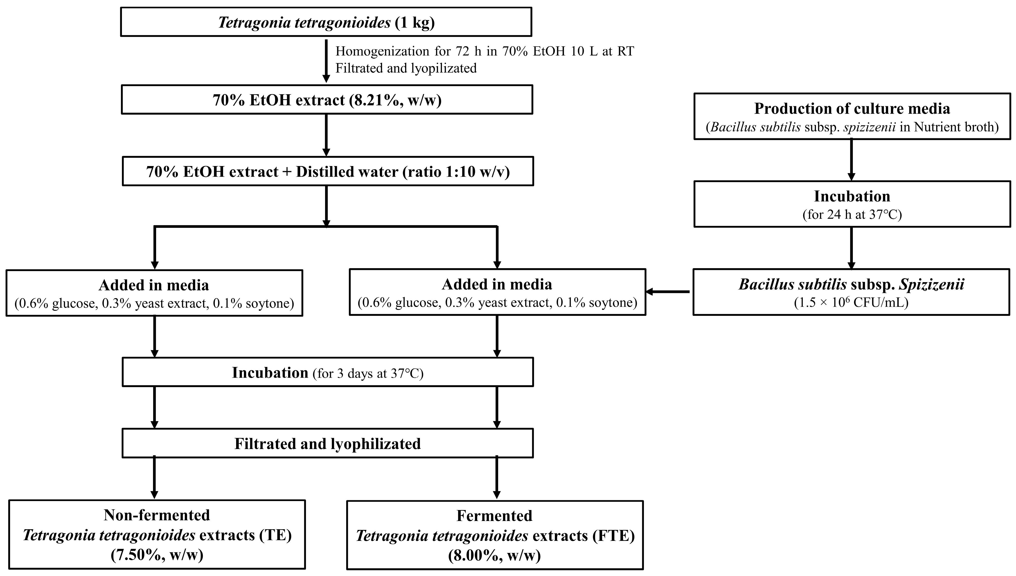
Figure 1. The preparation process for TEs and FTEs from T. tetragonioides.
Fermentation 09 00800 g001
Figure 2. The total polyphenol, flavonoid, and vanillic acid contents in the TEs and FTEs. (A) The TPC was analyzed as the mg gallic acid equivalent (GAE)/g of the TEs and FTEs. (B) The TFC was analyzed as the mg quercetin equivalent (QE)/g of the TEs and FTEs. (C) The results of the quantitative analysis of vanillic acid in the TEs and FTEs. Different letters indicate differences significant at p < 0.05, according to Duncan’s multiple range test.
Fermentation 09 00800 g002
Figure 3. The values for the RC50 of (A) DPPH and (B) ABTS radical-scavenging ability of the TEs and FTEs. The data are expressed as mean ± standard deviation (n = 3). Different letters indicate significant differences at p < 0.05, according to Duncan’s multiple range test.
Fermentation 09 00800 g003
Figure 4. The effect of the TEs and FTEs on cell viability and on LPS-induced nitric oxide production in RAW 264.7 cells. (A) An MTT assay was performed to analyze cell viability after treatment with TE or FTE (6.25, 12.5, 25, 50, 100, and 200 μg/mL) for 24 h. (B) RAW 264.7 cells were treated with different concentrations of each sample and LPS (100 ng/mL) for 24 h, and the NO content in the supernatant was measured with Griess reagent. All data are expressed as the mean ± SD of the experiment. # p < 0.05 compared to the control group; * p < 0.05 and ** p < 0.01 compared to the LPS control group.
Fermentation 09 00800 g004
Figure 5. The effect of FTEs and dexamethasone on LPS-induced COX-2, iNOS, and TNF-α expression in RAW 264.7 cells. (A) Western blotting using β-actin as a loading control was performed for the analysis of whole cell lysates. (B) The quantification of the band intensity ratios of COX-2, (C) iNOS, and (D) TNF-α relative to β-actin. The data are presented as the mean percentage compared to DW-treated cells. All data are expressed as the mean ± SD for the experiment. # p < 0.05 compared to the control group; * p < 0.05, ** p < 0.01, and *** p < 0.001 compared to the LPS control group. See also Figure S1.
Fermentation 09 00800 g005
Figure 6. The effect of FTEs on cell viability in HaCaT cells. An MTT assay was performed for analysis of cell viability after treatment with FTEs (10, 50, 100, and 200 μg/mL) for 24 h. All data are expressed as the mean ± SD for the experiment.
Fermentation 09 00800 g006
Figure 7. The effects of FTEs on the mRNA levels of FLG, HAS-1, and HAS-3. HaCaT cells were treated with FTEs (10, 50, and 100 μg/mL) for 24 h. The RNA was isolated, and (A) RT-PCR analysis was performed to determine the mRNA expression of FLG, HAS-1, and HAS-3. GAPDH was used as an internal control. (BD) The relative mRNA levels of FLG, HAS-1, and HAS-3 were quantified using the Image J program. All data are expressed as the mean ± SD for the experiment. *** p < 0.001 compared to the control group.
Fermentation 09 00800 g007
Table 1. Conditions for the HPLC analysis of the vanillic acid content in the TEs and FTEs.
InstrumentConditions
ColumnAgilent ZORBAX SB-C18
(4.6 mm × 250 mm, 5.0 µm, Agilent)
Column temp.27 °C
Mobile phase
(Gradient)
Time (min)A (1)B (2)
01000
451000
556040
652080
701000
DetectorShimadzu, SPD-M20A Diode Array Detector (334 nm)
Flow rate1.0 mL/min
Injection volume10 μL
Run time70 min
(1) 1% Acetic acid. (2) Methanol.
Table 2. The sequences of primers used in this study.
GeneSequences (5′ to 3′)
FLGFAGGGAAGATCCAAGAGCCCA
RACTCTGGATCCCCTACGCTT
HAS-1FCCACCCAGTACAGCGTCAAC
RCATGGTGCTTCTGTCGCTCT
HAS-3FTATACCGCGCGCTCCAA
RGCCACTCCCGGAAGTAAGACT
GAPDHFGCACCGTCAAGGCTGAGAAC
RATGGTGGTGAAGACGCCAGT
F: forward; R: reverse.
Disclaimer/Publisher’s Note: The statements, opinions and data contained in all publications are solely those of the individual author(s) and contributor(s) and not of MDPI and/or the editor(s). MDPI and/or the editor(s) disclaim responsibility for any injury to people or property resulting from any ideas, methods, instructions or products referred to in the content.