Article

Development of Non-Dairy Synbiotic Fruit Beverage Using Adansonia digatata (baobab) Fruit Pulp as Prebiotic

by
Patience T. Fowoyo
1,*,
Samuel T. Ogunbanwo
2,
Oluwatoyosi O. Popoola
2 and
Paulina O. Adeniji
3
1
Life Sciences Concentration, SOKA University of America, Aliso Viejo, CA 92656, USA
2
Department of Microbiology, University of Ibadan, Ibadan 200005, Nigeria
3
Department of Tourism Studies, Redeemer’s University, Ede 232101, Nigeria
*
Author to whom correspondence should be addressed.
Fermentation 2022, 8(12), 673; https://doi.org/10.3390/fermentation8120673
Submission received: 7 September 2022 / Revised: 8 October 2022 / Accepted: 17 October 2022 / Published: 25 November 2022
(This article belongs to the Section Fermentation for Food and Beverages)

Abstract

:
Probiotics improve gut health; however, their intake through diet is mainly in the form of dairy products, which represents a challenge to lactose-intolerant individuals and vegetarians. This study aimed to determine the prebiotic potential of baobab and to evaluate the potential of using fermented baobab-based beverages as functional foods. The prebiotic content of baobab fruit pulp was determined. Lactic acid bacteria (LAB) were isolated from raw milk samples, identified through phenotypic and molecular methods, and evaluated for their probiotic potential. Three potential non-dairy synbiotic functional beverages using baobab fruit pulp fermented with potential probiotic Limosilactobacillus fermentum and mixed with milk, water, and apple juice separately were produced. The growth and survival of probiotic L. fermentum in the beverages at room (25 °C) and refrigeration (4 °C) temperatures for 3 weeks were determined. Baobab fruit pulp contained phytochemicals, vitamins, fatty acids, inulin, and fructooligosaccharides. Sequence alignment of the LAB isolates identified homologous sequences of Lacticaseibacillus casei, Limosilactobacillus fermentum, Lactiplantibacillus plantarum, Lentilactobacillus buchneri, and Lactiplantibacillus pentosus with 97.2–98.5% similarity. All the lactic acid bacteria did not produce DNAse and gelatinase enzymes, exhibited antagonistic activity against test pathogenic organisms, and demonstrated tolerance to bile salt, simulated gastric juice, and acid. The viability of L. fermentum increased from an initial inoculum size of 106–108 CFU/mL in the baobab-based beverages and remained constant at 108 CFU/mL both at room and refrigeration temperatures. However, after three weeks, the viability of L. fermentum in the synbiotic beverages reduced to 107 CFU/mL. Refrigerated synbiotic beverages had more viable L. fermentum cells (8.04–8log10 CFU/mL) than those stored at room temperatures (7.95–7.7log10 CFU/mL) after three weeks of storage. This study has shown that baobab fruit pulp has prebiotic potential and can be used in the production of a non-dairy functional beverage.

1. Introduction

The human gastrointestinal tract comprises indigenous microbiota whose role is to protect the host from exogenous pathogenic infection, increase host nutrients, act as a xenobiotic, metabolize drugs, be involved in immunomodulation, and maintain the structural integrity of the gut mucosal barrier. However, gut dysbiosis occurs due to immune-mediated disorders caused by pathogenic bacteria in the gut, changes in dietary intake, stress, and antibiotic use [1]. When gut dysbiosis occurs, the exogenous intestinal pathogens might secrete harmful toxins that block the epithelial cell function and the host’s metabolic response to cause pathological disorders, including multi-system organ failure, colon cancer, and irritable bowel syndrome [1]. Previous studies have indicated that the overgrowth of pathogenic bacterial populations and the significant decline of health-promoting bacteria play an important role in innate intestinal inflammation and the pathogenesis of the gastrointestinal disease [2,3,4].
A proven way to remedy gut dysbiosis is through intake of diets containing living microorganisms (probiotics) with the same beneficial attributes as the indigenous microbiota. However, for a probiotic to exert its beneficial effect in the host, it has to be taken in through a delivery medium commonly called functional foods [5]. A delivery medium can be in the form of a pharmaceutical drug supplement or in the form of feed/food which, when consumed, can confer the required probiotic benefit on the host [5]. The two major mediums of delivery for probiotics are dairy and non-dairy probiotic carriers. The widely and commonly used carriers for probiotics are dairy products such as milk (fermented, powder) ice cream, yogurt, and cheese, while non-dairy probiotic carriers include fruits, rice, oatmeal, cereal-based products, and fruit juice [6].
Non-dairy probiotic carriers can serve dual roles as prebiotics and probiotic delivery mediums. In 2008, at the 6th Meeting of the International Scientific Association of Probiotics and Prebiotics (ISAPP), prebiotics were defined as a selectively fermented ingredient that results in specific changes in the composition and/or activity of the gastrointestinal microbiota, thus conferring benefit(s) upon host health [7]. Prebiotics also serve as food for probiotics. Non-dairy probiotic carriers serve as excellent functional foods as the challenges of lactose intolerance in the host, refrigeration as a means of preservation of the functional food, and allergies from cholesterol intake are eliminated [8]. Also, non-dairy products, due to their physical structures, establish a conducive environment for probiotics by limiting the adverse environmental conditions of the gastrointestinal tracts [8,9]. Plants contain polysaccharides such as inulin which are prebiotics and are capable of fermenting in the intestinal tract, thus enabling them to interact with probiotics, thereby enhancing the functionality of the organisms [10]. Plants possess oligosaccharides that deliver probiotics directly into the large intestines, thus preventing the digestion of probiotics in the gastrointestinal tract [11]. Prebiotics are present in significant amounts in several edible fruits, vegetables, and cereals and can alter the colonic microflora to a healthy composition by inducing beneficial luminal or systemic effects within the host [1].
Baobab (Adansonia digitata) is a potential source of polysaccharides such as sugar, carbohydrates, pectin (mainly soluble and insoluble fibers), proteins, vitamin C, calcium, and lipids [12]. These polysaccharides possess a prebiotic potential and thus can enhance the growth of probiotics in the large intestine, thus promoting the health of the gastrointestinal tract. The pectin also prevents the binding of pathogens to the intestinal wall, thereby chelating heavy metals [13]. Baobab fruit pulp contains a high concentration of potassium, copper, magnesium, and manganese [12]. Baobab is rich in fiber, low in fat and protein concentration, but contains different amino acids, thus making it ideal for fiber supplements in foods and increasing the nutritional profile of the food. Due to its low-sugar and high-fiber content, it possesses a low glycemic index which allows for easy digestibility and satiation [14]. The fruit pulp also contains stearic, palmitic, and arachidic acids, all of which contribute to its pharmacological use. Baobab is an excellent source of vitamins, micronutrients, and soluble fibers, having pre- and probiotic effects, thus serving as an intestinal regulator in the case of gastric disorders [15,16].
Lactose intolerant individuals interested in consuming foods that confer probiotic benefits can derive such health benefits from non-dairy foods. The development of synbiotic foods using baobab fruit pulp combined with lactic acid bacteria could ameliorate and eliminate the challenge of the inaccessibility of lactose-intolerant persons to functional foods. The prebiotic potential of baobab fruit pulp and its fit as a constituent in the formulation of a functional beverage were evaluated for the first time in this study. This study aimed to produce synbiotic beverages composed mainly of baobab fruit pulp and probiotic lactic acid bacteria (LAB) and to determine its efficacy as a functional food.

2. Materials and Methods

2.1. Sample Collection

Raw milk samples with nutritional content—dry matter (12.70% ± 0.25), non-fat dry matter (7.57% ± 0.39), fat (3.23% ± 0.09), protein (3.56% ± 0.31)—were obtained from a cattle farm in Ibadan, Nigeria using sterile sample bottles and transported in ice packs to the Microbiology Laboratory at the University of Ibadan for analysis. Baobab fruits grown in Ogbomosho, Nigeria were purchased from Molete Market, Ibadan, Nigeria, and identified at the Plant Anatomy herbarium of the Botany Department at the University of Ibadan as Adansonia digitata Linn belonging to the family Bombacaceae Yori Osi.

2.2. Isolation and Purification of Lactic Acid Bacteria

The method of Misganaw and Teketay [17] was employed in the isolation of LAB from milk samples. Each of the milk samples (1 mL) was homogenized in sterile, distilled water. Tenfold serial dilutions of the homogenized milk samples were carried out. Using the pour plate technique, 1 mL of the selected dilution factor (10−4) of each sample was inoculated unto previously prepared MRS agar (Oxoid Limited, Basingstoke, Hants, UK) in duplicates and incubated microaerophilically at 37 °C for 24–48 h. Pure cultures were obtained by repeated streaking of distinct representative colonies and characterized morphologically. The pure isolates were preserved on MRS agar slants at 4 °C.

2.3. Phenotypic and Molecular Characterization of Isolates

Pure isolates on MRS agar were characterized by examining their colonial morphology such as color, shape, edge, and consistency. The isolates were then subjected to microscopic and biochemical tests for further characterization and identification [18]. The following tests were carried out: Gram staining by Oyeleke and Manga [19], spore staining by Fawole and Oso [20], catalase testing by Oyeleke and Manga [19], motility testing by Shields and Cartcath [21], indole testing by MacFaddin [22], growth at different concentrations of sodium chloride (NaCl) by Ogundare et al. [23], oxidase testing by MacFaddin [22], gelatin hydrolysis testing by Leboffer and Pierce [24], carbohydrate fermentation testing by Harrigan and McCance [25], and starch hydrolysis testing by Seeley and Van Demark [26].
The method of Iruene et al. [27] was employed for the molecular characterization of the isolates. Genomic DNA was extracted from overnight cell cultures grown in MRS broth using a Quick-DNA™ Fungal/Bacterial Miniprep Kit (Zymo Research, Irvine, CA, USA) according to the manufacturer’s protocols. The final DNA concentration and purification were determined using an Epoch™ microplate spectrophotometer (Agilent Biotek, Santa Clara, CA, USA) and DNA quality was checked by 1% agarose gel electrophoresis. Polymerase chain reaction amplification of the 16S rRNA gene for presumptive LAB strains was carried out using bacterial universal primers—27 F:5′-AGA GTT TGA TCC TGG CTC AG-3′ and 1492 R:5′ GGT TAC CTT GTT ACG ACT T-3′. A polymerase chain reaction was performed in a 50 µL reaction containing 25 µL One Taq® 2X Master Mix with standard buffer, 1 µL forward primer, 1 µL reverse primer, and 22 µL RNase free water. Then 49 µL of the mixture was added into a sterile polymerase chain reaction tube, and 1 µL of gDNA was added and used as a template. The conditioning of the amplified gene fragment: pre-denaturation of the target DNA at 96 °C for 4 min followed by 30 cycles at 94 °C for 1 min, primer annealing at 51.5 °C for 1 min and 30 s, and primer extension at 68 °C for 8 min. Polymerase chain reaction was completed with 10 min elongation at 68 °C followed by cooling to 4 °C. The reactions were carried out in a 96 well thermal cycler—Applied Biosystems GeneAmp™ Polymerase chain reaction 9700 (Applied Biosystems, Thermofisher Scientific, Waltham, MA, USA). The size of the 16S rRNA gene polymerase chain reaction products was confirmed by electrophoresis on a 1% (w/v) agarose gel stained with GelRed and visualized using a Uvitec Uvisave Q9 gel documentation system (Uvitec, Cambridge, UK). Polymerase chain reaction products were purified using the QIAquick Polymerase chain reaction purification Kit (Qiagen, Hilden, Germany) according to the manufacturer’s instructions. The purified amplicons were sequenced using an automated DNA sequencer (ABI model 3350; Applied Biosystems) at Inqaba Biotech, Nigeria. The sequences of the bacterial isolates of this study were then compared to those in GenBank (National Centre for Biotechnology Information; https://blast.ncbi.nlm.nih.gov/Blast.cgi accessed on 15 July 2021) using the Basic Local Alignment Search Tool for nucleotide sequences (blastn).

2.4. Determination of Probiotic Properties of Lactic Acid Bacteria Isolates

All Gram-positive, catalase-negative, and non-spore-forming isolates were identified as lactic acid bacteria and screened for their probiotic potential.

2.4.1. Acid Tolerance of Lactic Acid Bacteria Isolates

The method employed by [28] was used in determining the acid tolerance of LAB isolates. The overnight broth culture of test isolates was serially diluted, and 1 mL of the seventh dilution factor was inoculated into MRS broth (Oxoid Limited, Basingstoke, UK) with the pH adjusted to 2.5 and 3.0 using concentrated hydrochloric acid. The survival of LAB was quantified using plate count on MRS agar after incubating microaerophilically at 37 °C at intervals of 0 and 4 h.

2.4.2. Bile Salt Tolerance of LAB Isolates

Tolerance to bile salts was carried out according to the method of Klingberg et al. [28]. MRS broth was supplemented with bile salt (Oxoid Limited) at concentrations of 0.3%, 0.5%, and 1% (w/v), and MRS broth without bile salt was used as the control. LAB isolates (18 h old) were inoculated into supplemented and non-supplemented MRS broth and incubated anaerobically at 37 °C for 0 and 4 h. The survival rate was determined using the pour plate method on MRS agar in duplicates.

2.4.3. Antagonistic Activity of LAB Isolates

Salmonella enterica ssp. enterica sv. Typhi, Pseudomonas aeruginosa, Listeria monocytogenes, Staphylococcus aureus, and Klebsiella pneumoniae were the pathogenic (test) microorganisms collected from University College Hospital (UCH), Ibadan. The LAB isolates were inoculated into sterile MRS broth and incubated microaerophilically at 37 °C for 48 h. The culture was centrifuged at 3260× g for 15 min using Sorvall™ ST 8 Small Benchtop Centrifuge (Thermofisher Scientific, Waltham, MA, USA), and the supernatant was filtered. The overnight broth culture of the pathogenic organisms was standardized to 0.5 McFarland standard and inoculated on Mueller Hinton agar (Oxoid Limited). Using the agar well diffusion method, a 5 mm cork borer was used to bore wells in the agar, and 0.5 mL of the filtered supernatant was dispensed into the wells and allowed to diffuse into the agar for 1 h. The plates were incubated at 37 °C for 24 h and the diameters of the zone of inhibition were measured as described by Nami et al. [29].

2.4.4. Tolerance of LAB Isolates to Simulated Gastric Juice

The viability of the organism in the presence of gastric juice was determined by suspending 3 mg/mL of pepsin (Thermofisher Scientific) in a sterile 0.85% saline solution adjusted to pH 2.5. The simulated gastric juice was inoculated with an 18–24 h-old culture of the test organism and incubated microaerophilically at 37 °C for 4 h. The viable cell population was determined on MRS agar using the pour plate method by Tokatli et al. [30].

2.5. Safety Assessment Tests on Lactic Acid Bacteria Isolates

DNase test, gelatinase test, and hemolytic activity test were carried out to determine the pathogenicity of the LAB isolates, which is a criterion in the selection of potential probiotics.

2.5.1. DNase Test

The protocol by UK Standards for Microbiology Investigation Protocol [31] was used. The overnight culture of each LAB isolate was inoculated on DNase agar (Oxoid Limited) and incubated at 37 °C for 24 h. The plates were flooded with concentrated HCl, left to stand for a few minutes, and then excess HCl was removed. Observation of clear zones around the colonies indicates a positive result.

2.5.2. Gelatinase Test

The method of [24] was used. Nutrient gelatin medium (Himedia, Mumbai, India) in test tubes was inoculated with an overnight culture of each test isolate and incubated at 37 °C for 24 h. After incubation the tubes were refrigerated. Partial or total liquefaction of the gelatin agar indicates a positive result.

2.5.3. Hemolytic Activity

Hemolytic activity was determined by employing the method described by [32]. Tryptic soy agar with 5% sheep blood agar plates (Carolina Biological Supply Company, Burlington, NC, USA) was inoculated with an overnight culture of LAB isolates and incubated anaerobically for 48 h. The plates were examined for clear, greenish, or no zones of clearance which depicted beta, alpha, and gamma hemolysis, respectively.

2.6. Preparation of Baobab Fruit Pulp

Baobab fruit pods were opened, and the pulp was separated from the seed. The pulp was pounded in a clean and surface-sterilized mortar and pestle before blending in a sterile blender into fine powder according to the UK Standards for Microbiology Investigation Protocol [31].

2.7. Phytochemical Screening of Baobab Fruit Pulp

Qualitative determination tests of tannin, phlobatannin, saponin, flavonoid, steroid, terpenoid, cardiac glycoside, cardenolide, alkaloids, anthraquinone, chalcones, and phenol were carried out employing the method described by Sofowora [33]. For quantitative determination of the phytochemical constituent of baobab pulp powder, the standard procedures outlined by AMC-RSC [34] and AOAC [35] were used.

2.8. Determination of Fatty Acid Content of Baobab Fruit Pulp

The fatty acid content was determined as described by AOAC [36]. The sample (2 g) was weighed into a 100 mL conical flask, and 20 mL of benzene was added, shaken thoroughly to extract all the fatty acids. The mixture was transferred into a 250 mL separatory funnel to separate the benzene extract from the aqueous extract. A 5 mL aliquot of the benzene extract was pipetted into a 15 mL test tube, and 2 mL of 10% copper acetate was added to develop a blue color. Standard concentrations of each fatty acid were prepared. The absorbance of the sample extract and the standard solutions at different concentrations were determined using Genesys™ 10S UV-Vis spectrophotometer (Thermofisher Scientific) at a wavelength defined for each fatty acid: lauric acid (640 nm), stearic acid (650 nm), palmitic acid (630 nm), arachidonic (690 nm), oleic acid (670 nm), linoleic acid (660 nm), linolenic acid (680 nm), ricinoleic acid (610 nm), dihydroxy stearic acid (655 nm).
The percentage of each fatty acid was obtained using the formula:
%   Fatty   acid = Absorbance   of   sample   ×   Gradient   factor   of   specific   fatty   acid   ×   Dilution   Factor Weight   of   Sample   × 10000

2.9. Determination of Vitamin Content of Baobab Fruit Pulp

The vitamin A, B, and C content of baobab fruit pulp was analyzed according to the standard method adopted by AOAC [37].

2.10. Determination of Mineral Content of Baobab Fruit Pulp

Minerals including the calcium, iron, sodium, and magnesium contents of baobab pulp were determined according to AOAC [38] standards.

2.11. Proximate Analysis of Baobab Fruit Pulp

The nutritional composition of baobab fruit pulp was analyzed to determine the crude protein, ash, ether extract, crude fiber, dry matter, and carbohydrate content using the method of AOAC [35].

2.12. Preparation of Baobab Fruit Pulp Beverages

Three (3) baobab fruit beverages (AM, AA, and AW) were produced. AM was prepared using baobab fruit pulp and milk in a ratio of 60:40. Baobab fruit pulp (60 g) was added to 40 mL of milk, and the mixture was blended. AA comprised of the baobab fruit pulp and apple juice in the ratio of 60:40. Baobab fruit pulp powder (60 g) was mixed with 40 mL of apple juice. The mixture was blended. AW is a 100% w/v mixture of baobab fruit pulp and deionized water. These beverages were fermented using the selected probiotic test organism (L. fermentum).

2.13. Preparation of Synbiotic Baobab Fruit Beverage

L. fermentum exhibited the best probiotic attributes, such as tolerance to low pH, bile salt tolerance, survival in gastric juice of pH 2.5, and antagonistic activity against selected pathogenic organisms, out of the five LAB species isolated from the milk samples and was selected as the choice species in producing the synbiotic beverages. The method of Johanningsmier et al. [39] was employed to prepare the inoculum concentration of the probiotic. A single colony of L. fermentum was inoculated into sterile MRS broth and incubated at 37 °C for 24 h. It was centrifuged at 3260× g for 10 min, the supernatant was decanted, and the pellets were washed thrice with sterile 0.85% normal saline; harvested cells were resuspended into 9 mL of 0.85% normal saline solution. Inocula count was determined using the pour plate method by incubating at 37 °C for 48 h. Viable counts were reported in CFU/mL. The beverages were pasteurized and inoculated with 1 mL (comprising of 6.3 × 10⁶ cells) of L. fermentum and incubated at 30 °C for 48 h for fermentation.

2.13.1. Determination of Growth Rate of L. fermentum in the Synbiotic Fruit Beverages

Over a 48 h period, the rate of growth of L. fermentum in the synbiotic beverages was determined at 12 h intervals using the pour plate technique. Each beverage (1 mL) was inoculated onto MRS agar plates and incubated at 37 °C for 48 h. Colonies that developed were enumerated as described by Mpofu et al. [40].

2.13.2. Determination of Total pH and Titratable Acidity of Synbiotic Fruit Beverages

The pH was determined using a Jenway 3520 digital pH meter (Jenway®, Staffordshire, OSA, UK). The total titratable acidity was determined as described by AOAC [41], 10 mL of each fruit juice was titrated against 0.1N NaOH using phenolphthalein as an indicator. Each ml is equivalent to 90.08 mg of lactic acid, and the acid equivalent is the volume of NaOH utilized. The titratable acidity was calculated as:
Volume   of   NaOH   × 0.1   NaOH   × 90.08 10

2.14. Determination of the Prebiotic Potential of Baobab Fruit Beverage

The prebiotic potential of the fruit pulp and beverages was determined using the method of Englyst [42]. The fructooligosaccharides were determined by weighing 1 g of baobab fruit pulp and baobab fruit beverage, respectively, into a 50 mL centrifuge tube; 2 mL dimethyl sulphoxide was added and capped. The mixture was stirred for about 2 min on an RT 2 basic magnetic stirrer (Thermofisher Scientific) to homogenize, and the tube was placed in a beaker of boiling water on a hot plate with a stirrer for 1 h. The tube was removed, without cooling, and 8 mL of sodium acetate buffer at pH 5.2 was added, pre-equilibrated at 50 °C, and vortex mixed using an LP Vortex mixer (Thermofisher Scientific). The tube was left at room temperature at about 35 °C until the content cooled to between 30 °C and 40 °C. α-amylase solution (0.5 mL) followed by 0.1 mL of sucrase borohydride solution was added and vortex mixed. The tube was capped and incubated for 16 h with continuous mixing for the first 1 h. Ethanol (40 mL) was added, and the tube was inverted and left for 1 h at room temperature. The mixture was centrifuged at 3260× g for 10 min, and the supernatant was removed by decantation or aspiration without disturbing the residue. The residue was washed twice using 50 mL 85% ethanol each time and mixed by inversion on a magnetic stirrer to suspend the residue and supernatant removed as before. Acetone (40 mL) was added to wash the residue, stirred for 5 min, and centrifuged at 3260× g for 10 min. The supernatant liquid was removed by aspiration and discarded. The residue in the tube was placed in a beaker of water at 65–75 °C on a stirrer hot plate with continuous stirring of the content until the residue appeared dry.
%   Fructooligosaccharide   = Weight   of   residue   Weight   of   sample × 100
Inulin content was determined by weighing 1 g of baobab fruit pulp and baobab fruit beverage, respectively, into a 100 mL volumetric flask, and 1 mL of 95% ethanol was added to wet the sample. About 10 mL of 0.5 M KOH was added and the mixture was held overnight at room temperature. The mixture was diluted to 100 mL with distilled water and again held overnight at room temperature. An aliquot (5 mL) of the diluted solution was pipetted out of the mixture into another 100 mL volumetric flask, and three drops of 0.1% phenolphthalein solution were added. The resulting solution was neutralized using 1 M HCl dropwise until neutral pH was achieved. Two milliliters of 0.2% sodium borohydride solution in 2% KI were added to the neutralized solution and made to volume with distilled water. Standard solutions of inulin in the range 0–10 ppm were prepared from 100 ppm stock solution. Inulin solution was subjected to the same treatment as the test sample. The absorbance of the sample and the standard solutions of different concentrations taken after 30 min of adding 0.2% sodium borohydride solution was determined in the spectrophotometer at a wavelength of 628 nm.
%   Inulin   = Absorbance   of   sample   ×   gradient   factor   ×   dilution   factor 10000

2.15. Estimation of the Shelf Life of Synbiotic Beverages

The shelf life of the synbiotic beverages was determined for 3 weeks at refrigeration temperature of 4 °C (RF) and room temperature of 25 °C (RT).

2.16. Statistical Analyses

Triplicate samples were analyzed. Data was expressed as mean value ± standard deviation. Statistical analysis was performed using a two-way analysis of variance (ANOVA) with the value of p < 0.05 considered significant. SigmaPlot® v12.5 (SYSTAT Software, Inc.,San Jose, CA, USA) was used for statistical data analysis.

3. Results

3.1. Occurrence of Lactic Acid Bacteria in Milk Samples

Twenty-three (23) LAB isolates were obtained from milk samples. The morphological and biochemical characteristics of the isolates showed that all were Gram-positive and tested negative for catalase, oxidase, starch hydrolysis, and gelatin liquefaction. The isolates were non-motile, non-spore formers, and fermented specific sugars (Table 1).
Sequences checked against the blastn database revealed similar sequences of between 97.2% and 98.5% similarity, as shown in Table 2. Five of the presumptively identified isolates were selected with given codes and GENBANK accession numbers YMI (NZ_AP012544), 16 (CP035054), 29VI (NZ_CP023174), 2 (GCA_000298115), and 27RI (NZ_CP022130), were closely affiliated to those in the database as demonstrated by the databank algorithm, and were identified as Lacticaseibacillus casei, Limosilactobacillus fermentum, Lactiplantibacillus plantarum, Lentilactobacillus buchneri, and Lactiplantibacillus pentosus.
Lactiplantibacillus plantarum (52%) was the highest occurring lactic acid bacteria isolated from the milk samples while Lacticaseibacillus casei (4%) had the least occurrence, as presented in Figure 1.

3.2. Probiotic Potential of Lactic Acid Bacteria Isolates

The LAB isolates (40–70%) tolerated the low pH of 2.5. L. fermentum (72%) had the highest survival rate while L. buchneri (40%) had the least survival rate (Figure 2). At pH 3.0, L. fermentum (80%) still had the highest survival rate, as shown in Figure 3.
The percentage of LAB isolates that survived different concentrations of bile salt (0.3%, 0.5%, and 1%) ranged from 40.7% to 93.3%. The percentage of L. casei that survived 0.3%, 0.5%, and 1% bile salt concentrations after 4 h were 59.3%, 50%, and 40.7%, respectively, while L. fermentum had the highest percentage survival rates of 85%, 70.6%, and 64.3% in 0.3%, 0.5%, and 1% bile salt concentrations, respectively, after 4 h of incubation (Figure 4, Figure 5 and Figure 6).
All LAB isolates tolerated simulated gastric juice at pH 2.5 after 4 h of incubation, with survival rates ranging between 64–90%. L. casei had the lowest survival rate at 64%, and L. fermentum had the highest survival rate at 90% (Figure 7).
L. fermentum exhibited antagonistic activity against all the pathogens. L. casei and L. pentosus did not inhibit Listeria monocytogenes and Klebsiella pneumoniae, while L. buchneri and L. plantarum had no inhibitory effect on Listeria monocytogenes (Table 3).

3.3. Safety Assessment Tests

All of the LAB isolates tested were non-hemolytic and did not produce DNase and gelatinase enzymes.

3.4. Phytochemical Analysis of Baobab Fruit Pulp

Baobab fruit pulp contained saponin, tannin, alkaloids, anthraquinone, glycosides, and phenols in appreciable quantity; phlobatannin and flavonoids were present in moderate amounts while steroids, terpenes, chalcones, and cardenolides were available in trace quantities (Table 4).
Linoleic (15.88%) and oleic acids (11.94%) were the predominating fatty acids in the baobab fruit pulp (Table 5).
Baobab fruit pulp contained vitamins A, B, and C, with vitamin C having the highest value at 224.65 mg/100 g (Table 6). Among all of the minerals, the calcium content was the highest with a value of 610 mg/100 g. Magnesium (280 mg/100 g), sodium (200 mg/100 g), and iron (18 mg/100 g) were present in lesser amounts (Table 6).
The proximate composition of baobab fruit pulp was protein (3.85%), ash (7.20%), fats (2.70%), crude fiber (3.40%), dry matter (93.07%), and carbohydrates (75.92%) (Table 7).
The nutrient composition of baobab beverages revealed protein (5.25%), ash (2.60%), crude fiber (0.90%), dry matter (74.15%), moisture content (25.85%), and carbohydrate (85.99%) as presented in Table 8.
The fructooligosaccharide (FOS) and inulin contents of baobab fruit pulp were (0.77% + 0.02) and (0.81% + 0.02), respectively.

3.5. Growth Rate of Probiotic L. fermentum in Synbiotic Beverages

L. fermentum grew in the beverages over a 48 h period with an increased survival rate. Microbial cell concentration of 8.12 log10 CFU/mL, which is the highest, was observed in AA and AM beverages (Figure 8).
The pH of the synbiotic beverages declined as time progressed from 0–48 h. The initial pH of the AA beverage was 5.86, but it decreased to 5.12 after 48 h. AM had an initial pH of 5.94 and final pH of 5.16, while the AW beverage had an initial and final pH of 6.01 and 5.07, respectively, after 48 h (Figure 9).
Figure 9. Effect of L. fermentum on the pH of the synbiotic beverages. The acidity of the beverages increased as time progressed. AW and AA beverages had the highest acid content of 3.60 mg/mL after 48 h (Figure 10). Key: AM—baobab fruit pulp and milk beverage; AA—baobab fruit pulp and apple juice beverage; AW—baobab fruit pulp and apple juice beverage.
Fermentation 08 00673 g009
Figure 10. Effect of L. fermentum on the total titratable acidity (TTA) of the beverages. The acidity of the synbiotic beverages increased after 48 h. AW and AA beverages had the highest acid content of 3.60 mg/mL after 48 h. Key: AM—baobab fruit pulp and milk beverage; AA—baobab fruit pulp and apple juice beverage; AW—baobab fruit pulp and apple juice beverage.
Fermentation 08 00673 g010

3.6. Effect of Storage Conditions on the Growth Rate of Potential Probiotics in the Synbiotic Beverages

L. fermentum was able to survive in the fruit drinks when stored at refrigeration and room temperatures for three weeks. After 7 days, L. fermentum in the synbiotic beverages demonstrated the highest survival rate as compared to after 14 and 21 days (Figure 11). However, the survival rate of L. fermentum in the refrigerated beverages was higher than those stored at room temperature.

3.7. Influence of Probiotic L. fermentum on the Chemical Properties of the Synbiotic Beverages

The pH of the beverages was determined after three weeks at room temperature and refrigeration temperature of 4 °C. There was a decline in the pH of the beverages after three weeks under both storage conditions; however, the reduction in pH of the beverages stored at room temperature was more than those refrigerated. The pH of AM ranged between 3.80–4.37. AA ranged between 3.70–4.40, while AW ranged between 4.30–4.58 at room temperature. After three weeks of storage at refrigeration temperature, the pH of AW beverage declined from 4.66 to 4.49, AM from 4.37 to 3.80, and AA reduced from 4.40 and 3.70 (Table 9).
The acidity of the synbiotic beverages increased under both storage conditions after three weeks however the acidification rate at room temperature was higher than at refrigeration temperature. AA had the highest acidic content (4.95 mg/mL) at room temperature, while AM had the highest acidity of 4.37 mg/mL after three weeks of storage.

3.8. Prebiotic Composition of Synbiotic Beverages

A decrease in the prebiotic content of the synbiotic beverages stored at room and refrigeration temperatures was observed after three weeks of fermentation. However, it was observed that the fructooligosaccharides, inulin, and lactate content of the synbiotic beverages stored at refrigeration temperature was higher than those stored at room temperature (Table 10).

4. Discussion

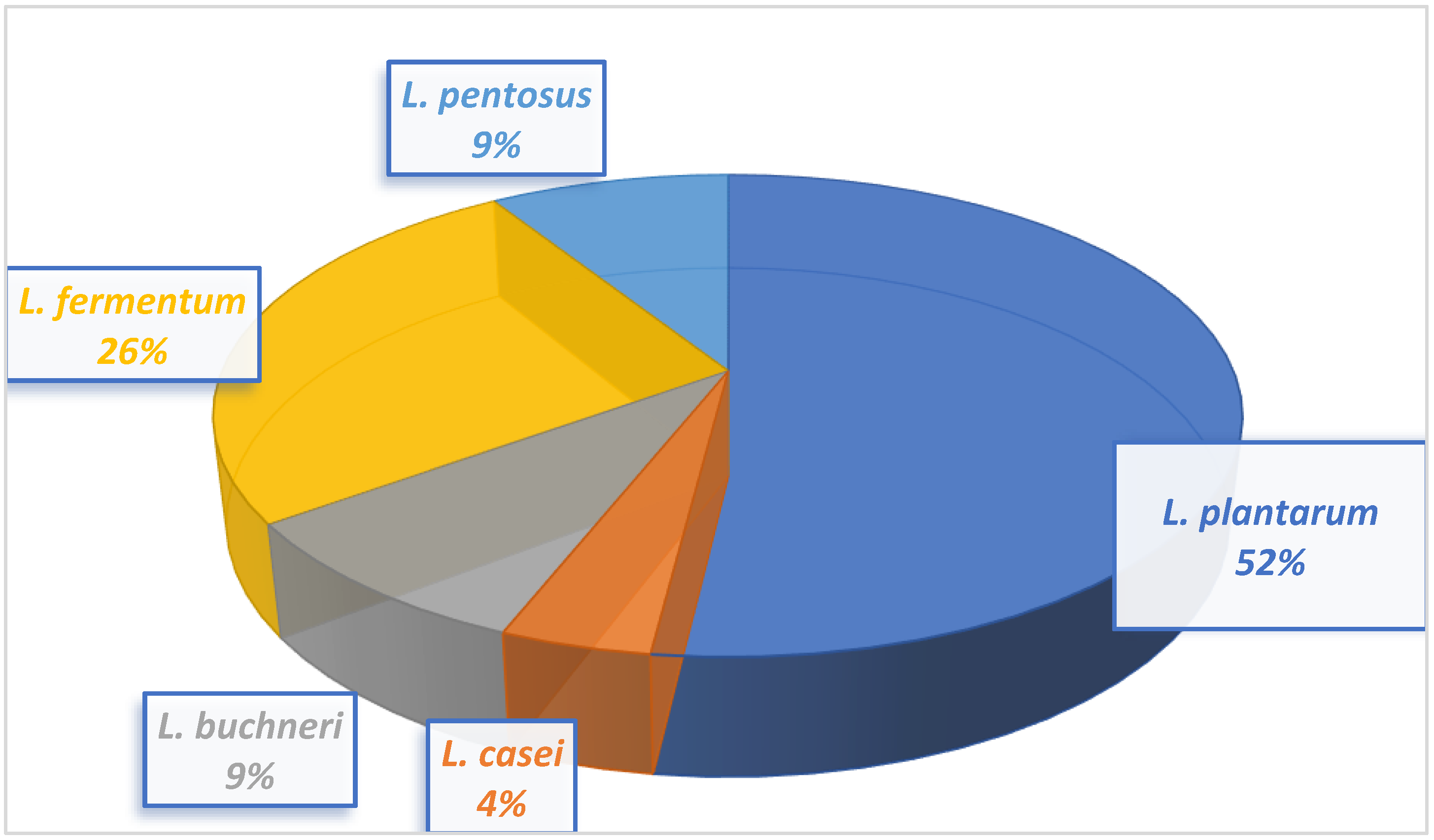
Twenty-three lactic acid bacteria isolated from milk samples were identified as L. buchneri, L. casei, L. fermentum, L. plantarum, and L. pentosus. The morphological and biochemical characteristics of the LAB isolates conform to that of Bergey’s Manual of Determinative Bacteriology [43]. The 16S rRNA gene sequencing revealed the isolates to be closely related to Lacticaseibacillus casei, Limosilactobacillus fermentum, Lactiplantibacillus plantarum, Lentilactobacillus buchneri, and Lactiplantibacillus pentosus.
Lactobacillus plantarum had the highest occurrence at 48%, and Lactobacillus casei had the lowest occurrence at 4%.
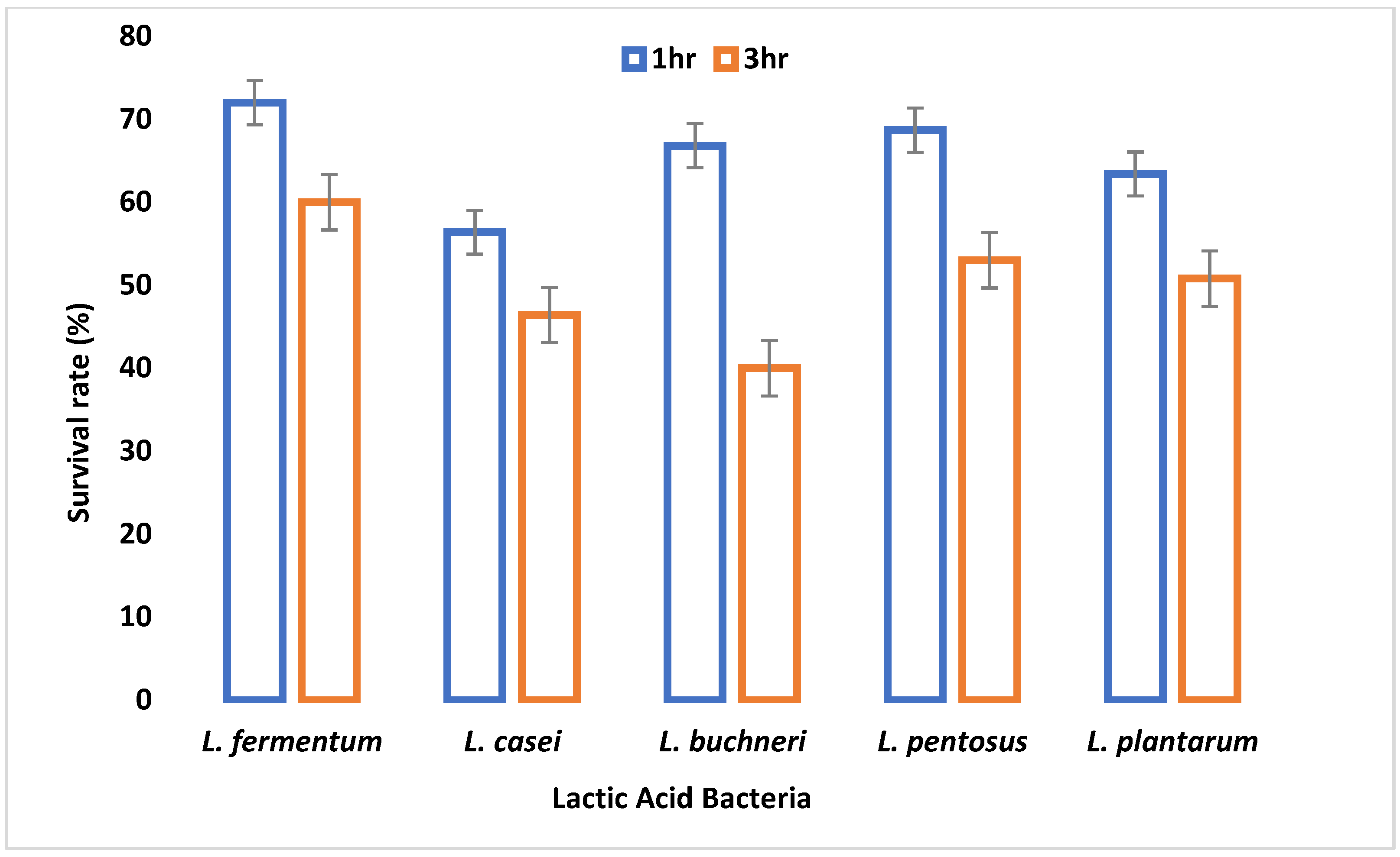
A major criterion used in probiotic selection is the ability to withstand acidity which invariably depicts its inability to inactivate due to the low pH of the gastrointestinal tract. All LAB isolates were able to survive at pH 2.5 and 3.0, although their viability started to decrease after 3 h of incubation. This demonstrates their potential to survive in the extremely acidic pH of the gastrointestinal tract. A similar study by Nami et al. [29] showed a decrease in the viability of the LAB isolates after 3 h of incubation at pH 3.0. All the organisms had a survival rate above 50% after 1 and 2 h of incubation. After 3 h of incubation, only 67% of the LAB isolates showed a viability of more than 50%. This agrees with the work of Hoque et al. [44] where all the Lactobacillus species isolated from yogurt were able to survive an acidic pH of 2.5 to 3.5. L. fermentum demonstrated the highest capacity to withstand low pH among the isolates tested. The adaptive acid tolerance reaction could be controlled by the production of some genes during adaptation, which comprises two different responses, pre-challenge adaptation and transient adaptation that occurs during low pH challenge.
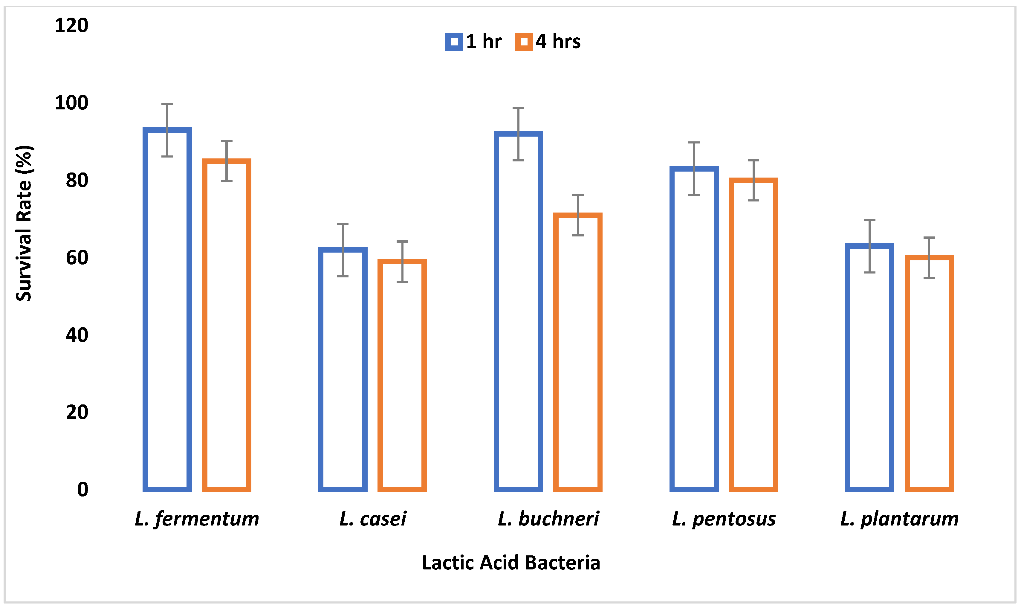
It takes 4 h for food to digest in the intestine [29]. All the isolates were able to resist bile salt concentrations of 0.3%, 0.5%, and 1% in varying degrees for 4 h. The ability to resist the effect of bile salt is an important criterion for probiotic selection, because the mean intestinal concentration of humans is supposedly 0.3% (w/v) [45]. This demonstrates that the LAB isolates met these probiotic criteria, although L. fermentum survived best. As the bile salt concentration increased and time progressed, the viability of the LAB species decreased, and this conforms to the work of Owusu-Kwarteng et al. [46], where all L. fermentum strains isolated from West African fermented millet dough were able to resist 0.3%–2% bile concentration with decreased viability.
Lactic acid bacteria are capable of lowering pH, producing antibacterial substances such as lactic and organic acids [47], and producing bacteriocins capable of inhibiting the growth of pathogenic and spoilage bacteria [48,49]. In this study, the metabolites produced by LAB present in the culture supernatant demonstrated antagonistic activity against the clinical pathogens and this lends credence to their antimicrobial potential.
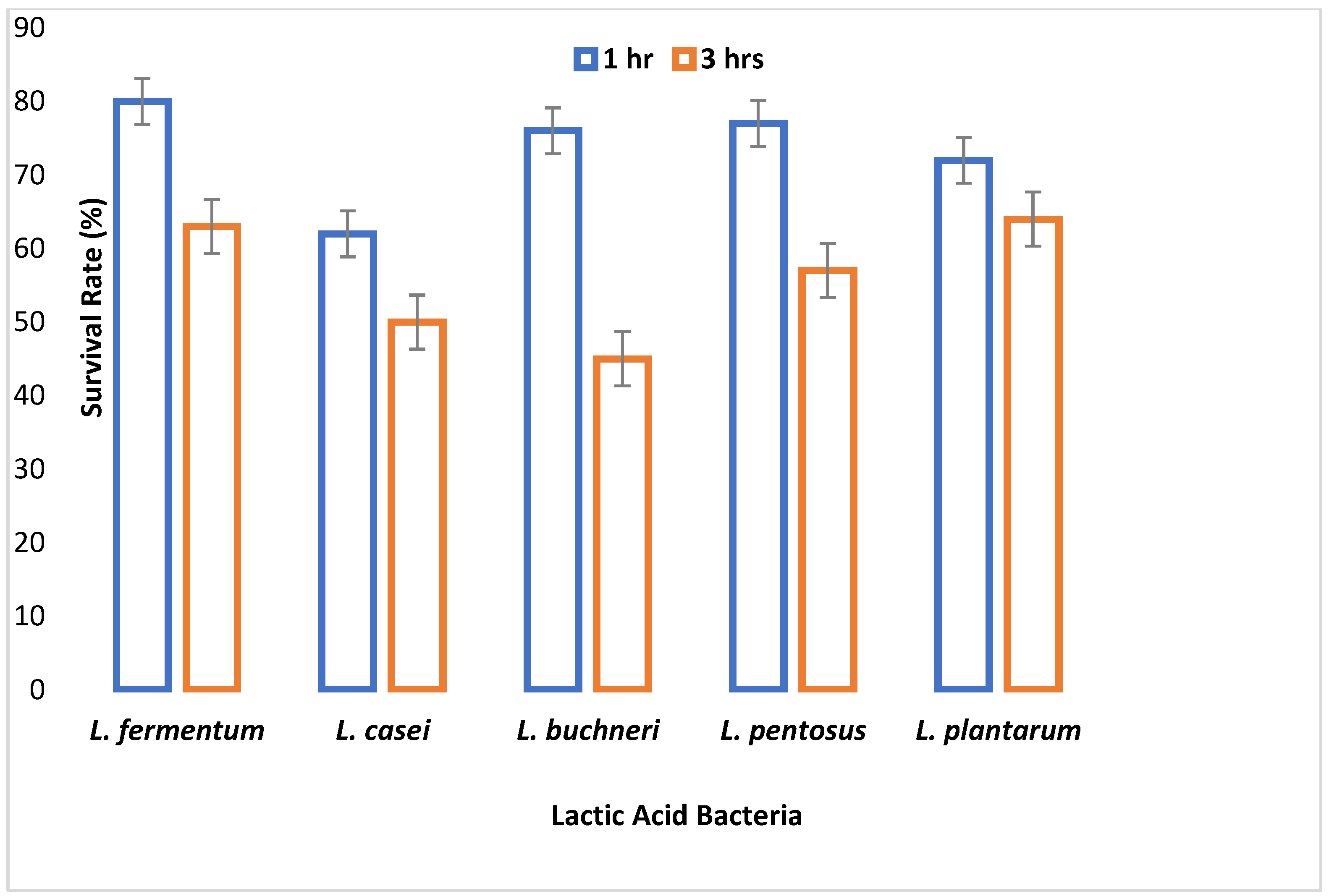
The small and large intestine is colonized by probiotic microorganisms and must be able to survive the harsh conditions therein to be functional. Gastric juice is secreted in the stomach where high acid persists. Therefore, the survival of an organism in gastric juice is a major criterion for probiotic selection. In this study, all tested LAB species were able to survive simulated gastric juice at pH 2.5 with a percentage survival rate ranging between 64% and 90% after 4 h. L. fermentum had the highest survival rate, while L. casei had the least survival rate. Reference [50] reported that the survival rate of Lactobacillus species in gastric juice differs with different strains.
Safety assessment of probiotic organisms is an important factor that must be considered before they are used in producing any functional food [51]. Thus, in the guidelines set by [52], safety assessment is a recommended criterion for choosing probiotics. In this study, the LAB isolates did not liquefy gelatin or produce DNase enzyme and were thereby incapable of hydrolyzing host DNA and unable to lyse red blood cells. The absence of DNase, gelatinase, and hemolytic activity is an indication that the organisms are GRAS organisms and non-virulent, which is a criterion for probiotic selection.
Baobab pulp contains considerable quantities of phytochemicals which makes it suitable as a dietary supplement and valuable for pharmaceuticals. Phytochemical compounds are secondary metabolites of plants that contribute to their use as pharmaceutical, nutritional, and dietary supplements [53]. These components are also referred to as nutraceuticals owing to their health-promoting properties as well as preventive and therapeutic use in the treatment of chronic diseases [54].
In this study, the fruit pulp of baobab contained a substantial amount of oleic and linoleic acid with a moderate amount of linolenic acid; therefore, baobab pulp can serve as a source of omega-3, -6, and -9 supplements and can help in improving health. Polyunsaturated fatty acids (PUFA), which are mainly linoleic and α-linolenic acids, are both synthesized by plants and are essential for the prevention of diseases and good health [55]. Fatty acids are valuable in the pharmaceutical and food industries. Linolenic acids (omega-3 supplements, omega-6 supplements) have been indicated for the prevention and treatment of cardiovascular diseases, improved concentration, memory, motivation, and motor abilities; they are also indicated during pregnancy for the reduction of postpartum, depression, and mood swings as well as improved health after birth [56]. They also serve both preventive and therapeutic purposes in conditions relating to premenstrual syndrome, diabetes, skin problems, and inflammation, to mention but a few [57]. Omega-9 supplements (oleic acid) are useful in increasing the healthy lifestyle of consumers and also help in combating total and bad cholesterol (LDL) and increasing the good ones (HDL), preventing coronary diseases and aging [58].
The amount of vitamin C in baobab pulp was very high (224.6 mg/100 g), and this shows that it is a good source of antioxidants. The vitamin C content in this research is higher and agrees with that of [59] who reported a range of 169–231 mg/100 g. Vitamins play an essential role in human nutrition and health but cannot be synthesized by humans and are therefore taken up in the diet. According to Aluko et al. [60], the daily intake of ascorbic acid in children and adults is about 17 mg/day and 30 mg/day, respectively; thus, baobab fruit can serve as a good source of ascorbic acid.
Minerals are inorganic nutrients required in minute quantity which are needed for the maintenance of physiochemical processes essential to life. Macronutrients (calcium and sodium) and micronutrients (iron and magnesium) are required for normal growth and development [61]. Deficiency of any of these minerals may result in impairment in cognitive performance, predisposition to infections due to lowered immunity, anemia, and many other conditions [62]. In this study, baobab was found to contain a higher amount of calcium and other essential nutrients such as iron, magnesium, and sodium. Consumption of this fruit can serve as a good source of micro and macronutrients.
Adequate nutrition is an important factor of a public health concern as it provides the basis for dietary assessment and formulation of healthier diets before consumption. High amounts of carbohydrates in food serve as a source of energy [63]. The result of this study showed that baobab is rich in carbohydrates (75.92%) which is comparable with the findings of Elmadfa et al. [64] that reported a carbohydrate content of 70.03% in baobab fruit pulp obtained from Adamawa State, Nigeria. Storage conditions, processing procedure, the difference in ripening age, well as the possibility of genetic variation may have been responsible for the result variation.
The human colon harbors about 1010–1012 microbial cells which are mostly anaerobes known as the gut microbiota. Their composition can be modulated by diet variation. Prebiotics are a nutritious, non-digestible group of carbohydrates that can be incorporated into diets so that when fermented by gut microbiota in the gastrointestinal tract, they can provide nutrients to stimulate the growth and activities of the gut microbiota. Carbohydrates in plants such as fructooligosaccharides (FOS) and inulin are the best-known sources of prebiotics. Baobab fruit pulp and the beverages produced in this study contained significant amounts of fructooligosaccharides and inulin. According to the study by Lockett et al. [65], the prebiotic potential of mango, banana, and apple peel powder at 0%, 2%, and 4% concentrations encouraged the proliferation of several test probiotics evaluated. Their study revealed that even at 0% concentration, the growth of the test probiotics increased significantly. This demonstrates that the concentration of prebiotics presents in baobab fruit pulp and beverages as determined in this study will support the growth of probiotics. Thus, they can be used to stimulate the activities of the gut microbiota and also to enhance host health by improving immune function, improving digestion, colonic integrity, and also down-regulate allergenic response [66]. Prebiotics can also improve the uptake of zinc, iron, and calcium and decrease colon cancer and cholesterol.
After inoculation of L. fermentum into the different beverages, pH reduced and acidity increased over time. This increased acidity may be attributed to the production of lactic acid by L. fermentum and conforms to the study by Sharma et al. [67], who also noticed a decrease in the pH of probiotic mutandabota (a traditional food in South Africa produced from baobab fruit) over some time after inoculation with Lactobacillus rhamnosus.
L. fermentum inoculated into the various baobab fruit beverages were able to survive after 48 h. The viability of the probiotic organism increased as time progressed, and this could be due to the presence of additional nutrients in the beverages which were able to support the growth of the organism. Over three weeks, the pH of the synbiotic fruit beverages stored at room and refrigeration temperatures decreased with an increase in acidity and subsequent decrease in viability of L. fermentum, and this may be associated with the increased production of lactic acid by L. fermentum, since fermentation commenced over the storage period. According to Othman et al. [68], lactic acid production is the metabolic end product of LAB fermentation which accumulates over time and inhibits LAB growth due to pH alteration into acidic conditions. L. fermentum in the synbiotic beverages survived best at refrigeration temperature than at room temperature.
Prebiotics are fermentable carbohydrates used by probiotics, and this is significant as it influences the composition and metabolic activities of the gut microbiota [69]. Although these carbohydrates are indigestible in the gastrointestinal tract, they can be broken down by the gut microbiota to produce short-chain fatty acids (SCFA) and lactate [70]. In this study, the test LAB isolates degraded the fructooligosaccharides and inulin content of the fruit beverages after three weeks with the production of lactate, and this may enhance the gut microbiota.

5. Conclusions

This study has shown that baobab fruit pulp can serve as a prebiotic as it contains fructooligosaccharides and inulin. The ability of the potential probiotics to remain viable and to survive in the synbiotic beverage produced has been established in this study. Thus, the synbiotic beverage produced with baobab bulb has the potential to serve as a non-dairy functional food and probiotic delivery medium for lactose intolerant individuals and vegetarians. Further studies may evaluate the in vivo probiotic potential of baobab-based functional foods on the gut microbiota.

Author Contributions

Conceptualization, S.T.O., O.O.P., P.T.F.; Methodology P.T.F., O.O.P.; Software O.O.P., validation; formal analysis P.T.F.; investigation P.T.F., S.T.O., O.O.P.; resources P.T.F.; data curation P.T.F., S.T.O.; writing - original draft preparation P.T.F., S.T.O.; writing- review and editing P.T.F., S.T.O., O.O.P.; visualization P.T.F., P.O.A.; supervision S.T.O.; project administration P.O.A.; funding acquisition, P.T.F. All authors have read and agreed to the published version of the manuscript.

Funding

The authors did not receive support from any organization for the submitted work.

Institutional Review Board Statement

Not applicable.

Informed Consent Statement

Not applicable.

Data Availability Statement

All data generated or analyzed during this study are included in this published article.

Conflicts of Interest

The authors have no competing interests to declare that are relevant to the content of this article.

References

  1. Lim, J.H.; Yoon, S.M.; Tan, P.L.; Yang, S.; Kim, S.H.; Park, H.J. Probiotic properties of Lactobacillus Plantarum LRCC5193, a plant-origin lactic acid bacterium isolated from Kimchi and its use in chocolates: Properties of chocolate of L. plantarum. J. Food Sci. 2018, 83, 2802–2811. [Google Scholar] [CrossRef] [PubMed]
  2. Singh, R.; Zogg, H.; Wei, L.; Bartlett, A.; Ghoshal, U.C.; Rajender, S.; Ro, S. Gut microbial dysbiosis in the pathogenesis of gastrointestinal dysmotility and metabolic disorders. J. Neurogastroenterol. Motil. 2021, 27, 19–34. [Google Scholar] [CrossRef]
  3. Lim, E. Preparation and functional properties of probiotic and oat-based synbiotic yogurts fermented with lactic acid bacteria. Appl. Biol. Chem. 2018, 6, 25–37. [Google Scholar] [CrossRef]
  4. Chassaing, B.; Koren, O.; Carvalho, F.A.; Ley, R.E.; Gewirtz, A.T. AIEC pathobiont instigates chronic colitis in susceptible hosts by altering microbiota composition. Gut 2014, 63, 1069–1080. [Google Scholar] [CrossRef] [PubMed]
  5. Chumphon, T.; Sriprasertsak, P.; Promsai, S. Development of rice as potential carriers for probiotic Lactobacillus amylovorus. Int. J. Food Sci. Technol. 2016, 51, 1260–1267. [Google Scholar] [CrossRef]
  6. Command, F.; Borges, F.; Burgain, F.; Guerin, J.; Revol-Junelles, A.M.; Gaiani, C. Food Matrix Design for Effective Lactic Acid Bacteria Delivery. Ann. Rev. Food Sci. Technol. 2019, 10, 285–310. [Google Scholar]
  7. Gibson, G.R.; Scott, K.P.; Rastall, R.A.; Tuohy, K.M.; Hotchkiss, A.; Dubert-Ferrandon, A.; Gareau, M.; Murphy, E.F.; Saulnier, D.; Loh, G. Dietary prebiotics: Current status and new definition. Food Sci. Technol. Bull. Funct. Foods 2010, 7, 1–19. [Google Scholar] [CrossRef] [Green Version]
  8. Ranadheera, C.S.; Vidanarachchi, J.K.; Rocha, R.S.; Cruz, A.G.; Ajlouni, S. Probiotic delivery through fermentation: Dairy versus non-dairy beverages. Fermentation 2017, 3, 67. [Google Scholar] [CrossRef] [Green Version]
  9. Subhashree, S.; Kavita, M.S.D. A study on suitable non-dairy food matrix for probiotic bacteria—A systematic review. Curr. Res. Nutr. Food Sci. 2019, 7, 5–16. [Google Scholar]
  10. Mudannayake, D.C.; Silva, K.; Wimalasiri, K.M.S.; Ajlouni, S. In Vitro Prebiotic Properties of Partially Purified Asparagus falcatus and Taraxacum javanicum Inulins. J. Food Nutr. Dis. 2016, 5, 1000213. [Google Scholar] [CrossRef]
  11. Anal, A.K.; Singh, H. Recent advances in microencapsulation of probiotics for industrial applications and targeted delivery. Trends Food Sci. Technol. 2007, 18, 240–251. [Google Scholar] [CrossRef]
  12. Chadare, F.J.; Linnemann, A.R.; Hounhouigan, J.D.; Nout, M.J.R.; Van Boekel, M.A.J.S. Baobab food products: A review on their composition and nutritional value. Crit. Rev. Food Sci. Nutr. 2009, 49, 254–274. [Google Scholar] [CrossRef] [PubMed]
  13. Rhoades, J.; Manderson, K.; Wells, A.; Hotchkiss, A.T., Jr.; Gibson, G.R.; Formentin, K.; Beer, M.; Rastall, R.A. Oligosaccharide-mediated inhibition of the adhesion of pathogenic Escherichia coli strains to human gut epithelial cells in vitro. J. Food Protect. 2008, 71, 2272–2277. [Google Scholar] [CrossRef] [PubMed]
  14. Bamalli, Z.; Abdulkarim, S.M.; Hasanah, M.G.; Karim, R. Baobab tree (Adansonia digitata L.) parts: Nutrition, applications in food and uses in ethnomedicine—A review. Ann. Nutr. Dis. Ther. 2014, 1, id1011. [Google Scholar]
  15. Shatkina, R.; Gurevich, S.D. Nutraceutical Composition. U.S. Patent 2010/0136064 A1, 3 December 2010. Available online: https://patents.google.com/patent/US8142823B2/en (accessed on 25 May 2021).
  16. Kamatou, G.P.P.; Vermaak, I.; Viljoen, A.M. An updated review of Adansonia digitata: A commercially important African tree. S. Afr. J. Bot. 2011, 77, 908–919. [Google Scholar] [CrossRef]
  17. Misganaw, W.; Teketay, W. Isolation and identification of lactic acid bacteria from raw cow milk. Int. J. Adv. Res. Biol. Sci. 2016, 3, 44–49. [Google Scholar]
  18. Onuorah, S.; Obika, I.; Odibo, F.J.C.; Orji, M. An assessment of the bacteriological quality of tsire-suya (grilled beef) sold in Awka, Nigeria. Am. J. Life Sci. Res. 2015, 3, 287–292. [Google Scholar]
  19. Oyeleke, S.B.; Manga, B.S. Essentials of Laboratory Practical in Microbiology; Tobest Publishers: Minna, Nigeria, 2008; pp. 28–62. [Google Scholar]
  20. Fawole, M.O.; Oso, B.A. Laboratory Manual of Microbiology; Spectrum Books Limited: Ibadan, Nigeria, 2004; pp. 24–33. [Google Scholar]
  21. Shields, P.; Cathcart, L. Motility Test Medium Protocol. Am. Soc. Microbiol. 2013, 16, 1–10. Available online: https://www.asmscience.org/content/education/protocol/protocol.3658?crawler=redirect&mimetype=application/pdf (accessed on 11 March 2020).
  22. MacFaddin, J. Oxidase Test. Biochemical Tests for Identification of Medical Bacteria; Lippincott Wilkins and Williams: Philadelphia, PE, USA, 2000; pp. 368–378. [Google Scholar]
  23. Ogundare, O.C.; Odetunde, S.K.; Omotayo, M.A.; Sokefun, O.O.; Akindiya, R.O.; Akinboro, A. Biopreservative application of bacteriocins obtained from samples Ictalurus punctatus and fermented Zea mays. Afr. J. Microbiol. Res. 2021, 15, 408–419. [Google Scholar]
  24. Leboffer, M.J.; Pierce, B.E. Microbiology Laboratory Theory and Application; Morton Publishing Company: Englewood, NJ, USA, 2010; pp. 8–38. [Google Scholar]
  25. Harrigan, W.F.; McCance, M.E. Laboratory Methods in Food and Dairy Microbiology; Academic Press: London, UK, 1976; pp. 25–29. [Google Scholar]
  26. Seelay, H.W.; Van Demark, P.J. Microbes in Action: A Laboratory Manual of Microbiology; W.H. Freeman & Co.: New York, NY, USA, 1972; pp. 10–450. [Google Scholar]
  27. Iruene, I.T.; Wafula, E.N.; Kuja, J.; Mathara, J.M. Phenotypic and genotypic characterization of lactic acid bacteria isolated from spontaneously fermented vegetable amaranth. Afr. J. Food Sci. 2021, 15, 254–261. [Google Scholar]
  28. Klingberg, T.D.; Axelsson, L.; Naterstad, K.; Elsser, D.; Budde, B.B. Identification of potential probiotic starter cultures for Scandinavian type fermented sausages. Int. J. Food Microbiol. 2005, 105, 419–431. [Google Scholar] [CrossRef] [PubMed]
  29. Nami, Y.; Haghshenas, B.; Yari Khosroushahi, A. Molecular identification and probiotic potential characterization of lactic acid bacteria isolated from human vaginal Microbiota. Adv. Pharm. Bull. 2018, 8, 683–695. [Google Scholar] [CrossRef] [PubMed]
  30. Tokatlı, M.; Gülgör, G.; Bağder Elmacı, S.; Arslankoz İşleyen, N.; Özçelik, F. In vitro properties of potential probiotic indigenous lactic acid bacteria originating from traditional pickles. BioMed Res. Int. 2015, 2015, 315819. [Google Scholar] [CrossRef] [PubMed] [Green Version]
  31. Public Health England. UK Standards for Microbiology Investigations; UK Health Security Agency: London, UK, 2019; pp. 1–51.
  32. Buxton, R. Blood agar and haemolysis protocol. Am. Soc. Microbial. 2005, 1–9. Available online: www.asmscience.org/content/education/protocol/protocol.2885 (accessed on 6 April 2021).
  33. Sofowora, A. Medicinal Plants and Traditional Medicines in Africa; John Wiley: Chichester, UK, 1993; pp. 1–200. [Google Scholar]
  34. AMC-RSC. Analytical Methods Committee of Royal Society of Chemistry; Royal Society of Chemistry: London, UK, 2002. [Google Scholar]
  35. AOAC. Official Methods of Analysis, 17th ed.; AOAC International: Rockville, MD, USA, 2003. [Google Scholar]
  36. AOAC. Official and Tentative Methods of the American Oil Chemists Society, 3rd ed.; Association of Official Analytical Chemists (AOAC) International: Rockville, MD, USA, 1978. [Google Scholar]
  37. AOAC. Official Methods of Analysis; AOAC International: Rockville, MD, USA, 2005. [Google Scholar]
  38. AOAC. Official Methods of Analysis, 13th ed.; Horwitz, W., Ed.; Association of Official Analytical Chemists (AOAC) International: Rockville, MD, USA, 1980. [Google Scholar]
  39. Johanningsmier, S.; McFeeters, R.F.; Fleming, H.P.; Thompson, R.L. Effects of Leuconostoc mesenteroides starter culture on fermentation of cabbage with reduced salt concentrations. J. Food Sci. 2007, 72, 166–172. [Google Scholar] [CrossRef]
  40. Mpofu, A.; Linnemann, A.R.; Sybesma, W.; Kort, R.; Nout, M.J.R.; Smid, E.J. Development of a locally sustainable functional food based on mutandabota, a traditional food in southern Africa. J. Dairy Sci. 2014, 97, 2591–2599. [Google Scholar] [CrossRef] [Green Version]
  41. AOAC. Official Methods of Analysis of the Association of Official Analytical Chemists; Association of Official Analytical Chemists (AOAC) International: Rockville, MD, USA, 2002. [Google Scholar]
  42. Englyst, J. Enzymatic gravimetric method of fructooligosaccharide determination. J. Food Sci. Agri. 1977, 9, 88–94. [Google Scholar]
  43. Buchanan, R.E.; Gibbons, N.E. Bergey’s Manual of Determinative Bacteriology; Williams & Wilkins Co.: Baltimore, MD, USA, 1974; pp. 1–1145. [Google Scholar]
  44. Hoque, M.Z.; Akter, F.; Hossain, K.M.; Rahman, M.S.M.; Billah, M.M.; Islam, K.M.D. Isolation, identification and analysis of probiotic properties of Lactobacillus sp. from selected regional yoghurts. World J. Dairy Food Sci. 2010, 5, 39–46. [Google Scholar]
  45. Graciela, F.; De, V.; María, P.T. Food Microbiology Protocols; Humana Press Inc.: Totowa, NJ, USA, 2001; pp. 1–511. [Google Scholar]
  46. Owusu-Kwarteng, J.; Tano-Debrah, K.; Akabanda, F.; Jespersen, L. Technological properties and probiotic potential of L. fermentum strains isolated from West African fermented millet dough. BioMed Cent. Microbiol. 2015, 15, 261. [Google Scholar] [CrossRef] [PubMed]
  47. Chakoosari, M.M.; Ghasemi, M.F.; Masiha, A.; Darsanaki, R.K.; Amini, A. Antimicrobial effect of lactic acid bacteria against common pathogenic bacteria. Med. Lab. J. 2015, 9, 4-1. [Google Scholar]
  48. Ogunbanwo, S.T.; Onilude, A.A. Effect of bacterinogenic Lactobacillus species on the shelf life of fufu, a traditional fermented cassava product. World J. Microbiol. Biotechnol. 2004, 20, 57–63. [Google Scholar] [CrossRef]
  49. Sure, K.P.; Kotnis, P.V.; Bhagwat, P.K.; Ranveer, R.C.; Dandge, P.B.; Sahoo, A.K. Production and characterization of bacteriocin produced by Lactobacillus viridescence NICM 2167. Braz. Arch. Biol. Technol. 2016, 59, 6150518. [Google Scholar]
  50. Cocoran, B.M.; Stanton, C.; Fitzgerald, G.F.; Ross, R.P. Survival of probiotic lactobacilli in acidic environments is enhanced in the presence of metabolizable sugars. Appl. Environ. Microbiol. 2005, 71, 3060–3067. [Google Scholar] [CrossRef]
  51. Gawai, K.M.; Prajapati, J.B. Safety aspects of fermented and probiotic foods. Int. J. Food Ferment. 2017, 6, 45–55. [Google Scholar] [CrossRef]
  52. FAO; WHO. Report of a Joint FAO/WHO Working Group on Drafting Guidelines for the Evaluation of Probiotics in Food, London, 2002; FAO: Rome, Italy; WHO: Geneva, Switzerland, 2002. [Google Scholar]
  53. Anani, K.; de Souza, C.; Akpagana, K.; Tower, G.H.N.; Arnason, J.T.; Gbeassor, M.; Hudson, J.B. Investigation of medicinal plants of Togo for antiviral and antimicrobial activities. Pharm. Biol. 2000, 38, 40–45. [Google Scholar] [CrossRef]
  54. Froni, C.; Facchiano, F.; Bartoli, M.; Pieretti, S.; Facchiano, A.D.; D’arcangelo, N.S.; Valle, G.; Nisini, R.; Beninati, S.; Tabolacci, C.; et al. Beneficial role of phytochemicals on oxidative stress and age-related diseases. BioMed Res. Int. 2019, 2019, 8748253. [Google Scholar] [CrossRef] [Green Version]
  55. Din, J.N.; Newby, D.E.; Flapan, A.D. Omega-3 fatty acids and cardiovascular disease—Fishing for a natural treatment. Br. Med. J. 2004, 328, 30–35. [Google Scholar] [CrossRef] [Green Version]
  56. Lottenberg, A.M.P. Importance of the dietary fat on the prevention and control of metabolic disturbances and cardiovascular disease. Arq. Bras. Endocrinol. E Metabol. 2009, 53, 595–607. [Google Scholar] [CrossRef] [Green Version]
  57. Mayser, P.; Grimm, H.; Grimminger, F. n-3 Fatty acids in psoriasis. Br. J. Nutr. 2002, 87, S77–S82. [Google Scholar] [CrossRef] [Green Version]
  58. Lin, M.H.; Khnykin, D. Fatty acid transporters in skin development, function and disease. Biochim. Biophys. Acta 2014, 1841, 362–368. [Google Scholar] [CrossRef] [Green Version]
  59. Almustafa, K.A.A. The Industrial and Nutritional Utilization of Baobab Pulp Adansonia Digitata L. Master’s Thesis, Food Science and Technology, University of Khartoum, Khartoum, Sudan, 2003. [Google Scholar]
  60. Aluko, A.E.; Kinyuru, J.; Chove, L.M.; Kahenya, P.; Owino, W. Nutritional quality and functional properties of baobab (Adansonia digitata) pulp from Tanzania. J. Food Res. 2016, 5, 23–31. [Google Scholar] [CrossRef] [Green Version]
  61. Othman, O.; Fabian, C.; Lugwisha, E. Post-Harvest physicochemical properties of soursop (Annona muricata l.) fruits of Coast Region, Tanzania. J. Food Nutri. Sci. 2014, 2, 220. [Google Scholar] [CrossRef] [Green Version]
  62. Soetan, K.O.; Olaiya, C.O.; Oyewole, O.E. The importance of mineral elements for humans, domestic animals and plants: A review. Afr. J. Food Sci. 2010, 4, 200–222. [Google Scholar]
  63. Chakravarty, I.; Ghosh, K. Micronutrient malnutrition-present status and future remedy. J. Indian Med. Assoc. 2000, 98, 539–542. [Google Scholar]
  64. Elmadfa, I.; Meyer, A.L. Importance of food composition data to nutrition and public health. Eur. J. Clin. Nutr. Suppl. 2010, 3, S4–S7. [Google Scholar] [CrossRef]
  65. Lockett, C.T.; Calvert, C.C.; Grivetti, L.E. Energy and micronutrient composition of dietary and medicinal wild plants consumed during drought Study of rural Fulani, northeastern Nigeria. Int. J. Food Sci. Nutr. 2000, 51, 195–208. [Google Scholar]
  66. Zahid, H.F.; Ranadheera, C.S.; Fang, Z.; Ajlouni, S. Utilization of Mango, Apple and Banana Fruit Peels as Prebiotics and Functional Ingredients. Agriculture 2021, 11, 584. [Google Scholar] [CrossRef]
  67. Sharma, S.; Agarwal, N.; Verma, P. Miraculous health benefits of prebiotics. Int. J. Pharm. Sci. Res. 2012, 3, 1544–1553. [Google Scholar]
  68. Othman, M.; Ariff, A.B.; Rios-Solis, L.; Halim, M. Extractive fermentation of lactic acid in lactic acid bacteria cultivation: A review. Front. Microb. 2017, 8, 2285. [Google Scholar] [CrossRef]
  69. Kaplan, H.; Hutkins, R.W. Fermentation of fructooligosaccharides by lactic acid bacteria and bifidobacteria. Appl. Env. Microbiol. 2000, 66, 2682–2684. [Google Scholar] [CrossRef] [Green Version]
  70. Davani-Davari, D.; Negahdaripour, M.; Karimzadeh, I.; Seifan, M.; Mohkam, M.; Masoumi, S.J.; Berenjian, A.; Ghasemi, Y. Prebiotics: Definition, types, sources, mechanisms, and clinical applications. Foods 2019, 8, 92. [Google Scholar] [CrossRef] [PubMed]
Figure 1. Occurrence of lactic acid bacteria isolates in milk samples. Lactobacillus plantarum was predominant among the lactic acid bacteria isolated from the milk samples.
Fermentation 08 00673 g001
Figure 2. Survival of the lactic acid bacteria isolates at pH 2.5 after 1 and 3 h, respectively. All isolates survived at pH of 2.5 but L. fermentum was the most resilient even after 3 h.
Fermentation 08 00673 g002
Figure 3. Survival of lactic acid bacteria isolates at pH 3.0 after 1 and 3 h, respectively. L. fermentum was well adapted to pH 3.0 far more than the other LAB isolates.
Fermentation 08 00673 g003
Figure 4. Survival of lactic acid bacteria isolates in 0.3% bile salt after 1 and 4 h, respectively. L. fermentum and L. buchneri survived well in 0.3% bile salt concentration after 1 h however after 4 h, the rate of survival of L. buchneri and L. fermentum decreased slightly.
Fermentation 08 00673 g004
Figure 5. Survival of lactic acid bacteria isolates in 0.5% bile salt after 1 and 4 h, respectively. After 1 h, L. fermentum isolates survived well in 0.5% bile salt although at the end of 4 h in 0.5% bile salt, L. pentosus was the most adapted.
Fermentation 08 00673 g005
Figure 6. Survival of lactic acid bacteria isolates in 1% bile salt after 1 and 4 h, respectively. L. fermentum isolates were well suited to adapt and survive in 1% bile salt.
Fermentation 08 00673 g006
Figure 7. All lactic acid bacteria isolates were able to tolerate simulated gastric juice but L. fermentum had the highest survival rate.
Fermentation 08 00673 g007
Figure 8. The growth rate of L. fermentum in the synbiotic beverages increased over a 48 h period. The highest growth rate of L. fermentum was observed in AA and AM beverages. Key: AM—baobab fruit pulp and milk beverage; AA—baobab fruit pulp and apple juice beverage; AW—baobab fruit pulp and apple juice beverage.
Fermentation 08 00673 g008
Figure 11. The survival of L. fermentum in the synbiotic fruit beverages after 7, 14, and 21 days at room temperature (RT) and refrigeration temperature (RF). Within 7 days of storage at room and refrigeration temperatures, all LAB isolates survived, with the number of L. fermentum cells in the synbiotic beverages being the highest. However, the survival of L. fermentum in the refrigerated synbiotic beverages was the highest. Key: AM—baobab fruit pulp and milk beverage; AA—baobab fruit pulp and apple juice beverage; AW—baobab fruit pulp and apple juice beverage.
Fermentation 08 00673 g011
Table 1. Morphological and biochemical characteristics of LAB isolates from fermented milk.
Carbohydrate Fermentation Tests
IsolatesColonial
Morphology
Gram’s ReactionEndospore StainingMotilityCatalaseOxidaseIndole4% NaCl6% NaClGelatineGlucoseMaltoseSucroseGalactoseSorbitolFructoseLactoseMannitolXyloseMannoseRiboseDextroseRhamnoseRaffinoseProbable Isolates
YM1Small, round, creamy white+++++++++++++L. casei
15, 9, 16, 3A, S54, 10Small, round creamy white +++++++++++++L. fermentum
14, 29VI, 29U2, 9AI, 29V2, 31V2, 27R2, IG1, 26Y2, IE1, 3A2, 22A1Small, round creamy white+++++++++++++++L. plantarum
2, MASmall, round creamy white++++++++++++++L. buchneri
27R1, 15B1Small, round creamy white++++++++++++++++L. pentosus
Key: YM1: Lacticaseibacillus casei isolated from fermented milk used in this study; 15, 9, 16, 3A, S54, 10: Limosilactobacillus fermentum isolated from fermented milk; 14, 29VI, 29U2, 9AI, 29V2, 31V2, 27R2, IG1, 26Y2, IE1, 3A2, 22A1: Lactiplantibacillus plantarum isolated from fermented milk in this study; 2, MA: Lentilactobacillus buchneri isolated from fermented milk; 27R1, 15B1: Lactiplantibacillus pentosus isolated from germented milk samples in this study; - represents negative reaction; + represents positive reaction.
Table 2. Molecular identification of lactic acid bacteria isolates.
Isolate CodeAccession NumberPercentage SimilarityClosest Relative
YMINZ_AP01254498%Lacticaseibacillus casei
16CP03505497.20%Limosilactobacillus fermentum
29VINZ_CP02317498.50%Lactiplantibacillus plantarum
2GCA_00029811598.50%Lentilactobacillus buchneri
27RINZ_CP02213098%Lactiplantibacillus pentosus
Table 3. Antagonistic activity of lactic acid bacteria against clinical pathogens.
Indicator Pathogenic OrganismsZones of Inhibition (mm) by Lactic Acid Bacteria
L. fermentumL. buchneriL. caseiL. plantarumL. pentosus
Salmonella typhi1514171313
Pseudomonas aeruginosa1913181719
Listeria monocytogenes14----
Staphylococcus aureus2015171613
Klebsiella pneumoniae1313-14-
Table 4. Phytochemical analysis of baobab fruit pulp.
Phytochemical CompoundsValue ± SD (mg)Inference
Saponin0.219 ± 0.003AA
Tanin0.010 ± 0.001AA
Phlobatannin0.006 ± 0.001MA
Alkaloids0.177 ± 0.002AA
Anthraquinone0.007 ± 0.001AA
Steroids0.002 ± 0.001TA
Terpenes0.002 ± 0.001TA
Glycosides0.114 ± 0.001AA
Chalcones0.002 ± 0.001TA
Cardenolides0.001 ± 0.001TA
Phenols0.129 ± 0.003AA
Flavonoids0.006 ± 0.001MA
AA = Present in an appreciable amount; MA = Present in a moderate amount; TA = Present in trace amount.
Table 5. Fatty acid composition of baobab fruit pulp.
Fatty AcidsValue ± SD (%)
Arachidonic1.16 ± 0.02
Behenic0.11 ± 0.01
Caproic0.07 ± 0.01
Caprylic0.03 ± 0.01
Capric0.03 ± 0.01
Erucic0.16 ± 0.01
Lauric1.90 ± 0.03
Linoleic15.88 ± 0.02
Linolenic1.26 ± 0.04
Lignoceric0.10 ± 0.01
Margaric0.22 ± 0.01
Myristic1.21 ± 0.04
Oleic11.94 ± 0.03
Palmitic4.52 ± 0.01
Palmitoleic0.09 ± 0.01
Stearic7.10 ± 0.01
Table 6. The vitamin and mineral content of baobab fruit beverages.
VitaminsValue ± SD (mg/100 g)
A3.88 ± 0.01
B0.55 ± 0.01
C224.65 ± 0.03
Mineral ComponentValue ± SD (mg/100 g)
Calcium610.0 ± 0.71
Magnesium280.0 ± 0.21
Sodium200.0 ± 0.05
Iron18.0 ± 0.01
Table 7. Proximate composition of baobab fruit pulp.
ContentAmount ± SD (%)
Crude protein4.75 ± 0.01
Ash7.22 ± 0.04
Ether extracts1.40 ± 0.01
Crude fiber3.60 ± 0.07
Dry matter43.78 ± 0.01
Carbohydrates39.25 ± 0.03
Table 8. Proximate composition of baobab fruit beverage.
ContentsAmount ± SD (%)
Crude protein5.41 ± 0.01
Ash2.60 ± 0.01
Crude fiber0.90 ± 0.01
Dry matter44.00 ± 0.07
Carbohydrates32.29 ± 0.01
Moisture Content14.80 ± 0.01
Table 9. Effect of storage on pH and titratable acidity of synbiotic beverages.
Synbiotic BeveragesWeek 1Week 2Week 3
pHTTApHTTApHTTA
RFRTRFRTRFRTRFRTRFRTRFRT
AW4.664.584.234.504.524.494.014.274.494.304.154.35
AA4.404.374.284.953.923.744.054.503.703.854.204.65
AM4.374.344.304.734.104.014.144.553.803.004.374.60
Key: AM—baobab fruit pulp and milk beverage; AA—baobab fruit pulp and apple juice beverage; AW—baobab fruit pulp and apple juice beverage.
Table 10. Prebiotic composition of the synbiotic beverages after fermentation.
Synbiotic BeveragesParametersRF (Mean ± SD)RT (Mean ± SD)
AAFructooligosaccharides0.17 ± 0.010.15 ± 0.01
Inulin0.13 ± 0.010.07 ± 0.01
Lactate0.19 ± 0.000.16 ± 0.00
AWFructooligosaccharides0.16 ± 0.010.14 ± 0.01
Inulin0.10 ± 0.010.07 ± 0.01
Lactate0.17 ± 0.000.16 ± 0.00
Key: AA—baobab fruit pulp and apple juice beverage; AW—baobab fruit pulp and apple juice beverage.
Publisher’s Note: MDPI stays neutral with regard to jurisdictional claims in published maps and institutional affiliations.

