Abstract
Health benefits of probiotics and production of inhibitors of angiotensin converting enzyme (ACE) released during milk fermentation are well known. That is why in this investigation the proteolytic profile and ACE inhibitory capacity of peptide fractions from protein hydrolysis of milk during fermentation processes was analyzed. Milk fermentation was carried out inoculating 106 CFU of L. rhamnosus GG, S. thermophilus SY-102 and with both bacteria. The proteolytic profile was determined using: TNBS, SDS-PAGE and SEC-HPLC techniques. In vitro ACE inhibition capacity was measured. The pH of 4.5 was reached at 56 h when the milk was fermented with L. rhamnosus, at 12 h with S. thermophillus and at 41 h in the co-culture. Production of free amino groups corresponded with the profile of low molecular weight peptides observed by SDS-PAGE and SEC-HPLC. Co-culture fermentation showed both the highest concentration of low molecular weight peptides and the ACE inhibitory activity (>80%). Results indicated that the combination of lactic cultures could be useful in manufacture of fermented milk with an added value that goes beyond basic nutrition, such as the production of ACE-inhibitory peptides.
1. Introduction
Fermented milks are products that have been generated since millennia and in the beginning, fermentation was the simplest, cheapest and safest technique for preserving milk []. The term “fermented milk” includes dairy products obtained from a technology equivalent to that of the manufacture of yogurt, but that uses different microorganisms than Lactobacillus delbrueckii subsp. bulgaricus and Streptococcus thermophilus [].
In addition to the research carried out on the technology of fermented milk, there are studies conducted to the diversification of starter cultures [,,]. These starter cultures are chosen for their potential in transforming lactose into lactic acid, generating flavors, and releasing peptides from protein metabolism. These peptide fractions have great technological importance due to their relationship with the generation of flavors and with the biological activity that they exert [,].
Among the changes that occur in milk during its fermentation is the decrease in pH, which can reach a value of 4 to 4.5 [,]. The production of undissociated acids (for example, lactic acid), peroxide and other substances with antimicrobial activity (bacteriocins), causes the inhibition of microbial development []. Another important product are the bioactive peptides, which are encrypted fractions in the primary sequence of milk proteins, mainly caseins []. One of the most important biological activities exerted by these peptides is the inhibition of the activity of angiotensin converting enzyme (ACE). This enzyme is involved in the production of angiotensin 2 in the human body, which is a metabolite with vasoconstrictor capacity and that causes an elevation in systolic pressure [,].
Various studies have reported the release of this type of compound during fermentation of milk with different lactic acid bacteria, including antihypertensive peptides released by L. rhamnosus GG [,,,]. Begunova et al. [] determined that the antihypertensive activity of the peptides released by a milk fermentation with a specie of L. rhamnosus, is greater than that compared to other types of lactobacilli, which release a higher concentration of antioxidant peptides. Likewise, it has been observed that the greater antihypertensive capacity is related with long fermentation time. In addition, the biological activity is also dependent to the size of the released peptides; smaller peptides have a greater antihypertensive capacity [,].
On the other hand, S. thermophillus is a lactic acid bacterium used as a co-culture in some fermented dairy products. This microorganism has demonstrated high effectiveness releasing fractions with antihypertensive capacity [,]. Its proteolytic system has great differences compared with other lactic acid bacteria and they have been discussed by several authors [,]. These differences are related to the auxotrophies of S. thermophilus, which have incidence in the reduced concentration of low molecular weight peptides produced during milk fermentation. Chang et al. [] described the potential of the PrtS proteinase exclusive to S. thermophillus spp. for releasing peptide sequences with antihypertensive capacity. Furthermore, in various investigations this microorganism has been used in co-culture combined with other lactic acid bacteria for the preparation of different fermented milks [,,]. That is why in this research the objective was to analyze the production of low molecular weight peptides with aromatic amino acids and it was related to peptide profile and inhibitory capacity of ACE of a milk fermentation with L. rhamnosus GG, S. thermophilus SY-102 and both bacteria in co-culture.
2. Materials and Methods
2.1. Culture Preparation
The bacteria Streptococcus thermophilus SY-102 and Lacticaseibacillus rhamnosus GG used were obtained from the Food Biotechnology Laboratory of the Universidad Autónoma Metropolitana campus Iztapalapa. Bacteria were conditioned in MRS broth incubating for 24 h at 42 °C. Then, one milliliter of the inoculated broth was added to 9 mL of skimmed milk powder solution (Dairy Gold at 10% (w/v)) pasteurized at 90 °C for 10 min. It was incubated for 24 h at 42 °C. From this solution, 1 mL was added to an Erlenmeyer flask with 100 mL of a 10% (w/v) pasteurized skim milk powder solution (90 °C for 10 min). Each microorganism was conditioned in the same way independently. After incubating for 24 h at 42 °C, the solution was refrigerated. This solution was the starter culture and before each inoculation a viable count was performed to determine the initial inoculum concentration for each fermentation.
2.2. Fermentation
For the fermentation process, three systems were prepared: one inoculated with Lacticaseibacillus rhamnosus GG (LLR), another inoculated with Streptococcus thermophillus SY-102 (LST) and a third inoculated with both microorganisms (LLS). The initial concentration of bacteria was approximately 1 × 10⁶ CFU for each of the systems. The solution for the fermentation was prepared with skimmed milk powder at 10% (w/v) added with 2% lactose (w/v). This solution was pasteurized at 90 °C for 10 min before inoculation and incubation at 42 °C. Pre-fermentations were performed to explore sampling points, fermentation time, and pH changes. In this way, the fermentation process was standardized. After the exploratory fermentations, it was sampled each two hours stopping when it reached a pH of 4.5. Samples were centrifuged at 24,600× g for 10 min at 4 °C (Eppendorf centrifuge) to eliminate biomass and high molecular weight proteins. The supernatants of the centrifuged samples were stored at −4 °C for later analysis. All measurements were made in triplicate.
2.3. Proteolytic Profile Analysis
Proteolytic profile characterization was carried out through three different studies. Free amino groups determination was performed with the TNBS technique to know the concentration of peptides produced. Tris-Tricine polyacrylamide gel electrophoresis (SDS-Tris-Tricine-PAGE) was used to separate peptides released during fermentation. Finally, with the objective to observe the presence of peptides with aromatic amino acids, especially those of low molecular weight produced during milk fermentation, an HPLC analysis by size exclusion column was carried out.
2.3.1. Free Amino Groups Analysis
Free amino groups derived from milk fermentation were measured with 2,4,6-trinitrobenzenesulphonic acid (TNBS). A volume of 125 µL of sample was mixed with 1 mL of phosphate buffer solution of 0.21 M, pH 8.2 in test tubes wrapped with aluminum foil. After that, 1 mL of 0.10% TNBS (Sigma Aldrich, San Louis, MO, USA) was added to phosphate buffer 0.21 M, pH 8.2, and each tube was agitated in vortex. Tubes were incubated for 1 h at 50 °C in the dark. The reaction was interrupted after 60 min, adding 2 mL of chloride acid 0.1 N. It was read in a spectrophotometer at 340 nm wavelength against control. Control was prepared with deionized water and a glycine concentration curve (0.05 to 0.25 mg/mL) was used.
2.3.2. Tris-Tricine Polyacrylamide Gel Electrophoresis (Tris-Tricine-SDS-PAGE)
The method proposed by Schägger and Von Jagow [] was used considering the modifications proposed by González-Olivares et al. []. The protein concentration of the samples was standardized at 150 ppm and analyzed with the Bradford method. Samples were deep frozen (−40 °C) and they were lyophilized (Labconco DrySystem/Freezone 4.5). The lyophilizates were re-suspended in 100 µL of Milli-Q deionized water (18.2 MΩ·cm). Electrophoresis was performed on a 16.5% T gel from a 30% T solution (acrylamide:bisacrylamide ratio 19:1 and 5% cross-linker, Bio-Rad, Hercules, CA, USA). The gels were stained with Comassie Blue G-250 (Bio-Rad, Hercules, CA, USA) and analyzed with Gel-Doc (BioRad, Hercules, CA, USA) software.
2.3.3. Peptides Separation by SEC-HPLC
The González et al. [] technique was used with some modifications. Samples were deep frozen (−40 °C) and they were lyophilized (Labconco DrySystem/Freezone 4.5). The lyophilizates were re-suspended in 100 µL of Milli-Q deionized water (18.2 MΩ·cm) and they were filtered using a syringe filter of 0.22 µm (Whatman® UNIFLO®) before HPLC analysis. A 20 µL of sample was injected into a modular HPLC equipment (Perkin Elmer, Series 200), with a size exclusion column (SEPAX Technologies, Inc., Newark, NJ, USA, SRT SEC-150, particle size 5μm, pore size 150 Å, 300 × 7.8 mm). The mobile phase used was a phosphate buffer pH 6.8. The run was carried out with an isocratic flow of 0.25 mL/min for 70 min at room temperature. The detection of residues with phenylalanine was carried out at 257 nm and at 280 nm for the detection of tryptophan and tyrosine residues. A diode array detector (Applied Biosystems 1000S) was used.
2.4. ACE-Inhibitory Activity
The inhibitory effect of ACE-I (EC 3.4.15.1; Sigma-Aldrich, San Louis, USA) was evaluated spectrophotometrically according to Cushman et al. (1977), with some modifications. The substratum, hippuril–histidyl–leucine (HHL; Sigma-Aldrich, San Louis, MO, USA), was dissolved in sodium borate buffer (0.1M, pH 8.3, with 0.3M sodium chloride) at a concentration of 5 mM substrate. Then 10 µL of ACE (EC 3.4.15.1, 5.1 U/mg; Sigma-Aldrich) was added to 100 µL substrate and 40 µL sample (AbsM). The reaction was carried out for 75 min at 37 °C. The formed hipuric acid was extracted with ethyl acetate, re-suspended in deionized water, and measured at 220 nm in a Power Wave XS UV-Biotek (software KC Junior, Kansas, MO, USA) spectrometer. The same treatment was performed for a sample of the positive control (AbsC) prepared instead with 40 µL of borate buffer instead, and for the negative control (AbsB) prepared with 50 µL of borate buffer and 100 µL of substratum (HHL). The inhibitory activity of ACE was calculated using the following formula according to absorbance of each measurement:
% ACE-Inhibition = [(AbsC − AbsM)/(AbsC − AbsB)] × 100
2.5. Statistical Analysis
Results were analyzed by one-way ANOVA (p = 0.05) and through Tukey’s contrast with the NCSS statistical software (NCSS 2007, v.0, Kaysville, UT, USA, 2007).
3. Results and Discussion
3.1. pH Changes during Fermentation
The pH decreased steadily in the three fermented milks of the study until reaching a pH between 4.5 and 5. In the LLR milk the pH was reached at 56 h, while for the LST and LLS milks the time was 12 and 41 h (Table 1).
Table 1.
pH and time of milk fermented by Lacticaseibacillus rhamnosus GG (LLR); Streptococcus thermophilus SY-102 (LST) and co-culture of both microorganisms (LLS).
The decreased time to reach the ending pH could be due to the type of fermentation process and the stress of the microorganism to environmental conditions. It is known that the decrease in pH is mainly due to two factors: lactic acid production from lactose conversion and biomass generation []. In the case of the rapidity of biomass generation, this is due to the metabolic capacity not only of the use of carbon but also of the hydrolysis of proteins. That is, the differences observed between fermentation times are dependent on the ability of each bacterium to use carbon and nitrogen sources [].
On the other hand, some research results showed that the pH decrease in a milk fermentation system is similar to that observed in this study [,,,]. Additionally, S. thermophilus SY-102 in LST showed a decrease at pH 4.51 in a period of 12 h. This decrease is due to the adaptation of the microorganism to the fermentation medium and to the source of free amino acids available for its growth [,]. Due to the fact in LLR milk the pH did not decrease beyond 5, it was decided to stop the fermentation at this value since in preliminary tests the same microorganism did not manage to lower the pH to 4.5. In the case of co-culture fermentation, the value of pH obtained after 41 h of fermentation was of 4.56. This value was obtained despite presence of L. rhamnosus in the medium. It is known that in symbiotic systems in which S. thermophilus participates, both the growth and the adaptation are dependent on the metabolism of the other microorganism (carbon source competition) since the release of some essential amino acids is necessary for their growth with the consequent delay in the formation of biomass and mainly of the lactose conversion [,,].
The decrease in pH in dairy systems fermented with S. thermophilus is retarded using two different carbon sources. First, lactose hydrolysis is carried out and it is known that this microorganism has a diauxic growth curve. This is since S. thermophilus can use the liberated galactose as a carbon source, fermenting it via the pentose phosphate pathway. However, in co-culture fermentation the acidification of the medium is limited by the fermentation of only the glucose released from the hydrolyzed disaccharide []. Additionally, the proteolytic capacity has a direct impact on the metabolic behavior of each microorganism due to its auxotrophies.
3.2. Proteolytic Profile
3.2.1. Free Amino Groups Determination by TNBS
During fermentation with Lacticaseibacillus rhamnosus GG, LLR fermentation showed a higher initial proteolysis compared to that observed in LST (Table 2). However, during fermentation, despite the concentration of free amino groups remaining apparently constant (from 0.544 ± 0.011 to 0.639 ± 0.006 mg/L), a significant difference (p = 0.05) was presented. These statistical results showed that there was an increase in the concentration of free amino groups at the end of the fermentation.
Table 2.
Free amino groups production during fermentation of milk with Lactticaseibacillus rhamnosus GG (LLR); Streptococcus thermophilus SY-102 (LST) and co-culture with both microorganisms (LLS).
In general, the differences in the concentration of free amino groups are due to the specificity of the proteolytic system of each microorganism. Some investigations similar to this work carried out with the genus Lactobacillus casei, showed that there is not variation in the concentration of free amino groups until 21 h of fermentation [,]. It has also been determined that the differences may be due to the method, since in the case of fermentations with casein hydrolysis, the decrease in free amino groups may be largely due to the precipitation of high molecular weight proteins. These proteins, despite being fractionated, undergo a flocculation process because they are in a medium close to their isoelectric point. This phenomenon is caused by the generation of lactic acid, which destabilizes the caseins [,].
At the beginning of the LST milk fermentation, a high proteolytic activity of the microorganism was found, which decreased after 8 and at the end of the process a concentration of free amino groups of 0.24 ± 0.012 mg/L was obtained. This behavior may be since some species of S. thermophilus have low proteolytic activity associated with cell wall proteases. Furthermore, it has been shown that increased proteolytic activity is due to intracellular peptidases that are released by cell lysis [,,,].
It was observed that the differences between the proteolytic systems of lactic acid bacteria are mainly due to the initial proteolysis []. Streptococcal species, having higher nutritional amino acid requirements compared to lactobacillus species, must have a greater capacity for initial protein hydrolysis and this is associated with the activity of the PrtS protease linked to the cell wall [,,,,,,,,,,,,,,,,]. Furthermore, unlike lactobacilli, most streptococcal peptidases and aminopeptidases are intracellular and this causes a lower hydrolysis rate. For this reason, the S. thermophilus peptidase system is more complex compared to that of lactobacilli, and all peptide cleavage is carried out within the cell with greatly decreased excretion of short-chain peptides [].
Despite the low proteolytic capacity of S. thermophilus SY-102, in LLS milk an increase in the concentration of free amino groups was observed throughout the fermentation process. The final concentration could be due to two important factors: (1) groups exposed because the partial denaturation of caseins for the formation of lactic acid, which cause a decrease in pH until the isoelectric point of caseins exposing their amino groups [] and (2) action of the proteolytic system of L. rhamnosus GG, which generates the initial release of peptides and free amino acids for the development and growth of S. thermophilus SY-102. Various investigations [,] showed that the growth and adaptation of streptococcal species are dependent on the metabolism of the other microorganism []. After having the amino acids necessary for its growth, the proteolytic action of S. thermophillus continues until the end of fermentation [].
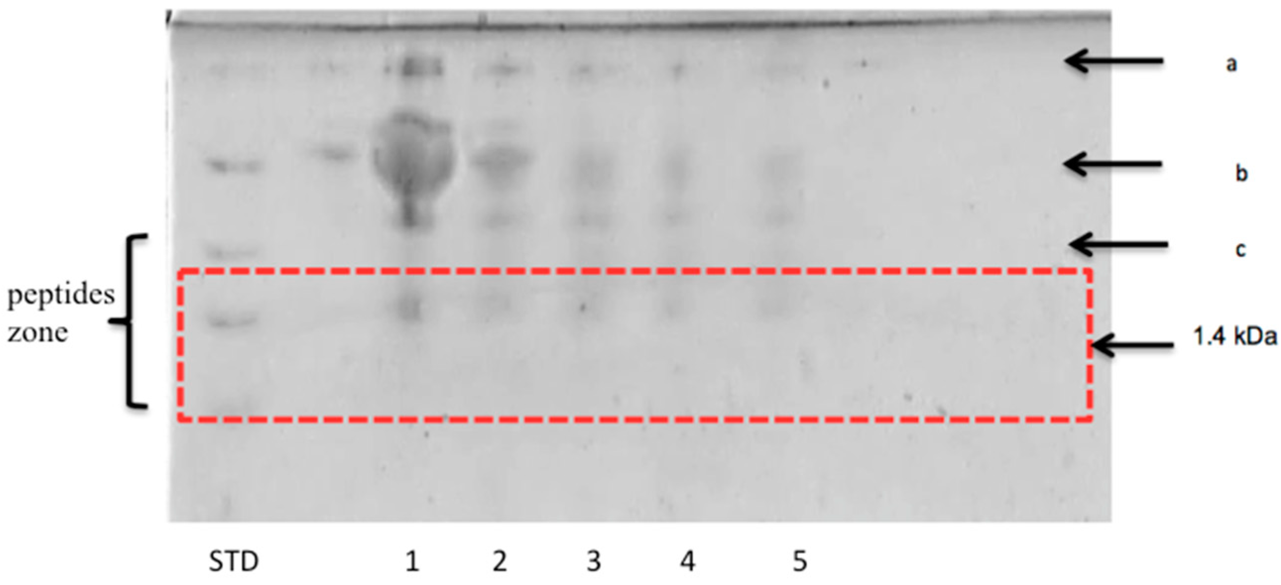
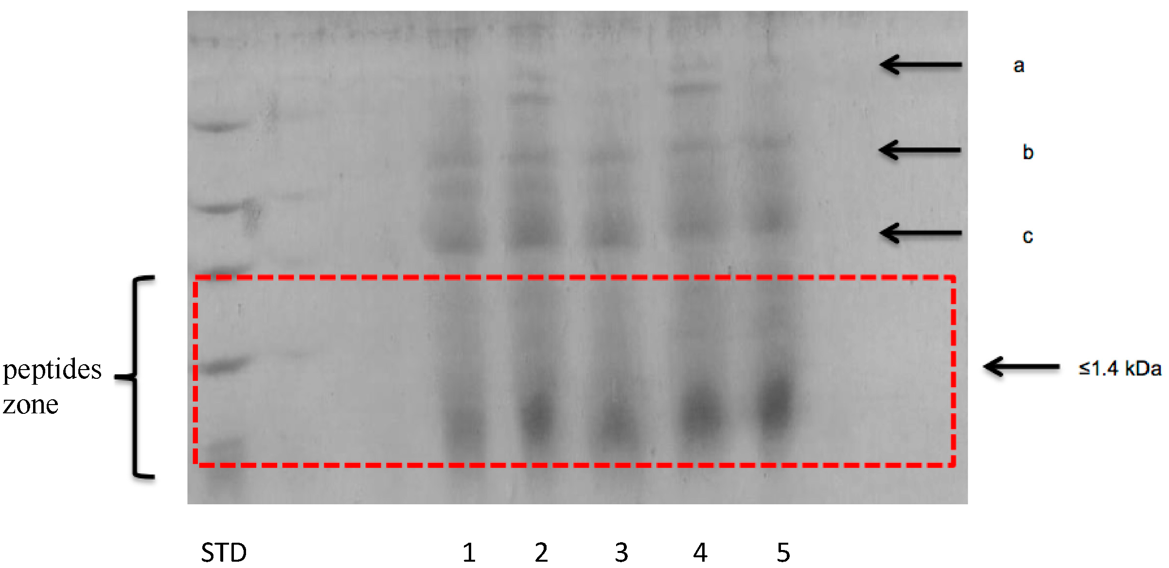
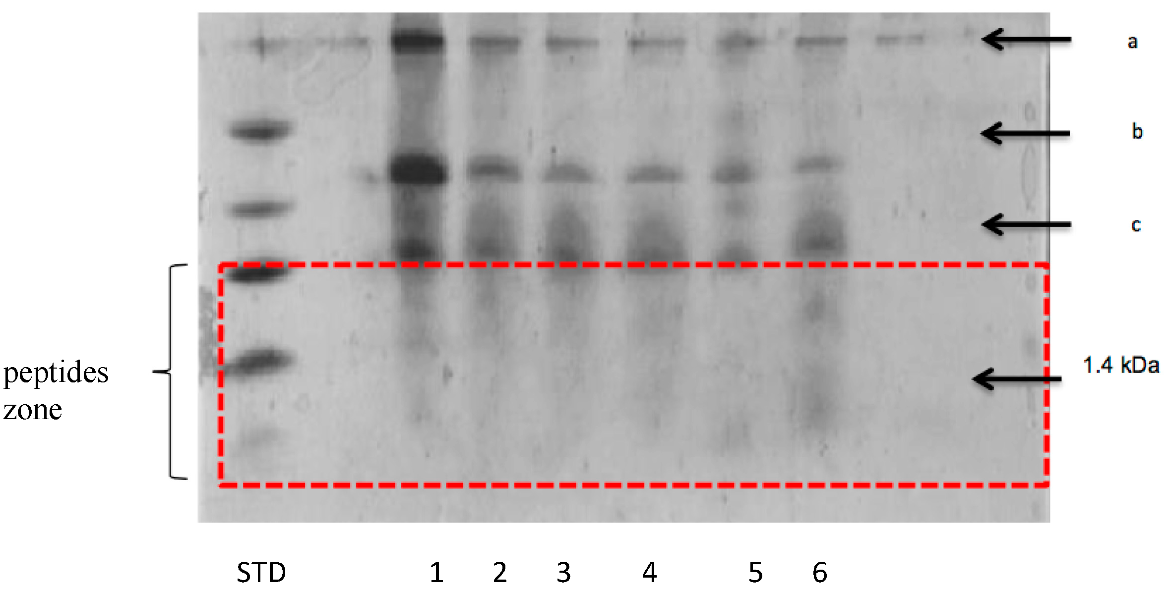
3.2.2. Peptide Separation byTris-Tricine SDS-PAGE
The electrophoresis gel to the samples obtained from LLR milk and showed bands corresponding to peptides less than 1.4 kDa (Figure 1). Many of the peptides with bioactive functions that are reported in the bibliography are less than 10 kDa []. An increase in the concentration of low molecular weight peptides and a decrease in both the concentration of caseins (c) and serum proteins (a and b) were observed.
Figure 1.
Peptide separation by SDS-PAGE of 10% skim milk fermented by Lacticaseibacillus rhamnosus GG. (a) caseins; (b) β-lactoglobulin; (c) α-lactoalbumin and (STD) peptide standard. Time of fermentation (hours): (1) 12; (2) 26; (3) 28; (4) 32; (5) 36 and (6) 56.
In the analysis, a decrease in the concentration of proteins with a molecular weight greater than 14.4 kDa was shown. Likewise, from the first time of monitoring, up to 5 peptides less than 14.4 kDa were identified. Several authors have highlighted that the origin of peptides from serum proteins is in general of greater importance because the fractionation of β-lactoglobulin is higher due to its concentration of proline, since the proteinases and peptidases of the proteolytic system of lactic bacteria are proline-specific [,].
In the case of LST (Figure 2), a high concentration of peptides less than 1.4 kDa was not observed despite having found a decrease in the concentration of proteins with a molecular weight greater than 14.4 kDa. These data coincide with the results obtained during the analysis of free amino groups by the TNBS method. Very high proteolysis was observed at the beginning of the fermentation, related to the decrease in the casein concentration.
Figure 2.
Peptide separation by SDS-PAGE of 10% skim milk fermented by Streptococcus thermophilus SY-102. (a) caseins; (b) β-lactoglobulin; (c) α-lactoalbumin and (STD) peptide standard. Time of fermentation (hours): (1) 2; (2) 4; (3) 8; (4) 10 and (5) 12.
It has been observed that due to its nutritional requirements, S. thermophilus is a species that has a high activity of PrtS proteinase at the beginning of fermentations to obtain the necessary amino acids for its survival [,]. This activity decreases through the fermentation time since the peptide cut is carried inside the cell. In contrast, in other lactic acid bacteria the peptide fractionation occurs by hydrolases linked to the cell wall and peptidases inside the cell [].
Figure 3 shows that in LLS milk the pattern of peptides released was different from that found in mono-culture systems, generating a higher concentration of low molecular weight peptides. At the same time, a decrease in the concentration of proteins with a molecular weight greater than 14.4 kDa was observed. The analysis demonstrated the release of peptides with molecular weight less than 1.4 kDa. It is well known that in some co-cultures fermentation of milk there is a protocooperation mechanism, which is regulated by the release of essential amino acids for one of the species present in the environment [].
Figure 3.
Peptide separation by SDS-PAGE of 10% skim milk fermented by Streptococcus thermophilus SY-102 and Lacticaseibacillus rhamnosus GG. (a) caseins; (b) β-lactoglobulin; (c) α-lactoalbumin and (STD) peptide standard. Time of fermentation (hours): (1) 2; (2) 12; (3) 15; (4) 25 and (5) 41.
The concentration of peptides obtained in the combined system increased throughout the study. These results are consistent with those observed in the analysis of free amino groups. It was also found that the release of peptides during fermentation followed the same pattern as the study carried out by Rojas-Ronquillo et al. [], where they also found that antihypertensive activity was related to the production of low molecular weight peptides.
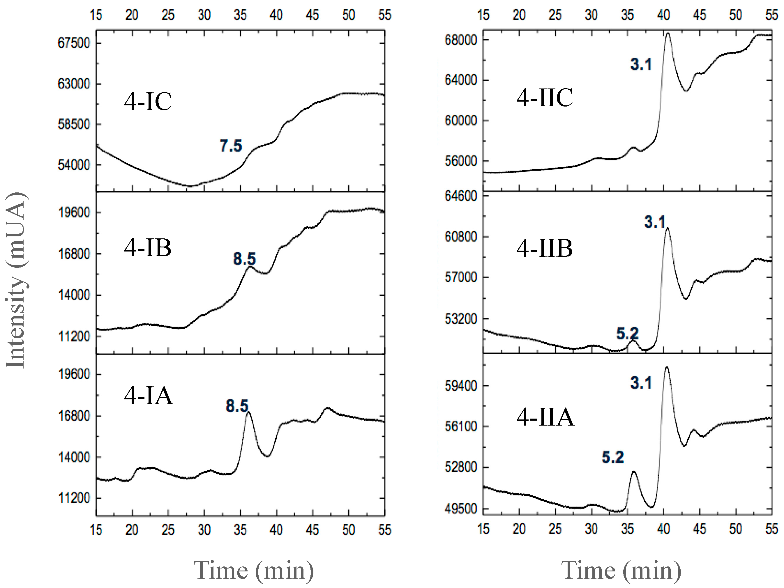
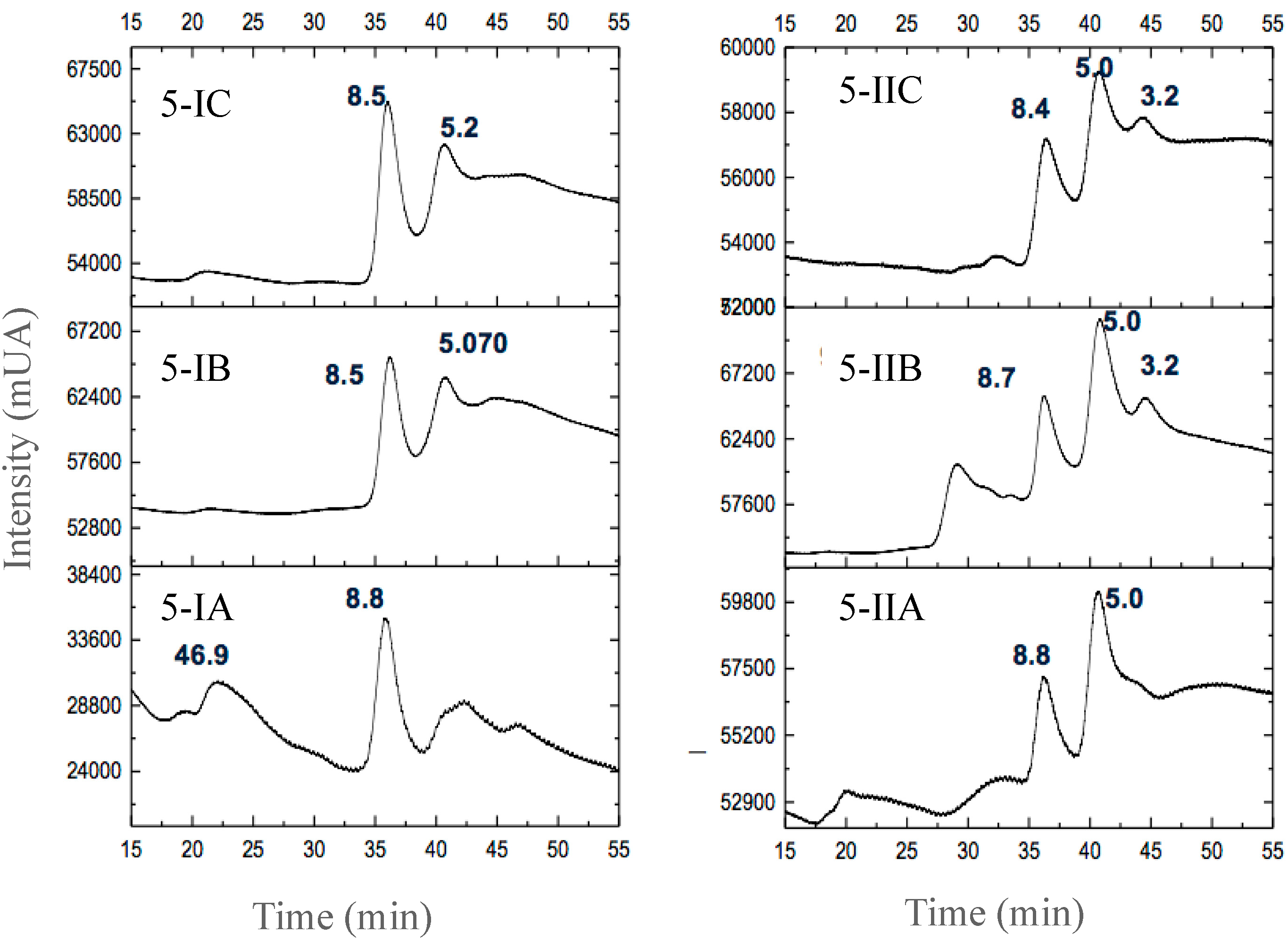
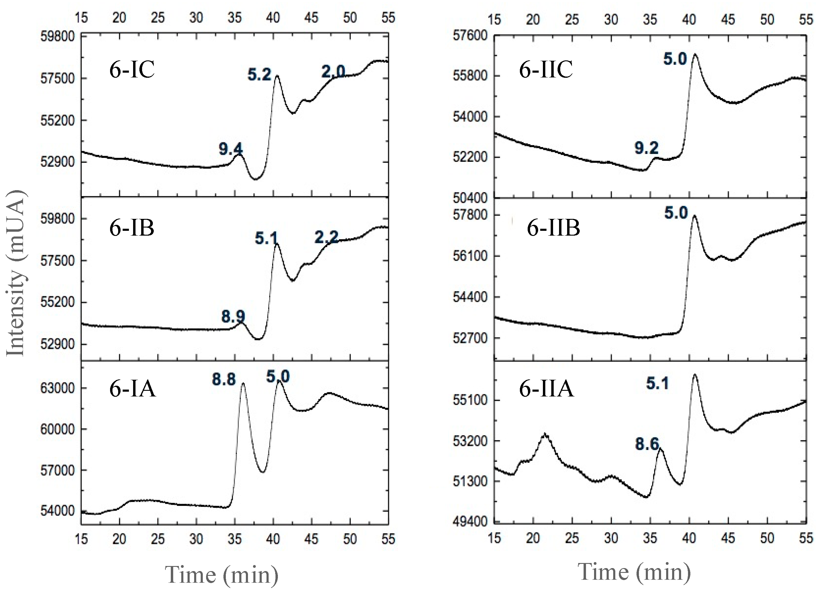
3.2.3. Separation of Peptides with Aromatic Amino Acid Residues
The peptide fractions corresponding to the beginning, the middle and the end of each fermentation were analyzed. Figure 4-I shows that at the beginning of the fermentation with L. rhamnosus GG (Figure 4-IA) at 257 nm, 8.5 kDa molecular weight fractions were found with the presence of phenylalanine, with a retention time (Rt) of 35 min. However, Figure 4-IB shows how the concentration of this peptide fraction decreased and completely disappeared at the end of the study (Figure 4-IC).
Figure 4.
Peptide separation by HPLC of 10% skim milk fermented by Lacticaseibacillus rhamnosus GG at 257 nm (4-I) and 280 nm (4-II). (A) beginning of fermentation, (B) middle of fermentation and (C) end of fermentation.
In the case of the analysis at 280 nm (Figure 4-II), at the beginning of the fermentation two peptide fractions with molecular weights of 5.2 and 3.3 kDa were observed with Rt at 35 and 40 min, respectively (Figure 4-IIA), with a presence of Trp and Tyr. However, after 21 h of fermentation (Figure 4-IIB), the 3.3 kDa fraction (Rt = 40 min) remained apparently constant throughout the fermentation process, which could indicate the presence of an accumulated peptide in the fermentation containing Tyr and Trp residues.
The presence of aromatic amino acids in the peptide fractions is due to the proteolytic system of each lactic acid bacteria. At the end of the proteolytic process, intracellular peptidases cut the peptide fractions inside the cell and excrete the fractions they do not use. Petidases such as PepX, PepO, PepF, and PepT can cut peptides in the hydrophobic part of the peptide sequence and sometimes the accumulated excreted peptides have aromatic amino acids in their structure [,,,]. Additionally, it is known that this type of structure is better coupled to the active site of ACE, causing its inhibition [].
In the analysis of the peptide fractions obtained during the fermentation with S. thermophilus, a protein of 46.9 kDa was observed with the presence of Phe (Figure 5-IA). This fraction could come from bovine serum albumin that was fractionated during fermentation or from some associated proteins in polymers, which is very common under certain temperature and pH conditions for some caseins, mainly β-casein [,].
Figure 5.
Peptide separation by HPLC of 10% skim milk fermented by Streptococcus termophilus SY-102 at 257 nm (5-I) and 280 nm (5-II). (A) beginning of fermentation (B) middle of fermentation and (C) end of fermentation.
In the same way, at the beginning of the fermentation, a peptide fraction of 8.8 kDa was observed, which remained in an apparently constant concentration (Figure 5-IB,IC). Many of these fractions could be produced from the first protein breakdown by the S. thermophilus SY-102 PrtS proteinase. From the start of the fermentation, two peptides with Tyr and Trp residues (Figure 5-IIA) with molecular weights of 8.8 and 5.0 kDa (Rt = 35 and 40 min, respectively) were identified. In the intermediate part of the fermentation a 3.2 kDa peptide was identified (Figure 5-IIB, IIC). At the end of the fermentation, these three peptides were identified and at the same time, an increase in the peptide concentration of 3.2 kDa was observed (Figure 5-IIC).
According to the chromatograms obtained from the co-culture fermentation (Figure 6-I,II), an increase in the apparent concentration of peptides with aromatic amino acid residues were observed. At the beginning of the fermentation (Figure 6-IA) two peptides of less than 10 kDa containing Phe (8.8 and 5.0 kDa with Rt = 35 and 40 min, respectively) were identified. The 8.8 kDa peptide concentration decreased in the intermediate stage of fermentation (Figure 6-IB) and another 9.4 kDa peptide (Rt = 30 min) appeared at the end of this (Figure 6-IC).
Figure 6.
Peptide separation by HPLC of 10% skim milk fermented by Lacticaseibacillus rhamnosus GG and Streptococcus termophilus SY-102 at 257 nm (6-I) and 280 nm (6-II). (A) beginning of fermentation (B) middle of fermentation, and (C) end of fermentation.
At the beginning of the fermentation, peptides with molecular weight less than 10 kDa were also found, with Tyr and Trp residues (Figure 6-IIA). At the end of the process there was an apparent increase in the concentration of the 5.0 kDa fraction (Figure 6-IIB). As in the analysis at 257 kDa, some fractions disappeared in the intermediate stage of fermentation (Figure 6-IIB) while others appeared at the end, mainly a 9.2 kDa fraction (Figure 6-IIC).
During the fermentation process in a co-culture system, the proteolytic system of L. rhamnosus GG broke casein to obtain essential amino acids for its metabolism, releasing low molecular weight peptide fractions that S. thermophillus SY-102 could metabolize. This behavior was described mainly for L. delbrueckii sub. bulgaricus and S. salivarius sub. thermophilus during the yogurt process [,,]. PrtS proteinase of S. thermophilus disintegrates the peptide fraction, using only some essential amino acids for its growth and survival in the medium. On the other hand, this microorganism contains special peptidases such as PepS, which breaks oligopeptides and releases residues with aromatic and hydrophobic amino acids, mainly Phe [,,,,].
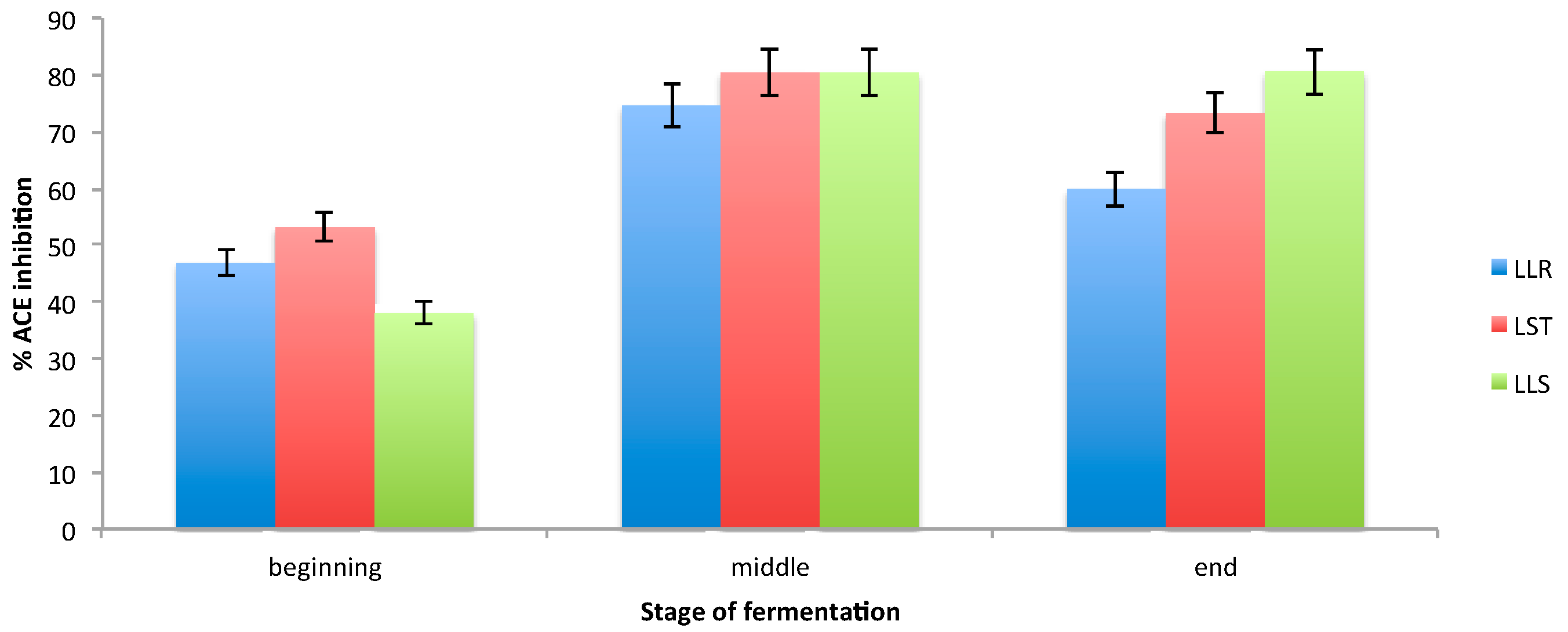
3.3. Determination of ACE Inhibition
At the beginning of fermentation in LLR milk (Figure 7) an ACE inhibition of 46.9% was determined. Inhibition increased in the middle of the process, since at 21 h of fermentation an inhibition of 74.6% was obtained. However, despite the accumulation of low molecular weight peptides, the percentage of inhibition decreased at the end (59.9%).
Figure 7.
ACE inhibitory activity from peptide fractions derived from 10% of skim milk fermented by Lacticaseibacillus rhamnosus GG (LLR), Streptococcus thermophilus SY-102 (LST) and co-culture of Lacticaseibacillus rhamnosus GG and Streptococcus thermophilus SY-102 (LLS).
It is known that at the beginning of fermentation L. rhamnosus requires essential aromatic amino acids (Phe, Trp) for its growth []. However, these needs apparently prevent the accumulation of peptides with aromatic amino acid residues, which can lead to decreased ACE inhibition.
On the contrary, the peptide fractions from fermentation LST showed an increase in ACE inhibition from 53.2% to 80.4% at 21 h and at the end of fermentation the inhibition decreased until 73.3%. These results could be due to the fact that S. thermophilus has different nutritional requirements than other lactic acid bacteria [,] and also has a very advanced system as an operon for the production of amino acids (leucine, isoleucine and valine) [,,,,,,,,,,,,,,,,,,,,,,,,]. Low molecular weight peptides are also generated, although during the experiment it was observed that the low molecular weight peptides were presented in a lower proportion than in the fermentation with L. rhamnosus. Furthermore, the null need for aromatic amino acid peptides by S. thermophilus allows their accumulation in the fermentation medium [,,].
In LLS milk, a 38% inhibition was observed; this percentage increased gradually, reaching 80.6% inhibition at 21 h and remained without significant change until the end of the process (41 h). This increase could be due to the presence of a specific intracellular peptidase of S. thermophilus that constantly generates peptides containing Phe [], since it has been reported that this microorganism excretes two and three amino acid peptides generated by the action of intracellular dipeptidases. [,,]. This increase in ACE inhibition is consistent with the results obtained during the electrophoresis analysis, in which accumulation of low molecular weight peptides was found. In the same way, this activity of inhibition could be related with the presence of peptides with aromatic amino acid residues observed in the HPLC analysis.
An increase in ACE inhibition has been observed during milk fermentation with lactic acid bacteria []. However, it is known that the antihypertensive capacity of fermented milks in which co-cultures were used is greater [] than in those produced with monocultures. Additionally, since S. thermophilus has the ability to release peptides with antihypertensive capacity from the beginning of protein hydrolysis [], it is likely that this activity will not be lost as long as this bacterium is found in combination with other microorganisms during lactic fermentation.
In the case of the co-culture fermentation studied in the present investigation, the microorganisms used showed a symbiotic growth as observed in proteolysis. The release of peptides by these microorganisms exhibited a behavior similar to that observed during fermentation with S. thermophilus, however, according to electrophoresis analysis, there is a greater diversity and concentration of peptides in the co-culture system and these residues could present other biological activities of importance, which were not studied in this investigation.
4. Conclusions
The combination of L. rhamnosus GG and S. thermophilus SY-102 in milk fermentation favors the acidification of the medium in shorter times, compared to fermented systems inoculated only with L. rhamnosus GG. Likewise, the use of a mixed system promotes greater proteolytic activity, favoring the increase in the concentration of low molecular weight peptides released by the hydrolysis of proteins. Furthermore, the co-culture fermentations do not produce negative effects on the release of peptides with aromatic amino acids, which creates an advantage in the bioactivity of these peptide fractions.
The antihypertensive activity of the peptide fractions obtained in this work is related to the presence of low molecular weight peptides as well as to the presence of aromatic amino acids in these fractions. In addition, using a mixed system for milk fermentation increases the antihypertensive capacity of the peptides derived from proteolysis. Co-culture fermentations are a field of study with challenges and opportunities yet to be discovered, especially in those investigations directed towards the elaboration of nutraceutical and functional food products.
Author Contributions
Conceptualization, J.L.S.-N.; L.G.G.-O.; G.M.R.-S. and A.E.C.-G.; methodology, G.M.R.-S., A.E.C.-G. and L.G.G.-O.; investigation, J.L.S.-N. and E.P.-E.; resources, A.C.-O., L.G.G.-O. and E.C.-L.; writing—original draft preparation, L.G.G.-O., J.R.-G. and E.C.-L.; writing—review and editing, A.A.-N., J.J.-O. and J.A.-M.; supervision, L.G.G.-O. All authors have read and agreed to the published version of the manuscript.
Funding
This work was supported by Consejo Nacional de Ciencia y Tecnología (CONACYT), Mexico, under Grant number CB-2014-241333.
Institutional Review Board Statement
Not applicable.
Informed Consent Statement
Not applicable.
Data Availability Statement
Not applicable.
Acknowledgments
The authors appreciate the support of researchers of the Biotechnology Laboratory at Universidad Autónoma Metropolitana for their technical support.
Conflicts of Interest
The authors declare no conflict of interest.
References
- Gamechu, T. Review in lactic acid bacteria function in milk fermentation and preservation. Afr. J. Food Sci. 2015, 9, 170–175. [Google Scholar] [CrossRef] [Green Version]
- Beltrán-Barrientos, L.M.; Hernández-Mendoza, A.; Torres-Llanez, M.J.; González-Córdova, A.F.; Vallejo-Córdoba, B. Fermented milk as antihypertensive functional food. J. Dairy Sci. 2016, 99, 4099–4110. [Google Scholar] [CrossRef] [PubMed] [Green Version]
- Sodini, I.; Lucas, A.; Oliveira, N.M.; Remeuf, F.; Corrieu, G. Effect of mil base and starter culture on acidification, texture and probiotic cell counts in fermented milk processing. J. Dairy Sci. 2002, 85, 2479–2488. [Google Scholar] [CrossRef]
- Nami, Y.; Haghshenas, B.; Vaseghi, R.; Jalaly, H.M.; Lotfi, H.; Eslami, S.; Amin Hejazi, M. Novel autochthonous lactobacilli with probiotic aptitudes as a main starter culture for probiotic fermented milk. LWT 2018, 98, 85–93. [Google Scholar] [CrossRef]
- Meng-Xin, H.; Ji-Nian, L.; Qian, G.; Ya-Qian, Z.; Hong-Mei, N. Probiotics biofilm-integrated electrospun nanofiber membranes: A new starter culture for fermented milk production. J. Agric. Food Chem. 2019, 67, 3198–3208. [Google Scholar] [CrossRef]
- Bhat, Z.F.; Kumar, S.; Bhat, H.F. Bioactive peptides of animal origin: A review. J. Food Sci. Technol. 2015, 52, 5377–5392. [Google Scholar] [CrossRef] [Green Version]
- Abd El-Fattah, A.M.; Sakr, S.S.; El-Dieb, S.M.; Elkashef, H.A.S. Bioactive peptides with ACE-I and antioxidant activity produced from milk proteolysis. Int. J. Food Prop. 2016, 20, 3033–3042. [Google Scholar] [CrossRef] [Green Version]
- Hammami, R.; Fliss, I.; Corsetti, A. Editorial: Application of protective cultures and bacteriocins for food biopreservation. Front. Microbiol. 2019, 10, 1561. [Google Scholar] [CrossRef] [Green Version]
- Chai, K.F.; Voo, A.Y.H.; Chen, W.N. Bioactive peptides from food fermentations: A comprehensive review of their sources, bioactivities, applications and future development. Compr. Rev. Food Sci. Food Saf. 2020, 19, 3825–3885. [Google Scholar] [CrossRef]
- Miralles, B.; Amigo, L.; Recio, I. Critical review and perspectives on food derived antihypertensive peptides. J. Agric. Food Chem. 2018, 66, 9384–9390. [Google Scholar] [CrossRef]
- Lee, S.Y.; Hur, S.J. Antihypertensive peptides from animal products, marine organisms, and plants. Food Chem. 2017, 228, 506–517. [Google Scholar] [CrossRef]
- Gonzalez-Gonzalez, C.R.; Tuohy, K.M.; Jauregi, P. Production of angiotensin-I-converting enzyme (ACE) inhibitory activity in milk fermented with probiotic strains: Effects of calcium, pH and peptides on the ACE-inhibitory activity. Int. Dairy J. 2011, 21, 615–622. [Google Scholar] [CrossRef]
- Nejati, F.; Rizzello, C.G.; Di Cagno, R.; Sheikh-Zeinoddin, M.; Diviccaro, A.; Minervini, F.; Gobbetti, M. Manufacture of a functional fermented milk enriched of Angiotensin-I Converting Enzyme (ACE)-inhibitory peptides and γ-amino butyric acid (GABA). LWT-Food Sci. Technol. 2013, 51, 183–189. [Google Scholar] [CrossRef]
- Elkhtab, E.; El-Alfy, M.; Shenana, M.; Mohamed, A.; Yousef, A.E. New potentially antihypertensive peptides liberated in milk during fermentation with selected lactic acid bacteria and kombucha cultures. J. Dairy Sci. 2017, 100, 9508–9520. [Google Scholar] [CrossRef]
- Cavalheiro, F.G.; Baptista, D.P.; Galli, B.D.; Negrão, F.; Eberlin, M.N.; Gigante, M.L. High protein yogurt with addition of Lactobacillus helveticus: Peptide profile and angiotensin-converting enzyme ACE-inhibitory activity. Food Chem. 2020, 333, 127482. [Google Scholar] [CrossRef]
- Begunova, A.V.; Savinova, O.S.; Glazunova, O.A.; Moiseenko, K.V.; Rozhkova, I.V.; Fedorova, T.V. Development of antioxidant and antihypertensive properties during growth of Lactobacillus helveticus, Lactobacillus rhamnosus and Lactobacillus reuteri on cow’s milk: Fermentation and peptidomics study. Foods 2021, 10, 17. [Google Scholar] [CrossRef]
- González-Olivares, L.G.; Añorve-Morga, J.; Castañeda-Ovando, A.; Contreras-López, E.; Jaimez-Ordaz, J. Peptide separation of commercial fermented milk during refrigerated storage. Food Sci. Technol. 2014, 34, 674–679. [Google Scholar] [CrossRef] [Green Version]
- Daliri, E.B.M.; Lee, B.H.; Park, B.J.; Kim, S.H.; Oh, D.H. Antihypertensive peptides from whey proteins fermented by lactic acid bacteria. Food Sci. Biotechnol. 2018, 27, 1781–1789. [Google Scholar] [CrossRef] [PubMed]
- de Souza Pereira, Á.M.; de Farias, D.R.B.; de Queiroz, B.B.; de Caldas Nobre, M.S.; Cavalcanti, M.T.; Salles, H.O.; Dos Santos, K.M.O.; de Medeiros, A.C.D.; Florentino, E.R.; Buriti, F.C.A. Influence of a Co-culture of Streptococcus thermophilus and Lactobacillus casei on the Proteolysis and ACE-Inhibitory Activity of a Beverage Based on Reconstituted Goat Whey Powder. Probiotics Antimicrob. Proteins 2017, 11, 273–282. [Google Scholar] [CrossRef] [PubMed]
- Rodríguez-Serrano, G.M.; García-Garibay, M.; Cruz-Guerrero, A.E.; Gómez-Ruiz, L.; Ayala-Nino, A.; Castaneda-Ovando, A.; Gonzalez-Olivares, L.G. Proteolytic System of Streptococcus thermophilus. J. Microbiol. Biotechnol. 2018, 28, 1581–1588. [Google Scholar] [CrossRef] [PubMed]
- Chang, O.K.; Roux, É.; Awussi, A.A.; Miclo, L.; Jardin, J.; Jameh, N.; Dary, A.; Humbert, G.; Perrin, C. Use of a free form of the Streptococcus thermophilus cell envelope protease PrtS as a tool to produce bioactive peptides. Int. Dairy J. 2014, 38, 104–115. [Google Scholar] [CrossRef]
- Buritti, F.; Cardarelli, H.; Filisetti, T.; Saad, S. Synbiotic potential of fresh cream cheese supplemented with inulin and Lactobacillus paracasei in co-culture with Streptococcus thermophilus. Food Chem. 2007, 104, 1605–1610. [Google Scholar] [CrossRef]
- Settachaimongkon, S.; Nout, M.J.R.; Antunes Fernandes, E.C.; Hettinga, K.A.; Vervoort, J.M.; van Hooijdonk, T.C.M.; Zwietering, M.H.; Smid, E.J.; van Valenberg, H.J.F. Influence of different proteolytic strains of Streptococcus thermophilus in co-culture with Lactobacillus delbrueckii subsp. bulgaricus on the metabolite profile of set-yoghurt. Int. J. Food Microbiol. 2014, 177, 29–36. [Google Scholar] [CrossRef] [PubMed]
- Santos, W.M.; de Caldas Nobre, M.S.; Cavalcanti, M.T.; dos Santos, K.M.O.; Salles, H.O.; Alonso Buriti, F.C. Proteolysis of reconstituted goat whey fermented by Streptococcus thermophilus in co-culture with commercial probiotic lactobacillus strains. Int. J. Dairy Technol. 2019, 72, 559–568. [Google Scholar] [CrossRef]
- Schägger, H.; von Jagow, G. Tricine-sodium dodecyl sulfate-polyacrylamide gel electrophoresis for the separation of proteins in the range from 1 to 100 kDa. Anal. Biochem. 1987, 166, 368–379. [Google Scholar] [CrossRef]
- González-Olivares, L.G.; Jiménez-Guzmán, J.; Cruz-Guerrero, A.; Rodríguez-Serrano, G.; Gómez-Ruíz, L.; García-Garibay, M. Bioactive peptides released by lactic acid bacteria in comercial fermented milks. Rev. Mex. Ing. Química 2011, 10, 179–188. [Google Scholar]
- Fitzgerald, R.J.; Murray, B.A. Bioactive peptides and lactic fermentations. Int. J. Dairy Technol. 2006, 59, 118–125. [Google Scholar] [CrossRef]
- Qiao, Y.; Leng, C.; Liu, G.; Zhang, Y.; Lv, X.; Chen, H.; Sun, J.; Feng, Z. Transcriptomic and proteomic profiling revealed global changes in Streptococcus thermophilus during pH-controlled batch fermentations. J. Microbiol. 2019, 57, 769–780. [Google Scholar] [CrossRef]
- Rajagopal, S.N.; Sandine, W.E. Associative growth and proteolysis of Streptococcus thermophilus and Lactobacillus bulgaricus in skim milk. J. Dairy Sci. 1990, 73, 894–899. [Google Scholar] [CrossRef]
- Donkor, O.N.; Henriksson, A.; Singh, T.K.; Vasiljevic, T.; Shah, N.P. ACE-inhibitory activity of probiotic yoghurt. Int. Dairy J. 2007, 17, 1321–1331. [Google Scholar] [CrossRef]
- Quirós, A.; Ramos, M.; Muguerza, B.; Delgado, M.A.; Miguel, M.; Aleixandre, A.; Recio, I. Identification of novel antihypertensive peptides in milk fermented with Enterococcus faecalis. Int. Dairy J. 2007, 17, 33–41. [Google Scholar] [CrossRef]
- Rojas-Ronquillo, R.; Cruz-Guerrero, A.; Flores-Nájera, A.; Rodríguez-Serrano, G.; Gómez-Ruiz, L.; Reyes-Grajeda, J.P.; Jiménez-Guzmán, J.; García-Garibay, M. Antithrombotic and angiotensin-converting enzyme inhibitory properties of peptides released from bovine casein by Lactobacillus casei Shirota. Int. Dairy J. 2012, 26, 147–154. [Google Scholar] [CrossRef]
- Yamamoto, E.; Watanabe, R.; Ichimura, T.; Ishida, T.; Kimura, K. Effect of lactose hydrolysis on the milk-fermenting properties of Lactobacillus delbrueckii ssp. bulgaricus 2038 and Streptococcus thermophilus 1131. J. Dairy Sci. 2021, 104, 1454–1464. [Google Scholar] [CrossRef]
- Markakiou, S.; Gaspar, P.; Johansen, E.; Zeidan, A.A.; Neves, A.R. Harnessing the metabolic potential of Streptococcus thermophilus for new biotechnological applications. Curr. Opin. Biotechnol. 2020, 61, 142–152. [Google Scholar] [CrossRef] [PubMed]
- Hafeez, Z.; Cakir-Kiefer, C.; Lecomte, X.; Miclo, L.; Dary-Mourot, A. The X-prolyl dipeptidyl-peptidase PepX of Streptococcus thermophilus initially described as intracellular is also responsible for peptidase extracellular activity. J. Dairy Sci. 2018, 102, 113–123. [Google Scholar] [CrossRef] [PubMed] [Green Version]
- Hu, T.; Cui, Y.; Qu, X. Analysis of the proteolytic system of Streptococcus thermophilus strains CS5, CS9, CS18 and CS20. Int. Dairy J. 2021, 118, 105025. [Google Scholar] [CrossRef]
- Fernandez-Espla, M.D.; Garault, P.; Monnet, V.; Rul, F. Streptococcus thermophilus cell wall-anchored proteinase: Release, purification, and biochemical and genetic characterization. Appl. Environ. Microbiol. 2000, 66, 4772–4778. [Google Scholar] [CrossRef] [PubMed] [Green Version]
- Ortiz-Chao, P.; Gómez-Ruiz, J.A.; Rastall, R.A.; Mills, D.; Cramer, R.; Pihlanto, A.; Korhonen, H.; Jauregi, P. Production of novel ACE inhibitory peptides from β-lactoglobulin using Protease N Amano. Int. Dairy J. 2009, 19, 69–76. [Google Scholar] [CrossRef]
- Leroy, F.; De Vuyst, L. Lactic acid bacteria as functional starter cultures for the food fermentation industry. Trends Food Sci. Technol. 2004, 15, 67–78. [Google Scholar] [CrossRef]
- Araujo, M.C.; Melo, R.L.; Cesari, M.H.; Juliano, L.; Carmona, A.K. Peptidase specificity characterization of C- and N- terminal catalytic sites of angiotensin-I-converting enzyme. Biochemistry 2000, 39, 8519–8525. [Google Scholar] [CrossRef]
- Fan, M.; Guo, T.; Li, W.; Chen, J.; Li, F.; Wang, C.; Shi, Y.; Li, D.X.; Zhang, S. Isolation and identification of novel casein-derived bioactive peptides and potential functions in fermented casein with Lactobacillus helveticus. Food Sci. Hum. Wellness 2019, 8, 156–176. [Google Scholar] [CrossRef]
- Innocente, N.; Biasutti, M.; Rita, F.; Brichese, R.; Comi, G.; Iacumin, L. Effect of indigenous Lactobacillus rhamnosus isolated from bovine milk on microbiological characteristics and aromatic profile of traditional yogurt. LWT-Food Sci. Technol. 2016, 66, 158–164. [Google Scholar] [CrossRef]
- Letort, C.; Juillard, V. Development of a minimal chemically-defined medium for the exponential growth of Streptococcus thermophilus. J. Appl. Microbiol. 2001, 91, 1023–1029. [Google Scholar] [CrossRef] [PubMed]
- Garault, P.; Letort, C.; Juillard, V.; Monnet, V. Branched-chain amino acid biosynthesis is essential for optimal growth of Streptococcus thermophilus in milk. Appl. Environ. Microbiol. 2000, 66, 5128–5133. [Google Scholar] [CrossRef] [PubMed] [Green Version]
- Fernandez-Espla, M.D.; Rul, F. PepS from Streptococcus thermophilus. A new member of the aminopeptidase T family of thermophilic bacteria. Eur. J. Biochem. 1999, 263, 502–510. [Google Scholar] [CrossRef] [PubMed] [Green Version]
- Rokka, T.; Syväoja, E.; Tuominen, J. Release of bioactive by enzymatic proteolysis of Lactobacillus GG fermented UHT milk. Milchwiissenschaft 1997, 66, 9. [Google Scholar]
Publisher’s Note: MDPI stays neutral with regard to jurisdictional claims in published maps and institutional affiliations. |
© 2021 by the authors. Licensee MDPI, Basel, Switzerland. This article is an open access article distributed under the terms and conditions of the Creative Commons Attribution (CC BY) license (https://creativecommons.org/licenses/by/4.0/).