Article

Influence of Oat β-Glucan on the Survival and Proteolytic Activity of Lactobacillus rhamnosus GG in Milk Fermentation: Optimization by Response Surface

by
María Isabel Chávez de la Vega
1,
Sergio Alatorre-Santamaría
1,
Lorena Gómez-Ruiz
1,
Mariano García-Garibay
1,2,
Francisco Guzmán-Rodríguez
1,
Luis Guillermo González-Olivares
3,*,
Alma Elizabeth Cruz-Guerrero
1,* and
Gabriela Mariana Rodríguez-Serrano
1,*
1
Departamento de Biotecnología, Universidad Autónoma Metropolitana-Iztapalapa, Ciudad de México C.P. 09340, Mexico
2
Departamento de Ciencias de la Alimentación, Universidad Autónoma Metropolitana-Lerma, Lerma de Villada C.P. 52006, Mexico
3
Centro de Investigaciones Químicas, Universidad Autónoma del Estado de Hidalgo, Mineral de la Reforma C.P. 42184, Mexico
*
Authors to whom correspondence should be addressed.
Fermentation 2021, 7(4), 210; https://doi.org/10.3390/fermentation7040210
Submission received: 16 August 2021 / Revised: 22 September 2021 / Accepted: 24 September 2021 / Published: 28 September 2021
(This article belongs to the Special Issue Lactic Acid Fermentation and the Colours of Biotechnology 3.0)

Abstract

:
β-glucans come from cereals that have been located within compounds with prebiotic activity. They have presented several bioactivities that have determined their high functional value. The aim of this study was to identify the influence of oat β-glucan on the survival and proteolytic activity of Lactobacillus rhamnosus GG in a milk fermentation through an experimental design to optimize the process. For β-glucan extraction after dry milling of oats, two methods were applied: with and without enzymatic inactivation of the semolina. The highest extraction yield (45.25 g/L) was obtained with enzymatic inactivation. For the optimization of survival and proteolytic activity, a central design composed of axial points with two factors on three levels was used. Control factors were β-glucan and inoculum concentrations. According to response surface, the best survival growth rate of probiotic was observed with 4.38% of inoculum and 22.46 g/L of β-glucan, and the highest production of free amino groups was observed with 4.18% of inoculum and 22.71 g/L of β-glucan. Thus, β-glucan promotes the proteolytic activity of Lb. rhamnosus GG in milk fermentation.

1. Introduction

Oats (Avena sativa L.) are a cereal whose whole grain has a high content of soluble dietary fiber, where β-glucan is included. It is a linear polymer of glucose units linked by glucosidic bonds β-(1 → 3) and β-(1 → 4), which is located mainly in the cell walls of the endosperm of the oat grain [1]. β-Glucan concentration of oats depends both on the culture conditions and growth. In addition, it is also regulated by the enzyme β-glucan endohydrolase (1 → 3, 1 → 4), which degrades the endosperm cell wall during germination or food preparation. Therefore, β-glucan concentration in the oat grain can vary from 1.8% to 5.5%, although the most common is to find a concentration of 4.0–5.5% [2].
Conversely, the definition of prebiotic has changed according to both the scientific discoveries and the origin of the compound considered as a prebiotic. Despite prebiotics being widely classified as dietary fibers, not all fibers are considered prebiotics [3]; this is why the current definition describes prebiotics as “ingredients that produce a selective stimulation of the growth and/or activities of one or a limited number of genera/species of microorganisms in the intestinal microbiota, giving benefits to the health of the host” [4]. Prebiotics have different characteristics, which are considered as functional; some of these characteristics include: being resistant to stomach pH, capacity for hydrolysis of gastric enzymes, and absorption in the gastrointestinal tract. Further, they can be fermented by the intestinal microbiota, and selectively stimulate the growth and/or activity of intestinal bacteria associated with the health of the host [5,6].
The β-glucans from cereals, mainly from barley and oats, have been considered as prebiotics [7,8], and the consumption of these compounds has been related to health benefits [9]. In addition to the fact that these compounds are part of the functional dietary fibers, their fermentation in the gastrointestinal tract could be the origin of those benefits [10,11,12,13]. However, the main importance is that they directly affect both the growth of probiotic microorganisms and the activation of their proteolytic system [14,15,16].
As a consequence of these interesting biological activities, numerous studies have linked the production of peptides with the presence of prebiotic substances, mainly in dairy matrices such as fermented milks and ripened cheeses [17,18], because in this kind of dairy products the proteolytic capacity of lactic acid bacteria plays an important technological and bioactive role [19,20]. The release of amino acids and small peptide fractions not only has a direct effect on the taste and aroma of fermented dairy products, but low molecular weight peptide fractions could have a bioactive effect on human metabolism [21,22,23]. For this reason, and based on all these characteristics, the definition of prebiotic can be expanded. Thus, the present study relates the selectivity of oat β-glucan on both the growth and proteolytic activity of L. rhamnosus GG, through an experimental design.

2. Materials and Methods

2.1. Sample Obtaining and Preparation

The oats (Avena sativa) that were used came from Productores Agropecuarios Tepexpan, Hidalgo. A quartering method was applied to reduce the amount of grains and 6 kg were obtained after cleaning (insects and stones elimination). Later, the grains were washed 4 times with water in a water to grain ratio of 1:1. In the last wash, and to disinfect the grains, a 1% (v/v) sodium hypochlorite solution was used, soaking them for 10 min before being rinsed with distilled water [24]. In the end, the oats were dried in a convection oven at 30 °C for 24 h until reaching a moisture level of 12%.

2.2. Oats Grinding

After cleaning and drying, 5 kg of oats were ground in a hammer mill (Chopin Moulin CD1, Chopin S.A., Villeneuve la Garenne, France). From this grinding, screening was carried out to obtain 3 fractions: flour with a 160 μ mesh, semolina with an 800 μ mesh, and husk, which corresponds to the biggest particles [25]. Fractions were stored at room temperature until their use and analysis.

2.3. Physicochemical Composition

The proximate composition of the fractions was determined by using the AOAC official methods [26]: moisture AOAC 925.19, fiber AOAC 991.43, protein AOAC 920.165, fat AOAC 948.22 and ashes AOAC 941.12. Carbohydrate content was calculated by subtraction. β-glucan determination was performed according AOAC method 995.16 using β-Glucan Assay Kit (Mixed Linkage) of Megazyme for dry samples.

2.4. β-Glucan Extraction and Quantification

For the extraction of β-glucan, a random sampling of each of the fractions obtained during the milling of the oats was carried out. The β-glucan extraction was made for 2 methods: (1) solubilization (M1) [27] using each fraction of the grinding, and (2) β-glucanases inactivation (M2) [28] just analyzing the semolina. For M1, a solution of 10 g in 100 mL of distilled water of each of the fractions was prepared. The mixtures were vigorously stirred for 3 h at 55 °C on a grill with stirring (Labtech CO, LTD, Guadalajara, Mexico). Then, the samples were centrifuged at 5500 rpm for 10 min and the supernatants were reserved. For the enzymatic inactivation method (M2), 27.5 g of semolina and 250 mL of 75% ethanol (v/v) were mixed and incubated at 80 °C for 4 h. The solvent was evaporated for 48 h at 40 °C, obtaining 6 g of dry matter. This solid fraction was mixed with 90 mL of distilled water at 40 °C, the pH was adjusted at 10 with 20% sodium carbonate (w/v) and was mixed for 30 min on a grill with stirring (Labtech CO, LTD). The mixture was left for 5 min at room temperature and centrifuged for 10 min at 2000× g. The supernatant was cooled to 20 °C, and the pH was adjusted to 4 with 20% (v/v) of an aqueous solution of hydrochloric acid. In the end, all the extracts obtained were stored at −4 °C until use. The β-glucan was quantified using the β-Glucan Assay Kit for wet samples (Mixed Linkage) (MegazymeTM; Sidney, Australia) with modifications (method adjusted for using half of samples and reagents).

2.5. Fermentation and Probiotic Growth

The fermentations were carried out in 25 mL Erlenmeyer flasks with 20 mL of 10% (w/v) Skim milk (BD Difco, Darmstadt, Germany) with β-glucan concentration and the corresponding percentage of L. rhamnosus GG (1 × 10⁶ CFU/mL), previously activated in MRS broth (BD Difco) for 24 h (Table 1). The fermentations were carried out at 37 °C for 24 h [29]. A sample of 5 mL of each flask was taken before and after the fermentation, before being centrifuged at 18,000 rpm for 20 min and stored at −4 °C for further analysis.
The viable cells quantification was made with a modified drop count method [30] with successive dilutions 1:10 in test tubes with 9 mL of 0.85% (w/v) saline solution (1 × 10⁻¹ to 1 × 10⁻⁶). Then, 5 μL were taken and inoculated in quadruple in Petri dishes divided into four quadrants with 10 mL of MRS agar BD (Difco). The dishes were incubated at 37 °C for 24 h. The count was made after 24 h in the quadrants with between 10 and 100 colonies. For the calculation of CFU/mL the Equation (1) was used
CFU/mL = (no. of colonies × inverse of dilution factor)/0.005
The growth is reported as the difference between the initial CFU/mL and final CFU/mL of each experiment. The control group was carried out in milk without β-glucan.

2.6. Free Amino Acid Groups Concentration Response (Proteolytic Activity of L. rhamnosus GG)

The proteolysis degree was determined by trinitrobenzenesulfonic acid method (TNBS) [31]. A phosphate buffer solution (0.21 M, pH 8.2) was added to test tubes, well covered with aluminum foil, and 250 μL of the sample was added to the tubes. For the blank, instead of the sample, deionized water was used and 2 mL of 0.1% (v/v) TNBS were added to each tube and the mixture was homogenized, before being incubated for 1 h at 50 °C in the dark. The reaction was stopped by adding 4 mL of 0.1 N HCl; this mixture was homogenized by vortex stirring and absorbance was read at 340 nm in a spectrophotometer (Shimadzu UV-1800 UV/Visible Scanning Spectrophotometer; 115 VAC, Kyoto, Japan). Every experiment was tested in triplicate at the beginning and end of the fermentation processes.

2.7. Optimization of Fermentation

2.7.1. Experimental Design

To analyze the effect of the β-glucan and inoculum concentration in the free amino groups production and probiotic growth, a central composite design with star points was applied. Twelve experiments were carried out, and the variables were codified (Table 1). Previous studies (data not shown) were done in order to choose the experimental conditions (factor levels). Statistical analysis was performed using the software Statgraphics 18-X24 to obtain the second-order polynomial model (Equation (2)) and the response surfaces.
Y = β 0 + i = 1 2 β i x i + i = 1 2 β i i x i 2 + i j = i + 1 β i j x i x j
where: Y was the predicted response (probiotic growth or free amino groups concentration), β0 was the constant coefficient, β1 and β2 were the linear coefficients, β11 and β22 were the quadratic coefficients, β12 was the cross-product coefficient, x1 (β-glucan concentration) and x2 (inoculum concentration) were the independent variables. Response surface plots were constructed to demonstrate the simultaneous effect of β-glucan concentration (x1) and inoculum concentration (x2) on the experimental dependent parameters (probiotic growth or free amino groups concentration).

2.7.2. Experimental Design Validation

An experimental verification was carried out to corroborate the optimization of the fermented process with the results of linear, square, and interaction coefficients. Both optimum probiotic growth and free amino groups concentration were determined through the statistical procedure applied. The best conditions (β-glucan and inoculum concentrations) and responses were chosen. The experimental values for each response were compared to the predicted data from the mathematical model by Tukey’s analysis.

3. Results

3.1. Proximate Compositional Analysis and Concentration of β-Glucan in Oat Milling Fractions

Table 2 shows the results of the proximate composition analysis of oat milling fractions. Each fraction has its own characteristics; for example, husk is characterized by a highest concentration of both protein and carbohydrates. Moreover, despite having the highest concentration of carbohydrates, fiber is found in the lowest proportion in flour and therefore, the concentration of β-glucan is lower than those exhibited by other fractions. A total β-glucan concentration of 4.71% was observed, which represented a proportion of 48.3% for the total fiber calculated in the oat grain (9.74%).

3.2. β-Glucan Extraction

In order to select the fraction of the highest releasing concentration of the β-glucan, the extraction was carried out on each of them according to M1 [27]. It was observed that in the semolina extract, the highest concentration of β-glucan was obtained (7.33 ± 0.15 g/L) compared to the husk extract (4.67 ± 0.13 g/L) and flour (3.90 ± 0.22 g/L), despite not having the highest concentration of this compound. Because semolina had a smaller particle size than the husk, the yield of β-glucan was higher in the former. Since semolina was the fraction with the easiest access to β-glucan, an extraction based on enzymatic inactivation was applied (M2). In this way, it was observed that the concentration of β-glucan extracted was approximately 6.5 times higher than in the extraction by solubilization (45.25 ± 0.09).

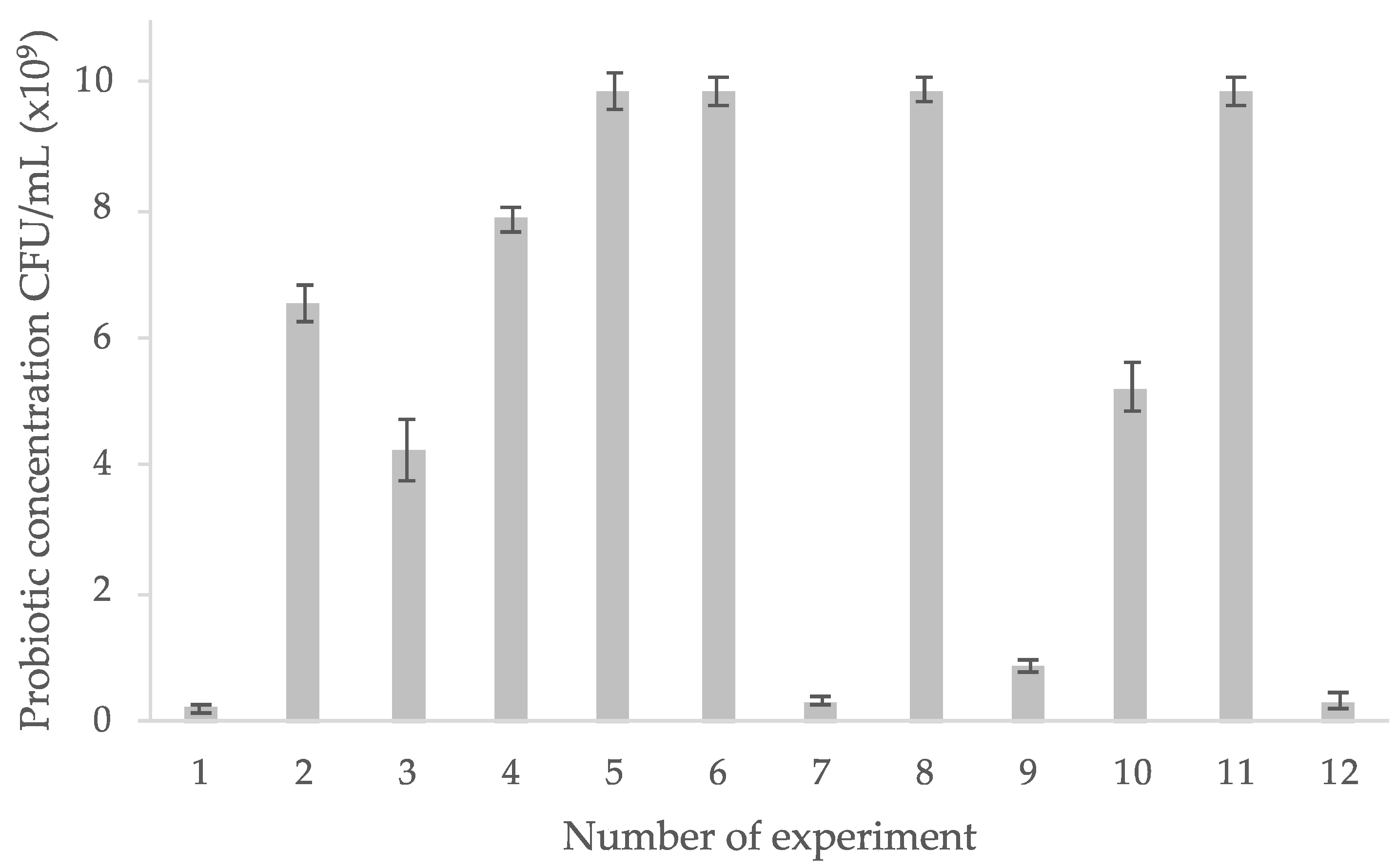
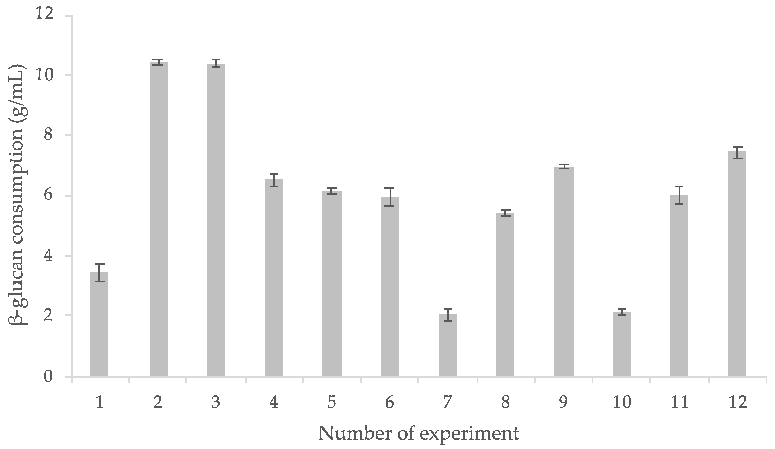
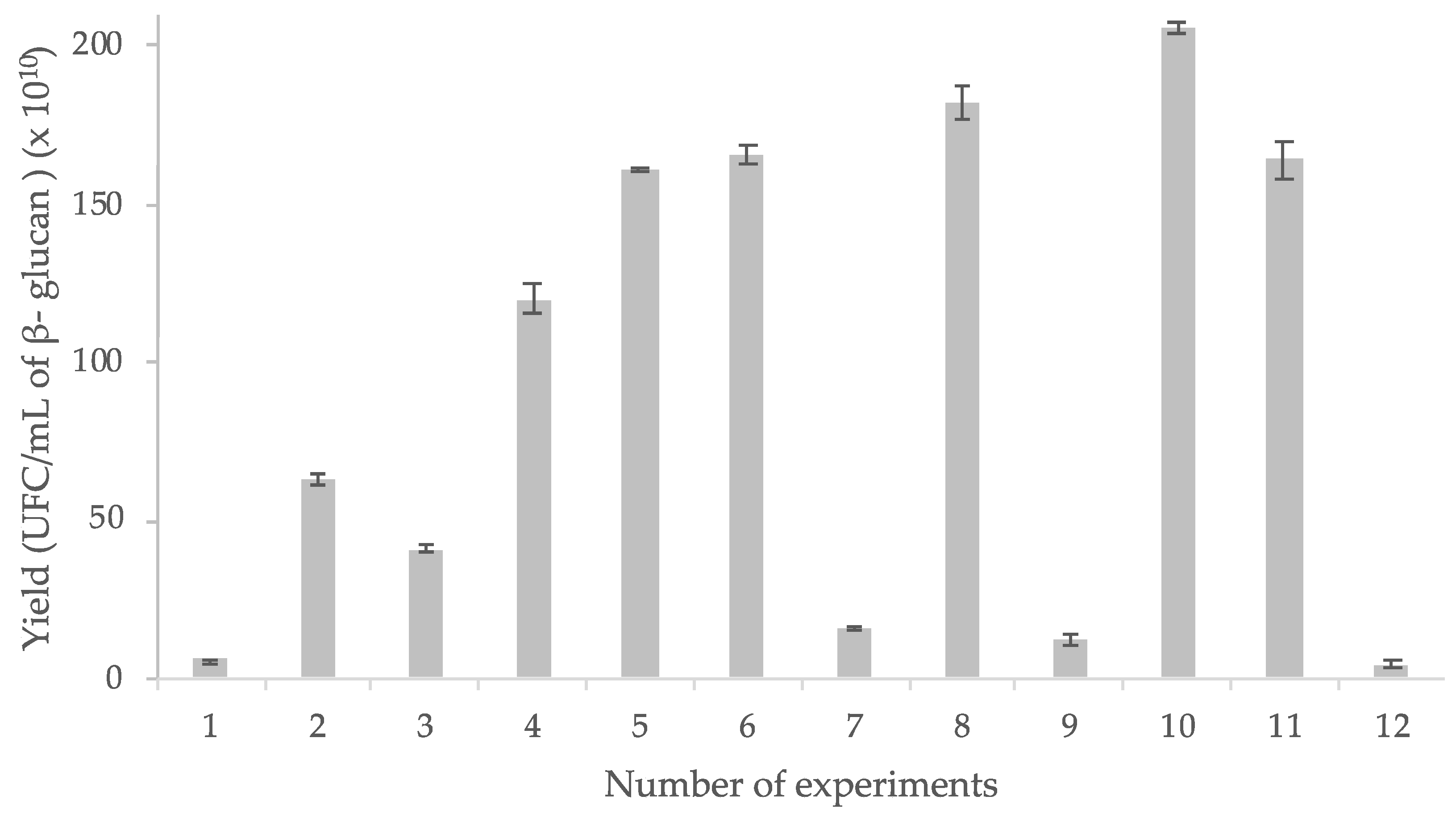
3.3. β-Glucan Intake and L. rhamnosus GG Survival

It was observed that the initial concentration of both; inoculum and β-glucan influenced the probiotic growth (Figure 1).
Experiments 1, 7, 9, and 12 showed the lowest probiotic concentration at the end of the study. In contrast, in the central points of the experimental design (experiments 5, 6, 8, and 11), which were made with 20 g/L of β-glucan and 4% of inoculum, the highest growth was observed. Therefore, the results demonstrated that the central points of the design of experiments were the most adequate to promote the growth of L. rhamnosus GG. In the control group, a growth of 0.9 × 10⁹ CFU/mL, was found.
In experiments 2 and 3, in which the initial concentrations of β-glucan and inoculum were higher, it was observed the best consumption of the prebiotic (Figure 2). In experiments 2 and 3, the initial concentrations of β-glucan and inoculum were the major and in them the highest consumption was obtained (Figure 2). In contrast, in experiments 7 and 10, in which there was a lower concentration of β-glucan (5.85 and 10 g/L respectively) and the most elevated initial inoculum concentrations (4 and 5% respectively), consumption was lesser.

3.4. Analysis of the Proteolytic Capacity of L. rhamnosus GG

According to the results obtained (Figure 3) in the central points of the experimental design with 20 g/L of β-glucan (5, 6, 8, and 11) a higher proteolytic activity was observed, and the free amino groups concentration was around 0.33 mg/L. In addition, in experiment 3, which had the highest concentration of prebiotic (34.41 g/L), free amino groups reached 0.179 mg/L; while in the experiment with the lowest concentration of prebiotic (experiment 7, 5.85 g/L) the concentration of free amino groups formed was close to 0.073 mg/L. Likewise, it was observed that the inoculum concentration, apparently, had an effect on the production of free amino groups. This is because in experiment 12, with a lower concentration of inoculum (2.58%), one of the lower proteolytic activities was observed.
However, to obtain the best probiotic growth and free amino groups production, it is necessary to combine prebiotic and inoculum concentrations. This is verified by comparing the results of experiment 1, which presented the lowest concentration of free amino groups, with the results of the experiments of the central points of the design. Thus, the higher yield, the maximum growth, and the best proteolytic activity are linked to the factors of the central points of the experimental design. The proteolytic activity of the control resulted in a concentration of 0.011 mg/L.

3.5. Experimental Design Results

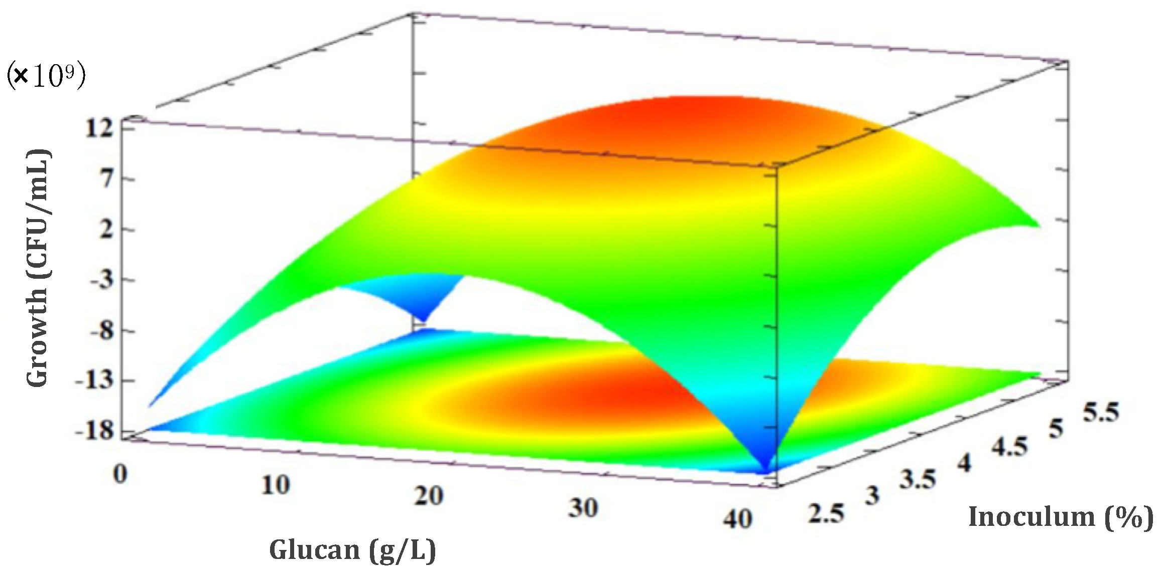
3.5.1. L. rhamnosus GG Growth Response

Response surface for L. rhamnosus GG derived from the central composite design is shown in Figure 4. It was observed that the optimal growth value was 1.04 × 10¹⁰ CFU/mL developed with 22.46 g/L of β-glucan and 4.38% of inoculum.
Equation (3) describes the model for growth with an R-squared of 97.53%. The percentage of inoculum was the factor with the highest significance. Although, due to the results obtained from the ANOVA (α = 0.05) all the terms were significant.
L = −5.79 × 10¹⁰ + (1.12 × 10⁹) (x1) + (2.47 × 10¹⁰) (x2) − (4.07 × 10⁷) (x1)2+ (1.30 × 10⁸)(x1) (x2) − (3.16 × 10⁹) (x2)2

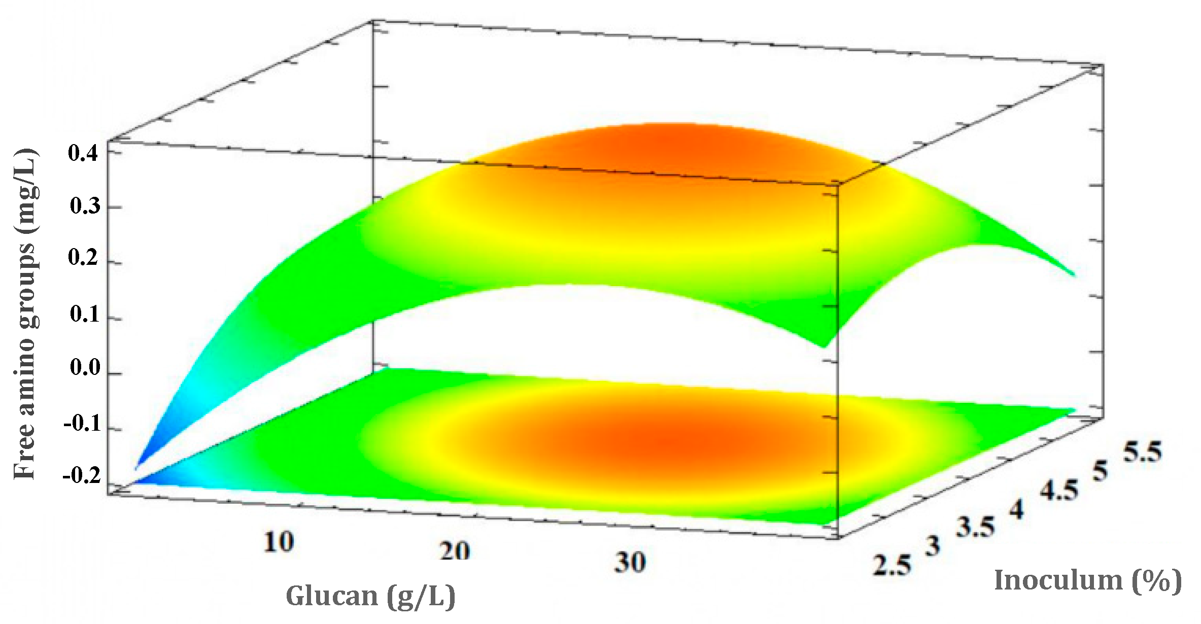
3.5.2. Free Amino Acid Groups Concentration Response (Proteolytic Activity of L. rhamnosus GG)

Regarding proteolysis (Figure 5), the response surface obtained showed that using 22.71 g/L of β-glucan in the culture medium combined with 4.18% of inoculum it was obtained the optimum production of free amino groups (0.33 mg/L).
Equation (4) describes the model for free amino groups generation with a R-square of 95.46%. According to the ANOVA (α = 0.05) all the conditions of the model had an effect during the proteolytic activity of L. rhamnosus GG, but inoculum concentration was the factor with the highest significance.
P = −1.6 + (0.04532) (x1) + (0.682) (x2) − (0.000834) (x1)2 − (0.00188) (x1)(x2) − (0.0766) (x2)2
It was observed that both inoculum initial concentration and prebiotic presence in the medium are implied in the proteolytic activity of the prebiotic.

3.5.3. Polynomial Model Validation

According to the experimental design, the optimal growth value of L. rhamnosus GG (1.04 × 10¹⁰ CFU/mL) is reached with 22.46 g/L of β-glucan and 4.38% of inoculum and the optimal concentration of free amino groups (0.33 mg/L) using 22.71 g/L of β-glucan combined with 4.18% of inoculum. Three confirmatory experiments were carried out under the optimal conditions. Comparison between the results of the confirmatory experiments and those predicted by the model showed no significant differences (Table 3).

4. Discussion

The proportion of β-glucan from oat calculated was similar to some studies, which have reported a proportion between 40.0 and 49.29% [15,32]. It is known that the highest concentration of β-glucan is found in the endosperm of the cereal (husk) [33], as found in this study. However, although the concentration of β-glucan in oats is dependent on the genotype, the nutrition of the crop, and the source of nitrogen supplemented during the development of the plant, it is the hot and humid climate that has the greatest influence on its concentration [34].
The concentration of β-glucan obtained by solubilization is, according to those reported in which the solubility of the β-glucan, related with the particle size having a great influence on the yield, in addition to the stirring time and the temperature [27]. Compared to other extraction methods, for example those using high pressures (>40 psi) and using temperatures above 100 °C, the yield could be improved up to three times. In this way, the properties of water change because hydrogen bonds are broken, increasing the solubility of β-glucan in short times [35].
During extraction of β-glucan obtained by enzymatic inactivation, the yield is related to the inactivation of glucanases, enzymes that break down β-glucan structures, because they can be inactivated by ethanol, lowering their activity from 80 U/kg to 70.5 U/kg [28]. Moreover, it has been reported that when changes in pH are applied (from pH = 10 to pH = 4), a higher concentration of β-glucan is obtained [36]. For that cause, enzymatic inactivation is the better method to use for obtaining β-glucan derived from cereals; indeed, this compound is a promising material to use in food processing, especially in those manufactured with some probiotic strains.
Studies have reported the importance of the presence of β-glucan in food matrices and its effect on the growth of different probiotic strains (L. fermentum, L. plantarum, and L. acidophilus) [14]. These authors observed that only on L. plantarum development there was a significant effect due to the presence of β-glucan. Furthermore, it has been determined that the presence of β-glucan is important during the first 24 h of fermentation because it activates the development of probiotics [37]. In addition, it is known that L. rhamnosus GG grows by hydrolyzing β-glucan when it is in the medium [8]. Additionally, it is known the presence of commercial prebiotics such as Regulact®, Oligomate 55®, and Frutafit® activate the L. rhamnosus GG development [38].
With the results obtained, it was observed that, in order to increase the yield of L. rhamnosus GG during fermentation, condition of central points from the experimental design could be used. In fact, yields obtained in this experiment were better than those reported in other study [39]. Authors found that the maximum yield of L. rhamnosus GG was 1.23 × 10⁹ CFU/g of the substrate in a medium enriched with 7.1% raw fiber from whole oat flour. This reflects the importance of having more available β-glucan substrates in the fermentation medium instead of oat flour.
In the case of the concentration of free amino groups, the results obtained were consistent with other authors who found a higher proteolytic activity of L. rhamnosus GG by adding agave juice during the manufacture of fermented milk [17]. Besides, another study also reports an increase in proteolytic activity using L. gasseri and a prebiotic extracted from Cudrania tricuspidata [40]. Thus, it is demonstrated that the presence of prebiotics stimulates the proteolytic activity of L. rhamnosus GG, which can lead to the production of metabolites such as bioactive peptides, bacteriocins, organic acids, among other molecules [41]. It is known that not only the prebiotic concentration influences the higher production of free amino groups, but the inoculum concentration is a factor that determines both the speed and the amount of free amino groups derived of the proteolytic process in fermented milk.
Inoculum concentration has been reported to influence the growth of some species of Lactobacillus. In a previous study, it was found that an initial inoculum amount used determines the final biomass concentration achieved. Counterintuitively, higher inoculum concentrations resulted in lower final biomass production, because higher initial biomass concentrations implied faster production and accumulation of lactic acid, which exhibits inhibition effects with a lower biomass net production. However, intermediate concentrations between 0.5 to 1 g/L of inoculum could provide better yield of biomass production of L. casei var rhmanosus in whey enriched with lactose after 10 h of fermentation [42]. Furthermore, other authors reported an effect on the growth of L. rhamnosus in the presence of β-glucan [6]. These results are consistent with those obtained in this work, which showed that the combination of both the initial inoculum concentration and the addition of β-glucan stimulate the biomass generation of L. rhamnosus GG.
In relation, it has been shown that an inoculum concentration of 1 × 10⁶ CFU/mL is enough to generate free amino groups concentrations close to 1 mg/L of fermented milk inoculated with L. rhamnosus [17,19,43]. Furthermore, other reports found that the higher production of free amino groups in the fermentation with L. casei is related to the higher inulin concentration assay (0.5% of inulin) [44]. This is the first report demonstrating the β-glucan influence on the proteolytic activity of L. rhamnosus GG.
It has been studied that prebiotics such as inulin are used as a carbon source (prebiotic) by different lactic acid bacteria in special for those with probiotic capacity. The use of prebiotics is directly related to an operon system integrated at probiotic metabolism, which is different in each bacterium. For example, L. casei contains the Lev operon, while in L. plantarum and L. paracasei the Fos operon is integrated. Both operons are related to inulin uptake [45]. For that reason, carbohydrate breakdown has a different mechanism of use and it is dependent on the strains utilized for fermentation [46]. In fact, different investigations have shown that some potential prebiotic materials integrated in production of fermented milks and ripened cheeses activate the proteolytic system of lactic acid bacteria in special those like-probiotics [17,47]. Recently, Habibi-Najafi et al. [48] and Pinto et al. [49] have reported the activation of the endoproteolytic system of different lactobacillus integrated into the manufacturing of symbiotic yogurt enriched by inulin, with activation being dependent on carbon source concentration. Furthermore, they conclude over the importance in the peptides production induced by the activation of some important peptidases, which are regulated by glucose.
In this sense, it has been reported the activation of two different peptidases due the metabolism of carbohydrates [50]. It has been demonstrated the glucose-dependence regulation by both pepR1 and pepQ genes, which encodes a transcriptional activator that regulates the expression of PepQ prolidase and PepR1 prolinase. This kind of lactobacillus peptidases are implied in the degradation of peptides within the cell to obtain essential amino acids, deriving peptide fractions from the cell, which could be measured as free amino groups in media. Morel et al. [51] propose a molecular mechanism of antagonist regulation of pepR1 and pepQ expression via the autoregulated PepR1 dependent of glucose, which is observed in Figure 6.
In addition, it is known that optimal pH of lactic acid bacteria proteinases is in a range of 5.0–5.5, and for that reason the rapid pH decrease because lactic acid formation favors the proteolytic activity [52]. In fact, the metabolism of lactic acid bacteria in the presence of β-glucan has not been studied, but according scientific reports, L. rhamnosus GG could have an operon related to the consumption of prebiotic or carbon sources such as β-glucan, which could regulate protein expression related of proteolytic system.
Finally, the validation of the model verified that both the greater growth of Lb. rhamnosus GG as the highest generation of free amino groups, and could be controlled with the combination of the concentration of β-glucan, and inoculum. Likewise, β-glucan, which is derived from oatsl is a promising material in the elaboration of symbiotic products due to the growth and the proteolytic activity which is improved in the probiotic.

5. Conclusions

This study marks the first report on the effect of β-glucan on growth and free amino groups production by L. rhamnosus GG during a milk fermentation process. The enzymatic inactivation method promotes greater extraction of β-glucan in oat semolina compared to the solubilization method. Likewise, both the growth of L. rhamnosus GG and its proteolytic activity are stimulated by the presence of β-glucan in a milk medium. Finally, the optimal conditions were found to stimulate the proteolytic activity and the growth of L. rhamnosus GG in a culture medium with milk added with β-glucan. These results are relevant for the formulation of a functional food in which a probiotic bacterium such as L. rhamnosus GG is incorporated in combination with β-glucan coupled with the bioactive potential of the peptides released during fermentation.
Further studies are needed in order to complete the information of peptides released during fermentation, should be done with the optimal conditions found in this study. This research provides some opportunities that could be explored in further experiments to increase the knowledge on peptides with potential bioactivity production by lactic acid bacteria and the real relationship between carbohydrates metabolism and proteolytic activity. Likewise, studies that demonstrate carbohydrate or prebiotics’ relationship to the biochemical regulation of expression of gene encoding peptidases in the proteolytic system of lactic acid bacteria are part of a research field little studied, and further studies would be beneficial.

Author Contributions

Conceptualization, G.M.R.-S. and M.I.C.d.l.V.; methodology, G.M.R.-S.; investigation, M.I.C.d.l.V., F.G.-R. and L.G.G.-O.; resources, G.M.R.-S., A.E.C.-G., M.G.-G. and L.G.-R.; writing—original draft preparation, M.I.C.d.l.V., G.M.R.-S., A.E.C.-G. and L.G.G.-O.; writing—review and editing S.A.-S.; supervision, G.M.R.-S. All authors have read and agreed to the published version of the manuscript.

Funding

This research received no external funding.

Institutional Review Board Statement

Not applicable.

Data Availability Statement

Not applicable.

Acknowledgments

The authors appreciate the support of CONACYT for the MSc. scholarship number 729460 granted to María Isabel Chávez de la Vega.

Conflicts of Interest

The authors declare no conflict of interest.

References

  1. Havrlentova, M.; Petrulakova, Z.; Burgarova, A.; Gago, F.; Hlinkova, A.; Šturdík, E. β-glucans and their significance for the preparation of functional foods—A review. Czech. J. Food Sci. 2011, 29, 1–14. [Google Scholar] [CrossRef] [Green Version]
  2. Henrion, M.; Francey, C.; Lê, K.-A.; Lamothe, L. Cereal B-Glucans: The Impact of Processing and How It Affects Physiological Responses. Nutrients 2019, 11, 1729. [Google Scholar] [CrossRef] [PubMed] [Green Version]
  3. Olveira, G.; Gibson, G.R.; Hutkins, R.; Sanders, M.E.; Prescott, S.L.; Reimer, R.A.; Salminen, S.J.; Scott, K.; Stanton, C.; Swanson, K.S.; et al. Expert consensus document: The International Scientific Association for Probiotics and Prebiotics (ISAPP) consensus statement on the definition and scope of prebiotics. Nat. Rev. Gastroenterol. Hepatol. 2017, 14, 491–502. [Google Scholar] [CrossRef] [Green Version]
  4. Valcheva, R.; Dieleman, L.A. Prebiotics: Definition and protective mechanisms. Best. Pract. Res. Clin. Gastroenterol. 2016, 30, 27–37. [Google Scholar] [CrossRef] [PubMed]
  5. Roberfroid, M.; Gibson, G.R.; Hoyles, L.; McCartney, A.L.; Rastall, R.; Rowland, I.; Wolvers, D.; Watzl, B.; Szajewska, H.; Stahl, B.; et al. Prebiotic effects: Metabolic and health benefits. Br. J. Nutr. 2010, 104, S1–S63. [Google Scholar] [CrossRef] [PubMed] [Green Version]
  6. Al-Sheraji, S.H.; Ismail, A.; Manap, M.Y.; Mustafa, S.; Yusof, R.M.; Hassan, F.A. Prebiotics as functional foods: A review. J. Funct. Foods 2013, 5, 1542–1553. [Google Scholar] [CrossRef]
  7. Zhu, F.; Du, B.; Xu, B. A critical review on production and industrial applications of beta-glucans. Food Hydrocoll. 2016, 52, 275–288. [Google Scholar] [CrossRef]
  8. Dong, J.L.; Yu, X.; Dong, L.E.; Shen, R.L. In vitro fermentation of oat β-glucan and hydrolysates by fecal microbiota and selected probiotic strains. J. Sci. Food Agric. 2017, 97, 4198–4203. [Google Scholar] [CrossRef]
  9. Jayachandran, M.; Chen, J.; Chung, S.; Xu, B. A critical review on the impacts of β-glucans on gut microbiota and human health. J. Nutr. Biochem. 2018, 61, 101–110. [Google Scholar] [CrossRef]
  10. Bozbulut, R.; Sanlier, N. Promising effects of β-glucans on glyceamic control in diabetes. Trends Food Sci. Technol. 2019, 83, 159–166. [Google Scholar] [CrossRef]
  11. Sima, P.; Vannucci, L.; Vetvicka, V. β-glucans and cholesterol (Review). Int. J. Mol. Med. 2018, 41, 1799–1808. [Google Scholar] [CrossRef] [Green Version]
  12. Korolenko, T.A.; Bgatova, N.P.; Ovsyukova, M.V.; Shintyapina, A.; Vetvicka, V. Hypolipidemic Effects of β-Glucans, Mannans, and Fucoidans: Mechanism of Action and Their Prospects for Clinical Application. Molecules 2020, 25, 1819. [Google Scholar] [CrossRef] [PubMed] [Green Version]
  13. Olvera-Rosales, L.-B.; Cruz-Guerrero, A.-E.; Ramírez-Moreno, E.; Quintero-Lira, A.; Contreras-López, E.; Jaimez-Ordaz, J.; Castañeda-Ovando, A.; Añorve-Morga, J.; Calderón-Ramos, Z.-G.; Arias-Rico, J.; et al. Impact of the Gut Microbiota Balance on the Health–Disease Relationship: The Importance of Consuming Probiotics and Prebiotics. Foods 2021, 10, 1261. [Google Scholar] [CrossRef] [PubMed]
  14. Arena, M.P.; Caggianiello, G.; Fiocco, D.; Russo, P.; Torelli, M.; Spano, G.; Capozzi, V. Barley β-glucans-containing food enhances probiotic performances of beneficial bacteria. Int. J. Mol. Sci. 2014, 15, 3025–3039. [Google Scholar] [CrossRef] [PubMed] [Green Version]
  15. Russo, P.; de Chiara, M.L.V.; Capozzi, V.; Arena, M.P.; Amodio, M.L.; Rascón, A.; Dueñas, M.T.; López, P.; Spano, G. Lactobacillus plantarum strains for multifunctional oat-based foods. LWT Food Sci. Technol. 2016, 68, 288–294. [Google Scholar] [CrossRef] [Green Version]
  16. Sharma, P.; Trivedi, N.; Gat, Y. Development of functional fermented whey–oat-based product using probiotic bacteria. 3 Biotech 2017, 7, 272. [Google Scholar] [CrossRef]
  17. Jaimez-Ordaz, J.; Martinez-Ramirez, X.; Cruz-Guerrero, A.E.; Contreras-Lopez, E.; Ayala-Nino, A.; Castro-Rosas, J.; González-Olivares, L.G. Survival and proteolytic capacity of probiotics in a fermented milk enriched with agave juice and stored in refrigeration. Food Sci. Technol. 2019, 39, 188–194. [Google Scholar] [CrossRef]
  18. Rolim, F.R.; Neto, O.C.F.; Oliveira, M.E.G.; Oliveira, C.J.; Queiroga, R.C. Cheeses as food matrixes for probiotics: In vitro and in vivo tests. Trends Food Sci. Technol. 2020, 100, 138–154. [Google Scholar] [CrossRef]
  19. Rojas-Ronquillo, R.; Cruz-Guerrero, A.; Flores-Nájera, A.; Rodríguez-Serrano, G.; Gómez-Ruiz, L.; Reyes-Grajeda, J.P.; Jiménez-Guzmán, J.; García-Garibay, M. Antithrombotic and angiotensin-converting enzyme inhibitory properties of peptides released from bovine casein by Lactobacillus casei Shirota. Int. Dairy J. 2012, 26, 147–154. [Google Scholar] [CrossRef]
  20. de Lourdes, E.; de Lima, C.; Roberta, H. Optimized fermentation of goat cheese whey with Lactococcus lactis for production of antilisterial bacteriocin-like substances. LWT Food Sci. Technol. 2017, 84, 710–716. [Google Scholar] [CrossRef]
  21. González-Olivares, L.G.; López-Cuellar, Z.L.; Añorve-Morga, J.; Franco-Fernández, M.J.; Castañeda-Ovando, A.; Contreras-López, E.; Jaimez-Ordaz, J.; Rodríguez-Serrano, G.M. Viability and proteolytic capacity of Lactobacillus bulgaricus 2772 and Lactobacillus rhamnosus GG during cheese ripening. J. Biosci. Med. 2014, 2014, 7–12. [Google Scholar]
  22. Aguilar-Toalá, J.E.; Santiago-López, L.; Peres, C.M.; Peres, C.; Garcia, H.S.; Vallejo-Cordoba, B.; Hernández-Mendoza, A. Assessment of multifunctional activity of bioactive peptides derived from fermented milk by specific Lactobacillus plantarum strains. J. Dairy Sci. 2017, 100, 65–75. [Google Scholar] [CrossRef]
  23. Pérez-Escalante, E.; Jaimez-Ordaz, J.; Castañeda-Ovando, A.; Contreras-López, E.; Añorve-Morga, J.; González-Olivares, L.G. Antithrombotic activity of milk protein hydrolysates by lactic acid bacteria isolated from commercial fermented milks. Braz. Archiv. Biol. Technol. 2018, 61, e18180132. [Google Scholar] [CrossRef] [Green Version]
  24. Fuentes, F.; Poblete, C.; Huerta, M.; Palape, I. Evaluación de la producción y calidad nutritiva de avena como forraje verde hidropónico en condiciones de desierto. Idesia 2011, 29, 75–81. [Google Scholar] [CrossRef] [Green Version]
  25. Pontieri, P.; Pepe, G.; Campiglia, P.; Meriai, F.; Basilicata, M.G.; Smolensky, D.; Calcagnile, M.; Troisi, J.; Romano, R.; Del Giudice, F.; et al. Comparision of content in phenolic compounds and antioxidant capacity in grains of white, red and black sorghum varieties grown in the Mediterranean area. ACS Food Sci. Technol. 2021, 1, 1109–1119. [Google Scholar] [CrossRef]
  26. Horwitz, W.; Latimer, G. Official Methods of Analysis of AOAC International, 18th ed.; AOAC International: Gaithersburg, MA, USA, 2005. [Google Scholar]
  27. Benito-Román, O.; Alonso, E.; Lucas, S. Optimization of the β-glucan extraction conditions from different waxy barley cultivars. J. Cereal Sci. 2011, 53, 271–276. [Google Scholar] [CrossRef]
  28. Beer, M.U.; Arrigoni, E.; Amado, R. Extraction of oat gum from oat bran: Effects of process on yield, molecular weight distribution, viscosity and (1→3)(1→4)-b-D-glucan content of the gum. Cereal Chem. 1996, 73, 58–62. [Google Scholar]
  29. Cruz-Guerrero, A.; Hernández-Sánchez, H.; Rodríguez-Serrano, G.; Gómez- Ruiz, L.; García-Garibay, M.; Figueroa-González, I. Commercial probiotic bacteria and prebiotic carbohydrates: A fundamental study on prebiotics uptake, antimicrobials production and inhibition of pathogens. J. Sci. Food Agric. 2014, 94, 2246–2252. [Google Scholar] [CrossRef] [PubMed]
  30. Herigstad, B.; Hamilton, M.; Heersink, J. How to optimize the drop plate method for enumerating bacteria. J. Microbiol. Methods 2001, 44, 121–129. [Google Scholar] [CrossRef]
  31. Alder-Nissen, J. Determination of the degree of hydrolysis of food protein hydrolysates by trinitrobenzensulfonic acid. J. Agric. Food Chem. 1979, 27, 1256–1262. [Google Scholar] [CrossRef] [PubMed]
  32. Bai, J.; Ren, Y.; Li, Y.; Fan, M.; Qian, H.; Wang, L.; Wu, G.; Zhang, H.; Qi, X.; Xu, M.; et al. Physiological functionalities and mechanisms of β-glucans. Trends Food Sci. Technol. 2019, 88, 57–66. [Google Scholar] [CrossRef]
  33. Wood, P.J. Cereal β-glucans in diet and health. J. Cereal Sci. 2007, 46, 230–238. [Google Scholar] [CrossRef]
  34. Hernandez-Campuzano, A.V.; Martínez-Rueda, C.G.; Estrada-Campuzano, G.; Dominguez-López, A. Effect of genotype and nitrogen fertilization on the yield, nitrogen content and β-glucans in the grain of oats (Avena sativa L.). Rev. Investig. Agropecu. 2018, 44, 88–95. [Google Scholar]
  35. Yoo, H.U.; Ko, M.J.; Chung, M.S. Hydrolysis of beta-glucan in oat flour during subcritical-water extraction. Food Chem. 2020, 308, 125670. [Google Scholar] [CrossRef] [PubMed]
  36. Gangopadhyay, N.; Hossain, M.B.; Rai, D.K.; Brunton, N.P. Optimisation of yield and molecular weight of β-glucan from barley flour using response surface methodology. J. Cereal Sci. 2015, 62, 38–44. [Google Scholar] [CrossRef]
  37. Arora, T.; Sharma, R.K. Prebiotic effectiveness of galactooligosaccharides and β-glucan in stimulation of growth of Lactobacillus acidophilus NCDC 13 in vitro. Curr. Top. Nutr. Res. 2011, 9, 67. [Google Scholar]
  38. Figueroa-Gonzalez, I.; Rodríguez-Serrano, G.; Gomez-Ruiz, L.; Garcia-Garibay, M.; Cruz-Guerrero, A. Prebiotic effect of commercial saccharides on probiotic bacteria isolated from commercial products. Food Sci. Technol. 2019, 39, 747–753. [Google Scholar] [CrossRef] [Green Version]
  39. Kocková, M.; Mendel, J.; Medveďová, A.; Šturdík, E.; Valík, Ľ. Cereals and pseudocereals as substrates for growth and metabolism of a probiotic strain Lactobacillus rhamnosus GG. J. Food Nutr. Res. 2013, 52, 25–36. [Google Scholar]
  40. Oh, N.S.; Lee, J.Y.; Kim, Y. The growth kinetics and metabolic and antioxidant activities of the functional synbiotic combination of Lactobacillus gasseri 505 and Cudrania tricuspidata leaf extract. Appl. Microbiol. Biotechnol. 2016, 100, 10095–10106. [Google Scholar] [CrossRef]
  41. Sanders, M.E.; Merenstein, D.J.; Reid, G.; Gibson, G.R.; Rastall, R.A. Probiotics and prebiotics in intestinal health and disease: From biology to the clinic. Nat. Rev. Gastroenterol. Hepatol. 2019, 16, 605–616. [Google Scholar] [CrossRef]
  42. Álvarez, M.M.; Aguirre-Ezkauriatza, E.J.; Ramírez-Medrano, A.; Rodríguez-Sánchez, Á. Kinetic analysis and mathematical modeling of growth and lactic acid production of Lactobacillus casei var. rhamnosus in milk whey. J. Dairy Sci. 2010, 93, 5552–5560. [Google Scholar] [CrossRef]
  43. Begunova, A.V.; Savinova, O.S.; Glazunova, O.A.; Moiseenko, K.V.; Rozhkova, I.V.; Fedorova, T.V. Development of antioxidant and antihypertensive properties during growth of Lactobacillus helveticus, Lactobacillus rhamnosus and Lactobacillus reuteri on Cow’s Milk: Fermentation and peptidomics study. Foods 2021, 10, 17. [Google Scholar] [CrossRef]
  44. Habibi Najafi, M.B.; Fatemizadeh, S.S.; Tavakoli, M. Effect of fat percentage and prebiotic composition on proteolysis, ACE-inhibitory and antioxidant activity of probiotic yogurt. Int. J. Nutr. Food Eng. 2017, 11, 622–628. [Google Scholar] [CrossRef]
  45. Buntin, N.; Hongpattarakere, T.; Ritari, J.; Douillard, F.P.; Paulin, L.; Boeren, S.; Shetty, A.; de Vos, W.M. An inducible operon is involved in inulin utilization in Lactobacillus plantarum strains, as revealed by comparative proteogenomics and metabolic profiling. Appl. Environ. Microbiol. 2017, 83, e02402-16. [Google Scholar] [CrossRef] [Green Version]
  46. Cui, Y.; Qu, X. Genetic mechanisms of prebiotic carbohydrate metabolism in lactic acid bacteria: Emphasis on Lacticaseibacillus casei and Lacticaseibacillus paracasei as flexible, diverse and outstanding prebiotic carbohydrate starters. Trends Food Sci. Technol 2021, 115, 486–499. [Google Scholar] [CrossRef]
  47. Ramírez-Godínez, J.; Gutiérrez-Rodríguez, J.F.; Contreras-López, E.; Rodríguez-Serrano, G.M.; Castañeda-Ovando, A.; Jaimez-Ordaz, J.; González-Olivares, L.G. Agave Juice improves survival and proteolytic activity of Lactobacillus rhamnosus GG during ripening of semi-ripened Mexican cheese. Food Sci. Technol. 2021. [Google Scholar] [CrossRef]
  48. Habibi-Najafi, M.B.; Sadat-Fatemizadeh, S.; Tavakoli, M. Release of proteolysis products with ACE-inhibitory and antioxidant activities in probiotic yogurt containing different levels of fat and prebiotics. Int. J. Pept. Res. Ther. 2019, 25, 367–377. [Google Scholar] [CrossRef]
  49. Pinto, G.; Picariello, G.; Addeo, F.; Chianese, L.; Scaloni, A.; Caira, S. Proteolysis and process-induced modifications in symbiotic yogurt investigated by peptidomics and phosphopeptidomics. J. Agric. Food Chem. 2020, 68, 8744–8754. [Google Scholar] [CrossRef] [PubMed]
  50. Savijoki, K.; Ingmer, H.; Varmanen, P. Proteolytic system of lactic acid bacteria. Appl. Microbiol. Biotechnol. 2006, 71, 394–406. [Google Scholar] [CrossRef] [PubMed]
  51. Morel, F.; Lamarque, M.; Bissardon, I.; Atlan, D.; Galinier, A. Autoregulation of the biosynthesis of the CcpA-like protein, PepR1 in Lactobacillus delbrueckii subsp bulgaricus. J. Mol. Microbiol. Biotechnol. 2001, 3, 63–66. [Google Scholar]
  52. Rodríguez-Serrano, G.M.; Garcia-Garibay, J.M.; Cruz-Guerrero, A.E.; Gomez-Ruiz, L.D.C.; Ayala-Nino, A.; Castañeda-Ovando, A.; González-Olivares, L.G. Proteolytic System of Streptococcus thermophilus. J. Microbiol. Biotechnol. 2018, 28, 1581–1588. [Google Scholar] [CrossRef] [PubMed]
Figure 1. Influence of the initial concentration of inoculum and β-glucan (according to the experimental design) over L. rhamnossus GG growth.
Fermentation 07 00210 g001
Figure 2. β-glucan consumption by L. rhamnosus GG under fermentation conditions according to the experimental design.
Fermentation 07 00210 g002
Figure 3. Proteolytic activity of L. rhamnosus GG in fermentation with β-glucan according to conditions of the experimental design.
Fermentation 07 00210 g003
Figure 4. Response surface derived from the combination effect of the β-glucan concentration and the percentage of inoculum over L. rhamnosus GG growth.
Fermentation 07 00210 g004
Figure 5. Response surface derived from the combination of the β-glucan concentration and the percentage of inoculum over free amino groups amount during milk fermentation by L. rhamnosus GG.
Fermentation 07 00210 g005
Figure 6. Mechanism of biosynthesis regulation of PepQ by PepR1 in L. bulgaricus in the absence of glucose (A) or in the presence of glucose (B). Glucose regulates production of PepQ through the binding of PepR1 to the single catabolite-responsible element (cre) box, which partly overlaps the −35 position of the PepR1 promoter (PpepR1) and which is located 16 nt upstream of the PepQ promoter (PpepQ). The position of the cre box target with regard to both promoters is essential for the inhibitory or stimulatory transcriptional effect resulting from the PepR1 binding. Image designed according to information extracted from Morel et al. [51].
Fermentation 07 00210 g006
Table 1. Selected experimental factors for β-glucan (x1) and inoculum concentration (x2) for milk fermentation.
Experimental FactorLevel
−α−101
x15.8510203034.41
x22.583455.41
Number of experimentFactor
x1x2
1−1−1
211
30
40
500
600
7−α0
800
91−1
10−11
1100
120−α
Factor x1 expressed in g/L and factor x2 in percentage of inoculum.
Table 2. Physicochemical composition and β-glucan content of oat fractions after milling.
Parameters
(g/100 g of Sample)
HuskSemolinaFlour
Moisture20.02 ±0.6221.02 ± 0.1419.05 ± 0.18
Ash5.24 ± 0.372.12 ± 0.161.16 ± 0.01
Fiber5.24 ± 0.374.21 ± 0.070.29 ± 0.03
Protein4.51 ± 1.3511.73 ± 0.118.94 ± 0.42
Fat3.23 ± 0.156.58 ± 0.406.90 ± 0.11
Carbohydrate25.70 ± 1.7554.35 ± 0.2363.67 ± 0.58
β-glucan3.35 ± 0.051.24 ± 0.060.12 ± 0.08
Table 3. Predicted and experimental values of the model.
VariablePredictedExperimentalP (α = 0.05)
Free amino groups (mg/L)0.330.33 ± 0.020.098
Growth (UFC/mL) × 10910.449.87 ± 0.070.059
Publisher’s Note: MDPI stays neutral with regard to jurisdictional claims in published maps and institutional affiliations.

