Article

Fermentation Profile and Probiotic-Related Characteristics of Bifidobacterium longum MC-42

by
Anna V. Begunova
1,
Irina V. Rozhkova
1,
Olga A. Glazunova
2,*,
Konstantin V. Moiseenko
2,
Olga S. Savinova
2 and
Tatyana V. Fedorova
2,*
1
Federal State Budgetary Scientific Institution “All-Russian Research Institute of Dairy Industry”, 115093 Moscow, Russia
2
Research Center of Biotechnology of the Russian Academy of Sciences, A.N. Bach Institute of Biochemistry, 119071 Moscow, Russia
*
Authors to whom correspondence should be addressed.
Fermentation 2021, 7(3), 101; https://doi.org/10.3390/fermentation7030101
Submission received: 26 May 2021 / Revised: 12 June 2021 / Accepted: 21 June 2021 / Published: 26 June 2021
(This article belongs to the Special Issue Fermented Foods and Microbes Related to Health)

Abstract

:
This article presents new data on Bifidobacterium longum MC-42—a strain that has been actively used for the preparation of commercial dairy products in Russia for almost 40 years. It was demonstrated that this strain possesses high activities of β-galactosidase, α-glucosidase, and leucine arylaminidase; inhibits the growth of pathogens such as Salmonella typhimurium, Staphylococcus aureus, and Escherichia coli; and can efficiently remove cholesterol from the cultural medium. The resistance of B. longum MC-42 determined for 15 commonly used antibiotics was in agreement with those previously reported for Bifidobacterium spp. The absence of frequently transmittable antibiotic resistance genes in the genome and the lack of undesirable activity of β-glucuronidase proved the safe use of B. longum MC-42 as a probiotic and starter culture. Additionally, the impact of two growth-promoting additives—yeast extract or milk protein hydrolysate containing supplementation—on the B. longum MC-42 fermentation profile was assessed. The introduction of these additives increases the maximum attainable viable cell count by orders of magnitude, significantly changed the profile of aminopeptidase activities in extracellular extracts, and influenced the antioxidant and antihypertensive properties of the obtained fermented products.

1. Introduction

In recent years, probiotics—living microorganisms providing health benefits upon adequate consumption—have gained incredible attention both from ordinary customers and professional scientists [1]. While it was originally thought that probiotics promote physical well-being primarily through the improvement of microbial balance, later research demonstrated that the action of probiotics on organism goes far beyond direct interaction with a consumer’s indigenous microflora [2]. The number of recently discovered properties of probiotics, often with strain-specificity such as improvement of intestinal barrier [3], production of beneficial enzymes (e.g., β-galactosidase and bile salt hydrolase) [4], modulation of immune system [5], synthesis of neurochemicals [6], and production of bioactive peptides [7], stimulates the search for new probiotic strains and reevaluation of old ones.
Bifidobacterium is a widely known genus of probiotic bacteria [8]. Being first introduced with breast milk, bifidobacteria constitutes 60–70% of the fecal bacteria in infancy and 30–40% in adulthood [9]. The long history of bifidobacteria consumption (both as part of fermented milk microflora and as commercially manufactured probiotic products) firmly established their safety and positive health benefits [10]. Currently, various strains of Bifidobacterium spp. are used in commercial products throughout the world. Although almost all of these strains were isolated many decades ago, an investigation on their beneficial properties are still actively carried out. Recently, systematical reviews regarding properties of several commercialized bifidobacterial strains, such as Bifidobacterium animalis subsp. lactis BB-12 [11], Bifidobacterium bifidum BGN4 [12], and Bifidobacterium breve M-16V [13], were published in scientific literature.
The strain Bifidobacterium longum MC-42 was isolated from the feces of infants by the staff of Scientific Research Institute of the Dairy Industry and patented in 1982 (USSR patent № 863639); at the time of isolation, it was identified as Bifidobacterium adolescentis. In the past 40 years, this strain was actively used (both alone and as part of a mixed starter culture) for the preparation of commercial dairy products and, currently, can be purchased in lyophilized form as a dietary supplement. Although advantageous technological properties of this strain such as the high viability of a lyophilized culture and the good sensory properties of the fermented products were time-tested, many aspects of its probiotic properties remain unclear. Only recently, the genome of B. longum MC-42 was sequenced [14], and the response of a human gut microbiome on short-term supplementation of this strain was investigated [15].
The aim of the current study was to investigate the B. longum MC-42 probiotic properties by evaluation of its profile of enzymatic activities, its ability to inhibit the growth of common pathogens, its resistance to common antibiotics, its possession of transmittable antibiotic resistance genes, and its capacity for cholesterol removal. Additionally, the fermentations of skim milk and skim milk supplemented with two different growth promoting additives (yeast extract and milk protein hydrolysate containing supplementation) performed by B. longum MC-42 were described in terms of strain’s growth characteristics, acidification capability, and proteolytic activity. The resulting fermented products were assessed in vitro for their antioxidant and antihypertensive properties.

2. Materials and Methods

2.1. Strain Reactivation and Preparation of Starting Inoculum

The strain B. longum MC-42 was obtained from the Microorganism Collection of the All-Russian Research Institute of the Dairy Industry (VNIMI, Moscow, Russia), where it was stored as a lyophilized culture at −80 °C. The strain was reactivated in commercial milk hydrolysate containing corn-lactose GMK-2 medium for bifidobacteria (Biokompas-S, Uglich, Russia) at 37 °C for 24–48 h (to achieve a turbidity of 0.5–1 McFarland standard). For all experiments, the starting inoculum was prepared by the addition of 3% (v/v) of a reactivated strain into 200 mL of a GMK-2 medium with subsequent incubation at 37 °C for 24–48 h (to achieve a turbidity of 0.5–1 McFarland standard) and adjustment to approximately 107 CFU·mL−1.
An evaluation of the enzymatic activities of B. longum MC-42 was carried out using the API ZYM kit (BioMerieux, Marcy-l’Étoile, France), according to the manufacturer instructions. For analysis, B. longum was grown in MRS broth for 72 h at 37 °C.

2.2. Assessment of Inhibition of Pathogens

The pathogenic bacterium strains Staphylococcus aureus ATCC-6538 and Escherichia coli ATCC-25922 were purchased from American Type Culture Collection (Manassas, VA, USA), and Salmonella typhimurium NCTC 00074 was purchased from National Collection of Type Cultures (Salisbury, UK). The strains were cultivated on agar slants (21.0 g·L−1 peptone, 6.5 g·L−1 NaCl, 6.25 g·L−1 glucose, 3.5 g·L−1 Na2HPO4, 0.6 g·L−1 KH2PO4, and 12.5 g·L−1 agar) for 24 h at 37 ± 2 °C. For inoculation, the cell suspension was washed off the agar slant and diluted to approximately 107 CFU·mL−1.
The antagonistic activity was evaluated by the co-culture method [16]. For the experimental samples, 20 mL of MRS broth was simultaneously inoculated with 1 mL of B. longum MC-42 and 1 mL of the pathogenic test-strain starting inocula; for the control samples, only inoculation with the pathogenic test-strain was performed. The incubation was carried out at 37 ± 2 °C, and samples were collected after 24 and 48 h. Pathogen cells were counted at SPA agar medium (Mikrogen, Moscow, Russia) at 37 °C for 24–48 h.

2.3. Antibiotic Resistance Assays

The antibiotic resistance of B. longum MC-42 was assessed by the disk diffusion method. MRS agar plates were inoculated with 1 mL of the B. longum MC-42 starting inoculum, and antibiotic disks (DI-PLS-50-01, NICF, St. Peterburg, Russia) were placed in the center of each plate. The plates were incubated under anaerobic conditions using Oxoid AnaeroJar 2.5 L (Thermo Fisher Scientific, USA) and GasPak sachets (BD Biosciences, San Jose, CA, USA) for 18–24 h at 37 °C, followed by measurement of the inhibition zone diameters.

2.4. Search for Genes of Interest in Genome

To search for genes of interest, the genome of B. longum MC-42 was downloaded from the NCBI GenBank database [17]; the GenBank assembly accession is GCA_001516925.1. Representative sequences for the antibiotic resistance genes were downloaded from the NCBI RefSeq database. The representative sequence of bile salt hydrolase (BSH) was that reported in [18]; GenBank accession is AF148138. The representative sequences of cholesterol transporters and cholesterol reductase were those proposed in [19]; the locus tags according to the Bifidobacterium bifidum PRL2010 genome (GenBank assembly accession is GCA_000165905.1) are BBPR_0676, BBPR_1704, BBPR_1348, BBPR_0146, BBPR_1508, and BBPR_0519. The search was performed using the BLAST [20] program, and the locus tags of the identified genes are shown in parentheses thorough the text.

2.5. Cholesterol-Removal Capacity Assay

To assess cholesterol-removal capacity, B. longum MC-42 was cultivated in MRS broth containing 1.5 mM of cholesterol (Sigma-Aldrich, St. Louis, MO, USA) at 37 °C for 24 h. Inoculation was performed with 1% of B. longum MC-42 culture. The residual concentration of cholesterol was determined by HPLC using a Gilson chromatographic system (Gilson Medical Electronics, Middleton, WI, USA) equipped with Luna C18 column (4.6 × 150 mm, 5 μm, Phenomenex, Torrance, CA, USA). The mobile phase comprised acetonitrile and water in a ratio of 90:10 (v:v). The flow rate of the mobile phase was 2 mL∙min−1. Detection was carried out using a diode array detector at the 210 nm wavelength.

2.6. Fermentations on Milk and Milk Supplemented with Growth-Promoting Additives

Fermentations of skim milk, skim milk containing 2 g·L−1 of yeast extract (YE; FBUN GNC PMB, Obolensk, Russia), and skim milk containing 2 g·L−1 of GMK-3 growth-promoting supplement for bifidobacteria (Biokompas-S, Uglich, Russia) were performed using the DASGIP bioreactor system (Eppendorf, Hamburg, Germany). The composition of GMK-3 was as follows (per 100 g): 66 g milk protein hydrolysate, 33 g corn extract, and 1 g ascorbic acid. For fermentation, the sterile growth media were aseptically inoculated with 1% (v/v) of the B. longum MC-42-starting inoculum and cultivated at 37 ± 2 °C for 72 h. Sampling was performed immediately after inoculation (0 h) and at 6, 16, 24, and 72 h of cultivation.
The extracellular extracts (EEs) were prepared as follows: if the pH of a sample was above 4.6, it was adjusted to 4.6 by the addition of trichloroacetic acid; the samples were centrifuged at 10,000× g for 20 min at 4 °C; the supernatant was filtered through a 0.45 µm syringe filter and stored at −80 °C until further analysis.
The protein content of EEs was determined using the Pierce BSA Protein Assay Kit (ThermoFisher, Rockford, IL, USA).

2.7. Enzymatic Activity Profile and Peptidase Assays

The lysine and leucine aminopeptidase activities (Lys-AA and Leu-AA, respectively) in EEs were determined with lysine p-nitroanilide (Lys–p-NA, Sigma-Aldrich, Buchs, Switzerland) and leucine p-nitroanilide (Leu–p-NA, Sigma-Aldrich, St. Louis, MO, USA) chromogenic substrates, respectively. X-prolyl-dipeptidyl aminopeptidase activity was measured with glycine–proline p-nitroanilide (Gly–Pro–p-NA, Sigma-Aldrich, St. Louis, MO, USA) as chromogenic substrate. The assay mixture contained 50 μL of the substrate (20 mM for Lys–p-NA and Leu–p-NA, and 10 mM for Gly–Pro–p-NA in methanol), 500 μL of 50 mM potassium phosphate buffer (pH 7.0), and 50 μL of sample (EE). After incubation at 37 °C for 4 h, the degree of hydrolysis was determined spectrophotometrically by measuring the absorbance of the colored product (p-nitroaniline) at 410 nm using a Lambda 35 spectrophotometer (PerkinElmer, Waltham, MA, USA) according to [21]. One unit of enzymatic activity was defined as an amount of the enzyme required to release 1 μmol of p-nitroaniline (ε410 = 8800 M−1·cm−1) per minute and per gram of protein under the assay conditions. All measurements were carried out in triplicate.

2.8. Proteolytic, Antioxidant, and Angiotensin-I-Converting Enzyme Inhibitory Activities

The antioxidant activity in EEs was determined by the oxygen radical absorbance capacity fluorescence method (ORAC) with generation of the peroxyl radical as described in [22] and Trolox equivalent antioxidant capacity (TEAC) assay with generation of the ABTS radical as described in [23]. The antioxidant capacity of samples against both ABTS and peroxyl radicals was expressed as an amount of Trolox molar equivalents.
Angiotensin-I-converting enzyme inhibitory (ACE-I) activity in EEs was determined in terms of half maximal inhibitory concentration (IC50), as described in [24]. ACE activity was measured using o-Aminobenzoyl-Phe-Arg-Lys(dinitrophenyl)-Pro (Sigma-Aldrich, St. Louis, MO, USA) as a substrate with internal fluorescence quenching. The measurements were carried out on a Synergy 2 microplate photometer–fluorometer (BioTek, Winooski, VT, USA).
The proteolytic activity was quantified by measurement of the amount of released amino groups in EEs using the 2,4,6-trinitrobenzenesulfonic acid solution (TNBS, Sigma-Aldrich, St. Louis, MO, USA) method. The optical density at 340 nm was measured using a Synergy 2 microplate photometer–fluorimeter (BioTek, Winooski, VT, USA). A calibration curve was prepared using L-leucine (L-Leu) as a standard (0.1–2.0 mM). The results were expressed as L-Leu molar equivalents (mM (Leu)).

3. Results and Discussion

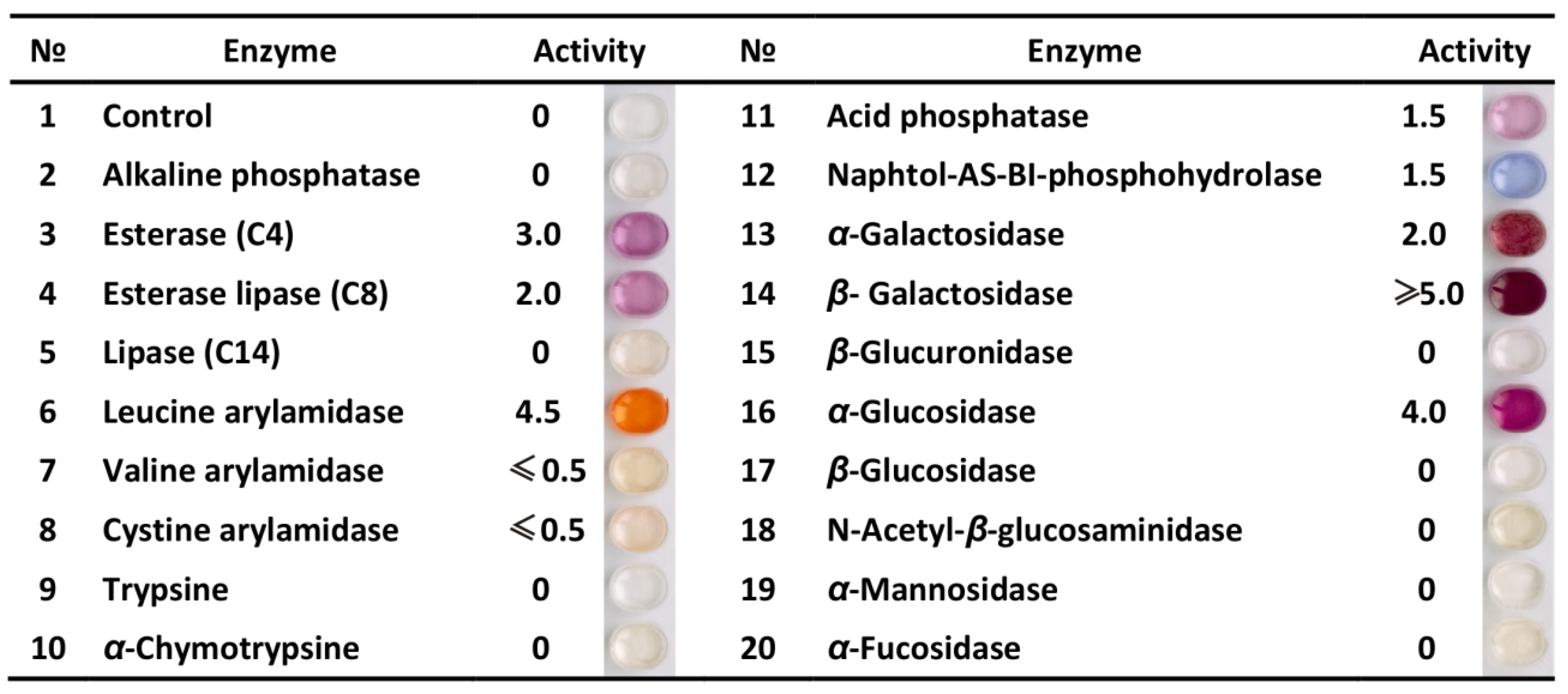
3.1. Profile of Enzymatic Activities

The enzymatic profile of probiotic strains plays an important role in both their health-promoting action on organism [25] and use for preparation of fermented food [26]. Several well-known examples of useful enzymatic activities of probiotics are glycoside hydrolase activities, which increase bioavailability of plant polysaccharides [27]; protease activities, which result in production of numerous bioactive peptides [7]; and β-galactosidase activity, which makes dairy products well tolerated by lactose-maldigesters [28]. In addition, in order to avoid the production of potentially toxic substances, probiotics must be evaluated for the production of undesirable enzymes, such as β-glucuronidase, a carcinogenic enzyme that can produce reactive metabolites negatively affecting the liver and increasing probability of colon carcinogenesis [29,30].
The semi-quantitative assessment of B. longum MC-42 enzymatic activities is presented in Figure 1. In total, 19 enzymes from the groups of glycoside-hydrolases, phosphatases, esterases, and proteases were assessed. In general, B. longum MC-42 demonstrated the high activities of leucine arylamidase, β-galactosidase, and α-glucosidase; moderate activities of esterase (C4), esterase lipase (C8), acid phosphatase, naphtol-AS-BI-phosphohydrolase, and α-galactosidase; and the absence of activities of alkaline phosphatase, lipase (C14), trypsin and α-chymotrypsin, β-glucosidase, N-acetyl-β-glucosaminidase, α-mannosidase, α-fucosidase, and most importantly, β-glucuronidase.
The enzymatic profile of B. longum MC-42 was generally similar to that of other B. longum strains. Typically, these strains have high activities of β-galactosidase and α-glucosidase, moderate-to-high activity of α-galactosidase, and almost absent activity of β-glucosidase. Additionally, weak or absent activities of N-acetyl-β-glucosaminidase, α-mannosidase, and α-fucosidase are characteristic for all Bifidobacterium spp. [31,32,33,34]. Weak protease activities are also typical for Bifidobacterium spp. As for B. longum MC-42 in the current study, almost no activities of trypsin and α-chymotrypsin were previously shown for strains of Bifidobacterium animalis, Bifidobacterium adolescentis, Bifidobacterium bifidum, Bifidobacterium breve, Bifidobacterium catenulatum, Bifidobacterium pseudocatenulatum, Bifidobacterium lactis, and B. longum [31,32,33,34,35,36]. At the same time, all of these strains demonstrated high activity of leucine aminopeptidase while the activities of valine and cystine aminopeptidases were more strain specific.

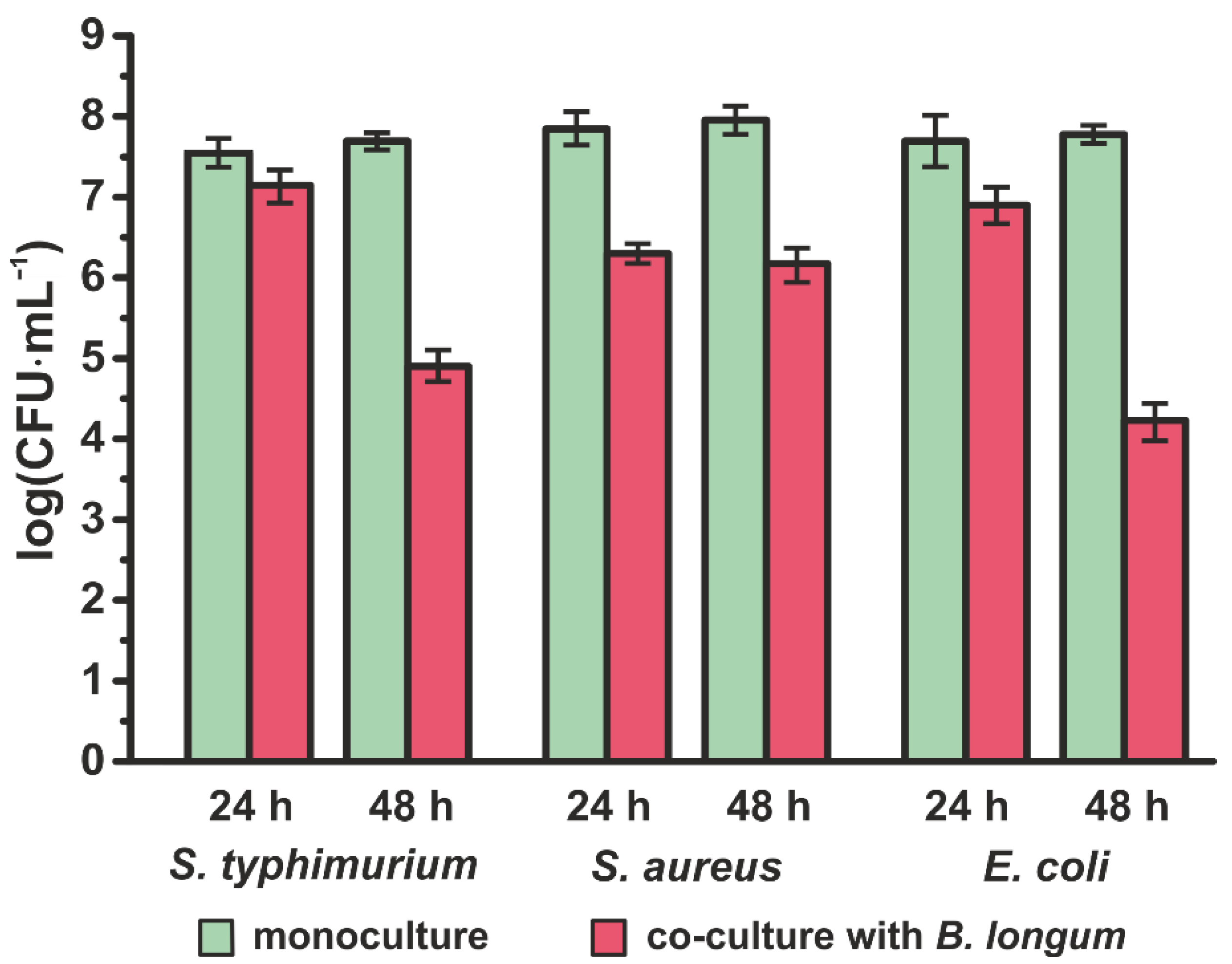
3.2. Inhibition of Pathogens

The ability to inhibit the growth of pathogenic and opportunistic bacteria is, arguably, the most demanded characteristic of a good probiotic strain. There are two main groups of mechanisms by which probiotics can inhibit pathogenic microflora: host-dependent and host-independent [37,38,39]. The mechanisms in the host-dependent group are highly reliant on the host’s physiology and include competition for binding sites, promotion of mucin secretion, induction of tight junction protein expression, and stimulation of immune response. The mechanisms in the host-independent group are based on the direct interaction of probiotics with pathogens and include competition for nutrients and production of organic acids, peroxide, and bacteriocins.
In this work, the host-independent inhibition of pathogenic microorganisms by B. longum MC-42 was assessed. The following pathogenic bacterial strains were used: S. typhimurium NCTC-00074, S. aureus ATCC-6538, and E. coli ATCC-25922. As seen in Figure 2, B. longum MC-42 almost equally inhibited the growth of S. typhimurium NCTC-00074 and E. coli ATCC-25922: the inhibition by approximately half an order of magnitude was observed after 24 h of co-cultivation, and that by approximately three orders of magnitude was observed after 48 h. The inhibition of S. aureus ATCC-6538 after 24 and 48 h of co-cultivation was the same and comprised approximately one and a half orders of magnitude.

3.3. Resistance to Antibiotics

Due to structural or functional characteristics, many microorganisms have an innate resistance to specific classes of antibiotics [40]. This so-called intrinsic antibiotic resistance can be passed only vertically from a parent to an offspring. While intrinsic antibiotic resistance can be a danger in the case of pathogenic and opportunistic microorganisms, it does not pose a particular problem considering probiotics [41]. Moreover, the moderate innate antibiotic resistance of certain probiotic strains prevents severe dysbiosis that can occur during the antibiotic treatment of infections. On the other hand, antibiotic resistance acquired by microorganism through a horizontal gene transfer—transmissible antibiotic resistance—now presents a major concern for safe use of probiotic strains [42]. Currently, it was demonstrated that some probiotic strains harboring antibiotic resistance genes can transmit them to microorganisms causing nosocomial and common infections [43].
The antibiotic resistance (AR) of B. longum MC-42 was tested with 15 commonly used antibiotics by qualitative disc-diffusion assay (Table 1). It was found that B. longum MC-42 was resistant only to lincomycin and the antibiotics belonging to the aminoglycoside structural group. The AR profile of B. longum MC-42 determined was in agreement with those previously reported for Bifidobacterium spp. [44,45]. The analysis of the B. longum MC-42 genome demonstrated an absence of a commonly transmissible by gut microbiota AR genes, such as aminoglycoside AR genes encoding the aminoglycoside-modifying enzymes aac(6′)-aph(2″) and aad(E) [46]; tetracycline AR genes encoding the ribosomal protection proteins—tet(M), tet(S), tet(W), tet(O), tet(Q), tet(32), tet(36), and tet(T); the tetracycline major facilitator superfamily efflux pumps tet(K) and tet(L) [47]; amphenicol AR genes encoding the chloramphenicol acetyltransferase—cat [48]; macrolide–lincosamide–streptogramin (MLS) AR genes encoding the 23S rRNA methylases—erm(A), erm(B), erm(C), erm(F) and erm(T)—and the macrolide major facilitator superfamily efflux pump mef(A) [49]. Hence, utilizing the previously proposed scheme for the antibiotic resistance assessment of bacteria [42], B. longum MC-42 can be considered an acceptable strain for use as a probiotic and starter culture.

3.4. Cholesterol-Removal Capacity

The recently discovered hypocholesterolemic effect of probiotic consumption offers a great opportunity for drug-free management of cholesterol levels in humans [50]. Currently, two main routes by which gut microbiota can significantly influence a host’s cholesterol homeostasis were proposed. The first route comprises deconjugation of bile salts by the action of bile salt hydrolases (BSHs, EC 3.5.1.24)—the microbial enzymes that catalyze hydrolysis of the amide bond in glycine/taurine-conjugated bile salts [4]. Deconjugation of bile salts decreases their reabsorption from the intestinal lumen and facilitates their excretion. To replenish the pool of bile salts, an endogenous cholesterol is used, which leads to its reduction in blood serum [51]. The second route involves decreasing the amount of cholesterol absorbed by human intestines via its microbial conversion into coprostanol [52]; however, compared to the bile salt deconjugation, less is known about this process at the molecular level.
The analysis of the B. longum MC-42 genome demonstrated the presence of a functional BSH encoding gene (locus_tag AS143_06365) that virtually guarantees the ability of this strain to lower a host’s cholesterol level by the deconjugation of bile salts. Additionally, several genes homologous to those that were previously proposed by Zanotti et al. [19] to participate in the conversion of cholesterol into coprostanol were determined: genes of the ABC family transporter (locus_tags AS143_00305, AS143_02140, and AS143_06580), a gene of the MFS family transporter (locus_tag AS143_02910), a gene of the EIIC component of the PTS system (locus_tag AS143_05800), and a gene of cholesterol reductase (locus_tag AS143_09220). However, since the presence of these genes only suggests—but not guarantees—the ability of B. longum MC-42 for cholesterol conversion, this ability was experimentally confirmed. It was determined that, during the cultivation of B. longum MC-42 on a cholesterol-containing medium, the cholesterol concentration was decreased by 54.8% (from 1.5 mM to 0.678 ± 0.014 mM) in 24 h.

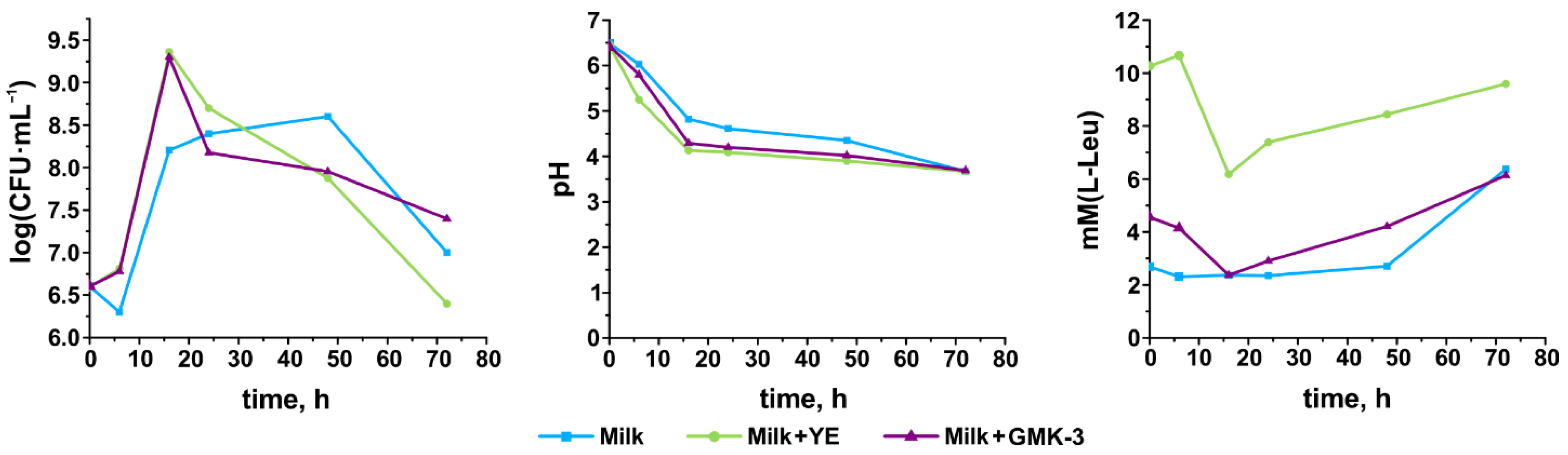
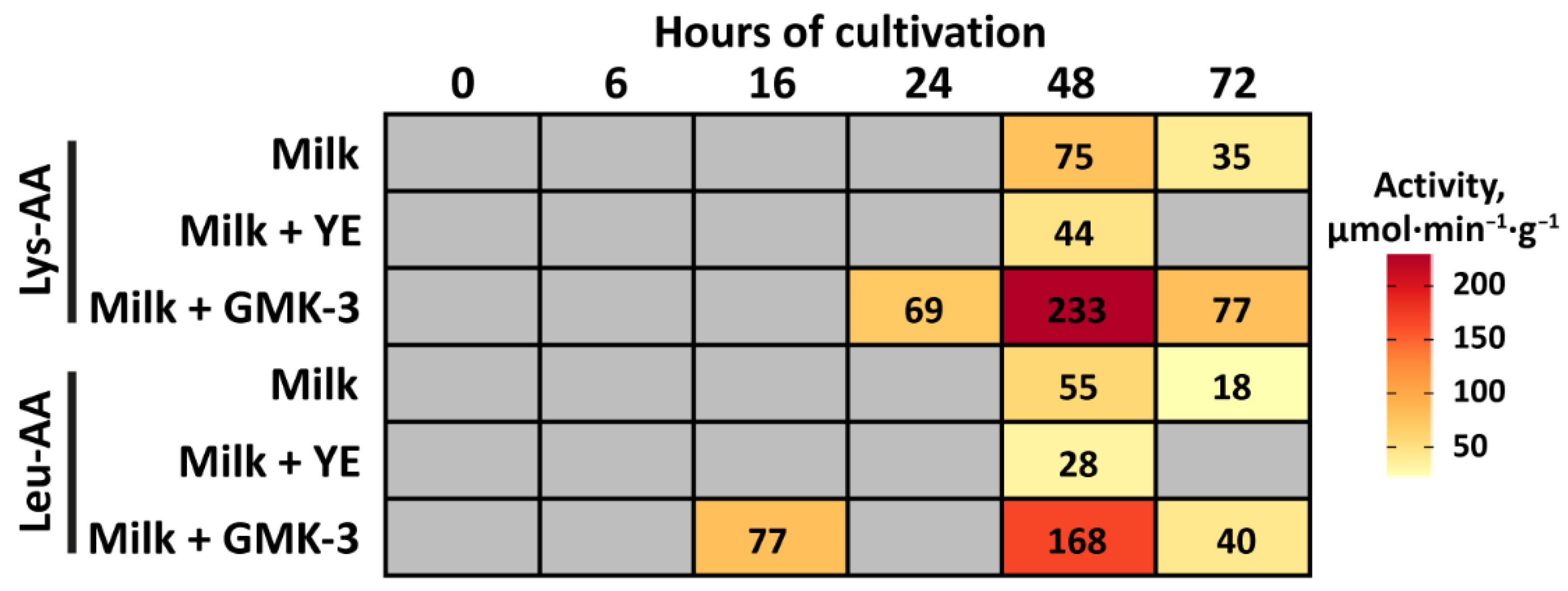
3.5. Growth Performance, Acidification Capability, and Degree of Proteolysis

As it was previously shown, in comparison with other lactic acid bacteria, Bifidobacteria spp. generally demonstrate slow or limited growth during milk fermentation [53]. Partially, this fact can be explained by the proposed inability of Bifidobacteria spp. to produce extracellular proteinases and, consequently, effectively hydrolyze proteins [54]. In agreement with this, it was demonstrated that yeast extract and hydrolysate of milk proteins are good growth promoters for various Bifidobacteria spp. [55].
The dynamic changes in the viable cell count, pH value, and degree of proteolysis during cultivation of B. longum MC-42 on skim milk, skim milk supplemented with YE, and skim milk supplemented with GMK-3 (containing hydrolysate of milk proteins and several other growth factors, see Section 2.6) are presented in Figure 3. The addition of YE and GMK-3 to the milk did not significantly alter the pH profile of fermentation. On all media, pH decreased from 6.5 to 3.7 units in a 72 h timespan, leading to the coagulation of milk proteins as a result of casein precipitation. Both YE and GMK-3 significantly stimulated the growth of B. longum MC-42: with these supplementations, the maximum attainable viable cell count (reached at 16 h of fermentation) comprised (2–2.3) × 109 CFU·mL−1, while on milk, this value (reached at 48 h of fermentation) did not exceed 4 × 10⁸ CFU·mL−1. For both YE- and GMK-3-supplemented milk, the degree of proteolysis decreased until 16 h of fermentation, suggesting the utilization of peptides already present in these media; however, after 16 h and until the end of fermentation, the degree of proteolysis grew steadily. For skim milk, the degree of proteolysis was the same until 48 h of fermentation, after which it increased at 72 h. It should be noted that the increase in the degree of proteolysis, observed after 16 h for YE- and GMK-3-supplemented milk and after 48 h for skim milk, coincided with a decrease in the viable cell count.
Since no trypsin and α-chymotrypsin activities were detected in the enzymatic profile of B. longum MC-42 (see Section 3.1), the observed increase in the degree of proteolysis can be attributed to the acid hydrolysis of proteins and to the possible action of some intracellular aminopeptidases released as a result of cell lysis [54].
The changes in aminopeptidase activity in the EEs of B. longum MC-42 during fermentation were assessed with three different substrates: Gly-Pro-p-NA, Lys-p-NA, and Leu-p-NA. It is known that the activity toward Lys-p-NA and Leu-p-NA chromogenic substrates can be detected in the presence of generic aminopeptidases PepC and PepN, which have been proposed to play a significant role in cell growth and peptide hydrolysis [54]. The aminopeptidase activity toward Gly-Pro-p-NA was absent on all studied media during the entire cultivation time; and Lys-AA and Leu-AA are shown in Figure 4. On all media, both, Lys-AA and Leu-AA were detected at the stage of cell death—after 16 h for YE- and GMK-3-supplemented milk and after 48 h for skim milk. The exception was Leu-AA for GMK-3-supplemented milk, which was detected at 16 h of fermentation when the maximum viable cell count was reached. Although it is generally accepted that the majority of aminopeptidases are intracellular, their extracellular release was previously reported [56]. Additionally, the prominent leucine arylamidase (aminopeptidase) activity was detected in the enzymatic API ZYM profile of B. longum MC-42 (see Section 3.1).
Generally, Lys-AA was 1.5–2.0 times higher than Leu-AA in all studied samples collected at the same time point. Supplementation of milk with different peptide sources resulted in different patterns of the aminopeptidase activities. In EEs from fermentation of YE-supplemented milk, both Lys-AA and Leu-AA were approximately 1.8 times lower compared to milk at 48 h and were totally absent at 72 h, while in EEs from fermentation of GMK-3-supplemented milk, these activities were approximately 2 times higher at 48 h and 3 times higher at 72 h compared to milk. This could be a result of different peptide profiles of YE and GMK-3; strain-specific differential regulation of aminopeptidases by peptide sources was previously reported for Bifidobacterium spp. in [57].

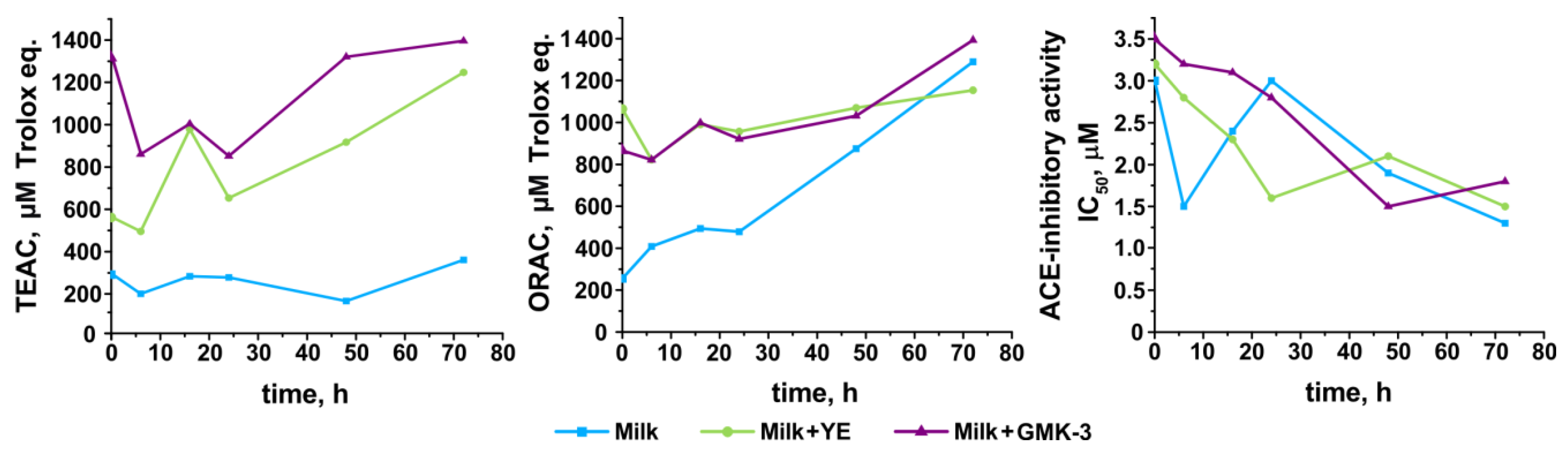
3.6. Development of Antioxidant and Antihypertensive Properties

The antioxidant and antihypertensive properties of the fermented milk are primarily attributed to the production of bioactive peptides during the fermentation process and, hence, can be species- or even strain-specific with respect to fermenting bacteria [58]. The activities of bioactive peptides are affected by both their amino acid compositions and sequences [59]. Generally, the ACE-I peptides are more sequence-specific than antioxidant ones, since they have to perform competitive inhibition at the catalytic site of the angiotensin-I-converting enzyme. Currently, the most popularly used assays for measurements of antioxidant activity are the TEAC and ORAC assays. However, for complex samples, the correlation between the ORAC and TEAC methods is low [60].
The development of antioxidant activity measured by the TEAC and ORAC assays during cultivation of B. longum MC-42 on different media is shown in Figure 5. For both YE- and GMK-3-supplemented milk, the general tendency of antioxidant activity development measured by TEAC an ORAC was similar: the activity decreased at 6 h of fermentation with a subsequent increase until the end of fermentation. However, while the TEAC assay showed higher antioxidant activity of the YE-supplemented milk compared to the GMK-3-supplemented milk (at almost all time points), the ORAC assay demonstrated comparable antioxidant activity for both supplementations. Interestingly, the antioxidant activity of the skim milk measured by the TEAC assay was almost the same during the entire cultivation; on the contrary, the antioxidant activity measured by the ORAC assay steadily grew and became comparable with that for the YE- and GMK-3-supplemented milk at the end of fermentation.
The development of ACE-I activity is shown in Figure 5. For skim milk, ACE-I activity increased (IC50 decreased) at 6 h of fermentation, after which it steadily decreased until 24 h with a subsequent increase until the end of fermentation. For the YE- and GMK-3 supplemented milk, ACE-I activity steadily increased until 24 h and 48 h of fermentation, respectively, after which it stayed at almost the same value.
The antioxidant activity profile generally agreed with the changes in the degree of proteolysis, suggesting extensive utilization of peptides (including antioxidant ones) from the media at the first hours of cultivation and subsequent peptide production by B. longum enzymes. Moreover, the significant increase in antioxidant and ACE-I activities after 24 h can also be related to the increase in Leu-AA and other aminopeptidases contributing to protein hydrolysis.

4. Conclusions

Currently, the amount of knowledge about the action of probiotics rapidly increases. This forces a reevaluation of long-used probiotic strains for their functional properties and safe use. In this work, for the first time, the properties of the bifidobacterial strain that was commercially used for decades in Russia, B. longum MC-42, were systematically reassessed. It was shown that this strain does not have frequently transmittable AR genes in its genome and does not possess harmful β-glucuronidase activity and, hence, can be regarded as generally safe for use according to the modern standards. At the same time, this strain can effectively lower the amount of cholesterol in a fermentation medium and can suppress the development of harmful bacteria. It was shown that the use of different growth-promoting additives results in the development of different patterns of antioxidant and ACE-I properties in final products. This can be explained by the different patterns of aminopeptidase activities in the cultural broth, although the exact mechanism for extracellular occurrence of these activities remains unclear.

Author Contributions

Conceptualization, T.V.F. and K.V.M.; investigation, A.V.B., O.S.S., O.A.G., K.V.M., I.V.R. and T.V.F.; data curation, T.V.F. and I.V.R.; writing—original draft preparation, T.V.F. and A.V.B.; writing—review and editing, K.V.M., O.A.G., I.V.R. and T.V.F.; visualization, K.V.M. and O.A.G.; supervision, T.V.F. and I.V.R. All authors have read and agreed to the published version of the manuscript.

Funding

This research was funded by the Ministry of Science and Higher Education of Russia (project № 0578-2019-0023).

Institutional Review Board Statement

Not applicable.

Informed Consent Statement

Not applicable.

Data Availability Statement

The data are contained within the article.

Conflicts of Interest

The authors declare no conflict of interest.

References

  1. Saarela, M.; Mogensen, G.; Fondén, R.; Mättö, J.; Mattila-Sandholm, T. Probiotic bacteria: Safety, functional and technological properties. J. Biotechnol. 2000, 84, 197–215. [Google Scholar] [CrossRef]
  2. Markowiak, P.; Śliżewska, K. Effects of probiotics, prebiotics, and synbiotics on human health. Nutrients 2017, 9, 1021. [Google Scholar] [CrossRef] [PubMed]
  3. Ohland, C.L.; MacNaughton, W.K. Probiotic bacteria and intestinal epithelial barrier function. Am. J. Physiol. Liver Physiol. 2010, 298, G807–G819. [Google Scholar] [CrossRef] [Green Version]
  4. Begley, M.; Hill, C.; Gahan, C.G.M. Bile salt hydrolase activity in probiotics. Appl. Environ. Microbiol. 2006, 72, 1729–1738. [Google Scholar] [CrossRef] [Green Version]
  5. Maldonado Galdeano, C.; Cazorla, S.I.; Lemme Dumit, J.M.; Vélez, E.; Perdigón, G. Beneficial effects of probiotic consumption on the immune system. Ann. Nutr. Metab. 2019, 74, 115–124. [Google Scholar] [CrossRef]
  6. Wang, H.; Lee, I.-S.; Braun, C.; Enck, P. Effect of probiotics on central nervous system functions in animals and humans: A systematic review. J. Neurogastroenterol. Motil. 2016, 22, 589–605. [Google Scholar] [CrossRef]
  7. Korhonen, H. Milk-derived bioactive peptides: From science to applications. J. Funct. Foods 2009, 1, 177–187. [Google Scholar] [CrossRef]
  8. Lugli, G.A.; Milani, C.; Duranti, S.; Mancabelli, L.; Mangifesta, M.; Turroni, F.; Viappiani, A.; van Sinderen, D.; Ventura, M. Tracking the taxonomy of the genus Bifidobacterium based on a phylogenomic approach. Appl. Environ. Microbiol. 2017, 84, e02249-17. [Google Scholar] [CrossRef] [Green Version]
  9. Arboleya, S.; Watkins, C.; Stanton, C.; Ross, R.P. Gut bifidobacteria populations in human health and aging. Front. Microbiol. 2016, 7, 1204. [Google Scholar] [CrossRef] [Green Version]
  10. Picard, C.; Fioramonti, J.; Francois, A.; Robinson, T.; Neant, F.; Matuchansky, C. Review article: Bifidobacteria as probiotic agents—Physiological effects and clinical benefits. Aliment. Pharmacol. Ther. 2005, 22, 495–512. [Google Scholar] [CrossRef]
  11. Jungersen, M.; Wind, A.; Johansen, E.; Christensen, J.; Stuer-Lauridsen, B.; Eskesen, D. The science behind the probiotic strain Bifidobacterium animalis subsp. lactis BB-12®. Microorganisms 2014, 2, 92–110. [Google Scholar] [CrossRef]
  12. Ku, S.; Park, M.; Ji, G.; You, H. Review on Bifidobacterium bifidum BGN4: Functionality and nutraceutical applications as a probiotic microorganism. Int. J. Mol. Sci. 2016, 17, 1544. [Google Scholar] [CrossRef] [Green Version]
  13. Wong, C.B.; Iwabuchi, N.; Xiao, J. Exploring the science behind Bifidobacterium breve M-16V in infant health. Nutrients 2019, 11, 1724. [Google Scholar] [CrossRef] [PubMed] [Green Version]
  14. Tupikin, A.E.; Kalmykova, A.I.; Kabilov, M.R. Draft genome sequence of the probiotic Bifidobacterium longum subsp. longum strain MC-42. Genome Announc. 2016, 4, e01411-16. [Google Scholar] [CrossRef] [Green Version]
  15. Naumova, N.; Alikina, T.; Tupikin, A.; Kalmykova, A.; Soldatova, G.; Vlassov, V.; Kabilov, M. Human gut microbiome response to short-term bifidobacterium-based probiotic treatment. Indian J. Microbiol. 2020, 60, 451–457. [Google Scholar] [CrossRef] [PubMed]
  16. Coman, M.M.; Verdenelli, M.C.; Cecchini, C.; Silvi, S.; Orpianesi, C.; Boyko, N.; Cresci, A. In vitro evaluation of antimicrobial activity of Lactobacillus rhamnosus IMC 501®, Lactobacillus paracasei IMC 502® and SYNBIO® against pathogens. J. Appl. Microbiol. 2014, 117, 518–527. [Google Scholar] [CrossRef]
  17. Clark, K.; Karsch-Mizrachi, I.; Lipman, D.J.; Ostell, J.; Sayers, E.W. GenBank. Nucleic Acids Res. 2016, 44, D67–D72. [Google Scholar] [CrossRef] [Green Version]
  18. Tanaka, H.; Hashiba, H.; Kok, J.; Mierau, I. Bile salt hydrolase of Bifidobacterium longum—Biochemical and genetic characterization. Appl. Environ. Microbiol. 2000, 66, 2502–2512. [Google Scholar] [CrossRef] [PubMed] [Green Version]
  19. Zanotti, I.; Turroni, F.; Piemontese, A.; Mancabelli, L.; Milani, C.; Viappiani, A.; Prevedini, G.; Sanchez, B.; Margolles, A.; Elviri, L.; et al. Evidence for cholesterol-lowering activity by Bifidobacterium bifidum PRL2010 through gut microbiota modulation. Appl. Microbiol. Biotechnol. 2015, 99, 6813–6829. [Google Scholar] [CrossRef] [PubMed]
  20. Altschul, S.F.; Gish, W.; Miller, W.; Myers, E.W.; Lipman, D.J. Basic local alignment search tool. J. Mol. Biol. 1990, 215, 403–410. [Google Scholar] [CrossRef]
  21. Exterkate, F.A. Location of peptidases outside and inside the membrane of Streptococcus cremoris. Appl. Environ. Microbiol. 1984, 47, 177–183. [Google Scholar] [CrossRef] [Green Version]
  22. Agarkova, E.Y.; Kruchinin, A.G.; Glazunova, O.A.; Fedorova, T.V. Whey protein hydrolysate and pumpkin pectin as nutraceutical and prebiotic components in a functional mousse with antihypertensive and bifidogenic properties. Nutrients 2019, 11, 2930. [Google Scholar] [CrossRef] [PubMed] [Green Version]
  23. Kruchinin, A.G.; Savinova, O.S.; Glazunova, O.A.; Moiseenko, K.V.; Agarkova, E.Y.; Fedorova, T.V. Hypotensive and hepatoprotective properties of the polysaccharide-stabilized foaming composition containing hydrolysate of whey proteins. Nutrients 2021, 13, 1031. [Google Scholar] [CrossRef] [PubMed]
  24. Torkova, A.A.; Ryazantseva, K.A.; Agarkova, E.Y.; Kruchinin, A.G.; Tsentalovich, M.Y.; Fedorova, T.V. Rational design of enzyme compositions for the production of functional hydrolysates of cow milk whey proteins. Appl. Biochem. Microbiol. 2017, 53, 669–679. [Google Scholar] [CrossRef]
  25. Indira, M.; Venkateswarulu, T.C.; Abraham Peele, K.; Nazneen Bobby, M.; Krupanidhi, S. Bioactive molecules of probiotic bacteria and their mechanism of action: A review. 3 Biotech 2019, 9, 306. [Google Scholar] [CrossRef]
  26. Marco, M.L.; Heeney, D.; Binda, S.; Cifelli, C.J.; Cotter, P.D.; Foligné, B.; Gänzle, M.; Kort, R.; Pasin, G.; Pihlanto, A.; et al. Health benefits of fermented foods: Microbiota and beyond. Curr. Opin. Biotechnol. 2017, 44, 94–102. [Google Scholar] [CrossRef]
  27. Cockburn, D.W.; Koropatkin, N.M. Polysaccharide degradation by the intestinal microbiota and its influence on human health and disease. J. Mol. Biol. 2016, 428, 3230–3252. [Google Scholar] [CrossRef]
  28. Oak, S.J.; Jha, R. The effects of probiotics in lactose intolerance: A systematic review. Crit. Rev. Food Sci. Nutr. 2019, 59, 1675–1683. [Google Scholar] [CrossRef]
  29. Nanno, M.; Morotomi, M.; Takayama, H.; Kuroshima, T.; Tanaka, R.; Mutai, M. Mutagenic activation of biliary metabolites of benzo(a)pyrene by β-glucuronidase-positive bacteria in human faeces. J. Med. Microbiol. 1986, 22, 351–355. [Google Scholar] [CrossRef] [Green Version]
  30. Davis, C.D.; Milner, J.A. Gastrointestinal microflora, food components and colon cancer prevention. J. Nutr. Biochem. 2009, 20, 743–752. [Google Scholar] [CrossRef] [Green Version]
  31. Arboleya, S.; Ruas-madiedo, P.; Margolles, A.; Solís, G.; Salminen, S.; Reyes-gavilán, C.G.D.L.; Gueimonde, M. Characterization and in vitro properties of potentially probiotic Bifidobacterium strains isolated from breast-milk. Int. J. Food Microbiol. 2011, 149, 28–36. [Google Scholar] [CrossRef] [PubMed] [Green Version]
  32. Delgado, S.; O’Sullivan, E.; Fitzgerald, G.; Mayo, B. In vitro evaluation of the probiotic properties of human intestinal Bifidobacterium species and selection of new probiotic candidates. J. Appl. Microbiol. 2008, 104, 1119–1127. [Google Scholar] [CrossRef] [PubMed]
  33. Desjardins, M.L.; Roy, D.; Goulet, J. Growth of bifidobacteria and their enzyme profiles. J. Dairy Sci. 1990, 73, 299–307. [Google Scholar] [CrossRef]
  34. Martínez-Villaluenga, C.; Gómez, R. Characterization of bifidobacteria as starters in fermented milk containing raffinose family of oligosaccharides from lupin as prebiotic. Int. Dairy J. 2007, 17, 116–122. [Google Scholar] [CrossRef] [Green Version]
  35. Kim, H.; Kim, J.S.; Kim, Y.G.; Jeong, Y.; Kim, J.E.; Paek, N.S.; Kang, C.H. Antioxidant and probiotic properties of lactobacilli and bifidobacteria of human origins. Biotechnol. Bioprocess Eng. 2020, 25, 421–430. [Google Scholar] [CrossRef]
  36. Kim, H.J.; Shin, S.I.; Lee, S.J.; Moon, T.W.; Lee, C.J. Screening and selection of Bifidobacterium strains isolated from human feces capable of utilizing resistant starch. J. Sci. Food Agric. 2018, 98, 5901–5907. [Google Scholar] [CrossRef]
  37. Corr, S.C.; Hill, C.; Gahan, C.G.M. Understanding the mechanisms by which probiotics inhibit gastrointestinal pathogens. Adv. Food Nutr. Res. 2009, 56, 1–15. [Google Scholar]
  38. Britton, R.A.; Versalovic, J. Probiotics and gastrointestinal infections. Interdiscip. Perspect. Infect. Dis. 2008, 2008, 1–10. [Google Scholar] [CrossRef]
  39. Isolauri, E.; Kirjavainen, P.V.; Salminen, S. Probiotics: A role in the treatment of intestinal infection and inflammation? Gut 2002, 50, iii54–iii59. [Google Scholar] [CrossRef]
  40. Cox, G.; Wright, G.D. Intrinsic antibiotic resistance: Mechanisms, origins, challenges and solutions. Int. J. Med. Microbiol. 2013, 303, 287–292. [Google Scholar] [CrossRef]
  41. Sharma, P.; Tomar, S.K.; Goswami, P.; Sangwan, V.; Singh, R. Antibiotic resistance among commercially available probiotics. Food Res. Int. 2014, 57, 176–195. [Google Scholar] [CrossRef]
  42. Álvarez-Cisneros, M.Y.; Ponce-Alquicira, E. Antibiotic resistance in lactic acid bacteria. In Antimicrobial Resistance—A Global Threat; IntechOpen: London, UK, 2019. [Google Scholar]
  43. Kumar, S.B.; Arnipalli, S.R.; Ziouzenkova, O. Antibiotics in food chain: The consequences for antibiotic resistance. Antibiotics 2020, 9, 688. [Google Scholar] [CrossRef]
  44. D’Aimmo, M.R.; Modesto, M.; Biavati, B. Antibiotic resistance of lactic acid bacteria and Bifidobacterium spp. isolated from dairy and pharmaceutical products. Int. J. Food Microbiol. 2007, 115, 35–42. [Google Scholar] [CrossRef]
  45. Moubareck, C.; Gavini, F.; Vaugien, L.; Butel, M.J.; Doucet-Populaire, F. Antimicrobial susceptibility of bifidobacteria. J. Antimicrob. Chemother. 2005, 55, 38–44. [Google Scholar] [CrossRef] [Green Version]
  46. Ammor, M.S.; Belén Flórez, A.; Mayo, B. Antibiotic resistance in non-enterococcal lactic acid bacteria and bifidobacteria. Food Microbiol. 2007, 24, 559–570. [Google Scholar] [CrossRef] [PubMed]
  47. Roberts, M.C. Update on acquired tetracycline resistance genes. FEMS Microbiol. Lett. 2005, 245, 195–203. [Google Scholar] [CrossRef] [PubMed]
  48. Schwarz, S.; Kehrenberg, C.; Doublet, B.; Cloeckaert, A. Molecular basis of bacterial resistance to chloramphenicol and florfenicol. FEMS Microbiol. Rev. 2004, 28, 519–542. [Google Scholar] [CrossRef] [Green Version]
  49. Roberts, M.C. Update on macrolide-lincosamide-streptogramin, ketolide, and oxazolidinone resistance genes. FEMS Microbiol. Lett. 2008, 282, 147–159. [Google Scholar] [CrossRef] [Green Version]
  50. Kumar, M.; Nagpal, R.; Kumar, R.; Hemalatha, R.; Verma, V.; Kumar, A.; Chakraborty, C.; Singh, B.; Marotta, F.; Jain, S.; et al. Cholesterol-lowering probiotics as potential biotherapeutics for metabolic diseases. Exp. Diabetes Res. 2012, 2012, 1–14. [Google Scholar] [CrossRef] [Green Version]
  51. Chand, D.; Avinash, V.S.; Yadav, Y.; Pundle, A.V.; Suresh, C.G.; Ramasamy, S. Molecular features of bile salt hydrolases and relevance in human health. Biochimica Biophysica Acta Gen. Subj. 2017, 1861, 2981–2991. [Google Scholar] [CrossRef] [PubMed]
  52. Kriaa, A.; Bourgin, M.; Mkaouar, H.; Jablaoui, A.; Akermi, N.; Soussou, S.; Maguin, E.; Rhimi, M. Microbial reduction of cholesterol to coprostanol: An old concept and new insights. Catalysts 2019, 9, 167. [Google Scholar] [CrossRef] [Green Version]
  53. Roy, D. Technological aspects related to the use of bifidobacteria in dairy products. Lait 2005, 85, 39–56. [Google Scholar] [CrossRef] [Green Version]
  54. Zhang, C.; Zhang, Y.; Li, H.; Liu, X. The potential of proteins, hydrolysates and peptides as growth factors for Lactobacillus and Bifidobacterium: Current research and future perspectives. Food Funct. 2020, 11, 1946–1957. [Google Scholar] [CrossRef]
  55. Poch, M.; Bezkorovainy, A. Growth-enhancing supplements for various species of the genus Bifidobacterium. J. Dairy Sci. 1988, 71, 3214–3221. [Google Scholar] [CrossRef]
  56. Shihata, A.; Shah, N.P. Proteolytic profiles of yogurt and probiotic bacteria. Int. Dairy J. 2000, 10, 401–408. [Google Scholar] [CrossRef]
  57. Meli, F.; Lazzi, C.; Neviani, E.; Gatti, M. Effect of protein hydrolizates on growth kinetics and aminopeptidase activities of some Bifidobacterium species. Anaerobe 2013, 22, 130–133. [Google Scholar] [CrossRef]
  58. Raveschot, C.; Cudennec, B.; Coutte, F.; Flahaut, C.; Fremont, M.; Drider, D.; Dhulster, P. Production of bioactive peptides by Lactobacillus species: From gene to application. Front. Microbiol. 2018, 9, 2354. [Google Scholar] [CrossRef] [Green Version]
  59. Udenigwe, C.C.; Aluko, R.E. Food protein-derived bioactive peptides: Production, processing, and potential health benefits. J. Food Sci. 2012, 77, R11–R24. [Google Scholar] [CrossRef]
  60. Zulueta, A.; Esteve, M.J.; Frígola, A. ORAC and TEAC assays comparison to measure the antioxidant capacity of food products. Food Chem. 2009, 114, 310–316. [Google Scholar] [CrossRef]
Figure 1. API ZYM enzymatic profile of B. longum MC-42.
Fermentation 07 00101 g001
Figure 2. The growth inhibition of S. typhimurium NCTC-00074, S. aureus ATCC-6538, and E. coli ATCC-25922 upon co-cultivation with B. longum MC-42.
Fermentation 07 00101 g002
Figure 3. Growth characteristics of B. longum MC-42 on different media: Milk—skim milk; Milk + YE—skim milk with addition of yeast extract, and Milk + GMK-3—skim milk with addition of hydrolysate containing supplementation GMK-3. The initial values of the degree of proteolysis were 2.74 mM (L-Leu) for Milk, 9.88 mM (L-Leu) for Milk + YE, and 3.77 mM (L-Leu) for Milk + GMK-3.
Fermentation 07 00101 g003
Figure 4. Lys-AA and Leu-AA of B. longum MC-42 during fermentation on different media: Milk—skim milk; Milk + YE—skim milk with addition of yeast extract, and Milk + GMK-3—skim milk with the addition of a hydrolysate containing GMK-3 supplementation. The aminopeptidase activities are expressed as µmol of p-NA released by aminopeptidase per minute and per gram of protein. The data are represented with a color-coded system where the highest value is shown in red and the lowest—in yellow.
Fermentation 07 00101 g004
Figure 5. Antioxidant and ACE-I properties of products obtained after fermentation by B. longum MC-42 of Milk—skim milk; Milk + YE—skim milk with addition of yeast extract, and Milk + GMK-3—skim milk with addition of hydrolysate containing supplementation GMK-3.
Fermentation 07 00101 g005
Table 1. The antibiotic resistance profile of B. longum MC-42.
AntibioticAmount, µgInhibition Zone
Diameter, mm
Resistance
Status
β-lactams (penams):
Ampicillin1028 ± 0.5Susceptible
Amoxicillin2028 ± 1Susceptible
Oxacillin112 ± 0.5Intermediate
Penicillin G1028 ± 1Susceptible
Fosfomycins:
Fosfomycin20023 ± 1Susceptible
Aminoglycosides:
Gentamicin12010 ± 0.5Resistant
Kanamycin A3011 ± 0.5Resistant
Neomycin307 ± 1Resistant
Tetracyclines:
Doxycycline3032 ± 1Susceptible
Tetracycline3028 ± 1Susceptible
Macrolides:
Azithromycin1514 ± 1Intermediate
Lincosamides:
Lincomycin158 ± 1Resistant
Amphenicols:
Chloramphenicol3026 ± 1Susceptible
Fluoroquinolones:
Levofloxacin514 ± 1Intermediate
Pefloxacin56 ± 1Resistant
Publisher’s Note: MDPI stays neutral with regard to jurisdictional claims in published maps and institutional affiliations.

